Note: Descriptions are shown in the official language in which they were submitted.
CA 02499094 2010-10-07
WO 2004/026347 PCT/US2003/029488
APPARATUS AND METHOD FOR SURGICAL BYPASS OF AQUEOUS
HUMOR
BACKGROUND OF THE INVENTION
Glaucoma is a disease condition of the eye in which increased intraocular
pressure (IOP) is created by dysfunction in the drainage mechanism for the
aqueous
humor. Aqueous humor is produced within the eye in the ciliary body and flows
within the anterior region of the eye. The aqueous humor normally flows
through a
network of tissues at the interior angle of the anterior chamber, named the
trabecular
meshwork and subsequently into a circular drainage space named Schlemm's
canal.
The aqueous humor continues its drainage path into collector channels and
finally into
aqueous veins to enter the venous system.
Typically in open angle glaucoma, the pathway for aqueous humor becomes
narrowed or occluded, increasing IOP and resulting in gradual nerve damage and
loss
of vision. Such conditions are usually treated by topical drugs in the form of
eye
drops, but may result in surgical treatment if drug treatment becomes
ineffective or if
patient compliance is an issue. Traditional glaucoma surgery, such as
trabeculotomy
or trabeculectomy, involves dissection of the eye and the forming of new holes
through the trabecular meshwork portion of the drainage pathway. The fluid is
channeled to a reservoir formed under the conjunctiva known as a bleb. While
blebs
are effective in removing the aqueous humor, bleb complications present the
highest
incidence of post-surgical complications due to irritation and infection.
A new class of surgical procedures aims to approach treatment of the ocular
drainage system from the scleral tissues without penetrating the interior
chamber of
the eye. These procedures are termed "non-penetrating" surgery and involve
careful
surgical dissection of the scleral tissues to access the tissues involved with
ocular
drainage. Deep sclerectomy is a form of this type of procedure in which a
portion of
intrascleral tissue is removed nearly to Descemet's membrane to allow
significant
aqueous flow from the anterior chamber to a bleb. Viscocanalostomy is another
non-
penetrating procedure, which increases the flow of aqueous humor form the
anterior
1
CA 02499094 2005-03-15
WO 2004/026347 PCT/US2003/029488
chamber into a surgically created intrascleral lake. Although non-penetrating
procedures present fewer direct complications than traditional surgeries, most
of the
procedures still require the surgical dissection of ocular tissues and a high
level of
surgical skill.
Various approaches and devices for glaucoma surgery involving the rerouting
of aqueous humor have been described in the art. One approach involves the
shunting
of aqueous humor through a tube in the anterior chamber into a reservoir
implanted on
the surface of the eye. See Mendez US 4,428,746, White US 4,554,918, Molteno
4,750,901, Ahmed US 5,071,408, US 5,411,473, US 5,616,118, US 5,681,275, US
5,785,674, US 6,261,256, Baerveldt, et al. US 5,178,604, US 5,397,300,
5,558,629,
6,050,970, Speckman US 5,338,291, Memmen US 5,370,607, Jacob US 5,882,327,
Odrich 6,41,666. A similar approach is to shunt the aqueous humor through a
tube
placed in the anterior chamber into a bleb on the surface of the eye. See
Worst US
5,180,362, Suson US 6,508,779, Wilcox WO 02/32343.
Another approach described in the art is the shunting of aqueous humor from
the anterior chamber to the tear film of the eye. See Ungerleider US
4,936,825; US
5,372,577; Wandel US 5,807,302; Brown US 6,595,945.
Another approach described in the art is placing a shunt for aqueous humor
through the trabecular meshwork to connect the anterior chamber and Schlemm's
canal. See Lynch et al. US 6,450,984, Hill US 6,533,768, WO 01/78656, and
Gharib
et al. US 2002 0165478.
SUMMARY OF THE INVENTION
The invention provides an apparatus for creating a tract within the scleral
tissues of an eye comprising an elongated body portion shaped to create a
tract which
forms a path for flow of aqueous humor into an ocular vein. The elongated body
portion has a proximal end and a distal end. The distal end may comprise a
mechanically cutting tip or an energy source to ablate tissue. The distal end
may be
visible by medical imaging methods such as ultra sound, or optical coherence
topography or visible under direct observation by an optical beacon at the
tip. The
apparatus may additionally accommodate a space-maintaining material for
placement
within the tract, such as hyaluronic acid or a cell proliferation inhibitor.
These space-
maintaining materials may also comprise a stent device made of hyaluronic
acid,
nickel, titanium alloy or other material.
2
CA 02499094 2011-04-07
The invention also provides a method for creating a path for flow of aqueous
humor of the
eye into an ocular vein comprising;
a. inserting an apparatus to form a tissue opening into an ocular vein on the
anterior
portion of the eye;
b. directing the apparatus to create a tract from the vein to a source of
aqueous
humor;
c. removing the apparatus;
d. closing the tissue opening while retaining flow through the tract between
the vein
and the source.
In another embodiment of the method the path is created by;
a. inserting the apparatus through a tissue opening in the eye into a source
of aqueous
humor;
b. directing the apparatus to create a tract from the source into an ocular
vein;
c. removing the apparatus while retaining flow through the tract between the
vein and
the source; and
d. optionally closing the tissue opening.
The source of aqueous humor typically will comprise the anterior chamber,
Schlemm's canal, the collector channel or a bleb.
In summary, an apparatus for creating a tract within the scleral tissues of an
eye
comprising:
an elongated body portion having a proximal end and distal end, said distal
end
comprising a beacon tip for visualizing said apparatus for guidance, said body
portion
shaped to create said tract wherein said tract forms a path for flow of
aqueous humor into
an ocular vein;
wherein said body portion comprises an outer sheath and an inner member
removable
during use of said outer sheath.
BRIEF DESCRIPTION OF THE DRAWINGS
Figure 1. Sectional view of the anterior portion of the human eye.
Figure 2. Cross-sectional view of the drainage system in the human eye.
3
CA 02499094 2011-04-07
Figure 3. Cross-sectional view of the drainage system in the human eye showing
a
microsurgical tool and tissue tract from an episcleral vein to the anterior
chamber.
Figure 4. Cross-sectional view of the drainage system in the human eye showing
tissue tract
from an episcleral vein to Schlemm's canal.
Figure 5. Cross-sectional view of the of an embodiment of the invention
comprising a
microcannula assembly.
Figure 6. View of an embodiment of a microcannula assembly incorporating fiber
optic core
for transmission of energy from a source.
Figure 7A. View of an embodiment of the invention comprising tube-stent
devices.
Figure 7B. View of a stent device comprising alternating filament loops.
20
30
3a
CA 02499094 2005-03-15
WO 2004/026347 PCT/US2003/029488
Figure 8A. Sectional view of the anterior portion of the human eye showing
bypass implant comprising shape memory material incorporating a reduced
diameter
segment to maintain flow control at a minimum level.
Figure 8B. Sectional view of the anterior portion of the human eye showing
laser
energy being applied externally to increase the diameter of the shape memory
bypass
implant.
Figure 9.. Cross-sectional view of microcannula assembly incorporating a
detachable tube stent as the distal sheath of the assembly.
Figure 10A. Sectional view of the anterior portion of the human eye showing
detachable tube stent assembly creating tract from an episcleral vein to the
anterior
chamber.
Figure 10B. Sectional view of the anterior portion of the human eye showing
tube
stent in place after removal of the cannula assembly.
DESCRIPTION OF INVENTION
The present invention to provide minimally invasive microsurgical tools and
methods, which enable surgical creation of a tissue tract within the tissues
of the eye
to directly connect a source of aqueous humor such as the anterior chamber, to
an
ocular vein, thereby forming a shunt or bypass for aqueous humor. The aqueous
veins, into which aqueous humor normally drains, are good candidates for this
procedure, however the invention is not limited to these specific vessels.
Furthermore, the tissue tract from the vein may be connected to any source of
aqueous
humor, including the anterior chamber, an aqueous collector channel, Schlemm's
canal, or a drainage bleb. Since the aqueous humor passes directly into the
venous
system, the normal drainage process for aqueous humor is restored. The tools
and
methods effectively bypass the small anatomical structures of the drainage
system,
such as the trabecular meshwork, Schlemm's canal and the collector channels,
which
have been identified with the mechanism of glaucoma. Furthermore, the
invention
describes devices and materials that can be implanted in the tract to maintain
the
tissue space and fluid flow post operatively.
The invention comprises tools, materials and related methods to surgically
create a bypass for aqueous humor. Figure 1 shows a sectional view of the
anterior
portion of the human eye for reference. The invention involves the steps of.
firstly,
identifying a candidate vein 4 on or within the eye of a living subject;
secondly,
inserting one or more tools into the eye to create a tissue tract in the
sclera 3, that
4
CA 02499094 2005-03-15
WO 2004/026347 PCT/US2003/029488
connects the vein to a source of aqueous humor such as the anterior chamber of
the
eye 1, or Schlemm's canal 2; thirdly, optionally inserting an implant or
material to
maintain the tract opening and fluid flow, and; lastly, closing the surgical
access site
as required. The tools and materials comprise the apparatus to create a tissue
tract
within scleral tissues of the eye such that the tract acts as a fluid path for
aqueous
humor to the vein.
A magnified cross-sectional view of the drainage system of the human eye is
shown in Figure 2 for reference. An episcleral vein 4 is identified suitable
for creating
the bypass. Candidate vessels can be found close to the surface of the eye,
such as but
not limited to, conjunctiva veins, anterior ciliary veins, and episcleral
veins. Surface
vessels can be directly visualized and accessed while subconjunctival vessels
may
require a small incision in the conjunctiva for access. Subsurface vessels
such as
collector channels 5 and aqueous veins 6 can be identified by high-resolution
medical
imaging methods such as high frequency ultrasound (HFU) or optical coherence
topography (OCT). The use of medical imaging is preferred in that veins which
are in
the most surgically desired location, can be selected. Vessels which are
angled toward
the anterior chamber 1, or Schlemm's canal 2, and having appropriate
dimensions and
minimal tortuosity, can be identified as candidates for bypass. The use of an
ultrasound or optical contrast agent, either delivered directly to the
candidate vein or
systemically to the subject, will facilitate vein identification. Doppler
imaging can be
used to assist identification of candidate blood vessels. Pressure changes
applied to
the anterior chamber will also facilitate vein identification and selection by
distinguishing vessels with the most direct connection to the aqueous drainage
system.
In some situations the blood/aqueous interface can be seen in episcleral
veins.
Changes in pressure in the chamber will cause this interface to move forward
and
backward within the vein. Furthermore, pressure changes can result in a change
of
the apparent vein diameter, which can be directly observed.
Cross-sectional views of the drainage system of the human eye with a
surgically created tissue tract are shown in Figures 3 and 4 for reference.
The use of
HFU or OCT imaging is desired to determine the optimal placement of the tissue
tract
7, from the candidate vein to the anterior chamber 1, or if desired, to a
collector
channel, Schlemm's canal 2, or an existing bleb. When forming a connection 9
of a
vein to the anterior chamber, any region of the anterior chamber in proximity
to the
vein may be used including the area at the anterior segment angle and the
corneal-
5
CA 02499094 2005-03-15
WO 2004/026347 PCT/US2003/029488
scleral junction. The surgeon may use imaging techniques to pre-plan the
'route of the
surgery and to verify locations, direction and placement of the microsurgical
tool 8,
during the procedure. The method may comprise first access to a vein 4, and
then
creating a tract 7, toward and into a source of aqueous humor such as the
anterior
chamber 1. Alternatively, the method may comprise access from the anterior
chamber
first, and then through the subsurface tissues to a vein. The advantage of
such a
method is that the tissue penetration may be accomplished through clear
cornea,
starting 180 from the bypass point and transversing through the anterior
chamber.
The microsurgical tool is placed at the correct site for penetration of the
tissues
through to the candidate vein. Small, clear corneal incisions are self-sealing
and
therefore do not require closure mechanisms. If the initial penetration of the
tool into
the eye is elsewhere, then the entry inscision may need to be closed after
removal of
the tool.
The microsurgical tool may comprise an elongated tool, such as a
microcannula, with a tip at one end which is directed into a vein. This may be
a
mechanically cutting tip such as a solid or hollow trocar-like member capable
of
creating a tunneled tract of controlled diameter through scleral tissues. In
another
embodiment, the tool may comprise a hollow tube with a sharpened distal edge
used
to core out a tissue tract. The removal of tissue may promote the subsequent
stability
of the tract and aid placement of an implant device into the channel.
Referring to Fig.
5, the tool may comprise an outer sheath 11 and inner member, with the outer
sheath
disposed axially about the inner member. The inner member may comprise a
trocar
10, solid rod, hollow rod or cylinder, needle, wire or optical fiber. The
inner member
may be designed to allow exchange during use to allow specific functions to be
brought to the tip of the tool once it is located in tissue. The microcannula
maybe
handled at its proximal end by a suitable accommodating mechanism such as luer
fitting 12.
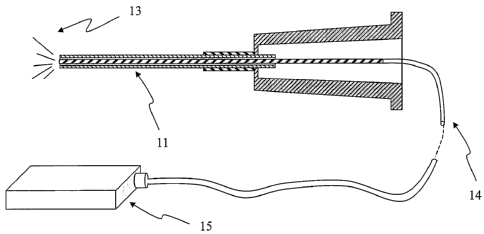
Referring to Fig. 6, an optical fiber inner member may be used to carry
visible
light to the tip of the fiber 13, which maybe disposed to reside at the tip of
the sheath
and hence may be used for direct visualization of the location of the tool
through
scleral tissues. In the case of an opaque outer sheath material, a cutout
section or
window near the distal tip of the sheath may be provided to visualize the
optical fiber
tip. The optical fiber may be fabricated from glass, fused silica, or plastic
that is
optically transparent to the wavelength of light used for visualization. The
described
6
CA 02499094 2005-03-15
WO 2004/026347 PCT/US2003/029488
optical beacon can provide an adjunct method of tracking the creation of the
tract,
aiding HFU or OCT imaging. Alternately, the optical fiber may be used to carry
energy for tissue ablation such as laser energy, in order to create the tract.
The tip
may also accommodate transmission of radio frequency or thermal energy to
ablate or
coagulate tissue. The fiber optic line 14 is connected to energy source 15.
The microsurgical tool is sized appropriate for access through small vessels
and to create controlled diameter tissue tracts. Diameters from 50-500 microns
are
useful, and in particular diameters from 50-200 microns are preferred. Outer
diameter
of a sheath member may correspond to these ranges and may comprise a wall
thickness between 10 and 100 microns. The microsurgical tool can comprise a
flexible microcannula to allow the distal tip to be advanced within the vein
toward a
source of aqueous humor such as the anterior chamber. The microsurgical tool
may
be fabricated from structural materials such as metals including steel,
titanium, and
nickel-titanium alloys, structural polymers including polyimide, polyethylene,
polyamide, polypropylene, polystyrene, polymethylmethacrylate,
polytetrafluoroethylene, and polysulfone. Several tools with different
material
composition and design may be used sequentially in the surgical procedure. For
example, one tool may be used to access the vessel and exchanged with a tool
to
create the scleral tissue tract. Alternatively, different types of inner
members such as
for cutting or light conduction may be used interchangeably within an outer
sheath.
The tool will accommodate features for orientation of the tract identified and
controlled by the clinical practitioner. The use of medical imaging to
coordinate or
verify the position and orientation of the tract aids accuracy and precision
of tract
placement. Tools which are in the field of view of the imaging system allow
for
identification of the tool's position while minimizing the creation of
artifacts into the
image. Selection of tool material and/or the use of contrast markers can
provide the
desired imaging properties for the tools.
The tract created may optionally be filled with a material to help maintain
the
patency and fluid flow of the tract. Such materials may comprise an anti-
fibrotic
material, anti-thrombotic agent, space maintaining material, tube-like stent
or similar
device to assure that the drainage tract remains patent. Anti-fibrotic
materials such as
hyaluronic acid and cellular proliferation inhibitors such as methotrexate,
sirolimus,
and paclitaxel, may be applied or released from a device within the tract.
Anti-
thrombotic agents such as heparin and tissue plasminogen activator may also be
7
CA 02499094 2005-03-15
WO 2004/026347 PCT/US2003/029488
applied or released from a device within the tract. Such materials may be in
the form
of microspheres, microparticles, microfibers, open-or closed-cell matrices,
foams,
gels and tubes, which may be designed to change their configuration in-situ
after
implantation. The materials may comprise degradable materials such as
hyaluronic
acid, collagen, glycosoaminoglycans and degradable synthetic polymers. The
materials may also comprise non-degradable materials with biocompatibility
suitable
for implant use including metals such as steel, titanium, and nickel-titanium
alloys,
and polymers such as polytetrafluoroethylene, polymethylmethacrylate,
polyimide,
polyethylene, polypropylene and polysulfone.
A tubular stent-like device may be placed within the tract to enlarge the
tract
diameter or provide stabilization through mechanical means. Referring to Fig..
7a,
there are examples shown of a simple tube 16 and a fenestrated tube 17. The
outer
sheath of the microsurgical tool may comprise a tube-stent, and can be left
behind
after the tool core is removed. Referring to Fig. 9, there is shown such a
microcannula assembly comprising a trocar tip core 10, the detachable stent
22,
driving cannula 23, and handle 24. The tube-stent may be pre-sized based on
pre-
surgical imaging or may be designed to be cut to size prior to or after
implantation.
The venous end of the tube-stent may be implanted to reside in the vein or
further
advanced to reside in a collector channel. The tube-stent may reside in the
entire
tissue tract between the vein and the aqueous humor source, or a portion of
the tract.
Several discrete tube-stents may also be used in stabilizing the tissue tract.
The diameter of the tube-stent maybe designed to allow expansion in-situ, for
example, by hydraulic pressure or thermal energy, or through the use of shape
memory materials for construction of the tube-stent. Referring to Fig. 8b, a
laser 20 is
used to increase the diameter of a shape memory implant 21. For long-term
stability,
preferred are tube-stents designed to be conformable to the tissue tract and
that do not
create mechanical loads on the tissue other than for dilation of the tract.
Mechanical
features of the tube-stent such as tissue interfacing porosity may be
incorporated to
aid retention. Tube-like stents may be comprised of permanent or biodegradable
materials. Suitable materials include metals such as steel, titanium and
nickel
titanium alloys, and biocompatible polymers such as hyaluronic acid, collagen,
glycosoaminoglycans, polylactic acid, polyglycolic acid,
polytetrafluoroethylene,
polymethylmethacrylate, polyimide, polyethylene, polypropylene and polysulfone
8
CA 02499094 2005-03-15
WO 2004/026347 PCT/US2003/029488
Furthermore, the tube-stent device can also have a design to provide a
controlled amount of flow restriction that would limit retrograde flow of
blood.
Devices with different flow resistance may be fabricated and chosen for
optimization
of aqueous flow by the practitioner. Referring to Fig. 8a, a shape memory
stent
incorporating a reduced diameter segment 19 connects the anterior chamber 2
with an
episcleral vein 4 via entry point 9. A valve may be incorporated into the tube
stent to
limit retrograde flow or to set a threshold pressure for flow. In another
embodiment,
the flow characteristics of the tube-stent may be varied after the procedure
upon
examination of the patient's IOP. Various energy sources such as laser light,
RF or
microwave may be directed at a portion of the implant to dilate or contract
discrete
segments to control flow. A photoreactive polymer or a pre-stressed polymer
similar
to heat shrink tubing may be employed to perform this function.
In a similar embodiment, the stent-like device may comprise a series of
filaments or wires. Referring to Fig. 7b, the device may be formed as a woven
tube or
a series of filament loops 18. The filament loops may be attached to each
other in an
alternating, or "zig zag" pattern, or may be attached to a linear member along
one
axis. The loops may be disposed at an angle to the axis, and be sufficiently
flexible to
maintain the tract opening without creating undue stress upon the surrounding
tissues.
Such an embodiment allows for the stent device to conform to changes in
diameter or
direction of the tract.
The invention also provides methods to surgically create an aqueous bypass in
the eye. The following methods are provided as explanatory and do not
constitute the
entire scope of methods which may be used in conjunction with the
microsurgical
tools described herein. Referring to Figs. 10a and 10b, in a first example,
the surgeon
will visually identify a candidate vein 4 on or about the surface of the eye.
Using high
resolution imaging techniques, a pathway from a target point along the axis of
the
vein to the desired endpoint, such as the anterior chamber 2, is mapped. A
surgical
tool comprising a handle 24, tube stent sheath 22, and trocar is used. The
trocar has a
distal point configured to pierce the tissues. The tube-stent will be of
correct length to
connect the chamber and vein. The vein is cannulated with the tool at the
target point,
and the tool oriented in the angle and direction that was plotted from the
imaging
session. The tool is advanced along the pathway until the tip is seen
penetrating the
anterior chamber at entry point 9, preferably above the iris. The trocar inner
member
and the tool are removed, leaving the tube-stent 22 behind. Alternatively, a
syringe
9
CA 02499094 2005-03-15
WO 2004/026347 PCT/US2003/029488
containing an antifibrotic hydrogel can be attached to the proximal end of the
sheath.
The hydrogel is applied into the tract at the same time that the tool,
including the
sheath, is being withdrawn, to aid in maintaining the tract. The access site
is then
sealed by any requisite surgical method.
In another example, an episcleral vein is located by visualization through a
surgical microscope. A target point is designated along the vein, at a distal
point of
sufficient diameter to accept the incoming microsurgical tool. An entry point
is
determined along the corneal limbus approximately 180 away from the candidate
vein. A gonio lens is used to visually inspect the anterior angle at the
bypass site to
choose a target entry point to connect to the vein. The microsurgical tool in
this
instance comprises a fiber optic inner member with a trocar like distal tip
and an
outer member comprising a tube-stent of the correct length to connect the
chamber
and vein. The tool is advanced through the clear cornea at the entry point and
advanced across the anterior chamber to the tissue entry point. The tool, is
then
advanced through the tissues guided toward the candidate vein by visualization
of the
beacon tip of the tool. The tool is advanced until the distal tip enters the
vein and then
continued until a sufficient portion of the tube stent distal end is within
the vessel to
maintain flow. If sized correctly, the proximal end of the tube stent will now
reside
just within the anterior chamber. The tool is withdrawn, leaving the tube
stent behind.
The entry point may be surgically closed or allowed to self-seal.
The procedure may also be performed on more than one venous site per eye as
may be required to provide adequate drainage. In practice, the procedure may
be
performed on one site, and the patient's IOP monitored post-surgically. If
more
pressure reduction is required, then a subsequent procedure may be performed
at
another target site. Multiple drainage paths can thereby be created.