Note: Descriptions are shown in the official language in which they were submitted.
CA 02499207 2005-03-16
WO 2004/024098 PCT/US2003/029414
METHOD FOR THE TREATMENT OF
NEPHRITIS USING ANTI-PDGF-DD ANTIBODIES
FIELD OF THE INVENTION
[0001] Embodiments of the invention described herein relate to antibodies
directed to
platelet derived growth factor-DD (PDGF-DD) and uses of such antibodies. The
antibodies of the
invention find use as diagnostics and as treatments for diseases associated
with the overproduction
of PDGF-DD. In particular, in accordance with embodiments of the invention,
the use of anti-
PDGF-DD antibodies for the treatment of nephritis and related disorders,
including diseases caused
by mesangial proliferation is provided.
BACKGROUND OF THE INVENTION
(0002] Nephritis is a group of kidney diseases that is a problem of growing
concern in
the United States and throughout the world. Nephritis can gradually progress
to kidney failure that
is ultimately fatal unless dialysis treatment or kidney transplantation is
received. The different
types of nephritis have different patterns of inheritance, and different rates
of progression.
Hereditary nephritis is manifested by microscopic traces of blood cells and
proteins in urine, and is
present and generally mild at birth. Another type of nephritis,
glomerulonephritis, is air
inflammation of the glomeruli, the Eltering units of the kidneys. Other forms
of nephritis may be
sequelae of infectious disease such as mononucleosis and Streptococcus (post-
infectious).
[0003] The symptoms of nephritis and other diseases related to proliferation
of
mesangial cells vary depending on the specific type of nephritis, but
typically includes the presence
of blood or proteins in the urine. In early stages of the disease, there may
be no signs or symptoms.
As the disease progresses, some or all of the following symptoms may occur:
high blood pressure,
excessive foaming of the urine, change in the color of the urine (to red or
dark brown), puffiness of
the eyes, hands, and feet, nausea and vomiting, difficulty breathing, and
headaches. These
symptoms may be used to identify the disease, to follow the course of
treatment, and to identify
what type of treatment is needed.
[0004] Injury to glomeruli can result in a variety of signs of the disease,
including but
not limited to proteinuria, hematuria, azotemia, oliguria, anuria, edema, and
hypertension. The
disease may also result in nephritic syndrome, acute nephritis, and rapidly
progressive
glomerulonephritis.
[0005] Many progressive renal diseases, including diabetic nephropathy, as
well as the
most frequent types of glomerulonephritides such as IgA-nephropathy are
characterized by
glomerular mesangial cell proliferation and/or matrix accumulation. Striker et
al., Lab havest
64:446-456 (1991). Some evidence now suggests that platelet derived growth
factors (PDGFs) and
the associated PDGF-system, may be involved in mesangial cell proliferation
and matrix
CA 02499207 2005-03-16
WO 2004/024098 PCT/US2003/029414
accumulation. Floege et al., supra (2001) and Floege et al., Am. J. Pathol.
154:169-79 (1999);
Gilbert et al., Kidney Int. 59:1324-32 (2001); Nakamura et al., Kidney Int.,
59:2134-45 (2001). In
addition, both PDGF 13-receptor subunit as well as PDGF B-chain are
overexpressed in renal
interstitial fibrosis. Kliem et al., Kidney Int. 49:666-78 (1996). Infusion of
large doses of the
dimer, PDGF-BB alone is able to induce interstitial fibrotic changes in normal
rat kidney. Tang et
al., Am. J. Pathol. 148:1169-80 (1996).
[0006] For two decades the platelet derived growth factor system consisted of
only two
PDGF chains, PDGF-A and -B, that are secreted as homo- or heterodimers and
bind to dimeric
PDGF receptors composed of a- and/or 13-chains. Whereas PDGF-A binds to the a-
chain only,
PDGF-B is a ligand for all receptor types. Floege e~ al., "Growth factors and
cytokines," in
Imnzunologic Renal Diseases (Neilson E.G. and Couser W.G., eds., 2d ed. 2001).
Recently two
other PDGF isoforms, designated PDGF-C and -D, were described that are
released as homodimers
only. According to current terminology, the homodimer form of PDGF-C is known
as "PDGF-CC"
and the homodimer form of PDGF-D is known as "PDGF-DD." LaRochelle et al.,
Nat. Cell Biol.
3:517-21 (2001); Li et al., Nat. Cell Biol. 2:302-09 (2000); and Bergsten et
al., Nat. Cell Biol.
3:512-16 (2001). The core chain of PDGF-CC appears to be largely a ligand for
the oca-PDGF
receptor, while PDGF-DD largely binds to the 1313-PDGF receptor. Id. In both
cases, some binding
has also been described to the a13-receptor. LaRochelle et al., supra (2001);
Bergsten et al., supra
(2001); Gilbertson et al., J. Biol. Chew. 276:27406-14 (2001). All four PDGF
isoforms, as well as
both receptor chains are expressed in the kidney, albeit in distinct spatial
arrangements. Floege et
al., supYa (2001); Changsirikulchai et al., Kidney Izzt. 62(6):2043-54 (2002);
Eitner et al., .I. Am.
Soc. Nephrol. 13(4):910-17 (2002).
[0007] PDGF-D is secreted as the disulphide-linked homodimer PDGF-DD, which is
activated upon limited proteolysis with dissociation of its CUB-domain to
become a specific
agonistic ligand for PDGF-1313- and a13-receptor. In developing and in adult
normal lcidneys, PDGF-
DD is expressed in visceral glomerular epithelial cells and some vascular
smooth muscle cells.
Changsirikulchai et al., supna (2002). In the developing mouse kidney, only
cells of the branching
ureter exhibited PDGF-DD immunoreactivity. Bergsten et al., supra (2001).
[0008] Diagnosis of nephritis is typically by identification of a family
history and/or
examination of the urinary sediment for the presence of red blood cells and
protein, specifically for
hematuria or albuminuria. Unfortunately, no specific treatment is known to
affect the underlying
pathological process or to alter the clinical course. Antibiotics,
anticoagulants, steroids, and
irnmunosuppressive agents have wrought no benefit. Control of hypertension is
suggested and
protein restriction may be of some use. When terminal ~ uremia occurs,
dialysis and even
-2_
CA 02499207 2005-03-16
WO 2004/024098 PCT/US2003/029414
transplantation of the kidney are necessary. Thus, a novel approach for the
treatment of nephritis is
needed.
SUMMARY OF THE INVENTION
[0009] Embodiments of the invention relate to the discovery that
administration of anti-
PDGF-DD antibodies, were highly effective at reducing proliferation of
glomerular cells and of
treating disorders associated with their proliferation.
[0010] Accordingly, one embodiment of the invention is the use of fully human
anti-
PDGF-DD antibodies, and anti-PDGF-DD antibody preparations with desirable
properties from a
therapeutic perspective, to inhibit the progression of nephritis and related
diseases. Preferably, the
antibodies have a heavy chain amino acid having a sequence selected from the
group consisting of
SEQ ID NOS: 2, 6, 10, 14, 18, 22, 26, 30, 34, 38, 42, 46, 50, 54, 58, 62, 66,
70, and 74. More
preferably, the antibodies further have a light chain amino acid having a
sequence selected from the
group consisting of SEQ ID NOS: 4, 8, 12, 16, 20, 24, 28, 32, 36, 40, 44, 48,
52, 56, 60, 64, 68,
and 72.
[0011] It will be appreciated that embodiments of the invention are not
limited to any
particular anti-PDGF-DD antibody, or any specific form of an antibody. For
example, the anti-
PDGF-DD antibody may be a full length antibody (e.g. having an intact human Fc
region) or an
antibody fragment (e.g. a Fab, Fab' or F(ab')2). In addition, the antibody may
be manufactured
from a hybridorna that secretes the antibody, or from a recombinantly produced
cell that has been
transformed or transfected with a gene or genes encoding the antibody.
[0012] In a preferred embodiment, the invention includes the treatment of
nephritis and
related diseases in humans, including but not limited to, mesangial
proliferative nephritis,
mesangial proliferative glomerulonephritis, mesangiocapillary
glomerulonephritis, systemic lupus
erythematosus, glomerular nephritis, renal failure, and diabetic nephropathy.
[0013] In one embodiment, the anti-PDGF-DD antibody forms a pharmaceutical
composition comprising an effective amount of the antibody, or a fragment
thereof, in association
with a pharmaceutically acceptable carrier or diluent. In an alternative
embodiment, an anti-PDGF-
DD antibody is linked to a radioisotope or a toxin. In another embodiment, the
anti-PDGF-DD
antibody or fragment thereof is conjugated to a therapeutic agent. The
therapeutic agent can be a
toxin or a radioisotope. Preferably, such antibodies can be used for the
treatment of diseases, such
as, for example, nephritis, progressive renal diseases, and related diseases,
such as mesangial
proliferative nephritis, mesangial proliferative glomerulonephritis,
mesangiocapillary
glomerulonephritis, systemic lupus erythernatosus, glomerular nephritis, renal
interstitial fibrosis,
renal failure, and diabetic nephropathy.
[0014] In another embodiment, the invention includes a method for treating
diseases or
conditions associated with the expression of PDGF-DD in a patient by
administering to the patient
-3-
CA 02499207 2005-03-16
WO 2004/024098 PCT/US2003/029414
an effective amount of an anti-PDGF-DD antibody. The patient is a mammalian
patient, preferably
a human patient. The disease or condition can be, for example, nephritis,
progressive renal
diseases, and related diseases, such as mesangial proliferative nephritis,
mesangial proliferative
glomerulonephritis, mesangiocapillary glomerulonephritis, systemic lupus
erythematosus,
glomerular nephritis, renal interstitial fibrosis, renal failure, or diabetic
nephropathy. Additional
embodiments include methods for the treatment of diseases or conditions
associated with the
expression of PDGF-DD in a mammal by identifying a mammal in need of treatment
for nephritis
and administering to the mammal a therapeutically effective dose of anti-PDGF-
DD antibodies.
[0015] Alternatively, anti-PDGF-DD antibodies may be administered to prevent a
mammal from contracting diseases or conditions associated with the expression
of PDGF-DD
including, but not limited to, nephritis or related diseases, and diseases
caused by mesangial
proliferation. Preferably the anti-PDGF-DD antibodies are fully human. The
disease or condition
can be nephritis and related diseases, including but not limited to,
nephritis, progressive renal
diseases, and related diseases, such as mesangial proliferative nephritis,
mesangial proliferative
glomerulonephritis, mesangiocapillary glomerulonephritis, systemic lupus
erythematosus,
glomerular nephritis, renal interstital fibrosis, renal failure, and diabetic
nephropathy.
[0016] In yet another embodiment, the invention includes a method for
inhibiting cell
proliferation associated with, or caused by, the expression of PDGF-DD by
contacting cells
expressing PDGF-DD with an effective amount of an anti-PDGF-DD antibody or a
fragment
thereof and incubating the cells and antibody, wherein the incubation results
in inhibited
proliferation of cells. In one embodiment, the cell proliferation is mesangial
cell proliferation.
Further, the mesangial cells can be human mesangial cells. In addition, the
method can be
performed in vivo.
[0017] In another embodiment, the invention is an article of manufacture
including a
container having a composition containing an anti-PDGF-DD antibody, and a
package insert or
label indicating that the composition can be used to treat conditions
characterized by the
overexpression of PDGF-D. Preferably a mammal and, more preferably, a human,
receives the
anti-PDGF-DD antibody. In a preferred embodiment, nephritis and related
diseases in humans are
treated, including but not limited to, nephritis, progressive renal diseases,
and related diseases, such
as mesangial proliferative nephritis, mesangial proliferative
glomerulonephritis, mesangiocapillary
glomerulonephritis, systemic lupus erythematosus, glomerular nephritis, renal
interstital fibrosis,
renal failure, and diabetic nephropathy.
[0018] Another embodiment is a method for identifying risk factors, of
disease,
diagnosis of disease, and staging of disease which involves identifying
overproliferation of
mesangial cells in the glomerulus using anti-PDGF-DD antibodies.
-4-
CA 02499207 2005-03-16
WO 2004/024098 PCT/US2003/029414
[0019] In one embodiment, the invention includes a method for diagnosing a
condition
associated with the expression of PDGF-DD in a cell by contacting the cell
with an anti-PDGF-DD
antibody, and detecting the presence of PDGF-DD. Preferred conditions include,
without
limitation, mesangial proliferative nephritis, mesangial proliferative
glomerulonephritis,
mesangiocapillary glomerulonephritis, systemic lupus erythematosus, glomerular
nephritis, renal
failure, and diabetic nephropathy.
[0020] In still another embodiment, the invention includes an assay lcit for
the detection
of PDGF-DD in mammalian tissues or cells to screen for nephritis and related
diseases in humans,
including but not limited to, mesangial proliferative nephritis, mesangial
proliferative
glomerulonephritis, mesangiocapillary glomerulonephritis, systemic lupus
erythematosus,
glomerular nephritis, renal failure, and diabetic nephropathy. The kit
includes an antibody that
binds to PDGF-DD and a means for indicating the reaction of the antibody with
PDGF-DD, if
present. Preferably the antibody is a monoclonal antibody. In one embodiment,
the antibody that
binds PDGF-DD is labeled. In another embodiment the antibody is an unlabeled
first antibody and
the means for indicating the reaction is a labeled anti-immunoglobulin
antibody. Preferably, the
antibody is labeled with a marker selected from the group consisting of: a
fluorochrome, an
enzyme, a radionuclide and a radiopaque material.
[0021] Yet another embodiment is the use of an anti-PDGF-DD antibody in the
preparation of a medicament for the treatment of nephritis and related
diseases. In one
embodiment, the disease is selected from the group comprising nephritis,
progressive renal
diseases, and related diseases, such as mesangial proliferative nephritis,
mesangial proliferative
glomerulonephritis, mesangiocapillary glomerulonephritis, systemic lupus
erythematosus,
glornerular nephritis, renal interstital fibrosis, renal failure, and diabetic
nephropathy.
BRIEF DESCRIPTION OF THE DRAWINGS
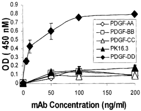
[0022] Figure 1 shows the characterization of anti-PDGF-DD mAb 6.4 specificity
by
ELISA.
[0023] Figure 2 shows the shows further characterization of anti-PDGF-DD mAb
6.4
specificity by ELISA.
[0024] Figure 3 shows the characterization of anti-PDGF-DD mAb specificity by
Western Blot Analysis.
[0025] Figure 4 is a line graph that shows that anti-PDGF-DD mAb 6.4 was able
to
neutralize PDGF-DD induced BrdU incorporation in NIH3T3 cells with an ICso of
approximately
75 ng/ml.
[0026] Figure 5 is a bar chart that shows that PDGF-DD acts as a growth factor
for
mesangial cells zn vitro. Data are means ~ SD of four independent experiments.
* indicates p<0.05
versus unstimulated control.
-5-
CA 02499207 2005-03-16
WO 2004/024098 PCT/US2003/029414
[0027] Figure 6 is a bar chart that shows the results of PDGF-DD-induced BrdU
incorporation in human mesangial cells.
[0028] Figure 7 is a graph that shows PDGF-DD expression in human serum for
patients
with various types of nephritis. A closed circle represents the PDGF-DD
concentration for an
individual clinical serum sample. PDGF-DD serum concentrations are grouped
according to the
patient disease indication. The number of patients (n) for a given clinical
indication is provided,
along with the mean PDGF-DD concentration in ng/ml.
[0029] Figure 8 shows immunochistochemical analysis of normal rat mesangium
cells
and the mesangium cells of rats with anti-Thy-1 induced nephritis. Elevated
anti-PDGF-DD
staining was found in rats with anti-Thy-1 induced nephritis. Mesangium,
tubules and surrounding
vasculature is shown. Mesangium cells included pericytes and renal tubules.
White and gray
arrows depict capillary and tubule staining respectively.
[0030] Figure 9 is a line graph that shows simulated fully human mAb kinetics
performed on rats. As shown, there is only a small peak to trough fluctuation
expected over 4 days,
even after a single dose.
[0031] Figure 10 is a line graph that shows transcript expression of PDGF-A, -
B, -C and
-D in the course of anti-Thyl.l nephritis relative to the expression in
untreated rats.
[0032] Figure 11 shows PDGF-DD protein was overexpressed during anti-Thy 1.1
nephritis in glomeruli. No PDGF-DD expression was noted in normal glomeruli
(Figure 11(A)),
whereas expression can be readily detected during mesangioproliferative
nephritis at day 7 after
disease induction (Figure 11(B)). No glomerular staining is present, when the
anti-PDGF-DD
antibody is replaced by an equal concentration of control IgG (Figure 11(C)).
Magnification is
600x.
[0033] Figures 12 A-H are bar charts that show glomerular changes on day 5 and
day 8
after disease induction in rats with mesangioproliferative anti Thy 1.1
nephritis treated with either
anti-PDGF-DD antibody, irrelevant control IgG or PBS alone.
[0034] Figure 13 is a bar graph that shows the results of glomerular
proliferation as
measured by BrdU incorporation in rats. Nephritic rats were treated with anti-
PDGF-DD mAb 6.4,
or control antibodies, or PBS. Healthy rats were treated with anti-PDGF-DD mAb
6.4 or control
antibodies.
[0035] Figure 14 is a bar graph that shows the results of glomerular
proliferation as
measured by PAS stain and quantitation of mitosis in rats. Nephritic rats were
treated with anti-
PDGF-DD mAb 6.4, or control antibodies, or PBS. Healthy rats were treated with
anti-PDGF-DD
mAb 6.4 or control antibodies.
[0036] Figure 15 is a bar graph that demonstrates the effect of anti-PDGF-DD
mAb 6.4
on mesangial cell mitosis in an acute rat anti-Thy-1 model. Anti-Thy-1 rats
were treated with anti-
-6-
CA 02499207 2005-03-16
WO 2004/024098 PCT/US2003/029414
PDGF-DD mAb 6.4, or control antibodies, or PBS. Healthy rats were treated with
anti-PDGF-DD
mAb 6.4 or control antibodies.
[0037] Figure 16 is a bar graph that demonstrates. the dose-responsive effects
of anti-
PDGF-DD mAb 6.4 on mitosis in glomerular cells in an acute rat Thy-1 model.
[0038] Figure 17 is a bar graph that demonstrates the dose-responsive effects
of anti-
PDGF-DD mAb 6.4 on BrdU incorporation in an acute rat Thy-1 model.
[0039] Figure 18 shows the immunohistochemical analysis of normal and diseased
human kidney tissue. Mesangium, tubules and surrounding vasculature is shown.
White and gray
arrows depict capillary and tubule staining respectively. Small black arrows
show punctate
inflammatory cell deposits in mesangium.
DETAILED DESCRIPTION
[0040] The invention described herein relates to methods for effectively
treating,
diagnosing, and/or staging nephritis and related conditions. Such conditions
include mesangial
proliferative nephritis, mesangial proliferative glomerulonephritis,
mesangiocapillary
glomerulonephritis, systemic lupus erythematosus, glomerular nephritis, renal
failure, and diabetic
nephropathy. In one particular embodiment, the invention includes
administering a therapeutically
effective amount of anti-PDGF-DD antibodies as a treatment for nephritis and
related conditions.
In preferred embodiments, the antibodies are fully human antibodies against
the dimer PDGF-DD.
[0041] Other embodiments of the invention relate to other compounds that
result in a
reduction of mesangioproliferative changes in vivo. Thus, compounds that
reduce the level of
PDGF-DD would be useful in treatment of nephritis. PDGF-D nucleic acids,
polypeptides,
antibodies, agonists, antagonists, and other related compound's uses are
disclosed more fully
below.
[0042] As described above, PDGF-D signals through a PDGF-B receptor and is
mitogenic for rat mesangial cells (MC). Low levels of PDGF-D mRNA were
detected in normal
rat glomeruli. However, incubation of cultured rat MCs with 100 ng/ml PDGF-DD
led to a 7-fold
increase in MC proliferation with a maximum after 24 hours. By real-time PCR,
PDGF-D mRNA
was detected in both cultured mesangial cells and glomeruli isolated from
normal rat kidney.
Following the induction of mesangioproliferative anti-Thy 1.1 nephritis in
rats, glomerular PDGF-
D mRNA and protein expression increased significantly from days 4 to 9 in
comparison to non-
nephritic rats as determined by real time PCR. Peak expression of PDGF-D mRNA
occurred
2 days later than peak PDGF-B mRNA expression. Additionally, PDGF-DD serum
levels
increased significantly in the nephritic animals on day 7.
[0043] To investigate the functional role of PDGF-DD during the nephritis,
neutralizing
fully human monoclonal anti-PDGF-DD antibodies were generated in Xenomouse~
(Abgenix,
Inc., Fremont, CA). Following the induction of anti-Thy 1.1 nephritis, rats
were treated on day 3
CA 02499207 2005-03-16
WO 2004/024098 PCT/US2003/029414
and day 5 after disease induction with 10 and 4 mg/kg fully human anti-PDGF-DD
antibody mAb
6.4 (n=15) or irrelevant human monoclonal antibody (n=15) or PBS (n=15) by
daily intraperitoneal
injection. On day 8 after disease induction antagonism of PDGF-DD led to a
significant reduction
of mitotic figures per 100 glomeruli (anti-PDGF-DD: 9.9 ~ 0.9; irrelevant IgG:
13.9 ~ 0.9; PBS:
14.7 1.0; p<0.0014) as well as of glomerular cells incorporating the thymidine
analog BrdU (anti-
PDGF-DD mAb 6.4: 1.62 0.23; irrelevant IgG: 2.88 0.28; PBS: 2.91 0.18;
p<0.0016).
Reduction of glomerular cell proliferation in the rats receiving anti-PDGF-DD
was not associated
with reduced glomerular expression of PDGF-B mRNA as determined by real time
PCR.
[0044] Injection of anti-PDGF-DD antibodies into normal rats did not affect
the
physiologic glomerular cell turnover as compared to normal rats receiving
irrelevant IgG. Thus,
PDGF-DD, produced by glomerular mesangial cells acts as a glomerular cell
mitogen both in vitro
and in vivo.
Seauence Listing
[0045] The heavy chain and light chain variable region nucleotide and amino
acid
sequences of representative human anti-PDGF-DD antibodies are provided in the
sequence listing,
the contents of which are summarized in Table 1 below.
Table 1
mAb Sequence SEQ
ID ID
No.: NO:
Nucleotide sequence encoding the variable region1
of the heavy chain
Amino acid sequence encoding the variable region2
6 of the heavy chain
4
. Nucleotide sequence encoding the variable region3
of the light chain
Amino acid sequence encoding the variable region4
of the light chain
Nucleotide sequence encoding the variable region5
of the heavy chain
Amino acid sequence encoding the variable region6
1 of the heavy chain
6
. Nucleotide sequence encoding the variable region7
of the light chain
Amino acid sequence encoding the variable region8
of the light chain
Nucleotide sequence encoding the variable region9
of the heavy chain
Amino acid sequence encoding the variable region10
11 of the heavy chain
1
. Nucleotide sequence encoding the variable region11
of the light chain
Amino acid sequence encoding the variable region12
of the light chain
Nucleotide sequence encoding the variable region13
of the heavy chain
Amino acid sequence encoding the variable region14
1 of the heavy chain
17
. Nucleotide sequence encoding the variable region15
of the light chain
Amino acid sequence encoding the variable region16
of the light chain
_g_
CA 02499207 2005-03-16
WO 2004/024098 PCT/US2003/029414
mAb Sequence SEQ ID
ID NO:
No.:
Nucleotide sequence encoding the variable region17
of the heavy chain
Amino acid sequence encoding the variable region18
of the heavy chain
1.18
Nucleotide sequence encoding the variable region19
of the light chain
Amino acid sequence encoding the variable region20
of the light chain
Nucleotide sequence encoding the variable region21
of the heavy chain
Amino acid sequence encoding the variable region22
of the heavy chain
1.19
Nucleotide sequence encoding the variable region23
of the light chain
Amino acid sequence encoding the variable region24
of the light chain
Nucleotide sequence encoding the variable region25
of the heavy chain
Amino acid sequence encoding the variable region26
of the heavy chain
1.23
Nucleotide sequence encoding the variable region27
of the light chain
Amino acid sequence encoding the variable region28
of the light chain
Nucleotide sequence encoding the variable region29
of the heavy chain
Amino acid sequence encoding the variable region30
of the heavy chain
1.24.1
Nucleotide sequence encoding the variable region31
of the light chain
Amino acid sequence encoding the variable region32
of the light chain
Nucleotide sequence encoding the variable region33
of the heavy chain
Amino acid sequence encoding the variable region34
of the heavy chain
1.25.1
Nucleotide sequence encoding the variable region35
of the light chain
Amino acid sequence encoding the variable region36
of the light chain
Nucleotide sequence encoding the variable region37
of the heavy chain
Amino acid sequence encoding the variable region38
1 of the heavy chain
29
. Nucleotide sequence encoding the variable region39
of the light chain
Amino acid sequence encoding the variable region40
of the light chain
Nucleotide sequence encoding the variable region41
of the heavy chain
Amino acid sequence encoding the variable region42
1 of the heavy chain
33
. Nucleotide sequence encoding the variable region43
of the light chain
Amino acid sequence encoding the variable region44
of the light chain
Nucleotide sequence encoding the variable region45
of the heavy chain
Amino acid sequence encoding the variable region46
1 of the heavy chain
38
1
. Nucleotide sequence encoding the variable region47
. of the light chain
Amino acid sequence encoding the variable region48
of the light chain
Nucleotide sequence encoding the variable region49
of the heavy chain
Amino acid sequence encoding the variable region50
1 of the heavy chain
39
1
. Nucleotide sequence encoding the variable region51
. of the light chain
Amino acid sequence encoding the variable region52
of the light chain
-9-
CA 02499207 2005-03-16
WO 2004/024098 PCT/US2003/029414
mAb Sequence SEQ
ID ID
' No.: NO:
Nucleotide sequence encoding the variable region53
of the heavy chain
Amino acid sequence encoding the variable region54
1 of the heavy chain
4
. Nucleotide sequence encoding the variable region55
of the light chain
Amino acid sequence encoding the variable region56
of the light chain
Nucleotide sequence encoding the variable region57
of the heavy chain
Amino acid sequence encoding the variable region58
of the heavy chain
1.46.1
Nucleotide sequence encoding the variable region59
of the light chain
Amino acid sequence encoding the variable region60
of the light chain
Nucleotide sequence encoding the variable region61
of the heavy chain
Amino acid sequence encoding the variable region62
1 of the heavy chain
1.48. Nucleotide sequence encoding the variable region63
of the light chain
Amino acid sequence encoding the variable region64
of the light chain
Nucleotide sequence encoding the variable region65
of the heavy chain
Amino acid sequence encoding the variable region66
4 of the heavy chain
1.
9.1 Nucleotide sequence encoding the variable region67
of the light chain
Amino acid sequence encoding the variable region68
of the light chain
Nucleotide sequence encoding the variable region69
of the heavy chain
Amino acid sequence encoding the variable region70
1 of the heavy chain
51
. Nucleotide sequence encoding the variable region71
of the light chain
Amino acid sequence encoding the variable region72
of the light chain
Nucleotide sequence encoding the variable region73
1 of the heavy chain
40
1
. Amino acid sequence encoding the variable region74
. of the heavy chain
Nucleotide sequence encoding the variable region75
of the heavy chain
Amino acid sequence encoding the variable region76
1 of the heavy chain
22
. Nucleotide sequence encoding the variable region77
of the light chain
Amino acid sequence encoding the variable region78
of the light chain
Nucleotide sequence encoding the variable region79
of the heavy chain
Amino acid sequence encoding the variable region80
1 of the heavy chain
59
. Nucleotide sequence encoding the variable region81
of the light chain
Amino acid sequence encoding the variable region82
of the light chain
Definitions
[0046] Unless otherwise defined, scientific and technical terms used in
connection with
the invention described herein shall have the meanings that are commonly
understood by those of
ordinary skill in the art. Further, unless otherwise required by context,
singular terms shall include
pluralities and plural terms shall include the singular. Generally,
nomenclatures utilized in
connection with, and techniques of, cell and tissue culture, molecular
biology, and protein and
-10-
CA 02499207 2005-03-16
WO 2004/024098 PCT/US2003/029414
oligo- or polynucleotide chemistry and hybridization described herein are
those well known and
commonly used in the art. Standard techniques are used for recombinant DNA,
oligonucleotide
synthesis, and tissue culture and transformation (e.g., electroporation,
lipofection). Enzymatic
reactions and purification techniques are performed according to
manufacturer's specifications or
as commonly accomplished in the art or as described herein. The foregoing
techniques and
procedures are generally performed according to conventional methods well
known in the art and
as described in various general and more specific references that are cited
and discussed
throughout the present specification. .See e.g., Sambrook et al. Molecular
Cloning: A Laboratory
Manual (3d ed., Cold Spring Harbor Laboratory Press, Cold Spring Harbor, N.Y.
(2001)). The
nomenclatures utilized in connection with, and the laboratory procedures and
techniques of,
analytical chemistry, synthetic organic chemistry, and medicinal and
pharmaceutical chemistry
described herein are those well known and commonly used in the art. Standard
techniques are used
for chemical syntheses, chemical analyses, pharmaceutical preparation,
formulation, and delivery,
and treatment of patients.
[0047] As utilized in accordance with the embodiments provided herein, the
following
terms, unless otherwise indicated, shall be understood to have the following
meanings:
[0048] Mesangial cells are cells found within the glomerular lobules of
mammalian
kidney where they serve as structural supports, may regulate blood flow, are
phagocytic and may
act as accessory cells, presenting antigen in immune responses.
[0049] Mesangial proliferative nephritis is glomerulonephritis with an
increase in
glomerular mesangial cells or matrix, or mesangial deposits. '
[0050] Mesangial proliferative glomerulonephritis is an inflammation of the
kidney
glomerulus (blood filtering portion of the kidney) due to the abnormal
deposition of IgM antibody
in the mesangium layer of the glomerular capillary.
[0051] Mesangiocapillary glomerulonephritis is a kidney disorder which results
in
kidney dysfunction. Inflammation of the glomeruli result from an abnormal
immune response and
the deposition of antibodies within the kidney (glomerulus). Symptoms include
cloudy urine
(pyuria), decreased urine output, swelling and hypertension. The disorder
often results in end-stage
renal disease.
[0052] The mesangium is the central part of the glomerulus between
capillaries.
Mesangial cells are phagocytic and for the most part separated from capillary
lumina by endothelial
cells. Extraglomerular mesangium are mesangial cells that fill the triangular
space between the
macula densa and the afferent and efferent arterioles of the juxtaglomerular
apparatus.
[0053] Glomerulonephritis is a variety of nephritis which is characterized by
inflammation of the capillary loops in the glomeruli of the kidney. It occurs
in acute, subacute and
chronic forms and may be secondary to infection or autoimmune disease.
-11-
CA 02499207 2005-03-16
WO 2004/024098 PCT/US2003/029414
[0054] The term "PDGF-DD "includes PDGF-DD in its full length and mature form,
along with its variants, and fragments thereof. Accordingly, PDGF-DD can
include, but is not
limited to, variants CG52053-O1, CG52053-02, CG52053-03, CG52053-04, CG52053-
O5,
CG52053-06, and CG52053-07. (CuraGen, New Haven, CT). More information can be
found in
PCT Publication WO 01125433 filed October 7, 1999.
[0055] The term "isolated polynucleotide" as used herein shall mean a
polynucleotide of
genomic, cDNA, or synthetic origin or some combination thereof, which by
virtue of its origin the
"isolated polynucleotide" (1) is not associated with all or a portion of a
polynucleotide in which the
"isolated polynucleotide" is found in nature, (2) is operably linked to a
polynucleotide which it is
not linked to in nature, or (3) does not occur in nature as part of a larger
sequence.
[0056] The term "isolated protein" referred to herein means a protein of cDNA,
recombinant RNA, or synthetic origin or some combination thereof, which by
virtue of its origin,
or source of derivation, the "isolated protein" (1) is not associated with
proteins found in nature,
(2) is free of other proteins from the same source, e.g. free of murine
proteins, (3) is expressed by a
cell from a different species, or (4) does not occur in nature.
[0057] The term "polypeptide" is used herein as a generic term to refer to
native protein,
fragments, or analogs of a polypeptide sequence. Hence, native protein,
fragments, and analogs are
species of the polypeptide genus. Preferred polypeptides in accordance with
the invention
comprise the human heavy chain immunoglobulin molecules and the human kappa
light chain
immunoglobulin molecules, as well as antibody molecules formed by combinations
comprising the
heavy chain immunoglobulin molecules with light chain immunoglobulin
molecules, such as the
kappa light chain imrnunoglobulin molecules, and vice versa, as well as
fragments and analogs
thereof.
[0058] The term "naturally occurring" as used herein as applied to an obj ect
refers to the
fact that an object can be found in nature. For example, a polypeptide or
polynucleotide sequence
that is present in an organism (including viruses) that can be isolated from a
source in nature and
which has not been intentionally modified by man in the laboratory or
otherwise is naturally
occurring.
[0059] The term °'operably linked" as used herein refers to positions
of components so
described are in a relationship permitting them to function in their intended
manner. A control
sequence "operably linked" to a coding sequence is ligated in such a way that
expression of the
coding sequence is achieved under conditions compatible with the control
sequences.
[0060] The term "control sequence" as used herein refers to polynucleotide
sequences
which are necessary to effect the expression and processing of coding
sequences to which they are
ligated. The nature of such control sequences differs depending upon the host
organism; in
prokaryotes, such control sequences generally include promoter, ribosomal
binding site, and
-12-
CA 02499207 2005-03-16
WO 2004/024098 PCT/US2003/029414
transcription termination sequence; in eukaryotes, generally, such control
sequences include
promoters and transcription termination sequence. The term "control sequences"
is intended to
include, at a minimum, all components whose presence is essential for
expression and processing,
and can also include additional components whose presence is advantageous, for
example, leader
sequences and fusion partner sequences.
[0061] The term "polynucleotide" as referred to herein means a polymeric form
of
nucleotides of at least 10 bases in length, either ribonucleotides or
deoxynucleotides or a modified
form of either type of nucleotide. The term includes single and double
stranded forms of DNA.
[0062] The term "oligonucleotide" referred to herein includes naturally
occurnng, and
modified nucleotides linked together by naturally occurring, and non-naturally
occurring
oligonucleotide linkages. Oligonucleotides are a polynucleotide subset
generally comprising a
length of 200 bases or fewer. Preferably oligonucleotides are 10 to 60 bases
in length and most
preferably 12, 13, 14, 15, 16, 17, 18, 19, or 20 to 40 bases in length.
Oligonucleotides are usually
single stranded, e.g. for probes; although oligonucleotides may be double
stranded, e.g. for use in
the construction of a gene mutant. Oligonucleotides of the invention can be
either sense or
antisense oligonucleotides.
[0063] The term "naturally occurring nucleotides" referred to herein includes
deoxyribonucleotides and ribonucleotides. The term "modified nucleotides"
referred to herein
includes nucleotides with modified or substituted sugar groups and the lilce.
The , term
"oligonucleotide linkages" referred to herein includes oligonucleotides
linkages such as
phosphorothioate, phosphorodithioate, phosphoroselenoate,
phosphorodiselenoate,
phosphoroanilothioate, phoshoraniladate, phosphoroamidate, and the like. See
e.g., LaPlanche et
al. Nuel. Acids Res. 14:9081 (1986); Stec et al. J. Arn. Chena. Soc. 106:6077
(1984); Stein et al.
Nucl. Acids Res. 16:3209 (1988); Zon et al. Anti-Cancer Drug Design 6:539
(1991); Zon et al.
Oligonucleotides and Analogues: A Practical Approach, pp. 87-108 (F. Eckstein,
Ed., Oxford
University Press, Oxford England (1991)); Stec et al. U.S. Patent No.
5,151,510; Uhlmann and
Peyman Cl2emical Reviews 90:543 (1990). An oligonucleotide can include a label
for detection, if
desired.
[0064] The term "selectively hybridize" referred to herein means to detectably
and
specifically bind. Polynucleotides, oligonucleotides and fragments thereof in
accordance with the
invention selectively hybridize to nucleic acid strands under hybridization
and wash conditions that
minimize appreciable amounts of detectable binding to nonspecific nucleic
acids. High stringency
conditions can be used to achieve selective hybridization conditions as known
in the art and
discussed herein. Generally, the nucleic acid sequence homology between the
polynucleotides,
oligonucleotides, and fragments of the invention and a nucleic acid sequence
of interest will be at
least 80%, and more typically with preferably increasing homologies of at
least 85%, 90%, 95%,
-13-
CA 02499207 2005-03-16
WO 2004/024098 PCT/US2003/029414
99%, and 100%. Two amino acid sequences are homologous if there is a partial
or complete
identity between their sequences. For example, 85% homology means that 85% of
the amino acids
are identical when the two sequences are aligned for maximum matching. Gaps
(in either of the
two sequences being matched) are allowed in maximizing matching; gap lengths
of 5 or less are
preferred with 2 or less being more preferred. Alternatively and preferably,
two protein sequences
(or polypeptide sequences derived from them of at least 30 amino acids in
length) are homologous,
as this term is used herein, if they have an alignment score of at more than 5
(in standard deviation
units) using the program ALIGN with the mutation data matrix and a gap penalty
of 6 or greater.
See M.O. Dayhoff, in Atlas of Protein Sequence ar2d Structure, Vol. 5, 101-110
and Supplement 2
to Vol. 5, 1-10 (National Biomedical Research Foundation 1972). The two
sequences or parts
thereof are more preferably homologous if their amino acids are greater than
or equal to 50%
identical when optimally aligned using the ALIGN program. The term
"corresponds to" is used
herein to mean that a polynucleotide sequence is homologous (i.e., is
identical, not strictly
evolutionarily related) to all or a portion of a reference polynucleotide
sequence, or that a
polypeptide sequence is identical to a reference polypeptide sequence. In
contradistinction, the
term "complementary to" is used herein to mean that the complementary sequence
is homologous
to all or a portion of a reference polynucleotide sequence. For illustration,
the nucleotide sequence
"TATAC" corresponds to a reference sequence "TATAC" and is complementary to a
"GTATA".
[0065] The following terms are used to describe the sequence relationships
between two
or more polynucleotide or amino acid sequences: "reference sequence,"
"comparison window,"
"sequence identity," "percentage of sequence identity," and "substantial
identity". A "reference
sequence" is a defined sequence used as a basis for a sequence comparison; a
reference sequence
may be a subset of a larger sequence, for example, as a segment of a full-
length cDNA or gene
sequence given in a sequence listing or may comprise a complete cDNA or gene
sequence.
Generally, a reference sequence is at least 18 nucleotides or 6 amino acids in
length, frequently at
least 24 nucleotides or 8 amino acids in length, and often at least 48
nucleotides or 16 amino acids
in length. Since two polynucleotides or amino acid sequences may each (1)
comprise a sequence
(i.e., a portion of the complete polynucleotide or amino acid sequence) that
is similar between the
two molecules, and (2) may further comprise a sequence that is divergent
between the two
polynucleotides or amino acid sequences, sequence comparisons between two (or
more) molecules
are typically performed by comparing sequences of the two molecules over a
"comparison
window" to identify and compare local regions of sequence similarity. A
"comparison window,"
as used herein, refers to a conceptual segment of at least 18 contiguous
nucleotide positions or 6
amino acids wherein a polynucleotide sequence or amino acid sequence may be
compared to a
reference sequence of at least 18 contiguous nucleotides or 6 amino acid
sequences and wherein
the portion of the polynucleotide sequence in the comparison window may
comprise additions,
-14-
CA 02499207 2005-03-16
WO 2004/024098 PCT/US2003/029414
deletions, substitutions, and the like (i.e., gaps) of 20 percent or less as
compared to the reference
sequence (which does not comprise additions or deletions) for optimal
alignment of the two
sequences. Optimal alignment of sequences for aligning a comparison window may
be conducted
by the local homology algorithm of Smith and Waterman, Adv. Appl. Math. 2:482
(1981), by the
homology alignment algorithm of Needleman and Wunsch, J. Mol. Biol. 48:443
(1970), by the
search for similarity method of Pearson and Lipman, Proc. Natl. Acad. Sci.
(U.S.A.) 85:2444
(1988), by computerized implementations of these algorithms (GAP, BESTFIT,
FASTA, and
TFASTA in the Wisconsin Genetics Software Package Release 7.0, (Genetics
Computer Group,
575 Science Dr., Madison, Wis.), Geneworks, or MacVector software packages),
or by inspection,
and the best alignment (i.e., resulting in the highest percentage of homology
over the comparison
window) generated by the various methods is selected.
[0066] The term "sequence identity" means that two polynucleotide or amino
acid
sequences are identical (i.e., on a nucleotide-by-nucleotide or residue-by-
residue basis) over the
comparison window. The term "percentage of sequence identity" is calculated by
comparing two
optimally aligned sequences over the window of comparison, determining the
number of positions
at which the identical nucleic acid base (e.g., A, T, C, G, U, or I) or
residue occurs in both
sequences to yield the number of matched positions, dividing the number of
matched positions by
the total number of positions in the comparison window (i.e., the window
size), and multiplying the
result by 100 to yield the percentage of sequence identity. The terms
"substantial identity" as used
herein denotes a characteristic of a polynucleotide or amino acid sequence,
wherein the
polynucleotide or amino acid comprises a sequence that has at least 85 percent
sequence identity,
preferably at least 90 to 95 percent sequence identity, more usually at least
99 percent sequence
identity as compared to a reference sequence over a comparison window of at
least 18 nucleotide
(6 amino acid) positions, frequently over a window of at least 24-48
nucleotide (8-16 amino acid)
positions, wherein the percentage of sequence identity is calculated by
comparing the reference
sequence to the sequence which may include deletions or additions which total
20 percent or less
of the reference sequence over the comparison window. The reference sequence
may be a subset
of a larger sequence.
[0067] As used herein, the twenty conventional amino acids and their
abbreviations
follow conventional usage. See Inafnunology - A Synthesis (2d ed., Golub, E.S.
and Gren, D.R.
eds., Sinauer Associates, Sunderland, Mass. 1991). Stereoisomers (e.g., D-
amino acids) of the
twenty conventional amino acids, unnatural amino acids such as a-, a-
disubstituted amino acids,
N-alkyl amino acids, lactic acid, and other unconventional amino acids may
also be suitable
components for polypeptides of the invention described herein. Examples of
unconventional
amino acids include: 4-hydroxyproline, y -carboxyglutamate, ~-N,N,N-
trimethyllysine, s-N-
acetyllysine, O-phosphoserine, N-acetylserine, N-formylmethionine, 3-
methylhistidine, 5-
-15-
CA 02499207 2005-03-16
WO 2004/024098 PCT/US2003/029414
hydroxylysine, a-N-methylarginine, and other similar amino acids and imino
acids (e.g., 4-
hydroxyproline). In the polypeptide notation used herein, the left-hand
direction is the amino
terminal direction and the right-hand direction is the carboxy-terminal
direction, in accordance
with standard usage and convention.
[0068] Similarly, unless specified otherwise, the left-hand end of single-
stranded
polynucleotide sequences is the 5' end; the left-hand direction of double-
stranded polynucleotide
sequences is referred to as the 5' direction. The direction of 5' to 3'
addition of nascent RNA
transcripts is referred to as the transcription direction; sequence regions on
the DNA strand having
the same sequence as the RNA and which are 5' to the 5' end of the RNA
transcript are referred to
as "upstream sequences"; sequence regions on the DNA strand having the same
sequence as the
RNA and which are 3' to the 3' end of the RNA transcript are referred to as
"downstream
sequences".
[0069] As applied to polypeptides, the term "substantial identity" means that
two
peptide sequences, when optimally aligned, such as by the programs GAP or
BESTFIT using
default gap weights, share at least 80 percent sequence identity, preferably
at least 90 percent
sequence identity, more preferably at least 95 percent sequence identity, and
most preferably at
least 99 percent sequence identity. Preferably, residue positions that are not
identical differ by
conservative amino acid substitutions. Conservative amino acid substitutions
refer to the
interchangeability of residues having similar side chains. For example, a
group of amino acids
having aliphatic side chains is glycine, alanine, valine, leucine, and
isoleucine; a group of amino
acids having aliphatic-hydroxyl side chains is serine and threonine; a group
of amino acids having
amide-containing side chains is asparagine and glutamine; a group of amino
acids having aromatic
side chains is phenylalanine, tyrosine, and tryptophan; a group of amino acids
having basic side
chains is lysine, arginine, and histidine; and a group of amino acids having
sulfur-containing side
chains is cysteine and methionine. Preferred conservative amino acids
substitution groups are:
valine-leucine-isoleucine, phenylalanine-tyrosine, lysine-arginine, alanine-
valine, glutamic-
aspartic, and asparagine-glutamine.
[0070] As discussed herein, minor variations in the amino acid sequences of
antibodies
or immunoglobulin molecules are contemplated as being encompassed by the
invention described
herein, providing that the variations in the amino acid sequence maintain at
least 75%, more
preferably at least 80%, 90%, 95%, and most preferably 99% of the originial
sequence. In
particular, conservative amino acid replacements are contemplated.
Conservative replacements are
those that take place within a family of amino acids that are related in their
side chains. Genetically
encoded amino acids are generally divided into families: (1) acidic=aspartate,
glutamate; (2)
basic=lysine, arginine, histidine; (3) non-polar=alanine, valine, leucine,
isoleucine, proline,
phenylalanine, methionine, tryptophan; and (4) uncharged polar=glycine,
asparagine, glutamine,
-16-
CA 02499207 2005-03-16
WO 2004/024098 PCT/US2003/029414
cysteine, serine, threonine, tyrosine. More preferred families are: serine and
threonine are aliphatic-
hydroxy family; asparagine and glutamine are an amide-containing family;
alanine, valine, leucine
and isoleucine are an aliphatic family; and phenylalanine, tryptophan, and
tyrosine are an aromatic
family. For example, it is reasonable to expect that an isolated replacement
of a leucine with an
isoleucine or valine, an aspartate with a glutamate, a threonine with a
serine, or a similar
replacement of an amino acid with a structurally related amino acid will not
have a major effect on
the binding or properties of the resulting molecule, especially if the
replacement does not involve
an amino acid within a framework site. Whether an amino acid change results in
a functional
peptide can readily be determined by assaying the specific activity of the
polypeptide derivative.
Assays are described in detail herein. Fragments or analogs of antibodies or
immunoglobulin
molecules can be readily prepared by those of ordinary skill in the art.
Preferred amino- and
carboxy-termini of fragments or analogs occur near boundaries of functional
domains. Structural
and functional domains can be identified by comparison of the nucleotide
andlor amino acid
sequence data to public or proprietary sequence databases. Preferably,
computerized comparison
methods are used to identify sequence motifs or predicted protein conformation
domains that occur
in other proteins of known structure andlor function. Methods to identify
protein sequences that
fold into a known three-dimensional structure are known. Bowie et al., Science
253:164 (1991).
Thus, the foregoing examples demonstrate that those of skill in the art can
recognize sequence
motifs and structural conformations that may be used to define structural and
functional domains in
accordance with the invention.
[0071) Preferred amino acid substitutions are those which: (1) reduce
susceptibility to
proteolysis, (2) reduce susceptibility to oxidation, (3) alter binding
affinity for forming protein
complexes, (4) alter binding affinities, and (4) confer or modify other
physicochemical or
functional properties of such analogs. Analogs can include various muteins of
a sequence other
than the naturally occurring peptide sequence. For example, single or multiple
amino acid
substitutions (preferably conservative amino acid substitutions) may be made
in the naturally
occurring sequence (preferably in the portion of the polypeptide outside the
domains) forming
intermolecular contacts. A conservative amino acid substitution should not
substantially change
the structural characteristics of the parent sequence (e.g., a replacement
amino acid should not tend
to break a helix that occurs in the parent sequence, or disrupt other types of
secondary structure
that characterizes the parent sequence). Examples of art-recognized
polypeptide secondary and
tertiary structures are described in Proteins, Structures arcd Molecular
Pri>2ciples (Creighton, ed.,
W. H. Freeman and Company, New York 1984); Iratroductiorc to Protein Structure
(Branden, C.
and Tooze, J. eds., Garland Publishing, New York, N.Y. 1991); and Thornton et
al., Nature
354:105 (1991).
-17-
CA 02499207 2005-03-16
WO 2004/024098 PCT/US2003/029414
[0072] The term "polypeptide fragment" as used herein refers to a polypeptide
that has
an amino-terminal and/or carboxy-terminal deletion, but where the remaining
amino acid sequence
is identical to the corresponding positions in the naturally occurring
sequence deduced, for
example, from a full-length cDNA sequence. Fragments typically are at least 5,
6, 8 or 10 amino
acids long, preferably at least 14 amino acids long, more preferably at least
20 amino acids long,
usually at least 50 amino acids long, and even more preferably at least 70
amino acids long. The
term "analog" as used herein refers to polypeptides which are comprised of a
segment of at least 25
amino acids that has substantial identity to a portion of a deduced amino acid
sequence and which
has at least one of the following properties: (1) specific binding to a PDGF-
DD dimer, under
suitable binding conditions, (2) ability to block appropriate PDGF-DD binding,
or (3) ability to
inhibit PDGF-DD expressing cell growth in vitro or in vivo. Typically,
polypeptide analogs
comprise a conservative amino acid substitution (or addition or deletion) with
respect to the
naturally occurring sequence. Analogs typically are at least 20 amino acids
long, preferably at least
50 amino acids long or longer, and can often be as long as a full-length
naturally occurring
polypeptide.
[0073] Peptide analogs are commonly used in the pharmaceutical industry as non-
peptide drugs with properties analogous to those of the template peptide.
These types of non-
peptide compound are termed "peptide mimetics" or "peptidomimetics." Fauchere,
.l. Adv. Drug
Res. 15:29 (1986); Veber and Freidinger, TINS p.392 (1985); and Evans et al.,
J. Med. Chern.
30:1229 (1987). Such compounds are often developed with the aid of
computerized molecular
modeling. Peptide mimetics that are structurally similar to therapeutically
useful peptides may be
used to produce an equivalent therapeutic or prophylactic effect. Generally,
peptidomimetics are
structurally similar to a paradigm polypeptide (i.e., a polypeptide that has a
biochemical property or
pharmacological activity), such as human antibody, but have one or more
peptide linkages
optionally replaced by a linkage selected from the group consisting of: --
CH2NH--, --CH2S--, --
CHZ-CHZ--, --CH=CH--(cis and trans), --COCHZ--, --CH(OH)CHZ--, and --CHZSO--,
by methods
well known in the art. Systematic substitution of one or more amino acids of a
consensus sequence
with a D-amino acid of the same type (e.g., D-lysine in place of L-lysine) may
be used to generate
more stable peptides. In addition, constrained peptides comprising a consensus
sequence or a
substantially identical consensus sequence variation may be generated by
methods known in the art
(Rizo and Gierasch Ann. Rev. Biochern. 61:387 (1992)); for example, by adding
internal cysteine
residues capable of forming intramolecular disulfide bridges which cyclize the
peptide.
[0074] "Antibody" or "antibody peptide(s)" refer to an intact antibody, or a
binding
fragment thereof that competes with the intact antibody for speciftc binding.
Binding fragments
are produced by recombinant DNA techniques, or by enzymatic or chemical
cleavage of intact
antibodies. Binding fragments include Fab, Fab', F(ab')Z, Fv, and single-chain
antibodies. An
-18-
CA 02499207 2005-03-16
WO 2004/024098 PCT/US2003/029414
antibody other than a "bispecific" or "bifunctional" antibody is understood to
have each of its
binding sites identical. An antibody substantially inhibits adhesion of a
receptor to a
counterreceptor when an excess of antibody reduces the quantity of receptor
bound to
counterreceptor by at least about 20%, 40%, 60% or 80%, and more usually
greater than about
85% (as measured in an iri vitro competitive binding assay).
[0075] The term "epitope" includes any protein determinant capable of specific
binding
to an immunoglobulin or T-cell receptor. Epitopic determinants usually consist
of chemically
active surface groupings of molecules such as amino acids or sugar side chains
and usually have
specific three-dimensional structural characteristics, as well as specific
charge characteristics. An
antibody is said to specifically bind an antigen when the dissociation
constant is <_1 ~.M, preferably
<_ 100 nM and most preferably <_ 10 nM.
[0076] The term "agent" is used herein to denote a chemical compound, a
mixture of
chemical compounds, a biological macromolecule, or an extract made from
biological materials.
[0077] "Active" or "activity" for the purposes herein refers to forms) of PDGF-
DD
polypeptide which retain a biological and/or an immunological activity of
native or naturally
occurring PDGF-DD polypeptides, wherein "biological" activity refers to a
biological function
(either inhibitory or stimulatory) caused by a native or naturally occurring
PDGF-DD polypeptide
other than the ability to induce the production of an antibody against an
antigenic epitope
possessed by a native or naturally occurring PDGF-DD polypeptide and an
"immunological"
activity refers to the ability to induce the production of an antibody against
an antigenic epitope
possessed by a native or naturally occurring PDGF-DD polypeptide.
[0078] "Treatment" refers to both therapeutic treatment and prophylactic or
preventative
measures, wherein the object is to prevent or slow down (lessen) the targeted
pathologic condition
or disorder. Those in need of treatment include those already with the
disorder as well as those
prone to have the disorder or those in whom the disorder is to be prevented.
[0079] "Mammal" refers to any animal classified as a mammal, including humans,
other
primates, such as monkeys, chimpanzees and gorillas, domestic and farm
animals, and zoo, sports,
laboratory, or pet animals, such as dogs, cats, cattle, horses, sheep, pigs,
goats, rabbits, rodents, etc.
For purposes of treatment, the mammal is preferably human.
[0080] "Carriers" as used herein include pharmaceutically acceptable carriers,
excipients, or stabilizers which are nontoxic to the cell or mammal being
exposed thereto at the
dosages and concentrations employed. Often the physiologically acceptable
carrier is an aqueous
pH buffered solution. Examples of physiologically acceptable carriers include
buffers such as
phosphate, citrate, and other organic acids; antioxidants including ascorbic
acid; low molecular
weight (less than about 10 residues) polypeptide; proteins, such as serum
albumin, gelatin, or
immunoglobulins; hydrophilic polymers such as polyvinylpyrrolidone; amino
acids such as
-19-
CA 02499207 2005-03-16
WO 2004/024098 PCT/US2003/029414
glycine, glutamine, asparagine, arginine or lysine; monosaccharides,
disaccharides, and other
carbohydrates including glucose, mannose or dextrins; chelating agents such as
EDTA; sugar
alcohols such as mannitol or sorbitol; salt-forming counterions such as
sodium; and/or nonionic
surfactants such as TWEENTM, polyethylene glycol (PEG), and PLURONICSTM.
[0081] Papain digestion of antibodies produces two identical antigen-binding
fragments,
called "Fab" fragments, each with a single antigen-binding site, and a
residual "Fc" fragment, a
designation reflecting the ability to crystallize readily. Pepsin treatment
yields an "F(ab')Z"
fragment that has two antigen-combining sites and is still capable of cross-
linking antigen.
[0082] "Fv" is the minimum antibody fragment that contains a complete antigen-
recognition and binding site of the antibody. This region consists of a dimer
of one heavy- and one
light-chain variable domain in tight, non-covalent association. It is in this
configuration that the
three CDRs of each variable domain interact to define an antigen-binding site
on the surface of the
VH-VL dimer. Collectively, the six CDRs confer antigen-binding specificity to
the antibody.
However, for example, even a single variable domain (e.g., the VH or VL
portion of the Fv dimer
or half of an Fv comprising only three CDRs specific for an antigen) may have
the ability to
recognize and bind antigen, although, possibly, at a lower affinity than the
entire binding site.
[0083] A Fab fragment also contains the constant domain of the light chain and
the first
constant domain (CH1) of the heavy chain. Fab fragments differ from Fab'
fragments by the
addition of a few residues at the carboxy terminus of the heavy chain CHl
domain including one or
more cysteines from the antibody hinge region. F(ab')z antibody fragments
originally were
produced as pairs of Fab' fragments which have hinge cysteines between them.
Other chemical
couplings of antibody fragments are also known.
[0084] "Solid phase" means a non-aqueous matrix to which the antibodies
described
herein can adhere. Examples of solid phases encompassed herein include those
formed partially or
entirely of glass (e.g., controlled pore glass), polysaccharides (e.g.,
agarose), polyacrylamides,
polystyrene, polyvinyl alcohol and silicones. In certain embodiments,
depending on the context,
the solid phases can comprise the well of an assay plate; in others it is a
purification column (e.g.,
an affinity chromatography column). This term also includes a discontinuous
solid phase of
discrete particles, such as those described in U.S. Patent No. 4,275,149.
[0085] The term "liposome" is used herein to denote a small vesicle composed
of
various types of lipids, phospholipids and/or surfactant which is useful for
delivery of a drug (such
as a PDGF-DD polypeptide or antibody thereto) to a mammal. The components of
the liposomes
are commonly arranged in a bilayer formation, similar to the lipid arrangement
of biological
membranes.
[0086] The term "small molecule" is used herein to describe a molecule with a
molecular weight below about 500 Daltons.
-20-
CA 02499207 2005-03-16
WO 2004/024098 PCT/US2003/029414
[0087] As used herein, the terms "label" or "labeled" refers to incorporation
of a
detectable marker, e.g., by incorporation of a radiolabeled amino acid or
attachment to a
polypeptide of biotinyl moieties that can be detected by marked avidin (e.g.,
streptavidin containing
a fluorescent marker or enzymatic activity that can be detected by optical or
colorimetric methods).
In certain situations, the label or marker can also be therapeutic. Various
methods of labeling
polypeptides and glycoproteins are known in the art and may be used. Examples
of labels for
polypeptides.include, but are not limited to, the following: radioisotopes or
radionuclides (e.g., 3H,
iaC~ isN~ ssS~ so~,~ 99TC' m~~ izsh 131n~ fluorescent labels (e.g., FITC,
rhodamine, lanthanide
phosphors), enzymatic labels (e.g., horseradish peroxidase, [3-galactosidase,
luciferase, allcaline
phosphatase), chemiluminescent, biotinyl groups, predetermined polypeptide
epitopes recognized
by a secondary reporter (e.g., leucine zipper pair sequences, binding sites
for secondary antibodies,
metal binding domains, epitope tags). In some embodiments, labels are attached
by spacer arms of
various lengths to reduce potential steric hindrance.
[0088] The term "pharmaceutical agent or drug" as used herein refers to a
chemical
compound or composition capable of inducing a desired therapeutic effect when
properly
administered to a patient. Other chemistry terms herein are used according to
conventional usage
in the art, as exemplified by The McGraw-Hill Dictionary of Cherraical Terms
(Parker, S., Ed.,
McGraw-Hill, San Francisco (1985))).
[0089] As used herein, "substantially pure" means an object species is the
predominant
species present (i.e., on a molar basis it is more abundant than any other
individual species in the
composition), and preferably a substantially purified fraction is a
composition wherein the object
species comprises at least about 50 percent (on a molar basis) of all
macromolecular species
present. Generally, a substantially pure composition will comprise more than
about 80 percent of
all macromolecular species present in the composition, more preferably more
than about 85%,
90%, 95%, and 99%. Most preferably, the object species is purified to
essential homogeneity
(contaminant species cannot be detected in the composition by conventional
detection methods)
wherein the composition consists essentially of a single macromolecular
species.
[0090] The term "patient" includes human and veterinary subjects.
Anti-PDGF-DD antibodies
[0091] Antibodies, or parts, fragments, mimetics, or derivatives thereof, may
be any type
of antibody or part which recognizes a PDGF-DD dimer. In certain embodiments,
it is preferred
that the antibody, or part thereof, can neutralize PDGF-DD. In additional
embodiments it is
preferred that the antibody, or part thereof, can reduce the symptoms
associated with PDGF-DD
and nephritis, including but not limited to inflammation, fluid retention,
tissue swelling, pain,
puffiness, high blood pressure, brain swelling, visual disturbances, low urine
volume, and urine
containing blood. According to one embodiment, the antibody can be anti-PDGF-
DD mAb 6.4, for
-21-
CA 02499207 2005-03-16
WO 2004/024098 PCT/US2003/029414
example. Further examples of such antibodies can be found in related United
States Patent
Application No.lO/041,860, filed January 7, 2002.
Antibody Structure
[0092] The basic antibody structural unit is known to comprise a tetramer.
Each
tetramer is composed of two identical pairs of polypeptide chains, each pair
having one "light"
(about 25 kDa) and one "heavy" chain (about 50-70 kDa). The amino-terminal
portion of each
chain includes a variable region of about 100 to 110 or more amino acids
primarily responsible for
antigen recognition. The carboxy-terminal portion of each chain defines a
constant region
primarily responsible for effector function. Human light chains are classified
as kappa and lambda
light chains. Heavy chains are classified as mu, delta, gamma, alpha, or
epsilon, and define the
antibody's isotype as IgM, IgD, IgA, and IgE, respectively. Within light and
heavy chains, the
variable and constant regions are joined by a "J" region of about 12 or more
amino acids, with the
heavy chain also including a "D" region of about 10 more amino acids. See
generally,
Fundamental Immunology Ch. 7 (Paul, W., ed., 2d ed. Raven Press, N.Y.
(1989))). The variable
regions of each light/heavy chain pair form the antibody binding site. Thus,
an intact antibody has
two binding sites. Except in bifunctional or bispecific antibodies, the two
binding sites are the
same.
[0093] The chains all exhibit the same general structure of relatively
conserved
framework regions (FR) joined by three hyper variable regions, also called
complementarity
determining regions or CDRs. The CDRs from the two chains of each pair are
aligned by the
framework regions, enabling binding to a specific epitope. From N-terminal to
C-terminal, both
light and heavy chains comprise the domains FRl, CDRl, FR2, CDR2, FR3, CDR3
and FR4. The
assignment of amino acids to each domain is in accordance with the definitions
of Kabat Sequences
of Proteins of Imrnunological Interest (National Institutes of Health,
Bethesda, Md. (1987 and
1991)), or Chothia & Lesk J. Mol. Biol. 196:901-917 (1987); Chothia et al.
Nature 342:878-883
(1989).
[0094] A bispecific or bifunctional antibody is an artificial hybrid antibody
having two
different heavy/light chain pairs and two different binding sites. Bispecific
antibodies can be
produced by a variety of methods including fusion of hybridomas or linking of
Fab' fragments.
See, e.g., Songsivilai ~ Lachrnann, Clin. Exp. Inamunol. 79: 315-321 (1990),
Kostelny et al., J.
Immunol. 148:1547-1553 (1992). Production of bispecific antibodies can be a
relatively labor
intensive process compared with production of conventional antibodies and
yields and degree of
purity are generally lower for bispecific antibodies. Bispecific antibodies do
not exist in the form
of fragments having a single binding site (e.g., Fab, Fab', and Fv).
[0095] It will be appreciated that such bifunctional or bispecific antibodies
are
contemplated and encompassed by the invention.
-22-
CA 02499207 2005-03-16
WO 2004/024098 PCT/US2003/029414
Human Antibodies and Humanization of Antibodies
[0096] Embodiments of the invention described herein also contemplate and
encompass
human antibodies. For treatment of a human, human antibodies avoid certain of
the problems
associated with antibodies that possess murine or rat variable and/or constant
regions. The
presence of such murine or rat derived proteins can lead to the rapid
clearance of the antibodies or
can lead to the generation of an immune response against the antibody by a
patient. In order to
avoid the utilization of murine or rat derived antibodies, it has been
postulated that one can develop
humanized antibodies or generate fully human antibodies through the
introduction of human
antibody function into a rodent so that the rodent would produce fully human
antibodies.
Human Antibodies
[0097] One method for generating fully human antibodies is through the use of
XenoMouse~ strains of mice that have been engineered to contain human heavy
chain and light
chain genes within their genome. For example, a XenoMouse~ mouse containing
245 kb and 190
kb-sized germline configuration fragments of the human heavy chain locus and
kappa light chain
locus is described in Green et al., Nature Genetics 7:13-21 (1994). The work
of Green et al. was
extended to the introduction of greater than approximately 80% of the human
antibody repertoire
through utilization of megabase-sized, germline configuration YAC fragments of
the human heavy
chain loci and kappa light chain loci, respectively. ~S"ee Mendez et al.,
Natuf°e Genetics 15:146-56
(1997) and U.S. Patent Application Serial No. 081759,620, filed December 3,
1996. Further,
XenoMouse~ mice have been generated that contain the entire lambda light chain
locus (U.S.
Patent Application Serial No. 60/334,508, filed November 30, 2001). And,
XenoMouse~ mice
have been generated that produce multiple isotypes (see, e.g., WO 00/76310).
XenoMouse~
strains are available from Abgenix, Inc. (Fremont, CA).
[0098] The production of XenoMouse~ mice is further discussed and delineated
in U.S.
Patent Application Serial Nos. 07/466,008, filed January 12, 1990, 071610,515,
filed November 8,
1990, 07/919,297, filed July 24, 1992, 07/922,649, filed July 30, 1992, filed
08/031,801, filed
March 15,1993, 08/112,848, filed August 27, 1993, 08/234,145, filed April 28,
1994, 08/376,279,
filed January 20, 1995, 08/430,938, April 27, 1995, 08/464,584, filed June 5,
1995, 081464,582,
filed June 5, 1995, 08/463,191, filed June 5, 1995, 08/462,837, filed June 5,
1995, 08/486,853,
filed June 5, 1995, 08/486,857, bled June 5, 1995, 08/486,859, filed June 5,
1995, 08/462,513,
filed June 5, 1995, 08/724,752, filed October 2, 1996, and 08/759,620, filed
December 3, 1996 and
U.S. Patent Nos. 6,162,963, 6,150,584, 6,114,598, 6,075,181, and 5,939,598 and
Japanese Patent
Nos. 3 068 180 B2, 3 068 506 B2, and 3 068 507 B2. See also Mendez et al.
Nature Genetics
15:146-156 (1997) and Green and Jakobovits J. Exp. Med., 188:483-495 (1998).
See also
European Patent No., EP 463,151 B 1, grant published June 12, 1996,
International Patent
Application No., WO 94/02602, published February 3, 1994, Tnternational Patent
Application No.,
-23-
CA 02499207 2005-03-16
WO 2004/024098 PCT/US2003/029414
WO 96/34096, published October 31, 1996, WO 98/24893, published June 11, 1998,
WO
00/76310, published December 21, 2000.
[0099] In an alternative approach, others, including GenPharm International,
Inc., have
utilized a "minilocus" approach. In the minilocus approach, an exogenous Ig
locus is mimicleed
through the inclusion of pieces (individual genes) from the Ig locus. Thus,
one or more VH genes,
one or more DH genes, one or more JH genes, a mu constant region, and a second
constant region
(preferably a gamma constant region) are formed into a construct for insertion
into an animal. This
approach is described in U.S. Patent No. 5,545,807 to Surani et al. and U.S.
Patent Nos. 5,545,806,
5,625,825, 5,625,126, 5,633,425, 5,661,016, 5,770,429, 5,789,650, 5,814,318,
5,877,397,
5,874,299, and 6,255,458 each to Lonberg and Kay, U.S. Patent No. 5,591,669
and 6,023.010 to
Krimpenfort and Berns, U.S. Patent Nos. 5,612,205, 5,721,367, and 5,789,215 to
Berns et al., and
U.S. Patent No. 5,643,763 to Choi and Dune, and GenPharm International U.S.
Patent Application
Serial Nos. 07/574,748, filed August 29, 1990, 07/575,962, filed August 31,
1990, 07/810,279,
filed December 17, 1991, 07/853,408, filed March 18, 1992, 07/904,068, filed
June 23, 1992,
07/990,860, filed December 16, 1992, 08/053,131, Eled April 26, 1993,
08/096,762, filed July 22,
1993, 08/155,301, fled November 18, 1993, 08/161,739, filed December 3, 1993,
08/165,699, filed .
December 10, 1993, 08/209,741, filed March 9, 1994. See also European Patent
No. 0 546 073 B1,
International Patent Application Nos. WO 92/03918, WO 92/22645, WO 92122647,
WO 92/22670,
WO 93/12227, WO 94/00569, WO 94/25585, WO 96/14436, WO 97/13852, and WO
98/24884
and U.S. Patent No. 5,981,175. See further Taylor et al., 1992, Chen et al.,
1993, Tuaillon et al.,
1993, Choi et al., 1993, Lonberg et al., (1994), Taylor et al., (1994), and
Tuaillon et al., (1995),
Fishwild et al., (1996).
[0100] The inventors of Surani et al., cited above and assigned to the Medical
Research
Counsel (the "MRC"), produced a transgenic mouse possessing an Ig locus
through use of the
minilocus approach. The inventors on the GenPharm International work, cited
above, Lonberg and
Kay, following the lead of the present inventors, proposed inactivation of the
endogenous mouse Ig
locus coupled with substantial duplication of the Surani et al. work.
[0101] An advantage of the minilocus approach is the rapidity with which
constructs
including portions of the Ig locus can be generated and introduced into
animals. Commensurately,
however, a significant disadvantage of the minilocus approach is that, in
theory, insufficient
diversity is introduced through the inclusion of small numbers of V, D, and J
genes. Indeed, the
published work appears to support this concern. B-cell development and
antibody production of
animals produced through use of the minilocus approach appear stunted.
Therefore, research
surrounding the invention described herein has consistently been directed
towards the introduction
of large portions of the Ig locus in order to achieve greater diversity and in
an effort to reconstitute
the immune repertoire of the animals.
-24-
CA 02499207 2005-03-16
WO 2004/024098 PCT/US2003/029414
[0102] Kirin has also demonstrated the generation of human antibodies from
mice in
which, through microcell fusion, large pieces of chromosomes, or entire
chromosomes, have been
introduced. See European Patent Application Nos.: 773 288 and 843 961.
(0103] Lidak Pharmaceuticals (now Xenorex) has also demonstrated the
generation of
human antibodies in SCID mice modified by injection of non-malignant mature
peripheral
leukocytes from a human donor. The modified mice exhibit an immune response
characteristic of
the human donor upon stimulation with an immunogen, which consists of the
production of human
antibodies. See U.S. Patent Nos. 5,476,996 and 5,698,767.
[0104] Human anti-mouse antibody (HAMA) responses have led the industry to
prepare
chimeric or otherwise humanized antibodies. While chimeric antibodies have a
human constant
region and a marine variable region, it is expected that certain human anti-
chimeric antibody
(HACA) responses will be observed, particularly in chronic or mufti-dose
utilizations of the
antibody. Thus, it would be desirable to provide fully human antibodies
against PDGF-DD in
order to vitiate concerns and/or effects of HAMA or HACA response.
Humanization and Display Technologies
[0105] As discussed above in connection with human antibody generation, there
are
advantages to producing antibodies with reduced immunogenicity. To a degree,
this can be
accomplished in connection with techniques of humanization and display
techniques using
appropriate libraries. It will be appreciated that marine antibodies or
antibodies from other species
can be humanized or primatized using techniques well known in the art. See
e.g., Winter and
Harris, Irnrnunol Today 14:43-46 (1993) and Wright et al., Cr~it, Reviews in
Itnrnuraol. 12:125-168
(1992). The antibody of interest may be engineered by recombinant DNA
techniques to substitute
the CH1, CH2, CH3, hinge domains, and/or the framework domain with the
corresponding human
sequence (see WO 92/02190 and U.S. Patent Nos. S,S30,101, S,S8S,089,
5,693,761, 5,693,792,
5,714,350, and 5,777,085). Also, the use of Ig cDNA for construction of
chimeric immunoglobulin
genes is known in the art (Liu et al., P.N.A.S. 84:3439 (1987) and J. Immunol.
139:3521 (1987)).
mRNA is isolated from a hybridoma or other cell producing the antibody and
used to produce
cDNA. The cDNA of interest may be amplified by the polymerase chain reaction
using specific
primers (U.S. Pat. Nos. 4,683,195 and 4,683,202). Alternatively, a library is
made and screened to
isolate the sequence of interest. The DNA sequence encoding the variable
region of the antibody is
then fused to human constant region sequences. The sequences of human constant
regions genes
may be found in Kabat et al., "Sequences of Proteins of Immunological
Interest," N.LH.
publication no. 91-3242 (1991). Human C region genes are readily available
from known clones.
The choice of isotype will be guided by the desired effector functions, such
as complement
fixation, or activity in antibody-dependent cellular cytotoxicity. Preferred
isotypes are IgGI, IgG3
-2S-
CA 02499207 2005-03-16
WO 2004/024098 PCT/US2003/029414
and IgG4. Either of the human light chain constant regions, kappa or lambda,
may be used. The
chimeric, humanized antibody is then expressed by conventional methods.
[0106] Antibody fragments, such as Fv, F(ab')2 and Fab may be prepared by
cleavage of the intact protein, e.g., by protease or chemical cleavage.
Alternatively, a truncated
gene is designed. For example, a chimeric gene encoding a portion of the
F(ab')z fragment would
include DNA sequences encoding the CH1 domain and hinge region of the H chain,
followed by a
translational stop codon to yield the truncated molecule.
[0107] Consensus sequences of heavy and light J regions may be used to design
oligonucleotides for use as primers to introduce useful restriction sites into
the J region for
subsequent linkage of V region segements to human C region segments. C region
cDNA can be
modified by site directed mutagenesis to place a restriction site at the
analogous position in the
human sequence.
[0108] Expression vectors include plasmids, retroviruses, YACs, EBV derived
episomes, and the lilce. A convenient vector is one that encodes a
functionally complete human CH
or CL immunoglobulin sequence, with appropriate restriction sites engineered
so that any VH or
VL sequence can be easily inserted and expressed. In such vectors, splicing
usually occurs
between the splice donor site in the inserted J region and the splice acceptor
site preceding the
human C region, and also at the splice regions that occur within the human CH
exons.
Polyadenylation and transcription termination occur at native chromosomal
sites downstream of the
coding regions. The resulting chimeric antibody may be joined to any strong
promoter, including
retroviral LTRs, e.g., SV-40 early promoter, (Okayama et al., Mol. Cell. Bio.
3:280 (1983)), Rous
sarcoma virus LTR (Gorman et al., P.N.A.S. 79:6777 (1982)), and moloney murine
leukemia virus
LTR (Grosschedl et al., Cell 41:885 (1985)). Also, as will be appreciated,
native Ig promoters and
the like may be used.
[0109] Further, human antibodies or antibodies from other species can be
generated
through display-type technologies, including, without limitation, phage
display, retroviral display,
ribosomal display, and other techniques, using techniques well known in the
art and the resulting
molecules can be subjected to additional maturation, such as affinity
maturation, as such
techniques are well known in the art. Wright and Harris, supra., Hanes and
Plucthau, PNAS USA
94:4937-4942 (1997) (ribosomal display), Parmley and Smith, Gene 73:305-318
(1988) (phage
display), Scott, TIBS 17:241-245 (1992), Cwirla et al., PNAS USA 87:6378-6382
(1990), Russel et
al., Nucl. Acids Res. 21:1081-1085 (1993), Hoganboom et al., Inarnunol.
Reviews 130:43-68 (1992),
Chiswell and McCafferty, TIBTECH 10:80-84 (1992), and U.S. Patent No.
5,733,743. If display
technologies are utilized to produce antibodies that are not human, such
antibodies can be
humanized as described above.
-26-
CA 02499207 2005-03-16
WO 2004/024098 PCT/US2003/029414
[0110] Using these techniques, antibodies can be generated to PDGF-DD
expressing
cells, PDGF-DD itself, forms of PDGF-DD, epitopes or peptides thereof, and
expression libraries
thereto (see e.g. U.S. Patent No. 5,703,057) which can thereafter be screened
as described above
for the activities described above.
Preparation of Antibodies
[0111] Through use of XenoMouse~ technology, fully human monoclonal antibodies
specific for the dimer form of PDGF-D were produced. Essentially, XenoMouseTM
lines of mice
were immunized with PDGF-DD; or fragements thereof, lymphatic cells (such as B-
cells) were
recovered from the mice that express antibodies, recovered cells were fused
with a myeloid-type
cell line to prepare immortal hybridoma cell lines, and such hybridoma cell
lines were screened and
selected to identify hybridoma cell lines that produced antibodies specific to
PDGF-DD. Further, a
characterization of the antibodies produced by such cell lines is described
herein, including
nucleotide and amino acid sequence analyses of the heavy and light chains of
such antibodies.
[0112] In preferred embodiments the antibody is selected from neutralizing
anti-PDGF-
DD mAbs 1.6, 1.9, 1.18, 1.19, 1.22, 1.29, 1.33, 1.40.1, 1.45, 1.46, 1.51,
1.59, and 6.4 described
herein. See PCT publication WO 03/057,857, dated July 17, 2003. Of course, the
disclosed
methods are not limited to use of any particular anti-PDGF-DD monoclonoal
antibody, but rather
encompass the use of any such antibody.
[0113] Alternatively, instead of being fused to myeloma cells to generate
hybridomas,
the recovered cells, isolated from immunized XenoMouseTM lines of mice, can be
screened further
for reactivity against the initial antigen, preferably PDGF-DD protein. Such
screening includes
ELISA with PDGF-DD-His protein, a competition assay with known antibodies that
bind the
antigen of interest, and in vitro binding to transiently transfected CHO cells
expressing full length
PDGF-DD. Single B cells secreting antibodies of interest are then isolated
using a PDGF-DD-
specific hemolytic plaque assay (Babcook et al., Proc. Natl. Acad. Sci. USA,
93:7843-7848
(1996)). Cells targeted for lysis are preferably sheep red blood cells (SRBCs)
coated with the
PDGF-DD antigen. In the presence of a B cell culture secreting the
immunoglobulin of interest and
complement, the formation of a plaque indicates specific PDGF-DD-mediated
lysis of the target
cells. The single antigen-specific plasma cell in the center of the plaque can
be isolated and the
DNA that encodes the antibody can then be isolated from the single plasma
cell. Using reverse-
transcriptase PCR, the DNA encoding the variable region of the antibody
secreted can be
specifically cloned. Such cloned DNA can then be further inserted into a
suitable expression
vector, preferably a vector cassette such as a pcDNA, more preferably such a
pcDNA vector
containing the constant domains of immunglobulin heavy and light chain. The
generated vector
can then be transfected into host cells, preferably CHO cells, and cultured in
conventional nutrient
media modified as appropriate for inducing promoters, selecting transformants,
or amplifying the
_27_
CA 02499207 2005-03-16
WO 2004/024098 PCT/US2003/029414
genes encoding the desired sequences. The isolation of multiple single plasma
cells that produce
antibodies specific to PDGF-DD is described herein. Further, the genetic
material that encodes the
specificity of the anti-PDGF-DD antibody is isolated, introduced into a
suitable expression vector
which is then transfected into host cells.
[0114] In general, it was found that antibodies produced by the above-
mentioned cell
lines possessed fully human IgG2 heavy chains with human Icappa light chains.
The antibodies had
high affinities, typically possessing Kd's of from about 10-6 through about 10-
" M, when measured
by either solid phase and solution phase. These mAbs can be stratified into
groups or "bins" based
on antigen binding competition studies. See PCT publication WO 03/048,731,
dated June 12,
2003.
[0115] Regarding the importance of affinity to therapeutic utility of anti-
PDGF-DD
antibodies, it will be understood that one can generate anti-PDGF-DD
antibodies, for example,
combinatorially, and assess such antibodies for binding affinity. One approach
that can be utilized
is to take the heavy chain cDNA from an antibody, prepared as described above
and found to have
good affinity to PDGF-DD, and combine it with the light chain cDNA from a
second antibody,
prepared as described above and also found to have good affinity to PDGF-DD,
to produce a third
antibody. The affinities of the resulting third antibodies can be measured as
described herein and
those with desirable dissociation constants are isolated and characterized.
Alternatively, the light
chain of any of the antibodies described above can be used as a tool to aid in
the generation of a
heavy chain that when paired with the light chain will exhibit a high affinity
for PDGF-DD, or vice
versa. These heavy chain variable regions in this library could be isolated
from naive animals,
isolated from hyperimmune animals, generated artificially from libraries
containing variable heavy
chain sequences that differ in the CDR regions, or generated by any other
methods that produce
diversity within the CDR regions of any heavy chain variable region gene (such
as random or
directed mutagenesis). These CDR regions, and in particular CDR3, may be a
significantly
different length or sequence identity from the heavy chain initially paired
with the original
antibody. The resulting library could then be screened for high affinity
binding to PDGF-DD to
generate a therapeutically relevant antibody molecule with similar properties
as the original
antibody (high affinity and neutralization). A similar process using the heavy
chain or the heavy
chain variable region can be used to generate a therapeutically relevant
antibody molecule with a
unique light chain variable region. Furthermore, the novel heavy chain
variable region, or light
chain variable region, can then be used in a similar fashion as described
above to identify a novel
light chain variable region, or heavy chain variable region, that allows the
generation of a novel
antibody molecule.
[0116] Another combinatorial approach that can be utilized is to perform
mutagenesis on
germ line heavy and/or light chains that are demonstrated to be utilized in
the antibodies in
-28-
CA 02499207 2005-03-16
WO 2004/024098 PCT/US2003/029414
accordance with the invention described herein, particularly in the
complementarity determining
regions (CDRs). The affinities of the resulting antibodies can be measured as
described herein and
those with desirable dissociation constants isolated and characterized. Upon
selection of a
preferred binder, the sequence or sequences encoding the same may be used to
generate
recombinant antibodies as described above. Appropriate methods of performing
mutagenesis on an
oligonucleotide are known to those skilled in the art and include chemical
mutagenesis, for
example, with sodium bisulfate, enzymatic misincorporation, and exposure to
radiation. It is
understood that the invention described herein encompasses antibodies with
substantial identity, as
defined herein, to the antibodies explicitly set forth herein, whether
produced by mutagenesis or by
any other means. Further, antibodies with conservative or non-conservative
amino acid
substitutions, as defined herein, made in the antibodies explicitly set forth
herein, are included in
embodiments of the invention described herein.
[0117] Another combinatorial approach that can be used is to express the CDR
regions,
and in particular CDR3, of the antibodies described above in the context of
framework regions
derived from other variable region genes. For example, CDRl, CDR2, and CDR3 of
the heavy
chain of one anti-PDGF-DD antibody could be expressed in the context of the
framework regions
of other heavy chain variable genes. Similarly, CDRl, CDR2, and CDR3 of the
light chain of an
anti-PDGF-DD antibody could be expressed in the context of the framework
regions of other light
chain variable genes. In addition, the germline sequences of these CDR regions
could be expressed
in the context of other heavy or light chain variable region genes. The
resulting antibodies can be
assayed for specificity and affinity and may allow the generation of a novel
antibody molecule.
[0118] As will be appreciated, antibodies prepared in accordance with the
invention
described herein can be expressed in cell lines other than hybridoma cell
lines. Sequences
encoding particular antibodies can be used for transformation of a suitable
mammalian host cell.
Transformation can be by any known method for introducing polynucleotides into
a host cell,
including, for example packaging the polynucleotide in a virus (or into a
viral vector) and
transducing a host cell with the virus (or vector) or by transfection
procedures known in the art, as
exemplified by U.S. Patent Nos.: 4,399,216, 4,912,040, 4,740,461, and
4,959,455. The
transformation procedure used depends upon the host to be transformed. Methods
for introduction
of heterologous polynucleotides into mammalian cells are well lrnown in the
art and include
dextran-mediated transfection, calcium phosphate precipitation, polybrene
mediated transfection,
protoplast fusion, electroporation, encapsulation of the polynucleotide(s) in
liposomes, and direct
microinj ection of the DNA into nuclei.
[0119] Mammalian cell lines available as hosts for expression are well known
in the art
and include many immortalized cell lines available from the American Type
Culture Collection
(ATCC), including but not limited to Chinese hamster ovary (CHO) cells, HeLa
cells, baby
-29-
CA 02499207 2005-03-16
WO 2004/024098 PCT/US2003/029414
hamster kidney (BHK) cells, monkey kidney cells (COS), human hepatocellular
carcinoma cells
(e.g., Hep G2), and a number of other cell lines. Cell lines of particular
preference are selected
through determining which cell lines have high expression levels and produce
antibodies with
constitutive PDGF-DD binding properties.
Additional Criteria for Antibody Therapeutics
[0120] As discussed herein, the function of the PDGF-DD antibody appears
important to
at least a portion of its mode of operation. By function, is meant, by way of
example, the activity
of the anti-PDGF-DD antibody in response to PDGF-DD. Accordingly, in certain
respects, it may
be desirable in connection with the generation of antibodies as therapeutic
candidates against
PDGF-DD that the antibodies may be made capable of effector function,
including complement-
dependent cytotoxicity (CDC) and antibody-dependent cellular cytotoxicity
(ADCC). There are a
number of isotypes of antibodies that are capable of the same, including,
without limitation, the
following: marine IgM, marine IgG2a, marine IgG2b, marine IgG3, human IgM,
human IgGl, and
human IgG3. It will be appreciated that antibodies that are generated need not
initially possess
such an isotype but, rather, the antibody as generated can possess any isotype
and the antibody can
be isotype switched thereafter using conventional techniques that are well
lrnown in the art. Such
techniques include the use of direct recombinant techniques (see, e.g., U.S.
Patent No. 4,816,397
and U.S. Patent No. 6,331,415), cell-cell fusion techniques (see, e.g., U.S.
Patent Nos. 5,916,771
and 6,207,418), among others.
[0121] In the cell-cell fusion technique, a myeloma or other cell line is
prepared that
possesses a heavy chain with any desired isotype and another myeloma or other
cell line is
prepared that possesses the light chain. Such cells can, thereafter, be fused
and a cell line
expressing an intact antibody can be isolated.
[0122] By way of example, the anti-PDGF-DD antibodies discussed herein are
human
anti-PDGF-DD IgG2 and IgG4 antibodies. If such antibody possessed desired
binding to the
PDGF-DD molecule, it could be readily isotype switched to generate a human
IgM, human IgGl,
or human IgG3, IgAl or IgGA2 isotypes, while still possessing the same
variable region (which
defines the antibody's specificity and some of its affinity). Such molecule
would then be capable
of fixing complement and participating in CDC.
[0123] Accordingly, as antibody candidates are generated that meet desired
"structural"
attributes as discussed above, they can generally be provided with at least
certain of the desired
"functional" attributes through isotype switching.
Epitope Mapping
hnmunoblot Analysis
[0124] The binding of the antibodies described herein to PDGF-DD can be
examined by
a number of methods. For example, PDGF-DD may be subjected to SDS-PAGE and
analyzed by
-30-
CA 02499207 2005-03-16
WO 2004/024098 PCT/US2003/029414
irnmunoblotting. The SDS-PAGE may be performed either in the absence or
presence of a
reduction agent. Such chemical modifications may result in the methylation of
cysteine residues.
Accordingly, it is possible to determine whether the PDGF-DD antibodies
described herein bind to
a linear epitope on PDGF-DD.
Surface-enhanced laser desor~tion/ionization
[0125] Epitope mapping of the epitope for the PDGF-DD antibodies described
herein
can also be performed using SELDI. SELDI ProteinChip~ arrays are used to
define sites of
protein-protein interaction. Antigens are specifically captured on antibodies
covalently
immobilized onto the Protein Chip array surface by an initial incubation and
wash. The bound
antigens can be detected by a laser-induced desorption process and analyzed
directly to determine
their mass. Such fragments of the antigen that bind are designated as the
"epitope" of a protein.
[0126] The SELDI process enables individual components within complex
molecular
compositions to be detected directly and mapped quantitatively relative to
other components in a
rapid, highly-sensitive and scalable manner. SELDI utilizes a diverse array of
surface chemistries
to capture and present large numbers of individual protein molecules for
detection by a laser-
induced desorption process. The success of the SELDI process is defined in
part by the
miniaturization and integration of multiple functions, each dependent on
different technologies, on
a surface ("chip"). SELDI BioChips and other types of SELDI probes are
surfaces "enhanced"
such that they become active participants in the capture, purification
(separation), presentation,
detection, and characterization of individual target molecules (e.g.,
proteins) or population of
molecules to be evaluated.
[0127] A single SELDI protein BioChip, loaded with only the original sample,
can be
read thousands of times. The SELDI protein BioChips from LumiCyte hold as many
as 10,000
addressable protein docking locations per 1 square centimeter. Each location
may reveal the
presence of dozens of individual proteins. When the protein composition
information from each
location is compared and unique information sets combined, the resulting
composition map reveals
an image with sets of features that are used collectively to define specific
patterns or molecular
"fingerprints." Different fingerprints may be associated with various stages
of health, the onset of
disease, or the regression of disease associated with the administration of
appropriate therapeutics.
[0128] The SELDI process may be described in further detail in four parts.
Initially, one
or more proteins of interest are captured or "docked" on the ProteinChip
Array, directly from the
original source material, without sample preparation and without sample
labeling. In a second step,
the "signal-to-noise" ratio is enhanced by reducing the chemical and
biomolecular "noise." Such
"noise" is reduced through selective retention of target on the chip by
washing away undesired
materials. Further, one or more of the target proteins) that are captured are
read by a rapid,
sensitive, laser-induced process (SELDI) that provides direct information
about the target
-31-
CA 02499207 2005-03-16
WO 2004/024098 PCT/US2003/029414
(molecular weight). Lastly, the target protein at any one or more locations
within the array may be
characterized ija situ by performing one or more on-the-chip binding or
modification reactions to
characterize protein structure and function.
Phase Display
[0129] The epitope for the PDGF-DD antibodies described herein can be
determined by
exposing the ProteinGhip Array to a combinatorial library of random peptide 12-
mer displayed on
Filamentous phage (New England Biolabs).
[0130] Phage display describes a selection technique in which a peptide is
expressed as
a fusion with a coat protein of a bacteriophage, resulting in display of the
fused protein on the
surface of the viriorl. Panning is carried out by incubation of a library of
phage displayed peptide
with a plate or tube coated with the target, washing away the unbound phage,
and eluting the
specifically bound phage. The eluted phage is then amplified and taken through
additional binding
and amplification cycles to enrich the pool in favor of binding sequences.
After three or four
rounds, individual clones binding are further tested for binding by phage
ELISA assays performed
on antibody-coated wells and characterized by specific DNA sequencing of
positive clones.
[0131] After multiple rounds of such panning against the PDGF-DD antibodies
described herein, the bound phage may be eluted and subjected to further
studies for the
identification and characterization of the bound peptide.
PDGF-DD A~onists and Antagonists
[0132] Embodiments of the invention described herein also pertain to variants
of a
PDGF-DD protein that function as either PDGF-DD agonists (mimetics) or as PDGF-
DD
antagonists. Preferably, the variants of PDGF-DD protein are useful for the
treatment of nephritis.
Variants of a PDGF-DD protein can be generated by mutagenesis, e.g., discrete
point mutation or
truncation of the PDGF-DD protein. An agonist of the PDGF-DD protein can
retain substantially
the same, or a subset of, the biological activities of the naturally occurring
form of the PDGF-DD
protein. An antagonist of the PDGF-DD protein can inhibit one or more of the
activities of the
naturally occurring form of the PDGF-DD protein by, for example, competitively
binding to a
downstream or upstream member of a cellular signaling cascade which includes
the PDGF-DD
protein. Thus, specific biological effects can be elicited by treatment with a
variant of limited
function. In one embodiment, treatment of a subject with a variant having a
subset of the
biological activities of the naturally occurring form of the protein has fewer
side effects in a subject
relative to treatment with the naturally occurring form of the PDGF-DD
protein.
[0133] Variants of the PDGF-DD protein that function as either PDGF-DD
agonists
(rnimetics) or as PDGF-DD antagonists can be identified by screening
combinatorial libraries of
mutants, e.g., truncation mutants, of the PDGF-DD protein for protein agonist
or antagonist
activity. In one embodiment, a variegated library of PDGF-D variants is
generated by
-32-
CA 02499207 2005-03-16
WO 2004/024098 PCT/US2003/029414
combinatorial mutagenesis at the nucleic acid level and is encoded by a
variegated gene library. A
variegated library of PDGF-D variants can be produced by, for example,
enzymatically ligating a
mixture of synthetic oligonucleotides into gene sequences such that a
degenerate set of potential
PDGF-D sequences is expressible as individual polypeptides, or alternatively,
as a set of larger
fusion proteins (e.g., for phage display) containing the set of PDGF-D
sequences therein. There
are a variety of methods which can be used to produce libraries of potential
PDGF-D variants from
a degenerate oligonucleotide sequence. Chemical synthesis of a degenerate gene
sequence can be
performed in an automatic DNA synthesizer, and the synthetic gene then ligated
into an appropriate
expression vector. Use of a degenerate set of genes allows for the provision,
in one mixture, of all
of the sequences encoding the desired set of potential PDGF-D variant
sequences. Methods for
synthesizing degenerate oligonucleotides are known in the art (see, e.g.,
Narang, TetYahedron 39:3
(1983); Itakura et al., Annu. Rev. Biochent. 53:323 (1984); Itakura et al.,
Science 198:1056 (1984);
Ike et al., Nucl. Acid Res. 11:477 (1983).
Design and Generation of Other Therapeutics
[0134] Moreover, based on the activity of the antibodies that are produced and
characterized herein with respect to PDGF-DD, the design of other therapeutic
modalities beyond
antibody moieties is facilitated. Such modalities include, without limitation,
advanced antibody
therapeutics, such as bispecific antibodies, immunotoxins, and radiolabeled
therapeutics,
generation of peptide therapeutics, gene therapies, particularly intrabodies,
antisense therapeutics,
and small molecules.
[0135] In connection with the generation of advanced antibody therapeutics,
where
complement fixation is a desirable attribute, it may be possible to sidestep
the dependence on
complement for cell killing through the use of bispecifics, immunotoxins, or
radiolabels, for
example.
[0136] For example, in connection with bispecific antibodies, bispecific
antibodies can
be generated that comprise (i) two antibodies one with a specificity to PDGF-
DD and another to a
second molecule that are conjugated together, (ii) a single antibody that has
one chain specific to
PDGF-DD and a second chain specific to a second molecule, or (iii) a single
chain antibody that
has specificity to PDGF-DD and the other molecule. Such bispecific antibodies
can be generated
using techniques that are well known for example, in connection with (i) and
(ii) see, e.g., Fanger
et al., Intrnurtol Methods 4:72-81 (1994) and Wright and Harris, supra and in
connection with (iii)
see, e.g., Traunecker et al., Irtt. J. Cancer (Suppl.) 7:51-52 (1992). In each
case, the second
specificity can be made to the heavy chain activation receptors, including,
without limitation,
CD16 or CD64 (see, e.g., Deo et al., 18:127 (1997)) or CD89 (see, e.g.,
Valerius et al., Blood
90:4485-4492 (1997)). Bispecific antibodies prepared in accordance with the
foregoing would be
-33-
CA 02499207 2005-03-16
WO 2004/024098 PCT/US2003/029414
likely to kill cells expressing PDGF-DD, and particularly those cells in which
the PDGF-DD
antibodies described herein are effective.
[0137] With respect to immunotoxins, antibodies can be modified to act as
immunotoxins utilizing techniques that are well known in the art. See, e.g.,
Vitetta, Inarnunol
Today 14:252 (1993). See also U.S. Patent No. 5,194,594. In connection with
the preparation of
radiolabeled antibodies, such modified antibodies can also be readily prepared
utilizing techniques
that are well known in the art. See, e.g., Junghans et al., in Cancer
Chenaother~apy and Biotlaerapy
655-686 (2d ed., Chafiier and Longo, eds., Lippincott Raven (1996)). See also
U.S. Patent Nos.:
4,681,581, 4,735,210, 5,101,827, 5,102,990 (RE 35,500), 5,648,471, and
5,697,902. Each of
immunotoxins and radiolabeled molecules would be likely to kill cells
expressing PDGF-DD, and
particularly those cells in which the antibodies described herein are
effective.
[0138] In connection with the generation of therapeutic peptides, through the
utilization
of structural information related to PDGF-DD and antibodies thereto, such as
the antibodies
described herein (as discussed below in connection with small molecules) or
screening of peptide
libraries, therapeutic peptides can be generated that are directed against
PDGF-DD. Design and
screening of peptide therapeutics is discussed in connection with Houghten et
al., Bioteclzniques
13:412-421 (1992), Houghten, PNAS USA 82:5131-5135 (1985), Pinalla et al.,
Bioteclaraiques
13:901-905 (1992), Blake and Litzi-Davis, BioConjugate Chern. 3:510-513
(1992). Immunotoxins
and radiolabeled molecules can also be prepared, and in a similar manner, in
connection with
peptidic moieties as discussed above in connection with antibodies.
[0139] Assuming that the PDGF-DD molecule (or a form, such as a splice variant
or
alternate form) is functionally active in a disease process, it will also be
possible to design gene
and antisense therapeutics thereto through conventional techniques. Such
modalities can be
utilized for modulating the function of PDGF-DD. In connection therewith the
antibodies, as
described herein, facilitate design and use of functional assays related
thereto. A design and
strategy for antisense therapeutics is discussed in detail in International
Patent Application No. WO
94/29444. Design and strategies for gene therapy are well known. However, in
particular, the use
of gene therapeutic techniques involving intrabodies could prove to be
particularly advantageous.
See, e.g., Chen et al., Hurnan Gene Therapy 5:595-601 (1994) and Marasco, Gene
Therapy 4:11-15
(1997). General design of and considerations related to gene therapeutics is
also discussed in
International Patent Application No.: WO 97/38137.
[0140] Small molecule therapeutics can also be envisioned. Drugs can be
designed to
modulate the activity of PDGF-DD, as described herein. Knowledge gleaned from
the structure of
the PDGF-DD molecule and its interactions with other molecules, as described
herein, such as the
antibodies described herein, and others can be utilized to rationally design
additional therapeutic
modalities. In this regard, rational drug design techniques such as X-ray
crystallography,
-34-
CA 02499207 2005-03-16
WO 2004/024098 PCT/US2003/029414
computer-aided (or assisted) molecular modeling (CAMM), quantitative or
qualitative structure-
activity relationship (QSAR), and similar technologies can be utilized to
focus drug discovery
efforts. Rational design allows prediction of protein or synthetic structures
which can interact with
the molecule or specific forms thereof which can be used to modify or modulate
the activity of
PDGF-DD. Such structures can be synthesized chemically or expressed in
biological systems.
This approach has been reviewed in Capsey et al., Genetically Eragiraee~ed
Hurvan Therapeutic
Drugs (Stockton Press, NY (1988)). Further, combinatorial libraries can be
designed and
synthesized and used in screening programs, such as high throughput screening
efforts.
Therapeutic Administration and Formulations
[0141] The anti-PDGF-DD compounds including, but not limited to, antibodies
and
fragments thereof are suitable for incorporation into pharmaceuticals that
treat organisms in need
of a compound that modulates PDGF-DD. These pharmacologically active compounds
can be
processed in accordance with conventional methods of galenic pharmacy to
produce medicinal
agents for administration to organisms, e.g., animals and mammals including
humans. In certain
embodiments, the active ingredients can be incorporated into a pharmaceutical
product with or
without modification. Additional embodiments include the manufacture of
pharmaceuticals or
therapeutic agents that deliver the pharmacologically active compounds,
described herein, by
several routes. For example, and not by way of limitation, DNA, RNA, and viral
vectors having
sequence encoding the antibodies or fragments thereof can be used in certain
embodiments.
Additionally, nucleic acids encoding antibodies or fragments thereof can be
administered alone or in
combination with other active ingredients.
[0142] It will be appreciated that administration of therapeutic entities
described herein
can be administered in admixture with suitable carriers, excipients,
stabilizers, and other agents
that are incorporated into formulations to provide improved transfer,
delivery, tolerance, and the
like. Pharmaceutically acceptable carriers include organic or inorganic
carrier substances suitable
for parenteral, enteral (for example, oral) or topical application that do not
deleteriously react with
the pharmacologically active ingredients of this invention. Suitable
pharmaceutically acceptable
carriers include, but are not limited to, water, salt solutions, alcohols, gum
arabic, vegetable oils,
benzyl alcohols, polyethylene glycols, gelatin, carbohydrates such as lactose,
amylose or starch,
magnesium stearate, talc, silicic acid, viscous paraffin, perfume oil, fatty
acid monoglycerides and
diglycerides, pentaerythritol fatty acid esters, hydroxy methylcellulose,
polyvinyl pyrrolidone, etc.
Additional Garners, excipients, and stabilizers include buffers such as TRIS
HCI, phosphate,
citrate, acetate and other organic acid salts; antioxidants such as ascorbic
acid; low molecular
weight (less than about ten residues) peptides such as polyarginine, proteins,
such as serum
albumin, gelatin, or immunoglobulins; hydrophilic polymers such as
polyvinylpyrrolidinone; amino
acids such as glycine, glutamic acid, aspartic acid, or arginine;
monosaccharides, disaccharides,
-35-
CA 02499207 2005-03-16
WO 2004/024098 PCT/US2003/029414
and other carbohydrates including cellulose or its derivatives, glucose,
mannose, or dextrins;
chelating agents such as EDTA; sugar alcohols such as rnannitol or sorbitol;
counterions such as
sodium andlor nonionic surfactants such as TWEEN, PLURONICS or
polyethyleneglycol. Many
more suitable vehicles are described in Rernmington's Pharmaceutical Sciences,
15th Edition,
Easton:Mack Publishing Company, pages 1405-1412 and 1461-1487(1975) and The
National
Formulary XIV, 14th Edition, Washington, American Pharmaceutical Association
(1975).
[0143] The route of antibody administration can be in accord with known
methods,
including, for example, but are not limited to, topical, transdermal,
parenteral, gastrointestinal,
transbronchial, and transalveolar. Parenteral routes of administration
include, but are not limited
to, electrical or direct injection or infusion such as direct injection into a
central venous line,
intravenous, intracerebral, intramuscular, intraperitoneal, intradermal,
intraarterial, intrathecal, or
intralesional routes. The antibody is preferably administered continuously by
infusion, by bolus
injection, or by sustained release systems as noted below. In a preferred
embodiment the
administration route can be subcutaneous injection. In an alternative
embodiment, the antibodies
are administered via the renal artery. Gastrointestinal routes of
administration include, but are not
limited to, ingestion and rectal. Transbronchial and transalveolar routes of
administration include,
but are not limited to, inhalation, either via the mouth or intranasally.
[0144] When used for in vivo administration, the antibody formulation may be
sterile.
This can be readily accomplished by filtration through sterile filtration
membranes, prior to or
following lyophilization and reconstitution. The antibody ordinarily will be
stored in lyophilized
form or in solution. In addition, the therapeutic composition can be pyrogen-
free and in a
parenterally acceptable solution having due regard for pH, isotonicity, and
stability. Therapeutic
antibody compositions generally are placed into a container having a sterile
access port, for
example, an intravenous solution bag or vial having a stopper pierceable by a
hypodermic injection
needle.
[0145] Sterile compositions for injection can be formulated according to
conventional
pharmaceutical practice as described in Remington's Pharmaceutical Sciences
(18t~' ed., Mack
Publishing Company, Easton, PA (1990)). The pharmaceutical preparations can be
sterilized and if
desired mixed with auxiliary agents, for example, lubricants, preservatives,
stabilizers, wetting
agents, emulsifiers, salts for influencing osmotic pressure, buffers,
antioxidants, coloring, flavoring
andlor aromatic substances and the like that do not deleteriously react with
the active compounds.
For example, dissolution or suspension of the active compound in a vehicle
such as water or
naturally occurring vegetable oil like sesame, peanut, or cottonseed oil or a
synthetic fatty vehicle
like ethyl oleate or the like may be desired.
[0146] Suitable compositions having the pharmacologically active compounds of
this
invention that are suitable for parenteral administration include, but are not
limited to,
-36-
CA 02499207 2005-03-16
WO 2004/024098 PCT/US2003/029414
pharmaceutically acceptable sterile isotonic solutions. Such solutions
include, but are not limited
to, saline and phosphate buffered saline for injection into a central venous
line, intravenous,
intramuscular, intraperitoneal, intradermal, or subcutaneous injection.
[0147] Compositions having the pharmacologically active compounds of this
invention
that are suitable for gastrointestinal administration include, but not limited
to, pharmaceutically
acceptable powders, pills or liquids for ingestion and suppositories for
rectal administration.
[0148] Suitable examples of sustained-release preparations include
semipenneable
matrices of solid hydrophobic polymers containing the polypeptide, which
matrices are in the form
of shaped articles, films or microcapsules. Examples of sustained-release
matrices include
polyesters, hydrogels (e.g., poly(2-hydroxyethyl-methacrylate) as described by
Langer et al., J.
Biomed Mater. Res., 15:167-277 (1981) and Langer, Chem. Tech., 12:98-105
(1982) or
poly(vinylalcohol)), polylactides (U.S. Pat. No. 3,773,919, EP 58,481),
copolymers of L-glutamic
acid and gamma ethyl-L-glutamate (Sidman et al., Biopolymef°s, 22:547-
556 (1983)), non-
degradable ethylene-vinyl acetate (Langer et al., supra), degradable lactic
acid-glycolic acid
copolymers such as the LUPRON DepotTM (injectable microspheres composed of
lactic acid-
glycolic acid copolymer and leuprolide acetate), and poly-D-(-)-3-
hydroxybutyric acid (EP
133,988).
[0149] While polymers such as ethylene-vinyl acetate and lactic acid-glycolic
acid
enable release of molecules for over 100 days, certain hydrogels release
proteins for shorter time
periods. When encapsulated proteins remain in the body for a long time, they
may denature or
aggregate as a result of exposure to moisture at 37°C, resulting in a
loss of biological activity and
possible changes in immunogenicity. Rational strategies can be devised for
protein stabilization
depending on the mechanism involved. For example, if the aggregation mechanism
is discovered
to be intermolecular S-S bond formation through disulfide interchange,
stabilization may be
achieved by modifying sulfhydryl residues, lyophilizing from acidic solutions,
controlling moisture
content, using appropriate additives, and developing specific polymer matrix
compositions.
[0150] Sustained-release compositions also include liposomally entrapped
antibodies of
the invention. Liposomes containing such antibodies are prepared by methods
known per se: U.S.
Pat. No. DE 3,218,121; Epstein et al., P~oc. Natl. Acad. Sci. US'A, 82:3688-
3692 (1985); Hwang et
al., Pf~oc. Natl. Acad. Sci. USA, 77:4030-4034 (1980); EP 52,322; EP 36,676;
EP 88,046; EP
143,949; 142,641; Japanese patent application 83-118008; U.S. Pat. Nos.
4,485,045 and 4,544,545;
and EP 102,324.
[0151] An effective amount of antibody to be employed therapeutically will
depend, for
example, upon the therapeutic objectives, the route of administration, and the
condition of the
patient. The dosage of the antibody will be determined by the attending
physician taking into
consideration various factors known to modify the action of drugs including
severity and type of
-37-
CA 02499207 2005-03-16
WO 2004/024098 PCT/US2003/029414
disease, body weight, sex, diet, time and route of administration, other
medications and other
relevant clinical factors. Accordingly, it will be necessary for the therapist
to titer the dosage and
modify the route of administration as required to obtain the optimal
therapeutic effect. Typically,
the clinician will administer antibody until a dosage is reached that achieves
the desired effect.
Therapeutically effective dosages may be determined by either in vitro or in
vivo methods. The
progress of this therapy is easily monitored by conventional assays or by the
assays described
herein.
[0152] Therapeutic efficacy and toxicity of such compounds can be determined
by
standard pharmaceutical procedures in cell cultures or experimental animals,
for example, ED50
(the dose therapeutically effective in 50% of the population). The data
obtained from treating the
rat model of nephritis or an alternative model may be used in formulating a
range of dosage for use
with other organisms, including humans. The dosage of such compounds lies
preferably within a
range of circulating concentrations that include the ED50 with no toxicity.
The dosage varies
within this range depending upon type of evectin, hybrid, binding partner, or
fragment thereof, the
dosage form employed, sensitivity of the organism, and the route of
administration.
[0153] Normal dosage concentrations of various antibodies or fragments thereof
can vary
from approximately 0.1 to 100 mg/kg. Desirable dosage concentrations include,
for example,
O.lmg/kg, 0.2mg/kg, 0.3mg/kg, 0.4mglkg, O.Smg/kg, 0.6mg/leg, 0.7mg/kg,
0.8mg/kg, 0.9mg/kg,
l.Omg/kg, l.5mg/kg, 2.Omg/kg, 2.Smg/kg, 3.Omg/leg, 3.Smg/kg, 4.Omg/kg,
4.Smg/kg, S.Omg/kg,
S.Smglkg, 6.Omg/kg, 6.Smg/kg, 7.Omg/kg, 7.Smg/kg, 8.Omg/kg, 8.Smg/kg,
9.Omg/kg, lOmg/kg,
l5mg/kg, 20mg/kg, 25mg/kg, 3pmg/kg, 35mglkg, 40mglkg, 45mg/kg, SOmg/kg,
SSmgIkg, 60mg/kg,
65mg/kg, 70mg/kg, 75mg/kg, 80mg/kg, 85mg/kg, 90mg/kg, 95mglkg, and 100mg/kg or
more. One
preferred dosage is 1 to lOmg/kg.
[0154] In some embodiments, the dose of antibodies or fragments thereof
produces a
tissue or blood concentration or both from approximately 0.1~,M to SOOmM,
preferably about 1 to
800~M, and more preferably greater than about 10~.M to about SOON,M.
Preferable doses are, for
example, the amount required to achieve a tissue or blood concentration or
both of lOwM, 15~.M,
20~.M, 25N,M, 30N,M, 351.iM, 40wM, 45N,M, SO~.M, SSNM, 60~M, 65~.M, 70~.M,
75~,M, 80NM,
85~,M, 90NM, 95~.M, 100EiM, 110~.~M, 120~.M, 130~M, 140NM, 145NM, 150NM,
160~M, 170NM,
180N,M, 190NM, 2001.iM, 220N.M, 240NM, 250N,M, 260wM, 280~M, 300N.M, 320~.M,
340N.M,
3601tM, 380~,M, 400wM, 420N.LVI, 4401iM, 460NM, 480~M, and SOON.M. In
alternative
embodiments, doses that produce a tissue concentration of greater than 800~.M
are can be used. A
constant infusion of the antibodies, hybrids, binding partners, or fragments
thereof can also be
provided so as to maintain a stable concentration in the tissues as measured
by blood levels.
[0155] Dosage and administration can be adjusted to provide sufficient levels
of the
active moiety or to maintain the desired effect. Embodiments herein include
both short acting and
-3 8-
CA 02499207 2005-03-16
WO 2004/024098 PCT/US2003/029414
long acting pharmaceutical compositions. Accordingly, embodiments include
schedules where
pharmaceutical compositions are administered approximately every 1, 2, 3, 4,
5, or 6 days, every
week, once every 2 weeks, once every 3 weeks, once every 4 weeks, once every 5
weeks, once
every 6 weeks, once every 7 weelcs, or once every 8 weeks. Depending on half
life and clearance
rate of the particular formulation, the pharmaceutical compositions described
herein can be
administered about once, twice, three, four, five, six, seven, eight, nine,
and ten or more times per
day.
[0156] Additional therapeutics may be administered in combination with,
before, or
after administration of the anti-PDGF-DD antibodies. These therapeutics may be
used to treat
symptoms of the disease or may decrease the side effects of the anti-PDGF-DD
antibodies. They
may also be used to enhance the activity of the anti-PDGF-DD antibodies. Any
type of therapeutic
may be used including, but not limited to, for example, antibiotics,
diuretics, anesthetics,
analgesics, anti-inflammatories, and insulin. Examples of agents that are
typically used to treat
glomerulonephritis and may be used in combination with the antibodies include
prednisone,
cyclophosphamide, chlorambucil, and blood thinning agents, such as, for
example, warfarin,
dipyradarnole, and aspirin.
Diagnostic Use
[0157] PDGF-DD has been found to be expressed at low levels in normal kidney
but its
expression is increased dramatically in postischemic kidney (Ichimura T,
Bonventre JV, Bailly V,
Wei H, Hession CA, Cate RL, Sanicola M., J. Biol. Chern. 273(7):4135-42
(1998)). As
immunohistochemical staining with anti-PDGF-DD antibody shows positive
staining of renal,
kidney, prostate and ovarian carcinomas (see below), PDGF-DD overexpression
relative to normal
tissues can serve as a diagnostic marker of such diseases.
[0158] Accordingly, embodiments of the invention are also useful for assays,
particularly in vitno diagnostic assays, for example, for use in determining
the level of PDGF-DD
in patient samples. Such assays rnay be useful for diagnosing diseases
associated with over
expression of PDGF-DD. In some embodiments, the disease is nephritis. The
patient samples can
be, for example, bodily fluids, preferably blood, more preferably blood serum,
synoival fluid, tissue
lysates, and extracts prepared from diseased tissues. Other embodiments of the
invention are
useful for diagnosing and staging nephritis and diseases related to mesangial
proliferation.
Monitoring the level of PDGF-DD may be used as a surrogate measure of patient
response to
treatment and as a method of monitoring the severity of the disease in a
patient. Elevated levels of
PDGF-DD compared to levels of other soluble markers would indicate the
presence of
postischemic kidney. The concentration of the PDGF-DD antigen present in
patient samples can
be determined using a method that specifically determines the amount of the
antigen that is present.
Such a method includes an ELISA method in which, for example, antibodies of
the invention may
-39-
CA 02499207 2005-03-16
WO 2004/024098 PCT/US2003/029414
be conveniently immobilized on an insoluble matrix, such as a polymer matrix.
Alternatively,
immunohistochemistry staining assays using anti-PDGF-DD antibodies may be used
to determine
levels of PDGF-DD in a sample. Using a population of samples that provides
statistically
significant results for each stage of progression or therapy, a range of
concentrations of the antigen
that may be considered characteristic of each stage of disease can be
designated.
[0159] In one embodiment, a sample of blood is taken from the subject and the
concentration of the PDGF-DD antigen present in the sample is determined to
evaluate the stage of
the disease in a subject under study, or to characterize the response of the
subject to a course of
therapy. The concentration so obtained is used to identify in which range of
concentrations the
value falls. The range so identified correlates with a stage of disease
progression or a stage of
therapy identified in the various populations of diagnosed subjects, thereby
providing a stage in the
subj ect under study.
[0160] Gene amplification and/or expression may be measured in a sample
directly, for
example, by conventional Southern blotting, Northern blotting to quantitate
the transcription of
mRNA (Thomas, Proc. Natl. Acad. Sci. USA, 77:5201-5205 (1980)), dot blotting
(DNA analysis),
or in situ hybridization, using an appropriately labeled probe, based on the
sequences provided
herein. Alternatively, antibodies may be employed that can recognize specific
duplexes, including
DNA duplexes, RNA duplexes, and DNA-RNA hybrid duplexes or DNA-protein
duplexes. The
antibodies in turn may be labeled and the assay can be carried out where the
duplex is bound to a
surface, so that upon the formation of duplex on the surface, the presence of
antibody bound to the
duplex 'can be detected.
[0161] For example, antibodies, including antibody fragments, can be used to
qualitatively or quantitatively detect the expression of PDGF-DD proteins. As
noted above, the
antibody preferably is equipped with a detectable, e.g., fluorescent label,
and binding can be
monitored by light microscopy, flow cytometry, fluorimetry, or other
techniques lrnown in the art.
These techniques are particularly suitable if the amplified gene encodes a
cell surface protein, e.g.,
a growth factor. Such binding assays are performed as known in the art.
[0162] In situ detection of antibody binding to the PDGF-DD protein can be
performed,
for example, by immunofluorescence or immunoelectron microscopy. For this
purpose, a tissue
specimen is removed from the patient, and a labeled antibody is applied to it,
preferably by
overlaying the antibody on a biological sample. This procedure also allows for
determining the
distribution of the marker gene product in the tissue examined. It will be
apparent for those skilled
in the art that a wide variety of histological methods are readily available
for in situ detection.
[0163] One of the most sensitive and most flexible quantitative methods for
quantitating
differential gene expression is RT-PCR, which can be used to compare mRNA
levels in different
sample populations, in normal and tumor tissues, with or without drug
treatment, to characterize
-40-
CA 02499207 2005-03-16
WO 2004/024098 PCT/US2003/029414
patterns of gene expression, to discriminate between closely related mRNAs,
and to analyze RNA
structure.
[0164] The first step is the isolation of mRNA from a target sample. The
starting
material is typically total RNA isolated from a disease tissue and
corresponding normal tissues,
respectively. Thus, mRNA can be extracted, for example, from frozen or
archived paraffm-
embedded and fixed (e.g. formalin-fixed) samples of diseased tissue for
comparison with normal
tissue of the same type. Methods for mRNA extraction are well known in the art
and are disclosed
in standard textbooks of molecular biology, including Ausubel et al., Cuf-
f~efzt Protocols of
Molecular Biology, John Wiley and Sons (1997). Methods for RNA extraction from
paraffin
embedded tissues are disclosed, for example, in Rupp and Locker, Lab Ifavest.,
56:A67 (1987), and
De Andres et al., BioTeclaniques, 18:42044 (1995). In particular, RNA
isolation can be performed
using purification kit, buffer set and protease from commercial manufacturers,
such as Qiagen,
according to the manufacturer's instructions. For example, total RNA from
cells in culture can be
isolated using Qiagen RNeasy mini-columns. Total RNA from tissue samples can
be isolated using
RNA Stat-60 (Tel-Test).
[0165] As RNA cannot serve as a template for PCR, the first step in
differential gene
expression analysis by RT-PCR is the reverse transcription of the RNA template
into cDNA,
followed by its exponential amplification in a PCR reaction. The two most
commonly used reverse
transcriptases are avilo myeloblastosis virus reverse transcriptase (AMV-RT)
and Moloney murine
leukemia virus reverse transcriptase (MMLV-RT). The reverse transcription step
is typically
primed using specific primers, random hexamers, or oligo-dT primers, depending
on the
circumstances and the goal of expression profiling. For example, extracted RNA
can be reverse-
transcribed using a GeneAmp RNA PCR kit (Perkin Elmer, CA, USA), following the
manufacturer's instructions. The derived cDNA can then be used as a template
in the subsequent
PCR reaction.
[0166] Although the PCR step can use a variety of thermostable DNA-dependent
DNA
polymerases, it typically employs the Taq DNA polymerase, which has a 5'-3'
nuclease activity but
lacks a 3'-5' endonuclease activity. Thus, TaqMan PCR typically utilizes the
5'-nuclease activity
of Taq or Tth polymerase to hydrolyze a hybridization probe bound to its
target amplicon, but any
enzyme with equivalent 5' nuclease activity can be used. Two oligonucleotide
primers are used to
generate an amplicontypical of a PCR reaction. A third oligonucleotide, or
probe, is designed to
detect nucleotide sequence located between the two PCR primers. The probe is
non-extendible by
Taq DNA polymerase enzyme, and is labeled with a reporter fluorescent dye and
a quencher
fluorescent dye. Any laser-induced emission from the reporter dye is quenched
by the quenching
dye when the two dyes are located close together as they are on the probe.
During the
amplification reaction, the Taq DNA polymerase enzyme cleaves the probe in a
-41-
CA 02499207 2005-03-16
WO 2004/024098 PCT/US2003/029414
template-dependent manner. The resultant probe fragments disassociate in
solution, and signal
from the released reporter dye is free from the quenching effect of the second
fluorophore. One
molecule of reporter dye is liberated for each new molecule synthesized, and
detection of the
unquenched reporter dye provides the basis for quantitative interpretation of
the data.
[0167] TaqMan RT-PCR can be performed using commercially available equipments,
such as, for example, ABI PRIZM 7700TM Sequence Detection SystemTM (Perkin-
Elmer-Applied
Biosystems, Foster City, CA, USA), or Lightcycler (Roche Molecular
Biochemicals, Mannheim,
Germany). In a preferred embodiment, the 5' nuclease procedure is run on a
real-time quantitative
PCR device such as the ABI PRIZM 7700TM Sequence Detection SystemTM. The
system
consists of a thermocycler, laser, charge-coupled device (CCD), camera and
computer. The system
amplifies samples in a 96-well format on a thermocycler. During amplification,
laser-induced
fluorescent signal is collected in real-time through fiber optics cables for
all 96 wells, and detected
at the CCD. The system includes software for running the instrument and for
analyzing the data.
[0168] 5'-Nuclease assay data are initially expressed as Ct, or the threshold
cycle. As
discussed above, fluorescence values are recorded during every cycle and
represent the amount of
product amplified to that point in the amplification reaction. The point when
the fluorescent signal
is first recorded as statistically significant is the threshold cycle (Ct).
The ~Ct values are used as
quantitative measurement of the relative number of starting copies of a
particular target sequence in
a nucleic acid sample when comparing the expression of RNA in a cell from a
diseased tissue with
that from a normal cell.
[0169] To minimize errors and the effect of sample-to-sample variation, RT-PCR
is
usually performed using an internal standard. The ideal internal standard is
expressed at a constant
level among different tissues, and is unaffected by the experimental
treatment. RNAs most
frequently used to normalize patterns of gene expression are mRNAs for the
housekeeping genes
glyceraldehyde-3-phosphate-dehydrogenase (GAPDH) and ~3-actin.
[0170] Differential gene expression can also be identified, or confirmed using
the
microarray technique. In this method, nucleotide sequences of interest are
plated, or arrayed, on a
microchip substrate. The arrayed sequences are then hybridized with specific
DNA probes from
cells or tissues of interest.
[0171] In a specific embodiment of the microarray technique, PCR amplified
inserts of
cDNA clones are applied to a substrate in a dense array. Preferably at least
10,000 nucleotide
sequences are applied to the substrate. The microarrayed genes, immobilized on
the microchip at
10,000 elements each, are suitable for hybridization under stringent
conditions. Fluorescently
labeled cDNA probes may be generated through incorporation of fluorescent
nucleotides by
reverse transcription of RNA extracted from tissues of interest. Labeled cDNA
probes applied to
the chip selectively hybridize to each spot of DNA on the array. After
stringent washing to remove
-42-
CA 02499207 2005-03-16
WO 2004/024098 PCT/US2003/029414
non-specifically bound probes, the chip is scanned by confocal laser
microscopy. Quantitation of
hybridization of each arrayed element allows for assessment of corresponding
mRNA abundance.
With dual color fluorescence, separately labeled cDNA probes generated from
two sources of RNA
are hybridized pairwise to the array. The relative abundance of the
transcripts from the two
sources corresponding to each specified gene is thus determined
simultaneously. The miniaturized
scale of the hybridization affords a convenient and rapid evaluation of the
expression pattern for
large numbers of genes. Such methods have been shown to have the sensitivity
required to detect
rare transcripts, which are expressed at a few copies per cell, and to
reproducibly detect at least
approximately two-fold differences in the expression levels (Schena et al.,
PPOC. Natl. Acad. Sci.
USA, 93(20)L106-49). The methodology of hybridization of nucleic acids and
microarray
technology is well known in the art.
(0172] Selected embodiments of the antibodies and methods are illustrated in
the
Examples below:
EXAMPLES
The following examples, including the experiments conducted and results
achieved are
provided for illustrative purposes only and are not to be construed as
limiting upon the
embodiments of the invention described herein.
Example 1
PDGF-DD Anti eg n Preparation
[0173] Recombinant human and marine PDGF-DD lacking the CUB-domain, that is
biologically active PDGF-DD p35, was produced as described in LaRochelle et
al., Nat Cell Biol
3:517-521 (2001). Human PDGF-CC was produced by the same protocol. PDGF-AA and
PDGF-
BB were purchased from R & D Systems (Minneapolis, MN).
Example 2
Aptamer Based Antagonist against PDGF
[0174] The synthesis and characterization of the PDGF-B aptamer (NX1975) has
been
described in detail. Green et al., Biochemistry 35:14413-14424 (1996).
Modifications of the
original DNA aptamer involved substitutions of certain nucleotides with 2-
fluoropyrimidines and
2'-O-methylpurines to improve nuclease resistance as well as coupling of the
aptamer to 40 kDa
polyethylene glycol (PEG) to prolong its plasma residence time in vivo. Floege
et al., Am JPatlaol
154:169-179 (1999).
Example 3
Anti-PDGF-DD Antibodies
[0175] Fully human anti-PDGF-DD monoclonal antibodies were generated as
described
in Yang et al., J. Leukoc. Biol. 66:401-410 (1999), with the following
modifications. Briefly, the°
human IgG2 bearing XenoMouse~ strain (8-10 weeks old) was immunized twice
weekly by
-43-
CA 02499207 2005-03-16
WO 2004/024098 PCT/US2003/029414
footpad injection with lOpg of VS-tagged soluble PDGF-DD, LaRochelle et al.,
Nat Cell Biol
3:517-521 (2001), in complete Freund's adjuvant. Yang et al., supra.
Hybridomas were generated
utilizing electro-cell fusion. Fully human isotype matched PK16.3 was used as
the negative
control.
Example 4
Characterization of the Fully Human Anti-PDGF-DD mAB 6.4
[0176] The specificity of fully human anti-PDGF-DD mAb 6.4 for PDGF-DD among
the
PDGFs was characterized by solid phase ELISA, western blot analysis, and NIH
3T3 BrdU
incorporation analysis.
Solid Phase ELISA
(0177] The specificity of the fully human anti-PDGF-DD was characterized by
solid-
phase ELISA. Briefly, Corning 96-well flat bottom high protein binding
polystyrene microtiter
plates were coated with SOOng/ml PDGF-AA, PDGF-BB, PDGF-CC, or PDGF-DD
overnight.
Plates were blocked with Assay Diluent (Pharmingen, San Diego, CA) for 1 hour.
Anti-PDGF-DD
mAb 6.4 or control mAb PK16.3 was then added at the indicated concentration
for 2 hours.
Primary mAb binding was detected using anti-human horseradish peroxidase
conjugated secondary
antibody with TMB Reagent (Pharmingen, San Diego, CA). Microtiter plates were
read at 450nm
with a Kinetic Microplate Reader (Molecular Devices, Sunnyvale, CA).
[0178] As shown in Figure 1, anti-PDGF-DD mAb 6.4 recognized PDGF-DD, but not
PDGF-AA, PDGF-BB or PDGF-CC. Control mAb PK16.3 showed no recognition of PDGF-
DD.
To confirm the ELISA result, western blot analysis was also performed.
[0179] Additionally, PDGF solid-phase ELISA was performed by coating Corning
96-
well flat-bottom high-protein binding polystyrene microtiter plates with
SOOng/ml human or murine
PDGF-DD overnight. Plates were blocked with Assay Diluent (Pharmingen, San
Diego, CA) for 1
hour. Anti-PDGF-DD mAb 6.4 was then added at the indicated concentration for 2
hours. Primary
mAb binding was detected using anti-human horse-radish peroxidase-conjugated
secondary
antibody with TMB reagent (Pharmingen). Microtiter plates were read at 450nm
with a Kinetic
Microplate Reader (Molecular Devices, Menlo Park, CA).
[0180] As shown in the Figure 2, anti-PDGF-DD mAb 6.4 antibody recognizes both
human and murine PDGF-DD.
Western Blot Analysis
[0181] For western blot analysis, PDGF-AA, PDGF-BB, PDGF-CC and PDGF-DD
(250 rig) were diluted in SDS-PAGE sample buffer, boiled and subjected to SDS-
PAGE gel
electrophoresis using a 16% SDS-polyacrylamide gels. Proteins were transferred
to Hybond-P
membranes (Amersham, Piscataway, N~ and filters were probed with PDGF-DD mAb
6.4 or
control mAb PK16.3 (0.85~.g/ml) for 12 hours. After washing, filters were
incubated with anti-
-44-
CA 02499207 2005-03-16
WO 2004/024098 PCT/US2003/029414
human horseradish peroxidase conjugated secondary antibody. Bands were
visualized by enhanced
chemiluminescence (Amersham, Piscataway, NJ).
[0182] Figure 3, shows that anti-PDGF-DD mAb 6.4 immunoblotted PDGF-DD, p35,
but not PDGF-AA, PDGF-BB or PDGF-CC. Control mAb PK16.3 recognized no PDGFs.
BIACor
kinetic measurements were used to determine that the affinity of anti-PDGF-DD
antibody 6.4 for
human PDGF-DD was 170pM and anti-PDGF-DD mAb 6.4 had at least a 20-fold lower
affinity for
murine PDGF-DD (data not shown).
NIH 3T3 BrdU Incorporation Assay
[0183] To test the ability of anti-PDGF-DD mAb 6.4 to neutralize PDGF-DD-
induced
mitogenic activity, a NIH 3T3 BrdU incorporation assay was used. The NIH 3T3
neutralization
assay was performed as described in LaRochelle et al., Nat Cell Biol 3:517-521
(2001), with the
following modifications. Briefly, NIH 3T3 cells were serum starved for 24
hours and monoclonal
antibody added at the indicated concentration. PDGF-DD was then added at
100ng/ml. After 18
hrs, BrdU was added for 5 hrs and the BrdU assay performed according to the
manufacturer's
specifications (Roche).
[0184] As shown in Figure 4, anti-PDGF-DD mAb 6.4 neutralized PDGF-DD-induced
BrdU incorporation with an ICso of approximately 75ng/ml. PDGF-BB-induced BrdU
incorporation was not affected at the highest concentrations tested (Sp,g/ml,
data not shown).
Control mAb PK16.3 did not affect PDGF-DD-induced BrdU incorporation. Taken
together, these
results demonstrate that anti-PDGF-DD mAb 6.4 is highly specific for PDGF-DD,
does not
recognize other PDGF family members and potently neutralizes PDGF-DD-induced
BrdU
incorporation.
Example 5
Effect of PDGF-DD on Mesan~ial Cell Proliferation in vitro
Mesan~ial Cell Culture Experiments
[0185) To study the effects of PDGF-DD on mesangial cell proliferation in
vitYO, Rat
mesangial cells were established in culture, characterized and maintained as
described previously.
Radeke et al., J Inamunol 153:1281-1292, (1994). Briefly, rat mesangial cells
were seeded in 96-
well plates (Nunc, Wiesbaden, Germany), grown to subconfluency and growth-
arrested for 48
hours in RPMI 1640 with 1% bovine serum albumin. After 48 hours, PDGF-DD (10-
200ng/ml)
and PDGF-BB (l0ng/ml and SOng/ml) together with PDGF-B-chain aptamer
(100ng/ml) or
sequence-scrambled aptamer (100ng/ml) were added and the cells were incubated
for 24 hours.
DNA synthesis was determined by BrdU incorporation and measured by a
calorimetric cell
proliferation ELISA (Roche, Mannheim, Germany) according to the instructions
of the
manufacturer.
-45-
CA 02499207 2005-03-16
WO 2004/024098 PCT/US2003/029414
[0186] Incubation of growth-arrested cultured rat mesangial cells with PDGF-DD
led to
a dose-dependent increase in proliferation (Figure 5). Data are means ~ SD of
four independent
experiments. Statistical significance (defined as p < 0.05) was evaluated
using ANOVA and
Bonferroni t-tests. * indicates p<0.05 versus unstimulated control.
[0187] Independence of the mitogenic PDGF-DD activity from PDGF-B was
demonstrated by incubating the cells with antagonistic PDGF-B aptamers or
sequence-scrambled
control aptamers simultaneously to PDGF-DD. While the aptamers blocked PDGF-BB
induced
proliferation, they had no effect on the mitogenic potential of PDGF-DD
(Figure 5). Similar data
were obtained with human mesangial cells (not shown).
Example 6
Effect of PDGF-DD and anti-PDGF-DD antibodies on Human Mesangial Cells (HMC)
[0188] Human Mesangial cells were serum starved and treated overnight with
BrdU
along with PDGF-DD or PDGF-BB at the following concentrations 100ng/mL,
250ng/mL, 1 p,g/mL.
For comparison, other mesenchymal cells, for example, NIH 3T3 ~broblasts, CCD
1070 foreskin
fibroblasts, and primary smooth-muscle cells, were treated with BrdU and
complete serum. BrdU
incorporation was detected by assay with an anti-BrdU antibody ELISA. As
Figure 6
demonstrates, PDGF-DD was found to induce the proliferation of primary human
mesangial cells
at concentrations above 100ng/mL. Figure 6 further illustrates that a ten-fold
difference was noted
in the concentrations of PDGF-DD and PDGF-BB that was required for similar
induction of BrdU
incorporation on human mesangial cells.
Example 7
PDGF-DD Levels in Nephritic Sera
[0189] A sandwich ELISA was developed to quantify PDGF-DD levels in human
serum.
The two fully human mAbs (anti-PDGF-DD mAbs 1.6 and 1.17) used in the sandwich
ELISA
recognized different epitopes on the PDGF-DD molecule (data not shown). Anti-
PDGF-DD mAb
1.6 was used as the capture antibody, and anti-PDGF-DD mAb 1.17 was used as
the detection
antibody.
[0190] The ELISA was performed as follows: 50p,1 of capture antibody (anti-
PDGF-DD
mAb 1.6) in coating buffer (0.1 M NaHC03, pH 9.6) at a concentration of 2pg/ml
was coated on
ELISA plates (Fisher). After incubation at 4°C overnight, the plates
were treated with 200p.1 of
blocking buffer (0.5% BSA, 0.1% Tween 20, 0.01% Thimerosal in PBS) for 1 hour
at 2S°C. The
plates were washed (3x) using 0.05% Tween 20 in PBS (washing buffer, WB).
Normal or patient
sera (Clinomics, Bioreclamation, Cooperative Human Tissue Network) were
diluted in blocking
buffer containing 50% human serum. The plates were incubated with serum
samples overnight at
4°C, washed with WB, and then incubated with 100~,1/well of
biotinylated detection anti-PDGF-
-46-
CA 02499207 2005-03-16
WO 2004/024098 PCT/US2003/029414
DD mAb 1.17 for 1 hour at 25°C. After washing, the plates were
incubated with HRP-streptavidin
for 15 min, washed as before, and then treated with 100~1/well of o-
phenylenediamine in H202
(Sigma developing solution) for color generation. The reaction was stopped
with 2M H2S04 and
analyzed using an ELISA plate reader at 492nm. The concentration of PDGF-DD in
serum
samples was calculated by comparison to a PDGF-DD standard curve using a four-
parameter curve
fitting program.
PDGF-DD Serum Levels in Type II Diabetic Patients with Nephritis
[0191] To determine whether PDGF-DD might be involved in nephritis, serum
levels
from patients with various types of nephritis, including type II diabetics
were surveyed. Serum
PDGF-DD concentrations were assessed using the quantitative PDGF-DD sandwich
ELISA
described above. The ELISA was specific for PDGF-DD and had a sensitivity of 4
ng/ml. Figure 7
summarizes the results of the study. A closed circle represents the PDGF-DD
concentration for an
individual clinical serum sample. PDGF-DD serum concentrations are grouped
according to the
patient disease indication. The number of patients (n) for a given clinical
indication is provided,
along with the mean PDGF-DD concentration in ng/ml.
[0192] As shown in Figure 7, PDGF-DD was elevated (mean = 11.4 ng/ml p<.001)
in 8
of 10 serum samples from patients with type II diabetes compared to 6% of
normal sera (n=50).
The mean serum levels of PDGF-DD in type II diabetes patients ranged from
around 4 to 24 ng/ml,
compared to a concentration of less than 4ng/ml in normal individuals. These
data demonstrate
that PDGF-DD is elevated in the sera of patients with type II diabetes
suggesting that PDGF-DD
may be a target to delay the onset of kidney disease/renal failure associated
with type II diabetes.
These results demonstrate that PDGF-DD levels are elevated four- to seven-fold
in the sera of
nephritis patients compared to the sera of normal individuals.
Example 8
Immunohistochemical Analysis of Rat Mesan ig um
[0193] Normal rat mesangium cells were compared with the mesangium cells of
rats
with anti-Thy-1 induced nephritis. Wistar rats were obtained from Charles
River.
Immunohistochemical staining was performed with anti-PDGF-DD sera followed by
detection with
goat anti-rabbit conjugated to horseradish peroxidase. Briefly, tissues were
deparaffinized using
conventional techniques, and treated with trypsin (0.15%) for 10 minutes at
37°C. After incubation
with primary antibody and anti-rabbit-HRP conjugate for 10 minutes each, a
solution of
diaminobenzidine (DAB) was applied onto the sections to visualize the
immunoreactivity. As
shown in Figure 8, immunohistochemical analysis revealed elevated anti-PDGF-DD
levels in rats
with anti-Thy-1 induced nephritis. Mesangium, tubules and surrounding
vasculature is shown.
Mesangium cells included pericytes and renal tubules. White and gray arrows
depict capillary and
tubule staining respectively.
-47-
CA 02499207 2005-03-16
WO 2004/024098 PCT/US2003/029414
Example 9
Simulated pharmacokinetics of a fully human anti-PDGF-DD mAb 6.4
[0194] Simulated fully human mAb kinetics in rats was performed. Male Wistar
rats
were dosed with l Omg/kg and Smg/kg of anti-PDGF-DD mAb 6.4 on day 3 and day
5, respectively.
Sera were harvested and human anti-PDGF-DD mAb levels were quantitated using a
human-
specific IgG ELISA. As indicated in Figure 9, there was not much peak to
trough fluctuation over
4 days, even after a single dose. These data correlated favorably with the pI~
simulated model of
human antibody clearance in rats, indicating that much of the anti-PDGF-DD mAb
6.4 remained in
circulation once administered.
[0195] In an additional experiment to analyze antibody clearance rates, forty-
nine (49)
rats were treated with varying levels of anti-PDGF-DD antibodies, control
antibodies, or PBS, as
described below.
Group animal # 1-10 Smg/kg anti-PDGF-DD antibodies
A
Group animal # 11-20lOmg/kg anti-PDGF-DD antibodies
B
Group animal # 21-3020mg/kg anti-PDGF-DD antibodies
C
Group animal # 31-4020mg/kg irrelevant control
D Ab
Group animal # 41-49PBS
E
[0196] In the following table, under "Circulating antibody," the left column
shows the
day 5 results for the 49 animals and the right column shows the day 8 sample
for the corresponding
animal.
Table 2
Anti-PDGF-DD Antibody Clearance
Circulating
antibody
(p,g/ml)
Animal Group
ID# Day 5 Day 8
1 Smg/kg anti-PDGF-DD 0.1 28.4
mAb
2 Smg/kg anti-PDGF-DD 37.3 9.4
mAb
3 Smg/kg anti-PDGF-DD <0.02 41.7
mAb
4 Smg/kg anti-PDGF-DD 55.0 80.2
mAb
Smg/leg anti-PDGF-DD 46.3 12.6
mAb
6 Smg/kg anti-PDGF-DD <0.02 30.0
mAb
7 Smg/kg anti-PDGF-DD 30.4 32.7
mAb
8 Smg/kg anti-PDGF-DD 32.7 32.5
mAb
9 Smg/lcg anti-PDGF-DD 50.5 42.7
mAb
Smg/kg anti-PDGF-DD 44.0 64.3
mAb
11 lOmg/kg anti-PDGF-DD 50.3 92.9
mAb
12 lOmg/kg anti-PDGF-DD 127.2 69.5
mAb
13 lOmg/kg anti-PDGF-DD 68.1 77.8
mAb
-48-
CA 02499207 2005-03-16
WO 2004/024098 PCT/US2003/029414
A G Circulating
i antibody
l ID# (~,g/ml)
n roup Day 5 Day 8
ma
14 lOmg/kg anti-PDGF-DD 58.2 119.0
mAb
15 lOmg/kg anti-PDGF-DD 89.0 13.5
mAb
16 lOmg/kg anti-PDGF-DD <0.02 12.1
mAb
17 lOmg/kg anti-PDGF-DD 0.1 160.4
mAb
18 lOmg/kg anti-PDGF-DD 115.6 51.9
mAb
19 lOmg/kg anti-PDGF-DD 86.0 31.4
mAb
20 lOmg/kg anti-PDGF-DD 44.7 48.2
mAb
21 20mg/kg anti-PDGF-DD 46.0 40.1
mAb
22 20mg/kg anti-PDGF-DD 253.6 73.9
mAb
23 20mg/kg anti-PDGF-DD 256.1 93.8
mAb
24 20mg/kg anti-PDGF-DD 309.9 254.0
mAb
25 20mg/kg anti-PDGF-DD 201.3 171.7
mAb
26 20mg/kg anti-PDGF-DD 0.3 15.0
mAb
27 20mg/kg anti-PDGF-DD 112.8 84.8
mAb
28 20mg/kg anti-PDGF-DD 187.9 66.8
mAb
29 20mg/kg anti-PDGF-DD 154.0 191.2
mAb
30 20mg/kg anti-PDGF-DD 186.7 94.8
mAb
31 20mg/kg irrelevant 104.2 49.1
control Ab
32 20mg/leg irrelevant 0.4 10.8
control Ab
33 20mg/kg irrelevant 117.0 91.7
control Ab
34 20mg/kg irrelevant 150.5 154.1
control Ab
35 20mg/kg irrelevant 149.9 124.7
control Ab
36 20mglkg irrelevant 162.2 156.2
control Ab
37 20mg/kg irrelevant 116.3 95.1
control Ab
38 20mg/kg irrelevant 176.2 49.9
control Ab
39 20mg/kg irrelevant 97.8 39.4
control Ab
40 20mg/kg irrelevant 0.1 <0.02
control Ab
41 PBS <0.02 <0.02
42 PBS <0.02 <0.02
43 PBS <0.02 <0.02
44 PBS <0.02 <0.02
45 PBS <0.02 <0.02
46 PBS 0.1 <0.02
47 PBS <0.02 <0.02
48 PBS <0.02 <0.02
49 PBS 19.1 <0.02
-49-
CA 02499207 2005-03-16
WO 2004/024098 PCT/US2003/029414
[0197] As shown in the above, table, the anti-PDGF-DD mAb 6.4 exhibited the
expected
circulating half life as calculated in the pharmacokinetic models.
Exam-ple 10
PDGF-DD Expression in Glomeruli during Mesan~ioproliferative Nephritis
[0198] To study the kinetics of PDGF-DD expression in glomeruli during anti-
Thy 1.1
nephritis, anti-Thy 1.1 mesangial proliferative glomerulonephritis was induced
in male Wistar rats
(Charles River, Sulzfeld, Germany) weighing 180 g by injection of 1 mg/kg
monoclonal anti-Thy
1.1 antibody (clone OX-7; European Collection of Animal Cell Cultures,
Salisbury, England).
Forty-five (45) rats received the anti-Thy 1.1 antibody and were sacrificed at
time points 4 h, day 1,
2, 4, 7, 9, '14, 21 and 28 after antibody injection (n = 5 each). Following
sacrifice, renal tissue as
well as isolated glomeruli were obtained. Glomerular isolation was performed
by differential
sieving. Johnson et al., J Clzn Invest 87:847-858 (1991). All glomerular
isolates were checked
microscopically and exhibited a purity of greater than 98%. In addition,
adrenal tissue was
obtained.
Glomerular RNA Extraction and Analyses
[0199] RNA was isolated from the glomeruli and the expression was measured by
real
time quantitative PCR. Briefly, total RNA was extracted from isolated rat
glomeruli and adrenal
gland with the guanidiniurn isothiocyanate/phenol/chloroform method using
standard procedures.
Chomczynski et al., Afaal Biocherra 162:156-159 (1987). The RNA content and
the purity of the
samples obtained was determined by UV spectrophotometry at 260 and 280 nm.
[0200] The cDNA syntheses were performed in a 30.1 reaction mix including l~.g
of
total RNA, 1~,1 of random-primer (6 nt, 250ng/~.1, Roche), 6~1 of M-MLV
reverse transcriptase
buffer (Invitrogen, Groningen, The Netherlands), 1.5.1 dNTP-mix (lOmM each,
Amersham
Pharmacia Biotech, Freiburg, Germany), 0.71 RNase-inhibitor (40U/~,1,
Prornega, Mannheim,
Germany), 1 ~l of M-MLV reverse trancriptase (200U/~,1, Invitrogen) and DEPC-
treated HaO. The
mix was incubated for 10 minutes at 25°C followed by 1 hour at
42°C.
[0201] Real time quantitative PCR was carried out using an ABI prism 7700
sequence
detector (Applied Biosystems, Weiterstadt, Germany). In each reaction 0.751
cDNA and 12.5.1
PCR Master Mix (Platinum Quantitative PCR SuperMix-UDG with ROX Reference Dye;
Invitrogen) were used in a total of 251 volume. The PCR conditions were
50°C for 2 minutes
followed by 40 cycles of 95°C for 15 seconds and 60°C for 1
minute. Taqman primers and probes
were designed from sequences in the Genbank database using the Primer Express
software
(Applied Biosystems). The sequences of primers and probes used in this study
are listed in Table 3
below.
-50-
CA 02499207 2005-03-16
WO 2004/024098 PCT/US2003/029414
Table 3
Primers and Probes.
Gene Forward rimer Reverse primer Ta man robe
Rat 5' - 5' - 5' -
GAPDH ACAAGATGGTGAAGGTCGAGAAGGCAGCCCTGGTAACGGATTTGGCCGTATCGG
GTG-3' (SEQ ID CC-3' (SEQ ID ACGC-3' (SEQ ID
N0:83) N0:84) N0:85)
Rat 5' - 5' - 5' -
PDGF-A TTCTTGATCTGGCCCCCATTGACGCTGCTGGTGTTACAGTGCAGCGCTTCACCT
T-3' (SEQ ID CAG-3' (SEQ ID CCACA-3' (SEQ TD
N0:86) N0:87) N0:88)
Rat 5' - 5' - 5' -
PDGF-B GCAAGACGCGTACAGAGGGAAGTTGGCATTGGTGCGTCCAGATCTCGCGGAACC
TG-3' (SEQ ID A-3' (SEQ ID, TCATCG-3' (SEQ
ID
N0:89) N0:90) N0:91)
Rat 5' - 5' - 5' -
PDGF-C CAGCAAGTTGCAGCTCTCGACAACTCTCTCATGCCGCGACAAGGAGCAGAACGG
CA-3' (SEQ ID GG-3' (SEQ ID AGTGCAA-3 (SEQ
ID
N0:92) N0:93) N0:94)'
Rat 5' - 5' - 5' -
PDGF-D ATCGGGACACTTTTGCGAGTGCCTGTCACCCGAATGTTGCGCAATGCCAACCTC
CT-3' (SEQ ID TT-3' (SEQ ID AGGAG-3' (SEQ ID
N0:95) N0:96) N0:97)
PGDF-DD Is Overexpressed in Glomeruli During Mesan~ioproliferative Nephritis
[0202] Following the induction of mesangioproliferative anti-Thy 1.1 nephritis
in rats,
glomerular PDGF-D mRNA expression initially decreased by 36% at 4 hours after
disease
induction, but then increased 2.4- to 2.9-fold between days 4 to 9 in
comparison to non-nephritic
rats (Figure 10). This latter peak paralleled that of glomerular PDGF-A mRNA
expression and
occurred with some delay after the maximum PDGF-B mRNA expression (Figure 10).
In contrast
to these three PDGF isoforms, PDGF-C mRNA was not upregulated during the first
28 days of
anti-Thy 1.1 nephritis.
[0203] To assess whether PDGF-D mRNA upregulation during anti-Thy 1.1
nephritis is
specific for the kidney, adrenal mRNA levels were also investigated, as the
adrenal gland has been
noted to be a prominent source of PDGF-D. LaRochelle et al., Nat Cell Biol
3:517-521 (2001). In
contrast to glomeruli, no significant change in the PDGF-D mRNA expression
level was observed
in the adrenal glands during the first 28 days of anti-Thy 1.1 nephritis (data
not shown). Despite
these latter findings, a dramatic upregulation PDGF-DD protein levels was
detected in the serum of
nephritic rats on day 8 after disease induction (27.7 ~ 14.5 ng PDGF-D/ml, n =
9) compared to the
levels in normal animals which were consistently below the detection limit (c
0.02 ng/ml, n=5).
Immunohistochernistry of PDGF-DD expression
[0204] By immunohistochemistry PDGF-DD expression in normal rat kidney was
conned to arterial and arteriolar vascular smooth muscle cells, whereas no
immunoreactivity was
noted in glomeruli (Figure 11(A)). Prominent glornerular overexpression of
PDGF-DD in the
-51-
CA 02499207 2005-03-16
WO 2004/024098 PCT/US2003/029414
expanded mesangium was present at day 7 after disease induction (Figure 11
(B)), whereas the
remaining staining pattern of the kidneys was not affected. No glomerular
staining was present,
when the anti-PDGF-DD antibody was replaced by an equal concentration of
control IgG
(Figure 11(C)).
Example 11
Interactions of PDGF-DD and PDGF-BB
[0205] Given that both PDGF-BB and PDGF-DD are overproduced in anti-Thy 1.1
nephritis (Figures 10 and 11) and given that antagonism of either results in a
reduction of
mesangioproliferative changes, potential interactions of the two PDGF isoforms
were assessed.
[0206] Antagonism of PDGF-DD with anti-PDGF-DD mAb 6.4 had no significant
effect
on glomerular PDGF-B- and PDGF-D mRNA levels on day 8 of the disease (Table
11). Also,
antagonism of PDGF-B by specific aptamers in this model led to no differences
of the glomerular
expression of PDGF-D mRNA on day 8 (3.18 ~ 0.58 increase over non-nephritic
rats in the aptamer
group versus 3.10 ~ 1.30 in the PEG40 control group, n = 5 each). Glomerular
PDGF-B mRNA
expression in the latter experiment, however, was mildly induced by PDGF-B
antagonism (3.31 ~
1.1 in the aptamer group versus 2.52 ~ 0.64 in the PEG40 control group, n = 5
each, expressions
relative to those in normal rats). Measurements were performed twice for each
sample.
Example 12
PDGF-DD antagonism ira vivo
[0207] To study the effects of PDGF-DD antagonism in vivo, rats were treated
with the
anti-PDGF-DD antibody 6.4, control IgG PK16.3 or PBS on days 3 and 5 after
induction of anti-
Thy 1.1 nephritis. Treatment consisted of intraperitoneal injections of the
antibodies dissolved in
800p.1 of 20 mM Tris-HCl/100 mM NaCl, pH 7.4. Treatment timing was chosen to
treat rats from
about one day after onset to the peak of mesangial cell proliferation, which
in the OX-7-induced
anti-Thy 1.1 nephritis model occurs between days 5 and 8 after disease
induction. The ira vivo
effects of three different dosages of the anti-PDGF-DD antibody were
investigated.
[0208] The average dosage of lOmg (day 3) plus 4mg (day 5) anti-PDGF-DD mAb
6.4
per kg body weight was chosen based on calculations that this would result in
serum levels of
higher than 50~.g/ml, or half maximal inhibition of PDGF-DD in vitro. To
verify that relevant
levels of anti-PDGF-DD mAb 6.4 or irrelevant control IgG2 PK16.3 were
achieved, human IgG2
serum levels were measured in treatment groups 1-4 on days 5 and 8. Animals
with levels below
30~,g/ml on day 5 were excluded from the analyses.
[0209] Altogether, seven groups of rats with sufficient human serum IgG2 in
the
antibody treated groups were studied: (1) Seven nephritic rats that received
Smg/kg body weight of
anti-PDGF-DD mAb 6.4 on day 3 and 2mg/kg on day 5; (2) Seven nephritic rats
that received
lOmg/lcg body weight of anti-PDGF-DD mAb 6.4 on day 3 and 4mglkg on day 5; (3)
Eight
-52-
CA 02499207 2005-03-16
WO 2004/024098 PCT/US2003/029414
nephritic rats that received 20mg/kg body weight of anti-PDGF-DD mAb 6.4, on
day 3 and 8mg/kg
on day 5; (4) Eight nephritic rats that received 20mg/kg body weight of
irrelevant control IgG on
day 3 and 8mglkg on day 5; (5) Nine nephritic rats that received equivalent
injections of PBS
alone; (6) Five non-nephritic, normal rats that received lOmg/kg body weight
of anti-PDGF-DD
mAb 6.4 on day 3 and 4mg/kg on day 5; and (7) Five non-nephritic, normal rats
that received
equivalent amounts of irrelevant control IgG.
[0210] In four randomly selected rats each from groups 1-5 renal biopsies for
histological evaluation were obtained on day 5 by intravital biopsy as
described. Floege et al., Ana
JPathol 154:169-179 (1999). In all rats, post mortem biopsy was obtained on
day 8 after disease
induction. The remaining cortical tissue of 2 or 3 rats from every group was
then pooled and used
to isolate glomeruli (see above). Urine collections were performed on day 7
after disease
induction. The thymidine analogue 5-bromo-2'-deoxyuridine (BrdU; Sigma,
Deisenhofen,
Germany; 100mg/kg body weight) was injected intraperitoneal 4 hours prior to
sacrifice on day 8.
Inhibition of PDGF-DD i~a vivo Reduces Pathological Mesan~ial Cell
Proliferation
[0211] Following the injection of anti-Thy 1.1 antibody, PBS treated animals
developed
the typical course of the nephritis, which is characterized by early
mesangiolysis and followed by a
phase of mesangial cell proliferation and matrix accumulation on days 5 and 8.
No obvious
adverse effects were noted following the repeated injection of anti-PDGF-DD
mAb 6.4 and all rats
survived and appeared normal until the end of the study. Serum levels of the
antibody that were
achieved in the nephritic groups are shown in Table 4. Albumin/creatinine
ratios in nephritic
groups and systolic blood pressures in all treatment groups were not
significantly different.
[0212] Urinary albumin levels were determined with an ELISA kit specific for
rat
albumin (Nephrat, Exocell, Philadelphia, PA). Urinary creatinine was
determined by the method of
two-point-kinetics with a Vitros 250 analyzer (Orthoclinical Diagnostics,
Neckargmiind, Germany).
All measurements were performed in duplicate. Blood pressure measurements were
performed by
the tail cuff method, using a programmed sphygmomanometer, BP-98A (Softron,
Tokyo, Japan).
Kitahara et al., JAm Soc Nephrol 13:1261-1270 (2002).
[0213] A considerable increase in albuminuria was present on day 7 in the
nephritic as
compared to non-nephritic rats (albumin/creatinine ratio: 15.5 ~ 5.6mg/~,mol
in nephritic rats
receiving PBS versus 0.3 ~ O.lmg/~mol in non-nephritic rats receiving control
IgG; p<0.01). No
significant differences were noted between the nephritic groups receiving
either PBS, control IgG
or the three dosages of anti-PDGF-DD mAb 6.4 (Table 4). Anti-PDGF-DD mAb 6.4
did not
induce proteinuria in non-nephritic rats.
[0214] No significant effects of the various anti-PDGF-DD mAb 6.4 doses or of
irrelevant control IgG on systemic blood pressure levels were observed and all
animals remained
normotensive on day 7 (Table 4).
-53-
CA 02499207 2005-03-16
WO 2004/024098 PCT/US2003/029414
Table 4
Human I~G2 antibody (anti-PDGF-DD mAb 6.4 or irrelevant control I~G2) levels
achieved ira vivo
urinary albumin/creatinine and systolic blood pressure
Human Urinary Systolic blood
IgG2
serum
level
[wg/ml] albumin/creatininepressure
Groups Day 5 Day 8 ratio [mg/pmol)[mmHG]
after after
disease disease Day 7 after Day 7 after
disease
inductioninductioninduction disease induction
Nephritic + mAb 42 + 9 39 + 26 17.9 + 9.4 112 + 11
6.4
mg/kg (day (n=7) (n=7) (n=7) (n=3)
3) +
2 rn /kg (day
5) '
Nephritic + mAb 75 + 29 65 + 36 18.0 + 6.7 136 + 7
6.4
mg/kg (day (n=7) (n=7) (n=7) (n=3)
3) +
4 m /kg (day
5)
Nephritic + mAb 188 + 112 + 20.5 + 23.3 131 + 21
6.4 85 72
mglkg (day (n=8) (n=8) (n=8) (n=4)
3) +
8 mg/kg (day
5)
Nephritic + Control134 + 95 + 47 15.7 + 4.7 119 + 7
IgG 29
20 mg/leg (day (n=8) (n=8) (n=8) (n=4)
3) +
8 mg/kg (day
5)
Nephritic + pBS <0.02 <0.02 15.5 + 5.6 132 + 15 (n=5)
(n=9) (n=9) (n=9)
(day 3 and day
5)
Normal + mAb 0.2 + 0.3 111 + 11
6.4
10 mg/kg (day n.d. n.d.
3) + (n=5) (n=3)
4 m /k (day 5)
Normal + Control
IgG 0 122 + 8
3 + 0
1
10 mglkg (day n.d. n.d. . (n=3)
3) + .
(n=5)
4 mg/k (day 5)
**Data are mean values + standard deviations. n.d.--- not determined.
[0215] Glomerular cell proliferation, as assessed by counting the number of
glomerular
mitoses was significantly reduced in a dose dependent manner on day 8 in rats
receiving the anti-
PDGF-DD mAb 6.4 as compared to rats receiving irrelevant IgG or PBS (Figure
12(A)).
Treatment was carried out on days 3 and 5. Normal, or non-nephritic rats, were
treated with anti
PDGF-DD antibody or irrelevant control IgG. * indicates p<0.05. Counting of
BrdU-positive
nuclei confirmed these findings with the most pronounced suppression of
proliferation on day 8 in
the 20 + 8mg anti-PDGF-DD antibody/kg treated group (Figure 12(B)). When data
of all three
groups receiving anti-PDGF-DD treatment were pooled, the antibody levels
achieved in vivo and '
BrdU-incorporating nuclei correlated negatively on day 5 (r = -0.53; p =
0.018) and day 8 (r = -
0.40; p = 0.081).
[0216] To assess the treatment effects on mesangial cells, renal sections were
immunostained for oc-smooth muscle actin, which is expressed by activated
mesangial cells only.
Johnson et al., J Clin Invest 87:847-858 (1991). The glomerular expression of
a-smooth muscle
actin was significantly reduced on day 8 in the rats receiving 10 + 4mg/kg and
20 + 8mg/kg anti-
-54-
CA 02499207 2005-03-16
WO 2004/024098 PCT/US2003/029414
PDGF-DD mAb 6.4 as compared to rats receiving irrelevant IgG or PBS (Figure
12(C)). To
specifically determine whether mesangial cell proliferation was reduced, anti-
PDGF-DD mAb 6.4
treated rats and control IgG or PBS- treated rats were double-immunostained
for BrdU and a.-
smooth muscle actin (Figure 12(D)). The data confirmed a marked decrease of
proliferating
mesangial cells on day 8 after disease induction in all three anti-PDGF-DD
antibody treated groups
with a maximum of 57% reduction of mesangial cell proliferation. The
rnesangiolysis scores were
similar in anti-PDGF-DD mAb 6.4 and control IgG treated rats (Figure 12(E)).
[0217] Inj ection of anti-PDGF-DD mAb 6.4 into normal rats did not affect the
physiologic glomerular cell turnover as compared to normal rats receiving
irrelevant IgG.
Inhibition of PDGF-DD in vivo Reduces Glomerular Monocyte/Macropha~e Influx
[0218] On day 5, but not day 8, all three dosages of anti-PDGF-DD mAb 6.4 led
to a
marked reduction of glomerular monocyte/macrophage influx (Figure 12(G)).
Treatment of normal
rats with either the specific anti-PDGF-DD antibody or irrelevant IgG had no
effect on the
glomerular monocyte/macrophage influx.
Inhibition of PDGF-DD in vivo Reduces Glomerular Matrix Accumulation
[0219] Treatment of the rats with either 10 + 4mg anti-PDGF-DD mAb 6.4/kg or
20 +
8mg anti-PDGF-DD mAb 6.4/kg resulted in a reduction of glomerular fibronectin
accumulation
compared to the nephritic controls (Figure 12(H)). In contrast, glomerular
accumulation of type I
collagen was not affected by anti-PDGF-DD antibody treatment in either of the
three nephritic
groups compared to the rats treated with control IgG or PBS (Figure 12(F)).
[0220] In normal rats, glomerular matrix expression was not affected by
treatment with
anti-PDGF-DD antibody or irrelevant IgG (Figure 12(H)).
Example 13
Efficacy of anti-PDGF-DD antibodies in vivo
[0221] The efficacy of anti-PDGF-DD mAb 6.4 to bind PDGF-DD in the anti-Thy-
1.1
antibody-induced mesangial proliferative glomerulonephritis model in rats was
assessed in vivo as
follows.
(0222] Male Wistar rats with a normal physiological state, 10 weeks old, and
weighing
approximately 150-200g (Charles River, Sulzfeld, Germany) were obtained. The
rats were first
separated into two groups, normal and those that were to be induced with anti-
Thy 1.1 mesangial
proliferative glomerulonephritis.
[0223] Animals were housed in the local animal facilities as follows: Rats
were
acclimated for seven (7) days and given food and tap water ad libituna.
Animals were examined
prior to initiation of the study to assure adequate health and suitability.
Animals that were found to
be diseased or unsuitable were not assigned to the study. During the course of
the study, a 12-hour
-55-
CA 02499207 2005-03-16
WO 2004/024098 PCT/US2003/029414
light/12-hour dark cycle was .maintained. A nominal temperature range of 20 to
23°C with a
relative humidity between 30% and 70% was also maintained.
[0224] Fully human anti-PDGF-DD mAb 6.4 was generated using Xenomouse~
technology as described above. Anti-Thy 1.1 mesangial proliferative
glomerulonephritis was
induced in the male Wistar rats by injection of lrng/kg monoclonal anti-Thy
1.1 antibody.
Following the induction of anti-Thy 1.1 nephritis, rats were treated on day 3
and day 5 after disease
induction with 10 and 4mg/kg mAb 6.4 (n=15) or irrelevant human monoclonal
antibody (n=15) or
PBS (n=15) by daily intraperitoneal injection. The remaining rats were
untreated. A total of five
groups of rats were studied. After treatment, the rats were analyzed by kidney
biopsy, urine
albumin, and tissue collection after sacrifice.
[0225] 1) Fifteen (15) nephritic rats that received a total of l4mg/kg
(lOmg/kg on day 3
and 4mg/kg on day 5 after disease induction) of anti-PDGF-DD mAb 6.4;
[0226] 2) Fifteen (15) nephritic rats that received a total of l4mg/kg
(lOmg/kg on day 3
and 4mg/kg on day 5 after disease induction) of an irrelevant isotype-matched
control antibody (not
an anti-PDGF-antibody);
[0227] 3) Fifteen (15) nephritic rats that received 800~.L bolus injections of
Tris
buffered saline alone;
[0228] 4) Five (5) normal rats that received a total of l4mg/kg of anti-PDGF-
DD mAb
6.4 (lOmg/kg on day 3 and 4mg/kg on day 5 after disease induction); and
[0229] 5) Five (5) normal untreated rats.
[0230] Table 5 provides a list of the study design for the five groups that
were tested.
Table 5
Study Design
Number Dose Volume
G of Animals
roup Type Treatment
Females Males (mg/kg) (pL)
1 Nephriticanti-PDGF-DD mAb 0 15 10 and 800
6.4 4
2 NephriticIrrelevant antibody0 15 10 and 800
4
3 NephriticPBS 0 15 - 800
4 Normal anti-PDGF-DD mAb 0 5 10 and 800
6.4 4
Normal No treatment 0 5 -
[0231] The purity of anti-PDGF-DD mAb 6.4 was greater than or equal to 90%.
All
vials were stored refrigerated, at 4°C until ready for use. Reserve
samples were retained at -80°C.
lOmg/kg and 4mg/kg body weight were injected intraperitoneally (i.p.) in Tris
buffered saline.
Dilutions in Tris buffered saline were such that a dose of lOmg/lcg and 4mg/kg
could be
administered i.p. in volume of 800~.L. Doses were administered once daily on
days 3 and 5 only.
-56-
CA 02499207 2005-03-16
WO 2004/024098 PCT/US2003/029414
[0232] The treatment duration was chosen to treat rats from about one day
after the
onset to the peak of mesangial cell proliferation, which for OX-7 induced anti-
Thy 1.1 nephritis
occurred between days 6 and 9 after disease induction.
[0233] The rats were observed daily for significant clinical signs, morbidity
and
mortality approximately 60 minutes after dosing rats. No body weight
measurements were
performed after initiation of the study. If the animal died prior to necropsy
then necropsy and
histology data were not included and tissues were not collected.
(0234] If an animal died during the necropsy, it was recorded as found dead
and
necropsy data was not used. However, tissues were collected into formalin for
potential
evaluation. Animals that were moribund, were killed and treated similarly.
[0235] All animals surviving to Day 8 were terminated using cervical
dislocation with
assessment of gross observations and collection of all scheduled tissues into
10% neutral buffered
fornialin, Methacarn solution and liquid nitrogen for histomorphologic
evaluation.
[0236] Staining procedures arid tissue preparation: Tissue for light
microscopy and
immunoperoxidase staining was fixed in methyl Carnoy's solution and embedded
in paraffin. Four
pin sections were stained with the periodic acid Schiff (PAS) reagent and
counterstained with
hematoxylin. In the PAS stained sections, the number of mitoses in over 30
cross sections (range
30-100) of consecutive cortical glomeruli containing more than 20 discrete
capillary segments each
was evaluated by an unbiased observer. Mesangiolysis was graded on a
semiquantitative scale as
described in Burg et al., Lab Invest 76:505-516 (1997): 0 = no mesangiolysis,
1 = segmental
mesangiolysis, 2 = global rnesangiolysis, 3 = microaneurysm.
[0237] Imzaamaopef~oxidase Staining.' Four ~.m sections of methyl Carnoy's
fixed biopsy
tissue were processed by an indirect immunoperoxidase technique as described
(Johnson et al,
1990). PDGF-DD was detected by a polyclonal rabbit antibody to human PDGF-D.
Primary
antibodies were identical to those described previously (Burg et al, 1997;
Yoshimura et al, 1991)
and included a murine monoclonal antibody (clone lA4) to a-smooth muscle
actin; a murine
monoclonal antibody (clone PGF-007) to PDGF-B-chain; a murine monoclonal IgG
antibody
(clone ED1) to a cytoplasmic antigen present in monocytes, macrophages and
dendritic cells;
affinity purified polyclonal goat anti-human/bovine type IV collagen IgG
preabsorbed with rat
erythrocytes; a polyclonal goat antibody to human type I collagen (Southern
Biotech Associates,
Birmingham, AL, USA); an affinity purified IgG fraction of a polyclonal rabbit
anti-rat fibronectin
antibody (Chemicon, Temecula, CA, USA); plus appropriate negative controls as
described
previously (Burg et al, 1997; Yoshimura et al, 1991). PDGF-DD was detected by
polyclonal rabbit
antibody to human PDGF-D. Sera was purified by Protein A Sepharose
chromatography. PDGF-C
cross reactivity was eliminated by absorption to a PDGF-C affinity column. The
resulting
immunoglobulin flow through was concentrated and did not react with PDGF-A, B
or C by ELISA
-57-
CA 02499207 2005-03-16
WO 2004/024098 PCT/US2003/029414
or western blot analysis. Evaluation of all slides was performed by an
observer, who was unaware
of the origin of the slides.
[0238] To obtain mean numbers of infiltrating leukocytes in glomeruli, more
than 50
consecutive cross sections of glomeruli were evaluated and mean values per
kidney were
calculated. For the evaluation of the inununoperoxidase stains for type I
collagen, fibronectin and
a-smooth muscle actin each glomerular area was graded semiquantitatively, and
the mean score per
biopsy was calculated. Each score reflects mainly changes in the extent rather
than intensity of
staining and depends on the percentage of the glomerular tuft area showing
focally enhanced
positive staining:
I = 0-25%
II = 25-50%
III= 50-75%
IV = >75%
[0239] Ifnrnunohistochemical Double-Staiyaing: Double immunostaining for the
identification of the type of proliferating cells was performed as reported
previously (Kliem et al,
1996; Hugo et al, 1996) by first staining the sections for proliferating cells
with a marine
monoclonal antibody (clone BU-1) against bromo-deoxyuridine containing
nuclease in Tris
buffered saline (Amersharn, Braunschweig, Germany) using an indirect
immunoperoxidase
procedure. Sections were incubated with the IgG1 mAb lA4 against a-smooth
muscle actin and
ED1 against monocytes/macrophages. Cells were identified as proliferating
mesangial cells or
monocytes/macrophages if they showed positive nuclear staining for BrdU and if
the nucleus was
completely surrounded by cytoplasm positive for a-smooth muscle actin or EDl
antigen. Negative
controls included omission of either of the primary antibodies in which case
no double-staining
was noted.
[0240] UYZ32~ Measu>"enients: Urinary protein (albuminuria) was measured using
the
Bio-Rad Protein Assay (Bio-Rad Laboratories GmbH, Miinchen, Germany) and
bovine serum
albumin (Sigma) as a standard. Blood pressure was measured by tail
phlethysmography.
[0241] Statistical Analysis from numerical data generated, arithmetic means
and
standard deviations was calculated. Statistical analyses were conducted on
data from animals
surviving to scheduled termination All values were expressed as means ~ SD.
Statistical
significance (defined as p < 0.05) was evaluated using Student t-tests or
ANOVA and Bonferroni t-
tests. Supplemental analyses were also performed to aid in interpretation of
the data, at the
discretion of the study director.
[0242] Results: Figure 13 shows the results of glomerular proliferation as
measured by
BrdU incorporation in a rat model of glomerulonephritis. Rats were treated
with BrdU six hours
before sacrifice. BrdU staining of nuclei was measured with anti-BrdU
antibody. The number of
-58-
CA 02499207 2005-03-16
WO 2004/024098 PCT/US2003/029414
mitoses observed in a rat model of nephritis were counted per 100 glomeruli.
Table 6 summarizes
the amount of BrdU Positive Nuclei per 100 glomeruli based on the five groups
tested. Table 6,
along with the corresponding graph in Figure 13, demonstrates that
administering anti-PDGF-DD
mAb 6.4 to animals with nephritis led to less glomerular cells incorporating
the thymidine analog
BrdU as compared to when irrelevant IgG and PBS were administered. Lane 1
shows the mitotic
index of diseased glomeruli. Lane 2 is the rat model of nephritis treated with
control antibodies.
Lane 3 is the rat model of nephritis treated with PBS. Lane 4 is a healthy rat
control treated with
anti-PDGF-DD mAb 6.4. Lane 5 is a healthy rat control treated with control
antibodies.
Table 6
Incorporation of BrdU by glomeruli
Group n ~ Treatment DayMean BrdU PositiveSD p vs.
Nuclei per 100 N+PBS
lomeruli
1: Ne 11 Anti-PDGF-DD 8 1.6 0.9 0.000
hritic mAb
2: Nephritic14 Irrelevant Ab 8 2.9 1.1 1.000
3: Nephritic14 PBS 8 2.9 0.7
4: Normal5 Anti-PDGF-DD 8 0.49 0.3 0.000
mAb
5: Normal4 Irrelevant Ab 8 0.54 0.1 0.000
[0243] Similarly, Figure 14 shows the results of glornerular proliferation as
measured by
PAS stain and quantitation of mitosis in a rat model of glomerulonephritis
when treated with anti-
PDGF-DD mAb 6.4. Four ~.M sections were stained with periodic acid-Schiff
reagent and counter
stained with hemoxylin. Mitoses were measured per 100 glomerular cells. The
number of mitoses
within 30-50 glomerular tufts was determined. Table 7 shows administration of
anti-PDGF-DD
mAb 6.4 led to significant reduction of mitoses per 100 glomeruli as compared
to irrelevant IgG
antibody and PBS. The corresponding graph in Figure 14 demonstrates that
administering anti-
PDGF-DD mAb 6.4 to animals with nephritis leads to fewer glomerular cells
undergoing mitosis as
compared to administering irrelevant IgG and PBS to animals with nephritis.
Lane 1 shows the
results observed in a rat model of nephritis treated with anti-PDGF-DD mAb
6.4. Lane 2 shows the
rat model of nephritis treated with control antibodies. Lane 3 shows the rat
model of nephritis
treated with PBS. Lane 4 shows a healthy rat control treated with anti-PDGF-DD
mAb 6.4. Lane 5
shows a healthy rat control treated with control antibodies.
-59-
CA 02499207 2005-03-16
WO 2004/024098 PCT/US2003/029414
Table 7
Measuring Mitoses (using PAS) in Glomeruli
Group n Treatment Day Mean mitoses SD p vs.
per N+PBS
100 lomeruli
1: Ne 15 Anti-PDGF-DD 8 9.9 3.5 0.004
hritic mAb
2: Nephritic15 Irrelevant Ab 8 14.7 3.9 0.559
3: Nephritic15 PBS 8 13.9 3.5
4: Normal5 Anti-PDGF-DD 8 3.6 2.1 0.000
mAb
5: Normal5 Irrelevant Ab 8 3.6 1.1 0.000
6: Nephritic5 Anti-PDGF-DD 5 14.7 5.6 0.373
mAb
7: Ne 5 Irrelevant Ab 5 22.9 15._30.71_1
htritic
8: Nephritic4 PBS 5 19.5 ~
9.5
[0244] As shown in Tables 6 and 7, treatment of nephritic rats with anti-PDGF-
DD mAb
6.4 reduced glomerular proliferation as measured by manual count of mitosis
(Table 7) as well as
by measuring BrdU incorporation (Table 6).
[0245] The mesangial cells were also counterstained with anti-smooth muscle
actin.
The number of mitoses within a given set of mesangial cells (30-50 glomerular
tuft) was
determined. The results of the double staining assays are provided below in
Table 8. Table 8,
along with corresponding Figure 15 demonstrates that anti-PDGF-DD mAb 6.4 is
effective at
reducing glomerular mitosis as compared to an irrelevant (non-PDGF-D) antibody
(PK16.3 IgG)
and PBS.
Table 8
BrdU + anti-sm-Actin
Group n TreatmentDay Mean BrdU+ cellsSD p vs.
per N+pBS
lomeruli
1: Nephritic11 anti-PDGF-D8 0.67 0.42 p< 0.05
2: Nephritic14 IgG 1.34 0.73 p< 0.05
3: Nephritic14 PBS 1.37 0.56
4: Normal5 anti-PDGF-D 0.04 0.01
5: Normal4 IgG 0.07 0.04
Example 14
Dose Responsive Effect of mAb 6.4 on an Acute Rat Thy-1 Model
[0246] The effect of anti-PDGF-DD mAb 6.4 on Thy-1-induced nephritis was also
investigated in a dose responsive manner. Following the induction of anti-Thy
1.1 nephritis, rats
were treated from day 3 to 8 after disease induction with 5, 10 and 20mg/kg
followed by 2, 4 and
8mg/kg respectively of either human anti-PDGF-DD mAb 6.4 (n=15) or irrelevant
human PK16.3
monoclonal antibody (n=15) or PBS (n=15) by daily intraperitoneal injection.
On day 8 after
disease induction antagonism of treatment with anti-PDGF-DD mAb 6.4 led to a
significant
reduction of mitotic figures per 100 glomeruli, as indicated by Figure 16 and
summarized in Table
-60-
CA 02499207 2005-03-16
WO 2004/024098 PCT/US2003/029414
9. Treatment with anti-PDGF-DD mAb 6.4 also led to a significant reduction in
glomerular cells
incorporating the thymidine analog BrdU as indicated by Figure 17 and
summarized in Table 10.
Therefore, treatment of a rat anti-Thy-1 model with 10 mg/kg anti-PDGF-DD mAb
6.4 was able to
inhibit proliferation by 40 to 70% in a somewhat dose-responsive manner.
Table 9
Group Treatment Dose Mitoses per 100
glomeruli
1: NephriticAnti-PDGF-DD mAb 5 and 2mg/kg 10.9
6.4
2: NephriticAnti-PDGF-DD mAb 10 and 4mg/kg8.9
6.4
3: NephriticAnti-PDGF-DD mAb 20 and 8mg/kg6.5
6.4
4: Nephriticirrelevant PK16.3 20 and 8mg/kg18.6
mAb
5: NephriticPBS -- 14.3
Table 10
Group Treatment Dose BrdU+ cells per
glomeruli
1: NephriticAnti-PDGF-DD mAb 5 and 2mg/kg 1.4
6.4
2: NephriticAnti-PDGF-DD mAb 10 and 4mg/kg1.6
6.4
3: NephriticAnti-PDGF-DD mAb 20 and 8mg/kg1.0
6.4
4: Nephriticirrelevant PK16.3 20 and 8mg/kg2.1
mAb
5: NephriticPBS -- 2.3
Table 11
Glomerular PDGF-B- and PDGF-D-mRNA expression in anti-PDGF-D-mAb treated rats
PDGF-B mRNA PDGF-D mRNA
Groups [relative to expression[relative to expression
in in
normal rats + controlnormal rats + control
I G] I G]
Nephritic + mAb 1.60 (1.3-1.9) 1.90 (1.5-2.2)
6.4
mg/kg (day 3) (n=2) (n=2)
+
2 mg/kg (day 5)
Nephritic + mAb
6.4 1.25 (1.0-1.6) 1.40 (1.2-1.5)
mg/leg (day (n=3) (n=3)
3) +
4 m /k (day 5)
Nephritic + mAb
6.4 1.35 (1.1-1.5) 1.45 (1.2-1.7)
mg/kg (day (n=2) (n=2)
3) +
8 mg/kg (day 5)
Nephritic + Control1.40 (l.l-1.7) 1.60 (1.4-1.7)
IgG
20 mg/kg (day (n=3) (n=3)
3) +
8 mglkg (day 5)
Nephritic + PBS 1.45 (l.l-1.8) 2.10 (1.5-2.6)
(day 3 and day (n=3) (n=3)
5)
Normal + mAb 6.4 0.95 (0.7-1.1) 1.10 (1.0-1.2)
10 mg/kg (day (n=2) (n=2)
3) +
4 mglkg (day 5)
Normal + Control
IgG 1.0 1.0
10 mg/kg (day (n=2) (n=2)
3) +
4 m /k (day 5)
[0247] **Data are means (and ranges) of pooled fractions within each treatment
group.
-61-
CA 02499207 2005-03-16
WO 2004/024098 PCT/US2003/029414
Example 15
T_mmunohistochemical analysis of human kidney disease tissues
[0248] Human kidney disease tissues were tested for the presence of PDGF-DD by
immunohistochemical analysis.
[0249] Immunohistochemical staining was performed with rabbit anti-PDGF-DD IgG
that does not recognize PDGF-AA, PDGF-BB or PDGF-CC. Staining was followed by
detection
with goat anti-rabbit conjugated to horseradish peroxidase (anti-rabbit-HRP
conjugate). After
incubation with anti-rabbit-HRP conjugate, a solution of diaminobenzidine
(DAB) was applied
onto the sections to visualize the immunoreactivity.
[0250] In active glomerular nephritis, tubule (most likely proximal) staining
was
evident. Some glomeruli also stained positive (about 10-20% of
mesangiurns/field). A tissue from
a patient with chronic allograft rej ection also stained positive showing
tubule and vascular staining.
Cellular deposits were also detected in the mesangium suggestive of
proinflammatory mast cells
and cellular deposition. Control rabbit IgG did not stain.
[0251] PDGF-DD staining was also evident in drug-induced interstitial
nephritis. Here,
increased tubular staining, prominent staining of mesangial cells, and some
staining of infiltrating
proinflammatory cells was observed. No significant staining of normal human
kidney was
observed except perhaps very weak tubular staining. In nephrosclerosis, PDGF-
DD staining of
tubules was noted (data not shown). No staining was observed in ischemic
tubular injury (data not
shown). These results suggest elevation of PDGF-DD in many human kidney
pathologies,
suggestive of its role in kidney disease. PDGF-DD may be involved in changes
in tubular
interstitium, mesangial proliferation, and active inflammatory processes (see
Figure 18). White
and gray arrows depict capillary and tubule staining respectively. Small black
arrows show
punctate inflammatory cell deposits in mesangium.
Example 16
Analyzin~ the risk for developing, the diagnosis of, and staQin~ of nephritis
with ELISA
[0252] Serum levels of PDGF-DD from patients afflicted with nephritis is
analyzed.
The concentration of PDGF-DD is assessed using a quantitative sandwich ELISA
with 2 fully
human mAbs raised against PDGF-DD. It is found that PDGF-DD levels are
elevated four to seven
fold in the sera of nephritis patients compared to normal patients. These
differences in the level of
PDGF-DD can accordingly help form diagnostics and help practitioners track
staging of nephritis
and related diseases.
Example 17
Treatment of nephritis in a human with anti-PDGF-DD antibodies
[0253] A practitioner administers an effective amount of anti-PDGF-DD
antibodies to a
patient in need, such that the patient in need has symptomatic relief or the
nephritis is effectively
-62-
CA 02499207 2005-03-16
WO 2004/024098 PCT/US2003/029414
treated. The administration and dosage is specific to the patient. The
administration of the anti-
PDGF-DD antibodies is through subcutaneous injection.
[0254] The various methods and techniques described above provide a number of
ways
to carry out numerous embodiments. Of course, it is to be understood that not
necessarily all
objectives or advantages described may be achieved in accordance with any
particular embodiment
described herein. Thus, for example, those skilled in the art will recognize
that the methods may
be performed in a manner that achieves or optimizes one advantage or group of
advantages as
taught herein without necessarily achieving other objectives or advantages as
may be taught or
suggested herein.
[0255) Furthermore, the skilled artisan will recognize the interchangeability
of various
features from different embodiments. Similarly, the various features and steps
discussed above, as
well as other known equivalents for each such feature or step, can be mixed
and matched by one of
ordinary skill in this art to perform methods in accordance with principles
described herein.
[0256] Although the methods described herein have been disclosed in the
context of
certain embodiments and examples, it will be understood by those skilled in
the art that these
methods extend beyond the specifically disclosed embodiments to other
alternative embodiments
and/or uses and obvious modifications and equivalents thereof. Accordingly,
the methods
described herein are not intended to be limited by the specific disclosures of
preferred
embodiments herein, but instead by reference to claims attached hereto.
-63-
CA 02499207 2005-03-16
WO 2004/024098 PCT/US2003/029414
SEQUENCE LISTING
<110> ABGENIX, INC.
FLOEGE, Juergen .
GAZIT, Gadi
KEYT, Bruce
LAROCHELLE, William, J.
LICHENSTEIN, Henri
<120> METHOD FOR'THE TREATMENT OF NEPHRITIS
USING ANTI-PDGF-DD ANTIBODIES
<130> ABGENTX.052VPC
<150> US 60/411,137
<151> 2002-09-16
<160> 97
<170> FastSEQ for Windows Version 4.0
<210> 1
<211> 377
<212> DNA
<213> homo Sapiens
<400> 1
caggtgcagc tggtgcagtc tggggctgaggtgaagaagcctggggcctc agtgaaggtc60
tcctgcaagg cttctggata caccttcaccagttatgatatcaactgggt gcgacaggcc120
actggacaag ggcttgagtg gatgggatggataaaccctaatagtggtaa cacagactat180
gcacagaagt tccagggcag agtcaccatgaccagggacacctccataag cacagcctac240
atggagctga gcagcctgag atctgaggacacggccatatattattgtgt gagaggcttt300
ggatacagct ataattacga ctactattacggtatggacgtctggggcca agggaccacg360
gtcaccgtct cctcagt 377
<210> 2
<211> 125
<2l2> PRT
<213> homo Sapiens
<400> 2
Gln Val Gln Leu Val Gln Ala Glu Lys Lys Pro Gly
Ser Gly Val Ala
1 5 10 15
Ser Val Lys Val Ser Cys Ser Gly Thr Phe Thr Ser '
Lys Ala Tyr Tyr
20 25 30
Asp Ile Asn Trp Val Arg Thr Gly Gly Leu Glu Trp
Gln Ala Gln Met
35 40 45
Gly Trp Ile Asn Pro Asn Asn Thr Tyr Ala Gln Lys
Ser Gly Asp Phe
50 55 60
Gln Gly Arg Val Thr Met Asp Thr Ile Ser Thr A1a
Thr Arg Ser Tyr
65 70 75 80
Met Glu Leu Ser Ser Leu Glu Asp Ala Ile Tyr Tyr
Arg Ser Thr Cys
85 90 95
Val Arg Gly Phe Gly Tyr Asn Tyr Tyr Tyr Tyr Gly
Ser Tyr Asp Met
100 105 110
Asp Val Trp Gly Gln Gly Val Thr Ser Ser
Thr Thr Val
115 120 125
-1-
CA 02499207 2005-03-16
WO 2004/024098 PCT/US2003/029414
<210> 3
<211> 325
<212> DNA
<213> homo sapiens
<400> 3
gaaattgtgt tgacgcagtc tccaggcaccctgtctttgtctccagggga aagagccacc60
ctctcctgca gggccagtca gagtgttagtagtagttacttagcctggta ccagcagaag120
cctggccagg ctcccaggct cctcatctatgctacatccagcagggccac tggcatccca180
gacaggttca gtggcagtgg gtctgggacagacttcactctcaccatcag cagactggag240
cctgaagatt ttgcagtgta ttactgtcagcagtatggtagttcaccgtg cagttttggc300
caggggacca agctggaaat caagc 325
<210> 4
<211> 108
<212> PRT
<213> homo Sapiens
<400> 4
Glu Ile Val Leu Thr Gln Gly Thr Ser Leu Ser Pro
Ser Pro Leu Gly
1 5 10 15
Glu Arg Ala Thr Leu Ser Ala Ser Ser Va1 Ser Ser
Cys Arg Gln Ser
20 25 30
Tyr Leu Ala Trp Tyr Gln Pro Gly Ala Pro Arg Leu
Gln Lys Gln Leu
35 40 45
Ile Tyr Ala Thr Ser Ser Thr Gly Pro Asp Arg Phe
Arg Ala Ile Ser
50 55 60
Gly Ser Gly Ser Gly Thr Thr Leu Ile Ser Arg Leu
Asp Phe Thr Glu
65 70 75 80
Pro Glu Asp Phe A1a Val Cys Gln Tyr Gly Ser Ser
Tyr Tyr Gln Pro
85 90 95
Cys Ser Phe G1y Gln Gly Leu Glu Lys
Thr Lys Ile
100 105
<210> 5
<211> 379
<212> DNA
<213> homo Sapiens
<400> 5
gaggtgcagc tggtggagtc tgggggaggc ctggtcaagc ctggggggtc cctgagactc 60
tcctgtgcag cctctggatt caacttcaga acctataaca tgaactgggt ccgccaggct 120
ccagggaagg ggctggagtg ggtctcatcc attagtagta gtagtagtaa catatactac 180
gcagactcag tgaagggccg attcaccatc tccagagaca acgccaagaa ctcactgtat 240
ctgcaaatga acagcctgag agccgaggac acggctgtat attactgtgc gagagatatt 300
atgattacgt ttgggggaat tatcgcctcg ttctactttg actactgggg ccagggaacc 360
ctggtcaccg tctcctcag 379
<210> 6
<211> 126
<212> PRT
<213> homo Sapiens
<400> 6
Glu Val Gln Leu Val Glu Ser Gly Gly Gly Leu Val Lys Pro Gly Gly
1 5 10 15
Ser Leu Arg Leu Ser Cys Ala Ala Ser Gly Phe Asn Phe Arg Thr Tyr
20 25 30
Asn Met Asn Trp Val Arg Gln Ala Pro Gly Lys Gly Leu Glu Trp Val
35 40 45
-2-
CA 02499207 2005-03-16
WO 2004/024098 PCT/US2003/029414
Ser Ser Ile Ser Ser Ser Ser Ser Asn Ile Tyr Tyr Ala Asp Ser Val
50 55 60
Lys Gly Arg Phe Thr Ile Ser Arg Asp Asn Ala Lys Asn Ser Leu Tyr
65 70 75 80
Leu Gln Met Asn Ser Leu Arg Ala Glu Asp Thr Ala Val Tyr Tyr Cys
85 90 95
Ala Arg Asp Ile Met Ile Thr Phe Gly Gly Ile Ile Ala Ser Phe Tyr
100 105 110
Phe Asp Tyr Trp Gly Gln Gly Thr Leu Val Thr Val Ser Ser
115 120 125
<210> 7
<211> 322
<212> DNA
<213> homo Sapiens
<400> 7
gacatccaga tgacccagtc tccatcctcc ctgtctgcat ctgtaggaga cagagtcacc 60
atcacttgcc gggcaagtca gggcattaga aatgatttag gctggtttca gcagaaacca 120
gggaaagccc ctaagcgcct gatctatgct gcatccagtt tgcaaagtgg ggtcccatca 180
aggttcagcg gcagtggatc tgggacagaa ttcactctca caatcagcag cctgcagcct 240
gaagattttg caacttatta ctgtctacag cataatagtt acccgctcac tttcggcgga 300
gggaccaagg tggagatcaa ac 322
<210> 8
<211> 107
<212> PRT
<213> hamo Sapiens
<400> 8
Asp Ile Gln Met Thr Gln Ser Pro Ser Ser Leu Ser Ala Ser Val Gly
1 5 10 15
Asp Arg Val Thr Ile Thr Cys Arg Ala Ser Gln Gly Ile Arg Asn Asp
20 25 30
Leu Gly Trp Phe Gln Gln Lys Pro Gly Lys Ala Pro Lys Arg Leu Ile
35 40 45
Tyr Ala Ala Ser Ser Leu Gln Ser Gly Val Pro Ser Arg Phe Ser G1y
50 55 60
Ser G1y Ser Gly Thr Glu Phe Thr Leu Thr Ile Ser Ser Leu Gln Pro
65 70 75 80
Glu Asp Phe Ala Thr Tyr Tyr Cys Leu Gln His Asn Ser Tyr Pro Leu
85 90 95
Thr Phe Gly Gly G1y Thr Lys Val Glu Ile Lys
100 105
<210> 9
<211> 361
<212> DNA
<213> homo Sapiens
<400> 9
gaggtgcagc tggtgcagtc tggaggaggc ttgatccagc ctggggggtc cctgagactc 60
tcctgtgcag cctctgggtt caccgtcagt agcaactaca tgagctgggt ccgccaggct 120
ccagggaagg ggctggagtg ggtctcagtt atttatagcg gtggtagcac atactacgca 180
gactccgtga agggccgatt caccatctcc agagacaatt ccaagaacac gctgtatctt 240
caaatgaaca gcctgagagc cgaggacacg gccgtgtatt actgtgcggg aacggtgact 300
acgaattact actacggtat ggacgtctgg ggccaaggga ccacggtcac cgtctcctca 360
g 361
-3-
CA 02499207 2005-03-16
WO 2004/024098 PCT/US2003/029414
<210> 10
<211> 120
<212> PRT
<213> homo sapiens
<400> 10
Glu Val Gln Leu Val Gln Ser Gly Gly Gly Leu Ile Gln Pro Gly Gly
1 5 10 15
Ser Leu Arg Leu Ser Cys Ala Ala Ser Gly Phe Thr Val Ser Ser Asn
20 25 30
Tyr Met Ser Trp Val Arg G1n Ala Pro Gly Lys Gly Leu Glu Trp Val
35 40 45
Ser Val Ile Tyr Ser Gly Gly Ser Thr Tyr Tyr Ala Asp Ser Val Lys
50 55 60
Gly Arg Phe Thr Tle Ser Arg Asp Asn Ser Lys Asn Thr Leu Tyr Leu
65 70 75 80
Gln Met Asn Ser Leu Arg Ala Glu Asp Thr Ala Val Tyr Tyr Cys Ala
85 90 95
Gly Thr Val Thr Thr Asn Tyr Tyr Tyr Gly Met Asp Val Trp Gly Gln
100 105 110
Gly Thr Thr Val Thr Val Ser Ser
115 120
<210> 11 i
<211> 334
<212> DNA
<213> homo sapiens
<400> 11
gatattgtga tgactcagtc tccactctccctgcccgtcacccctggaga gccggcctcc60
atctcctgca ggtctagtca gagcctcctgcaaagtaatggatacaacta tttggattgg120
tacctgcaga agccagggca gtctccacagctcctgatctatttgggttc taatcgggcc180
tccggggtcc ctgacaggtt cagtggcagtggatcaggcacagattttac actgaaaatc240
agcagagtgg aggctgagga tgttggggtttattactgcatgcaagctct acaaactctc300
actttcggcg gagggaccaa ggtggagatcaaac 334
<210> 12
<211> 111
<212> PRT
<'213> homo sapiens
<400> 12
Asp Ile Val Met Thr Gln Leu Ser Pro Val Thr Pro
Ser Pro Leu Gly
1 5 ZO 15
Glu Pro Ala Ser Ile Ser Ser Ser Ser Leu Leu Gln
Cys Arg Gln Ser
20 25 30
Asn Gly Tyr Asn Tyr Leu Tyr Leu Lys Pro G1y Gln
Asp Trp Gln Ser
35 40 45
Pro Gln Leu Leu I1e Tyr Ser Asn Ala Ser Gly Val
Leu Gly Arg Pro
50 55 60
Asp Arg Phe Ser Gly Ser Gly Thr Phe Thr Leu Lys
Gly Ser Asp Ile
65 70 75 80
Ser Arg Val Glu Ala Glu Gly Val Tyr Cys Met Gln
Asp Val Tyr Ala
85 90 95
Leu Gln Thr Leu Thr Phe Gly Thr Val Glu Ile Lys
Gly Gly Lys
100 105 110
<210> 13
<211> 379
-4-
CA 02499207 2005-03-16
WO 2004/024098 PCT/US2003/029414
<212> DNA
<2l3> homo Sapiens
<400> 13
caggtgcagc tggtggagtc ggggggaggcgtggtccagcctgggaagtc cctgagactc60
tcctgtgcag cgtctggatt ca.ccttcagtagctatggcatgcactgggt ccgccaggct120
ccaggcaagg ggctggagtg ggtggcagttatatggtatgatggaagtaa taaatactat180
gcagactccg tgaagggccg attcaccatctccagagacaattccaagaa cacgctgtat240
ctgcaaatga acagcctgag agccgaggacacggctgtgtattactgtgc gagagatcaa300
ggatacagat atgctggtta ctactacgactacggtatggacgtctgggg ccaagggacc360
acggtcaccg tctcctcag 379
<210> 14
<211> 126
<212> PRT
<213> homo Sapiens
<400> 14
Gln Val Gln Leu Val Glu Gly Gly Val Gln Pro Gly
Ser Gly Val Lys
1 5 10 15
Ser Leu Arg Leu Ser Cys Ser Gly Thr Phe Ser Ser
A1a Ala Phe Tyr
20 25 30
Gly Met His Trp Val Arg Pro Gly Gly Leu G1u Trp
Gln Ala Lys Val
35 40 45
Ala Val Ile Trp Tyr Asp Asn Lys Tyr Ala Asp Ser
Gly Ser Tyr Val
50 55 60
Lys Gly Arg Phe Thr Ile Asp Asn Lys Asn Thr Leu
Ser Arg Ser Tyr
65 70 75 80
Leu Gln Met Asn Ser Leu Glu Asp Ala Val Tyr Tyr
Arg Ala Thr Cys
85 90 95
A1a Arg Asp Gln Gly Tyr Ala Gly Tyr Tyr Asp Tyr
Arg Tyr Tyr Gly
100 105 110
Met Asp Val Trp Gly Gln Thr Val Val Ser Ser
Gly Thr Thr
115 120 125
<210> 15
<211> 322
<212> DNA
<213> homo Sapiens
<400> 15
gacatccaga tgacccagtc tccatcctcc ctgtctgcat ctgtaggaga cagagtcacc 60
atcacttgcc gggcaagtca gggcattaga aatgatttag gctggtatca gcagaaacca 120
gggaaagccc ctaagcgcct gatctatgct gcatccagtt tgcaaagtgg ggtcccatca 180
aggttcagcg gcagtggatc tgggacagaa ttcactctca caatcagcag cctgcagcct 240
gaagattttg caacttatta ctgtctacag cataatagtt acccgctcac tttcggcgga 300
gggaccaagg tggagatcaa ac 322
<210> 16
<211> 107
<212> PRT
<213> homo Sapiens
<400> 16
Asp Ile Gln Met Thr Gln Ser Pro Ser Ser Leu Ser Ala Ser Val Gly
1 5 10 15
Asp Arg Val Thr Ile Thr Cys Arg Ala Ser Gln Gly Ile Arg Asn Asp
20 25 30
Leu Gly Trp Tyr Gln Gln Lys Pro Gly Lys Ala Pro Lys Arg Leu Ile
35 40 45
-5-
CA 02499207 2005-03-16
WO 2004/024098 PCT/US2003/029414
Tyr Ala Ala Ser Ser Leu Gln Ser Gly Val Pro Ser Arg Phe Ser Gly
50 55 60
Ser Gly Ser Gly Thr Glu Phe Thr Leu Thr Ile Ser Ser Leu Gln Pro
65 70 75 80
Glu Asp Phe Ala Thr Tyr Tyr Cys Leu Gln His Asn Ser Tyr Pro Leu
85 90 95
Thr Phe Gly Gly Gly Thr Lys Val Glu Ile Lys
100 105
<210> 17
<211> 379
<212> DNA
<213> homo sapiens
<400> 17
caggtgcagc tggtgcagtc gggggctgaggtgaagaagcctggggcctc agtgaaggtc60
tcctgcaagg cttctggata caccttcaccagttatgatatcaactgggt gcgacaggcc120
actggacaag ggcttgagtg gatgggatggatgaacccaaacagtggtaa cacaggctat180
gcacagaagt tccagggcag agtcaccatgaccaggaacacctccataag cacagcctac240
atggagctga gcagcctgag atctgaggacacggccgtgtattactgtgc gagagagggt300
atagcagtgg ctgggacata ctactactactacggtatggacgtctgggg ccaagggacc360
acggtcaccg tctcctcag 379
<210> 18
<211> 126
<212> PRT
<213> homo Sapiens
<400> 18
Gln Val Gln Leu Val Gln Ala Glu Lys Lys Pro Gly
Ser Gly Va1 Ala
1 5 10 15
Ser Val Lys Val Ser Cys Ser Gly Thr Phe Thr Ser
Lys Ala Tyr Tyr
20 25 30
Asp Ile Asn Trp Val Arg Thr Gly Gly Leu Glu Trp
Gln Ala Gln Met
35 40 45
Gly Trp Met Asn Pro Asn Asn Thr Tyr Ala Gln Lys
Ser Gly Gly Phe
50 55 60
Gln Gly Arg Val Thr Met Asn Thr Ile Ser Thr Ala
Thr Arg Ser Tyr
65 70 75 80
Met Glu Leu Ser Ser Leu Glu Asp Ala Val Tyr Tyr
Arg Ser Thr Cys
85 90 95
Ala Arg Glu Gly Ile Ala Gly Thr Tyr Tyr Tyr Tyr
Val Ala Tyr Gly
l00 105 110
Met Asp Val Trp Gly G1n Thr Val Val Ser Ser
Gly Thr Thr
115 120 125
<210> 19
<211> 322
<2l2> DNA
<213> homo Sapiens
<400> 19
gacatccaga tgacccagtc tccatcctcc ctgtctgcat ctgtaggaga cagagtcacc 60
atcacttgcc gggcaagtca gggcattaga aatgatttag gctggtatca gcagaaacca 120
gggaaagccc ctaagcgcct gatctatgct gcatccagtt tgcaaagtgg ggtcccatca 180
aggttcagcg gcagtggatc tgggacagaa ttcactctca caatcagcag cctgcagcct 240
gaagattttg caacttattt ctgtctacag cataatagtt acccattcac tttcggccct 300
gggaccaaag tggatatcaa ac 322
-6-
CA 02499207 2005-03-16
WO 2004/024098 PCT/US2003/029414
<210> 20
<211> 107
<212> PRT
<213> homo sapiens
<400> 20
Asp Ile Gln Met Thr Gln Ser Pro Ser Ser Leu Ser Ala Ser Val Gly
1 5 10 15
Asp Arg Val Thr Ile Thr Cys Arg Ala Ser Gln Gly Ile Arg Asn Asp
20 25 30
Leu Gly Trp Tyr Gln Gln Lys Pro Gly Lys Ala Pro Lys Arg Leu Ile
35 40 45
Tyr Ala Ala Ser Ser Leu Gln Ser Gly Val Pro Ser Arg Phe Ser Gly
50 55 60
Ser Gly Ser Gly Thr Glu Phe Thr Leu Thr Ile Ser Ser Leu Gln Pro
65 70 75 80
Glu Asp Phe Ala Thr Tyr Phe Cys Leu Gln His Asn Ser Tyr Pro Phe
85 90 95
Thr Phe Gly Pro Gly Thr Lys Val Asp Ile Lys
100 l05
<210> 21
<211> 379
<212> DNA
<213> homo Sapiens
<400> 21
caggtgcagc tggtgcagtc tggggctgaggtgaagaagcctggggcctc agtgaaggtc60
tcctgcaagg cttctggata caccttcaccagttatgatatcaactgggt gcgacaggcc120
actggacaag ggcttgagtg gatgggatggatgaaccctaacagtggtaa cacaggctat180
gcacagaagt tccagggcag agtcaccatgaccaggaacacctccataag cacagcctac240
atggagctga gcagcctgag atctgaggacacggccgtgtattactgtgc gagagacgtt300
atgattacgt ttgggggagt tatcgtgcactacggtatggacgtctgggg ccaagggacc360
acggtcaccg tctcctcag 379
<210> 22
<211> 126
<212> PRT
<213> homo Sapiens
<400> 22
Gln Val Gln Leu Val Gln Ala Glu Lys Lys Pro Gly
Ser Gly Val Ala
1 5 10 15
Ser Val Lys Val Ser Cys Ser Gly Thr Phe Thr Ser
Lys Ala Tyr Tyr
20 25 30
Asp Ile Asn Trp Val Arg Thr Gly Gly Leu Glu Trp
Gln Ala Gln Met
35 40 45
Gly Trp Met Asn Pro Asn Asn Thr Tyr Ala Gln Lys
Ser Gly Gly Phe
50 55 60
Gln Gly Arg Val Thr Met Asn Thr Ile Ser Thr Ala
Thr Arg Ser Tyr
65 70 75 80
Met Glu Leu Ser Ser Leu Glu Asp Ala Val Tyr Tyr
Arg Ser Thr Cys
85 90 95
Ala Arg Asp Val Met Ile Gly Gly Ile Val His Tyr
Thr Phe Va1 Gly
100 , 105 110
Met Asp Val Trp Gly Gln Thr Val Val Ser Ser
Gly Thr Thr
115 l20 125
<210> 23
CA 02499207 2005-03-16
WO 2004/024098 PCT/US2003/029414
<211> 322
<212> DNA
<213> homo Sapiens
<400> 23
gacatccaga tgacccagtc tccatcctcc ctgtctgcat ctgtaggaga cagagtcacc 60
atcacttgcc gggcaagtca gggcattaga aatgatttag gctggtatca gcagaaacca 120
gggaaagccc ctaagcgcct gatctatgct gcatccagtt tgcaaagtgg ggtcccatca 180
aggttcagcg gcagtggatc tgggacagat ttcactctca caatcagcag cctgcagcct 240
gaagattttg caacttatta ctgtctacag cataatagtg acccgtgcag ttttggccag 300
gggaccaagy tggagatcag ac 322
<210> 24
<211> 107
<212> PRT
<213> homo Sapiens
<400> 24
Asp Ile Gln Met Thr Gln Ser Pro Ser Ser Leu Ser Ala Ser Val Gly
Z 5 10 15
Asp Arg Val Thr Ile Thr Cys Arg Ala Ser Gln Gly Tle Arg Asn Asp
20 25 30
Leu Gly Trp Tyr Gln Gln Lys Pro Gly Lys Ala Pro Lys Arg Leu Ile
35 40 45
Tyr Ala Ala Ser Ser Leu Gln Ser Gly Val Pro Ser Arg Phe Ser Gly
50 55 60
Ser Gly Ser Gly Thr Asp Phe Thr Leu Thr Ile Ser Ser Leu Gln Pro
65 70 75 80
Glu Asp Phe Ala Thr Tyr Tyr Cys Leu G1n His Asn Ser Asp Pro Cys
85 90 95
Ser Phe Gly Gln Gly Thr Lys Leu Glu Ile Arg
100 105
<210> 25
<211> 379
<212> DNA
<213> homo Sapiens
<400> 25
gaggtgcagc tggtgcagtc tggagcagag gtgaaaaagc ccggggagtc tctgaagatc 60
tcctgtgagg gttctggata cagctttacc agctactgga tcggctgggt gcgccagatg 120
cccgggaaag gcctggagtg gatggggatc atctatcctg gtgactctga taccagatac 180
agcccgtcct tccaaggcca ggtcaccatc tcagccgaca agtccatcag caccgcctac 240
ctgcagtgga gcagcctgaa ggcctcggac accgccatgt attactgtgc gagacatgta 300
tcgtattact atgtttcggg gagttattat aacgtctttg actactgggg ccagggaacc 360
ctggtcaccg tctcctcag 379
<210> 26
<211> 126
<212> PRT
<213> homo sapiens
<400> 26
Glu Val Gln Leu Val Gln Ser Gly A1a Glu Val Lys Lys Pro Gly Glu
1 5 10 15
Ser Leu Lys Ile Ser Cys Glu Gly Ser Gly Tyr Ser Phe Thr Ser Tyr
20 25 30
Trp Ile Gly Trp Val Arg Gln Met Pro Gly Lys Gly Leu Glu Trp Met
35 40 45
Gly Ile Ile Tyr Pro Gly Asp Ser Asp Thr Arg Tyr Ser Pro Ser Phe
_g_
CA 02499207 2005-03-16
WO 2004/024098 PCT/US2003/029414
50 55 60
Gln Gly Gln Val Thr Tle Ser Ala Asp Lys Ser Ile Ser Thr Ala Tyr
65 70 75 80
Leu Gln Trp Ser Ser Leu Lys Ala Ser Asp Thr Ala Met Tyr Tyr Cys
85 90 95
Ala Arg His Val Ser Tyr Tyr Tyr Val Ser Gly Ser Tyr Tyr Asn Val
100 105 110
Phe Asp Tyr Trp Gly Gln Gly Thr Leu Val Thr Val Ser Ser
115 120 125
<210> 27
<211> 322
<212> DNA
<213> homo sapiens
<400> 27
gacatccaga tgacccagtc tccatcctccctgtctgcatctgtaggaga cagagtcacc60
atcacttgcc gggcaagtca gggcattagaaatgatttaggctggtatca gcagatacca120
gggaaagccc ctaagcgcct gatctatgctgcatccagtttgcaacgtgg ggtcccatca180
aggttcagcg gcagtggatc tgggacagaattcactctcacaatcagcag cctgcagcct240
gaagattttg caacttatta ctgtctacagcataatagttacccgtggac gttcggccaa300
gggaccaagg tggaaatcaa ac 322
<210> 28
<211> 107
<212> PRT
<213> homo Sapiens
<400> 28
Asp Ile Gln Met Thr Gln Ser Ser Ser Ala Ser Val
Ser Pro Leu Gly
1 5 10 15
Asp Arg Val Thr Ile Thr Ala Ser G1y Ile Arg Asn
Cys Arg G1n Asp
20 25 30
Leu Gly Trp Tyr Gln Gln Gly Lys Pro Lys Arg Leu
Ile Pro Ala 21e
35 40 45
Tyr Ala Ala Ser Ser Leu Gly Val Ser Arg Phe Ser
Gln Arg Pxo Gly
50 55 60
Ser Gly Ser Gly Thr Glu Leu Thr Ser Ser Leu Gln
Phe Thr Ile Pro
65 70 75 80
Glu Asp Phe Ala Thr Tyr Leu Gln Asn Ser Tyr Pro
Tyr Cys His Trp
85 90 95
Thr Phe Gly Gln Gly Thr Glu Ile
Lys Val Lys
100 105
<210> 29
<211> 379
<212> DNA
<213> homo Sapiens
<400> 29
caggtgcagc tggtggagtc tgggggaggc gtggtccagc ctgggaggtc cctgagactc 60
tcctgtgcag cgtctggatt cagtttcagt agctatggca tgcactgggt ccgccaggct 120
ccaggcaagg ggctggagtg ggtggcagat atatggtatg atggaagtaa taaatactat 180
gcagactccg tgaagggccg attcaccatc tccagagaca attccaagaa cacgctgtat 240
ctgcaaatga acagcctgag agccgaggac acggctgtgt attattgtgc gagagatcag 300
ggatacagct atggttacgt ctactacgac tacggtatgg acgtctgggg ccaagggacc 360
acggtcaccg tctcctcag 379
<2l0> 30
-9-
CA 02499207 2005-03-16
WO 2004/024098 PCT/US2003/029414
<211> 126
<212> PRT
<213> homo Sapiens
<400> 30
Gln Val Gln Leu Val Glu Ser Gly Gly Gly Val Val Gln Pro G1y Arg
1 5 10 15
Ser Leu Arg Leu Ser Cys Ala Ala Ser Gly Phe Ser Phe Ser Ser Tyr
20 25 30
Gly Met His Trp Val Arg Gln Ala Pro Gly Lys Gly Leu Glu Trp Val
35 40 45
Ala Asp Ile Trp Tyr Asp Gly Ser Asn Lys Tyr Tyr Ala Asp Ser Val
50 55 60
Lys Gly Arg Phe Thr Ile Ser Arg Asp Asn 5er Lys Asn Thr Leu Tyr
65 70 75 80
Leu Gln Met Asn Ser Leu Arg Ala Glu Asp Thr Ala Val Tyr Tyr Cys
85 90 95
A1a Arg Asp Gln Gly Tyr Ser Tyr Gly Tyr Val Tyr Tyr Asp Tyr Gly
100 105 110
Met Asp Val Trp Gly Gln Gly Thr Thr Val Thr Val Ser Ser
115 120 125
<210> 31
<211> 322
<212> DNA
<213> homo Sapiens
<400> 31
gacatccaga tgacccagtc tccatcctcc ctgtctgcat ctgtaggaga cagagtcacc 60
atcacttgcc gggcaagtca gggcattaga aatgatttag gctggtatca gcagaaacca 120
gggaaagccc ctaagcgcct gatctatgct gcatccagtt tgcaaagtgg ggtcccatca 180
aggttcagcg gcagtggatc tgggacagag ttcactctca caatcagcag cctgcagcct 240
gaagattttg caacttatta ctgtctacag cataatagtt acccgtggac gttcggccaa 300
gggaccaagg tggaaatcaa ac 322
<210> 32
<211> 107
<2l2> PRT
<213> homo Sapiens
<400> 32
Asp Ile Gln Met Thr Gln Ser Pro Ser Ser Leu Ser Ala Ser Val Gly
1 5 10 15
Asp Arg Val Thr Ile Thr Cys Arg Ala Ser Gln Gly Ile Arg Asn Asp
20 25 30
Leu Gly Trp Tyr Gln Gln Lys Pro Gly Lys Ala Pro Lys Arg Leu Ile
35 40 45
Tyr Ala Ala Ser Ser Leu Gln Ser Gly Val Pro Ser Arg Phe Ser Gly
50 55 60
Ser Gly Ser Gly Thr Glu Phe Thr Leu Thr I1e Ser Ser Leu G1n Pro
65 70 75 80
Glu Asp Phe Ala Thr Tyr Tyr Cys Leu Gln His Asn Ser Tyr Pro Trp
85 90 95
Thr Phe Gly Gln Gly Thr Lys Val Glu Ile Lys
100 105
<210> 33
<211> 379
<212> DNA
-10-
CA 02499207 2005-03-16
WO 2004/024098 PCT/US2003/029414
<213> homo sapiens
<400> 33 '
gaggtgcagc tggtgcagtc gggagcagaggtgaaaaagcccggggagtc tctgaagatc60
tcctgtaagg gttctggata caggtttaccagctactggatcggctgggt gcgccagatg120
cccgggaaag gcctggagtg gatggggatcatctatcctggtgactctga taccagatac180
agcccgtcct tccaaggcca ggtcaccatctcagccgacaagtccatcag caccgcctac240
ctgcagtgga gcagcctgaa ggcctcggacaccgccatgtattactgtgc gagacatgga300
tcgtattatt atggttcgga gacttattataatgtctttgactactgggg ccagggaacc360
ctggtcaccg tctcctcag 379
<210> 34
<211> 126
<2l2> PRT
<213> homo Sapiens
<400> 34
Glu Val G1n Leu Val Gln Ala Glu Lys Lys Pro Gly
Ser Gly Val Glu
1 5 10 15
Ser Leu Lys Ile Ser Cys Ser Gly Arg Phe Thr Ser
Lys Gly Tyr Tyr
20 25 30
Trp Ile Gly Trp Val Arg Pro Gly Gly Leu Glu Trp
Gln Met Lys Met
35 40 45
Gly Ile Ile Tyr Pro Gly Asp Thr Tyr Ser Pro Ser
Asp Ser Arg Phe
50 55 60
Gln Gly Gln Val Thr Ile Asp Lys Ile Ser Thr Ala
Ser Ala Ser Tyr
65 70 75 80
Leu Gln Trp Ser Ser Leu Ser Asp Ala Met Tyr Tyr
Lys Ala Thr Cys
85 90 95
Ala Arg His Gly Ser Tyr Gly Ser Thr Tyr Tyr Asn
Tyr Tyr Glu Val
100 105 110
Phe Asp Tyr Trp Gly Gln Leu Val Val Ser 5er
Gly Thr Thr
115 120 125
<210> 35
<211> 322
<212> DNA
<213> homo Sapiens
<400> 35
gacatccaga tgacccagtc tccatcctcc ctgtctgcat ctgtaggaga cagagtcacc 60
atcacttgcc gggcaagtca gggcattaga aatgatttag gctggtatca gcagaaacca 120
gggaaagccc ctaagcgcct gatctatgct gcatccagtt tgcaaagtgg ggtcccatca 180
aggttcagcg gcagtggatc tgggacagaa ttcactctca caatcagcag cctgcagcct 240
gaagattttg caacttatta ctgtctacag cataatagtt acccgtggac gttcggccaa 300
gggaccaagg tggaaatcaa ac 322
<210> 36
<211> 107
<2l2> PRT
<213> homo sapiens
<400> 36
Asp Ile Gln Met Thr Gln Ser Pro Ser Ser Leu Ser Ala Ser Val Gly
1 5 10 15
Asp Arg Val Thr Ile Thr Cys Arg Ala Ser Gln Gly Ile Arg Asn Asp
20 25 30
Leu Gly Trp Tyr Gln Gln Lys Pro Gly Lys Ala Pro Lys Arg Leu Ile
35 40 45
Tyr Ala Ala Ser Ser Leu Gln Ser Gly Val Pro Ser Arg Phe Ser Gly
-11-
CA 02499207 2005-03-16
WO 2004/024098 PCT/US2003/029414
50 55 60
Ser Gly Ser Gly Thr Glu Phe Thr Leu Thr Ile Ser Ser Leu Gln Pro
65 70 75 80
Glu Asp Phe Ala Thr Tyr Tyr Cys Leu Gln His Asn Ser Tyr Pro Trp
85 90 95
Thr Phe Gly Gln G1y Thr Lys Val G1u Ile Lys
100 105
<210> 37
<211> 388
<212> DNA
<213> homo sapiens
<400> 37 '
gaggtgcagc tggtgcagtc gggagcagaggtgaaaaagcccggggagtc tctgaagatc60
tcctgtaagg gttctggata cagctttaccagctactggatcggctgggt gcgccagatg120
cccgggaaag gcctggagtg gatggggatcatctatcctggtgactctga taccagatac180
agcccgtcct tccaaggcca ggccaccatctcagccgacaagtccatcag caccgcctac240
ctgcagtgga gcagcctgaa ggcctcggacaccgccatgtattactgtgc gagacacgtg300
gatgtagggg ctacgattgg gggatattactattactaccacggtatgga cgtctggggc360
caagggacca cggtcaccgt ctcctcag 388
<210> 38
<211> 129
<212> PRT
<213> homo Sapiens
<400> 38
Glu Val Gln Leu Val Gln Ala Glu Lys Lys Pro Gly
Ser Gly Val Glu
1 5 10 15
Ser Leu Lys Ile Ser Cys Ser Gly Ser Phe Thr Ser
Lys Gly Tyr Tyr
20 25 30
Trp Ile Gly Trp Val Arg Pro Gly Gly Leu Glu Trp
Gln Met Lys Met
35 40 45
Gly Ile Ile Tyr Pro Gly Asp Thr Tyr Ser Pro Ser
Asp Ser Arg Phe
50 55 60
Gln Gly Gln Ala Thr Ile Asp Lys Ile Ser Thr Ala
Ser Ala Ser Tyr
65 70 75 80
Leu Gln Trp Ser Ser Leu Ser Asp Ala Met Tyr Tyr
Lys Ala Thr Cys
85 90 95
Ala Arg His Val Asp Val Thr Ile Gly Tyr Tyr Tyr
Gly Ala Gly Tyr
100 105 110
Tyr His Gly Met Asp Val Gln Gly Thr Val Thr Val
Trp Gly Thr Ser
115 120 125
Ser
<210> 39
<211> 340
<212> DNA
<213> homo sapiens
<400> 39
gatattgtga tgactcagtc tccactctcc ctgcccgtca cccctggaga gccggcctcc 60
atctcctgca ggtctagtca gagcctcctg catagtaatg gatacaacta tttggattgg 120
tacctgcaga agccagggca gtctccacaa ctcctgatct atttgggttc taatcgggcc 180
tccggggtcc ctgacaggtt cagtggcagt ggatcaggca cagattttac actgaaaatc 240
agcagagtgg aggctgacga tgttggggtt tattactgca tgcaagctct acaatctctc 300
atgtgcagtt ttggccaggg gaccaagctg gagatcaaac 340
-12-
CA 02499207 2005-03-16
WO 2004/024098 PCT/US2003/029414
<210> 40
<211> 113
<212> PRT
<213> homo Sapiens
<400> 40
Asp Ile Val Met Thr Gln Ser Pro Leu Ser Leu Pro Val Thr Pro Gly
1 5 10 15
Glu Pro Ala Ser Tle Ser Cys Arg Ser Ser Gln Ser Leu Leu His Ser
20 25 30
Asn Gly Tyr Asn Tyr Leu Asp Trp Tyr Leu Gln Lys Pro Gly Gln Ser
35 40 45
Pro Gln Leu Leu Ile Tyr Leu Gly Ser Asn Arg Ala Ser Gly Val Pro
50 55 60
Asp Arg Phe Ser Gly Ser Gly Ser Gly Thr Asp Phe Thr Leu Lys Ile
65 70 75 80
Ser Arg Val Glu Ala Asp Asp Val Gly Val Tyr Tyr Cys Met Gln A1a
85 90 95
Leu Gln Ser Leu Met Cys Ser Phe Gly Gln Gly Thr Lys Leu Glu Ile
100 105 110
Lys
<2l0> 41
<211> 382
<212> DNA
<213> homo Sapiens
<400> 4l
caggttcagc tggtgcagtc gggagctgaggtgaagaagcctggggcctc agtgaaggtc60
tcctgcaagg cttctggtta cacctttaccagctatggtatcagctgggt gcgacaggcc120
cctggacaag ggcttgagtg gatgggatggatcagcgcttacaatggtaa cacaaactat180
gcacagaagc tccagggcag agtcaccatgaccacagacacatccacgag cacagcctac240
atggagctga ggagcctgag atctgacgacacggccgtgtattactgtgc gagagatcat300
tactatgata gtagtgatta tctctactactactacggtttggacgtctg gggccaaggg360
accacggtca ccgtctcctc ag 382
<2l0> 42
<21l> 127
<212> PRT
<213> homo sapiens
<400> 42
Gln Val Gln Leu Val Gln Ala Glu Lys Lys Pro Gly
Ser Gly Val Ala
1 5 10 15
Ser Val Lys Val Ser Cys Ser Gly Thr Phe Thr Ser
Lys Ala Tyr Tyr
20 25 30
Gly Ile Ser Trp Val Arg Pro Gly Gly Leu Glu Trp
Gln Ala Gln Met
35 40 45
Gly Trp Ile Ser A1a Tyr Asn Thr Tyr Ala Gln Lys
Asn Gly Asn Leu
50 55 60
Gln Gly Arg Val Thr Met Asp Thr Thr Ser Thr Ala
Thr Thr Ser Tyr
65 70 75 80
Met Glu Leu Arg Ser Leu Asp Asp Ala Val Tyr Tyr
Arg Ser Thr Cys
85 90 95
Ala Arg Asp His Tyr Tyr Ser Asp Leu Tyr Tyr Tyr
Asp Ser Tyr Tyr
100 105 110
Gly Leu Asp Val Trp Gly Thr Thr Thr Val Ser Ser
Gln Gly Val
115 120 125
-13-
CA 02499207 2005-03-16
WO 2004/024098 PCT/US2003/029414
<210> 43
<211> 322
<212> DNA
<213> homo Sapiens
<400> 43
gacatccaga tgacccagtc tccatcctccctgtctgcatctgtaggaga cagagtcacc60
atcacttgcc gggcgagtca gggcattagcaattatttagcctggtatca gcagaaacca120
gggaaagttc ctaagctcct gatctatgctgcatccactttgcaatcagg ggtcccatct180
cggttcagtg gcagtggatc tgggacagatttcactctcaccatcagcag cctgcagcct240
gaagatgttg caacttatta ctgtcaaaagtataacagtgccccgctcac tttcggcgga300
gggaccaagg tggagatcaa ac 322
<2l0> 44
<211> 107
<212> PRT
<213> homo sapiens
<400> 44
Asp Ile Gln Met Thr Gln Ser Ser Ser Ala Ser Val
Ser Pro Leu Gly
1 5 10 15
Asp Arg Val Thr Ile Thr Ala Ser Gly Ile Ser Asn
Cys Arg Gln Tyr
20 25 30
Leu Ala Trp Tyr Gln Gln Gly Lys Pro Lys Leu Leu
Lys Pro Val Ile
35 40 45
Tyr Ala Ala 5er Thr Leu Gly Val Ser Arg Phe Ser
Gln Ser Pro Gly
50 55 60
Ser Gly Ser Gly Thr Asp Leu Thr Ser Ser Leu Gln
Phe Thr Ile Pro
65 70 75 80
Glu Asp Val Ala Thr Tyr Gln Lys Asn Ser Ala Pro
Tyr Cys Tyr Leu
85 90 95
Thr Phe Gly Gly Gly Thr Glu 21e
Lys Val Lys
100 105
<210> 45
<211> 382
<212> DNA
<213> homo Sapiens
<400> 45
caggtgcagc tggtggagtc ggggggaggc gtggtccagc ctgggaggtc cctgagactc 60
tcctgtgcag cgtctggatt caccttcagt agctatggca tgcactgggt ccgccaggct 120
ccaggcaagg ggctggagtg ggtggcaatt atatggtatg atggaaatga taaatactat 180
gcagactccg tgaagggccg cttcaccgtc tccagagaca attccaagaa cacgctgtat 240
ctgcaaatga acagcctgag agccgaggac acggctgtgt attactgtgc gagaggatat 300
tactatgata gtagtgatta tctctactac tactacggta tggacgtctg gggccaaggg 360
accacggtca ccgtctcctc ag 382
<210> 46
<2l1> 127
<212> PRT
<213> homo Sapiens
<400> 46
Gln Val Gln Leu Val Glu Ser Gly Gly Gly Val Val Gln Pro Gly Arg
1 5 10 15
Ser Leu Arg Leu Ser Cys Ala Ala Ser Gly Phe Thr Phe Ser Ser Tyr
20 25 30
-14-
CA 02499207 2005-03-16
WO 2004/024098 PCT/US2003/029414
Gly Met His Trp Val Arg Gln Ala Pro Gly Lys Gly Leu Glu Trp Val
35 40 45
Ala Ile Ile Trp Tyr Asp Gly Asn Asp Lys Tyr Tyr Ala Asp Ser Va1
50 55 60
Lys Gly Arg Phe Thr Val Ser Arg Asp Asn Ser Lys Asn Thr Leu Tyr
65 70 75 80
Leu Gln Met Asn Ser Leu Arg Ala Glu Asp Thr Ala Val Tyr Tyr Cys
85 90 95
Ala Arg Gly Tyr Tyr Tyr Asp Ser Ser Asp Tyr Leu Tyr Tyr Tyr Tyr
100 105 110
Gly Met Asp Val Trp Gly Gln Gly Thr Thr Val Thr Val Ser Ser
115 120 125
<210> 47
<211> 322
<212> DNA
<213> homo Sapiens
<400> 47
gacatccaga tgacccagtc tccatcctccctgtctgcatctgtaggaga cagagtcacc60
atcacttgcc gggcgagtca gggcattagcaattatttagcctggtatca gcagaaacca120
gggaaagttc ctaacctcct gatctatgctgcatccactttgcaatcagg ggtcccatct180
cggttcagtg gcagtggatc tgggacagatttctctctcaccatcagcag cctgcagcct240
gaagatgttg cagcttatta ctgtcaaaagtgtaacagtgccccgtggac gttcggccaa300
gggaccacgg tggagatcaa ac 322
<210> 48
<211> 107
<212> PRT
<213> homo Sapiens
<400> 48
Asp Ile Gln Met Thr Gln Ser Ser Ser Ala Ser Val
Ser Pro Leu Gly
1 5 10 15
Asp Arg Va1 Thr Ile Thr Ala Ser G1y Ile Ser Asn
Cys Arg Gln Tyr
20 25 30
Leu Ala Trp Tyr Gln Gln Gly Lys Pro Asn Leu Leu
Lys Pro Va1 Ile
35 40 45
Tyr Ala Ala Ser Thr Leu Gly Val Ser Arg Phe Ser
Gln Ser Pro Gly
50 55 60
Ser Gly Ser Gly Thr Asp Leu Thr Ser Ser Leu Gln
Phe Ser Ile Pro
65 70 75 80
Glu Asp Va1 Ala Ala Tyr Gln Lys Asn Ser Ala Pro
Tyr Cys Cys Trp
85 90 95
Thr Phe Gly Gln Gly Thr Glu Ile
Thr Val Lys
100 105
<210> 49
<211> 379
<2l2> DNA
<213> homo Sapiens
<400> 49
gaggtgcagc tggtgcagtc gggaacagag gtgaaaaagc ccggggagtc tctgaagatc 60
tcctgtaagg gttctggata caggtttacc agctactgga tcggctgggt gcgccagatg 120
cccgggaaag gcctggagtg gatggggatc atctatcctg gtgactctga taccagatac 180
agcccgtcct tccaaggcca ggtcaccatc tcagccgaca agtccatcag caccgcctac 240
ctgcagtgga gcagcctgaa ggcctcggac accgccatgt attactgtgc gagacatgga 300
tcgtattact ataattcggg gagttattat aacgtctttg actactgggg ccagggaacc 360
-15-
CA 02499207 2005-03-16
WO 2004/024098 PCT/US2003/029414
ctggtcaccg tctcctcag 379
<210> 50
<211> 126
<212> PRT
<213> homo sapiens
<400> 50
Glu Val Gln Leu Val Gln Ser Gly Thr Glu Val Lys Lys Pro Gly Glu
1 5 10 15
Ser Leu Lys Ile Ser Cys Lys G1y Ser Gly Tyr Arg Phe Thr Ser Tyr
20 25 30
Trp Tle Gly Trp Val Arg Gln Met Pro Gly Lys Gly Leu Glu Trp Met
35 40 45
Gly Ile Ile Tyr Pro Gly Asp Ser Asp Thr Arg Tyr Ser Pro Ser Phe
50 55 60
Gln Gly Gln Val Thr Ile Ser Ala Asp Lys Ser Ile Ser Thr Ala Tyr
65 70 75 80
Leu Gln Trp Ser Ser Leu Lys Ala Ser Asp Thr Ala Met Tyr Tyr Cys
85 90 95
Ala Arg His Gly Ser Tyr Tyr Tyr Asn Ser Gly Ser Tyr Tyr Asn Val
100 105 110
Phe Asp Tyr Trp Gly Gln Gly Thr Leu Val Thr Val Ser Ser
115 120 125
<210> 51
<211> 322
<212> DNA
<213> homo Sapiens
<400> 51
gacatccaga tgacccagtc tccatcctcc ctgtctgcat ctgtaggaga cagagtcacc 60
atcacttgcc gggcaagtca gggcattaga aatgatttag gctggtatca gcagaaacca 120
gggaaagccc ctaagcgcct gatctatgct gcatccagtt tgcaaagtgg ggtcccatca 180
aggttcagcg gcagtggatc tgggacagaa ttcactctca caatcagcag cctgcagcct 240
gaagattttg caacttatta ctgtctacag cataatagtt acccgtggac gttcggccaa 300
gggaccaagg tggaaatcaa ac 322
<210> 52
<211> 107
<212> PRT
<213> homo Sapiens
<400> 52
Asp I1e Gln Met Thr Gln Ser Pro Ser Ser Leu Ser Ala Ser Val Gly
1 5 10 15
Asp Arg Val Thr Ile Thr Cys Arg Ala Ser Gln Gly I1e Arg Asn Asp
20 25 30
Leu Gly Trp Tyr Gln Gln Lys Pro Gly Lys Ala Pro Lys Arg Leu Ile
35 40 45
Tyr Ala Ala Ser Ser Leu Gln Ser Gly Val Pro Ser Arg Phe Ser Gly
50 55 60
Ser Gly Ser Gly Thr Glu Phe Thr Leu Thr Ile Ser Ser Leu Gln Pro
65 70 75 80
Glu Asp Phe Ala Thr Tyr Tyr Cys Leu Gln His Asn Ser Tyr Pro Trp
85 90 95
Thr Phe Gly Gln Gly Thr Lys Val Glu Ile Lys
100 105
-16-
CA 02499207 2005-03-16
WO 2004/024098 PCT/US2003/029414
<210> 53
<211> 376
<212> DNA
<213> homo Sapiens
<400> 53
caggtgcagc tggtgcagtc gggggctgaggtgaagaagcctggggcctc agtgaaggtc60
tcctgcaagg cttctggata caccttcaccagttatgatatcaactgggt gcgacaggcc120
actggacaag ggcttgagtg gatgggatggatgaaccctaacagtggtaa cacaggctat180
gcacagaagt tccagggcag agtcaccatgaccaggaacacctccataag cacagcctac240
atggagctga gcagcctgag atctgaggacacggccgtgtattactgtgc gagaggcagt300
ggatacagct atggttacga ctactactacggtatggacgtctggggcca agggaccacg360
gtcaccgtct cctcag 37~
<210> 54
<211> 125
<212> PRT
<213> homo Sapiens
<400> 54
Gln Val Gln Leu Val Gln Ala Glu Lys Lys Pro Gly
Ser Gly Val Ala
1 5 10 15
Ser Val Lys Val Ser Cys Ser Gly Thr Phe Thr Ser
Lys Ala Tyr Tyr
20 25 30
Asp Ile Asn Trp Val Arg Thr Gly Gly Leu Glu Trp
Gln Ala Gln Met
35 40 45
Gly Trp Met Asn Pro Asn Asn Thr Tyr Ala G1n Lys
Ser Gly Gly Phe
50 55 60
Gln Gly Arg Val Thr Met Asn Thr Ile Ser Thr Ala'Tyr
Thr Arg Ser
65 70 75 80
Met Glu Leu Ser Ser Leu Glu Asp Ala Val Tyr Tyr
Arg Ser Thr Cys
85 90 95
A1a Arg Gly Ser Gly Tyr Gly Tyr Tyr Tyr Tyr G1y
Ser Tyr Asp Met
100 105 110
Asp Val Trp Gly Gln Gly Val Thr Ser Ser
Thr Thr Val
115 120 125
<210> 55
<211> 322
<212> DNA
<213> homo Sapiens
<400> 55
gacatccaga tgacccagtc tccatcctcc ctgtctgcat ctgtaggaga cagagtcacc 60
atcaattgcc gggcgagtca gggcattagc aatgatttag cctggtatca gcagaaacca 120
gggaaagttc ctaagctcct gatctatgct gcatccactt tgcaattagg ggtcccatct 180
cggttcagtg gcagtggatc tgggacagat ttcactctca ccatcagcag cctgcagcct 240
gaagatgttg caacttatta ctgtcaaaag tataacagtg ccccattcac tttcggccct 300
gggaccaaag tggatatcaa ac 322
<210> 56
<211> 107
<212> PRT
<213> homo sapiens
<400> 56
Asp I1e Gln Met Thr Gln Ser Pro Ser Ser Leu Ser Ala Ser Val Gly
1 5 10 l5
Asp Arg Val Thr Ile Asn Cys Arg Ala Ser Gln Gly Ile Ser Asn Asp
20 25 30
-17-
CA 02499207 2005-03-16
WO 2004/024098 PCT/US2003/029414
Leu Ala Trp Tyr Gln Gln Lys Pro Gly Lys Val Pro Lys Leu Leu Ile
35 40 45
Tyr Ala Ala Ser Thr Leu Gln Leu Gly Val Pro Ser Arg Phe Ser Gly
50 55 60
Ser Gly Ser G1y Thr Asp Phe Thr Leu Thr Ile Ser Ser Leu Gln Pro
65 70 75 80
Glu Asp Val Ala Thr Tyr Tyr Cys Gln Lys Tyr Asn Ser Ala Pro Phe
85 90 95
Thr Phe Gly Pro Gly Thr Lys Val Asp Ile Lys
100 105
<210> 57
<211> 379
<212> DNA
<213> homo Sapiens
<400> 57
caggtgcagc tggtgcagtc gggggctgaggtgaagaagcctggggcctc agtgaaggtc60
tcctgcaagg cttctggata ctccttcaccagttatgatatcaactgggt gcgacaggcc120
actggacaag ggcttgagtg gatgggatggatgaaccctaacaatggtaa cacaggctat180
gcacagaagt tccagggcag agtcaccatgaccaggaacacctccataag cacagcctac240
atggagctga gcagcctgag atctgaggacacggccgtgtattactgtgc gagagatatt300
gtagtggtgg taactgctac ggactactactacggtatggacgtctgggg ccaagggacc360
acggtcaccg tctcctcag 379
<210> 58
<211> 126
<212> PRT
<213> homo Sapiens
<400> 58
Gln Val Gln Leu Val Gln Ala Glu Lys Lys Pro Gly
Ser Gly Val Ala
l 5 10 15
Ser Val Lys Val Ser Cys Ser Gly Ser Phe Thr Ser
Lys Ala Tyr Tyr
20 25 30
Asp Ile Asn Trp Val Arg Thr Gly Gly Leu Glu Trp
G1n Ala Gln Met
35 40 45
Gly Trp Met Asn Pro Asn Asn Thr Tyr Ala Gln Lys
Asn Gly Gly Phe
50 55 60
Gln Gly Arg Val Thr Met Asn Thr Ile Ser Thr Ala
Thr Arg Ser Tyr
65 70 75 80
Met Glu Leu Ser Ser Leu Glu Asp AIa Val Tyr Tyr
Arg Ser Thr Cys
85 90 95
Ala Arg Asp Ile Val Val Thr Ala Asp Tyr Tyr Tyr
Val Val Thr Gly
100 105 110
Met Asp Val Trp Gly Gln Thr Val Val Ser Ser
Gly Thr Thr
115 120 125
<210> 59
<211> 322
<212> DNA
<213> homo Sapiens
<400> 59
gacatccaga tgacccagtc tccatcctcc ctgtctgcat ctgtaggaga cagagtcacc 60
atcacttgcc gggcaagtca gggcattaga aatgatttag gctggtatca gcagaaacca 120
gggaaagccc ctaagcgcct gatttttgct gcatccagtt tgccaagtgg ggtcccatca 180
aggttcagcg gcagtggatc tgggacagaa ttcactctca caatcagcag cctgcagcct 240
gaagattttg caacttatta ctgtctacag catagtggtt accctccgac gttcggccaa 300
-18-
CA 02499207 2005-03-16
WO 2004/024098 PCT/US2003/029414
gggaccaagg tggaaatcaa ac 322
<210> 60
<211> 107
<212> PRT
<213> homo Sapiens
<400> 60
Asp Tle Gln Met Thr Gln Ser Pro Ser Ser Leu Ser Ala Ser Val Gly
1 5 10 15
Asp Arg Val Thr Ile Thr Cys Arg Ala Ser Gln Gly Ile Arg Asn Asp
20 25 30
Leu Gly Trp Tyr Gln Gln Lys Pro Gly Lys Ala Pro Lys Arg Leu Ile
35 40 45
Phe Ala Ala Ser Ser Leu Pro Ser Gly Val Pro Ser Arg Phe Ser Gly
50 55 60
Ser Gly Ser Gly Thr Glu Phe Thr Leu Thr Tle Ser Ser Leu Gln Pro
65 70 75 80
Glu Asp Phe Ala Thr Tyr Tyr Cys Leu Gln His Ser Gly Tyr Pro Pro
85 90 95
Thr Phe Gly G1n Gly Thr Lys Val Glu Ile Lys
100 105
<210> 61
<211> 376
<212> DNA
<213> homo Sapiens
<400> 61
caggttcagc tggtgcagtc gggagctgaggtgaagaagcctggggcctc agtgaaggtc60
tcctgcaagg cttctggtta cacctttaccagctatggtatcagctgggt gcgacaggcc120
cctggacaag ggcttgagtg gatgggatggatcagcgcttacaatggtaa cacaaactat180
gcacagaagc tccagggcag agtcaccatgaccacagacacatccacgag cacagcctac240
atggagctga ggagcctgag atctgacgacacggccgtgtattactgtgc gagagatgtt300
gaatattact atgatggtag tggttattactactttgactactggggcca gggaaccctg360
gtcaccgtct cctcag 376
<210> 62
<211> 125
<212> PRT
<213> homo Sapiens
<400> 62
Gln Val Gln Leu Val Gln Ala Glu Lys Lys Pro Gly
Ser Gly Val Ala
1 5 10 15
Ser Val Lys Val Ser Cys Ser Gly Thr Phe Thr 5er
Lys Ala Tyr Tyr
20 25 30
Gly Ile Ser Trp Val Arg Pro Gly Gly Leu Glu Trp
Gln Ala Gln Met
35 40 45
Gly Trp Ile Ser A1a Tyr Asn Thr Tyr Ala Gln Lys
Asn Gly Asn Leu
50 55 60
Gln Gly Arg Val Thr Met Asp Thr Thr Ser Thr Ala
Thr Thr Ser Tyr
65 70 75 80
Met Glu Leu Arg Ser Leu Asp Asp Ala Va1 Tyr Tyr
Arg Ser Thr Cys
85 90 95
Ala Arg Asp Val Glu Tyr Asp Gly G1y Tyr Tyr Tyr
Tyr Tyr Ser Phe
100 105 110
Asp Tyr Trp Gly Gln Gly Val Thr Ser Ser
Thr Leu Val
115 120 125
-19-
CA 02499207 2005-03-16
WO 2004/024098 PCT/US2003/029414
<2l0> 63
<211> 322
<212> DNA
<213> homo sapiens
<400> 63
gacatccaga tgacccagtc tccatcttccgtgtctgcatctgtaggaga cagagtcacc60
atcacttgtc gggcgagtca gggtattagcagctggttagcctggtatca gcagaaacca120
gggaaagccc ctaagctcct gatctatgctgcatccattttgcaaagtgg ggtcccatca180
aggttcagcg gcagtggatc tgggacagatttcactctcaccatcagcag cctgcagcct240
gaggattttg catcttacta ttgtcaacagtctaacagtttccctcggac gttcggccaa300
gggaccaagg tggagatcaa ac 322
<210> 64
<211> 107
<212> PRT
<213> homo Sapiens
<400> 64
Asp Ile Gln Met Thr Gln Ser Ser Ser Ala Ser Val
Ser Pro Val Gly
1 5 10 15
Asp Arg Val Thr Ile Thr Ala Ser Gly Ile Ser Ser
Cys Arg Gln Trp
20 25 30
Leu Ala Trp Tyr G1n Gln Gly Lys Pro Lys Leu Leu
Lys Pro Ala Ile
35 40 45
Tyr Ala Ala Ser Ile Leu Gly Val Ser Arg Phe Ser
Gln Ser Pro Gly
50 55 60
Ser Gly Ser Gly Thr Asp Leu Thr Ser Ser Leu Gln
Phe Thr Ile Pro
65 70 75 80
Glu Asp Phe Ala Ser Tyr Gln Gln Asn Ser Phe Pro
Tyr Cys Ser Arg
85 90 95
Thr Phe Gly Gln Gly Thr Glu I1e
Lys Val Lys
100 105
<210> 65
<211> 382
<212> DNA
<213> homo sapiens
<400> 65
caggtgcagc tggtgcagtc gggggctgag gtgaagaagc ctggggcctc agtgaaggtc 60
tcctgcaagg cttctggata caccttcacc agttatgata tcaactgggt gcgacaggcc 120
actggacaag ggcttgagtg gatgggatgg atgaacccta acagtggtga cacaggctat 180
gcacagaagt tccagggcag agtcaccatg accaggaaca cctccataag cacagcctac 240
atggagctga gcagcctgag atctgaggac acggccgtgt atttctgtgc gagaatgagg 300
gatatagtgg ctacgagcta ttactactac ttctacggta tggacgtctg gggccaaggg 360
accacggtca ccgtctcctc ag 382
<210> 66
<211> 127
<212> PRT
<213> homo Sapiens
<400> 66
Gln Val Gln Leu Val Gln Ser Gly Ala Glu Val Lys Lys Pro Gly Ala
1 5 10 15
Ser Val Lys Val Ser Cys Lys Ala Ser Gly Tyr Thr Phe Thr Ser Tyr
20 25 30
Asp Ile Asn Trp V,al Arg Gln Ala Thr Gly Gln Gly Leu Glu Trp Met
-20-
CA 02499207 2005-03-16
WO 2004/024098 PCT/US2003/029414
35 40 45
Gly Trp Met Asn Pro Asn Ser Gly Asp Thr Gly Tyr Ala Gln Lys Phe
50 55 60
Gln Gly Arg Val Thr Met Thr Arg Asn Thr Ser Ile Ser Thr Ala Tyr
65 70 75 80
Met Glu Leu Ser Ser Leu Arg Ser Glu Asp Thr Ala Val Tyr Phe Cys
85 90 95
Ala Arg Met Arg Asp Ile Val Ala Thr Ser Tyr Tyr Tyr Tyr Phe Tyr
100 105 110
Gly Met Asp Val Trp Gly Gln Gly Thr Thr Val Thr Val Ser Ser
115 120 125
<210>
67
<2l1>
334
<212>
DNA
<213> Sapiens
homo
<400>
67
gatattgtgatgactcagtc tccactctccctgcccgtcacccctggaga gccggcctcc60
~
atctcctgcaggtctagtca gagcctcctgcatagtaatggatacaacta tttggattgg120
tacctgctgaagccagggca gtctccacagctcctgatctatttgggttc tagtcgggcc180
tccggggtccctgacaggtt cagtggcagtggatcaggcacagattttac actgaaaatc240
agcagagtggaggctgagga tgttggggtttattactgcatgcaaactct acaaactatc300
accttcggccaagggacacg actggagattaaac 334
<210>
68
<2ll>
111
<212>
PRT
<213> Sapiens
homo
<400>
68
Asp Ile Met Thr Gln Ser Leu Ser Pro Val Thr Pro
Val Pro Leu Gly
1 5 10 15
Glu Pro Ser Ile Ser Cys Ser Ser Ser Leu Leu His
Ala Arg Gln Ser
20 25 30
Asn Gly Asn Tyr Leu Asp Tyr Leu Lys Pro Gly Gln
Tyr Trp Leu Ser
35 40 45
Pro Gln Ser Ser Ala Ser Gly Val
Leu Leu Arg Pro
Ile Tyr
Leu Gly
50 55 60
Asp Arg Ser Gly Ser Gly Gly Thr Phe Thr Leu Lys
Phe Ser Asp Ile
65 70 75 80
Ser Arg Glu Ala Glu Asp Gly Val Tyr Cys Met Gln
Val Val Tyr Thr
85 90 95
Leu Gln Ile Thr Phe Gly Gly Thr Leu Glu Ile Lys
Thr Gln Arg
100 105 110
<210> 69
<211> 379
<212> DNA
<213> homo Sapiens
<400> 69
gaggtgcagc tggtgcagtc gggagctgag gtgaaaaagc ccggggagtc tctgaagatc 60
tcctgtaagg gttctggata cagctttacc agctactgga tcggctgggt gcgccagatg 120
cccgggaaag gcctggagtg gatggggatc atctatcctg gtgactctga tgccaaatac 180
agcccgtcct tccaaggcca ggtcaccatc tcagccgaca agtccatcag caccgcctac 240
ctgcagtgga gcagcctgaa ggcctcggac accgccatgt attactgtgc gagacactat 300
gattacgttt ggaggaatta tcggtataca gggtggttcg acccctgggg ccagggaacc 360
ctggtcaccg tctcctcag 379
-21-
CA 02499207 2005-03-16
WO 2004/024098 PCT/US2003/029414
<210> 70
<211> 126
<212> PRT
<213> homo sapiens
<400> 70
Glu Val Gln Leu Val Gln Ser Gly Ala Glu Val Lys Lys Pro Gly Glu
1 5 10 15
Ser Leu Lys Ile Ser Cys Lys Gly Ser Gly Tyr Ser Phe Thr Ser Tyr
20 25 30
Trp Ile Gly Trp Val Arg Gln Met Pro Gly Lys Gly Leu Glu Trp Met
35 40 45
Gly I1e Tle Tyr Pro Gly Asp Ser Asp Ala Lys Tyr Ser Pro Ser Phe
50 55 60
Gln Gly Gln Val Thr Ile Ser Ala Asp Lys Ser Tle Ser Thr Ala Tyr
65 70 ~ 75 ~ 80
Leu Gln Trp Ser Ser Leu Lys Ala Ser Asp Thr Ala Met Tyr Tyr Cys
' 85 90 95
Ala Arg His Tyr Asp Tyr Val Trp Arg Asn Tyr Arg Tyr Thr G1y Trp
100 105 110
Phe Asp Pro Trp Gly Gln Gly Thr Leu Val Thr Val,Ser Ser
115 120 125
<210> 71
<211> 325
<212> DNA
<213> homo Sapiens
<400> 71
gaaattgtgt tgacgcagtc tccaggcaccctgtctttgtctccagggga aagagccacc60
ctctcctgca gggccagtca gagtgttagcagcagctacttagcctggta ccagcagaaa120
cctggccagg ctoccaggct cctcatctatggtgcatccaacagggccac tggcatccca180
gacaggttca gtggcagtgg gtctgggacagacttcactctcaccatcag cagactggag240
cctgaagatt ttgcagtgta ttactgtcagcagtatggtagctcactatt cactttcggc300
cctgggacca aagtggatat caaac 325
<210> 72
<211> 108
<212> PRT
<213> homo Sapiens
<400> 72
Glu Ile Val Leu Thr Gln Gly Thr Ser Leu Ser Pro
Ser Pro Leu Gly
1 5 10 15
Glu Arg Ala Thr Leu Ser Ala Ser Ser Val Ser Ser
Cys Arg Gln Ser
20 25 30
Tyr Leu Ala Trp Tyr Gln Pro Gly Ala Pro Arg Leu
Gln .Lys Gln Leu
35 40 45
Ile Tyr Gly A1a Ser Asn Thr Gly Pro Asp Arg Phe
Arg Ala Tle Ser
50 55 60
Gly Ser Gly Ser Gly Thr Thr Leu Ile Ser Arg Leu
Asp Phe Thr Glu
65 70 75 80
Pro Glu Asp Phe Ala Val Cys Gln Tyr Gly Ser Ser
Tyr Tyr Gln Leu
85 90 95
Phe Thr Phe Gly Pro G1y Val Asp Lys
Thr Lys Ile
100 105
<210> 73
CA 02499207 2005-03-16
WO 2004/024098 PCT/US2003/029414
<211> 379
<212> DNA
<213> homo Sapiens
<400> 73
caggtgcagc tggtgcagtc gggggctgaggtgaagaagcctggggcctc agtgaaggtc60
tcctgcaagg cttctggata caccttcaccacttatgatatcaactgggt gcgacaggcc120
actggacaag ggcttgagtg gatgggatggatgaaccctaacagtggtaa cacaggctat180
gcacagaagt tccagggcag agtcaccatgaccaggaacacctccctaag cacagcctac240
atggagctga gcagcctgag atctgaggacacggccgtgtattactgtgc gagagatatt300
gtagtggtgg tagctgctac caactactacaacggtatggacgtctgggg ccaagggacc360
acggtcaccg tctcctcag 379
<210> 74
<2l1> 126
<212> PRT
<213> homo Sapiens
<400> 74
Gln Val Gln Leu Val Gln Ala Glu Lys Lys Pro Gly
Ser Gly Val Ala
1 5 10 15
Ser Val Lys Val Ser Cys Ser G1y Thr Phe Thr Thr
Lys Ala Tyr Tyr
20 25 30
Asp Ile Asn Trp Val Arg Thr Gly Gly Leu Glu Trp
Gln Ala Gln Met
35 40 45
Gly Trp Met Asn Pro Asn Asn Thr Tyr Ala Gln Lys
Ser Gly Gly Phe
50 55 60
Gln Gly Arg Val Thr Met Asn Thr Leu Ser Thr Ala
Thr Arg Ser Tyr
65 70 75 80
Met Glu Leu Ser Ser Leu Glu Asp Ala Val Tyr Tyr
Arg Ser Thr Cys
85 90 95
Ala Arg Asp Ile Val Val Ala Ala Asn Tyr Tyr Asn
Val Va1 Thr Gly
100 105 110
Met Asp Val Trp Gly Gln Thr Val Val Ser Ser
Gly Thr Thr
115 120 125
<210> 75
<211> 560
<212> DNA
<213> homo Sapiens
<400> 75
tcaggtgcag ctggagcagt cgggagcaga ggtgaaaaag cccggggagt ctctgaagat 60
ctcctgtaag ggttctggat ataattttat cagctactgg atcggctggg tgcgccagat 120
gcccgggaaa ggcctggagt ggatggggat catctctcct ggtgactctg ataccagata 180
cagcccgtcc ttccaaggcc aggtcaccat ctcagccgac aagtccatca gcaccgccta 240
cctgcagtgg agcagcctga aggcctcgga caccgccatg tattactgtg cgagacagta 300
tgattacgtt tgggggagtt atcggtatac agggtggttc gacccctggg gccagggaac 360
cctggtcacc gtctcctcag cctccaccaa gggcccatcg gtcttccccc tggcgccctg 420
ctccaggagc acctccgaga gcacagcggc cctgggctgc ctggtcaagg actacttccc 480
cgaaccggtg acggtgtcgt ggaactcagg cgctctgacc agcggcgtgc acaccttccc 540
agctgtccta cagtcctcag 560
<210> 76
<211> 186
<2l2> PRT
<213> homo Sapiens
<400> 76
Gln Val Gln Leu Glu Gln Ser Gly Ala Glu Val Lys Lys Pro Gly Glu
-23-
CA 02499207 2005-03-16
WO 2004/024098 PCT/US2003/029414
1 5 10 15
Ser Leu Lys Ile Ser Cys Lys Gly Ser Gly Tyr Asn Phe Ile Ser Tyr
20 25 30
Trp Ile Gly Trp Val Arg Gln Met Pro Gly Lys Gly Leu Glu Trp Met
35 40 45
Gly Ile I1e Ser Pro Gly Asp Ser Asp Thr Arg Tyr Ser Pro Ser Phe
50 55 60
Gln Gly Gln Val Thr Ile Ser Ala Asp Lys Ser Ile Ser Thr Ala Tyr
65 70 75 80
Leu Gln Trp Ser Ser Leu Lys Ala Ser Asp Thr Ala Met Tyr Tyr Cys
85 90 95
Ala Arg G1n Tyr Asp Tyr Val Trp Gly Ser Tyr Arg Tyr Thr Gly Trp
100 105 110
Phe Asp Pro Trp Gly Gln Gly Thr Leu Val Thr Val Ser Ser Ala Ser
115 120 125
Thr Lys Gly Pro Ser Val Phe Pro Leu Ala Pro Cys Ser Arg Ser Thr
130 135 140
Ser Glu Ser Thr Ala Ala Leu G1y Cys Leu Val Lys Asp Tyr Phe Pro
145 150 155 160
Glu Pro Val Thr Val Ser Trp Asn Ser Gly Ala Leu Thr Ser Gly Val
165 170 175
His Thr Phe Pro Ala Val Leu Gln Ser Ser
180 185
<210>
77
<211>
359
<212>
DNA
<213> Sapiens
homo
<400>
77
gaaacgcagctgacgcagtc tccagccaccctgtctgtgtctccaggggaaagagccacc60
ctctcctgcagggccagtca gagtgttagcagcaacttagcctggtaccagcagaaacct120
ggccaggctcccaggctcct catctatggtgcatccaccagggccattggtatcccagcc180
aggttcagtggcagtgggtc tgggacagagttcactctcaccatcagcagcctgcagtct240
gaagattttgcagtttatta ctgtcagcagtataataactggccgctcactttcggcgga300
gggaccaaggtggagatcaa acgaactgtggctgcaccatctgtcttcatcttcccgcc359
<210>
78
<211>
119
<212>
PRT
<213> Sapiens
homo
<400>
78
Glu Thr Leu Thr Gln Ser Ala Thr Ser Val Pro Gly
Gln Pro Leu Ser
1 5 10 15
Glu Arg Thr Leu Ser Cys Ala Ser Ser Val Ser Asn
Ala Arg Gln Ser
20 25 30
Leu Ala Tyr Gln Gln Lys Gly Gln Pro Arg Leu Ile
Trp Pro Ala Leu
35 40 45
Tyr Gly Ser Thr Arg Ala Gly Ile Ala Arg Ser G1y
Ala Ile Pro Phe
50 55 60
Ser Gly Gly Thr Glu Phe Leu Thr Ser Ser G1n Ser
Ser Thr Ile Leu
65 70 75 80
Glu Asp Ala Val Tyr Tyr Gln Gln Asn Asn Pro Leu
Phe Cys Tyr Trp
85 90 95
Thr Phe Gly Gly Thr Lys Glu Ile Arg Thr Ala Ala
Gly Val Lys Val
100 105 110
Pro Ser Phe Ile Phe Pro
Val
115
-24-
CA 02499207 2005-03-16
WO 2004/024098 PCT/US2003/029414
<210>
79
<211>
514
<212>
DNA
<213> Sapiens
homo
<400>
79
gagcagaggtgaaaaagccc ggggagtctctgaagatctcctgtaagggt tctggatata60
attttatcagctactggatc ggctgggtgcgccagatgcccgggaaaggc ctggagtgga120
tggggatcatctctcctggt gactctgataccagatacagcccgtccttc caaggccagg180
tcaccatctcagccgacaag tccatcagcaccgcctacctgcagtggagc agcctgaagg240
cctcggacaccgccatgtat tactgtgcgagacagtatgattacgtttgg gggagttatc300
ggtatacagggtggttcgac ccctggggccagggaaccctggtcaccgtc tcctcagcct360
ccaccaagggcccatcggtc ttccccctggcgccctgctccaggagcacc tccgagagca420
cagcggccctgggctgcctg gtcaaggactacttccccgaaccggtgacg gtgtcgtgga480
actcaggcgctctgaccagc ggcgtgcacacctt 514
<210>
80
<211>
170
<212>
PRT
<213> Sapiens
homo
<400>
80
Ala Glu Lys Lys Pro Gly Ser Leu Ile Ser Cys Lys
Va1 Glu Lys Gly
1 5 10 15
Ser Gly Asn Phe Ile Ser Trp Tle Trp Val Arg G1n
Tyr Tyr Gly Met
20 25 30
Pro G1y Gly Leu Glu Trp Gly Ile Ser Pro Gly Asp
Lys Met Ile Ser
35 40 45
Asp Thr Tyr Ser Pro Ser Gln Gly Val Thr Ile Ser
Arg Phe Gln Ala
50 55 60
Asp Lys Ile Ser Thr Ala Leu Gln Ser Ser Leu Lys
Ser Tyr Trp Ala
65 70 75 80
Ser Asp Ala Met Tyr Tyr Ala Arg Tyr Asp Tyr Val
Thr Cys Gln Trp
85 90 95
Gly Ser Arg Tyr Thr Gly Phe Asp Trp Gly Gln Gly
Tyr Trp Pro Thr
100 105 110
Leu Val Val Ser Ser A1a Thr Lys Pro Ser Val Phe
Thr Ser Gly Pro
115 120 125
Leu Ala Cys Sex Arg Ser Ser G1u Thr Ala Ala Leu
Pro Thr Ser Gly
130 135 140
Cys Leu Lys Asp Tyr Phe Glu Pro Thr Val Ser Trp
Val Pro Val Asn
145 150 155 160
Ser Gly Leu Thr Ser Gly His Thr
Ala Val
165 170
<210> 81
<211> 462
<212> DNA
<213> homo Sapiens
<400> 81
gaaatagaga tgacgcagtc tccagccacc ctgtctgtgt ctccagggga aagagccacc 60
ctttcctgca gggccagtca gagtgttagc agcaatttag cctggtacca gcagaaacct 120
ggccaggctc ccaggctcct catctatggt gcatccacca gggccattgg tatcccagcc 180
aggttcagtg gcagtgggtc tgggacagag ttcactctca ccatcagcag cctgcagtct 240
gaagattttg cagtttatta ctgtcagcag tataataact ggccgctcac tttcggcgga 300
gggaccaagg tggagatcaa acgaactgtg gctgcaccat ctgtcttcat cttcccgcca 360
tctgatgagc agttgaaatc tggaactgcc tctgttgtgt gcctgctgaa taacttctat 420
cccagagagg ccaaagtaca gtggaaggtg gataacgccc tc 462
-25-
CA 02499207 2005-03-16
WO 2004/024098 PCT/US2003/029414
<210> 82
<211> 154
<212> PRT
<213> homo sapiens
<400> 82
Glu Ile Glu Met Thr Gln Ser Pro Ala Thr Leu Ser Val Ser Pro Gly
1 5 10 15
Glu Arg Ala Thr Leu Ser Cys Arg Ala Ser Gln Ser Val Ser Ser Asn
20 25 30
Leu Ala Trp Tyr Gln Gln Lys Pro Gly Gln Ala Pro Arg Leu Leu Ile
35 40 45
Tyr Gly Ala Ser Thr Arg Ala Ile Gly Ile Pro Ala Arg Phe Ser G1y
50 55 ~ 60
Ser Gly Ser Gly Thr G1u Phe Thr Leu Thr Ile Ser Ser Leu Gln Ser
65 70 75 80
Glu Asp Phe Ala Val Tyr Tyr Cys Gln Gln Tyr Asn Asn Trp Pro Leu
85 90 95
Thr Phe Gly Gly Gly Thr Lys Val Glu Tle Lys Arg Thr Val Ala Ala
100 105 110
Pro Ser Val Phe Ile Phe Pro Pro Ser Asp Glu Gln Leu Lys Ser Gly
115 120 125
Thr Ala Ser Val Val Cys Leu Leu Asn Asn Phe Tyr Pro Arg Glu Ala
130 135 140
Lys Val Gln Trp Lys Val Asp Asn Ala Leu
145 150
<210> 83
<211> 21
<212> DNA
<213> rattus norvegicus
<400> 83
acaagatggt gaaggtcggt g 21
<210> 84
<21l> 20
<212> DNA
<213> rattus norvegicus
<400> 84
agaaggcagc cctggtaacc 20
<2l0> 85
<211> 22
<212> DNA
<213> rattus norvegicus
<400> 85
cggatttggc cgtatcggac gc 22
<210> 86
<211> 19
<2l2> DNA
<213> rattus norvegicus
<400> 86
ttcttgatct ggcccccat 19
-2 6-
CA 02499207 2005-03-16
WO 2004/024098 PCT/US2003/029414
<210> 87
<211> 21
<212> DNA
<213> rattus norvegicus
<400> 87
ttgacgctgc tggtgttaca g 21
<210> 88
<211> 23
<212> DNA
<213> rattus norvegicus
<400> 88
cagtgcagcg cttcacctcc aca 23
<210> 89
<211> 20
<212> DNA
<213> rattus norvegicus
<400> 89
gcaagacgcg tacagaggtg 20
<210> 90
<211> 19
<212> DNA
<213> rattus norvegicus
<400> 90
gaagttggca ttggtgcga 19
<210> 91
<211> 24
<212> DNA
<213> rattus norvegicus
<400> 91
tccagatctc gcggaacctc atcg 24
<210> 92
<211> 20
<212> DNA
<213> rattus norvegicus
<400> 92
cagcaagttg cagctctcca 20
<210> 93
<2ll> 20
<212> DNA
<213> rattus norvegicus
<400> 93
gacaactctc tcatgccggg 20
<210> 94
<211> 25
<212> DNA
<213> rattus norvegicus
-27-
CA 02499207 2005-03-16
WO 2004/024098 PCT/US2003/029414
<400> 94
cgacaaggag cagaacggag tgcaa 25
<210> 95
<211> 20
<212> DNA
<213> rattus norvegicus
<400> 95
atcgggacac ttttgcgact 20
<210> 96
<211> 20
<212> DNA
<213> rattus norvegicus
<400> 96
gtgcctgtca cccgaatgtt 20
<210> 97
<211> 23
<212> DNA
<213> rattus norvegicus
<400> 97
ttgcgcaatg ccaacctcag gag 23
-28-