Note: Descriptions are shown in the official language in which they were submitted.
CA 02499355 2005-03-16
WO 2004/027028 PCT/US2003/029231
TITLE OF THE INVENTION
COMPOSITIONS, METHODS AND KITS FOR DETECTION
OF AN ANTIGEN ON A CELL AND IN A BIOLOGICAL MIXTURE
BACKGROUND OF THE INVENTION
Each year in the United States alone, hundreds of millions of red blood
cell (RBC) antigen typings are performed on donated units of blood and the
patients that
are to receive them. In addition, equivalent numbers of patient antisera are
screened for
the presence of pre-existing anti-RBC antibodies, the specificities of which
must be
identified prior to the selection of compatible blood. The technology used in
blood banks
for doing these tests is essentially the same as the one demonstrated by
Landsteiner over
100 years ago -- the agglutination of RBCs by an appropriate antisera. Assay
systems of
this type are labor intensive and typically require teams of highly-trained
medical
technologists manually shaking test tubes over magnifying mirrors and
assessing
agglutination patterns by eye. Consequently, blood banks require significantly
more
bench technologists per test than any other type of clinical laboratory, as
reflected in the
10- to 100-fold greater cost per test for the transfusion laboratory than
those for other
areas of laboratory medicine. In addition, blood donation facilities, blood
banks, and
hospital transfusion services across the country are facing a growing shortage
of skilled
staff to perform such tests due to the lack of qualified and interested
candidates. This is
particularly concerning given the extraordinary importance of accurate pre-
transfusion
testing and the ability to provide blood components to patients in a timely,
often
emergent, basis.
As opposed to other forms of laboratory testing such as those in clinical
chemistry, coagulation, and hematology, blood bank testing has defied the
development
of rapid, high-throughput automation. The methods for blood bank automation
that are
currently available require, in essence, the use of a machine that detects the
agglutination
of red cells, but agglutination (or some variant thereof) is still the end-
point much as it
was nearly 100 years ago. Reasons for the difficulty in developing truly
automated blood
typing systems are multiple, but in large part have to do with the need to
work with intact
CA 02499355 2005-03-16
WO 2004/027028 PCT/US2003/029231
cells in order to detect the presence of specific polymorphic molecules on
their surfaces.
This is in contrast to other laboratory tests that simply count numbers of
cells or measure
the concentrations of soluble plasma proteins or electrolytes.
While it is true that flow cytometric testing also detects cell-surface
phenotype, the indications for such tests do not, in general, require rapid
real-time results
such as those required in transfusion medicine where the goal is to prevent
the transfusion
of incompatible blood, often during emergencies such as trauma or
unanticipated surgery,
where time and accuracy are of the essence. Furthermore, essential differences
in the
nature of blood bank testing have precluded the development of "point-of-care"
testing
devices, such as those now available for glucose or electrolyte determinations
or for the
rapid "on-the-scene" diagnosis of myocardial infarction. The development of
novel
blood bank testing methods could lead to the development of small, portable
devices for
pre-transfusion testing that could facilitate "point-of-care" (e.g.,
battlefield) testing not
possible using conventional approaches.
Another significant issue in blood banking testing is the growing
unavailability of complete panels of high-quality immunological reagents for
typing.
Supplies of conventional sources come from donated human polyclonal antisera
that are
difficult to quality control and are dwindling in supply due to growing
ethical concerns
regarding the deliberate hyperimmunization of reagent donors. Because immune
responses to many blood group antigens are mounted only in humans (who lack
the
particular antigen) and not in animals (e.g., mice, whose immune systems
generally
cannot detect the subtle human polymorphisms to which the antisera needs to be
directed), efforts to produce monoclonal typing reagents have required the
ability to
transform human B-cells, which is a very inefficient and expensive endeavor.
Therefore,
the availability of endless supplies of well-characterized monoclonal RBC
antibodies,
analogous to those which revolutionized the automation of other inununological-
based
assays, such as those for endocrinology or infectious diseases, has been
problematic in
the field of transfusion medicine.
More than 20 million units of blood are collected in the United States
annually, with worldwide collections exceeding 40 million units. Blood
collection centers
(e.g., American Red Cross, hospital-based donor centers), hospitals, and other
blood
2
CA 02499355 2005-03-16
WO 2004/027028 PCT/US2003/029231
banks and transfusion centers all have on-going needs to type blood quickly
and
accurately in a high-throughput manner. Small, automated, blood typing
instruments
would also have "point-of-care" applications in physician offices such as
those of
obstetricians in which a patient's Rh type needs to be determined in order to
properly
administer Rh(D)-immune globulin. Each unit of blood that is collected is
typed for at
least 3 (i.e., A, B, Rh(D)) antigens and often the blood is tested for
detection of many
more antigens (e.g., Rh(C), Rh(c), Rh(E), Rh(e), K, Fya, Fyb, M, N, S, s, Jka,
Jkb, and the
like).
Upon receipt of units by a blood bank, standards require that each unit be
retested for A and B to ensure proper labeling. Each collected unit of blood
is separated
into red cells, platelets, and plasma in order to treat 3 different patients
with different
needs: Approximately twice as many patients are typed for A, B, and Rh(D) (and
often
other antigens) than those who actually receive blood (i.e.,
crossmatch/transfusion ratio
is approximately 2). In addition, blood samples are collected every seventy-
two hours on
hospitalized patients in order to have fresh samples available for cross-
matching purposes
such that many patients are typed and retyped many times during their
hospitalization.
Therefore, the number of blood typings performed worldwide annually is in the
hundreds
of millions of tests.
As noted previously, essentially all methods for RBC typing, whether
manual or automated, use agglutination as the endpoint. The disadvantages of
manual
methods include labor costs, low throughput, and human error. Disadvantages of
current
automated methods include inability to multiplex testing reactions and
relatively low
throughput when compared to other laboratory testing. Additionally,
significant
disadvantages of both current manual and automated methods include their
reliance on
conventional sources of antisera, which sources are dwindling in supply and
can
potentially transmit human disease, or the few human or mouse hybridoma-
produced
antibodies which are difficult and expensive to produce. The present invention
provides
endless supplies of inexpensive phage-displayed anti-RBC reagents that can be
used not
only in an automated "phenotyping-by-reagent genotyping" technology as
disclosed
herein, but that are also compatible with conventional manual and automated
3
CA 02499355 2005-03-16
WO 2004/027028 PCT/US2003/029231
agglutination methods using anti-M13 antibody as the agglutinating (i.e.
"Coombs")
agent (e.g., US Patent No. 5,985,543, to Siegel).
In sum, there is a long-felt and acute need for improved blood typing
methods and reagents therefore, which will allow the automation of such tests
thereby
lowering costs, improving efficiency and accuracy, and obviating the need for
current
difficult to obtain reagents. The present invention meets these needs.
BRIEF SUMMARY OF THE INVENTION
The invention includes a method of detecting the presence of an antigen-
bearing moiety on a cell. The method comprises, a) contacting a cell with a
bacteriophage expressing an antibody known to specifically bind with the
antigen-bearing
moiety wherein the bacteriophage comprises a nucleic acid and wherein the
sequence of
the nucleic acid is at least partially known; b) denaturing any bacteriophage
specifically
bound with the cell to release the nucleic acid; and c) detecting the nucleic
acid, wherein
detecting the nucleic acid detects the presence of the antigen-bearing moiety
on the cell,
thereby detecting the presence of the antigen-bearing moiety on the cell.
In one aspect, the method further comprises amplifying the nucleic acid
prior to step (c).
In another aspect, the method further comprises washing the cell between
step (a) and step (b).
In yet another aspect, the cell is a red blood cell and the antigen-bearing
moiety is a red blood cell antigen.
In a further aspect, the red blood cell antigen is selected from the group
consisting of A, B, Rh(D), Rh(C), Rh(c), Rh(E), Rh(e), K, Fya, Fy', M, N, S,
s, Jka, and
Jkb.
In one aspect, the cell is a white blood cell and the antigen-bearing moiety
'is selected from the group consisting of a lymphocyte antigen, a monocyte
antigen, and a
granulocyte antigen.
In yet another aspect, the cell is a platelet and the antigen-bearing moiety
is a platelet antigen.
4
CA 02499355 2005-03-16
WO 2004/027028 PCT/US2003/029231
In a further aspect, the platelet antigen is selected from the group
consisting of HPA-la, HPA-lb, HPA-2a, HPA-2b, HPA-3a, HPA-3b, HPA-4a, HPA-4b,
HPA-5a, HPA-5b, HPA-6b, HPA-7b, HPA-8b, HPA-9b, HPA-10b, Gova, and Govb.
In another aspect, the nucleic acid comprises a sequence complementary
to a molecular beacon probe.
In a further aspect, the sequence is complementary to a sequence selected
from the group consisting of the sequence of SEQ ID NO:3 and the sequence of
SEQ ID
NO:4.
In yet a further aspect, the sequence of the molecular beacon probe is
selected from the group consisting of the sequence of SEQ ID NO:7, the
sequence of
SEQ ID NO:8, the sequence of SEQ ID NO:9, and the sequence of SEQ ID NO:10.
In another aspect, the molecular beacon probe comprises a fluorophore.
In one aspect, the nucleic acid is amplified using polymerase chain
reaction (PCR).
In another aspect, the PCR comprises using a primer selected from the
group consisting of the sequence of SEQ ID NO:1 and the sequence of SEQ ID
NO:2.
In yet another aspect, the nucleic acid is amplified by transcription using
immuno-detection amplified by T7 RNA (IDAT).
The invention includes a method of detecting the presence of at least two
different antigen-bearing moieties on a cell. The method comprises: a)
contacting a cell
with a first bacteriophage expressing an antibody known to specifically bind
with a first
antigen-bearing moiety wherein the first bacteriophage comprises a first
nucleic acid and
wherein the sequence of the first nucleic acid is at least partially known; b)
contacting
the cell with a second bacteriophage expressing an antibody known to
specifically bind
with a second antigen-bearing moiety wherein the second bacteriophage
comprises a
second nucleic acid and wherein the sequence of second the nucleic acid is at
least
partially known and wherein the sequence of the first nucleic acid is
detectably different
from the sequence of the second nucleic acid; c) detecting the binding of the
first
bacteriophage with the antigen-bearing moiety by detecting the presence of the
first
nucleic acid, wherein detecting the first nucleic acid detects the presence of
the first
antigen-bearing moiety on the cell; d) detecting the binding of the second
bacteriophage
5
CA 02499355 2005-03-16
WO 2004/027028 PCT/US2003/029231
with the antigen-bearing moiety by detecting the presence of the second
nucleic acid,
wherein detecting the second nucleic acid detects the presence of the second
antigen-
bearing moiety on the cell; thereby detecting the presence of at least two
different
antigen-bearing moieties on the cell.
The invention also includes a method of detecting the presence of an anti-
red blood cell antibody in human serum. The method comprises: a) contacting a
human
red blood cell expressing at least one human red blood cell antigen on the
surface of the
cell with the serum; b) washing the cell to remove any antibody bound non-
specifically
with the cell; c) contacting the cell with a bacteriophage expressing an anti-
humanglobulin reagent wherein the bacteriophage comprises a nucleic acid and
wherein
the sequence of the nucleic acid is at least partially known; d) denaturing
any
bacteriophage specifically bound with the cell to release the nucleic acid;
and e)
detecting the nucleic acid, wherein detecting the nucleic acid detects the
presence of the
anti-red blood cell antibody in the serum.
In one aspect, the anti-humanglobulin reagent is selected from the group
consisting of an anti-human IgG, an anti-human 1gM, an anti-human kappa/lambda
light
chain antibody, a staphylococcal Protein A, a streptococcal Protein G, and a
peptostreptococcal Protein L.
In another aspect, the method further comprises amplifying the nucleic
acid before step (e).
In yet another aspect, the antibody is selected from the group consisting of
an autoantibody and an alloantibody.
The invention includes a method of detecting the presence of anti-red
blood cell autoantibody in a human. The method comprises: a) obtaining a red
blood
cell from the human; b) washing the cell to remove any antibody bound non-
specifically
with the cell; c) contacting the cell with a bacteriophage expressing an anti-
humanglobulin reagent wherein the bacteriophage comprises a nucleic acid and
wherein
the sequence of the nucleic acid is at least partially known; d) denaturing
any
bacteriophage specifically bound with the cell to release the nucleic acid;
and e) detecting
the nucleic acid, wherein detecting the nucleic acid detects the presence of
the anti-red
blood cell autoantibody in the human.
6
CA 02499355 2005-03-16
WO 2004/027028 PCT/US2003/029231
In one aspect, the method further comprises amplifying the nucleic acid
before step (e).
The invention includes a method of detecting the presence of a ligand in a
sample. The method comprises: a) contacting a cell with a bacteriophage
expressing a
receptor known to specifically bind with the ligand wherein the bacteriophage
comprises
a nucleic acid and wherein the sequence of the nucleic acid is at least
partially known; b)
denaturing any bacteriophage specifically bound with the cell to release the
nucleic acid;
and c) detecting the nucleic acid, wherein detecting the nucleic acid detects
the presence
of the ligand in the sample.
In one aspect, the ligand is present on a cell.
In another aspect, the sample is a biological sample obtained from a
human.
In yet another aspect, the biological sample is a cell sample.
In a further aspect, the cell sample comprises a red blood cell and the
ligand is a red blood cell antigen and further the receptor is an antibody.
In another aspect, the method further comprises amplifying the nucleic
acid prior to detection in step (c).
The invention includes a kit for detecting the presence of an antigen-
bearing moiety on a cell. The kit comprises a bacteriophage expressing an
antibody
known to specifically bind with the antigen-bearing moiety wherein the
bacteriophage
comprises a nucleic acid and wherein the sequence of the nucleic acid is at
least partially
known. The kit further comprises an applicator, and an instructional material
for the use
of the kit.
In one aspect, the antigen-bearing moiety is a red blood cell antigen
selected from the group consisting of A, B, Rh(D), Rh(C), Rh(c), Rh(E), Rh(e),
K, Fya,
Fyb, M, N, S, s, Jka, and Jkb.
In another aspect, the kit further comprises a molecular beacon probe
wherein the nucleic acid sequence of the probe is selected from a sequence
complementary to a sequence selected from the group consisting of the sequence
of SEQ
ID NO:3 and the sequence of SEQ ID NO:4.
7
CA 02499355 2005-03-16
WO 2004/027028 PCT/US2003/029231
In yet another aspect, the sequence of the molecular beacon probe is
selected from the group consisting of the sequence of SEQ ID NO:7, the
sequence of
SEQ ID NO:8, the sequence of SEQ ID NO:9, and the sequence of SEQ ID NO:10.
In a further aspect, the kit further comprises a PCR primer.
In yet a further aspect, the sequence of the primer is selected from the
group consisting of the sequence of SEQ ID NO:1 and the sequence of SEQ ID
NO:2.
BRIEF DESCRIPTION OF THE DRAWINGS
For the purpose of illustrating the invention, there are depicted in the
drawings certain embodiments of the invention. However, the invention is not
limited to
the precise arrangements and instrumentalities of the embodiments depicted in
the
drawings.
Figure 1 is a diagrammatical outline of technical plan illustrating use of
(A) phage-displayed anti-RBC antibodies, (B) phage DNA amplification, and (C)
phage
DNA detection.
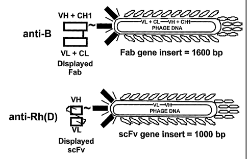
Figure 2 is a diagram of a schematic representation of anti-B (top) and
anti-Rh(D) (bottom) phage-displayed human monoclonal RBC antibodies.
Figure 3 is an image depicting phenotyping RBCs for the blood group B
and Rh(D) antibodies in a multiplex phage antibody assay. Four possible RBC
phenotypes (positive or negative for the blood group B antigen and positive or
negative
for the Rh(D) antigen) were incubated with phage displayed anti-B alone, anti-
Rh(D)
alone, anti-B and anti-Rh(D) together, or buffer. After washing away unbound
phage
reagent, RBCs were resuspended in anti-M13 phage antibody, an aliquot of the
cell
suspension was removed, diluted 200-fold in water, and 2-microliters of the
diluted
phage/lysed RBCs were subjected to PCR. The balance of the anti-M13
resuspended
RBC samples were placed in microtiter plate wells and assayed for
agglutination as
described elsewhere herein (e.g., Siegel et al., 1997, J. Immunol. Meth.
206:73-85). Note
that agglutination (top panel, wells with large crosslinked cell pellets) only
occurs with
the appropriate antibody/cell phenotype combination as expected. Most notably,
only the
appropriate antibody sequence was detected (1600-bp product with RBCs that
expressed
blood group B antigen; 1000-bp product with RBCs that expressed the Rh(D)
antigen)
8
CA 02499355 2005-03-16
WO 2004/027028 PCT/US2003/029231
and there was no detectable background (i.e., no anti-B DNA product with type
0 RBCs
which do not express group A or B antigens; and no anti-Rh(D) DNA product was
detected using Rh(D)-negative cells). For PCR amplification of the inserts,
the forward
primer ("5-prime LC") was as follows: 5'-AAGACAGCTATCGCGATTG-3' (SEQ ID
NO: 1); and the reverse primer ("GBACK") was as follows: 5'-
GCCCCCTTATTAGCGTTTGCCATC-3' (SEQ ID NO:2).
Figure 4, comprising Figures 4A and 4B, depict a diagram illustrating
various phagemid constructs for anti-B-expressing phage particles (Figure 4A)
and anti-
Rh(D)-expressing phage particles (Figure 4B). The diagram illustrates cloning
of inserts
of about 140 basepairs in size (more specifically, 142 bp) into the anti-B
phagemid
("B140") or anti-Rh(D) phagemid ("D140") downstream of the 20-bp T7 RNA
polymerase promoter site. The 142-bp inserts are identical except for an
internal 33-bp
region to which B-or Rh(D)-specific molecular beacons or microarrayed oligos
hybridize
(`B-Beacon/Oligo" and "D-Beacon/Oligo", respectively). B140 and D140 can be
amplified by PCR with an identical set of oligonucleotide primers ("PCR-F" and
"PCR-
R") or transcribed using T7 RNA polymerase. The sequence of the "B 140" insert
is 5'-
TGCTATGTCACTTCCCCTTGGTTCTCTCATCTGGCCTGGTGCAATAGGCCCTGC
ATGCACTGGATGCACTCTATCCCATTCTGCAGCTTCCTCATTGATGGTCTCTTT
TAACATTTGCATGGCTGCTTGATGTCCCCCCACT-3' (SEQ ID NO:3) and the
sequence of the "D140" insert is 5'-
TGCTATGTCACTTCCCCTTGGTTCTCTCATCTGGCCTGGTGCAATAGGCCCTGC
ATGCACTGGATGCACTCTGTTTTACCTCATTATCCTTCTGCCAGCGCTAGCTTT
TAACATTTGCATGGCTGCTTGATGTCCCCCCACT-3' (SEQ ID NO:4). The
forward PCR primer ("PCR-F") is: 5'-TGCTATGTCACTTCCCCTTGGTTCTCT-3'
(SEQ ID NO:5) and the reverse PCR primer ("PCR-R") sequence is: 5-
AGTGGGGGGACATCAAGCAGCCATGCAAAT-3' (SEQ ID NO:6). The B-Beacon
and D-Beacon sequences are as follows, showing the fluorescent derivatives and
the stem
structures in lower case. The "B-Beacon" sequence is as follows: 6-FAM-
gcgagcATCCCATTCTGCAGCTTCCTCATTGATGGTCTCgctcgc-DABCYL (SEQ ID
NO:7. The "D-Beacon" is: TAMRA-
cgagcGTTTTACCTCATTATCCTTCTGCCAGCGCTAGCgctcgc-DABCYL (SEQ ID
9
CA 02499355 2005-03-16
WO 2004/027028 PCT/US2003/029231
NO:8). The upper case letters in the beacon sequences represent the respective
sequences
in B140 and D140 to which the beacons anneal. Therefore, the upper case
letters are the
sequences of the oligonucleotides that are used for the DNA array detection.
That is, a B-
oligo is: 5'-ATCCCATTCTGCAGCTTCCTCATTGATGGTCTC-3' (SEQ ID NO:9), and
a "D-oligo" is: 5'-GTTTTACCTCATTATCCTTCTGCCAGCGCTAGC-3' (SEQ ID
NO: 10).
DETAILED DESCRIPTION OF THE INVENTION
Definitions
As used herein, each of the following terms has the meaning associated
with it in this section.
The articles "a" and "an" are used herein to refer to one or to more than
one (i.e., to at least one) of the grammatical object of the article. By way
of example, "an
element" means one element or more than one element.
By the term "antigen-bearing moiety" as used herein, is meant a molecule
to which an antibody binds. The antigen-bearing moiety may be a membrane bound
protein which is selected from the group consisting of an antigen and a
receptor. In
another aspect, the membrane bound protein is an antigen, such as a red blood
cell
antigen, such as Rh antigen. When the antigen-bearing moiety is a
carbohydrate, it may
be a carbohydrate expressed on a glycolipid, for example, a P blood group
antigen or
other antigen.
As used herein, amino acids are represented by the full name thereof, by
the three letter code corresponding thereto, or by the one-letter code
corresponding
thereto, as indicated in the following table:
Full Name Three-Letter Code One-Letter Code
Aspartic Acid Asp D
Glutamic Acid Glu E
Lysine Lys K
Arginine Arg R
Histidine His H
CA 02499355 2005-03-16
WO 2004/027028 PCT/US2003/029231
Tyrosine Tyr Y
Cysteine Cys C
Asparagine Asn N
Glutamine Gln Q
Serine Ser S
Threonine Thr T
Glycine Gly G
Alanine Ala A
Valine Val V
Leucine Leu L
Isoleucine Ile I
Methionine Met M
Proline Pro P
Phenylalanine Phe F
Tryptophan Trp W
As used herein, to "alleviate" a disease means reducing the severity of one
or more symptoms of the disease.
"Antisense" refers particularly to the nucleic acid sequence of the non-
coding strand of a double stranded DNA molecule encoding a protein, or to a
sequence
which is substantially homologous to the non-coding strand. As defined herein,
an
antisense sequence is complementary to the sequence of a double stranded DNA
molecule encoding a protein. It is not necessary that the antisense sequence
be
complementary solely to the coding portion of the coding strand of the DNA
molecule.
The antisense sequence may be complementary to regulatory sequences specified
on the
coding strand of a DNA molecule encoding a protein, which regulatory sequences
control
expression of the coding sequences.
The terms "bacteriophage" and "phage" are used interchangeably herein
and refer to viruses which infect bacteria. By the use of the terms
"bacteriophage library"
or "phage library" as used herein, is meant a population of bacterial viruses
comprising
heterologous DNA, i.e., DNA which is not naturally encoded by the bacterial
virus.
11
CA 02499355 2005-03-16
WO 2004/027028 PCT/US2003/029231
By the term "applicator," as the term is used herein, is meant any device
including, but not limited to, a hypodermic syringe, a pipette, and the like,
for
administering the bacteriophage expressing a receptor (e.g., an antiglobulin
reagent, an
antibody, an anti-antibody, and the like), a cell, a sample, primers,
molecular beacon
probe, dNTPs, T7 RNA polymerase, and the like, of the invention to a cell, a
sample, and
the like.
"Biological sample," or simply "sample", as that term is used herein,
means a sample, such as one that is, but need not be, obtained from an animal,
which
sample is to be assessed for the presence of a biological organism, or
component thereof,
such that the sample can be used to assess the presence, absence and/or level,
of an
antigen, or ligand, of interest according to the methods of the invention.
Such sample
includes, but is not limited to, any biological fluid (e.g., blood, lymph,
semen, sputum,
saliva, phlegm, tears, and the like), fecal matter, a hair sample, a nail
sample, a brain
sample, a kidney sample, an intestinal tissue sample, a tongue tissue sample,
a heart
tissue sample, a mammary gland tissue sample, a lung tissue sample, an adipose
tissue
sample, a muscle tissue sample, and any sample obtained from an animal that
can be
assayed for the presence or absence of an antigen. Further, the sample can
comprise an
aqueous sample (e.g., a water sample) however obtained, to be assessed for the
presence
of an organism, or a component thereof, such as a drinking water sample,
before or after
any treatment, wherein the presence of a biological organism (e.g., a
Cryptosporidium
organism) is assessed.
As used herein, the term "fragment" as applied to a nucleic acid, may
ordinarily be at least about 20 nucleotides in length, preferably, at least
about 30
nucleotides, more typically, from about 40 to about 50 nucleotides,
preferably, at least
about 50 to about 80 nucleotides, even more preferably, at least about 80
nucleotides to
about 90 nucleotides, yet even more preferably, at least about 90 to about
100, even more
preferably, at least about 100 nucleotides to about 150 nucleotides, yet even
more
preferably, at least about 150 to about 200, even more preferably, at least
about 200
nucleotides to about 250 nucleotides, yet even more preferably, at least about
250 to
about 300, more preferably, from about 300 to about 350 nucleotides,
preferably, at least
12
CA 02499355 2005-03-16
WO 2004/027028 PCT/US2003/029231
about 350 to about 360 nucleotides, and most preferably, the nucleic acid
fragment will
be greater than about 365 nucleotides in length.
As used herein, the term "fragment" as applied to a polypeptide, may
ordinarily be at least about 20 amino acids in length, preferably, at least
about 30 amino
acids, more typically, from about 40 to about 50 amino acids, preferably, at
least about 50
to about 80 amino acids, even more preferably, at least about 80 amino acids
to about 90
amino acids, yet even more preferably, at least about 90 to about 100, even
more
preferably, at least about 100 amino acids to about 120 amino acids, and most
preferably,
the amino acid fragment will be greater than about 123 amino acids in length.
By the term "Fab/phage" as used herein, is meant a phage particle which
expresses the Fab portion of an antibody.
By the term "scFv/phage" are used herein, is meant a phage particle which
expresses the Fv portion of an antibody as a single chain.
"Phage," or "phage particle," as these terms are used herein, include that
contain phage nucleic acid encoding, inter alia, an antibody. This is because,
as would
be appreciated by the skilled artisan, unlike peptide phage display (where the
peptide
DNA insert is small and it is actually cloned into the phage DNA), the larger
scFv or Fab
DNA inserts are actually cloned into, among other things, a plasmid. Thus, the
nucleic
acid encoding the antibody, e.g., a plasmid such as, but not limited to,
pComb3, not only
20' comprises a plasmid origin of replication, but also a phage (e.g., M13)
origin of
replication sequence and an M13 packaging sequence, so that when the nucleic
acid is
produced, a helper phage can be used to provide the required phage (e.g., M13)
proteins
in trans to make "phage-like" particles. That is, these particles resemble
phage on the
outside, but on the inside they contain plasmid (also referred to as a
"phagemid") DNA.
In other words, the phagemid DNA need not encode any M13 phage proteins,
except a
piece of M13 gene III fused to the DNA for antibody or peptide. Thus, it
should be
understood that the terms "phage," "phage particle," "phage-like particle" and
"phagemid" are used interchangeably herein.
A "disease" is a state of health of an animal wherein the animal cannot
maintain homeostasis, and wherein if the disease is not ameliorated, then the
animal's
health continues to deteriorate.
13
CA 02499355 2005-03-16
WO 2004/027028 PCT/US2003/029231
In contrast, a "disorder" in an animal is a state of health in which the
animal is able to maintain homeostasis, but in which the animal's state of
health is less
favorable than it would be in the absence of the disorder. Left untreated, a
disorder does
not necessarily cause a further decrease in the animal's state of health.
"Homologous" as used herein, refers to the subunit sequence similarity
between two polymeric molecules, e.g., between two nucleic acid molecules,
e.g., two
DNA molecules or two RNA molecules, or between two polypeptide molecules. When
a
subunit position in both of the two molecules is occupied by the same
monomeric
subunit, e.g., if a position in each of two DNA molecules is occupied by
adenine, then
they are homologous at that position. The homology between two sequences is a
direct
function of the number of matching or homologous positions, e.g., if half
(e.g., five
positions in a polymer ten subunits in length) of the positions in two
compound
sequences are homologous then the two sequences are 50% homologous, if 90% of
the
positions, e.g., 9 of 10, are matched or homologous, the two sequences share
90%
homology. By way of example, the DNA sequences 5'ATTGCC3' and 5'TATGGC3'
share 50% homology.
"Instructional material," as that term is used herein, includes a publication,
a recording, a diagram, or any other medium of expression which can be used to
communicate the usefulness of the nucleic acid, peptide, and/or compound of
the
invention in the kit for detecting the presence of an antigen-bearing moiety
on a cell of
interest, and/or for detecting an autoantibody in serum. The instructional
material of the
kit may, for example, be affixed to a container that contains the nucleic
acid, peptide,
and/or compound of the invention or be shipped together with a container which
contains
the nucleic acid, peptide, and/or compound. Alternatively, the instructional
material may
be shipped separately from the container with the intention that the recipient
uses the
instructional material and the compound cooperatively.
An "isolated nucleic acid" refers to a nucleic acid segment or fragment
which has been separated from sequences which flank it in a naturally
occurring state,
e.g., a DNA fragment which has been removed from the sequences which are
normally
adjacent to the fragment, e.g., the sequences adjacent to the fragment in a
genome in
which it naturally occurs. The term also applies to nucleic acids that have
been
14
CA 02499355 2005-03-16
WO 2004/027028 PCT/US2003/029231
substantially purified from other components that naturally accompany the
nucleic acid,
e.g., RNA or DNA or proteins, which naturally accompany it in the cell. The
term
therefore includes, for example, a recombinant DNA which is incorporated into
a vector,
into an autonomously replicating plasmid or virus, or into the genomic DNA of
a
prokaryote or eukaryote, or which exists as a separate molecule (e.g., as a
cDNA or a
genomic or cDNA fragment produced by PCR or restriction enzyme digestion)
independent of other sequences. It also includes a recombinant DNA that is
part of a
hybrid gene encoding additional polypeptide sequence.
"Recombinant polynucleotide" refers to a polynucleotide having
sequences that are not naturally joined together. An amplified or assembled
recombinant
polynucleotide may be included in a suitable vector, and the vector can be
used to
transform a suitable host cell.
A recombinant polynucleotide may serve a non-coding function (e.g.,
promoter, origin of replication, ribosome-binding site, etc.) as well.
A host cell that comprises a recombinant polynucleotide is referred to as a
"recombinant host cell." A gene that is expressed in a recombinant host cell
wherein the
gene comprises a recombinant polynucleotide, produces a "recombinant
polypeptide."
A "recombinant polypeptide" is one that is produced upon expression of a
recombinant polynucleotide.
A "vector" is a composition of matter which comprises an isolated nucleic
acid and which can be used to deliver the isolated nucleic acid to the
interior of a cell.
Numerous vectors are known in the art including, but not limited to, linear
polynucleotides, polynucleotides associated with ionic or amphiphilic
compounds,
plasmids, and viruses. Thus, the term "vector" includes an autonomously
replicating
plasmid or a virus. The term should also be construed to include non-plasmid
and non-
viral compounds which facilitate transfer of nucleic acid into cells, such as,
for example,
polylysine compounds, liposomes, and the like. Examples of viral vectors
include, but
are not limited to, adenoviral vectors, adeno-associated virus vectors,
retroviral vectors,
and the like.
"Expression vector" refers to a vector comprising a recombinant
polynucleotide comprising expression control sequences operatively linked to a
CA 02499355 2005-03-16
WO 2004/027028 PCT/US2003/029231
nucleotide sequence to be expressed. An expression vector comprises sufficient
cis-
acting elements for expression; other elements for expression can be supplied
by the host
cell or in an in vitro expression system. Expression vectors include all those
known in
the art, such as cosmids, plasmids (e.g., naked or contained in liposomes) and
viruses that
incorporate the recombinant polynucleotide.
By describing two polynucleotides as "operably linked" is meant that a
single-stranded or double-stranded nucleic acid moiety comprises the two
polynucleotides arranged within the nucleic acid moiety in such a manner that
at least one
of the two polynucleotides is able to exert a physiological effect by which it
is
characterized upon the other. By way of example, a promoter operably linked to
the
coding region of a gene is able to promote transcription of the coding region.
Preferably, when the nucleic acid encoding the desired protein further
comprises a promoter/regulatory sequence, the promoter/regulatory is
positioned at the 5'
end of the desired protein coding sequence such that it drives expression of
the desired
protein in a cell. Together, the nucleic acid encoding the desired protein and
its
promoter/regulatory sequence comprise a "transgene."
As used herein, the term "promoter/regulatory sequence" means a nucleic
acid sequence which is required for expression of a gene product operably
linked to the
promoter/regulatory sequence. In some instances, this sequence may be the core
promoter sequence and in other instances, this sequence may also include an
enhancer
sequence and other regulatory elements which are required for expression of
the gene
product. The promoter/regulatory sequence may, for example, be one which
expresses
the gene product in a tissue specific manner.
A "constitutive" promoter is a nucleotide sequence which, when operably
linked with a polynucleotide which encodes or specifies a gene product, causes
the gene
product to be produced in a living human cell under most or all physiological
conditions
of the cell.
An "inducible" promoter is a nucleotide sequence which, when operably
linked with a polynucleotide which encodes or specifies a gene product, causes
the gene
product to be produced in a living human cell substantially only when an
inducer which
corresponds to the promoter is present in the cell.
16
CA 02499355 2005-03-16
WO 2004/027028 PCT/US2003/029231
A "tissue-specific" promoter is a nucleotide sequence which, when
operably linked with a polynucleotide which encodes or specifies a gene
product, causes
the gene product to be produced in a living human cell substantially only if
the cell is a
cell of the tissue type corresponding to the promoter.
A "polyadenylation sequence" is a polynucleotide sequence which directs
the addition of a poly A tail onto a transcribed messenger RNA sequence.
A "polynucleotide" means a single strand or parallel and anti-parallel
strands of a nucleic acid. Thus, a polynucleotide may be either a single-
stranded or a
double-stranded nucleic acid.
The term "nucleic acid" typically refers to large polynucleotides.
The term "oligonucleotide" typically refers to short polynucleotides,
generally, no greater than about 50 nucleotides. It will be understood that
when a
nucleotide sequence is represented by a DNA sequence (i.e., A, T, G, C), this
also
includes an RNA sequence (i.e., A, U, G, C) in which "U" replaces "T."
In the context of the present invention, the following abbreviations for the
commonly occurring nucleic acid bases are used. "A" refers to adenosine, "C"
refers to
cytidine, "G" refers to guanosine, "T" refers to thymidine, and "U" refers to
uridine.
Conventional notation is used herein to describe polynucleotide
sequences: the left-hand end of a single-stranded polynucleotide sequence is
the 5'-end;
the left-hand direction of a double-stranded polynucleotide sequence is
referred to as the
5'-direction.
The direction of 5' to 3' addition of nucleotides to nascent RNA
transcripts is referred to as the transcription direction. The DNA strand
having the same
sequence as an mRNA is referred to as the "coding strand"; sequences on the
DNA strand
which are located 5' to a reference point on the DNA are referred to as
"upstream
sequences"; sequences on the DNA strand which are 3' to a reference point on
the DNA
are referred to as "downstream sequences."
A "portion" of a polynucleotide means at least at least about twenty
sequential nucleotide residues of the polynucleotide. It is understood that a
portion of a
polynucleotide may include every nucleotide residue of the polynucleotide.
17
CA 02499355 2005-03-16
WO 2004/027028 PCT/US2003/029231
"Primer" refers to a polynucleotide that is capable of specifically
hybridizing to a designated polynucleotide template and providing a point of
initiation for
synthesis of a complementary polynucleotide. Such synthesis occurs when the
polynucleotide primer is placed under conditions in which synthesis is
induced, i.e., in
the presence of nucleotides, a complementary polynucleotide template, and an
agent for
polymerization such as DNA polymerase. A primer is typically single-stranded,
but may
be double-stranded. Primers are typically deoxyribonucleic acids, but a wide
variety of
synthetic and naturally occurring primers are useful for many applications. A
primer is
complementary to the template to which it is designed to hybridize to serve as
a site for
the initiation of synthesis, but need not reflect the exact sequence of the
template. In such
a case, specific hybridization of the primer to the template depends on the
stringency of
the hybridization conditions. Primers can be labeled with, e.g., chromogenic,
radioactive,
or fluorescent moieties and used as detectable moieties.
"Probe" refers to a polynucleotide that is capable of specifically
hybridizing to a designated sequence of another polynucleotide. A probe
specifically
hybridizes to a target complementary polynucleotide, but need not reflect the
exact
complementary sequence of the template. In such a case, specific hybridization
of the
probe to the target depends on the stringency of the hybridization conditions.
Probes can
be labeled with, e.g., chromogenic, radioactive, or fluorescent moieties and
used as
detectable moieties.
"Recombinant polynucleotide" refers to a polynucleotide having
sequences that are not naturally joined together. An amplified or assembled
recombinant
polynucleotide may be included in a suitable vector, and the vector can be
used to
transform a suitable host cell.
A recombinant polynucleotide may serve a non-coding function (e.g.,
promoter, origin of replication, ribosome-binding site, etc.) as well.
A "recombinant polypeptide" is one which is produced upon expression of
a recombinant polynucleotide.
"Polypeptide" refers to a polymer composed of amino acid residues,
related naturally occurring structural variants, and synthetic non-naturally
occurring
analogs thereof linked via peptide bonds, related naturally occurring
structural variants,
18
CA 02499355 2005-03-16
WO 2004/027028 PCT/US2003/029231
and synthetic non-naturally occurring analogs thereof. Synthetic polypeptides
can be
synthesized, for example, using an automated polypeptide synthesizer.
The term "protein" typically refers to large polypeptides.
The term "peptide" typically refers to short polypeptides.
Conventional notation is used herein to portray polypeptide sequences: the
left-hand end of a polypeptide sequence is the amino-terminus; the right-hand
end of a
polypeptide sequence is the carboxyl-terminus.
As used herein, the term "reporter gene" means a gene, the expression of
which can be detected using a known method. By way of example, the Escherichia
coli
lacZ gene may be used as a reporter gene in a medium because expression of the
lacZ
gene can be detected using known methods by adding a chromogenic substrate,
e.g., o-
nitrophenyl ,8-D-galactopyranoside, to the medium (Gerhardt et al., eds.,
1994, Methods
for General and Molecular Bacteriology, American Society for Microbiology,
Washington, DC, p. 574).
A "receptor" is a compound that specifically binds with a ligand. This
term includes a protein, such as an antibody, an antiglobulin reagent, and the
like, that
when expressed by a phage and contacted with its cognate ligand, binds
specifically
therewith.
The term "ligand," as used herein, refers to any protein or proteins that can
interact with a receptor binding domain, thus having a "binding affinity" for
such domain.
Ligands can be soluble or membrane bound, and they can be a naturally
occurring
protein, or synthetically or recombinantly produced. The "ligand" can also be
a
nonprotein molecule that acts as ligand when it interacts with the receptor
binding
domain. Interactions between the ligand and receptor binding domain include,
but are
not limited to, any,covalent or non-covalent interactions. The receptor
binding domain is
any region of the receptor molecule that interacts directly or indirectly with
the ligand.
By the term "specifically binds," as used herein, is meant a molecule, e.g.,
a protein, a nucleic acid, an antibody, a compound, and the like, which
recognizes and
binds a specific molecule, but does not substantially recognize or bind other
molecules in
a sample. For instance, an antibody which recognizes and binds a cognate
ligand (i.e., an
19
CA 02499355 2005-03-16
WO 2004/027028 PCT/US2003/029231
antigen-bearing moiety present on a cell) in a sample, but does not
substantially
recognize or bind other molecules in the sample.
To "treat" a disease as the term is used herein, means to reduce the
frequency of the disease or disorder reducing the frequency with which a
symptom of the
one or more symptoms disease or disorder is experienced by an animal.
Description
The invention relates to methods for detecting the presence of a molecule
of interest on a cell or in a biological sample. Typically, a red blood
antigen expressed
on a RBC surface is detected, but the invention encompasses detecting the
presence of
numerous antigens of interest on a wide plethora of cells, including, but not
limited to,
red and white blood cells, as well as platelets, and cells used for
transplantation therapy,
and the identification of antigens on cells for forensic purposes (e.g., hair,
skin, nail,
sperm, saliva, and other cells), among many other uses.
The invention also relates to detection of an antigen of interest in a
biological sample. Such a sample includes an aqueous sample to detect the
presence of
any organism, or component thereof, in the sample.
The invention relates to using an antibody, specific for a known antigen,
displayed by a phage (e.g., an M13, T7, lambda, eukaryotic, and the like), to
detect the
presence of the antigen on a cell or in a biological sample. More
specifically, phage
specifically bound with a cell are detected by assaying for the nucleic acid
contained in
the phage particle. That is, the nucleic acid sequence of the nucleic acid
contained in the
phagemid is at least partially known, such that techniques for detecting
nucleic acids can
be used to assess the presence of the sequence, thereby detecting, in a novel
process
referred to herein as "phenotyping-by-reagent-genotyping", the antigen.
Essentially, the bacteriophage nucleic acid acts like a tag for detecting an
antigen recognized by the antibody encoded by the phage. In this way, the high
sensitivity and high throughput screening properties of nucleic acid detection
methods
can be applied to the immunological detection of an antigen, thereby combining
the
advantages of both technologies. The crucial features of this approach are
that the
specificity of the antibody displayed by the bacteriophage and the nucleic
acid sequence,
CA 02499355 2005-03-16
WO 2004/027028 PCT/US2003/029231
or a portion thereof, of the DNA contained within the phage, both be known. It
would be
understood, based upon the disclosure provided herein, that the precise nature
of the
antigen, be it a protein, carbohydrate, lipid, or any other compound,
recognized by the
antibody, need not be known, only that the specificity of the antibody for
that antigen be
known. For instance, where an antibody is known to bind with and identify a
cancer cell
(or any cell associated with a disease), but not bind with an otherwise
identical cell that is
not cancerous (or associated with a disease), the antibody can be used to
detect a cancer
(or disease state) using the methods of the invention. That is, the antibody
binding with a
test cell or a biological sample, can be detected by detecting the nucleic
acid present in
the phage particle encoding the antibody portion, thereby detecting a cancer
cell, without
having to know the precise nature of the antigen present on the cancer (or
disease-
associated) cell.
The invention further relates to detection of multiple antigens of interest
on a cell in a single tube assay. That is, bacteriophage that encode
antibodies specific for
at least two different antigens can be used to detect those antigens on a
cell. More
specifically, each phage encodes an antibody that specifically binds with a
known antigen
and each phage encodes an antibody that recognizes a different antigen, or
antigen-
moiety. Further, each phage contains a DNA molecule comprising a sequence that
is
known, wherein the sequence differs between the phage. Using this approach,
the
presence of a plurality of antigens of interest can be readily assessed by
simply using a
panel of phage, each displaying an antibody specific for one of the antigens,
where the
nucleic acid molecule of each phage comprises a known sequence that is
distinguishable
from that of any other phage in the panel. In this way, multiple antigens can
be assayed
for using a single reaction step. This "multiplexing" method is not possible
using
conventional methods that identify the binding of antigen-specific antibodies
to a cell
since the secondary anti-antibody antibody used to detect the antigen-specific
antibodies
typically cross-reacts with all the antigen-binding antibodies, or it cannot
be determined
which antigen-specific antibody the second antibody is bound with. In the case
of
conventional methods for phenotyping red blood cells, in which antibodies
directly
agglutinate the appropriate cell type (i.e., no secondary antibody needed), if
mixed
together, it would likewise not be possible to determine which antigen-
specific antibody
21
CA 02499355 2005-03-16
WO 2004/027028 PCT/US2003/029231
was responsible for the agglutination. This multiplex approach allows the
rapid
simultaneous detection of a plurality of antigens using only a single sample.
Further, the invention relates to identification of anti-red blood cell
antibodies in serum. That is, a panel of RBCs, expressing various known
antigens on
their surfaces, can be contacted with a serum sample. Reagent RBCs, expressing
characterized antigens, are commercially available (e.g., Johnson & Johnson,
Raritan,
NJ). The cells are then washed to remove any antibodies non-specifically
adhering to the
cells and the cells are then contacted with bacteriophage displaying an anti-
globulin
reagent.
Additionally, autoantibodies present in a patient can be detected by
obtaining RBCs from the patient, washing them to remove any antibodies and/or
complement that is non-specifically bound with the cells, and the cells can
then be
contacted with a phage expressing an antihumanglobulin reagent. Thus, by
detecting a
nucleic acid sequence contained by the phage, the presence of autoantibody on
the patient
cells, as well as the presence of complement deposited on the cells due to the
autoantibody, can be readily detected according to the novel "phenotyping-by-
reagent-
genotyping" methods disclosed herein.
Conventionally, screening and identification of serum antibodies using
reagent red cells displaying known antigens is referred to in the art as an
"antiglobulin
test", one such test is a Coombs reaction. These assays detect the presence of
an
antibody, or complement deposited thereby, on a cell of interest. Because
complement,
while not an antibody, is considered a "globulin", the reagents used to detect
antibodies
and/or complement are referred to in the art as "antiglobulin" reagents.
These assays, which detect antibodies and/or complement fragments (e.g.,
C3d) on patient red cells to detect anti-red cell autoantibodies, or the
complement they
deposit, and also to detect patient alloantibodies, or the complement they
deposit, can be
used to identify autoantibodies, alloantibodies, or both, that could be
destroying
autologous cells or transfused cells in a hemolytic transfusion reaction.
As used herein, an "antiglobulin reagent" is a reagent that can detect
antibodies, complement, or both. Thus, the present invention includes, as
would be
understood by one skilled in the art armed with the teachings provided herein,
22
CA 02499355 2005-03-16
WO 2004/027028 PCT/US2003/029231
antiglobulin reagents comprising, among others, e.g., anti-antibody
antibodies, anti-
complement antibodies, Protein A, Protein G, or Protein L, that is, the
invention
encompasses expression by phage of a wide plethora of reagents that would be
understood by the skilled artisan to specifically bind with a globulin, such
as antibody,
complement, and the like. That is, the present invention includes using an
antiglobulin
reagent expressed by a phage including, but not limited to, an "anti-antibody
antibody",
an anti-complement, and any reagent known to bind a globulin (e.g., an
antibody,
complement, and the like). Additionally, phage expressing Protein A, or an
immunoglobulin-binding domain thereof, have been described previously (e.g.,
Djojonegoro et al., 1994, Bio/Technol. 12:169-172). Such antiglobulin reagent-
expressing phage can be used in the methods disclosed herein as would be
understood by
one skilled in the art armed with the teachings provided herein.
The invention relates to identifying autoantibodies in a serum sample
obtained from a patient, or autoantibodies or complement fragments pre-
deposited on
patient cells in vivo, both characteristics of a disease such as, but not
limited to,
autoimmune hemolytic anemia. That is, serum obtained from the patient is
contacted
with an aliquot of reagent RBCs, such as those that are commercially
available. RBC
autoantibodies bind to common antigens present on essentially all red cells,
not just of the
patient. Thus, the patient cannot be transfused with blood from another human
since the
autoantibodies present in the patient serum with also react with the donor
RBCs.
Because the patient's RBCs are already be coated with the autoantibodies,
those
autoantibodies already on the cells from having been bound in vivo can be
detected
according to the methods of the invention by assaying the cells directly using
antihumanglobulin reagent expressed on a phage. Alternatively, detecting
autoantibodies is performed the same way as is detection of alloantibodies -by
contacting the patient serum with reagent red cells. In the case of
alloantibodies, only
certain reagent RBCs will bind the antibodies, and knowing the precise
phenotype of
those cells identifies the antigen specificity. In the case of autoantibodies,
typically all
reagent red cells will bind the antibodies because the autoantigens are
present on all cells.
Any antibody specifically bound with the RBCs is then detected according to
the
methods of the invention such as, as more fully disclosed elsewhere herein, by
contacting
23
CA 02499355 2005-03-16
WO 2004/027028 PCT/US2003/029231
the cells with a phage expressing an antiglobulin reagent and detecting the
binding of the
phage with the cells by detecting a nucleic acid contained by the phage, i.e.,
by
performing "phenotyping-by-reagent-genotyping" according to the methods of the
invention. In this way, autoantibodies present in human serum can be readily
detected
using the methods disclosed herein analogous to the conventional "indirect
antiglobulin
test". Furthermore, by contacting patient RBCs with antiglobulin-expressing
phage
particles and detecting the binding of the phage with the cells by detecting a
nucleic acid
contained by the phage, one can detect the presence of in vivo-deposited
autologous
antibodies and/or complement fragments on patient RBCs. This assay is
analogous to the
conventional "direct antiglobulin test".
Further, the invention relates to performing compatibility testing between
patient serum and red cells drawn from prospective units of blood to be
transfused to the
patient (i.e., patient/donor "crossmatching"). That is, an aliquot of RBCs
from a
prospective unit of donor blood can be contacted with a serum sample from a
potential
transfusion recipient. The cells are then washed to remove any antibodies non-
specifically adhering to the cells and the cells are then contacted with
bacteriophage
displaying an antiglobulin reagent. Thus, the present invention provides
methods for
detecting an alloantibody in a patient that is to be transfused thereby
allowing proper
patient/donor crossmatching to prevent incompatible transfusion.
I. Methods
A. Methods of detecting an antigen
The invention includes a method for detecting the presence of an antigen-
bearing moiety on a cell. The method comprises contacting a cell with a
bacteriophage
expressing an antibody that is known to specifically bind with the antigen-
bearing moiety
when it is present on a cell. Such phage-displayed antibodies, as well as
methods for
their production, are well-known in the art, and are described in, among
others, U.S.
Patent No. 5,876,925, No. 5,985,543, and No. 6,255,455, all to Siegel. These
antibody-
displaying bacteriophage are exemplified herein by phage displaying anti-Rh(D)
and anti-
B specific antibodies. However, the skilled artisan would understand, based
upon the
disclosure provided herein, that the invention is not limited to these, or any
other,
24
CA 02499355 2011-06-14
WO 2004/027028 PCTNS2003/029231
particular antibodies displayed on the specific bacteriophage disclosed
herein. Rather,
the antibody displayed by the phage can be specific for any cell component and
techniques for producing phage-displaying antibodies to antigens of interest
are well-
known in the art, and are encompassed in the present invention.
The procedures for making a bacteriophage library comprising
heterologous DNA are well known in the art and are described herein in, as
well as in for
example, in Sambrook et al., supra. Bacteriophage which encode a desired
antibody can
be engineered such that the antibody protein is displayed on the surface
thereof in such a
manner that it is available for binding to its corresponding binding protein,
e.g., the
antigen against which the antibody is directed. Thus, when bacteriophage which
express
a specific antibody are incubated in the presence of a cell which expresses
the
corresponding antigen, the bacteriophage will bind to the cell. Bacteriophage
which do
not express the antibody will not bind to the cell. Such panning techniques
are well
known in the art and are described for example, in Wright et al. (supra).
Processes such as those described above, have been developed for the
production of human antibodies using M13 bacteriophage display (Burton et al.,
1994,
Adv. Immunol. 57:191-280). Methods relating to production of such display
libraries,
and the screening thereof, are set forth in U.S. Patent No. 6,255,455, to
Siegel.
Essentially, a cDNA
library is generated from mRNA obtained from a population of antibody-
producing cells.
The mRNA encodes rearranged inununoglobulin genes and thus, the CDNA encodes
the
same. Amplified cDNA is cloned into M 13 expression vectors (or phagemids with
M 13
packaging signals) creating a library of phage which express human Fab (or
scFv)
fragments on their surface. Phage which display the antibody of interest are
selected by
antigen binding and are propagated in bacteria to produce soluble human Fab
(or scFv)
immunoglobulin. Thus, in contrast to conventional monoclonal antibody
synthesis, this
procedure immortalizes DNA encoding human inununoglobulin rather than cells
which
express human immunoglobulin.
Although the bacteriophage displaying antibodies of interest herein are
exemplified by M13 phage, the present invention is not limited to these, or
any other,
vector displaying an antibody. Instead, one skilled in the art would
appreciate, armed
CA 02499355 2005-03-16
WO 2004/027028 PCT/US2003/029231
with the teachings provided herein, that any vector that can display an
antibody, wherein
the vector comprises a nucleic acid the sequence of which is at least
partially known, can
be used in the methods disclosed herein. Therefore, while the antibody-
displaying
bacteriophage disclosed herein are exemplified by M13, other bacteriophage,
such as
lambda phage or T7 phage, can also be useful in the method of the invention.
Lambda
phage display libraries have been generated which display peptides encoded by
heterologous DNA on their surface (Sternberg et al., 1995, Proc. Natl. Acad.
Sci. USA
92:1609-1613) as have T7 phage display libraries (Hansen et al., 2001, Int. J.
Oncol.
19:1303-1309).
Moreover, it is contemplated that the method of the invention may be
extended to include viruses other than bacteriophage, such as eukaryotic
viruses. In fact,
eukaryotic viruses can be generated which encode genes suitable for delivery
to a
mammal and which encode and display an antibody capable of targeting a
specific cell
type or tissue into which the gene is to be delivered. For example, retroviral
vectors have
been generated which display functional antibody fragments (Russell et al.,
1993, Nucl.
Acids Res. 21:1081-1085). These, and any other vector expressing an antibody
can be
used in the methods of the invention and are encompassed thereby.
Furthermore, while the method of the invention as exemplified herein
describes using phage which encode the Fab portion or an scFv portion of an
antibody
molecule, the method should not be construed to be limited solely to the use
of phage
encoding Fab or scFv antibodies. Fab molecules comprise the entire Ig light
chain, that
is, they comprise both the variable and constant region of the light chain,
but include only
the variable region and first constant region domain (CH1) of the heavy chain.
Single
chain antibody molecules comprise a single chain of protein comprising the Ig
Fv
fragment. An Ig Fv fragment includes only the variable regions of the heavy
and light
chains of the antibody, having no constant region contained therein. Phage
libraries
comprising scFv DNA may be generated following the procedures described in
Marks et
al., 1991, J. Mol. Biol. 222:581-597. Panning of phage so generated for the
isolation of a
desired antibody is conducted as described herein for phage libraries
comprising Fab
DNA.
26
CA 02499355 2005-03-16
WO 2004/027028 PCT/US2003/029231
The invention should also be construed to include synthetic phage display
libraries in which the heavy and light chain variable regions may be
synthesized such that
they include nearly all possible specificities. Therefore, antibody-displaying
libraries can
be "natural" or "synthetic" (Barbas, 1995, Nature Medicine 1:837-839; de Kruif
et al.,
1995, J. Mol Biol. 248:97-105). Antibody-displaying libraries comprising
"natural"
antibodies are generated as described in, e.g., U.S. Patent No. 5,876,925, to
Siegel.
Antibody-displaying libraries comprising "synthetic" antibodies are generated
following
the procedure described in Barbas (1995, supra) and the references cited
therein.
The skilled artisan would appreciate, based upon the disclosure provided
herein, that the red blood cell antibodies to which antibodies can be
generated using
methods known in the art and can then be used in the method of the invention
include,
but are not limited to, Rh antigens, including Rh(D), Rh(C), Rh(c), Rh(E),
Rh(e), and
other non-Rh antigens, including red blood cell antigens in the Kell, Duffy,
Lutheran and
Kidd blood groups.
Thus, the method of the invention can be used for detection of any RBC
antigen or other cell antigen, such as, but not limited to, tumor-specific
antigen, bacterial
antigens, and the like. The method of the invention is also useful for typing
platelets by
generating phage antibodies specific for a number of clinically important
platelet
antigens, notably, HPA-la/lb, HPA-2a/2b, HPA-3a/3b, and the like.
The invention is further useful for typing donor white blood cells for HLA
antigens for the purposes of matching donors and recipients for potential
transplant
matching in the case of both solid (for example, kidney, heart, liver, lung)
and non-solid
(for example, bone marrow) organ or tissue transplanting.
In addition, the methods of the present invention can be used for forensic
purposes, to detect any antigen of interest in a sample, where the sample can
be, but is not
limited to, bone, hair, skin, semen, saliva, or any other sample that can be
obtained from
an organism or biological sample. The only feature required is that the sample
contain an
antigen that can be specifically recognized by an antibody expressed by a
bacteriophage,
or other antibody-displaying vector. Thus, the present invention is not
limited in any way
to the detection of any particular antigen; instead, the invention encompasses
detecting a
27
CA 02499355 2005-03-16
WO 2004/027028 PCT/US2003/029231
wide plethora of antigens of interest using the novel "phenotyping-by-reagent-
genotyping" detection methods disclosed herein.
Thus, the invention encompasses detecting an antigen of interest on a red
blood cell, referred to herein as "phenotyping," by detecting the binding of a
phage
expressing an anti-red blood cell antibody, where the phage is detected by
detecting a
known sequence present in the nucleic acid contained by the phage particle,
which is
referred to herein as "phenotyping-by-reagent-genotyping." Further, the
invention
includes screening of patient sera for anti-red blood cell antibodies using
phage particles
that display anti-human IgG (or anti-IgM or anti-kappa/lambda light chain
antibody
which would pick up any Ig isotype). Again, the phage bound with the RBCs is
detected
by detecting a nucleic acid sequence present in the nucleic acid contained by
the phage.
Additionally, the invention encompasses using the phenotyping-by-
reagent-genotyping method in an immune assay, whether the antigen being
detected is on
a cell or not (e.g., antigens such as, but not limited to, any measured for
research or
clinical purposes from a cytokine to HCG for a pregnancy test). That is, the
present
invention combines the specificity conferred by immunoglobulins for a given
substance,
which specificity takes into account any post-translational modification
(e.g.,
phosphorylation, glycosylation, and,the like), with the sensitivity conferred
by nucleic
acid detection methods - as well as the ability to perform multiplex assays.
That is, a
sample being assayed would be applied such that its components are affixed to
a solid
support, such as coating the well of a plate for an ELISA, nitrocellulose
filter, bead, or
any other solid support, and the phage expressing a protein that specifically
binds with a
cognate ligand can be allowed to bind with the components affixed to the solid
support.
Any phage specifically bound to a cognate ligand can be detected by detecting
a known
nucleic acid sequence specified by the nucleic acid contained within the
phage. Thus, the
presence of any ligand of interest can be detected using the "phenotyping-by-
reagent-
genotyping" method disclosed herein even where the sample being assayed does
not
comprise a cell.
Moreover, the skilled artisan would appreciate, based upon the disclosure
provided herein, that the invention encompasses the phenotyping of other blood
cells
(e.g., platelets, white cells, and the like) and the detection of antibodies
to those cells in
28
CA 02499355 2005-03-16
WO 2004/027028 PCT/US2003/029231
the blood (e.g., anti-platelet auto- or alloantibodies, anti-HLA antibodies,
etc.), such that
the present invention is not limited to red blood cells. Indeed, the invention
is not limited
to blood cells at all, but can be used to detect any molecule of interest
present on any kind
of cell. Thus, one skilled in the art would appreciate, based upon the
disclosure provided
herein, that the present invention includes, but is not limited to, detecting
a molecule of
interest on a cell where flow cytometry would otherwise be used such that the
wide
plethora of antibodies now available (e.g., hundreds of anti-CD antibodies,
such as anti-
CD4 or CD-8 for helper/suppressor T cells, anti-CD20 for B cells, and the
like) can be
expressed on a phage and used to detect, according to the novel methods
disclosed
elsewhere herein, whether the antigen is present in a cell. The present
invention includes
using antibodies to be developed in the future to antigens of interest as
these are
developed and used according to the methods disclosed herein.
The skilled artisan would appreciate, based upon the teachings provided
herein, that detection of any molecule of interest, for instance, with regard
to forensic
application of the methods disclosed herein, provides an important advantage
over
present methods in that many antigens important for identifying the origin of
fluids
(blood or soluble substances in saliva, and the like) are carbohydrates (like
the A and B
antigens). Using genetic testing on the miniscule spot for DNA cannot amplify
the DNA
that encodes carbohydrates because DNA does not encode carbohydrates which are
products of post-translational modification of proteins. Prior art methods
relating to
carbohydrate detection are limited to detecting the DNA for the enzymes (e.g.,
the
glycosytransferases) that are responsible for assembling the sugar moieties
onto the
protein or lipid. The problem with conventional detection assays is that the
ultimate
expression of a particular sugar is the result of the inheritance of a number
of enzymes
that act in precise sequence to assemble the chains such that the genes for
all of the
enzymes would need to be detected in order to identify the identity of the
person the
sample was derived from. For example, in order for an individual to be blood
group A,
the enzyme that adds N-acetylgalactosamine onto its precursor sugar is
required, as is the
enzyme (a fucosyl transferase) to assemble the precursor sugar. Other
carbohydrates
(like P) are even more complicated in their structures and assembly. If the
sample
comprises a mixture of secretions in one spot from different individuals, DNA
testing
29
CA 02499355 2005-03-16
WO 2004/027028 PCT/US2003/029231
would pick up all enzymes and the test would not be able to distinguish
whether one
person had all the enzymes and could make a particular sugar antigen or if the
sample
comprised DNA from various persons who could each only produce the various
sugar
components. Unlike conventional nucleic acid-based testing, the present
invention
provides the advantage of combining the exquisite specificity of an antibody
that is
capable of recognizing a complex structure, such as a glycan, and the ability
to detect
miniscule quantities of a nucleic acid; thus, detection of the nucleic acid
contained by the
phage, combined with the specificity of an antibody, provide a novel assay
with the
extraordinary sensitivity and specificity required in forensic uses.
One skilled in the art, based upon the disclosure provided herein, would
understand that while the term "phenotyping" is generally used in the art to
detecting a
characteristic demonstrated by a cell, or organism, the term as used herein
with regard to
"phenotyping-by-reagent-genotyping", relates to the identification of any
antigen of
interest, whether or not the antigen is associated with a cell, by detecting a
nucleic acid
sequence. Thus, for instance, the identification of a drug in a dried spot on
a car door
using a phage-displayed anti-drug antibody according to the methods of the
invention,
would be "phenotyping" as the term is used herein. Therefore, the methods of
the
invention, where an antibody expressed by a phage binds with a cognate antigen
and the
antigen is detected by assaying for a nucleic acid sequence present in the
phage DNA, is
"phenotyping" as used herein.
Indeed, the skilled artisan, armed with the teachings provided herein,
would realize that the present invention is not limited to detection of an
"antigen" using
phage-displayed antibody (which antibody is then detected by detecting a
nucleic acid
sequence encoded by the phage DNA). Instead, the present invention encompasses
using
a non-antibody protein expressed by a phage, which protein specifically binds
with a
cognate ligand present on a cell, in a sample, or both. Many such binding
pairs are well-
known in the art and have been identified using a wide variety of assays,
including yeast
two- and three-hybrid binding assays, among a wide plethora of other assays.
Thus,
where a binding pair is known in the art, one of the two molecules can be
expressed by
the phage (the binding pair protein expressed by the phage is referred to
herein as the
"receptor") and the presence of the other member of the binding pair (referred
to as the
CA 02499355 2005-03-16
WO 2004/027028 PCT/US2003/029231
"ligand" or "target") can be detected by detecting a nucleic acid sequence
contained by
the phage expressing the receptor protein. The ligand that is to be detected
by its cognate
receptor/binding partner expressed by the phage can include, but is not
limited to, a
hormone, or a portion of a hormone where the portion can bind with the
receptor
displayed by the phage. Further, the methods of the present invention can be
used to,
inter alia, measure the expression of a hormone receptor on a cell by
assessing the
amount of a phage displaying the hormone, or portion thereof, which binds with
the cell
being assayed. The phage specifically bound with the cell due to the
receptor/ligand
(hormone receptor/hormone expressed by the phage, respectively) interaction
can be
detected by detecting a nucleic acid sequence present in the nucleic acid
contained by the
phage as more fully disclosed elsewhere herein.
One skilled in the art would understand, based upon the disclosure
provided herein, that the present invention encompasses detection of a
molecule of
interest that is not associated with a cell. That is, the present invention
includes assaying
for the presence of a molecule of interest in any sample where the sample can
be applied
to a solid support such that the molecule of interest can be immobilized. A
phage
expressing an receptor known to bind specifically with that molecule (herein
referred to
as a "ligand" or "target" molecule) can then be contacted with the immobilized
sample
and the binding of any phage can be detected by assaying for the presence of a
nucleic
acid sequence contained by the phage as more fully described elsewhere
wherein. In this
way, the present invention can be used to detect a molecule of interest
(ligand) present in
any sample using the "phenotyping-by-reagent-genotyping" methods disclosed
herein.
The skilled artisan would also appreciate, based upon the disclosure
provided herein, that a phage can readily expresses a peptide that is known to
detect
cancer cells but where it is not known what component on the cancer cell the
peptide
binds with. Thus, the protein known to bind cancer cells can be used to detect
a cancer
cell even though the identity of the ligand/binding partner that binds with
the protein is
not known, by detecting bound phage by detecting a nucleic acid sequence
contained by
the phage, all as more fully disclosed elsewhere herein.
Additionally, where the phage is used to detect the binding of serum
antibodies to a reagent red blood cell, the phage can express Staph Protein A,
or a portion
31
CA 02499355 2005-03-16
WO 2004/027028 PCT/US2003/029231
thereof, instead of anti-IgG, to detect immunoglobulins bound with the RBCs.
Therefore,
the skilled artisan would appreciate, based upon the disclosure provided
herein, that a
wide plethora of molecules can be expressed by the phage to detect a cognate
binding
partner present on a cell, in a tissue or aqueous sample, and the like, and
the present
invention is not in any way limited to phage expressing an antibody, or to
detection of an
antigen on a cell, as exemplified elsewhere herein. That is, once a binding
pair is known,
the skilled artisan, armed with the disclosure provided herein, would readily
be able to
detect one of the binding pair using the methods of the invention, i.e., by
expressing one
member of the binding pair on a phage and contacting the phage with a sample,
then
detecting any phage specifically bound with the sample by detecting a nucleic
acid
sequence encoded by the phage nucleic acid. This allows the rapid and
sensitive
detection of a molecule of interest, or various molecules of interest where
multiplexing is
used, where the molecule is not a nucleic acid, by detecting a nucleic acid.
The specific conditions under which the antibody, or receptor, displayed
by the bacteriophage is allowed to specifically bind with an antigen, or
ligand, of interest
will depend on the specific antigen-antibody and/or receptor-ligand complex
involved in
the reaction. The skilled artisan would understand, based upon the disclosure
provided
herein, that such conditions can be readily determined for each
antigen/binding pair being
detected and the antibody/receptor being used to do so, as is exemplified
herein for
detection of Rh(D) and B antigens on intact red blood cells using phage
expressing
antibodies specific for these antigens. These techniques for determining
binding
conditions are routinely practiced in the art, and are therefore not described
further
herein.
Once the bacteriophage expressing the antibody (or receptor) are
specifically bound with the cell, or ligand in a sample, via the interaction
between the
antigen-bearing moiety on a molecule of interest present on the cell (ligand)
and the
antibody expressed by the phage (receptor), the presence of bound phage is
detected by
detecting the nucleic acid contained in the bacteriophage particle. For the
Ml3 phage
exemplified herein, the nucleic acid is a single-stranded DNA molecule, but
the present
invention is not limited to any particular nucleic acid; rather, any nucleic
acid can be
detected using techniques well-known in the art (e.g., as described in
Sambrook et al.,
32
CA 02499355 2005-03-16
WO 2004/027028 PCT/US2003/029231
1989, Molecular Cloning: A Laboratory Manual, Cold Spring Harbor Laboratory,
New
York; and Ausubel et al., 1997, Current Protocols in Molecular Biology, John
Wiley &
Sons, New York), some of which are disclosed herein, as well as techniques to
be
developed in the future, and these various techniques are all encompassed in
the
invention.
The present invention also encompasses amplification of the nucleic acid
to assist in its detection. However, the present invention is not limited to
methods
requiring the amplification of the nucleic acid. Instead, the skilled artisan,
based upon
the disclosure provided herein, would appreciate that detection methods which
do not
require amplification of the nucleic acid are encompassed in the invention.
Such
detection methods include, but are not limited to, detection of a nucleic acid
directly
transferred to a chip wherein a fluorescent (or enzyme)-labeled
oligonucleotide
complementary to the phage(mid) sequence can detect the unamplified nucleic
acid.
Thus, while Figure 1 is illustrative of the various techniques that can be
used to detect the
nucleic acid sequence of interest, the invention is not limited to procedures
that require
amplification prior to detection of the sequence. Therefore, PCR, IDAT, or
other
amplification reactions are preferred, but not required, to practice the
invention.
The skilled artisan would understand, once armed with the teachings
provided herein, that, as exemplified herein, the nucleic acid can be
amplified using
convention polymerase chain reaction assays. That is, a set of primer
sequences can be
developed based on the known sequence of the nucleic acid contained by the
bacteriophage. As discussed elsewhere herein, the primers can be specific for
any
portion of the nucleic acid, either the unique sequence comprised in the
portion of the
nucleic acid encoding the CDR3 portion of the antibody, or any other sequence
present in
the nucleic acid. Thus, one primer can be complementary to a generic sequence
contained in the phage DNA (irrespective of antibody specificity) and the
other primer
can be complementary to, e.g., a sequence specific to that phage, such as, but
not limited
to, the CDR3 hypervariable region of the antibody's heavy chain (i.e., the
sequence that
is unique for a given antibody).
Detection of the amplified nucleic acid indicates the presence of the
antigen recognized by the specific antibody encoded by the bacteriophage. The
33
CA 02499355 2005-03-16
WO 2004/027028 PCT/US2003/029231
production of PCR primers, and probes that hybridize with the sequence
amplified by the
PCR, are well-known in the art, and these methods are described in, among
others,
Sambrook et al. (1989, Molecular Cloning: A Laboratory Manual, Cold Spring
Harbor
Laboratory, New York), and Ausubel et al. (1997, Current Protocols in
Molecular
Biology, John Wiley & Sons, New York).
Additionally, the skilled artisan would appreciate, based upon the
disclosure provided herein, that sequences can be inserted into the nucleic
acid encoding
the antibody expressed by the bacteriophage, which inserted sequence can then
be
detected using various assays known in the art. For instance, as discussed
elsewhere
herein, "molecular beacons", or as used herein, "beacons" or "beacon
sequences," are
stem-and-loop-structured oligonucleotides with a fluorescent label at the 5'
end and a
universal quencher at the 3' end (see, e.g., Tyagi and Kramer, 1996, Nature
Biotech.
14:303-308; Broude, 2002, Trends in Biotechnology 20:249-256). When the stem
is
closed (in the absence of complementary nucleic acid), the fluorophore and
quencher are
in close proximity and fluorescent energy is absorbed by the quencher and
fluorescence is
quenched and not detectable. In the presence of complementary nucleic acid,
the loop of
the beacon hybridizes and the fluorophore and quencher separate such that
quenching
does not occur. Photons are then emitted from the fluorophore, unquenched, at
the
wavelength specific for that fluorophore and fluorescence is then detectable.
By
combining a number of beacons in one tube, each with a different fluorophore
at their 5'
ends, multiple DNA (Tyagi et al, 1998, Nature Biotech. 16:49-53) or RNA (de
Baar et
al., 2001, J. Clin. Microbiol. 39:1895-1902) targets can be simultaneously
detected by
measuring the spectrum of colors emitted from the reaction vessel.
Molecular beacons of two, or more, different colors can be incorporated
into a PCR and/or a transcription reaction (e.g., IDAT) to detect the presence
of antibody-
specific DNA. As described elsewhere herein, the nucleic acid of each
bacteriophage,
encoding an antibody specific for an antigen of interest, can be modified to
insert a
unique beacon sequence and each molecular beacon probe can be conjugated to a
unique
quencher/fluorophore pair such that each beacon, when bound with its
complementary
sequence, will fluoresce at a unique frequency. In this way, each beacon can
be used to
detect an antibody binding with an antigen such that the "multiplex" reaction
can yield
34
CA 02499355 2005-03-16
WO 2004/027028 PCT/US2003/029231
results demonstrating which antigens are present on a cell being examined by
assessing
which fluorophores are present in the sample. The design and production of
such
"beacon" sequences, and nucleic acid sequences comprising sequences
complementary
thereto, are well known in the art.
Armed with the disclosure provided herein, the skilled artisan would
understand that the present invention is not limited in the number of
molecules of interest
that can be detected in a single multiplex reaction. That is, the design of
unique
sequences that can be detected and distinguished from the each other in a
single reaction
is well-known in the art. Further, one skilled in the art would appreciate,
based upon the
disclosure provided herein, that various technologies, such as, but not
limited to,
microchip arrays, slot blots, use of beacon probes, and other high-throughput
assays
allowing the processing of many samples, and providing the capability for
multiplex
assays, can be used in the methods of the present invention as exemplified
herein, as
known in the art, or using techniques to be developed in the future, the use
of which can
be readily contemplated based upon the disclosure provided herein. That is,
current chip
technology already provides that the number of antigens that can be assayed on
a single
chip exceeds the number of known red blood cell antigens. Further, where the
cycling
parameters of various PCR reactions are compatible, a single tube comprising
numerous
primer pairs can be used to multiplex the PCR reactions. Thus, multiplexing
the reactions
relating to the methods of the invention would appear to only be limited as to
the number
of spots on the chips, since the binding of phage to cells, the number of
primers that can
be used perform PCR in a single tube, and the like, do not limit the number
molecules
that can be assayed for using the methods of the invention.
The skilled artisan would understand, based upon the disclosure provided
herein, that the invention encompasses amplification of the nucleic acid of
interest (i.e.,
the nucleic acid contained by the bacteriophage expressing the antibody to the
antigen-
bearing moiety of interest which is bound to the cell by the specific binding
of the
antibody with its cognate antigen) using any method known in the art, as well
as methods
to be developed in the future. PCR amplification was discussed previously
herein, and is
exemplified elsewhere herein, as is IDAT, which is amplification of the
nucleic acid
using a transcription-based method. However, these are exemplary amplification
CA 02499355 2005-03-16
WO 2004/027028 PCT/US2003/029231
methods only, and the present invention is not in any way limited to these, or
any other,
method for amplifying the nucleic acid contained by the phage of interest.
The invention also encompasses detecting the nucleic acid once it has
been amplified. One skilled in the art would appreciate, once armed with the
teachings
provided herein, that any method for detection of a nucleic acid known in the
art, or to be
developed in the future, can be used to detect the nucleic acid in the method
of the
invention. Such detection methods include, but are not limited to, real-time
PCR using
fluorescent probes, detecting amplicons of the predicted size using size
separation
techniques (e.g., agarose gel electrophoresis), Southern and Northern blotting
techniques,
hybridization to oligonucleotide microarrays, and use of "molecular beacon"
probes,
discussed more fully elsewhere herein. Further, as more fully disclosed
elsewhere herein,
techniques to automate, accelerate, or otherwise improve the detection of the
nucleic acid
sequence of interest are contemplated. Such techniques include, but are not
limited to,
"electric field-accelerated hybridization to oligonucleotide microarrays" (Su
et al., 2002,
Electrophoresis 23:1551-1557), which provides rapid results, e.g., time from
application
of DNA (or RNA) to readout is less than about 10 minutes. Thus, techniques to
improve
the efficiency of the detection step are encompassed in the invention as would
be
understood by the skilled artisan.
B. Detection of multiple antigens
The present invention encompasses a method for detecting the presence of
at least two different antigen-bearing moieties on a cell. The method
comprises
contacting at least two different bacteriophage, each encoding and expressing
an antibody
that specifically binds an antigen, where the two antibodies do not bind the
same antigen.
Any phage that are non-specifically bound with the cell are removed (e.g., by
washing
the cell), and the presence of any bound bacteriophage is detected by
detecting the
nucleic acid present in the phage. That is, are more fully described elsewhere
herein, the
sequence, or a portion thereof, of the nucleic acid present in the phage
particle is exposed
and the presence of the nucleic acid (i.e., the presence of its known nucleic
acid
sequence) is detected using methods well-known in the art. Because each
bacteriophage
comprises a nucleic acid sequence that is distinguishable from those present
in other
36
CA 02499355 2005-03-16
WO 2004/027028 PCT/US2003/029231
bacteriophages present in the same sample, the presence of various antigens
can be
detected in a single sample mixture. Such "multiplex" assays are not possible
using
antibody-based detection methods, since the reagents used to detect the
presence of
antibodies bound with the cell cannot readily distinguish between each
antibody. Further,
conventional blood typing does not use reagents that detect the presence of
antibodies
bound with the cell since many blood typing reagents, typically the decavalent
IgMs,
directly agglutinate the cells. In those assays, one cannot multiplex the
reaction it would
not be possible to determine which reagent caused the agglutination. However,
methods
based on detecting multiple, unique nucleic acid sequences, make assaying for
various
antigens, by detecting the nucleic acid sequences present within phage
particles bound
to/linked with those antigens via an antibody molecule expressed by the phage,
possible
as demonstrated herein.
One skilled in the art would appreciate, based upon the disclosure
provided herein, that the various bacteriophage, each displaying a different
antibody
recognizing an antigen distinct from the antigens recognized by any other
phage-
displayed antibody present in the sample, can be contacted with the cell being
assayed
simultaneously, in the same reaction mixture. However, the bacteriophage can
be
contacted with the cell in serial fashion, such that each bacteriophage
contacted with the
cell, any unbound bacteriophage is removed, and the next bacteriophage can be
contacted
with the cell, the unbound phage removed, and on and on, until all of the
bacteriophage
have been allowed to bind with the cell such that all of the antigens of
interest have been
assayed for on the cell. All the bound phage can then be treated to release
the nucleic
acids present within, and the various nucleic acid sequences present in the
sample can be
detected as discussed more fully elsewhere herein. Because each bacteriophage
expressing a unique antibody contains a nucleic acid comprising a known
sequence that
is distinct from the sequences of all the other bacteriophage nucleic acids
used in the
assay, the binding of each bacteriophage can be determined separately from all
the others.
Thus, the presence of each antigen assayed for can be determined by detecting
the unique
nucleic acid sequence associated with the bacteriophage displaying the
antibody that
bound with that antigen because detecting various nucleic acid sequences in a
sample
does not interfere with the detection of other, unrelated, sequences in that
same sample.
37
CA 02499355 2005-03-16
WO 2004/027028 PCT/US2003/029231
The skilled artisan would appreciate, based upon the disclosure provided
herein, that where speed is desired, different antigens can be assayed for in
a single
reaction mixture. Moreover, where greater sensitivity of the assay is desired,
e.g., where
forensic detection of a small sample is involved, or where the particular
combination of
phage required for the assay are somehow incompatible with the same
amplification
scheme or conditions, then the various reactions can be performed serially.
Thus, while it
is preferred that PCR be performed by adding all the relevant primers into one
tube and
amplifying all the fragments at once, the invention also encompasses methods
where each
antigen/ligand is identified in serial fashion using the same sample. In
designing the
primers and the stretches of phage(mid) DNA to amplify. it is therefore
preferable to
design specific sequences (tags) to be amplified in the phage DNA, rather than
exploiting
the difference in antibody or peptide sequence, since one can make them
compatible in
terms of multiplexing and cycling conditions. As exemplified herein for
detection of B
and Rh(D) antigens on an RBC using anti-B and anti-Rh(D) displayed by phage,
the
primers can be designed to be used in a single reaction and the phage were
added together
to the RBCs and the PCR was performed in a single tube to produce both 1100 bp
and
1600 bp amplicons. While this is the preferred method, the invention is not
limited to
this particular scheme.
Therefore, a number of different phage-displayed antibodies (e.g.,
antibodies specific for various blood group antigens) can be contacted
simultaneously
with a sample of RBCs. The unbound phage are removed, and the nucleic acids of
the
phage bound with the cells are assayed to determine which phage bound with the
cells.
Since each bacteriophage contains a unique sequence "tag", nucleic acid
methods can be
used to determine which phage, and therefore, which antigens, are present on
the cells.
This "multiplex" method is a vast improvement over prior art methods which
require that
each antigen be assayed for separately, thereby requiring additional reagents,
increasing
the technical difficulty and length of the assay, and introducing more
opportunity for
errors in requiring additional steps and manipulations.
Accordingly, a number of different phage-displayed blood group
antibodies can be contacted simultaneously to the same sample of red cells and
the
differences in antibody nucleotide sequence can be exploited to determine
which ones
38
CA 02499355 2005-03-16
WO 2004/027028 PCT/US2003/029231
bound and which ones did not, as demonstrated herein using anti B and anti-
Rh(D)
antibodies displayed on different phage. Such "multiplexing" is not possible
by
agglutination methods as one could never tell which antibody(ies) caused the
agglutination.
That such a methodology is possible, i.e., that the simultaneous binding of
multiple anti-RBC antibodies can be detected by the amplification and
detection of
antibody DNA, is demonstrated by the data disclosed herein where a model
system
comprising phage-displayed anti-blood group B and anti-Rh(D) human monoclonal
antibodies was employed. However, the skilled artisan, based upon the
disclosure
provided herein, would readily appreciate that such "multiplexing" strategy is
not limited
to any particular antibodies, but can be used to detect multiple red blood
cell antigens
using a wide plethora of antibody-displaying phage, where each phage comprises
a DNA
sequence that can be detectably distinguished from the nucleic acid of other
phage
encoding antibodies having different specificities, or even phage encoding
antibodies
having the same specificities, so long as the nucleic acids of the phage can
be
distinguished from one another. Indeed, these methods are not limited to red
blood cells
or their antigens, but can be readily applied to any system where it is
desirable to detect
the presence of multiple antigens on a cell, or in a sample.
The skilled artisan would appreciate, as more fully discussed elsewhere
herein, that where several antibody-displaying phage, each reactive with a
different
antigen of interest, can be used in a "multiplex" reaction where the antigens
are detected
in a single reaction, and/or within the same sample, the primers are selected
such that the
regions amplified by each primer pair (i.e., forward and reverse primers and,
if desired,
probe for the amplicon produced therefrom) are each distinguishable from each
other.
C. Detection of antibody in serum
The present invention includes a method for detecting the presence of
autoantibodies or alloantibodies in serum, more specifically, for detecting
anti-red blood
cell antibodies present in human serum (indirect antiglobulin test). The
method
comprises contacting a human red blood cell expressing at least one red blood
cell
antigen with a serum sample to be assayed. The cell is washed to remove non-
39
CA 02499355 2005-03-16
WO 2004/027028 PCT/US2003/029231
specifically bound antibodies and the cell is then contacted with
bacteriophage displaying
an antiglobulin reagent on its surface. Where there is a human antibody (IgG,
IgM, and
the like) bound with the cell, the bacteriophage will bind via the
antiglobulin reagent
displayed by the phage. The presence of phage specifically bound with the cell
(via
binding with the human antibody on the cell) can then be detected as disclosed
herein
based on detection of a known nucleic acid sequence present in the
bacteriophage. In this
way, where the antigen composition of a panel of cells is known, this
reference panel of
cells can be used to assay for the presence of antibodies to these antigens in
any sample
by simply and rapidly detecting the nucleic acid of a bacteriophage displaying
an
antiglobulin on its surface, such that "phenotyping-by-genotyping" can be used
to
increase the efficiency and sensitivity, as well as to automate, assays that
were previously
performed using antibody-based detection methods.
D. Detection of antibody or complement fragments on red blood cells
The present invention includes a method for detecting the presence of
autoantibodies, alloantibodies, or complement fragments bound to the surface
of red
blood cells, more specifically, for the diagnosis of autoimmune hemolytic
anemia or for
the determination of alloimmune destruction of transfused red blood cells
(direct
antiglobulin test). The method comprises washing a sample of red blood cells
to remove
non-specifically bound antibodies and then contacting the cells with
bacteriophage
displaying an antiglobulin reagent on its surface. Where there is human
antibody or
complement bound with the cell, the bacteriophage will bind via the
antiglobulin reagent
displayed by the phage. The presence of phage specifically bound with the cell
(via
binding with the human antibody or complement on the cell) can then be
detected as
disclosed herein based on detection of a known nucleic acid sequence present
in the
bacteriophage. In this way, "phenotyping-by-genotyping" can be used to
increase the
efficiency and sensitivity, as well as to automate, assays that were
previously performed
using antibody-based detection methods.
E. Performing donor/recipient compatibility testing
CA 02499355 2005-03-16
WO 2004/027028 PCT/US2003/029231
The present invention includes a method for assuring compatibility, i.e.,
non-reactivity, between antibodies in patient sera and an aliquot of red blood
cells drawn
from a unit of blood intended for transfusion (crossmatching). The method
comprises
contacting a sample of characterized donor red blood cells with a patient
serum sample to
be tested. The cells are washed to remove non-specifically bound antibodies
and the cell
is then contacted with bacteriophage displaying an antiglobulin reagent on its
surface.
Where there is human antibody bound with the cell, such as would be the case
with an
incompatible crossmatch, the bacteriophage will bind via the antiglobulin
reagent
displayed by the phage. The presence of phage specifically bound with the cell
(via
binding with the human antibody on the cell) can then be detected as disclosed
herein
based on detection of a known nucleic acid sequence present in the
bacteriophage. In this
way, "phenotyping-by-genotyping" can be used to increase the efficiency and
sensitivity,
as well as to automate, assays that were previously performed using antibody-
based
detection methods.
II. Kits
The invention includes various kits which comprise a compound, such as a
bacteriophage displaying an antibody with known specificity for an antigen of
interest, a
primer pair for amplifying a known nucleic acid sequence present in the phage,
a
molecular beacon for detecting a known sequence present in the nucleic acid
contained in
the bacteriophage, a reagent for use in an IDAT reaction (e.g., T7 RNA
polymerase,
DNA polymerase I, dNTPs, and the like), and/or compositions of the invention,
an
applicator, and instructional materials which describe use of the compound to
perform the
methods of the invention, and any combination of the preceding components.
Although
exemplary kits are described below, the contents of other useful kits will be
apparent to
the skilled artisan in light of the present disclosure. Each of these kits is
included within
the invention.
In one aspect, the invention includes a kit for detecting the presence of an
antigen-bearing moiety on a cell. The kit is used pursuant to the methods
disclosed in the
invention. Briefly, the kit may be used to contact a bacteriophage displaying
an antibody
that specifically binds with the antigen-bearing moiety when it is present on
a cell. This
41
CA 02499355 2005-03-16
WO 2004/027028 PCT/US2003/029231
is because, as more fully disclosed elsewhere herein, binding of the
bacteriophage with
the cell, and subsequent detection of a nucleic acid sequence known to be
present in the
phage, indicates that the phage bound with the cell, thereby indicating that
the antibody
displayed by the phage bound with its cognate antigen, thus, in turn,
indicating that the
antigen is present on the cell, thereby detecting the antigen by this novel
"phenotyping-
by-genotyping" method of the invention.
The kit further comprises an applicator useful for administering the
bacteriophage, PCR primers, molecular beacons, and the like, to a sample. The
particular
applicator included in the kit will depend on, e.g., the method used to detect
the antigen
using "phenotyping-by-genotyping" as disclosed herein, and such applicators
are well-
known in the art and may include, among other things, a pipette, a syringe, a
dropper, and
the like. Moreover, the kit comprises an instructional material for the use of
the kit.
These instructions simply embody the disclosure provided herein.
In one aspect, the kit further comprises a bacteriophage expressing an
antibody that specifically binds a red blood cell antigen, such as, but not
limited to, RBC
antigens A, B, Rh(D), Rh(C), Rh(c), Rh(E), Rh(e), K, Fya, Fyb, M, N, S, s,
Jka, Jkb
Further, in another aspect, the kit further comprises a molecular beacon
probe wherein the nucleic acid sequence of the probe is complementary with a
sequence
such as, for instance, of the sequence of SEQ ID NO:3 and the sequence of SEQ
ID
NO:4, as exemplified herein. These sequences are contained within the nucleic
acid
contained by the bacteriophage such that sequences hybridizing therewith can
detect the
presence of phage(mid) nucleic acid. More specifically, the kit comprises a
molecular
beacon probe having a sequence such as, but not limited to, the sequence of
SEQ ID
NO:7, SEQ ID NO:8, SEQ ID NO:9, and SEQ ID NO:10.
In yet another aspect, the kit comprises a PCR primer than can amplify the
nucleic acid sequence present in the phage. Such a PCR primer includes, but is
not
limited to, a primer comprising the sequence of SEQ ID NO:1 and the sequence
of SEQ
ID NO:2.
The kit includes a pharmaceutically-acceptable carrier. The composition
is provided in an appropriate amount as set forth elsewhere herein.
42
CA 02499355 2005-03-16
WO 2004/027028 PCT/US2003/029231
Additional kits, such as those for detecting complement, and auto- and
allo-antibodies in a sample, as well as kits for detecting any ligand of
interest where a
known ligand/receptor binding pair is known, are also included as would be
readily
appreciated by one skilled in the art based upon the disclosure provided
herein.
The invention is now described with reference to the following Examples.
These Examples are provided for the purpose of illustration only and the
invention should
in no way be construed as being limited to these Examples, but rather should
be
construed to encompass any and all variations which become evident as a result
of the
teaching provided herein.
EXAMPLES
Current technologies used in blood collection facilities, blood banks, and
transfusion service laboratories are extraordinarily labor intensive, prone to
human error,
and an order of magnitude more expensive per test that those in other clinical
laboratories. Coupled with a growing shortage of skilled medical
technologists,
dwindling supplies of human plasma-derived phenotyping reagents, and an
inherent
difficulty in fully automating 1950's-based agglutination methodologies, the
ability to
perform the hundreds of millions of pre-transfusion tests per year in a rapid,
accurate, and
cost-effective manner is a significant challenge.
The present invention relates to the development of novel molecular
technologies and reagents pertinent thereto, to develop a new class of
renewable,
inexpensive, high-quality blood bank testing reagents that function in a
rapid, high-
throughput, automatable assay system.
A central feature of the novel technologies disclosed herein are red blood
cell antigen-specific monoclonal antibodies displayed on the surface of
bacteriophage
particles. The naturally-occurring presence of unique DNA sequences within the
phage
particles has been exploited to develop an assay system in which the phenotype
of a red
cell is determined by assaying the genotype of the detecting reagent, i.e.,
the phage
bearing an antibody that specifically binds with an antigen present on a red
blood cell.
Such a strategy offers extraordinary sensitivity and specificity, requires
minute amounts of testing materials and reagents, is easily adapted to
automation, and is
43
CA 02499355 2011-06-14
WO 20041027028 PCT/US2003/029231
amenable to multiplexing strategies thereby offering the ability to perform
simultaneous
antigen profiling of a red cell sample in a single reaction vessel, all which
offers
substantial improvement over prior art methods.
A panel of phage-displayed antibody reagents specific for clinically-
significant red cell antigens is developed using antibody phage display
library
technologies. Examples of these reagents and methodologies for their
production have
described previously (see, e.g., U.S. Patent Nos. 5,876,925, and 6,255,455),
and are exemplified by the reagents
used herein. These phage-display antibody reagents have been demonstrated to
be
superior to conventional blood bank reagents and can be used with all
currently-available
agglutination-based blood typing methods. Moreover, a novel blood typing
platform
based on this new generation of anti-red blood cell antibodies is disclosed,
which novel
platform makes full use of the coupled phenotypic/genotypic properties of
these novel
reagents.
Thus, the data disclosed herein demonstrates that the present invention
overcomes several long-standing technical hurdles in the field of blood
typing. The data
disclosed herein demonstrate development of a new class of renewable,
inexpensive,
high-quality blood bank testing reagents and methodologies pertaining thereto,
that
function in a rapid, high-throughput, automatable assay system.
A feature of the novel technology disclosed herein are RBC antigen-
specific monoclonal antibodies displayed on the surface of filamentous phage
particles
that are isolated using a number of technologies well known in the art, and
such
technologies as are developed in the future. The phage particles physically
link the
phenotype of an antibody displayed on the phage (the antigen-binding moiety)
with its
genotype (the unique sequence of DNA within the particle that encodes the
amino acid
sequence of that particular antigen-binding moiety). Additionally, the phage
particle can
link the phenotype of the antibody displayed on its surface and the DNA
present in the
phage particle in that another portion of the DNA, which does not encode the
antigen-
binding portion of the molecule but which is associated therewith (Le., a
beacon
sequence), can be detected such that detecting the identity of the antigen
bound by the
44
CA 02499355 2005-03-16
WO 2004/027028 PCT/US2003/029231
antibody displayed on the phage can be readily determined by detecting the
presence of
the beacon.
Thus, the naturally-occurring presence of unique DNA sequences within
the particles has been exploited herein by developing a novel assay system in
which the
phenotype of an RBC being assayed is determined by assaying the genotype of
the
detecting reagent, i.e., the antibody-displaying phage and the DNA molecule
encoding
such antibody, or another unique DNA sequence (i.e., a beacon sequence) within
the
DNA contained by the phage. The rationale behind the development of this novel
"phenotyping-by-reagent-genotyping" approach is the recognition that
methodologies
which use nucleic acid detection schemes offer the highest sensitivity and
specificity,
require minute amounts of testing materials and reagents, and are readily
adaptable to
automation.
Furthermore, nucleic acid-based assays are amenable to multiplexing
strategies which, in the case of blood typing, would offer the possibility of
simultaneously determining the antigen profile of a given RBC sample in a
single
reaction vessel. The ability to multiplex typing reactions using the
technology proposed
in this research application would represent a significant advantage for both
blood
collection facilities and transfusion services which historically have been
limited to the
conventional "one tube/one result" agglutination methodology. Therefore, the
novel
assays described herein allow the detection of multiple antigen-bearing
moieties present
on an RBC to be readily and quickly detected.
Pha e-display technology
At the core of the proposed technology are RBC antigen-specific
monoclonal antibodies which are displayed on the surface of filamentous
bacteriophage
particles (reviewed in Siegel, 2001, Transfusion Med. Rev. 15:35-52). In
contrast to
expensive and time-consuming conventional cellular methods for generating
monoclonal
antibodies from B-lymphocytes, antibody phage display works by immortalizing
the
immunoglobulin genes rather than the cells from which they were derived. By
using
molecular methods instead of cell transformation, "libraries" of phage
particles are
produced from populations of B-cells, each particle displaying a particular
antibody
CA 02499355 2005-03-16
WO 2004/027028 PCT/US2003/029231
specificity on the outside and containing the antibody's unique DNA sequence
on the
inside.
Methods for selecting phage particles specific to particular cell-surface
antigens from such libraries have been described previously (e.g., Siegel et
al., 1997, J.
Immunol. Meth. 206:73-85; U.S. Patent No. 5,876,925, to Siegel) and hundreds
of unique
human anti-Rh(D) monoclonal phage-displayed antibodies have been produced to
date
(e.g., Siegel et al., 1997, J. Immunol. Meth. 206:73-85; Chang and Siegel,
1998, Blood
91:3066-3078; U.S. Patent No. 6,255,455, to Siegel). Although monoclonal
antibodies
produced in this way can be expressed as soluble antibody molecules (unlinked
to phage)
that can agglutinate RBCs using the conventional indirect antiglobulin (i.e.,
Coombs)
reaction (see Siegel and Silberstein, 1994, Blood 83:2334-2344), it has been
established
that the actual phage particles displaying the recombinant monoclonal
antibodies can be
used in agglutination reactions by substituting anti-M13 phage antibody for
the Coombs
reagent (Siegel et al., 1997, J. Immunol. Meth. 206:73-85; U.S. Patent No.
5,985,543, to
Siegel). An advantage of this method in agglutination assays using intact
phage
displaying the antibody is increased sensitivity since as few as approximately
10 anti-
Rh(D)-expressing phage particles (compare with about 150 - 1000 conventional
IgG) are
needed to induce agglutination due to the greater degree of crosslinking by
anti-M13
afforded by the relatively large size (approximately 0.5 microns) of the
particles.
More importantly, for commercial application, is the ability of such phage-
displayed antibodies to direct their own replication within E. coli, allowing
enough
reagent to be produced for use in conventional red cell typing of nearly
500,000 units of
blood for a reagent cost of a few dollars (see Siegel et al., 1997, J.
Immunol. Meth.
206:73-85).
The substitution of conventional blood bank typing reagents with phage-
displayed recombinant antibodies in agglutination assays is a vast improvement
over
prior art Coombs-based agglutination methodologies in and of itself for the
reasons stated
above - the ability to clone human antibodies without the need to B-cell
transformation,
greater assay sensitivity, inexpensive production in bacterial culture, and
others (Siegel,
2001, Transfusion Med. Rev. 15:35-52). However, the data disclosed herein
demonstrate
the further dramatic improvement upon the phage-based technology by exploiting
the
46
CA 02499355 2005-03-16
WO 2004/027028 PCT/US2003/029231
naturally-occurring presence of unique DNA sequences contained within the
antibody-
expressing phage particles to facilitate high-throughput automation and
multiple-antigen
typing in a single reaction vessel (multiplexing). The method of the invention
can
comprise the various steps illustrated in Figure 1, and is more fully
disclosed elsewhere
herein.
Using antibody phage-display and other technologies available in the art, a
set of novel monoclonal reagents specific for clinically-significant RBC
antigens can be
cloned, produced, and the performance characteristics thereof can be validate
according
to the teachings provided herein, as well as methods known in the art and to
be developed
in the future. For instance, previous studies demonstrated the production and
isolation of
such reagents with specificities for RBC antigens B, anti-Rh(D), M and N (see,
e.g.,
Chang and Siegel, 2001, Transfusion. 41:6-12; Siegel et al., 1997, J. Immunol.
Meth.
206:73-85; Chang and Siegel, 1998, Blood 91:3066-3078; Czerwinski et al.,
1995,
Transfusion. 35:137-144; Czerwinski et al., 1999, Transfusion. 39:364-371).
Such
methods can be applied to develop, among others, anti-A, anti-Rh(C, c, E, e),
as well as
antibodies in the Kell, Duffy, Kidd, and Ss blood groups. These reagents can
be used in
conventional manual and automated agglutination assays, as well as in the
novel methods
disclosed herein.
An index set of anti-blood group B and anti-Rh(D) phage was produced
and unique DNA sequence tags (i.e., beacon sequences), oligonucleotide primer
and
hybridization sites, and polymerase promoters are inserted into the DNA that
codes for
each antibody. The performance characteristics of a number of nucleic acid
amplification/detection schemes is assessed to identify and quantify the RBC
binding of
each reagent as exemplified herein using group B and anti-Rh(D) phage
reagents.
The data disclosed herein demonstrate that polymerase chain reaction
(PCR) and agarose gel electrophoresis can be used to simultaneously detect and
differentiate the binding of two different anti-RBC antibody specificities.
These data
demonstrate that screening using these methods can be performed rapidly, and
can be
scaled, and automated for commercial application.
Amplification of phage DNA Using the Polymerase Chain Reaction:
47
CA 02499355 2005-03-16
WO 2004/027028 PCT/US2003/029231
In one aspect, the binding of a RBC-specific phage-displayed antibody,
e.g., a phage particle expressing anti-Rh(D), was detected through the
addition of
oligonucleotide primers specific to the anti-Rh(D)'s nucleic acid sequence
exposed when,
for example, the bound phage particles were heated to denature the phage coat.
One
primer can be complementary to a generic sequence contained in the phage DNA
(irrespective of antibody specificity) and the other primer can be
complementary to, e.g.,
a sequence specific to that phage, such as, but not limited to, the CDR3
hypervariable
region of the antibody's heavy chain (i.e., the sequence that is unique for a
given
antibody). The measurement of the resultant amplified antibody DNA can
indicate the
presence of that antibody's cognate antigen on the surface of a cell being
examined.
Without wishing to be bound by any particular theory, a number of different
phage-
displayed blood group antibodies can be contacted simultaneously to the same
sample of
red cells and the differences in antibody nucleotide sequence can be exploited
to
determine which ones bound and which ones did not as demonstrated herein using
anti B
and anti-Rh(D) antibodies displayed on different phage. Such "multiplexing" is
not
possible by agglutination methods as one could never tell which antibody(ies)
caused the
agglutination.
That such a methodology is possible, i.e. that the simultaneous binding of
multiple anti-RBC antibodies can be detected by the amplification and
detection of
antibody DNA, is demonstrated by the data disclosed herein where a model
system
comprising phage-displayed anti-blood group B and anti-Rh(D) human monoclonal
antibodies was employed. However, the skilled artisan, based upon the
disclosure
provided herein, would readily appreciate that such "multiplexing" strategy is
not limited
to any particular antibodies, but can be used to detect multiple red blood
cell antigens
using a wide plethora of antibody-displaying phage, where each phage comprises
a DNA
sequence that can be detectably distinguished from the nucleic acid of other
phage
encoding antibodies having different specificities, or even phage encoding
antibodies
having the same specificities, so long as the nucleic acids of the phage can
be
distinguished from one another. Using PCR and agarose gel electrophoresis to
amplify
and then detect unique coding sequences within each type of phage particle
based on,
e.g., size of the amplicons, the data disclosed herein demonstrate that a
sample of RBCs
48
CA 02499355 2005-03-16
WO 2004/027028 PCT/US2003/029231
was simultaneously phenotyped for B and Rh(D) with extraordinary sensitivity.
That is,
the single assay detected the equivalent of 20 attograms of conventional IgG
and required
10,000-fold fewer RBCs (135 picoL or about 1500 total RBCs) than a
conventional
agglutination reaction.
In practice, however, a rapid, scaleable, and automatable DNA readout can
be used instead of agarose gel electrophoresis. Many methods are well-known in
the art,
and several such methods are discussed more fully elsewhere herein.
Nonetheless, the
skilled artisan would understand, once armed with the teachings of the
invention, that a
wide plethora of methods to detect nucleic acids can be used in the methods of
the
invention, and the invention is not in any way limited to the methods
exemplified and
discussed herein.
Amplification of phase DNA Using Transcription-Mediated Amplification
In addition to using PCR for phage DNA amplification step (step B in
Figure 1), methods based on detection of transcription of phage antibody DNA,
instead of
its amplification, can be used in the methods of the invention. More
specifically,
immunodetection by this method has been used to detect the binding of
antibodies to
which oligonucleotides containing the T7 RNA polymerase promoter site have
been
chemically-conjugated with glutaraldehyde as described in Zhang et al. (2001,
Proc. Natl.
Acad. Sci. USA 98:5497-5502). This technique for the transcription of DNA that
is
attached in vivo to an antibody by virtue of its physical association in phage
particles can
be used as an alternative to PCR and other amplification techniques. This
technology has
been termed IDAT, which stands for immuno-detection amplified by T7 RNA (Zhang
et
al., 2001, Proc. Natl. Acad. Sci. USA 98:5497-5502). By placing the T7 RNA
polymerase promoter site upstream from an arbitrary sequence tag in the
phagemid DNA,
the addition of T7 RNA polymerase and NTPs rapidly (100 bases per second)
produces
tag transcripts through the consecutive and progressive binding of T7 enzymes
to their
promoter.
Since T7 RNA polymerase binding to RNA products does not occur,
amplification is linear not exponential as in PCR. For RBC phenotyping, such
linear
amplification provides an advantage over PCR (and certainly over conventional
agglutination methods) in that quantitative information (i.e., relative
antigen copy number
49
CA 02499355 2005-03-16
WO 2004/027028 PCT/US2003/029231
per cell) about multiple antigens can be determined simultaneously from a
single sample
of cells. An example, among others, of where such quantification can be useful
in blood
banking is the detection of "weak Rh(D)" phenotypes as reviewed in Mollison et
al.
(1997, In: Blood Transfusion in Clinical Medicine, 10th ed., Blackwell
Scientific
Publications, Oxford, England).
An additional advantage of transcription-based detection methods, such as,
but not limited to, IDAT, over PCR is elimination of temperature cycling once
the
antibody phage DNA is released from the particles. Elimination of temperature
cycling
reactions simplify instrument design and lowers cost of the assay.
Nevertheless, PCR
and transcription methods each have advantages and disadvantages that are well-
known
in the art such that the skilled artisan can readily determine which method,
or any other
method, can be used for any particular assay and the conditions desired
therefor. This is
because PCR, transcription, and many other methods to detect a nucleic acid,
can be used
successfully in the methods of the present invention and the skilled artisan
would
appreciate what method to employ based on art-recognized factors.
Detection of phage DNA Using Molecular Beacons:
Molecular beacons are stem-and-loop-structured oligonucleotides with a
fluorescent label at the 5' end and a universal quencher at the 3' end (see,
e.g., Tyagi and
Kramer, 1996, Nature Biotech. 14:303-308; Broude, 2002, Trends in
Biotechnology
20:249-256). When the stem is closed (in the absence of complementary nucleic
acid),
the fluorophore and quencher are in close proximity and fluorescent energy is
absorbed
by the quencher and fluorescence is quenched and not detectable. In the
presence of
complementary nucleic acid, the loop of the beacon hybridizes and the
fluorophore and
quencher separate such that quenching does not occur. Photons are then emitted
from the
fluorophore, unquenched, at the wavelength specific for that fluorophore and
fluorescence is then detectable. By combining a number of beacons in one tube,
each
with a different fluorophore at their 5' ends, multiple DNA (Tyagi et al,
1998, Nature
Biotech. 16:49-53) or RNA (de Baar et al., 2001, J. Clin. Microbiol. 39:1895-
1902)
targets can be simultaneously detected by measuring the spectrum of colors
emitted from
the reaction vessel.
CA 02499355 2005-03-16
WO 2004/027028 PCT/US2003/029231
Molecular beacons of two different colors are incorporated into the PCR
and transcription reactions to detect the presence 'of antibody-specific DNA.
As
described elsewhere herein, anti-Rh(D) and anti-B phage DNA are modified to
contain
short DNA sequences that can be amplified (or transcribed) and subsequently
detected
using molecular beacons as described elsewhere herein. The design an
production of
such "beacon" sequences, and nucleic acid sequences comprising sequences
"complementary" thereto are well known in the art. Indeed, software programs
are
commercially available to assist in the design of such sequences, including
the molecular
beacon probe sequences complementary to a sequence of interest.
Further, such beacons and sequences that bind therewith, such as those
exemplified in Figure 4, comprise the following sequences: the sequence of the
"B 140"
insert is 5'-
TGCTATGTCACTTCCCCTTGGTTCTCTCATCTGGCCTGGTGCAATAGGCCCTGC
ATGCACTGGATGCACTCTATCCCATTCTGCAGCTTCCTCATTGATGGTCTCTTT
TAACATTTGCATGGCTGCTTGATGTCCCCCCACT-3' (SEQ ID NO:3) and the
sequence of the "D140" insert is 5'-
TGCTATGTCACTTCCCCTTGGTTCTCTCATCTGGCCTGGTGCAATAGGCCCTGC
ATGCACTGGATGCACTCTGTTTTACCTCATTATCCTTCTGCCAGCGCTAGCTTT
TAACATTTGCATGGCTGCTTGATGTCCCCCCACT-3' (SEQ ID NO:4). The
forward PCR primer ("PCR-F") is: 5'-TGCTATGTCACTTCCCCTTGGTTCTCT-3'
(SEQ ID NO:5) and the reverse PCR primer ("PCR-R") sequence is: 5-
AGTGGGGGGACATCAAGCAGCCATGCAAAT-3' (SEQ ID NO:6). The B-Beacon
and D-Beacon sequences are as follows, showing the fluorescent derivatives at
the ends
and the stem structures in lower case letters. The "B-Beacon" sequence is as
follows: 6-
FAM-gcgagcATCCCATTCTGCAGCTTCCTCATTGATGGTCTCgctcgc-DABCYL
(SEQ ID NO:7. The "D-Beacon" is: TAMRA-
cgagcGTTTTACCTCATTATCCTTCTGCCAGCGCTAGCgctcgc-DABCYL (SEQ ID
NO:8). The upper case letters in the beacon sequences represent the respective
sequences
in B140 and D140 to which the beacons anneal. Therefore, the upper case
letters are the
sequences of the oligonucleotides that are used for the DNA array detection.
That is, a
"B-oligo" is: 5'-ATCCCATTCTGCAGCTTCCTCATTGATGGTCTC-3' (SEQ ID
51
CA 02499355 2005-03-16
WO 2004/027028 PCT/US2003/029231
NO:9), and a "D-oligo" is: 5'-GTTTTACCTCATTATCCTTCTGCCAGCGCTAGC-3'
(SEQ ID NO:10).
The present invention is not limited to these exemplary sequences; rather,
the invention encompasses such additional sequences as can be readily designed
by the
skilled artisan once armed with the disclosure provided herein. That is, the
design and
use of beacon sequences are well-known in the art and are not discussed
further herein
and the sequences disclosed herein are merely an example of the sequences that
can be
used to practice the invention. For instance, many fluorescer-quencher pairs
are known
in the art, including, but not limited to, those exemplified herein which
encompass 6-
carboxyfluorescein (6-FAM), 6-carboxytetramethylrhodamine (TAMRA), and DABCYL
(a non-fluorescent chromophore that serves as a universal quencher for any
fluorophore
in a molecular beacon: 4-(4-dimethylaminophenylazo)-benzoic acid). Such
molecules
are well known in the art, and are described in, e.g., US Patent Nos.
6,395,517, and
6,615,063, and are not discussed further herein.
Detection of phage DNA Using Oligonucleotide Microarrays:
In addition to molecular beacons, hybridization of fluorescent RBC phage
antibody amplicons (from PCR) or transcripts (produced using IDAT) to arrays
of
complementary oligonucleotide probes can be used to indirectly quantify the
amount (if
any) of bound antibody in a sample. Further, although the use of conventional
methods
for hybridization to such microarrays are diffusion limited and may require
several hours
to obtain adequate fluorescent signals, this process can be accelerated by 2-3
orders of
magnitude through the application of an electric field across the surface of
an inexpensive
indium tin oxide-coated glass slide as described in Su et al. (2002,
Electrophoresis
23:1551-1557). This process, known in the art as "electric field-accelerated
hybridization
to oligonucleotide microarrays" provides rapid results, e.g., time from
application of
DNA (or RNA) to readout is less than about 10 minutes. Therefore, electric
field-
accelerated hybridization can be used to further enhance the rapid detection
of antigens of
interest present on a cell (e.g., a red blood cell, a platelet, and the like).
The present invention is not limited to blood typing, but has wide
potential uses in many other areas of transfusion medicine, such as, but not
limited to,
52
CA 02499355 2005-03-16
WO 2004/027028 PCT/US2003/029231
platelet antigen testing, and has broad application in transplantation
immunology (HLA
antigen typing) and particularly forensic medicine, where multiplexing of
reactions can
provide the most amount of information from minute amounts of testing samples.
In
addition, the construction of antiglobulin reagents (e.g., anti-IgG, -IgM, -C3
complement
component) expressed on phage particles can be used to perform serum screening
for pre-
formed anti-RBC antibodies, reverse group typing, or to perform
direct/indirect Coombs
tests using a methodology that detects the antiglobulin reagents' associated
DNA. The
antiglobulin phage reagents can be isolated from immune murine phage display
libraries,
or through the cloning of pre-existing hybridoma immunoglobulin mRNA using
techniques well-known in the art.
Anti-blood group B and anti-Rh(D) typing using huge DNA analysis
The data disclosed herein demonstrate detection of anti-blood group B and
anti-Rh(D) antigens on RBCs using the novel methods of the invention. That is,
two
phage displayed human monoclonal antibodies - an anti-blood group B and an
anti-
Rh(D) - both previously isolated from the panning of phage display libraries
constructed
from immunized individuals (Chang and Siegel, 2001, Transfusion. 41:6-12;
Siegel et al.,
1997, J. Immunol. Meth. 206:73-85) were used demonstrating the multiplexing
detection
of these two antigens.
For the purposes of this study, one antibody (the anti-B termed FB5.7) was
expressed as a phage displayed Fab fragment and the other (the anti-Rh(D),
termed
E1M2) as a single-chain Fv (scFv) fragment (Figure 2). These antibodies were
described
previously in Chang and Siegel, 2001, Transfusion. 41:6-12; Siegel et al.,
1997, J.
Immunol. Meth. 206:73-85; Chang and Siegel, 1998, Blood 91:3066-3078; and U.S.
Patent No. 5,876,925, No. 5,985,543, and No. 6,255,455, all to Siegel. These
data
demonstrate that various antibody forms (e.g., Fab, scFv, and the like) can be
readily
used in the methods of the invention.
PCR amplification of the antibody coding regions of the corresponding
phagemid DNA was predicted to produce products of different lengths (i.e.,
1600 bp and
1000 bp) and agarose gel electrophoresis was then be used to genetically
determine the
53
CA 02499355 2005-03-16
WO 2004/027028 PCT/US2003/029231
presence of anti-B and/or anti-Rh(D) antibodies instead of conventional
antibody-based
detection methods based on the different sizes of the predicted amplicons.
Before performing binding assays of the phage displayed reagents with
RBCs, a series of PCR reactions with serial dilutions of the anti-B or anti-
Rh(D) phage
preparations were performed to validate the novel genetic detection method and
to
determine its sensitivity. PCR of the phagemid antibody coding regions
produced the
predicted product sizes of 1600 bp for the anti-B-encoding Fab DNA and 1000 bp
for the
anti-Rh(D)-encoding scFv DNA. Remarkably, the sensitivity of detection when
visualizing only 10% of the total PCR reaction products, was about 100 phage
antibody
particles. This value represents the equivalent of only 1.7 x 10⁻²² moles or
approximately
2 x 10-17g of IgG (about 20 attograms), a startling level of sensitivity not
reached by
previous methods for blood typing.
For PCR amplification of the inserts, the forward primer ("5-prime LC")
was as follows: 5'-AAGACAGCTATCGCGATTG-3' (SEQ ID NO:1); and the reverse
primer ("GBACK") was as follows: 5'-GCCCCCTTATTAGCGTTTGCCATC-3' (SEQ
ID NO: 2).
To determine whether this genetic assay of using anti-B and anti-Rh(D)
phage-displayed antibodies could be used to correctly phenotype RBCs, an
experiment
was performed, which demonstrated perfect concordance between the known
phenotypes
of the reagent RBCs, the conventional agglutination-based test results
performed using
the phage antibodies, and the novel genetic testing method results (Figure 3).
Therefore,
the data disclosed herein demonstrate the effectiveness of the novel
"phenotyping-by-
reagent genotyping" as well as the ability to multiplex phenotype
determinations.
Furthermore, using the PCR protocol disclosed herein, the assay is
remarkably sensitive given that the results shown in the lanes of the agarose
gel depicted
in Figure 3 represent only 10% of the total reaction product, and the number
of RBCs
added to each PCR reaction was only about 1500, or the equivalent of 135 picoL
of
RBCs. In contrast, conventional methods utilize approximately 10,000 times
more
RBCs per agglutination assay.
Development of pha e-displayed anti-RBC typing reagents
54
CA 02499355 2005-03-16
WO 2004/027028 PCT/US2003/029231
The methods utilized to clone, produce, and validate the performance
characteristics of phage-displayed anti-RBC monoclonal antibodies have been
the focus
of numerous publications (e.g., Siegel, 2002, In: Methods in Molecular
Biology:
Antibody Phage Display: Methods and Protocols, vol. 178, pp. 219-226, Aitkem &
O'Brien, eds., Humana Press, Totowa, NJ; Siegel, 2000, In: Phage Display of
Proteins
and Peptides: A Laboratory Manual, vol. 23, pp. 23.21-23.32, Cold Spring
Harbor Press,
Cold Spring Harbor, NY; Siegel and Chang, 1997, In: Antibody Engineering: New
Technologies, Applications, & Commercialization, IBC, Boston, MA), as well as
several
issued U.S. patents (see, supra). Specimens (residual peripheral blood, spleen
tissue,
bone marrow, and the like) from which RBC antigen specificities other than B,
Rh(D),
M, and N were isolated, have already been archived using residual diagnostic
patient
material.
Rapid and scaleable phage antibody detection methodology
The phagemid DNA of anti-RBC blood group, e.g., anti-B and anti-Rh(D),
antibodies are modified such that the phage antibodies each contain a unique
tag that can
be amplified by PCR or transcribed by T7 RNA polymerase and subsequently
detected
by a corresponding pair of unique molecular beacons or microarrayed
oligonucleotides.
The tags are inserted in the phagemid DNA outside of the anti-B or anti-Rh(D)
coding
region so as not to disrupt antibody expression and display on the phage coat
(see, e.g.,
Figure 4). A selected number of nucleic acid amplification/detection schemes
are
performed using the modified set of anti-RBC phage-displayed antibodies in
order to
assess the performance characteristics in order to maximize the efficiency of
rapid,
multiplexed, RBC phenotyping.
For the modification of phagemid DNA, B140 and D140 were sequenced.
PCR-F and PCR-R, along with B-Beacon/Oligo in kinetic PCR (molecular beacon)
assays to measure the level of HIV gag cDNA (O'Doherty et al., 2000, J. Virol.
74:10074-10080) have been performed. B140 and D140 are ligated into anti-B or
anti-
Rh(D) phagemids using standard cloning techniques. Antibody-expressing phage
particles are produced from their modified DNAs and their binding properties
are
validated as described previously (Chang and Siegel, 1998, Blood 91:3066-3078;
Siegel,
2002, In: Methods in Molecular Biology: Antibody Phage Display: Methods and
CA 02499355 2005-03-16
WO 2004/027028 PCT/US2003/029231
Protocols, vol. 178, pp. 219-226, Aitkem & O'Brien, eds., Humana Press,
Totowa, NJ;
Siegel, 2000, In: Phage Display of Proteins and Peptides: A Laboratory Manual,
vol. 23,
pp. 23.21-23.32, Cold Spring Harbor Press, Cold Spring Harbor, NY; Siegel and
Chang,
1997, In: Antibody Engineering: New Technologies, Applications, &
Commercialization, IBC, Boston, MA).
Amplification of phage DNA by PCR experiments are performed on
RBC/phage-incubated samples as described previously elsewhere herein, except
for use
of ABI 7700 spectrofluorimetric thermal cycler, addition of one or both of B-
BEACON
or D-BEACON, or use of PCR fluorescein labeling mix (dNTPs spiked with
fluorescein
dUTP) depending on detection method. For amplification of phage DNA by
transcription
a series of experiments analogous to those performed using PCR are performed
using the
following RNA amplification procedure: Phage particles are heated to 94 C for
2
minutes to denature the phage coat and release the single-stranded phagemid
DNA.
Since T7 RNA polymerase requires double-stranded DNA as template, DNA
polymerase
I, dNTPs, and the Not I-containing reverse primer used for cloning D140/B140
are used
to synthesize second-strand DNA during RNA synthesis. To initiate RNA
amplification,
amplification buffer containing T7 RNA polymerase (Zhang et al., 2001, Proc.
Natl.
Acad. Sci. USA 98:5497-5502) is added in the presence of one or both molecular
beacons
or fluorescein-12-UTP depending on the detection method as described below.
For the detection of amplified phage DNA using molecular beacons, B-
Beacon (FAM-labeled) and D-Beacon (TAMRA-labeled) stem-and-loop structures are
present both singly and in combination during PCR amplicon formation and
during RNA
transcription. The shortest time-to-positivity (fewest PCR cycles/shortest
time for RNA
transcription), where positivity is at least 2 logs of fluorescence above
background, is
determined. Initially, sensitivity assays are performed using serial dilutions
of phage and
followed by binding experiments using antigen-negative and positive RBCs. The
ability
to multiplex reactions with anti-B and anti-Rh(D) is assessed and titering
numerous
variables are examined including relative concentrations of each beacon,
amount of
inputted phage antibodies, number of RBCs, time of RBC/phage incubation, and
number
of washes. In addition, the effect on the fluorescent signal as a function of
antigen copy
number per RBC is assessed (e.g., compare Rh(D) phenotypes R2R2, R1R1, R1r,
D",
56
CA 02499355 2011-06-14
WO 2004/027028 PCT/1JS2003/029231
partial Rh(D), and the like.) (Mollison et al., 1997, In: Blood Transfusion in
Clinical
Medicine, 10th ed., Blackwell Scientific Publications, Oxford, England) and
quantification of antigen density with exponential (PCR) and linear
(transcription)
amplification are compared.
For the detection of amplified phage DNA on electric-field enhanced
oligonucleotide microarrays, oligonucleotides corresponding to the hybridizing
nucleotides of B-Beacon and D-Beacon arc synthesized and applied to indium tin
oxide-
coated glass slides using an arrayer. Slides are processed, incubated with
fluorescently-
labeled PCR amplicons or RNA transcripts, and washed as described (Su et al.,
2002,
Electrophoresis 23:1551-1557) and analyzed using a ScanArray 5000 microarray
scanner.
Similar to the approach taken in the molecular beacon experiments
described above, the detection of anti-B- and -Rh(D)-associated phage DNA in
the
shortest time is optimized by varying similar parameters. Because RBCs with
and
without each antigen are included, test samples with one or both (or neither)
phage
antibody, and a nlicroarray with multiple spots, a number of internal positive
and
negative controls are present that will permit an accurate assessment of
signal/noise ratio.
Based on previous experience, it is estimated that less than 10 minutes from
the time of
sample application to hybridization and readout are required.
Routine molecular cloning methods are used and troubleshooting is
straightforward. Furthermore, it is unlikely that there is any adverse affect
on antibody
expression or display resulting from the introduction of B 140/D 140. Other
nucleotide
sequences have been successfully cloned into the Not I site of pComb3X without
any
untoward effects. Furthermore, there are other convenient unique restriction
sites into
which B140/D140 (or an alternative set of tags) can be cloned, if necessary or
desired.
The important features of the assays is the relative tine-to-positivity for
the
amplification/detection strategy used and it lends itself to multiplexing of
RBC
phenotyping. PCR studies disclosed herein demonstrated sensitivity and
specificity. The
transcription procedure, although linear, offers the simplicity of isothermal
amplification
reactions and, with an input of 10' -109 template DNAs per sample, sensitivity
will likely
not be a limiting factor. Indeed, previous studies utilizing transcription
methods with
glutaraldehyde-conjugated oligonucleotideJmonoclonal antibodies demonstrated
109 to
57
* Trade-mark
CA 02499355 2005-03-16
WO 2004/027028 PCT/US2003/029231
1011 -fold greater sensitivity than ELISA assays and enhanced
chemiluminescence-
Western blot assays, respectively, with a reported ability to detect as little
as a few copies
of antigen in a cell lysate (Zhang et al., 2001, Proc. Natl. Acad. Sci. USA
98:5497-5502).
Further, the methods disclosed herein present vast improvement over
instruments, such as, but not limited to, the Olympus PK7200 automated
analyzer, which
are considered state-of-the-art for a device that uses hemagglutination
technology. This
is because with a reported throughput of several hundred specimens per hour,
the
methods disclosed herein represent feasible and ultimately superior in that
time-to-
positivity (including RBC/phage incubation times) is in the 30-minute range,
reactions
take place in a 96-well format, and multiple antigen determinations
(multiplexing) can
take place in a single well.
Detection of auto- and alloantibodies in serum
This assay is performed in a manner similar to the standard indirect
antiglobulin test (see, e.g., Mollison, 1997, In: Blood Transfusion in
Clinical Medicine,
10th ed., Blackwell Scientific Publications, Oxford, England) with the
substitution of
antiglobulin expressing phage particles for the conventional antiglobulin
reagent, and the
detection of bound phage reagent as disclosed herein based on detection of a
known
nucleic acid sequence present in the bacteriophage. Briefly, members of a
panel of
reagent red blood cells of known antigen composition are each incubated with
an aliquot
of patient sera. Cells are washed to remove non-specifically bound antibodies
and the
cells are then contacted with bacteriophage displaying an antiglobulin
reagent. The
antiglobulin reagent can be specific for all human immunoglobulin isotypes if
desired, or
specific for only one class such as IgM or IgG. Using algorithms well known in
the field
of immunohematology, the specificity or specificities of anti-red blood cell
antibodies
present in the patient sera is determined based on the pattern of reactivity
of sera with
panel red blood cells.
Detection of antibody or complement fragments on red blood cells
This assay is performed in a manner similar to the standard direct
antiglobulin test (see, e.g., Mollison, 1997, Blood Transfusion in Clinical
Medicine, 10th
58
CA 02499355 2011-06-14
WO 20041027028 PCT/US2003/029231
ed., Blackwell Scientific Publications, Oxford, Bngland) with the substitution
of
antiglobulin expressing phage particles for the conventional antiglobulin
reagent, and the
detection of bound phage reagent as disclosed herein based on detection of a
known
nucleic acid sequence present in the bacteriophage. Briefly, a sample of red
blood cells is
washed to remove non-specifically bound antibodies and then contacted with
bacteriophage displaying an antiglobulin reagent on its surface. The
antiglobulin reagent
preparation can comprise molecules specific for IgG, for complement C3d, or
both (e.g.,
anti-IgG antibody, anti-C3d antibody, both, and the like). Where there is
human antibody
or complement bound with the cell, the bacteriophage binds via the
antiglobulin reagent
displayed by the phage. The presence of phage specifically bound with the cell
(via
binding with the human antibody or complement on the cell) is then detected as
disclosed
herein based on detection of a known nucleic acid sequence present in the
bacteriophage.
Performing donorhrecipient compatibility testing
This assay is performed in a manner similar to the standard Coombs
crossmatch test (see, e.g., Mollison, 1997, In: Blood Transfusion in Clinical
Medicine,
10th ed., Blackwell Scientific Publications, Oxford, England) with the
substitution of
antiglobulin expressing phage particles for the conventional antiglobulin
reagent, and the
detection of bound phage reagent as disclosed herein based on detection of a
known
nucleic acid sequence present in the bacteriophage. Briefly, the method
comprises
contacting a sample of donor red blood cells with a patient serum sample. The
cells are
washed to remove non-specifically bound antibodies and the call is then
contacted with
bacteriophage displaying an antiglobulin reagent (e.g., anti-IgM or anti-IgG)
on its
surface. Where there is human antibody bound with the cell, such as would be
the case
with an incompatible crossmatch, the bacteriophage binds via the antiglobulin
reagent
displayed by the phage. The presence of phage specifically bound with the cell
(via
binding with the human antibody on the cell) is detected as disclosed herein
based on
detection of a known nucleic acid sequence present in the bacteriophage.
59
CA 02499355 2005-03-16
WO 2004/027028 PCT/US2003/029231
While this invention has been disclosed with reference to specific
embodiments, it is apparent that other embodiments and variations of this
invention may
be devised by others skilled in the art without departing from the true spirit
and scope of
the invention. The appended claims are intended to be construed to include all
such
embodiments and equivalent variations.
CA 02499355 2005-03-16
WO 2004/027028 PCT/US2003/029231
SEQUENCE LISTING
<110> Siegel, Donald L.
<120> COMPOSITIONS, METHODS AND KITS FOR DETECTION OF AN ANTIGEN ON A CELL
AND IN A BIOLOGICAL MIXTURE
<130> 053893-5051
<150> 60/411,693
<151> 2002-09-18
<160> 10
<170> Patentln version 3.2
<210> 1
<211> 19
<212> DNA
<213> Artificial
<220>
<223> forward primer "5-prime LC"
<400> 1
aagacagcta tcgcgattg 19
<210> 2
<211> 24
<212> DNA
<213> Artificial
<220>
<223> reverse primer "GBACK"
<400> 2
gcccccttat tagcgtttgc catc 24
<210> 3
<211> 142
<212> DNA
<213> Artificial
<220>
<223> B140
<400> 3
tgctatgtca cttccccttg gttctctcat ctggcctggt gcaataggcc ctgcatgcac 60
tggatgcact ctatcccatt ctgcatcttc ctcattgatg gtctctttta acatttgcat 120
ggctgcttga tgtcccccca ct 142
<210> 4
1
CA 02499355 2005-03-16
WO 2004/027028 PCT/US2003/029231
<211> 142
<212> DNA
<213> Artificial
<220>
<223> D140
<400> 4
tgctatgtca cttccccttg gttctctcat ctggcctggt gcaataggcc ctgcatgcac 60
tggatgcact ctgttttacc tcattatcct tctgccagcg ctagctttta acatttgcat 120
ggctgcttga tgtcccccca ct 142
<210> 5
<211> 27
<212> DNA
<213> Artificial
<220>
<223> forward PCR primer "PCR-F"
<400> 5
tgctatgtca cttccccttg gttctct 27
<210> 6
<211> 30
<212> DNA
<213> Artificial
<220>
<223> reverse PCR primer "PCR-R"
<400> 6
agtgggggga catcaagcag ccatgcaaat 30
<210> 7
<211> 45
<212> DNA
<213> Artificial
<220>
<223> B-Beacon
<400> 7
gcgagcatcc cattctgcag cttcctcatt gatggtctcg ctcgc 45
<210> 8
<211> 45
<212> DNA
<213> Artificial
<220>
2
CA 02499355 2005-03-16
WO 2004/027028 PCT/US2003/029231
<223> D-Beacon
<400> 8
gcgagcgttt tacctcatta tccttctgcc agcgctagcg ctcgc 45
<210> 9
<211> 33
<212> DNA
<213> Artificial
<220>
<223> B-oligo
<400> 9
atcccattct gcagcttcct cattgatggt ctc 33
<210> 10
<211> 33
<212> DNA
<213> Artificial
<220>
<223> D-oligo
<400> 10
gttttacctc attatccttc tgccagcgct agc 33
3