Note: Descriptions are shown in the official language in which they were submitted.
CA 02499475 2005-03-18
WO 2004/026357 PCT/US2003/030125
-1-
THERAPEUTIC AGENT DELIVERY DEVICE WITH PROTECTIVE
SEPARATING LAYER
Cross-Reference to Related Applications
This application is a continuation-in-part of pending U.S. Application Serial
No. 09/948,989, filed September 7, 2001, which claims priority to
U.S. Provisional Application Serial No. 60/314,259, filed August 20, 2001 both
of which are incorporated herein in their entirety.
Field of the Invention
The invention relates to a therapeutic agent delivery device for delivery of
agents, such as drugs, to a patient, and more particularly, the invention
relates to a
device having therapeutic agents separated by a protective layer.
Description of the Related Art
Implantable medical devices are often used for delivery of a beneficial
agent, such as a drug, to an organ or tissue in the body at a controlled
delivery
rate over an extended period of time. These devices may deliver agents to a
wide
variety of bodily systems to provide a wide variety of treatments.
One of the many implantable medical devices which have been used for
local delivery of beneficial agents is the coronary stmt. Coronary stems are
typically introduced percutaneously, and transported transluminally until
positioned at a desired location. These devices are then expanded either
mechanically, such as by the expansion of a mandrel or balloon positioned
inside
the device, or expand themselves by releasing stored energy upon actuation
within
the body. Once expanded within the lumen, these devices, called stems, become
encapsulated within the body tissue and remain a permanent implant.
Known stmt designs include monofilament wire coil stems (U.S. Pat. No.
4,969,458); welded metal cages (U.S. Pat. Nos. 4,733,665 and 4,776,337); and,
CA 02499475 2005-03-18
WO 2004/026357 PCT/US2003/030125
_2_
most prominently, thin-walled metal cylinders with axial slots formed around
the
circumference (U.S. Pat. Nos. 4,733,665; 4,739,762; and 4,776,337). Known
construction materials fox use in stems include polymers, organic fabrics and
biocompatible metals, such as stainless steel, gold, silver, tantalum,
titanium, and
shape memory alloys, such as Nitinol.
Of the many problems that may be addressed through stmt-based local
delivery of beneficial agents, one of the most important is restenosis.
Restenosis is
a major complication that can arise following vascular interventions such as
angioplasty and the implantation of stems. Simply defined, restenosis is a
wound
healing process that reduces the vessel lumen diameter by extracellular matrix
deposition, neointimal hyperplasia, and vascular smooth muscle cell
proliferation,
and which may ultimately result in renarrowing or even reocclusion of the
Lumen.
Despite the introduction of improved surgical techniques, devices, and
pharmaceutical agents, the overall restenosis rate is still reported in the
range of
25 % to 50 % within six to twelve months after an angioplasty procedure. To
treat
this condition, additional revascularization procedures are frequently
required,
thereby increasing trauma and risk to the patient.
One of the techniques under development to address the problem of
restenosis is the use of surface coatings of various beneficial agents on
stems.
U.S. Pat. No. 5,716,981, for example, discloses a stmt that is surface-coated
with
a composition comprising a polymer carrier and paclitaxel (a well-known
compound that is commonly used in the treatment of cancerous tumors). The
patent offers detailed descriptions of methods for coating stmt surfaces, such
as
spraying and dipping, as well as the desired character of the coating itself:
it
should "coat the stmt smoothly and evenly" and "provide a uniform,
predictable,
prolonged release of the anti-angiogenic factor." Surface coatings, however,
can
provide little actual control over the release kinetics of beneficial agents.
These
coatings are necessarily very thin, typically 5 to 8 microns deep. The surface
area
of the stent, by comparison is very large, so that the entire volume of the
CA 02499475 2005-03-18
WO 2004/026357 PCT/US2003/030125
-3-
beneficial agent has a very short diffusion path to discharge into the
surrounding
tissue.
Increasing the thickness of the surface coating has the beneficial effects of
improving drug release kinetics including the ability to control drug release
and to
allow increased drug loading. However, the increased coating thickness results
in
increased overall thickness of the stent wall. This is undesirable for a
number of
reasons, including increased trauma to the vessel wall during implantation,
reduced
flow cross-section of the lumen after implantation, and increased
vulnerability of
the coating to mechanical failure or damage during expansion and implantation.
Coating thickness is one of several factors that affect the release kinetics
of the
beneficial agent, and limitations on thickness thereby limit the range of
release
rates, duration of drug delivery, and the like that can be achieved.
In addition to sub-optimal release profiles, there are further problems with
surface coated stems. The fixed matrix polymer carriers frequently used in the
device coatings typically retain approximately 30 % of the beneficial agent in
the
coating indefinitely. Since these beneficial agents are frequently highly
cytotoxic,
sub-acute and chronic problems such as chronic inflammation, late thrombosis,
and late or incomplete healing of the vessel wall may occur. Additionally, the
carrier polymers themselves are often highly inflammatory to the tissue of the
vessel wall. On the other hand, use of biodegradable polymer carriers on stmt
surfaces can result in the creation of "virtual spaces" or voids between the
stent
and tissue of the vessel wall after the polymer carrier has degraded, which
permits
differential motion between the stmt and adjacent tissue. Resulting problems
include micro-abrasion and inflammation, stent drift, and failure to re-
endothelialize the vessel wall.
Another significant problem is that expansion of the stmt may stress the
overlying polymeric coating causing the coating to plastically deform or even
to
rupture, which may therefore effect drug release kinetics or have other
untoward
effects. Further, expansion of such a coated scent in an atherosclerotic blood
CA 02499475 2005-03-18
WO 2004/026357 PCT/US2003/030125
-4-
vessel will place circumferential shear forces on the polymeric coating, which
may
cause the coating to separate from the underlying stent surface. Such
separation
may again have untoward effects including ernbolization of coating fragments
causing vascular obstruction.
In addition, it is not currently possible to deliver some drugs with a surface
coating due to sensitivity of the drugs to water, other compounds, or
conditions in
the body which degrade the drugs. For example, some drugs lose substantially
all
their activity when exposed to water for a period of time. When the desired
treatment time is substantially longer than the half life of the drug in water
the
drug cannot be delivered by know coatings. Other drugs, such as protein or
peptide based therapeutic agents, lose activity when exposed to enzymes, pH
changes, or other environmental conditions. These drugs which are sensitive to
compounds or conditions in the body often cannot be delivered using surface
coatings .
IS Accordingly, it would be desirable to provide a beneficial agent delivery
device for delivery of agents, such as drugs, to a patient while protecting
the agent
from compounds or conditions in the body which would degrade the agent.
Summary of the Invention
The present invention relates to medical device for delivery of therapeutic
agents where the therapeutic agents are protected from degradation by a
protective
layer.
In one aspect the present invention is directed to an implantable medical
device comprising an implantable device body having a plurality of holes
therein; a
therapeutic agent contained within the plurality of holes in the device body;
and a
protective layer of material provided in the plurality of holes and arranged
to
protect the therapeutic agent from compounds or conditions in the body which
would degrade the agent. In a preferred embodiment the implantable medical
device is a stem.
CA 02499475 2005-03-18
WO 2004/026357 PCT/US2003/030125
-5-
In preferred embodiments, the protective layer is a pharmaceutically
acceptable bioerodible matrix that allows said therapeutic agent to be
released as
the matrix erodes.
In another preferred embodiment, the therapeutic agent is a first therapeutic
agent provided in a first therapeutic agent layer adjacent said protective
layer and
said protective layer is a bioerodible matrix that prevents the therapeutic
agent
from being released until the protective layer has substantially eroded.
In yet another preferred embodiment, the implantable medical device
further comprises a second therapeutic agent provided in a second therapeutic
agent layer, wherein said protective layer separates the first therapeutic
agent layer
from a second therapeutic agent layer, and said first and second therapeutic
agent
layers each comprising a therapeutic agent disposed in a pharmaceutically
acceptable bioerodible matrix.
Preferably the bioerodible matrix comprises pharmaceutically acceptable
polymers, that may be selected from the group consisting of polylactie acid,
polyglycolic acid, polylactic-co-glycolic acid, polylactic acid-co-
caprolactone,
polyethylene glycol, polyethylene oxide, polyvinyl pyrrolidone,
polyorthoesters,
polysaccharides, polysaccharide derivatives, polyhyaluronic acid, polyalginic
acid,
chitin, chitosan, cellulose, hydroxyehtylcellulose, hydroxypropylcellulose,
carboxymethylcellulose, polypeptides, polylysine, polyglutamic acid, albumin,
polyanhydrides, polyhydroxy alkonoates, polyhydroxy valerate, polyhydroxy
butyrate, proteins, and polyphosphate esters.
Alternatively, the bioerodible matrix is selected from the group consisting
of phosphatidylcholine, phosphatidylethanolamine, phosphatidylserine,
sphingomyelin, dimyristoyl phosphatidylcholine, dipalmitoyl
phosphatidylcholine,
distearoyl phosphatidylcholine, distearoyl phosphatidylglycerol, dipalmitoyl
phosphatidyl-glycerol, dimyristoyl phosphatidylserine, distearoyl
phosphatidylserine, dipalmitoyl phosphatidylserine, fatty acids, and fatty
acid
esters.
CA 02499475 2005-03-18
WO 2004/026357 PCT/US2003/030125
-6-
In a preferred embodiment, the bioerodible matrix further comprises
additives for controlling the rate of erosion.
In another preferred embodiment, the bioerodible matrix substantially
prevents the ingress of water or enzymes.
Preferably the bioerodible matrix erodes by hydrolysis, dissolution, or
enzymatic degradation. Alternatively, the protective layer erodes by
physically
breaking apart when the first therapeutic agent layer is substantially eroded.
In one embodiment, at least one therapeutic agent is homogeneously
dispersed in said bioerodible matrix. In an alternative embodiment, the
therapeutic
agent is heterogeneously disposed in said bioerodible matrix, preferably as a
solid
particle dispersion, encapsulated agent dispersion, an emulsion, a suspension,
a
liposome, niosome, or a microparticle, wherein said niosome, liposome or
microparticle comprise a homogeneous or heterogeneous mixture of the
therapeutic agent.
In another preferred embodiment, the first and second therapeutic agents
are homogeneously dispersed in each of said first and second therapeutic agent
layers. Alternatively, the first and second therapeutic agents are
heterogeneously
disposed in each of said first and second therapeutic agent layers, preferably
as a
solid particle dispersion, encapsulated agent dispersion, and emulsion, a
suspension, a liposome or a microparticle, wherein said liposome or
microparticle
comprise a homogeneous or heterogeneous mixture of the therapeutic agent.
Preferably, the therapeutic agent is selected from the group consisting of
antineoplastic agents, neoplastic agents, antiproliferative agents, antisense
compounds, immunosuppresants, angiogenic agents, angiogenic factors,
antiangiogenic agents, and anti-inflammatory agents, or combinations thereof.
In still another preferred embodiment, the protective layer further
comprises an activating or a deactivating agent, wherein the activating or
deactivating agent prevents the loss of biological function of the first or
second
therapeutic agents, preferably the activating or deactivating agents are
selected
CA 02499475 2005-03-18
WO 2004/026357 PCT/US2003/030125
from the group consisting of antacids, buffers, enzyme inhibitors, hydrophobic
additives, and adjuvants, more preferably the activating or deactivating agent
is an
antacid that protects one of said first and second therapeutic agents from a
deactivating decrease in pH. Alternatively, the protective layer comprises an
activating or deactivating agent that prevents deactivating interactions
between said
first and second therapeutic agents.
In one of its method aspects, the present invention is directed to a method
for delivering a drug to a patient which method comprises placement within the
patient's artery or vein of an implantable medical device as described above.
In another of its method aspects, the present invention is directed to a
method for delivering a drug to a patient using an implantable medical device
as
described above, wherein said drug delivery method is used to treat restenosis
in
the patient after the patient has received percutaneous transluminal coronary
angioplasty and intraluminal stem placement.
Brief Description of the Drawing Figures
The invention will now be described in greater detail with reference to the
preferred embodiments illustrated in the accompanying drawings, in which like
elements bear like reference numerals, and wherein:
FIG. 1 is a perspective view of a therapeutic agent delivery device in the
form of an expandable stmt;
FIG. 2 is a cross sectional view of a portion of a therapeutic agent delivery
device having a beneficial agent contained in an opening in layers;
FIG. 3 is a cross sectional view of a portion of a therapeutic agent delivery
device having therapeutic agent layers, protective layers, and a barrier layer
contained in an opening in the device;
FIG. 4 is a cross sectional view of a portion of a therapeutic agent delivery
device having beneficial agent layers having varying concentrations of
therapeutic
agent;
CA 02499475 2005-03-18
WO 2004/026357 PCT/US2003/030125
_g_
FIG. 5 is a cross sectional view of a portion of a therapeutic agent delivery
device having therapeutic agent layers, protective layers, a barrier layer,
and a cap
layer contained in an opening in the device; and
FIG. 6 is a cross sectional view of a portion of a therapeutic agent delivery
device having a therapeutic agent and a protective material in a single layer
and a
separate cap layer.
Detailed Description of the Invention
The present invention relates to a beneficial agent delivery device for
delivery of agents, such as drugs, to a patient. More particularly, the
invention
relates to a medical device having one or more therapeutic agents separated or
protected from compounds or conditions within the body which would degrade the
agents) by one or more protective layers.
First, the following terms, as used herein, shall have the following
meanings
The term "beneficial agent" as used herein are intended to have their
broadest possible interpretation and is used to include any therapeutic agent
or
drug, as well as inactive agents such as barrier layers, carrier layers,
therapeutic
layers or protective layers.
The terms "drug" and "therapeutic agent" are used interchangeably to refer
to any'therapeutically active substance that is delivered to a bodily conduit
of a
living being to produce a desired, usually beneficial, effect. The present
invention
is particularly well suited for the delivery of antineoplastic, angiogenic
factors,
immuno-suppressants, and antiproliferatives (anti-restenosis agents) such as
paclitaxel and Rapamycin for example, and antithrombins such as heparin, for
example.
The therapeutic agents used in the present invention include classical low
molecular weight therapeutic agents commonly referred to as drugs including
all
classes of action as exemplified by, but not limited to: antineoplastic,
immuno-
CA 02499475 2005-03-18
WO 2004/026357 PCT/US2003/030125
-9-
suppressants, antiproliferatives, antithrombins, antiplatelet, antilipid, anti-
inflammatory, angiogenic, anti-angiogenic, vitamins, ACE inhibitors,
vasoactive
substances, antimitotics, metello-proteinase inhibitors, NO donors,
estradiols, anti-
sclerosing agents, alone or in combination. Therapeutic agent also includes
higher
molecular weight substances with drug like effects on target tissue sometimes
called biologic agents including but not limited to: peptides, lipids, protein
drugs,
enzymes, oligonucleotides, ribozymes, genetic material, prions, virus,
bacteria,
and eucaryotic cells such as endothelial cells, monocyte/macrophages or
vascular
smooth muscle cells to name but a few examples. The therapeutic agent may also
be a pro-drug, which metabolizes into the desired drug when administered to a
host. In addition, the therapeutic agents may be pre-formulated as a
microcapsules, microspheres, microbubbles, liposomes, niosomes, emulsions,
dispersions or the like before it is incorporated into the therapeutic layer.
The
therapeutic agent may also be radioactive isotopes or agents activated by some
other form of energy such as Iight or ultrasonic energy, or by other
circulating
molecules that can be systemically administered.
The term "matrix" or "biocompatible matrix" are used interchangeably to
refer to a medium or material that, upon implantation in a subject, does not
elicit a
detrimental response sufficient to result in the rejection of the matrix. The
matrix
typically does not provide any therapeutic responses itself, though the matrix
may
contain or surround a therapeutic agent, a therapeutic agent, an activating
agent or
a deactivating agent, as defined herein. A matrix is also a medium that may
simply provide support, structural integrity or structural barriers. The
matrix may
be polymeric, non-polymeric, hydrophobic, hydrophilic, lipophilic,
amphiphilic,
and the like.
The term "bioerodible" refers to a matrix, as defined herein, that is
bioresorbable and/or can be broken down by either chemical or physical
process,
upon interaction with a physiological environment. The bioerodible matrix is
broken into components that are metabolizable or excretable, over a period of
time
CA 02499475 2005-03-18
WO 2004/026357 PCT/US2003/030125
-10-
from minutes to years, preferably less than one year, while maintaining any
requisite structural integrity in that same time period.
The term "pharmaceutically acceptable" refers to a matrix or an additive,
as defined herein, that is not toxic to the host or patient. When in reference
to a
matrix, it provides the appropriate storage and/or delivery of therapeutic,
activating or deactivating agents, as defined herein, and does not interfere
with the
effectiveness or the biological activity of the agent.
The term "substantially eroded" refers to an erodable layer that has been
broken down or absorbed into the system nearly completely. In a substantially
eroded layer, at least about 75 % of the original layer is eroded away,
preferably,
90 % of the material is eroded and more preferably 95 % of the material is
eroded
away.
The term "substantially prevents or retards", as used in herein, refers to
a process, such as water absorption, that is nearly stopped, but is probably
not
completely stopped from occurring. For this example, water absorption is
substantially prevented if the rate at which water is absorbed is decreased by
at
least about 10 % , more preferably by at least about 20 % and even more
preferably
by at least about 50 % , when compared to a standard.
The term "protective layer" refers to a matrix which serves to prevent or
retard the occurrence of any process that would act to degrade or deactivate a
drug, which is either contained in the same layer, or is contained in another
adjacent Layer. The protective layer is preferably bioerodible.
The term "erosion" refers to the process by which the components of a
medium or matrix are bioresorbed and/or degraded and/or broken down by either
chemical or physical process. For example in reference to polymers, erosion
can
occur by cleavage or hydrolysis of the polymer chains, such that the molecular
weight of the polymer is lowered. The polymer of lower molecular weight will
have greater solubility in water and is therefore dissolved away. In another
CA 02499475 2005-03-18
WO 2004/026357 PCT/US2003/030125
-11-
example, erosion occurs by physically breaking apart upon interaction with a
physiological environment.
The term "erosion rate" is a measure of the amount of time it takes for
the erosion process to occur and is usually report in unit area per unit time.
The term "degrade" or "deactivate" refers to any process that causes an
active component, such as a therapeutic agent, to become unable, or less able,
to
perform the action which it was intended to perform when incorporated in the
device.
The term "polymer" refers to molecules formed from the chemical union
of two or more repeating units, called monomers. Accordingly, included within
the term "polymer" may be, for example, dimers, trimers and oligomers. The
polymer may be synthetic, naturally-occurring or semisynthetic. In preferred
form,
the term "polymer" refers to molecules which typically have a MW greater than
about 3000 and preferably greater than about 10,000 and a MW that is less than
about 10 million, preferably less than about a million and more preferably
less
than about 200,000. Examples of polymers include but are not limited to, poly-
a-
hydroxy acid esters such as, polylactic acid, polyglycolic acid, polylactic-co-
glycolic acid, polylactic acid-co-caprolactone; polyethylene glycol and
polyethylene oxide, polyvinyl pyrrolidone, polyorthoesters; polysaccharides
and
polysaccharide derivatives such as polyhyaluronic acid, polyalginic acid,
chitin,
chitosan, cellulose, hydroxyehtylcellulose, hydroxypropylcellulose,
carboxymethylcellulose; polypeptides, and proteins such as polylysine,
polyglutamic acid, albumin; polyanhydrides; polyhydroxy alkonoates such as
polyhydroxy valerate, polyhydroxy butyrate, and the like.
The term "lipid", as used herein, refers to a matrix that comprises
preferably non-polymeric small organic, synthetic or naturally-occurring,
compounds which are generally amphipathic and biocompatible. The lipids
typically comprise a hydrophilic component and a hydrophobic component.
Exemplary lipids include, for example, fatty acids, fatty acid esters, neutral
fats,
CA 02499475 2005-03-18
WO 2004/026357 PCT/US2003/030125
-12-
phospholipids, glycolipids, aliphatic alcohols, waxes, terpenes, steroids and
surfactants. Term lipid is also meant to include derivatives of lipids. More
specifically the term lipids includes but is not limited to
phosphatidylcholine,
phosphatidylethanolamine, phosphatidylserine, sphingomyelin as well as
synthetic
phospholipids such as dimyristoyl phosphatidylcholine, dipalmitoyl
phosphatidylcholine, distearoyl phosphatidylcholine, distearoyl
phosphatidylglycerol, dipalmitoyl phosphatidyl-glycerol, dimyristoyl
phosphatidylserine, distearoyl phosphatidylserine and dipalmitoyl
phosphatidylserine.
The term "hydrogel" refers to cross-linked polymeric material in which
the liquid component is water. Hydrogels may be prepared by cross-linking
certain polymers and lipids disclosed herein.
The term "additives" refers to pharmaceutically acceptable compounds,
materials, and compositions that may be included in a matrix along with a
1S therapeutic agent. An additive may be encapsulated in or on or around a
matrix.
It may be homogeneously or heterogeneously disposed, as defined herein, in the
matrix. Some examples of additives are pharmaceutically acceptable excipients,
adjuvants, carriers, antioxidants, preservatives, buffers, antacids, and the
like,
such as those disclosed in Remington: The Science and Practice of Pharmacy,
Gennaro, ed., Mack Publishing Co., Easton, Pa., 19th ed., 1995.
The term "holes" refers to holes of any shape and includes both through-
openings and recesses.
The term "reaction environment" or "environment" refers to the area
between a tissue surface abutting the device and the first intact layer of
beneficial
2S agent within a hole in the medical device.
The term "activating and deactivating agents" refers to a compound or
material or medium that serves to prepare a reaction medium or environment for
an active component. This may include the process of activating a compound
(for
example an enzyme) within the reaction environment. It may also include
altering
CA 02499475 2005-03-18
WO 2004/026357 PCT/US2003/030125
-13-
the pH or other physiological condition of the environment. This may further
include the process of degrading a compound from~the reaction environment or
preventing deactivation or degradation. Some examples of activating and
deactivating agents include, but axe not limited to inorganic and organic
acids and
S bases, (preferably inorganic) buffers, RNAase, catalysts, kinases, and the
like.
The term "homogeneously disposed" refers to a component which is
mixed uniformly in a matrix in such a manner that the component is
macroscopically indistinguishable from the matrix itself. An example of a
homogeneously disposed component is a drug formulation such as a
microemulsion in which small beads of oil are dispersed uniformly in water.
The term "heterogeneously disposed" refers to a component which is
mixed non-uniformly into a matrix in such a manner that the component is
macroscopically distinguishable from the matrix itself. An example of a
heterogeneously disposed component is a simple emulsion in which the beads of
IS oil in the water are large enough to cause a turbidity to the solution and
can be
seen settling out of solution over time. Heterogeneously disposed compositions
also include encapsulated formulations where a component, such as a protective
layer, is layered onto or around a therapeutic agent or a therapeutic layer,
forming
a protective shell.
Implantable Medical Devices with Holes
FIG. 1 illustrates a medical device 10 according to the present invention
in the form of a stent design with large, non-deforming struts 12 and links
14,
which can contain holes 20 without compromising the mechanical properties of
the
struts or links, or the device as a whole. The non-deforming struts 12 and
links 14
may be achieved by the use of ductile hinges 16 which are described in detail
in
U.S. Patent No. 6,241,762 which is incorporated hereby by reference in its
entirety. The holes 20 serve as large, protected reservoirs for delivering
various
beneficial agents to the device implantation site.
CA 02499475 2005-03-18
WO 2004/026357 PCT/US2003/030125
-14-
The relatively large, protected openings 20, as described above, make
the expandable medical device of the present invention particularly suitable
for
delivering larger molecules or genetic or cellular agents, such as, for
example,
protein drugs, enzymes, antibodies, antisense oligonucleotides, ribozymes,
gene/vector constructs, and cells (including but not limited to cultures of a
patient's own endothelial cells). Many of these types of agents are
biodegradable
or fragile, have a very short or no shelf life, must be prepared at the time
of use,
or cannot be pre-loaded into delivery devices such as stems during the
manufacture
thereof for some other reason. The large holes 20 in the expandable device of
the
present invention form protected areas or receptors to facilitate the loading
of such
an agent either at the time of use or prior to use, and to protect the agent
from
abrasion and extrusion during delivery and implantation.
The volume of beneficial agent that can be delivered using holes 20 is
about 3 to 10 times greater than the volume of a 5 micron coating covering a
stmt
with the same stent/vessel wall coverage ratio. This much larger beneficial
agent
capacity provides several advantages. The larger capacity can be used to
deliver
multi-drug combinations, each with independent release profiles, for improved
efficacy. Also, larger capacity can be used to provide larger quantities of
less
aggressive drugs and to achieve clinical efficacy without the undesirable side-
effects of more potent drugs, such as retarded healing of the endothelial
layer.
Holes also decrease the surface area of the beneficial agent bearing
compounds to which the vessel wall surface is exposed. For typical devices
with
beneficial agent openings, this exposure decreases by a factors ranging from
about
6:1 to 8:1, by comparison with surface coated stems. This dramatically reduces
the exposure of vessel wall tissue to polymer carriers and other agents that
can
cause inflammation, while simultaneously increasing the quantity of beneficial
agent delivered, and improving control of release kinetics.
FIG. 2 shows a cross section of a medical device 10 in which one or
more beneficial agents have been loaded into the opening 20 in discrete layers
30.
CA 02499475 2005-03-18
WO 2004/026357 PCT/US2003/030125
-15-
Examples of some methods of creating such layers and arrangements of layers
are
described in U.S. Patent Application No. 09/948,989, filed on September 7,
2001,
which is incorporated herein by reference in its entirety.
According to one example, the total depth of the opening 20 is about 125
to about 140 microns, and the typical layer thickness would be about 2 to
about 50
microns, preferably about 12 microns. Each typical layer is thus individually
about twice as thick as the typical coating applied to surface-coated stems.
There
would be at least two and preferably about ten to twelve such layers in a
typical
opening, with a total beneficial agent thickness about 25 to 28 times greater
than a
typical surface coating. According to one preferred embodiment of the present
invention, the openings have an area of at least 5 x 10⁻⁶ square inches, and
preferably at least 7 x 10⁻⁶ square inches.
Since each layer is created independently, individual chemical
compositions and pharmacokinetic properties can be imparted to each layer.
Numerous useful arrangements of such layers can be formed, some of which will
be described below. Each of the layers may include one or more agents in the
same or different proportions from layer to layer. The layers may be solid,
porous, or filled with other drugs or excipients.
FIG. 3 shows an arrangement of layers provided in a through opening 20
in which layers 40 of a therapeutic agent in a biodegradable carrier material,
are
alternated with layers 42 of the biodegradable carrier material alone, with no
active agent loaded, and a barrier layer 44 is provided at the inwardly facing
surface. Such an arrangement releases therapeutic agent in three programmable
bursts or waves achieving a stepped or pulsatile delivery profile. The use of
carrier material layers without active agent creates the potential for
synchronization of drug release with cellular biochemical processes for
enhanced
efficacy. The biodegradable carrier layers 42 and/or the barrier layer 44 may
also
be protective layers, as will be described below.
CA 02499475 2005-03-18
WO 2004/026357 PCT/US2003/030125
-16-
Alternatively, different layers could be comprised of different therapeutic
agents altogether, creating the ability to release different therapeutic
agents at
different points in time. The layers of beneficial agent provide the ability
to tailor
a delivery profile to different applications. This allows the medical device
according to the present invention to be used for delivery of different
beneficial
agents to a wide variety of locations in the body.
A further alternative is illustrated in FIG. 4. Here the concentration of
the same therapeutic agent is varied from layer to layer, creating the ability
to
generate release profiles of arbitrary shape. Progressively increasing the
concentration of agent in the layers 50 with increasing distance from the
outwardly
facing surface 56, for example, can produce a release profile with a constant
release rate, also called a zero order release profile, which would be
impossible to
produce using known thin surface coating materials and techniques.
Certain types of drugs cannot be delivered by surface coatings or other
known methods because of sensitivity of the drugs to compounds or conditions
within the body which tend to degrade the drugs. For example, some drugs lose
substantially all of their activity when exposed to water for a short period
of time.
Therefore, it is not possible to deliver these drugs over an extended period
of time
because the activity of the drug is substantially reduced by the time of
delivery.
Other drugs degrade in the presence of other compounds or conditions within
the
body including exposure to enzymes, pH changes, or other environmental
conditions.
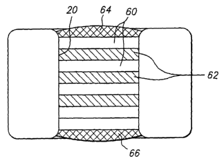
FIG. 5 illustrates an arrangement of layers of a therapeutic agent 60
layered between layers 62 of a protective material which protects the
therapeutic
agents from compounds or conditions within the body which would degrade the
therapeutic agent. Examples of protective interlayers 62 will be discussed in
detail
below. FIG. 5 also illustrates a protective layer in the form of a cap layer
64
provided at a tissue contacting surface of medical device. The cap layer 64
blocks
or retards biodegradation of subsequent layers and/or blocks or retards
diffusion of
CA 02499475 2005-03-18
WO 2004/026357 PCT/US2003/030125
-17-
the beneficial agent in that direction for a period of time which allows the
delivery
of the medical device to a desired location in the body. The barrier layer 64
may
also function to prevent hydration of inner layers of beneficial agent and
thus
prevent swelling of the inner layers when such layers are formed of
hygroscopic
materials. FIG. 5 also illustrates a barrier layer 66. When the medical device
10
is a stent which is implanted in a lumen, the barrier layer 66 is positioned
on a side
of the opening 20 facing the inside of the lumen. The barrier layer 66
prevents the
therapeutic agent 60 from passing into the lumen and being carried away
without
being delivered to the lumen tissue.
In the embodiment of FIG. 5, the protective layers 62 prevent or retard
the flow of water (or other compounds) to the therapeutic layers 60 in a
manner
which will be described in further detail below. The protective layers 62
prevent
or reduce the loss of biological function of the therapeutic agent by reducing
contact of water with the therapeutic agent until a desired delivery time.
FIG. 6 illustrates a further embodiment of the invention in which the
opening 20 in the medical device 10 is filled with a therapeutic agent and a
protective agent in the same layer or layers 70. In this embodiment, the
therapeutic agent layer and the protective agent layer are incorporated in the
same
layer. Optionally, a barrier layer 72 may be provided as in the embodiment of
FIG. 5.
Beneficial Agent Formulations
Beneficial agents include any therapeutic agent or drug, as well as
inactive agents such as barrier Layers, carrier layers, therapeutic layers or
protective layers.
Therapeutic Layer Formulations
The therapeutic agent layers of the present invention are beneficial agents
comprised of a matrix and at least one therapeutic agent. The matrix of the
CA 02499475 2005-03-18
WO 2004/026357 PCT/US2003/030125
-I8-
therapeutic agent layers can be made from pharmaceutically acceptable
polymers,
such as those typically used in medical devices. Such polymers are well known
and include but are not limited to poly-a-hydroxy acid esters such as,
polylactic
acid, polyglycolic acid, polylactic-co-glycolic acid, polylactic acid-co-
caprolactone; polyethylene glycol and polyethylene oxide; polyvinyl
pyrrolidone;
polyorthoesters; polysaccharides and polysaccharide derivatives such as
polyhyaluronic acid, polyalginic acid, chitin, chitosan, cellulose,
hydroxyehty1ce11ulose, hydroxypropylcellulose, carboxymethylcellulose;
polypeptides, and proteins such as polylysine, polyglutamic acid, albumin;
polyanhydrides; polyhydroxy alkonoates such as polyhydroxy valerate,
polyhydroxy butyrate, and the like, and copolymers thereof. The polymers and
copolymers can be prepared by methods well known in the art (see, for example,
Rempp and Merril: Polymer Synthesis, 1998, John Wiley and Sons) in or can be
used as purchased from Alkermes, in Cambridge, MA or Birmingham Polymer
Inc., in Birmingham, Alabama.
The preferred co-polymer for use in the present invention are
poly(lactide-co-glycolide) (PLGA) polymers. The rate at which the polymer
erodes is determined by the selection of the ratio of lactide to glycolide
within the
copolymer, the molecular weight of each polymer used, and the crystallinity of
the
polymers used.
Bioerodible polymers may also be used to form barrier layers that erode
at a rate that can be predetermined base on the composition and that contain
no
therapeutic agent.
Additives in Protective laXer and Therapeutic layer Formulations
Typical additives that may be included in a bioerodible matrix are well
known to those skilled in the art (see Remington: The Science and Practice of
Pharmacy, Gennaro, ed., Mack Publishing Co., Easton, Pa., 19th ed., 1995) and
include but are not limited to pharmaceutically acceptable excipients,
adjuvants,
CA 02499475 2005-03-18
WO 2004/026357 PCT/US2003/030125
-19-
carriers, antioxidants, preservatives, buffers, antacids, emulsifiers, inert
fillers,
fragrances, thickeners, tacki~ers, opacifiers, gelling agents, stabilizers,
surfactants, emollients, coloring agents, and the like.
Typical formulations for therapeutic agents incorporated in these medical
devices are well known to those skilled in the art and include but are not
limited to
solid particle dispersions, encapsulated agent dispersions, and emulsions,
suspensions, liposomes or microparticles, wherein said liposome or
microparticle
comprise a homogeneous or heterogeneous mixture of the therapeutic agent.
The amount of the drug that is present in the device, and that is required
to achieve a therapeutic effect, depends on many factors, such as the miumum
necessary dosage of the particular drug, the condition to be treated, the
chosen
location of the inserted device, the actual compound administered, the age,
weight,
and ,response of the individual patient, the severity of the patient's
symptoms, and
the like.
The appropriate dosage level of the therapeutic agent, for more
traditional routes of administration, are known to one skilled in the art.
These
conventional dosage levels correspond to the upper range of dosage levels for
compositions, including a physiologically active substance and traditional
penetration enhancer. However, because the delivery of the active substance
occurs at the site where the drug is required, dosage levels significantly
lower than
a conventional dosage level may be used with success. Ultimately, the
percentage
of therapeutic agent in the composition is determined by the required
effective
dosage, the therapeutic activity of the particular formulation, and the
desired
release profile. In general, the active substance will be present in the
composition
in an amount from about 0.0001 % to about 99 % , more preferably about 0.01 %
to
about 80 % by weight of the total composition depending upon the particular
substance employed. However, generally the amount will range from about
0.01 % to about 75 % by weight of the total composition, with levels of from
about
25 % to about 75 % being preferred.
CA 02499475 2005-03-18
WO 2004/026357 PCT/US2003/030125
-20-
Protective LaXer Formulations
The protective layers of the present invention are beneficial agents
comprised of a bioerodible matrix and optionally contain additional additives,
therapeutic agents, activating agents, deactivating agents, and the like.
Either a
property of the chosen material of the protective layer, or a chemical
embedded in
the protective Layer provides protection from deactivating processes or
conditions
for at least one therapeutic agent. In addition to the polymer materials
described
above, the protective layer may also be comprised of pharmaceutically
acceptable
lipids or lipid derivatives, which are well known in the art and include but
are not
limited to fatty acids, fatty acid esters, lysolipids, phosphocholines,
(Avanti Polar
Lipids, Alabaster, Ala.), including 1-alkyl-2-acetoyl-sn-glycero 3-
phosphocholines, and 1-alkyl-2-hydroxy-sn-glycero 3-phosphocholines;
phosphatidylcholine with both saturated and unsaturated lipids, including
dioleoylphosphatidylcholine; dimyristoyl-phosphatidylcholine;
dipentadecanoylphosphatidyicholine; dilauroylphosphatidyl-choline;
dipalmitoylphosphatidylcholine (DPPC); distearoylphosphatidylcholine (DSPC);
and diarachidonylphosphatidylcholine (DAPC); phosphatidyl-ethanolamines, such
as dioleoylphosphatidylethanolamine, dipahnitoyl-phosphatidylethanolamine
(DPPE) and distearoylphosphatidylefhanolamine (DSPE); phosphatidylserine;
phosphatidylglycerols, including distearoylphosphatidylglycerol (DSPG);
phosphatidylinositol; sphingolipids such as sphingomyelin; glucolipids;
sulfatides;
glycosphingolipids; phosphatidic acids, such as dipahmitoylphosphatidic acid
(DPPA) and distearoylphosphatidic acid (DSPA); palmitic acid; stearic acid;
arachidonic acid; oleic acid; lipids bearing polymers, such as chitin,
hyaluronic
acid, polyvinylpyrrolidone or polyethylene glycol (PEG), also referred to
herein as
"pegylated lipids", with preferred lipids bearing polymers including DPPE-PEG
(DPPE-PEG), which refers to the lipid DPPE having a PEG polymer attached
thereto, including, for example, DPPE-PEG5000, which refers to DPPE having
attached thereto a PEG polymer having a mean average molecular weight of about
CA 02499475 2005-03-18
WO 2004/026357 PCT/US2003/030125
-21-
5000; lipids bearing sulfonated mono-, di-, oligo- or polysaccharides;
cholesterol,
cholesterol sulfate and cholesterol hemisuccinate; tocopherol hemisuccinate;
lipids
with ether and ester-linked fatty acids; polymerized lipids (a wide variety of
which
are well known in the art); diacetyl phosphate; dicetyl phosphate;
stearylamine;
cardiolipin; phospholipids with short chain fatty acids of about 6 to about 8
carbons in length; synthetic phospholipids with asymmetric acyl chains, such
as,
for example, one acyl chain of about 6 carbons and another acyl chain of about
12
carbons; ceramides; non-ionic liposomes including niosomes such as
polyoxyethylene fatty acid esters, polyoxyethylene fatty alcohols,
polyoxyethylene
fatty alcohol ethers, polyoxyethylated sorbitan fatty acid esters, glycerol
polyethylene glycol oxystearate, glycerol polyethylene glycol ricinoleate,
ethoxylated soybean sterols, ethoxylated castor oil, polyoxyethylene-
polyoxypropylene polymers, and polyoxyethylene fatty acid stearates; sterol
aliphatic acid esters including cholesterol sulfate, cholesterol butyrate,
cholesterol
iso-butyrate, cholesterol palmitate, cholesterol stearate, lanosterol acetate,
ergosterol palmitate, and phytosterol n-butyrate; sterol esters of sugar acids
including cholesterol glucuronide, lanosterol glucuronide, 7-
dehydrocholesterol
glucuronide, ergosterol glucuronide, cholesterol gluconate, lanosterol
gluconate,
and ergosterol gluconate; esters of sugar acids and alcohols including lauryl
glucuronide, stearoyl glucuronide, myristoyl glucuronide, lauryl gluconate,
myristoyl gluconate, and stearoyl gluconate; esters of sugars and aliphatic
acids
including sucrose acetate isobutyrate (SAIB), sucrose laurate, fructose
laurate,
sucrose palritate, sucrose stearate, glucuronic acid, gluconic acid and
polyuronic
acid; saponins including sarsasapogenin, smilagenin, hederagenin, oleanolic
acid,
and digitoxigenin; glycerol dilaurate, glycerol trilaurate, glycerol
monolaurate,
glycerol dipalmitate, glycerol and glycerol esters including glycerol
tripalmitate,
glycerol monopalmitate, glycerol distearate, glycerol tristearate, glycerol
monostearate, glycerol monomyristate, glycerol dimyristate, glycerol
trimyristate;
long chain alcohols including n-decyl alcohol, lauryl alcohol, myristyl
alcohol,
CA 02499475 2005-03-18
WO 2004/026357 PCT/US2003/030125
-22-
cetyl alcohol, and n-octadecyl alcohol; 1,2-dioleoyl-sn-glycerol; 1,2-
dipalmitoyl-
sn-3-succinylglycerol; 1,3-dipalmitoyl-2-succinylglycerol; 1-hexadecyl-2-
palmitoylglycerophosphoethanolamine and palmitoylhomocysteine, and/or
combinations thereof.
If desired, a cationic lipid may be used, such as, for example, N-[1-(2,3-
dioleoyloxy)propyl]-N,N,N-trimethylammonium chloride (DOTMA), 1,2-
dioleoyloxy-3-(trimethylammonio)propane (DOTAP); and I,2-dioleoyl-3-(4'-
trimethylammonio)butanoyl-sn-glycerol (DOTB). If a cationic lipid is employed
in
the lipid compositions, the molar ratio of cationic lipid to non-cationic
lipid may
be, for example, from about 1:1000 to about 1:100. Preferably, the molar ratio
of
cationic lipid to non-cationic lipid may be from about 1:2 to about 1:10, with
a
ratio of from about 1:1 to about 1:2.5 being preferred. Even more preferably,
the
molar ratio of cationic lipid to non-cationic lipid may be about l:l.
These lipid materials are well known in the art and can be used as
purchased from Avanti, Burnaby, B.C. Canada.
The preferred lipids for use in the present invention are phosphatidyl-
choline, phosphatidylethanolamine, phosphatidylserine, sphingomyelin as well
as
synthetic phospholipids such as dimyristoyl phosphatidylcholine, dipalmitoyl
phosphatidylcholine, distearoyl phosphatidylcholine, distearoyl phosphatidyl-
glycerol, dipalmitoyl phosphatidylglycerol, dimyristoyl phosphatidylserine,
distearoyl phosphatidylserine and dipalmitoyl phosphatidylserine.
The rate at which the bioerodible matrix erodes is determined by the
choice of lipid, the molecular weight, and the ratio of the chosen materials.
The protective layer can erode by either chemical or physical erosion
mechanisms. If the layer erodes by a physical mechanism, the layer is
typically a
thin film from about 0.1 ~crn to about 3 ~,m of a non-polymeric material
embedded
between two polymeric layers. In this instance, the structural integrity of
the
protective layer is maintained by the presence of both of these polymeric
layers.
When the polymeric material closest to the luminal surface erodes away, the
CA 02499475 2005-03-18
WO 2004/026357 PCT/US2003/030125
-23-
protective layer breaks apart by the physical forces exerted on it from the
remaining polymeric layer. In another embodiment, the protective layer is
eroded
by chemical interactions, dissolution in water, hydrolysis, or reaction with
enzymes.
One function of the protective layer is to protect one or more therapeutic
agents from deactivating or degrading conditions. The protection may come from
the properties of the material when, for example, a hydrophobic protective
layer
would protect a water sensitive agent from water by resisting the influx of
moisture. The protective layer may also act as a physical barrier. For
example, a
protective layer comprised of a hydrogel may allow water to be absorbed by the
gel, and allow any agents contained within the gel to diffuse out of the gel
into the
reaction environment. The hydrogel, however, would prevent enzymes from
penetrating the layer, thereby protecting any agents contained within from the
enzyme. Finally the protective layer does not have to act as a barrier. The
protective layer may protect a therapeutic agent by releasing an agent, such
as an
activating agent or a deactivating agent, into the reaction environment prior
to the
release of the therapeutic agent.
A therapeutic agent may be incorporated directly in the protective layer.
The therapeutic agent can be heterogeneously or homogeneously dispersed in the
protective layer. The therapeutic agent can be a drug, or a drug formulated
into a
microcapsule, niosome, liposome, microbubble, microsphere, or the like. In
addition, the protective layer may contain more than one therapeutic agent.
For
example, a water sensitive drugs, such as a limus, or any other drug that must
be
administered through intravenous, intramuscular, or subcutaneously, could be
incorporated in a hydrophobic matrix such as SAIB, or fatty acid ester.
A therapeutic agent may also be disposed in a therapeutic agent layer,
separate from the protective layer. In this case the protective layer may be
adjacent to the therapeutic agent layer and may serve to prevent or retard
processes
that would degrade or deactivate the therapeutic agent until the protective
layer has
CA 02499475 2005-03-18
WO 2004/026357 PCT/US2003/030125
-24-
substantially eroded. In this instance the protective layer is a barrier
between a
therapeutic layer and the reaction environment. This barrier may be a
hydrophobic barrier that resists water absorption. The hydrophobic barrier
would
be used in conjunction with water-sensitive drugs as described above.
Alternatively, the protective layer maybe a hydrogel that resists the
absorbance of
enzymes. An enzyme resistant barrier would used to protect an drug such as a
I~NA, RNA, peptide or protein based therapeutic agent.
The protective layer may optionally include activating and deactivating
agents for the purpose of preparing the reaction envirorunent for the
subsequent
release of a therapeutic agent. These activating and deactivating agents are
well
known to those skilled in the art and include but are not limited to antacids,
buffers, enzyme inhibitors, hydrophobic additives, and adjuvants. For example,
Mg(OH)2 in particles of about 0.5 ~,m to about 5 ~,m more preferably, about 1
~,m
incorporated in a PLGA polymer layer could be used in conjunction with any
acid
senstive drug. An example of an activating agent is chymotrypsin, which may be
incorporated in polyvinyl pyrrolidone layer. The chymotrypsin, could be used
to
convert a pro-drug to an active drug.
Preferred Embodiments
In one embodiment, the protective layer of the present invention is
essentially hydrophobic and can prevent or retard the absorption of water.
This is
especially advantageous for the delivery of water sensitive drugs such as a
limus.
Some examples of hydrophobic, bioerodible matrix materials are lipids, fatty
acid
esters, such as glycerides. The erosion rate is controlled by varying the
hydrophilic-lipophilic balance (HLB). Alternatively, the hydrophobic
protective
layer may encapsulate the therapeutic agent, and the encapsulated particles
may be
dispersed in either a polymer or lipid matrix.
In another embodiment, the protective layer may contain an antacid, or
pH retaining agent, that protects a therapeutic agent from a deactivating
reduction
CA 02499475 2005-03-18
WO 2004/026357 PCT/US2003/030125
-25-
in pH. Polymers comprised of monomer units of lactide, glycolide,
caprolactone,
13-hydroxy valerate, trimethylene carbonate, dioxanone, 13-hydroxy butyrate
and
other co-hydroxyalkyl carboxylic acids are degraded by water in hydrolysis in
vivo
and in vitro to produce free acid groups in such a quantity that the
microclimate
within the polymer matrix, and sometimes the external environment, becomes
acidic with a pH of less than or equal to six during the process of polymer
degradation. Some therapeutic agents that can be advantageously delivered in
local, sustained fashion from such polymers are sensitive to an acidic
environment
in that their biological activity is attenuated or eliminated as the pH
decreases
during the polymer matrix degradation required to release the agent from the
delivery matrix. Examples of such acid sensitive agents are RNA oligomers with
phosphodiester-ribose linkages or morpholino-imidate linkages (so-called
"anti-sense oligo's), limus's (like sirolimus and everolimus) and generally
therapeutic agents that have chemical functionality that undergo acid
catalyzed
hydrolysis (such as ester, amide, urea, Spiro ester, anhydride and carbonate)
or
that contain functional groups that can be protonated at pH less than or equal
to six
to render the agent biologically inactive, such as amino and imino groups
(such as
the deactivation of bio-active proteins).
To mitigate the effects of acidity generated during polymer degradation
and in vivo resorption, both within the matrix (the micro-climate) and outside
the
matrix (the environment), it is envisioned to include an acid scavenger,
antacid or
neutralization agent capable of maintaining the pH at equal to or greater than
six or
above a threshold pH where the particular agent become therapeutically
ineffective. Inorganic antacids contemplated include metal hydroxides,
particularly divalent metal hydroxides like Mg(OH)2 and Ca(OH)~, and Ba(OH)Z,
monovalent bicarbonates and carbonates like NaHC03 and Na2C03, divalent
carbonates like ZnC03, monovalent and divalent hydrogen phosphates and
dihydrogen phosphates like NazHP04 and Na2HP04, monovalent salts of
carboxylic acids, like sodium acetate. Additionally, organic bases such as
organic
CA 02499475 2005-03-18
WO 2004/026357 PCT/US2003/030125
-26-
amines are envisioned as acid scavengers, such as triethanol amine,
ethanolamine,
morpholine, pyrimidine and purine bases, poly ethyleneimine, nucleosides,
amino
acids and poly amino acids, particularly poly lysine and poly hydroxylysine,
poly
arginine and peptides containing lysine, hydroxy lysine, arginine and/or
histidine
units.
Inorganic antacids are contemplated to be incorporated into the polymer
matrix by standard polymer processing techniques such as solvent casting,
molding, blending, milling and extrusion. The amount of antacid will be enough
to provide for acid neutralization during some or all of the time the acid
sensitive
agent or combination of agents are released in therapeutically relevant
dosages and
pharmacokinetic profiles. The antacid may be incorporated into the polymeric
drug delivery matrix in amounts up to where the desired physical
characteristics
are compromised for the desired application, or may be used at lower levels.
Antacids may be used alone or in combination with other antacids. For polymers
containing lactide and/or glycolide (the so-called PLGA family of polymers),
the
amount of antacid will generally not exceed 10 % by weight and may preferably
be
used at 1-6 % by weight. The antacid need not be used at the stoichiometric
level
calculated for complete polymer degradation or hydrolysis, but may provide
beneficial protection for the acid sensitive agents at less than
stoichiometric values,
particularly if all the agent is delivered prior to complete degradation of
the
polymer to its constituent monomer or co-monomer units.
In still another embodiment, the protective layer protects a therapeutic
agent from a deactivating or degrading enzyme. An enzyme inhibitor can be
incorporated into the protective layer, so that it is introduced to the
reaction
environment as the protective layer erodes. The therapeutic agent would then
enter an environment with less enzyme than would be present if the inhibitor
were
not incorporated in the protective layer. Alternatively the protective layer
may be
made of a hydrogel material, such as calcium alginate, (made by adding Ca(OH)2
to polyalginic acid) that allows small molecules to diffuse into and out of
the gel,
CA 02499475 2005-03-18
WO 2004/026357 PCT/US2003/030125
-27-
but substantially prevents larger molecules from entering the protective
layer.
DNA, RNA, peptide and protein based therapeutics would be protected using
hydrogel barriers.
Uses for Implantable Medical Devices
Although the present invention has been describe with reference to a
medical device in the form of a stent, the medical devices of the present
invention
can also be medical devices of other shapes useful for site-speciftc and time-
release
delivery of drugs to the body and other organs and tissues. The drugs may be
delivered to the vasculature including the coronary and peripheral vessels for
a
variety of therapies, and to other lumens in the body. The drugs may increase
lumen diameter, create occlusions, or deliver the drug for other reasons.
Medical devices and stems, as described herein, are useful for the
prevention of amelioration of restenosis, particularly after percutaneous
transluminal coronary angioplasty and intraluminal stent placement. In
addition to
the timed or sustained release of anti-restenosis agents, other agents such as
anti-
inflammatory agents may be incorporated in to the multi-layers incorporated in
the
plurality of holes within the device. This allows for site-specific treatment
or
prevention any complications routinely associated with stmt placement that are
known to occur at very specific times after the placement occurs.
The methods for loading beneficial agents into openings in an expandable
medical device may include known techniques such as dipping and coating and
also known piezoelectric micro jetting techniques. Micro-injection devices may
be
used to deliver precise amounts of one or more liquid beneficial agents
including
protective layers, therapeutic agent layers, and any other layers to precise
locations
on the expandable medical device in a known manner. The beneficial agents may
also be loaded by manual injection devices.
CA 02499475 2005-03-18
WO 2004/026357 PCT/US2003/030125
-28-
EXAMPLES
In the examples below, the following abbreviations have the following
meanings. If an abbreviation is not defined, it has its generally accepted
meaning.
mL - milliliters
M - Molar
wt. - weight
vol. - volume
,uL - microliters
,um - micrometers
nm - nanometers
DMSO = Dimethyl sulfoxide'
NMP - N-methylpyrrolidone
DMAC = Dimethyl acetamide
Example 1
Formulation com~risin~ a Therapeutic Aeent within the Protective Laver
A first mixture of poly(lactide-co-glycolide) (PLGA) (Birmingham
Polymers, Inc), lactide:glycolide::85:15, (M~> 100,000 Daltons) 7% wt. and a
suitable organic solvent, such as DMSO, NMP, or DMAC 93 % wt. is prepared.
The mixture is loaded dropwise into holes in the stem, then the solvent is
evaporated to begin formation of the barrier layer. A second barrier layer is
laid
over the first by the same method of filling polymer solution into the hole
followed
by solvent evaporation. The process is continued until five individual layers
have
been laid down to form the barrier layer.
A second mixture of a limus, such as sirolimus, 3 % solids basis, and
dipalmitoyl phosphatidylcholine (DPPC), 7 % solids basis, in a suitable
organic
solvent, such as DMSO, is introduced into holes in the stmt over the barrier
layer.
The solvent is evaporated to form a drug filled protective layer and the
filling and
evaporation procedure repeated until the hole is filled to about 75 % of its
total
volume with drug in protective layer layered on top of the barrier layer.
Three layers of a third solution, of poly(lactide-co-glycolide) (PLGA),
lactide:glycolide::50:50, (M~ c 80,000 Daltons) 7% wt. and a suitable organic
CA 02499475 2005-03-18
WO 2004/026357 PCT/US2003/030125
-29-
solvent, such as DMSO, are then laid down over the drug in matrix layer to
provide a cap layer.
Following implantation of the filled stmt in vivo, the cap layer degrades
allowing the limus to be delivered. The barrier layer prevents the therapeutic
agent from being delivered out the barrier layer side of holes in the stmt.
Example 2
Formulation Comprising Therapeutic Agents in Therapeutic Agent
Layers and a Protective Layer Se~parating_the Therapeutic Agent Lavers
A first mixture of poly(lactide-co-glycolide) (PLGA),
lactide: glycolide: :85:15, (M~ > 100,000 Daltons) 7 % wt. and a suitable
organic
solvent, such as DMSO, 93 % wt. is prepared. The mixture is loaded drop-wise
into holes in the stmt, and the solvent is then evaporated to form the barrier
layer.
A second barrier layer is laid over the first by the same method of filling
polymer
solution into the hole followed by solvent evaporation. The process is
continued
until five individual layers have been laid down to form the barrier layer.
A second mixture of an PCN-1 ribozyme, 8 % solids basis, and
poly(vinylpyrrolidone) (PVP), molecular weight 8,000 daltons, 2% solids basis,
in
an mixed solvent of RNA-ase / DNA-ase free water, 50% vol., and dimethyl
sulfoxide (DMSO), 50% vol., is introduced into holes in the stmt over the
barrier
layer. The solvent is evaporated to form a therapeutic agent layer and the
filling
and evaporation procedure repeated until the hole is filled sufficiently.
Three layers of a third solution, SAIB, (Eastman Chemicals) 7 % wt. and
a suitable organic solvent, such as DMSO, are then laid down over the drug in
matrix layer to provide a protective layer.
A fourth mixture of PLGA, lactide:glycolide::50:50, (M,, = 80,000
Daltons) 5 % wt. , Dexamethasone, 5 % wt. , and a suitable organic solvent,
such as
DMSO, 90 % wt. is prepared. The mixture is then loaded into the holes and the
CA 02499475 2005-03-18
WO 2004/026357 PCT/US2003/030125
-30-
solvent is evaporated to form a second therapeutic agent layer. This process
is
continued until five layers have been laid down.
A fifth mixture of PLGA, lactide:glycolide::50:50, (M~ c 80,000
Daltons) 7 % and a suitable organic solvent, such as DMSO, are then laid down
over the second therapeutic agent layer to provide a cap layer.
Following implantation of the filled stmt in vivo, the cap layer degrades
allowing the Dexmethasone to be delivered. The protective layer protects the
PCN-1 ribozyne from degrading while the Dexamethasone is delivered. After the
protective layer degrades, the PCN-1 ribozyme is then delivered.
Example 3
Formulation Comprising a Theraper utic Agent in a Therapeutic Agent
Layer and a Protective LaXer Containing~an Activating Agent
A first mixture of high molecular weight poly(lactide-co-glycolide)
(PLGA), lactide:glycolide: :50:50 (M~ > 100,000 Daltons), 7 % wt. and a
suitable
organic solvent, such as DMSO, 93 % wt. is prepared. The mixture is loaded
drop-wise into holes in the stmt, then the solvent is evaporated to form the
barrier
layer. A second barrier layer is laid over the first by the same method of
filling
polymer solution into the hole followed by solvent evaporation. The process is
continued until five individual layers have been laid down to form the
complete
barrier layer.
A second mixture of chymotrypsin, 3 % solids basis, and polyvinyl
pyrrolidone, 7% solids basis, in a solvent mixture of water:DMS0::50:50 is
introduced into holes in the stmt over the barrier layer. The solvent is
evaporated
to form an activating ester hydrolytic enzyme filled protective layer and the
filling
and evaporation procedure repeated until the hole is filled to about 20 % of
its total
volume with enzyme in activating layer.
Three layers of a third solution, of poly(lactide-co-glycolide) (PLGA),
lactide:glycolide::50:50, (M~ = 80,000 Daltons) 7% wt. and a suitable organic
CA 02499475 2005-03-18
WO 2004/026357 PCT/US2003/030125
-31-
solvent, such as DMSO, are then laid down over, the enzyme in matrix layer to
provide a time delay.
A fourth solution of a pro-drug paclitaxel-polyglutamic acid (PTX-PGA)
conjugate (where a free hydroxyl group on paclitaxel is covalently bonded via
an
ester linkage to the PGA), 1 % wt. and poly(lactide-co-glycolide) (PLGA),
lactide:glycolide::50:50, (M~ = 80,000 Daltons) 7% wt. and a suitable organic
solvent, such as DMSO, 92 % wt. is prepared. The mixture is filled into holes
in
the stmt over the protective layer, then the solvent is evaporated to form the
pro-drug layer. A pro-drug layer is laid over the first by the same method of
filling polymer solution into the hole followed by solvent evaporation. The
process is continued until six individual layers have been laid down to form
the
pro-drug layer.
Following implantation of the filled stmt in vivo, the pro-drug is released
first and partitions into the arterial tissue. After a delay time while the
protection
layer degrades, the protected chymotrypsin is released and enzymatically
hydrolyzes the ester bond of the pro-drug to activate release of the drug
paclitaxel
in the tissue.
Example 4
Formulation Comprising a Therapeutic Agent in a Therapeutic Agent
Layer and a Protective Layer Containing a Deactivating Agent
A first mixture of poly-lactide, 5 % wt. and a suitable organic solvent,
such as DMSO, 95 % wt. is prepared. The mixture is loaded drop-wise into holes
in the stmt, then the solvent is evaporated to form the barrier layer. A
second
barrier layer is laid over the first by the same method of filling polymer
solution
into the hole followed by solvent evaporation. The process is continued until
five
individual layers have been laid down to form the complete barrier layer.
A second mixture of citric acid, 8 % solids basis, and polyvinyl
pyrrolidone, 2% solids basis, in a solvent mixture of water:DMS0::50:50 is
CA 02499475 2005-03-18
WO 2004/026357 PCT/US2003/030125
-32-
introduced into holes in the stmt over the barrier layer. The solvent is
evaporated
to form a deactivating compound containing layer capable of catalyzing the
hydrolysis of phosphodiester bonds and depolymerizing and deactivating RNA
oligomers. The filling and evaporation procedure is repeated until the hole is
filled to about 20 % of its total volume with enzyme in activating layer.
Three layers of a third solution, of poly(lactide-co-glycolide) (PLGA),
lactide:glycolide::50:50, 7% wt. and a suitable organic solvent, such as DMSO,
are then laid down over the deactivating compound containing layer to provide
a
separating layer by the same fill and evaporate sequence.
A fourth mixture of PCN-1 ribozyme, 8 % solids basis, and polyvinyl
pyrrolidone, 2% solids basis, in a solvent mixture of water:DMS0::50:50 is
introduced into holes in the stmt over the separation layer. The solvent is
evaporated to form an anti-sense oligonucleotide filled polymer therapeutic
agent
layer and the filling and evaporation procedure repeated until the hole is
filled to
about 20 % of its total volume.
A fifth mixture of PLGA lactide:glycolide::50:50, (M~ = 80,000
Daltons) 7 % and a suitable organic solvent, such as DMSO, are then laid down
over the therapeutic agent layer to provide a cap layer.
Following implantation of the filled stmt in vivo, the PCN-1 ribozyme is
released first and partions into the arterial tissue and provides a
therapeutic effect.
After a delay time while the protection layer degrades, the protected citric
acid is
released and catalytically hydrolyzes the phosphodiester ester bond of
ribozyme
oligonucleotide backbone and terminates its therapeutic activity.
Example 5
Formulation Comnrisin~ a Therapeutic Agent in a Therapeutic Agent
LaXer and a Protective Lair Containing; an Antacid
A first mixture of poly(lactide-co-glycolide) (PLGA),
lactide:glycolide::85:15, (M~ > 100,000 Daltons) 7 % wt. and a suitable
organic
CA 02499475 2005-03-18
WO 2004/026357 PCT/US2003/030125
-33-
solvent, such as DMSO, 93 % wt. is prepared. The mixture is loaded drop-wise
into holes in the stent, then the solvent is evaporated to form the barrier
layer. A
second barrier layer is laid over the first by the same method of filling
polymer
solution into the hole followed by solvent evaporation. The process is
continued
until five individual layers have been laid down to form the complete barrier
layer.
A second mixture of sirolimus, 3 % solids basis,
poly(lactide-co-glycolide) (PLGA), lactide:glycolide::50:50, (M~ = 80,000
Daltons) 7 % wt, and magnesium hydroxide, 0.35 % wt (5 % wt based on PLGA)
is introduced into holes in the stent over the barrier layer. The solvent is
evaporated to form a drug protecting layer containing drug and an antacid and
the
filling and evaporation procedure repeated until the hole is filled to about
60 % of
its total volume with protecting layer.
Three layers of a third solution, of poly(lactide-co-glycolide) (PLGA),
lactide:glycolide::50:50, (M~ = 80,000 Daltons) 7% wt. and a suitable organic
solvent, such as DMSO, are then laid down.
A fourth mixture of PLGA lactide:glycolide::50:50, (M~ c 80,000
Daltons) 7 % and a suitable organic solvent, such as DMSO, are then laid down
over the therapeutic agent layer to provide a cap layer.
Following implantation of the filled stmt in vivo, PLGA polymer
degrades via hydrolysis and sirolimus is released, as well as acidic
byproducts
(lactic and glycolic acids as well as acid function terminated PLGA
oligomers).
The acidic byproducts are immediately and continuously neutralized by the
action
of magnesium hydroxide over the time the sirolimus is released, thus
protecting
the sirolimus from acid catalyzed degradation.
CA 02499475 2005-03-18
WO 2004/026357 PCT/US2003/030125
-34-
Example 6
Measurement of Paclitaxel Release Rates from a Medical Device with
Multiple Therapeutic A eg nt Layers
A solution of phosphate buffered saline (PBS) is prepared by dissolving
five "Phosphate Buffered Saline Tablets" (Sigma-Aldrich Co., catalog #P-4417)
in
1000 mL deionized water to provide a solution with a pH of 7.4, 0.01 M in
phosphate buffer, 0.0027 M in potassium chloride and 0.137 M in sodium
chloride. This PBS solution is used as a Release Solution.
The elution rate of drug from the multilayered stmt of Example 1 is
determined in a standard sink condition experiment.
A first 10 mL screw capped vial is charged with release solution, 3 mL,
then placed in a shaking water bath held at 37 ° C until temperature
has
equilibrated. The above stmt containing a drug in matrix layer in between two
protection layers is placed into the release solution, shaking at 60 cycles
per
minute commenced, and the stmt is held immersed in the release solution for a
period of time. The stmt is then placed in a second screw capped vial is
charged
with release solution, 3 mL, at 37 °C, and held for a period of time.
The first
release solution is called sample #1. From time to time, the stmt is removed
from
release solution in one vial and placed into fresh solution in the next vial
to
generate a series of samples containing varying amounts of drug eluted from
the
stmt.
The amount of paclitaxel in a given release solution sample is determined
by High Pressure Liquid Chromatography (HPLC). The following conditions are
used:
Analysis Column: Sym. C18 (5 ,um, 3.9 x 150 mm, Waters Corp., MA)
Mobile phase: Water / Acetonitrile :: 55 % vol. / 45 % vol.
Flow Rate: 1 mL / minute
Temperature: 25 °C
Detection wavelength: 227 nm
CA 02499475 2005-03-18
WO 2004/026357 PCT/US2003/030125
-35-
Injection volume: 50 ,uL
Retention time: 10.5 minutes
By comparison with a calibration curve generated from known stock
solutions, the amount of paclitaxel eluted into the release solution during
any time
period of the experiment can be calculated.
Methods and results for measuring release profiles are published in A.
Finkelstein et al., "The Conor Medsystems Stent: A programmable Drug Delivery
Device," TCT 2001 Conference, Washington, D.C., September 2001.
While the invention has been described in detail with reference to the
preferred embodiments thereof, it will be apparent to one skilled in the art
that
various changes and modifications can be made and equivalents employed,
without
departing from the present invention.