Note: Descriptions are shown in the official language in which they were submitted.
CA 02499547 2005-03-18
WO 2004/026133 PCT/IL2003/000753
1
METHOD, APPARATUS AND SYSTEM FOR CHARACTERIZING SLEEP
FIELD AND BACKGROUND OF THE INVENTION
The present invention relates to methods, apparati and systems for
characterizing sleep and, more particularly, to methods, apparati and systems
for an
efficient determination of sleep stages, body positions and/or sleep disorders
of a
sleeping subject, using only data derived from signals of electrical activity
recorded
of a chest of a sleeping subject, such as electrocardiogram (ECG) signals,
reflecting
cardiac electrical activity, and signals inherently associated with ECG
signals,
l0 reflecting autonomic nervous system activity and electrical activity of
muscles, other
than the heart muscle itself, present in the chest of the sleeping subject.
The growing interest in sleep and its disorders, including their influence on
health, well-being and public safety (such as in car accidents) have caused a
continuously increasing need to perform sleep investigations for both research
and
clinical purposes. Substantial research has been undertaken directed toward
understanding the nature of sleep and of sleep disorders. These researches
yielded
considerable information concerning human patterns of sleep and wakefulness,
and of
physiological activities occurring during human sleep. In addition,
substantial
information has been obtained concerning various sleep disorders.
2o It is common to divide the sleep of a normal healthy individual into a
succession of three states of being, known as Wakefulness, Rapid-Eye-Movement
(REM) sleep and Non-REM (NREM) sleep. NREM sleep is subdivided into four
sleep stages, which are enumerated from Stagel to Stage-4 according to the
increasing threshold to the influence of external stimuli, these stages are
also known
as the depth of sleep.
NREM and REM sleep alternate throughout the night in cycles: each sleep
cycle lasts about 90-120 minutes; normally each cycle starts with NREM
followed by
REM. The night contains 4-5 sleep cycles, where, within each cycle along the
night,
the relative duration of REM sleep increases and the relative duration of NREM
decreases. Altogether, the period of NREM sleep represents more than 60 % of
the
night sleep and REM around 30 %. Normally, REM sleep Erst occurs about 90
minutes after sleep-onset (beginning of Stage-1), at the end of the first
sleep cycle.
CA 02499547 2005-03-18
WO 2004/026133 PCT/IL2003/000753
2
This first REM period is short and might be easily overlooked. Each subsequent
cycle lasts approximately the same time with shorter and lighter stages of
NREM and
extending REM periods as the night goes on. Thus, towards morning hours sleep
becomes lighter (longer stage 2) and individuals dream more (longer REM). A
person may complete between four and six cycles in a typical night's sleep.
The
overall percentage of the duration of NREM stages and the REM stage is
typically
about 70 % of NREM and about 30 % of REM in a healthy adult person.
The percentage of REM sleep is highest during infancy and early childhood,
drops off during adolescence and young adulthood, and remains stable
thereafter.
l0 Total sleep time is longest during early infancy (newborns sleep about 18
hour a day)
and sleep times decreases gradually to normal adult values, around 8 hours a
night.
Paradoxically, the sleep needs during adolescence are increased while the
social and
curricular needs at this age cause sleep deprivation. In the old age sleep
needs do not
change, however the ability to sleep is somewhat reduced. NREM sleep becomes
lighter, REM remains stable at about 25-30 % of total sleep time, the sleep
latency
increases, and generally sleep is more fragmented than in younger individuals.
Monitoring an individual's sleep pattern is crucial for diagnosing sleep
disorders,
follow up results of treatment of sleep disturbances, and conducting research
in the
field of sleep.
To date, sleep stages are monitored and examined clinically with a
polysomnograph (PSG), which provides data regarding the electrical activity of
brain,
muscles and eye movement during sleep. The PSG data are analyzed according to
a
gold standard procedure attributed to Rechtschaffen and Kales (R&K)
[Rechtschaffen
A., Kales A., eds., "A manual of standardized terminology, techniques and
scoring
system for sleep staging in human subjects", Washington DC: US Goveniment
Printing Office, NIH Publication 204, 1968]. The R&K criteria are primarily
based
on the analysis of three collected bio-signals: (i) electroencephalogram
(EEG), (ii)
electrooculogram (EOG), and (iii) electromyogram (EMG). The standard procedure
is as follows: EEG signals are derived primarily from the cortex of the brain.
At the
3o same time an EMG signal which monitors muscle activity, generally from one
of the
muscles of the mandible (submental) is measured, together with left eye and
right eye
EOG (signals produced by eyeball movements relative to the skull). These EEG,
CA 02499547 2005-03-18
WO 2004/026133 PCT/IL2003/000753
3
EMG and EOG signals are conventionally recorded on a multi-channel
physiological
recorder.
The number of physiologic inputs which are required in the PSG procedure
may vary. Specifically, the monitored signals include EEG (2-4 leads), EOG (2
leads), EMG (chin and limbs; 1-3 or more leads), airflow, respiratory effort
(1-2
leads), oxygen saturation, electrocardiogram (ECG), body position and a
microphone.
Data is stored during the sleep, and the analysis is typically done off line,
according
to the standard R&K criteria.
For Stage-1 sleep, which is often considered to be first in the sequence (in
l0 models where walcing is not included), there is some slowing in EEG
frequency, the
brain activity is similar to that of wakefulness, there is a slow rolling eye
movements
and a certain decrease in EMG amplitude. The eyes are closed during Stage-1
sleep,
but if aroused from it, a person may feel as if he or she has not slept. Stage-
1 usually
lasts a few minutes.
Stage-2 is a period of light NREM sleep during which PSG readings are
characteristic. EEG signal displays Sleep spindles and biphasic waves-K
complexes,
EOG signals shows no eye movements in normal subjects free of pharmacological
treatments and the EMG signal amplitude is lower than during wakefulness. I~-
complexes are spontaneous and can be induced by means of sudden auditory
stimuli.
The heart rate slows, and body temperature decreases. At this point, the body
prepares to enter deep sleep. Stages-1 and -2 are collectively known as Light
Sleep
(LS).
Stages-3 and -4 are deep sleep stages, with Stage-4 being more intense than
Stage-3. These stages are known as Slow-Wave-Sleep (SWS). During SWS,
especially during Stage-4, the EEG is characterized by slow waves of high
amplitude
and pattern synchronization. EOG shows no eye movements and the EMG amplitude
is significantly lower than during wakefulness.
REM sleep is distinguishable from NREM sleep by changes in physiological
states, including its characteristic Rapid-Eye-Movements. However, EEG signal
3o shows wave patterns in REM to be similar to Stage-1 sleep and wakefulness
with
mixed frequencies and low amplitude desynchronized activity. The eye movements
are rapid and similar to the wakefulness eye movements. The skeletal, weight
bearing
CA 02499547 2005-03-18
WO 2004/026133 PCT/IL2003/000753
4
muscles become atonic-the EMG amplitude is extremely low. During normal REM
sleep, heart rate and respiration speed up and become eiTatic, while the face,
fingers
and legs may twitch. Intense dreaming occurs during REM sleep and there is
increased metabolism in certain brain regions. Paradoxically, paralysis occurs
simultaneously in the major voluntary muscle groups and the muscles of the
upper
airways. It is generally thought that REM-associated muscle paralysis is meant
to
keep the body from acting out the dreams that occur during this stage.
The waking stage is referred to as relaxed wakefulness, during this time
period, which varies according to the environmental conditions and
individual's
characteristics the body prepares for sleep. Normally, as a person becomes
sleepier,
the body begins to slow down. Muscles begin to relax, and eye movement slows
to a
roll and the responsiveness to external stimuli decreases steeply with sleep
onset.
During sleep the muscles of the upper part of the throat relax. For healthy
individual, the upper part of the throat remains open enough to permit the
flow of air
into the lungs. Some individuals, however, suffer from increased upper airway
resistance.
Several sleep disorders and symptoms are associated with increased upper
airway resistance, for example, snoring and obstructive apnea. The ability to
maintain upper airway patency during the normal respiratory cycle is the
result of a
2o delicate equilibrium between the forces that promote airway closure and
dilation.
Factors predisposing upper airway obstruction include anatomic narrowing,
abnormal
mechanical linkage between airway dilating muscles and airway walls, muscle
weakness, and abnormal neural regulation.
Despite the misleadingly benign clinical presentation, the pathological
consequences of sleep apnea, especially in children, may be severe, and some
pathological consequences are still being uncovered. Several immediate
consequences of upper airway obstruction during sleep are recognized. These
include, sleep fragmentation, increased work of breathing, alveolar
hypoventilation
and intermittent hypoxemia.
3o Many sleep disorders, in particular snoring, sudden. infant death syndrome
and
obstructive sleep apnea syndrome, are position-dependent. Knowing the body
CA 02499547 2005-03-18
WO 2004/026133 PCT/IL2003/000753
position during sleep is important for study, diagnosis and treatment strategy
of such
sleep disorders.
Other disorders or disturbances are also related to frequent body position
changes during sleep.
5 Quality of sleep, which is closely related to the amount of body position
changes [De Koninck J. et al., "Sleep positions in the young adult and their
relationship with the subjective quality of sleep", 1983, Sleep, 6: 52].
Pulmonary
blood flow, which was suggested to be influenced by gravity, and its
distribution was
shown to depend on body posture [Halcim T.S. et al., "Effect of body posture
on
to spatial distribution of pulmonary blood flow", JAppl Physiol., 1988,
64(3):1160-70].
Very recently, a connection was found between sleep position and kidney
stones [Bijan S., Lu A.F., and Stoller M.L., "Correlation of unilateral
urolithiasis with
sleep posture"' The J. Uf°ol., 2001, 165:1085-1087].
Furthermore, in ST monitoring and other ECG-based applications, where
measurements of ECG segments are relevant (e.g. ischemia), movement of the
subject
is considered artifact [Adams M.G., Drew B.J., "Body position effects on the
ECG -
implication for ischemia monitoring", 1997, J electrocard, 30:285-291].
Knowing
changes in the body position may be of advantage so as to screen out movement
artifacts.
Hence, in addition to the above physiologic inputs, a standard whole night
PSG procedure often includes body position monitoring, for example, using
specific
sensors or visual means, such as a video camera. Determination of body
position
during sleep may also assists in diagnosing sleep disorders originating from
frequent
body position changes during sleep.
Whether or not the body position monitoring is included, the PSG procedure is
uncomfortable for the subject, artifacts in the acquired signals are very
frequent and
cause difficulties in data interpretation with the need to redo the study or
to increase
greatly the time required for the interpretation. Automatic data scoring,
although
available, is generally not very reliable. Thus, often an expert is reviewing
the
acquired data and analyses/scores it epoch by epoch. This manual data
interpretation
is cumbersome and tinted with subjectivity. Standard sleep studies are thus
expensive
and cumbersome, their reliability is often limited, especially when the data
collected
CA 02499547 2005-03-18
WO 2004/026133 PCT/IL2003/000753
6
is of bad quality and the interpretation is automatic. In addition, the sleep
of the
subject is influenced by both the requirement to sleep in the laboratory and
the
multitude of sensors used, which leads to an undesired effect of a measurement
influencing the results of the measurement.
It is recognized that sleep is accompanied by cardiocirculatory changes which
are a direct consequence of alterations in the autonomic nervous system (ANS).
Broadly speaking, during sleep parasympathetic activity is increased while
sympathetic activity decreases with phasic activations-deactivations in REM
sleep
[Parmeggiani P.L. and Morrison A.R., "Alterations in human functions during
sleep",
l0 Cezztz°al Regulation of Autonomic Functions, Lowey A.D. and Spyer
K.M., eds.,
Oxford University Press, 1990, 367].
Recently, analysis of ECG signals in general and Heart-Rate-Variability
(HRV) in particular, have been used to quantify the behavior of the ANS,
thereby to
characterize different sleep stages using different ANS behavior [Berlad I,
Shlitner A,
Ben-Haim S, Lavie P. "Power spectrum analysis and heart rate variability in
stage 4
and REM sleep: Evidence for state specific changes in autonomic dominance", J.
Sleep Res. 1993, 2:88; Baharav et al. "Fluctuations in autonomic nervous
activity
during sleep displayed by power spectrum analysis of heart rate variability",
Neurology 1995, 45:1183; Bonnet M.H., and Arand, D.L., "Heart rate
variability:
sleep stage, time of night, and arousal influence", EEG Cli. Neuroplzy 1997,
102:390;
Scholtz U.J., Bianchi A.M., Cerutti S, Kubicki S., "Vegetative Background of
Sleep:
Spectral Analysis of the Heart Rate Variability" Physiol Behav, 1997, 62:1037;
Baharav A., Shinar Z., Sivan Y., Toledo E., Keselbrener L., and Akselrod S.,
"Autonomic changes associated with sleep onset investigated by time-frequency
decomposition of heart rate variability", Sleep 1998, 21:208; Monti A, Medigue
C,
Nedelcoux H, Escourrou P., "Autonomic control of the cardiovascular system
during
sleep in normal subjects" Eur JAppl Plzysiol, 2002, 87:174].
The ANS plays a cardinal role in the control of cardiovascular function. Heart
rate (HR), heart excitability and contractility are under the constant
influence of the
parasympathetic-sympathetic balance. Parasympathetic nerves and sympathetic
fibers
innervate the Sino-Atrial (SA) node; the parasympathetic influence is
inhibitory while
the sympathetic influence is excitatory. The parasympathetic fibers to the SA
node
CA 02499547 2005-03-18
WO 2004/026133 PCT/IL2003/000753
7
are driven by inhibitory and excitatory inputs from peripheral receptors
(baroreceptors, chemoreceptors, cardiac, pulmonary and airway receptors).
Behavioral adaptive influence of the heart rate at the sinus node is mediated
by
supramedullary inputs to the cardiovagal neurons. The origin of the
sympathetic
irmervation of the heart is located at the T2-T5 segment of the spinal cord
and the
preganglionic fibers synapse in the cervical ganglia; the post synaptic
ganglionic
fibers innervate the SA node (predominantly Right sympathetics increase HR) as
well
as the Atrio-Venticular (AV) node (predominantly Left sympathetics - increase
AV
conduction and cardiac contractility).
to Normal cardiac function is regulated by the complex balance of the
sympathetic and parasympathetic outflows to the heart. This balance is also
responsible for the susceptibility to arrhythmias: while vagal activity has a
protective
role, sympathetic activity lowers the threshold to ventricular fibrillation.
Normal
heart function, heart rate included, is modulated by the fluctuations in the
sympathetic
and parasympathetic flow to the heart. These fluctuations induce beat-to-beat
variability in heart rate and arterial pressure. Hence, the analysis of the
instantaneous
fluctuations in cardiovascular variables supplies valuable information on the
autonomic control in an intact organism.
The early methods of analysis of HRV to study the ANS employed algorithms
2o based on Fast-Fourier-Transform (FFT) [Akselrod et al. "Power spectrum
analysis of
heart rate fluctuations: a quantitative probe of beat to beat cardiovascular
control",
Science 1981, 213:220] and autoregressive methods [Malliani et al.
"Cardiovascular
neural regulation explored in the frequency domain", Ciy-culatiofa, 1991,
84:482].
These pioneer methods require stationary signals for a relatively long time
period,
hence allow for estimation of the autonomic function under steady state
conditions.
However, it has been realized that spreading eventual time-dependent changes
in
frequency content over the entire time window, results in obscuring any
insight into
the time axis within the trace length.
To overcome the non physiologic assumption of stationary conditions, new
3o mathematical methods have been developed. Their quantitative description is
based
on the use of time-frequency spectral decomposition of the simultaneous HR,
blood
pressure (BP) and respiratory signals. A sequential estimation of power
spectra, such
CA 02499547 2005-03-18
WO 2004/026133 PCT/IL2003/000753
8
as the use of a time shifted shout time Fourier transform [Nawab S.H. and
Quatieri
T.F., "Short-Time Fourier Transform", Advanced Topics in Signal ProcessifZg,
Lim
and Oppenheim, eds, Englewood Cliffs, NJ, Prentice Hall 1988, 289] represents
the
most straightforward attempt to overcome this limitation. However it suffers
from the
intrinsic compromise, which involves its loss in time resolution within the
power
spectrum of each sub-trace, as well as its severe limitation regarding the
minimum
frequency it can focus on.
Various approaches have been recently developed in order to overcome these
limitations (to this end see, e.g., a review by Cohen L., entitled "Time-
frequency
l0 distributions" and published in P~oc. IEEE 1989, 77:941). These approaches
include,
Selective Discrete Algorithm (SDA) [I~eselbrener L and Akselrod S. "Selective
discrete Fourier transform algorithm for time-frequency analysis: Methods and
application on simulated and cardiovascular signals" IEEE Ts°a~2s.
Bionxed. Eng. 1996,
43:789], modified Wigner-Ville [Novak P and Novak V, "Time-frequency Mapping
of the Heart Rate, Blood Pressure and Respiratory Signals", Medical &
Biological
Ef2giraeerifZg ayad Cofyaputihg, 1993, 31:103], time-dependent autoregression
[Bianchi
et al., "Time-Variant Power Spectrum Analysis for the detection of Transient
episode
in HRV Signals", IEEE T~a~zsactio~zs ofZ BioTnedical Eng., 1993, 40: 136], and
Wavelets [Meyer Y., "Wavelets: Algorithms and applications", Ed. SIAM,
2o Philadelphia 1993].
The above studies were primarily aimed at investigating autonomic activity (in
steady and non-steady conditions) using HRV analysis, and when focusing on
sleep,
previous studies were primarily directed at investigating sleep physiology by
means
of HRV analysis. However, prior art methods fail to exploit HRV for the
purpose of
scoring sleep, in general, and determining the various sleep stages in
particular.
There is thus a widely recognized need for, and it would be highly
advantageous to have, a method, apparatus and system for determining sleep
stages of
a subject, based on data derived solely from electrical signals recorded of a
chest of a
sleeping subject, and devoid of the limitations associated with prior art
3o methodologies.
CA 02499547 2005-03-18
WO 2004/026133 PCT/IL2003/000753
9
SUMMARY OF THE INVENTION
According to one aspect of the present invention there is provided a method of
determining a Slow-Wave-Sleep (SWS) period and a Non-SWS (NSWS) period from
signals of electrical activity recorded of a chest of a sleeping subject, the
signals being
measured over a plurality of epochs, the method comprising: extracting a
series of
cardiac R-R intervals from the signals and obtaining a time-frequency
decomposition
from the series of cardiac R-R intervals; and using the time-frequency
decomposition
to determine the SWS period; thereby determining the SWS period and the NSWS
to period of the sleeping subject.
According to another aspect of the present invention there is provided a
method of determining a Rapid-Eye-Movement (REM) sleep and a Non-REM
(NREM) sleep from signals of electrical activity recorded of a chest of a
sleeping
subject, the signals being measured over a plurality of epochs, the method
comprising:
extracting a plurality of electromyogram (EMG) parameters from the signals;
and
using the plurality of EMG parameters to determine at least one REM period;
thereby
determining the REM sleep and the NREM sleep of the sleeping subject.
According to yet another aspect of the present invention there is provided a
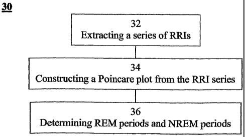
method of determining a REM sleep and an NREM sleep from signals of electrical
2o activity recorded of a chest of a sleeping subject, the signals being
measured over a
plurality of epochs, the method comprising: extracting a series of cardiac R-R
intervals
from the signals; constructing a Poincare plot of the series of cardiac R-R
intervals;
and using the Poincare plot to determine the REM sleep and the NREM sleep of
the
sleeping subject.
According to further features in preferred embodiments of the invention
described below, the method further comprising calculating a plurality of
moments
with respect to a predetermined line along the Poincare plot, each of the
plurality of
moments being calculated within a predetermined time-window.
According to still further features in the described preferred embodiments the
3o REM sleep is defined by a plurality of epochs, each characterized by a
moment which
is below a predetermined threshold.
CA 02499547 2005-03-18
WO 2004/026133 PCT/IL2003/000753
According to still further features in the described preferred embodiments the
method further comprising normalizing each of the plurality of moments.
According to still another aspect of the present invention there is provided a
method of determining sleep stages from signals of electrical activity
recorded of a
5 chest of a sleeping subject, the signals being measured over a plurality of
epochs, the
method comprising: extracting a series of cardiac R-R intervals from the
signals and
obtaining a time-frequency decomposition from the series of cardiac R-R
intervals;
using the time-frequency decomposition to determine at least one SWS period
and at
least one NSWS period; from the at Ieast one NSWS period, determining at least
one
l0 sleep-onset (SO) period and a plurality of non-sleep periods; extracting a
plurality of
EMG parameters from a portion of the signals, the portion corresponds to a
NSWS
period other than the at least one SO period and other than the plurality of
non-sleep
period; using the plurality of EMG parameters to determine at least one REM
period
thereby obtaining also at least one light-sleep (LS) period defined as a NSWS
period
other than the at least one SO period, other than the plurality of non-sleep
periods and
other than the at least one REM period; thereby determining the sleep stages
of the
sleeping subject.
According to further features in preferred embodiments of the invention
described below, the method further comprising determining, from the at least
one LS
2o period, at least one Stage-2 period thereby obtaining also a Stage-1
period, the Stage-1
period being defined as a LS period other than the at least one Stage-2.
According to still further features in the described preferred embodiments the
obtaining the time-frequency decomposition comprises calculating, for each
epoch, at
least one time-dependent power spectrum component selected from the group
consisting of a very-low-frequency (VLF) power spectrum, a low-frequency (LF)
power spectrum and a high-frequency (HF) power spectrum.
According to still further features in the described preferred embodiments the
SWS period is defined by a plurality of epochs, each characterized by at least
one
power parameter which is below a predetermined threshold, the at least one
power
3o parameter is selected from the group consisting of the VLF power spectrum,
the LF
power spectrum, the HF power spectrum, and a combination between two of the
VLF,
the LF and the HF power spectra.
CA 02499547 2005-03-18
WO 2004/026133 PCT/IL2003/000753
11
According to still further features in the described preferred embodiments at
least one of the VLF, the LF and the HF power spectra are calculated within a
window
along the series of cardiac R-R intervals, the window being characterized by a
duration which is a function of a respective frequency.
According to still further features in the described preferred embodiments the
method further comprising determining a frequency resolution.
According to still further features in the described prefeiTed embodiments the
frequency resolution is from O.OOI Hz to 0.03 Hz.
According to still further features in the described preferred embodiments the
to method further comprising determining a time resolution.
According to still further features in the described preferred embodiments the
time resolution is from 1 second to 30 seconds.
According to still further features in the described preferred embodiments the
method further comprising determining an onset and a termination of the time-
dependent power spectra.
According to still further features in the described preferred embodiments at
least one of the VLF, the LF and the HF power spectra are calculated by a
wavelet
transform.
According to still further features in the described preferred embodiments the
wavelet transform is selected from the group consisting of a discrete wavelet
transform
and a continuous wavelet transform.
According to still further features in the described preferred embodiments at
least one of the VLF, the LF and the HF power spectra are calculated by a
selective
discrete spectral transform.
According to still further features in the described preferred embodiments the
selective discrete spectral transform is selected from the group consisting o~
a Fourier
transform, a Haar transform, a Hartley transform, a sine transform, a cosine
transform,
and a Hadamard transform.
According to still further features in the described preferred embodiments the
3o determining at least one SO period comprises calculating at least one SO
parameter
and defining the SO period to be at Ieast one epoch being characterized by at
least one
CA 02499547 2005-03-18
WO 2004/026133 PCT/IL2003/000753
12
SO parameter which is above a predetermined threshold, over a predetermined
time
range.
According to still further features in the described prefeiTed embodiments the
method further comprising calculating the predetermined frequency limits.
According to still further features in the described preferred embodiments the
calculating the predetermined frequency limits comprises obtaining a steady
state
power spectrum from series of cardiac R-R intervals, and applying a minimum-
cross-
entropy method on the steady state power spectrum, so as to provide the
frequency
limits.
l0 According to still further features in the described preferred embodiments
the
minimum-cross-entropy method is executed so as to separate between frequency
peaks
of the steady state power spectrum.
According to still further features in the described preferred embodiments the
method further comprising normalizing the at least one SO parameter.
According to still further features in the described preferred embodiments the
method further comprising analyzing the at least one SO parameter using a
plurality of
statistical quantities.
According to still further features in the described preferred embodiments the
method further comprising: (a) filtering the series of cardiac R-R intervals
using a
low-pass-filter, thereby providing a first series of signals; and (b) defining
the at least
one awakening period as a plurality of epochs each associated with at least
one of the
first series of signals which is below a predetermined threshold.
According to still further features in the described preferred embodiments the
low-pass-filter is at about 0.01 Hz.
According to still further features in the described preferred embodiments the
method further comprising: (a) filtering the series of cardiac R-R intervals
using a
band-pass-filter, thereby providing a second series of signals; and (b)
defining the at
least one arousal period as a plurality of epochs each associated with at
least one of the
second series of signals which is below a predetermined threshold.
According to still further features in the described preferred embodiments the
extracting a plurality of EMG parameters is effected by at least one procedure
selected
CA 02499547 2005-03-18
WO 2004/026133 PCT/IL2003/000753
13
from the group consisting of: eliminating P waves, eliminating T waves and
eliminating QRS-complexes from the signals.
According to still further features in the described preferred embodiments the
eliminating P waves and the eliminating T waves from the signals is by high
pass
filtering.
According to still further features in the described preferred embodiments the
high pass filtering is at a threshold frequency of about 10 Hz.
According to still further features in the described preferred embodiments the
eliminating QRS-complexes is by a combination of gating and/or subtraction.
l0 According to still further features in the described preferred embodiments
the
REM sleep is defined by a plurality of epochs, each characterized by at least
one of the
plurality of EMG parameters which is below a predetermined threshold.
According to still further features in the described preferred embodiments the
at least one Stage-2 period is defined by a plurality of epochs, each
associated to a
cardiac R-R interval corresponding to a K-complex.
According to still another aspect of the present invention there is provided a
method of determining a body position or a change in the body position from
signals
of electrical activity recorded of a chest of a sleeping subject, the signals
being
characterized by QRS complexes, the method comprising: extracting R-wave
durations from the QRS complexes, thereby obtaining an R-wave duration (RWD)
function; and using the RWD function to determine the body position or the
change in
the body position of the sleeping subject.
According to still further features in the described preferred embodiments the
change in the body position is defined when a change of the RWD function is
above a
predetermined threshold.
According to still further features in the described preferred embodiments the
change of the RWD function is calculated using at least one local average of
the RWD
function.
According to still further features in the described preferred embodiments the
change of the RWD function is defined as a difference between two local
averages of
the RWD function.
CA 02499547 2005-03-18
WO 2004/026133 PCT/IL2003/000753
14
According to still further features in the described preferred embodiments the
two body positions, comprise a first body position, defined when a value of
the RWD
function is high and a second body position, defined when a value of the RWD
function is low.
According to still further features in the described preferred embodiments the
method further comprises defining at least two segments of each of the QRS
complexes and determining width of each of the at least two segments, thereby
obtaining, for each QRS complex, a set of widths, the set being representative
of the
body position.
l0 According to still further features in the described preferred embodiments
each
of the segments has a first endpoint and a second endpoint, the first and the
second
endpoints being characterized by a zero nth-order derivative of a respective R-
wave of
the QRS complex, where n is a positive integer.
According to still further features in the described preferred embodiments the
four body positions comprise: a first body position, defined when a value of
the left
segment is high and a value of the right segment is high; a second body
position,
defined when a value of the left segment is low and a value of the right
segment is
high; a third body position, defined when a value of the left segment is high
and a
value of the right segment is low; and a fourth body position, defined when a
value of
2o the left segment is low and a value of the right segment is low.
According to still further features in the described prefeiTed embodiments the
method further comprises applying a clustering procedure on each the sets of
widths,
so as to define a plurality of clusters, each one of the plurality of clusters
corresponding to a different body position.
According to still further features in the described preferred embodiments the
clustering procedure is selected from the group consisting of graph theory
procedure,
density estimation procedure, Potts-spins-based procedure, hierarchical
procedure and
partitional procedure.
According to still further features in the described preferred embodiments the
3o partitioned procedure is selected from the group consisting of a K means
procedure, an
adaptive I~-means procedure, hard C-means procedure and fuzzy C-means
procedure.
CA 02499547 2005-03-18
WO 2004/026133 PCT/IL2003/000753
According to still further features 'in the described prefeiTed embodiments
the
hierarchical procedure is selected from the group consisting of a nearest
neighbor
procedure and a minimal spanning tree procedure.
According to still another aspect of the present invention there is provided a
5 method of characterizing a sleep of a sleeping subject, the method
comprising:
calculating at least one autonomic balance index (ABI), each corresponding to
a
different sleep stage of the sleeping subject and being calculated using a
weight of the
sleep stage and at least one power parameter; and using the at least one ABI
for
characterizing the sleep of the sleeping subject.
l0 According to still further features in the described preferred embodiments
if
one or more of the at least one ABI is larger than a predetermined threshold
then
determining an obstructive sleep apnea for the sleeping subject.
According to still further features in the described preferred embodiments the
method further comprises summing the at least two ABIs thereby obtaining a
total
15 ABI.
According to still further features in the described preferred embodiments if
the total ABI is larger than a predetermined threshold then determining an
obstructive
sleep apnea for the sleeping subject.
According to still further features in the described preferred embodiments the
method further comprises determining periods of the SWS using time=frequency
decomposition of the series of cardiac R-R intervals.
According to still further features in the described preferred embodiments the
method further comprises determining periods of the REM sleep using a
plurality of
EMG parameters extracted from the signals.
According to still further features in the described preferred embodiments the
method further comprises determining periods of the REM sleep using a Poincare
plot
of the series of cardiac R-R intervals.
According to still further features in the described preferred embodiments the
method further comprises: obtaining also periods of the LS, defined as a
period other
than the SWS period, other than the SO periods, other than the non-sleep
periods and
other than the REM periods.
CA 02499547 2005-03-18
WO 2004/026133 PCT/IL2003/000753
16
According to still another aspect of the present invention there is provided a
v
method of determining a sleep apnea from signals of electrical activity
recorded of a
chest of a sleeping subject, the signals being measured over a plurality of
epochs, the
method comprising: (a) extracting a series of cardiac R-R intervals from the
signals;
(b) determining awakening periods of the sleeping subject and excluding
cardiac R-R
intervals corresponding to the awakening periods from the series of cardiac R-
R
intervals; (c) obtaining a power spectrum from the series of cardiac R-R
intervals; and
(d) using the power spectrum to determine the sleep apnea of the sleeping
subject.
According to still further features in the described preferred embodiments the
l0 method further comprises determining body positions or a change in a body
position
of the sleeping subj ect prior to step (b), and executing steps (b)-(d)
separately for each
one of the body positions.
According to still further features in the described preferred embodiments the
method further comprises discarding signals corresponding to abnormal heart
beats of
the sleeping subject, prior to step (a).
According to still further features in the described preferred embodiments the
method further comprises interpolating the signals so as to compensate missing
heart
beats of the sleeping subject, prior to step (a).
According to still further features in the described preferred embodiments the
obtaining the power spectrum is by a discrete transform.
According to still further features in the described preferred embodiments the
discrete transform is selected from the group consisting of a steady state
discrete
transform and a time-dependent discrete transform.
According to still further features in the described preferred embodiments
step
(d) comprises obtaining, for each period other than the awakening period, a
power
spectrum component of the power spectrum, and if the power spectrum component
is
above a predetermined threshold then identifying sleep apnea for the period.
According to still further features in the described preferred embodiments the
method further comprises: employing a pattern recognition procedure on a
portion of
3o the series of cardiac R-R intervals, so as to identify representative
patterns of sleep
apnea; and identifying periods corresponding to the representative patterns as
sleep
apnea periods.
CA 02499547 2005-03-18
WO 2004/026133 PCT/IL2003/000753
17
According to still further features in the described preferred embodiments the
portion of the series of cardiac R-R intervals corresponds to body positions
having
durations lower than a predetermined threshold.
According to still further features in the described preferred embodiments the
predetermined threshold equals about 200 seconds plus total awakening time in
a
respective body position.
According to still further features in the described preferred embodiments the
portion of the series of cardiac R-R intervals corresponds to periods
characterized by a
power spectrum component which is below a predetermined threshold, the power
spectrum component is power of signals being at a frequency range representing
sleep
apnea.
According to an additional aspect of the present invention there is provided
an
apparatus for determining a Slow-Wave-Sleep (SWS) period and a Non-SWS (NSWS)
period from signals of electrical activity recorded of a chest of a sleeping
subject, the
signals being measured over a plurality of epochs, the apparatus comprising:
an R-R
extractor for extracting a series of cardiac R-R intervals from the signals; a
decomposer for obtaining a time-frequency decomposition from the series of
cardiac
R-R intervals; and an SWS determinator, for determining the SWS period using
the
time-frequency decomposition; thereby to determine the SWS period and the NSWS
2o period of the sleeping subject.
According to yet an additional aspect of the present invention there is
provided
an apparatus for determining a REM sleep and a NREM sleep from signals of
electrical activity recorded of a chest of a sleeping subject, the signals
being measured
over a plurality of epochs, the apparatus comprising: an EMG extractor for
extracting
a plurality of EMG parameters from the signals; and a REM determinator for
using the
plurality of EMG parameters to determine the REM sleep and the NREM sleep of
the
sleeping subj ect.
According to still an additional aspect of the present invention there is
provided an apparatus for determining a REM sleep and a NREM sleep from
signals
of electrical activity recorded of a chest of a sleeping subject, the signals
being
measured over a plurality of epochs, the apparatus comprising: an R-R
extractor, for
extracting a series of cardiac R-R intervals from the signals; a plotter, for
constructing
CA 02499547 2005-03-18
WO 2004/026133 PCT/IL2003/000753
18
a Poincare plot of the series of cardiac R-R intervals; and a REM
determinator, for
using the Poincare plot to determine the REM sleep and the NREM sleep of the
sleeping subject.
According to still further features in the described preferred embodiments the
apparatus further comprising electronic-calculating functionality for
calculating a
plurality of moments with respect to a predetermined line along the Poincare
plot,
each of the plurality of moments being calculated within a predetermined time-
window.
According to still further features in the described preferred embodiments the
l0 apparatus further comprising electronic-calculating functionality for
normalizing each
of the plurality of moments
According to a further aspect of the present invention there is provided an
apparatus for determining sleep stages from signals of electrical activity
recorded of a
chest of a sleeping subject, the signals being measured over a plurality of
epochs, the
apparatus comprising: a R-R extractor for extracting a series of cardiac R-R
intervals
from the signals; a decomposer, for obtaining a time-frequency decomposition
from
the series of cardiac R-R intervals; a SWS determinator for using the time-
frequency
decomposition to determine at least one SWS period and at least one NSWS
period; a
SO determinator for determining at least one SO period onset period from the
at least
one NSWS period; a non-sleep determinator for determining plurality of non-
sleep
periods from the at least one NSWS period; an EMG extractor, for extracting a
plurality of EMG parameters from a portion of the signals, the portion.
corresponds to
a NSWS period other than the at least one SO period and other than the
plurality of
non-sleep periods; a REM determinator for using the plurality of EMG
parameters to
determine at least one REM period thereby to obtain also at least one LS
period
defined as a NSWS period other than the at least one SO period, other than the
plurality of non-sleep periods and other than the at least one REM period;
thereby to
determine the sleep stages of the sleeping subject.
According to still further features in the described preferred embodiments the
apparatus further comprising a Stage-2 determinator for determining, from the
at least
one LS period, at least one Stage-2 period, thereby to obtain also a Stage-1
period, the
Stage-1 period being defined as a LS period other than at least one Stage-2
period.
CA 02499547 2005-03-18
WO 2004/026133 PCT/IL2003/000753
19
According to still further features in the described preferred embodiments the
apparatus further comprising electronic-calculating functionality for
normalizing the at
least one SO parameter.
According to still further features in the described preferred embodiments the
apparatus further comprising a statistical analyzer for analyzing the at least
one SO
parameter using a plurality of statistical quantities.
According to yet a further aspect of the present invention there is provided a
system for determining a SWS period and a NSWS period of a sleeping subject,
the
system comprising: an apparatus for providing signals of electrical activity
of a chest
l0 of the sleeping subject, measured over a plurality of epochs; an R-R
extractor for
extracting a series of cardiac R-R intervals from the signals; a decomposer
for
obtaining a time-frequency decomposition from the series of cardiac R-R
intervals;
and an SWS determinator, for determining the SWS period using the time-
frequency
decomposition; thereby to determine the SWS period and the NSWS period of the
sleeping subject.
According ~to still a further aspect of the present invention there is
provided a
system for determining a REM sleep and a NREM sleep of a sleeping subject, the
system comprising: an apparatus for providing signals of electrical activity
of a chest
of the sleeping subject, measured over a plurality of epochs; an EMG extractor
for
extracting a plurality of EMG parameters from the signals; and a REM
detenninator
for using the plurality of EMG parameters to determine the REM sleep and the
NREM
sleep of the sleeping subject.
According to still a further aspect of the present invention there is provided
a
system for determining a REM sleep and a NREM sleep of a sleeping subject, the
system comprising: an apparatus for providing signals of electrical activity
of a chest
of the sleeping subject, measured over a plurality of epochs; an R-R
extractor, for
extracting a series of cardiac R-R intervals from the signals; a plotter, for
constructing
a Poincare plot of the series of cardiac R-R intervals; and a REM
determinator, for
using the Poincare plot to determine the REM sleep and the NREM sleep of the
sleeping subject.
According to still further features in the described preferred embodiments the
apparatus for providing signals comprises cardiac electrodes, adapted for
attachment
CA 02499547 2005-03-18
WO 2004/026133 PCT/IL2003/000753
to a plurality of predetermined locations on the chest of the sleeping
subject, the
plurality of predetermined locations are selected so as to substantially
optimize heart-
beat reads from the signals.
According to still further features in the described preferred embodiments the
5 apparatus fox providing signals comprises a single Lead, adapted for
attachment to a
predetermined location on the chest of the sleeping subject, the predetermined
location
is selected so as to substantially optimize heart-beat reads from the signals.
According to still further features in the described preferred embodiments
each
of the plurality of predetermined locations is adjacent to a different muscle.
to According to still further features in the described preferred embodiments
at
Least two of the plurality of predetermined locations are adjacent to the same
muscle.
According to still further features in the described preferred embodiments the
system further comprising electronic-calculating functionality for calculating
a
plurality of Moments with respect to a predetermined line along the Poincare
plot,
15 each of the plurality of Moments being calculated within a predetermined
time-
window.
According to still further features in the described preferred embodiments the
plurality of Moments is a plurality of Moments of inertia.
According to still further features in the described preferred embodiments the
20 REM determinator is programmed to define the R.EM period by a plurality of
epochs,
each characterized by a Moment which is below a predetermined threshold.
According to still further features in the described preferred embodiments the
predetermined line along the Poincare plot is a straight line, forming a
predetermined
angle with respect to an axis of the Poincare plot.
According to still further features in the described preferred embodiments the
predetermined angle equals about 45 degrees.
According to still further features in the described preferred embodiments the
system further comprising electronic-calculating functionality for normalizing
each of
the plurality of Moments.
According to still a further aspect of the present invention there is provided
a
system for determining sleep stages of a sleeping subject, the system
comprising: an
apparatus for providing signals of electrical activity of a chest of the
sleeping subject,
CA 02499547 2005-03-18
WO 2004/026133 PCT/IL2003/000753
21
measured over a plurality of epochs; an R-R extractor for extracting a series
of cardiac
R-R intervals from the signals; a decomposer, for obtaining a time-frequency
decomposition from the series of cardiac R-R intervals; a SWS determinator for
using
the time-frequency decomposition to determine at least one SWS period and at
least
one NSWS period; a SO determinator for determining at least one SO period
onset
period from the at least one NSWS period; a non-sleep detenninator for
determining a
plurality of non-sleep periods from the at least one NSWS period; an EMG
extractor,
for extracting a plurality of EMG parameters from a portion of the signals,
the portion
corresponds to a NSWS period other than the at Least one SO period and other
than the
plurality of non-sleep period; a REM determinator for using the plurality of
EMG
parameters to determine at least one REM period thereby to obtain also at
Least one LS
period defined as a NSWS period other than the at least one SO period, other
than the
plurality of non-sleep periods and other than the at least one REM period;
thereby to
determine the sleep stages of the sleeping subject.
According to still further features in the described preferred embodiments the
apparatus for providing signals is an ECG apparatus.
According to still further features in the described preferred embodiments the
apparatus for providing signals comprises cardiac electrodes, adapted for
attachment
to a plurality of predetermined locations on the chest of the sleeping
subject, the
plurality of predetermined locations are selected so as to substantially
optimize heart-
beat reads from the signals and to substantially optimize EMG reads from the
signals.
According to still further features in the described preferred embodiments the
apparatus for providing signals comprises a single lead, adapted for
attachment to a
predetermined location on the chest of the sleeping subject, the predetermined
location
is selected so as to substantially optimize heart-beat reads from the signals
and to
substantially optimize EMG reads from the signals.
According to still further features in the described preferred embodiments the
system further comprising a Stage-2 determinator for determining, from the at
least
one LS period, at least one Stage-2 period, thereby to obtain also a Stage-1
period, the
Stage-1 period being defined as a LS period other than at least one Stage-2
period.
According to still further features in the described preferred embodiments the
decomposer is operable to calculate, for each epoch, at least one time-
dependent
CA 02499547 2005-03-18
WO 2004/026133 PCT/IL2003/000753
22
power spectrum component selected from the group consisting of a VLF power
spectrum, a LF power spectrum and a HF power spectrum.
According to still further features in the described preferred embodiments the
SWS determinator is programmed to define the SWS period by a plurality of
epochs,
each characterized by at least one power parameter which is below a
predetermined
threshold, the at least one power parameter is selected from the group
consisting of the
VLF power spectrum, the LF power spectrum, the HF power spectrum, and a
combination between two of the VLF, the LF and the HF power spectra.
According to still further features in the described preferred embodiments the
to combination is a ratio.
According to still further features in the described preferred embodiments the
predetermined threshold is constant.
According to still further features in the described preferred embodiments the
predetermined threshold is a first function of an average value of the at
least one
power parameter.
According to still further features in the described preferred embodiments the
first function is a linear function.
According to still further features in the described preferred embodiments the
predetermined threshold varies with time.
2o According to still further features in the described preferred embodiments
the
decomposer is operable to calculate the VLF, the LF and the HF power spectra
within
a window along the series of cardiac R-R intervals, the window being
characterized by
a duration which is a function of a respective frequency.
According to still further features in the described preferred embodiments the
function of the respective frequency is inversely related to the respective
frequency.
According to still further features in the described preferred embodiments the
window has an aperture selected from the group consisting of: a rectangular
aperture,
a Hamming aperture, a Harming aperture, a Blackman aperture, a Gaussian
window, a
Lorentzian window, a sinc window, a power of a sine window and a power of a
cosine
3o window.
According to still further features in the described preferred embodiments the
decomposer comprises a wavelet processor.
CA 02499547 2005-03-18
WO 2004/026133 PCT/IL2003/000753
23
According to still further features in the described preferred embodiments the
wavelet processor is selected from the group consisting of a discrete wavelet
processor
and a continuous wavelet processor.
According to still further features in the described preferred embodiments the
decomposes comprises a selective discrete spectral processor.
According to still further features in the described preferred embodiments the
decomposes further comprises a spectral transform selector for selecting a
transfonn
from the group consisting of: a Fourier transform, a Haar transform, a Hartley
transform, a sine transform, a cosine transform, and a Hadamard transform.
l0 According to still further features in the described preferred embodiments
the
SO determinator comprises electronic-calculating functionality for calculating
at least
one SO parameter and for defining the SO period to be at least one epoch being
by at
least one SO parameter which is above a predetermined threshold, over a
predetermined time range.
According to still further features in the described preferred embodiments the
predetermined time range is from 2 epochs to 10 epochs.
According to still further features in the described preferred embodiments the
at least one SO parameter comprises at least one integrated power spectrum
calculated
by integrating at least one of the power spectra over predetermined frequency
limits.
According to still further features in the described preferred embodiments the
at least one SO parameter further comprises at least one time-dependent power
ratio
calculated using the at least one integrated power spectrum.
According to still further features in the described preferred embodiments the
SO determinator further comprises electronic-calculating functionality for
calculating
the predetermined frequency limits.
According to still further features in the described preferred embodiments the
system further comprising electronic-calculating functionality for normalizing
the at
least one SO parameter.
According to still further features in the described preferred embodiments the
system further comprising a statistical analyzer for analyzing the at least
one SO
parameter using a plurality of statistical quantities.
CA 02499547 2005-03-18
WO 2004/026133 PCT/IL2003/000753
24
According to still further features in the described preferred embodiments the
plurality of statistical quantities selected from the group consisting of an
average, a
variance and a t-test.
According to still further features in the described prefeiTed embodiments the
plurality of non-sleep periods comprises at least one awalcening period and/or
at least
one arousal period.
According to still further features in the described preferred embodiments the
non-sleep determinator comprises: (a) a low-pass filter for filtering the
series of
cardiac R-R intervals, thereby to provide a first series of signals; and (b)
an awakening
l0 period definer for defining the at least one awakening period as a
plurality of epochs
each associated with at least one of the first series of signals which is
below a
predetermined threshold.
According to still further features in the described preferred embodiments the
low-pass-filter is at about 0.01 Hz.
According to still fiuther features in the described preferred embodiments the
predetermined threshold is about 0.85 of an averaged value of the first series
of
signals.
According to still further features in the described preferred embodiments the
non-sleep determinator comprises: (a) a band-pass-filter for filtering the
series of
cardiac R-R intervals, thereby providing a second series of signals; and (b)
an arousal
period definer for defining the at least one arousal period as a plurality of
epochs each
associated with at least one of the second series of signals which is below a
predetermined threshold.
According to still further features in the described preferred embodiments the
band-pass-filter is characterized by a lower band limit of about 0.05 Hz and
an upper
band limit of about 0.2 Hz.
According to still further features in the described preferred embodiments the
predetermined threshold is about 0.85 of an averaged value of the second
series of
signals.
According to still further features in the described preferred embodiments the
predetermined profile is characterized by a specific width and a specific
depth.
CA 02499547 2005-03-18
WO 2004/026133 PCT/IL2003/000753
According to still further features in the described preferred embodiments the
EMG extractor comprises an eliminator for eliminating at least one signal
selected
from the group consisting of: a P wave, a T wave and a QRS-complex.
According to still further features in the described preferred embodiments the
5 eliminator comprises at least one high pass filter for filtering out the P
wave and the T
wave.
According to still further features in the described preferred embodiments the
high pass filter is characterized by a threshold frequency of about 10 Hz.
According to still further features in the described preferred embodiments the
to eliminator is operable comprises to eliminate the QRS-complex by a
combination of
gating and/or subtraction.
According to still further features in the described preferred embodiments the
plurality of EMG parameters are selected from the group consisting of
normalized
amplitude (mrEMG), normalized mean power (nPWR), zero-crossing average (ZC),
15 median frequency (MF), mean power frequency (MPF), Expected Zero Crossing
(EZC), power variance (PVAR), turns (NT) and Complexity (Cmplx).
According to still further features in the described preferred embodiments the
REM detenninator is programmed to define the REM period by a plurality of
epochs,
each characterized by at least one of the plurality of EMG parameters Which is
below a
2o predetermined threshold.
According to still further features in the described preferred embodiments the
Stage-2 determinator is programmed to define the at least one Stage-2 period
by a
plurality of epochs, each associated to a cardiac R-R interval corresponding
to a K-
complex.
25 According to still further features in the described preferred embodiments
the
cardiac R-R interval corresponding to the K-complex is characterized by a
specific
width and a specific depth.
According to still another aspect of the present invention there is provided
an
apparatus for determining a body position or a change in the body position
from
signals of electrical activity recorded of a chest of a sleeping subject, the
signals being
QRS complexes, the apparatus comprising: an RWD extractor for extracting R-
wave
durations from the QRS complexes, thereby to obtain an R-wave duration
function a
CA 02499547 2005-03-18
WO 2004/026133 PCT/IL2003/000753
26
body position determinator for determining the body position or the change in
the
body position of the sleeping subject using the RWD function.
According to still further features in the described preferred embodiments the
apparatus further comprises a segment calculator for defining at least two
segments of
each of the QRS complexes and determining width of each of the at least two
segments, thereby to obtain, for each QRS complex, a set of widths, the set
being
representative of the body position.
According to still another aspect of the present invention there is provided
an
apparatus of characterizing a sleep of a sleeping subject, the apparatus
comprising: an
to ABI calculator, for calculating at least one ABI, each corresponding to a
different
sleep stage of the sleeping subject, the ABI calculator is operable to
calculated the
ABI using a weight of the sleep stage and at least one power parameter; and a
sleep
characterizes for characterizing the sleep of the sleeping subj ect using the
at least one
ABI.
According to still further features in the described preferred embodiments the
apparatus further comprises an obstructive sleep apnea determinator, for
determining
an obstructive sleep apnea for the sleeping subject if one or more of the at
least one
ABI is larger than a predetermined threshold.
According to still further features in the described preferred embodiments the
ABI calculator is operable to sum the at least two ABIs thereby to provide a
total ABI.
According to still further features in the described preferred embodiments the
apparatus further comprises an obstructive sleep apnea determinator, for
determining
an obstructive sleep apnea for the sleeping subject if the total ABI is larger
than a
predetermined threshold.
According to still fizrther features in the described preferred embodiments
the
sleep stage is selected from the group consisting of a SWS, a REM sleep and an
LS.
According to still further features in the described preferred embodiments the
apparatus further comprises: an R-R extractor, for extracting a series of
cardiac R-R
internals from signals of electrical activity recorded of a chest of the
sleeping subj ect;
3o a decomposes, for obtaining a time-frequency decomposition from the series
of cardiac
R-R intervals; and an SWS determinator for using the time-frequency
decomposition
to determine periods of the SWS.
CA 02499547 2005-03-18
WO 2004/026133 PCT/IL2003/000753
27
According to still further features in the described preferred embodiments the
apparatus further comprises an EMG extractor, for extracting a plurality of
EMG
parameters from signals of electrical activity recorded of a chest of the
sleeping
subject; the portion corresponds to a NSWS period other than the at least one
SO
period and other than the plurality of non-sleep period; a REM detenninator
for using
the plurality of EMG parameters to determine periods of the REM sleep.
According to still further features in the described preferred embodiments the
apparatus fiu-ther comprises an R-R extractor, for extracting a series of
cardiac R-R
intervals from the signals; a plotter, for constructing a Poincare plot of the
series of
to cardiac R-R intervals; and a REM determinator, for using the Poincare plot
to
determine periods of the REM sleep.
According to still further features in the described preferred embodiments the
apparatus further comprises: a SO determinator for determining periods of SO
from
the signals; a non-sleep determinator for determining periods of non-sleep
from the
signals; an EMG extractor, for extracting a plurality of EMG parameters from a
portion of the signals, the portion corresponding to periods other than the
SWS
periods, other than the SO periods and other than the non-sleep periods; a REM
determinator for using the plurality of EMG parameters to determine periods of
the
REM sleep, thereby to obtain also periods of the LS defined as periods other
than the
2o SWS periods, other than the SO periods, other than non-sleep periods and
other than
the RElVI periods.
According to still another aspect of the present invention there is provided
an
apparatus for determining a sleep apnea from signals of electrical activity
recorded of
a chest of a sleeping subject, the signals being measured over a plurality of
epochs, the
apparatus comprising: an R-R extractor for extracting a series of cardiac R-R
intervals
from the signals; a non-sleep determinator for determining awakening periods
of the
sleeping subj ect and excluding cardiac R-R intervals corresponding to the
awakening
periods from the series of cardiac R-R intervals; a decomposer for calculating
a power
spectrum from the series of cardiac R-R internals; and a sleep apnea
determinator for
3o using the power spectrum and determining the sleep apnea of the sleeping
subject.
CA 02499547 2005-03-18
WO 2004/026133 PCT/IL2003/000753
28
According to still further features in the described preferred embodiments the
apparatus further comprises a body positions determinator for determining body
positions or a change in a body position of the sleeping subject.
According to still further features in the described preferred embodiments the
apparatus further comprises a discrete transformer for obtaining the power
spectrum.
According to still further features in the described preferred embodiments the
apparatus further comprises a pattern recognition functionality for
identifying
representative patterns of sleep apnea.
According to still another aspect of the present invention there is provided a
l0 system for determining a body position or a change in the body position of
a sleeping
subject, the system comprising: an apparatus for providing signals of
electrical activity
of a chest of the sleeping subject, characterized by QRS complexes; an R-wave
duration (RWI~) extractor for extracting R-wave durations from the QRS
complexes,
thereby to obtain an R-wave duration function a body position determinator for
determining the body position or the change in the body position of the
sleeping
subject using the RWD function.
According to still further features in the described preferred embodiments the
RWD extractor is operable to define the change in the body position when a
change of
the RWD function is above a predetermined threshold.
2o According to still further features in the described preferred embodiments
the
predetermined threshold is a standard deviation of the RWD function.
According to still further features in the described preferred embodiments the
body position determinator is operable to calculate at least one local average
of the
RWD function.
According to still further features in the described preferred embodiments the
body position determinator is operable to calculate a difference between two
local
averages of the RWD function.
According to still further features in the described preferred embodiments the
body position is one of two body positions.
According to still further features in the described preferred embodiments the
body position determinator is operable to define a first body position, when a
value of
CA 02499547 2005-03-18
WO 2004/026133 PCT/IL2003/000753
29
the RWD function is high and a second body position, when a value of the RWD
function is low.
According to still further features in the described preferred embodiments the
system further comprises a segment calculator for defining at least two
segments of
each of the QRS complexes and determining width of each of the at least two
segments, thereby to obtain, for each QRS complex, a set of widths, the set
being
representative of the body position.
According to still further features in the described preferred embodiments the
segment calculator is operable to calculate nth-order derivatives of R-waves
of the
l0 QRS complex, where n is a positive integer, and further wherein the segment
calculator is operable to locate zeros of the nth-order derivatives.
According to still further features in the described preferred embodiments the
at least two segments comprise a left segment and a right segment and the body
position is one of four body positions.
According to still further features in the described preferred embodiments the
body position determinator is operable to define: a first body position, when
a value of
the left segment is high and a value of the right segment is high; a second
body
position, when a value of the left segment is low and a value of the right
segment is
high; a third body position, when a value of the left segment is high and a
value of the
right segment is low; and a fourth body position, when a value of the left
segment is
low and a value of the right segment is low.
According to still another aspect of the present invention there is provided a
system for determining a sleep apnea of a sleeping subject, the system
comprising: an
apparatus for providing signals of electrical activity of a chest of the
sleeping subject,
measured over a plurality of epochs; an R-R extractor for extracting a series
of cardiac
R-R intervals from the signals; a non-sleep determinator for determining
awakening
periods of the sleeping subject and excluding cardiac R-R intervals
corresponding to
the awakening periods from the series of cardiac R-R intervals; a decomposes
for
calculating a power spectrum from the series of cardiac R-R intervals; and a
sleep
apnea determinator for using the power spectrum and determining the sleep
apnea of
the sleeping subject.
CA 02499547 2005-03-18
WO 2004/026133 PCT/IL2003/000753
According to still further features in the described preferred embodiments the
system further comprises a body positions deteuminator for determining body
positions
or a change in a body position of the sleeping subject.
According to still further features in the described preferred embodiments the
5 system further comprises a discrete transformer for obtaining the power
spectrum.
According to still further features in the described preferred embodiments the
discrete transformer is selected from the group consisting of a steady state
discrete
transformer and a time-dependent discrete transformer.
According to still further features in the described preferred embodiments the
l0 discrete transformer is operable to perform a transform selected from the
group
consisting of a discrete Fourier transform, a discrete Hartley transform, a
discrete sine
transform, a discrete cosine transform, a discrete Hadamard transform, a
discrete Haar
transform and a discrete wavelet transform.
According to still further features in the described preferred embodiments the
15 sleep apnea determinator is operable to obtain a power spectrum component
of the
power spectrum, and to identify sleep apnea if the power spectrum component is
above a predetermined threshold.
According to still further features in the described preferred embodiments the
power spectrum component is power of signals being at a frequency range
2o representing sleep apnea.
According to still further features in the described preferred embodiments the
frequency range is from about 0.01 Hz to about 0.04 Hz.
According to still further features in the described preferred embodiments the
predetermined threshold is about half of a total power of the power spectrum.
25 According to still further features in the described preferred embodiments
the
system further comprises a pattern recognition functionality for identifying
representative patterns of sleep apnea.
According to still further features in the described preferred embodiments the
representative patterns are characterized by a U-shape of the cardiac R-R
intervals.
30 The present invention successfully addresses the shortcomings of the
presently
known configurations by providing methods, apparati and systems for
determining
sleep stages, body positions and/or sleep disorders, based on data derived
solely from
CA 02499547 2005-03-18
WO 2004/026133 PCT/IL2003/000753
31
electrical signals recorded of a chest of a sleeping subject. The methods,
apparati and
systems of the invention enjoy properties far exceeding those characterizing
prior art
techniques.
Unless otherwise defined, all technical and scientific teens used herein have
the same meaning as commonly understood by one of ordinary skill in the art to
which this invention belongs. Although methods and materials similar or
equivalent
to those described herein can be used in the practice or testing of the
present
invention, suitable methods and materials are described below. All
publications,
patent applications, patents, and other references mentioned herein are
incorporated
to by reference in their entirety. In case of conflict, the patent
specification, including
definitions, will control. In addition, the materials, methods, and examples
are
illustrative only and not intended to be limiting.
Implementation of the method, apparatus and system of the present invention
involves performing or completing selected tasks or steps manually,
automatically, or
a combination thereof. Moreover, according to actual instrumentation and
equipment
of preferred embodiments of the method, apparatus and system of the present
invention, several selected steps could be implemented by hardware or by
software on
any operating system of any firmware or a combination thereof. For example, as
hardware, selected steps of the invention could be implemented as a chip or a
circuit.
As software, selected steps of the invention could be implemented as a
plurality of
software instructions being executed by a computer using any suitable
operating
system. In any case, selected steps of the method, apparatus and system of the
invention could be described as being performed by a data processor, such as a
computing platform for executing a plurality of instl-uctions.
BRIEF DESCRIPTION OF THE DRAWINGS
The invention is herein described, by way of example only, with reference to
the accompanying drawings. With specific reference now to the drawings in
detail, it
is stressed that the particulars shown are by way of example and for purposes
of
illustrative discussion of the preferred embodiments of the present invention
only, and
are presented in the cause of providing what is believed to be the most useful
and
readily understood description of the principles and conceptual aspects of the
CA 02499547 2005-03-18
WO 2004/026133 PCT/IL2003/000753
32
invention. In this regard, no attempt is made to show structural details of
the
invention in more detail than is necessary for a fundamental understanding of
the
invention, the description talcen with the drawings making apparent to those
skilled in
the art how the several forms of the invention may be embodied in practice.
In the drawings:
FIG. 1 is a flowchart of a method of determining a SWS period and a NSWS
period from signals of electrical activity recorded of a chest of a sleeping
subject,
according to the present invention;
FIG. 2 is a flowchart of a method of determining a REM sleep and NREM
l0 sleep from signals of electrical activity recorded of a chest of a sleeping
subject, using
a plurality of electromyogram parameters, according to the present invention;
FIG. 3 is a flowchart of a method of determining a REM sleep and NREM
sleep from signals of electrical activity recorded of a chest of a sleeping
subject, using
a Poincare plot, according to the present invention;
FIG. 4 is a flowchart of a method of determining various sleep stages from
signals of electrical activity recorded of a chest of a sleeping subject,
according to the
present invention;
FIG. 5 is a flowchart of a method of characterizing a sleep of a sleeping
subject, according to the present invention;
FIG. 6 is a flowchart of a method of determining a body position or a change
in the body position of a sleeping subject, according to the present
invention;
FIG. 7 shows a QRS complex, where two segments are defined for the R
wave, a left segment, designated L-RWD and defined between the R wave peak
(circle) and its left inflection point (asterisk), and a right segment,
designated R-RWD
and defined between the R wave peals and its right inflection point
(asterisk); the R-
wave duration, designated RWD is defined as between the two inflection points,
according to the present invention;
FIG. ~ is a flowchart of a method of determining a sleep apnea, according to
the present invention;
FIG. 9 is a schematic illustration of an apparatus for determining a SWS
period and a NSWS period from signals of electrical activity recorded of a
chest of a
sleeping subject, according to the present invention;
CA 02499547 2005-03-18
WO 2004/026133 PCT/IL2003/000753
33
FIG. 10 is a schematic illustration of an apparatus for determining a REM
sleep and a NREM sleep from signals of electrical activity recorded of a chest
of a
sleeping subject, using a plurality of electromyogram parameters, according to
the
present invention;
FIG. 11 is a schematic illustration of an apparatus for determining a REM
sleep and an NREM sleep from signals of electrical activity recorded of a
chest of a
sleeping subject, using a Poincare plot, according to the present invention;
FIG. 12 is a schematic illustration of an apparatus for determining various
sleep stages from signals of electrical activity recorded of a chest of a
sleeping subject,
to according to the present invention;
FIG. 13 is a schematic illustration of an apparatus for determining a body
position or a change in the body position from signals of electrical activity
recorded of
a chest of a sleeping subject, according to the present invention;
FIG. 14 is a schematic illustration of an apparatus for characterizing a sleep
of
a sleeping subj ect, according to the present invention;
FIG. 15 is a schematic illustration of an apparatus for determining a sleep
apnea from signals of electrical activity recorded of a chest of a sleeping
subject,
according to the present invention;
FIG. 16 shows the output of the wavelet transform of the RRI series of one
2o subject, together with the sleep stages as determined by standard criteria;
FIG. 17 shows average LF/HF ratio during SWS, as a function of the average
LF/HF ratio throughout the entire night;
FIGs. 18a-c show typical Poincare plots for two minutes data, which, were
identified as REM sleep;
FIG. 19 shows the steady state power spectrum and the frequency thresholds
for a single subject calculated for a first sleep onset study detailed in
Example 3,
below;
FIG. 20a is a three-dimensional plot of the time-frequency decomposition as
obtained by the SDA, at a time range of 80 epochs from the beginning of the
sleep
3o study calculated for the first sleep onset study of Example 3;
CA 02499547 2005-03-18
WO 2004/026133 PCT/IL2003/000753
34
FIG. 20b is a contour plot of the time-frequency decomposition as obtained by
the SDA, at a time range of 80 epochs from the beginning of the sleep study
calculated for the first sleep onset study of Example 3;
FIG. 21 a shows integrated total power as a function of time calculated for
the
first sleep onset study of Example 3;
FIG. 21b shows integrated LF power as a function of time calculated for the
first sleep onset study of Example 3;
FIG. 21 c shows integrated HF power as a function of time calculated for the
first sleep onset study of Example 3;
l0 FIG. 21d shows ratio between integrated LF and total power as a function of
time calculated for the first sleep onset study of Example 3;
FIG. 21e shows ratio between integrated HF and total power as a function of
time calculated for the first sleep onset study of Example 3;
FIG. 21f shows ratio between integrated LF and integrated HF power as a
function of time calculated for the first sleep onset study of Example 3;
FIG. 22 shows a behavior of the averaged LF, HF and total power over a series
of 15 points, starting 5 minutes before sleep-onset (SO) and ending 9 minutes
after
SO calculated for the first sleep onset study of Example 3;
FIG. 23 shows a behavior of the ratios LF/T, HF/T and LF/HF, over the 15
points series calculated for the first sleep onset study of Example 3;
FIG. 24a shows integrated VLF power as a function of time calculated for a
second sleep onset study detailed in Example 4, below;
FIG. 24b shows integrated LF power as a function of time calculated for the
second sleep onset study of Example 4;
FIG. 24c shows integrated LF power as a function of time calculated for the
second sleep onset study of Example 4;
FIG. 24d shows EEG power spectrum in Delta frequency band as a function of
time calculated for the second sleep onset study of Example 4;
FIG. 24e shows EEG power spectrum in Alpha frequency band as a function
of time calculated for the second sleep onset study of Example 4;
FIG. 24f shows sleep stages as measured and identified based on standard
sleep scoring criteria, for the second sleep onset study of Example 4;
CA 02499547 2005-03-18
WO 2004/026133 PCT/IL2003/000753
FIGS. 25a-f show the same parameters of Figure 24, for a different subject,
which also participated in the second sleep onset study of Example 4;
FIG. 26a shows normalized power spectrum of the RRI series, averaged over
all the subjects who participated in the second sleep onset study of Example
4, with
5 reference set on the time that the Delta power reached two thirds of its
average value
after SO;
FIG. 26b shows normalized power spectrum of the RRI series, averaged over
all the subjects who participated in the second sleep onset study of Example
4, with
reference set on the time that the Alpha power reached two thirds of its
average value
to before SO;
FIG. 27 shows RRI series during the 5th sleep hour of a healthy subject;
FIG. 28 shows RRI series during the 4th sleep hour of a subject having
arrhythmia;
FIG. 29 shows the total power during various sleep stages in obstructive sleep
15 apnea syndrome (OSAS) patients and normal subjects;
FIGS. 30a-c are charts of VLF power (a), LF power (b) and HF power (c), in
normal subj acts;
FIG. 31 shows LFIHF power parameter during wakefulness for normal
subjects and OSAS patients;
2o FIG. 32 shows LF/HF power parameter during wakefulness, LS, SWS and
REM sleep as calculated globally fox normal subjects and OSAS patients;
FIG. 33 shows correlation between the total autonomic balance index (ABI)
and respiratory disturbance index (RDI);
FIG, 34 shows time dependence of the RWD function, calculated from lead II
25 ECG, for a single subject, before a filtering procedure;
FIG. 35 shows the time dependence of the RWD function, calculated from lead
II ECG, for a single subject, after the filtering procedure;
FIG. 36a-c show a two-dimensional phase space, constructed from the RWD
function of Figure 32, for the purpose of classifying 4 different groups;
30 FIGs. 37a-c show results of dissection of an RRI series into a plurality of
segments, where each segment corresponds to a different body position; and
CA 02499547 2005-03-18
WO 2004/026133 PCT/IL2003/000753
36
FIGs. 38a-d shows power spectra of three adjacent segments of the RRI series.
DESCRIPTION OF THE PREFERRED EMBODIMENTS
The present invention is of a method, apparatus and a system for determining
sleep stages, body positions and/or sleep disorders of a sleeping subject
using signals
of electrical activity recorded of a chest of a sleeping subject, such as
electrocardiogram (ECG) signals, reflecting cardiac electrical activity, and
signals
inherently associated with ECG signals, reflecting electrical activity of
muscles, other
than the heart muscle itself, present in the thorax of the sleeping subject.
Specifically,
l0 the present invention can be used to determine, REM sleep, and NREM sleep
of a
sleeping subject. More specifically, the present invention can be used to
determine
REM sleep, Slow-Wave-Sleep, sleep onset, non-sleep periods and light sleep
periods
of the sleeping subject. Further, the present invention can be used for
determining
sleep apnea and correlate occurrences of all the above sleep stages with sleep
apnea
and the body position of the sleeping subject.
The principles and operation of a method, apparatus and a system for
determining sleep stages of a sleeping subject according to the present
invention may
be better understood with reference to the drawings and accompanying
descriptions.
Before explaining at least one embodiment of the invention in detail, it is to
be
2o understood that the invention is not limited in its application to the
details of
construction and the arrangement of the components set forth in the following
description or illustrated in the drawings. The invention is capable of other
embodiments or of being practiced or carried out in various ways. Also, it is
to be
understood that the phraseology and terminology employed herein is for the
purpose
of description and should not be regarded as limiting.
It the embodiments described below, signals, reflecting electrical activity of
muscles, are recorded from the sleeping subject and analyzed thereafter. It is
to be
understood, that the recording procedure may be any known procedure for
recording
signals of electrical activity of muscles present in the thorax of the
sleeping subject.
3o Specifically, the signals may be recorded from the front side, the back
side, the left
side or the right side of the sleeping subject. The teens "thorax" and "chest"
are used
interchangeably throughout the specification.
CA 02499547 2005-03-18
WO 2004/026133 PCT/IL2003/000753
37
According to one aspect of the invention, there is provided a method of
determining a Slow-Wave-Sleep (SWS) period and a Non-SWS (NSWS) period from
signals of electrical activity recorded of a chest of a sleeping subject,
measured over a
plurality of epochs. The method is generally referred to herein as method 10,
and is
illustrated in the flowchart of Figure 1.
The signals of electrical activity may be recorded by an appropriate medical
equipment, such as, but not limited to, an ECG apparatus or equivalent. The
recorded
signals may be, for example, ECG signals, reflecting cardiac electrical
activity. More
specifically, the signals may reflect electrical activity associated with
heart-beats of
the sleeping subject.
Referring now to Figure 1, in a first step of method 10, designated by Block
12, a series of cardiac R-R intervals is extracted from the ECG signals. ECG
signals
include, iTZte~ alia, the so-called P waves, T waves and QRS complexes, which
QRS
complexes include Q waves, R-waves and S waves. An R-R interval (RRI) is the
elapsed time between two successive R-waves of the ECG signals. Two known
definitions exist for the R peak: (i) the highest (absolute value) peak in the
QRS
complex; and (ii) the first positive peak in the QRS complex. It should be
understood,
that, in all the embodiments detailed herein, any of the above definitions may
be used
when extracting the RRIs.
2o The RRI series serves for measuring heart rate changes, commonly referred
to
as Heart-Rate-Variability (HRV). The heartbeat changes are a direct
consequence of
alterations in the autonomic nervous system (ANS), which alterations, as
further
demonstrated hereinafter, can be used to determine many sleep stages, e.g.,
SWS.
The procedure of extracting RRI series from the ECG signals is well known in
the art
and can be executed, either manually or automatically, e.g., by an R-wave
detector
which, in one embodiment, can be associated with the medical apparatus which
provides the signals.
In a second step of method 10, designated by Block 14, a time-frequency
decomposition is obtained from the RRI series. The time-frequency
decomposition
may be obtained in any way known in the art for calculating the frequency
content of
the RRI series. According to a preferred embodiment of the present invention,
the
time-frequency decomposition is obtained by calculating, for each epoch, at
least one
CA 02499547 2005-03-18
WO 2004/026133 PCT/IL2003/000753
38
time-dependent power spectrum component. The power spectrum components
include, but are not limited to, a very-low-frequency (VLF) power spectrum, a
low-
frequency (LF) power spectrum and a high-frequency (HF) power spectrum. As a
general rule, the frequency bands reflect different activities of the
autonomic nervous
system. Specifically, high-frequencies reflect the fast reacting
parasympathetic
activity while low- and very-low-frequencies reflect both the parasympathetic
and the
slow reacting sympathetic activities. As fuuther detailed and exemplified
hereinunder,
the SWS, as well as other sleep stages, can be characterized by various
autonomic
activities, which are expressed through HRV. A detailed description of a
method of
l0 obtaining the time-frequency decomposition, according to a preferred
embodiment of
the present invention, is provided hereinafter.
In a third step of method 10, designated by Block 16, the time-frequency
decomposition is used for determining the SWS period. Once the SWS periods are
determined, the NSWS periods are defined as all the sleep periods other than
the SWS
periods.
According to a preferred embodiment of the present invention, the SWS
period is defined by a plurality of epochs, each characterized by at least one
power
parameter which is below a predetermined threshold. The power parameters are
preferably the VLF, LF and HF power spectra or any combination thereof. For
2o example, one power parameter may be the ratio LF/HF, and another power
parameter
may be the ratio VLF/HF. The ratio LF/HF is also known as the sympathovagal
balance.
The predetermined threshold, which is used for separating the SWS periods
from the NSWS periods may be chosen in more than one way. In addition,
according
to a preferred embodiment of the present invention, more than one threshold
may be
used, so as to construct a set of criteria for identifying the SWS periods.
For example, it has been confirmed [Baharav A, Kotagal S, Gibbons V, et al.,
"Fluctuations in autonomic nervous activity during sleep displayed by power
spectrum analysis of heart rate variability", Neurology, 45:1183-1187, 1995]
that
3o during SWS sleep there is an enhanced parasympathetic activity. Which
enhanced
parasympathetic activity is expressed by an increased percentage of the HF
power at
the expense of a reduction in the percentage of the LF power. Thus, in one
preferred
CA 02499547 2005-03-18
WO 2004/026133 PCT/IL2003/000753
39
embodiment, a constant threshold may be imposed on the value of the LF power
and/or the VLF power. A typical numerical value for this threshold is below
the
median (e.g., at about one third) of the possible range of the LF and/or VLF
powers.
As used herein, the term about refers to ~10 %.
Beside a constant threshold, the SWS period may also be identified using a
threshold which varies from one sleeping subject to another, using a parameter
which
is particular to the sleeping subject, in a manner described herein.
Hence, in another preferred embodiment, one power parameter may be
averaged over the entire sleep of the sleeping subject. This average power
parameter,
l0 which can be considered as a particular power average for the sleeping
subject, may
be used for choosing the threshold. In other words, the threshold, separating
between
the SWS periods and the NSWS periods, is a function of the particular power
average
of the sleeping subject. For example, supposing that the power parameter is a
ratio
between LF power and HF power. Then, denoting the average of LF/HF for the
entire
sleep of the sleeping subject by (LF/HF), the predetermined threshold is a
function of
(LF/HF). According to a preferred embodiment of the present invention the
threshold
may be a linear function of (LF/HF~ where the parameters of linear function
are
determined from experimental measurements, as further demonstrated in the
Examples section that follows.
As can be understood from the above discussion, the thresholds) of the above
embodiments are time-independent. However, in yet another preferred embodiment
A
of the present invention, the thresholds) may also vary with time. During
sleep, the
power balance between the VLF, LF and HF power gradually increases. In this
preferred embodiment, the numerical values of the above threshold may be
adapted to
the overall tendency of power balance to change over the sleep. For example,
if a
constant threshold is used, this constant threshold is selected to be smaller
during the
beginning of the sleep and higher towards the end of the sleep. If the
threshold is a
function of some average power parameter (e.g., (LF/HF)), the parameters of
the
function are selected so that the value of the function is smaller during the
beginning
of the sleep and higher towards the end of the sleep.
It is to be understood, that in addition to the above examples for selecting
the
thresholds separating between the SWS periods and the NSWS periods, other
CA 02499547 2005-03-18
WO 2004/026133 PCT/IL2003/000753
thresholds may be used, independently or in combination with the above
examples to
construct an optimal set of criteria.
Beside SWS periods, the present invention successfully addresses the problem
of determining other sleep stages. Following is a description of two methods,
referred
s to herein as method 20 and method 30, which, as will be explained, may be
use
independently or in combination to determine REM sleep and NREM sleep.
Reference is now made to Figure 2, which is a flowchart of method 20 for
determining REM sleep and NREM sleep from signals of electrical activity
recorded
of a chest of a sleeping subject, measured over a plurality of epochs.
l0 As further detailed hereinbelow, the recorded signals may comprise, for
example, signals inherently associated with ECG signals, reflecting electrical
activity
of muscles, other than the heart muscle itself, present in the chest of the
sleeping
subject. Similarly to method 10, the signals of electrical activity may be
recorded by
an appropriate medical equipment, such as, but not limited to, an ECG
apparatus or
is equivalent.
In a first step of method 20, designated by Block 22, a plurality of
electromyogram (EMG) parameters is extracted from the recorded signals. The P
waves, the T waves and the QRS complexes of the heart-beat signals are
generated by
an electric dipole created by current flow between polarized and depolarized
regions
20 of the heart. The EMG information, on the other hand, is expressed through
other
deflections appearing, e.g., in ECG signals. Hence, according to a preferred
embodiment of the present invention the first step is executed by eliminating
P waves,
T waves and QRS complexes, from the ECG signals. This may be done, for example
by a combination of gating and/or subtraction techniques. A gating is a
process of
25 selecting a portion of the signal according to predetermined criteria,
while a
subtraction is a process of subtracting an average pattern from the signal.
According
to a preferred embodiment of the present invention, the gating criteria can
be,
selection of the signal segment between T wave and P wave, and the subtraction
criteria can be subtraction of the average QRS pattern from the original
signal. In
30 addition, the P waves, the T waves and the QRS complexes may also be
eliminated by
high pass filtering at a predetermined threshold. A typical high-pass
threshold is
about 10 Hz.
CA 02499547 2005-03-18
WO 2004/026133 PCT/IL2003/000753
41
In a second step of method 20, designated by Bloclc 24, the EMG parameters
are used to determine at least one REM period, which may be defined, for
example, as
a plurality of epochs, each characterized by at least one EMG parameter which
is
below a predetermined threshold. Once the REM periods are determined, the NREM
periods are defined as all the sleep periods other than the REM periods.
It would be appreciated that the efficiency of method 20 depends on the
procedure by which the signals are recorded, e.g., using leads of an ECG
apparatus.
In standard ECG leads, however, EMG signals are suppressed, typically by
positioning different electrodes on different muscles or by using appropriate
filters.
l0 Therefore, prior to the execution of method 20, the locations of the
electrodes on
sleeping subject are preferably selected so as to optimize those signals which
correspond to the EMG parameters. This may be done, for example, by
positioning
two electrodes on a single skeleton muscle of the chest of the subject (e.g.,
Pectoralis
Major or Pectoralis Minor).
Many EMG parameters are known [to this end see, e.g., Bartolo A, Roberts C,
Dzwonczyk R R and Goldman E: "Analysis of diaphragm EMG signals comparison
of gating vs. subtraction for removal of ECG contamination", J. Appl. Phsiol.,
80:1898-1902, 1996], and can be used for identifying the REM sleep. According
to a
preferred embodiment of the present invention the EMG parameters include, but
are
not limited to:
(i) a normalized amplitude (mrEMG), which may be defined as:
tn~EMG = ~ ~Nl~s(i)I , where s(i) is the ith sample of the EMG signal and N is
the
signal's section length;
(ii) a normalized mean power (nPWR), which may be defined as:
1 1 fH
nPWIZ = N ~P( f ) , where P(f) is the power spectrum at frequency f,
fH fL .f=rL
and fL and fH are the low- and high-frequency limits, respectively;
(iii) a zero-crossing average (ZC), which may be defined as the number of
intersects between the EMG signal and the zero level, divided by 2N;
CA 02499547 2005-03-18
WO 2004/026133 PCT/IL2003/000753
42
(iv) a median frequency (MF), which may be defined from the equation:
MF fN
P(f) _ ~P(f)~
f=fL f=MF
(v) a mean power frequency (MPF), which may be defined as:
MPF= ~f P(f) ~P(f);
f=fL f=fa
(vi) an expected zero crossing (EZC), which may be defined as:
EZC = ~ f ZP(f ) ~l'(f )
f=fL f=fL
(vii) a power variance (PVAR), which may be defined as:
fN
PVAR = ~[P( f ) - P( f )]2 N f , where Nfis the number of frequencies between
fL
f=fL
and fM at which the power, P, is calculated and where an overline represents
an
to average;
(viii) a turns parameter (NT), which may be defined as the number of
changes in EMG signal derivative sign, divided by 2N; and
(ix) a complexity parameter (Cmplx), which may be defined as:
fN fH
Cmplx = ~ f 4P(f ) ~ P(f )
f=fL f=fL
A typical value for the low-frequency, fL, is about 20 Hz, and a typical value
for the high-frequency, fH, is such that the frequency range fL <_ f <_ fH
includes about
95 % of the total power in the frequencies from fL to one half of the EMG
sample rate.
Reference is now made to Figure 3, which is a flowchart of method 30 for
determining REM sleep and NREM sleep from signals of electrical activity
recorded
of a chest of a sleeping subject, as further detailed hereinabove.
In a first step of method 30, designated by Block 32, an RRI series is
extracted
from the ECG signals, as detailed hereinabove, with respect to the first step
of method
10.
In a second step of method 30, designated by Block 34, a Poincare plot is
constructed from the RRI series. A Poincare plot is a graph generated from a
vector
of data. Typically, a Poincare plot is a two-dimensional graph in which a
particular
point on the graph represents a dependence of one datum of the vector on a
preceding
CA 02499547 2005-03-18
WO 2004/026133 PCT/IL2003/000753
43
datum of the same vector, where the latter datum (the preceding) may be
referred to
as "the cause" and the former datum may be refeiTed to as "the effect". In
other
words, the Poincare plot represents the dependence of a data set on its
history. The
gap between "the cause" and "the effect" may vary. According to a preterrea
embodiment of the present invention, the gap is from about one heart-beat to
about 10
heart-beats or more.
In a third step of method 30, designated by Block 36, the Poincare plot is
used
for determining the REM sleep and the NREM sleep of the sleeping subject.
While
reducing the present invention to practice, it has been unexpectedly uncovered
that
l0 the REM sleep is related to certain moments, each calculated for points of
the
Poincare plot, which are selected within a predetermined time-widow (e.g., a
two-
minute time-window, a three-minute time-window, etc.).
Many moments may be defined on the Poincare plot for the purpose of
determining a REM sleep, as further detailed below. One such moment is a
moment
of inertia. Broadly speaking, the moment of inertia is calculated by
performing a
summation of a plurality of squared distances of a plurality of points from a
point, a
line or a plane of reference, where each term in the summation is weighted by
a
respective mass. In one embodiment, all the points on the Poincare plot have
equal
"masses". Hence, the moment of inertia is defined by IM = m~Di2, where Di is a
distance of the ith point of the Poincare plot from a predetermined line along
the plot,
m is an arbitrary mass parameter and the summation is over at least a portion
of the
points. The predetermined line may be, for example, a straight line, forming a
predetermined angle (e.g., 45°) with respect to one axis of the
Poincare plot.
Irrespective of the type of moments being chosen, the calculated moments
may be normalized by dividing each moment by the total number of points. In
addition, some of the points of the Poincare plot, failing to obey some
statistical
requirement, may be excluded from the calculation. For example, in embodiments
in
which the moments of inertia are used the statistical requirement may be that
the
distance, D, is smaller than the average of absolute D plus one standard
deviation of
D.
According to a preferred embodiment of the present invention, the REM sleep
is defined by a plurality of epochs, each characterized by a moment which is
below a
CA 02499547 2005-03-18
WO 2004/026133 PCT/IL2003/000753
44
predetermined threshold. Once the REM sleep is determined, the NREM sleep is
defined as all the sleep periods other than the REM periods.
As stated, method 20 and method 30 may be executed either independently or
in combination. Specifically, each of method 20 and method 30 may be solely
executed to identify the REM sleep, or, alternatively, both method 20 and
method 30
may be executed to determine the REM sleep of the same sleeping subject, and
the
respective results may be processed using statistical techniques so as to
improve the
accuracy of the analysis. It should be understood, that both method 20 and
method 30
may be employed on the same dataset, where for each method a different portion
of
l0 the dataset is used. For example, if the dataset includes ECG signals
recorded of the
chest of the sleeping subject using an ECG apparatus, then, for method 30, the
R-
waves of the ECG are used, while for method 20, the EMG portion of the ECG is
used. The EMG portion of the ECG is also referred to in the literature as the
"noise",
the "baclcground" or the "contamination" of the ECG. As further demonstrated
in the
Examples section that follows, information may be simultaneously extracted
both
from this "noise" and from the heart-beat signal using non-standard leads of
the ECG
apparatus (e.g., by locating both ECG electrodes on the Pectoralis Major or
the
Pectoralis Minor). Hence, the present invention successfully exploits both the
ECG
itself and its associated "noise" which is EMG signals associated with
electrical
2o activity of the chest muscles other than the heart itself.
Methods of determining specific sleep stages of a sleeping subject (e.g.,
methods 10, 20 and 30) may also be combined to obtain a powerful tool for
providing
a detailed and substantially complete analysis of the various sleep stages of
the
subject over the entire sleep.
Hence, according to another aspect of the present invention, there is provided
a method of determining sleep stages from signals of electrical activity
recorded of a
chest of a sleeping subject, measured over a plurality of epochs (hereinafter
the
dataset). The method is generally referred to herein as method 40, and is
illustrated in
the flowchart of Figure 4.
3o Method 40 is directed at determining the sleep stages in a manner that each
epoch identified as a sleep stage is marked and excluded from the dataset.
This
approach has the advantage that (i) no epoch is identified as belonging to
more than
CA 02499547 2005-03-18
WO 2004/026133 PCT/IL2003/000753
one sleep stage; and (ii) sleep stages which are difficult to be distinguish
may be
better identified, because the corresponding epochs propagate through the
steps of the
analysis and are therefore being filtered by different criteria.
Referring now to Figure 4, in a first step of method 40, designated by Block
5 42, a series of RRIs is extracted from the ECG signals, as further detailed
hereinabove. In a second step, designated by Block 44, a time-frequency
decomposition is obtained from the RRI series. As already stated, the time-
frequency
decomposition may be obtained in any known way, and a detailed description of
a
method of obtaining such decomposition is provided hereinafter. Similarly to
method
l0 10, the time-frequency decomposition is preferably obtained by calculating,
for each
epoch, at least one time-dependent power spectrum component, each of which may
independently be a VLF, an LF, an HF power spectrum or any other frequency-
band
power spectrum.
In a third step, designated by Block 46, the time-frequency decomposition is
15 used for determining at least one SWS period and at least one NSWS period.
The
SWS and the NSWS periods may be determined, e.g., by executing the respective
method steps of method 10. Block 47 represents the epochs which are identified
and
marked as SWS periods. Once the SWS periods are determined the marked epochs
are excluded from the dataset, while the remaining epochs (NSWS) are used for
the
2o following steps.
In a forth step, designated by Block 48, at least one sleep-onset (SO) period
and a plurality of non-sleep periods are determined from the remaining epochs.
SO is
commonly referred to as a transition between quiet wakefulness and sleep. The
SO
periods are preferably defined as at least one epoch which is characterized by
at least
25 one SO parameter which is above a predetermined threshold, over a
predetermined
time range (typically 2-10 epochs). As can be understood, the SO parameters
are
selected so as to characterize a transition between quiet wakefulness and
sleep.
Although it is known that SO detection is a complicated task, it has been
found by the
inventors of the present invention that the time-frequency decomposition may
be used
3o to detect unique changes of autonomic function involved in the process of
falling
asleep. These unique changes are preferably used as parameters for determining
the
SO periods.
CA 02499547 2005-03-18
WO 2004/026133 PCT/IL2003/000753
46
Thus, in one embodiment, the SO parameters are calculated by integrating,
over predetermined frequency limits, at least one of the power spectra
calculated in
the second step of method 40. In another embodiment, the SO parameters are
defined
as time-dependent power ratios calculated using the integrated power spectra.
The
time-dependent power ratios may be, for example, a ratio between two
integrated
power spectra or a ratio between an integrated power spectrum and an
integrated total
power.
Beside integration limits which are the frequency thresholds defining the
various power spectrum components, other integration limits may be used so as
to
l0 . optimize the ability of the SO parameters to characterize transition
between quiet
wakefulness and sleep.
One procedure for calculating the integration limits, according to a preferred
embodiment of the present invention, is by obtaining a steady state power
spectrum
from the RRI series and employing a method lrnown as a minimum-cross-entropy
method so as to separate between frequency pealcs of the steady state power
spectrum.
The steady state power spectrum may be obtained by any known mathematical
transform such as, but not limited to, a Fourier transform. The minimum-cross-
entropy method is found, e.g., in the following publications, the contents of
all of
which are hereby incorporated by reference: Kullbaclc, S., "Information Theory
and
2o Statistics", John Wiley, New York, 1959; Seth, A.K., Kapur J.N., "A
comparative
assessment of entropic and non-entropic methods of estimation", Maximum
Entf°opy
and Bayesian Methods, Fougere, P.F. (Ed.), Kluwer Academic Publishers, 451-
462,
1990; Brink, A.D., Pendoclc N.E., "Minimum Cross-Entropy Threshold Selection",
Patt. Recog. 29:179-188, 1996. The advantage of using the minimum-cross-
entropy
threshold method is that this method, without assuming any a priof°i
knowledge about
the original spectrum distribution, sets the optimal integration limits so
that the
difference in the information content between the original and segmented
spectra is
minimized. A more detailed description of the minimum-cross-entropy method is
provided in Appendix 1 of the Examples section.
3o According to a preferred embodiment of the present invention,
irrespectively
of the method in which the SO parameters are calculated, each SO parameter is
normalized and/or analyzed by calculating a plurality of statistical
quantities. The
CA 02499547 2005-03-18
WO 2004/026133 PCT/IL2003/000753
47
statistical quantities include, but are not limited to, an average, a variance
and a t-test.
The normalization may be, for example, by dividing each SO parameter by its
average. The advantage of normalizing the SO parameters is to minimize
influence of
variation in HRV power and ratio values.
As stated, in the fourth step the plurality of non-sleep periods are
determined.
Broadly speaking, non-sleep periods are accompanied first by an acceleration
of the
heart-rate (i.e., a decrement of the RRI values) and second by a deceleration
of the
heart-rate (i.e., an increment of the RRI values), where the RRI decrement is
slower
than its increment. In addition, before a non-sleep period the RRI values are
typically
l0 above the RRI mean value. As further detailed hereinunder, these
characteristics are
used for the purpose of determining the epochs of non-sleep periods from the
RRI
series.
There are different types of non-sleep periods occurring during sleep, which,
according to a preferred embodiment of the present invention, can be
determined by
method 40. These include, but are not limited to, awakening periods and
arousal
periods. For a detailed definition of awakenings and arousals during sleep the
reader
is referred to an article by Bonnet M. et al., entitled "EEG arousals: scoring
rules and
examples: a preliminary report from the Sleep Disorders Atlas Task Force of
the
American Sleep Disorders Association", published in Sleep, 15(2):173-84, 1992.
The main difference between awakenings and arousals is at the scale at which
these non-sleep periods affect the ECG signal. Specifically the awakening
periods,
which are typically characterized by trace duration of at least 30 seconds,
affect the
ECG signal in the low frequencies region while the arousals periods, which are
typically characterized by trace duration of 5-10 seconds, affect the ECG
signal in the
intermediate-high frequencies region.
Thus, according to a preferred embodiment of the present invention, the RRI
series is filtered using a low-pass-filter thereby providing a first series of
signals.
Then, the awakening periods are defined as a plurality of epochs each
associated with
at least one of the first series of signals which is below a predetermined
threshold.
Similarly, for the purpose of determining the arousal periods, the RRI series
is
preferably filtered using a band-pass-filter thereby providing a second series
of
signals. Then, the arousal periods are defined as a plurality of epochs each
associated
CA 02499547 2005-03-18
WO 2004/026133 PCT/IL2003/000753
48
with at least one of the second series of signals which is below a
predetermined
threshold.
Typical thresholds for the awalcening and arousals periods are about 0.X5 of
the averaged value of the first series and the second series of signals,
respectively. A
typical cutoff frequency for the low-pass-filter is about 0.01 Hz, and typical
cutoff
frequencies of the band-pass-filter are 0.05 Hz for the low limit and about
0.2 Hz for
upper band limit.
Block 49 represents the epochs which are identified and marked in the fourth
step. Once the SO periods and the non-sleep periods are identified, the
corresponding
to epochs are excluded from the dataset, while the remaining epochs are used
for the
following steps.
Thus, in a fifth step, designated by Block 50 of Figure 4, a plurality of EMG
parameters is extracted from the remaining portion of the dataset. This step
is
preferably executed similarly to the respective step of method 20. In a sixth
step,
designated by Block 52 and preferably executed similarly to the respective
step of
method 20, the EMG parameters are used to determine at least one REM period,
e.g.,
as a plurality of epochs, each characterized by at least one EMG parameter
which is
below a predetermined threshold. Block 53, represents the epochs which are
identified and marked as REM.
Once the REM periods are identified, the corresponding epochs are excluded
from the dataset, while the remaining epochs are used for a seventh step of
method
40. In the seventh step, designated by Block 55, all the remaining epochs
(once the
sixth step is completed) are defined as Light-Sleep (LS) periods.
In a preferred embodiment, designated by Block 56, at least one Stage-2
period is defined from the remaining portion of the dataset (the LS portion),
thereby
obtaining also a Stage-1 period which is defined as all the LS epochs other
than those
identified as Stage-2 epochs. Block 57 represents the epochs which are
identified and
marked as Stage-2 and Blocks 58 and 59 represent all the remaining epochs
which are
defined (Block 58) and marked (Block 59) as Stage-1.
3o According to a preferred embodiment of the present invention the Stage-2
periods are defined by a plurality of epochs, each associated to an RRI
corresponding
to a K-complex, which is characterized by a specific width and a specific
depth.
CA 02499547 2005-03-18
WO 2004/026133 PCT/IL2003/000753
49
Similarly to the detection of the non-sleep periods, the specific shape of the
RRI may
be identified, for example, by employing at least one filter at a
predetermined
frequency threshold.
Thus, method 40 with its various steps, allows for a determination of many
sleep stages and transitions of the sleeping subject, including: SWS, NSWS,
SO, non-
sleep, REM, LS, Stage-2 and Stage-1. One of ordinarily skill in the art would
appreciate that the above sleep stages and transitions, as determined by
methods 10,
20, 30 and 40, may also be used for determining all the sleep parameters which
are
presently measured in a standard PSG procedure. These parameters include, but
are
1o not limited to, sleep latency to SO, REM latency from SO to REM onset,
sleep
architecture (which is typically the percentage and absolute duration of LS,
SWS
and/or REM sleep), wake after SO, total sleep time, sleep efficiency (which is
typically the ratio between the total sleep time and the time from the
beginning to the
end of the sleep study), and sleep fragmentation by arousals and awakenings
(which is
typically the ratio between the number of arousals and/or awakenings to the
total
sleep time).
A particular advantage of the present embodiments of the invention is that the
number of leads which are used for the measurement procedure is much smaller
than
the number of leads in a standard PSG procedure. For example, all the above
parameters may be determined using a single lead. It is to be understood,
however,
that using more than one lead is not excluded from the invention.
A detailed description of a method of obtaining the time-frequency
decomposition, according to a preferred embodiment of the present invention,
is now
provided. The method, referred to herein as Selective Discrete Algorithm
(SDA), was
developed by I~eselbrener L. and Akselrod S. and is found, e.g., in U.S.
Patent No.
5,797,840 and in an article entitled "Selective discrete Fourier transform
algorithm for
time-frequency analysis: Methods and application on simulated and
cardiovascular
signals" published in IEEE Ty~aszs. Bionaed. Eng., 43:789, 1996, both of which
are
hereby incorporated by reference.
The SDA is a variable window method for time-dependent spectral analysis.
This algorithm has been extensively validated on physiological signals (e.g.,
physiological signals in humans modulated by the ANS) under a variety of
transient
CA 02499547 2005-03-18
WO 2004/026133 PCT/IL2003/000753
conditions. Generally speaking, the power spectrum of physiological signals in
humans modulated by the ANS can be divided into the VLF range (below 0.04 Hz),
the LF range (from 0.04 Hz to 0.15 Hz) and the HF range (above 0.15 Hz
displaying a
peak at about 0.2 Hz for adults and a peals at about 0.4 Hz for children). The
HF
5 range is mediated by the fast reacting parasympathetic nervous system, the
LF range
is mediated by both the parasympathetic nervous system and the slower reacting
sympathetic nervous system and the VLF range is mediated by thermoregulation
and
unknown systems.
The SDA is directed at determining the power content of frequencies of
l0 interest embedded in the physiological signal. The essence of the SDA
derives from a
basic rule according to which the amount of information which is required to
estimate
the power of fluctuations is a decreasing function of the frequency of
interest. More
specifically, in order to estimate the power of a high frequency fluctuation,
only a
short string of data is required, while a low frequency fluctuation demands a
much
15 wider time window.
Hence, according to a preferred embodiment of the present invention, a
selective windowed time-frequency (t-f) analysis is performed for providing
the time-
dependent power spectrum of the RRI series. For each time of interest and for
each
frequency of interest, a minimal time-window is chosen over the relevant
digitized
20 signal, as further detailed hereinbelow. According to a preferred
embodiment of the
present invention, a series of windows are generated along the signal within
which the
power spectrum of the frequencies under investigation is to be analyzed. Then,
the
power spectrum for a particular frequency within each window is determined.
According to a preferred embodiment of the present invention, the duration of
25 the windows is generally a decreasing function of the frequency under
investigation,
preferably inversely proportional to the frequency. Hence, low frequencies are
investigated using long time windows while high frequencies are investigated
using
short time windows. The t-f analysis can be at a wide range of resolutions,
both in
frequency and in time. Typically, the frequency resolution is in the order of
0.005 Hz
30 at the low frequency end of the spectrum, with time resolution in the order
of one
minute. For the higher frequency end, frequency resolution is in the order of
0.02 Hz
CA 02499547 2005-03-18
WO 2004/026133 PCT/IL2003/000753
51
with time resolution of a few seconds. The time and frequency resolutions
preferably
reach intermediate values around the center of the t-f plane.
The selective windowed t-f analysis may be implemented by more than one
way, for example, in one embodiment a wavelet transform is used, in another
embodiment a selective discrete spectral transforni is used, and the like.
In the embodiment in which wavelet transform is used, the aperture, duration
and the time resolution between consecutive windows are defined by a prototype
function lz(t), a scale parameter, a, and a shift parameter, b, according to
the wavelet
transform JIZav(t)f(t)dt. Further information on wavelet processing, is found
in an
to article by Daubaechies L, entitled "The Wavelet Transform, Time Frequency
Localization and Signal Analysis", published in IEEE TrafZSactions on
Ihfo~naation
Theory, Vol. 36. No. 5, 1990 the contents of which are hereby incorporated by
reference.
As well known in the art, for a large scale parameter value, the prototype
function is stretched such that the prototype wavelet acts as a low frequency
function
while, for a small scale parameter value, the prototype function is contracted
such that
the wavelet function acts a high frequency function. Hence, depending on the
value
assigned to scaling parameter, a, the wavelet function dilates or contracts in
time,
causing the corresponding contraction or dilation in the frequency domain.
Thus, the
wavelet transform provides a flexible time-frequency resolution and analyzes
higher
frequencies with better time resolution but poorer frequency resolution than
lower
frequencies.
In the embodiment in which a selective discrete spectral transform is used, a
predetermined number of data points are selected from the windows. Based on
the
data points, the power spectrum of the frequency within the windows is
calculated,
using a mathematical transform, which may be, for example, a Fourier
transform, a
Haar transform, a Hartley transform, a sine transform, a cosine transform, a
Hadamard
transform, and the like. According to a preferred embodiment of the present
invention the data points are selected by employing a low pass filter and
undersampling technique such as moving average. Typically, the same number of
data points is provided, irrespective of the duration of the windows, so as
not to
generate artifacts or normalization problems.
CA 02499547 2005-03-18
WO 2004/026133 PCT/IL2003/000753
52
As mentioned hereinabove, the duration of windows is preferably inversely
related to the frequency under investigation. Depending on the type of the
selective
windowed t-f analysis which is used, the duration of windows typically lies
from
about 2 periods to about 10 periods of the frequency under investigation. The
windows can have different apertures including, but not limited to, a
rectangular
aperture, a Hanmming aperture, a Harming aperture, a Blaclcman aperture, a
Gaussian
window, a Lorentzian window, a sinc window, any power of a sine window, any
power of a cosine window, any derivative of these windows, and the like.
Some corrections may be employed to the obtained power spectra, depending
to on the combination of the type of transform and the aperture of the window.
For
example, if the Fourier transform is used with a rectangular window, then, to
ensure
the highest possible frequency resolution by minimizing side lobes, the
obtained
power spectra are preferably corrected by dividing by the corresponding sinc
function.
The calculated power spectra may be represented for example, in a 3D form, a
2D contour map form and the like. For example, if a power spectrum is
represented
by a 3D time dependent power spectrum graph, a first axis of the graph may
represent
frequencies, a second axis may represent time and a third axis may represent
the
power spectrum. Irrespective of the selected representation, the t-f
resolution is
2o substantially inhomogeneous, so that an optimal time-resolution is achieved
for each
frequency. Specifically, low frequencies have high frequency resolution and
reduced
time resolution, while high frequencies have lesser frequency and better time
resolution.
While conceiving the present invention it has been realized that the sleep of
a
sleeping subject can be characterized using indices derived from the time-
frequency
decomposition.
Hence, according to another aspect of the present invention, there is provided
a method of characterizing a sleep of a sleeping subject. The method,
generally
referred to herein as method 110, is illustrated in the flowchart of Figure 5.
In a first
3o step of method 110, designated by Block 112 at least one autonomic balance
index
(ABI), is calculated and in a second step, designated by block 114, the ABIs
are used
to characterize the sleep as further detailed hereinbelow.
CA 02499547 2005-03-18
WO 2004/026133 PCT/IL2003/000753
53
According to a preferred embodiment of the present invention each ABI
characterizes a different sleep stage and is calculated (e.g., by
multiplication) using a
weight of the sleep stage and at least one power parameter. According to a
preferred
embodiment of the present invention the power parameters may be, for example,
the
VLF, LF and HF power spectra. The power parameters may also be a combination
(e.g., a ratio) between two of the VLF, LF and HF power spectra. The sleep
stages
can include any known sleep stage, such as, but not limited to, the sleep
stages
mentioned above.
Thus, according to a preferred embodiment of the present invention each the
l0 ABI has the following general from:
ABIs = WSPs,
where the subscript S represents a particular sleep stage, W is the weight of
the
respective sleep stage and P is the power parameter.
The weight, W, may be the percentage of the sleep stage, S, from the entire
sleep. For example, if the power parameter is the ratio LF/HF and an ABI of
the
REM sleep is to be calculated, then the percentage of the REM sleep from the
entire
sleep is multiplied by ratio LF/HF of the REM sleep.
According to a preferred embodiment of the present invention, for each power
parameters, a total ABI may be calculated, e.g., using the equation: ABI
=~ABIS ,
s
2o where the surmnation is over a plurality of sleep stages, such as, but not
limited to,
LS, SWS and REM. Such a combination may be used to provide a general score of
the entire sleep of the sleeping subject.
The ABI may be used for many applications in medicine for the purpose of
quantifying a specific sleep stage and/or the entire sleep of a subject. The
quantification may then be associated to certain observed sleep phenomena, and
used
for a comparison between different groups of subjects.
For example, the ABIs may be used for determining sleep apnea of the
sleeping subject. As demonstrated in the example section that follows, there
is a
strong correlation between sleep apnea syndrome and the ABI of the present
invention. Hence, according to a preferred embodiment of the present invention
if
one or more of the ABIs is larger than a predetermined threshold then an
obstructive
sleep apnea is determined. The predetermined threshold depends on the type of
ABI
CA 02499547 2005-03-18
WO 2004/026133 PCT/IL2003/000753
54
which is used as the apnea discriminator. Typical values for this threshold
include,
without limitations, 0.05 for ABIsWS and 0.08 for ABIWake, ABILS and ABI~M.
As stated, the present invention is also directed at determining body
positions,
e.g., for the purpose of identifying sleep disorders such as snoring, sleep
apnea
multiple body position changing and the like. It is recognized that there is a
relationship between the QRS complex of the ECG signal and the anatomical
position
of the heart in the chest. A straightforward example is the effect of
respiration, which
modulates the angle of the mean electric axis of the heart, and can be applied
(using
two leads) to obtain an electrocardiogram-derived-respiration sigmal [Moody
G.B. et
1o al., "Derivation of respiratory signals from mufti-lead ECGs", 1985, Coynp.
Ca~diol.,
12:113-116].
While conceiving the present invention it has been hypothesized and while
reducing the present invention to practice it has been realized that different
body
positions affect the shape of the QRS complex of the ECG signal, and, more
particularly the width of the R-wave of the QRS complex.
Hence, according to an additional aspect, there is provided a method of
determining a body position or a change in the body position. The method,
referred to
herein as method 120 is illustrated in the flowchart of Figure 6. Similarly to
the
above methods, the input to method 120 is series of signals of electrical
activity
2o recorded of the chest, as further detailed hereinabove. The body positions
of the
sleeping subject are then determined using information derived from these
signals,
preferably devoid of any visual means such as video camera and the like. The
advantage of method 120 is the ability to combine the determination of body
positions
with other determinations (e.g., of the various sleep stages) in a single
measurement.
In addition, as further detailed hereinafter and demonstrated in the Examples
section
that follows, the method and its combining with the above methods can be fully
automatic, so that the efforts to determine, for example, which sleep stage
corresponds to which body position is minimized.
Referring to Figure 6, in a first step of method 120, designated by Block 122,
3o at least one R-wave duration (RWD) is extracted from the QRS complexes of
the
signals. The RWD may be extracted by any way known in the art. For example, a
robust definition of the RWD, which can be applied to different shapes of QRS
and
CA 02499547 2005-03-18
WO 2004/026133 PCT/IL2003/000753
represent a consistent characteristic of the QES complex is disclosed in an
article by
Shinar et al., entitled "R-Wave Duration as a Measure of Body Position Changes
During Sleep" published in Computers in Cardiology 26:49-52 (1999), the
contents of
which are hereby incorporated by reference.
5 Figure 7, shows a typical QRS complex, where the peak of the R-wave is
marked with a circle. Two inflection points are marked by asterisks to the
left and the
right of the peals. According to a preferred embodiment of the present
invention the
RWD is preferably defined as the time between two inflection points adjacent
to the
R-wave peak. It is to be understood, however, that any other definition of the
RWD is
l0 within the scope of the present invention.
Irrespectively of the method by which the RWD is defined, once all the QRS
complexes are processed, and for each complex, an RWD is extracted, an RWD
function is defined.
As used herein, an RWD function refers to a mathematical quantity which
15 returns, for each heart beat (i.e., for each instant of the sleep) the
respective RWD. A
typical graphical representation of the RWD function is exemplified in Figure
35, in
the Examples section that follows.
In a second step of method 120, designated by Block 114, the RWD function
is used for determining the body position or the change in the body position
of the
20 sleeping subject.
It has been uncovered by the Inventors of the present invention that a sudden
change in the RWD function corresponds to a change in the body position. Thus,
according to a preferred embodiment of the present invention a change in the
body
position is defined when a change of RWD function is above a predetermined
25 threshold. One way to determine a sudden change in the RWD function is by
defining a moving window and calculating several statistical quantities
(average,
standard deviation, etc.) within the moving window, so that each QRS complex
or a
group of QRS complexes (e.g., an epoch of sleep) is characterized by a set of
statistical quantities. The relations between the statistical quantities in
the set are then
30 used for determining whether or not a change in the body position has
occurred.
For example, in one embodiment, two averages and two standard deviations
are calculated separately within the left hand and the right hand sides of the
moving
CA 02499547 2005-03-18
WO 2004/026133 PCT/IL2003/000753
56
window. Then, a change in the body position is identified if the difference
between
the two averages is larger than the sum of the two standard deviations. A
skilled
artisan will appreciate that other similar criteria may be used for locating a
sudden
change in the RWD function, such as, but not limited to, using a derivative of
the
RWD function or by transforming the RWD using a mathematical transform which
is
sensitive to sudden changes.
Additionally, as stated, the RWD function can be exploited for determining
body position (rather than only a change in the body position). As
demonstrated in
the Examples section that follows, the RWD function may be exploited in more
than
l0 one way to determine the body position of the sleeping subject. Although
any
individual may lie in many different positions while sleeping, in practice,
for all
presently known purposes, especially for diagnosis and study of sleep
disorders, there
is a discrete and finite number of body positions of interest. Hence, the
ability of the
QRS complex to serve as a discriminator between different body positions
depends on
the method by which this function is discretized. The Inventors of the present
invention have found a unique method of discretizing the RWD function, by
defining
segments of the QRS complex and using these segments to span a well defined
discrete basis of states, in which each state is representative of a different
body
position.
2o Hence, according to a preferred embodiment of the present invention method
120 further comprises an optional step, designated by Block 116, in which at
least two
segments are defined for each of the QRS complexes, so that each QRS complex
is
attributed to a set of segments, or, more particularly, to a set of widths, as
each
segment is characterized by its width.
It will be appreciated that the number of segments of each QRS complex and
the number of leads, through which the QRS complexes are measured, determine
the
size of the discrete basis of state, hence the number of different states body
positions
which can be identified. Generally, as each QRS complex is attributed to a set
of
numbers, the entire data set is composed of mufti-dimensional points. Such
data set is
3o not uniformly occupied by the mufti-dimensional points. Instead, some
regions in the
space are sparse while others are crowded by clusters of points. The present
invention successfully exploits this non-uniformity for the purpose of
identifying
CA 02499547 2005-03-18
WO 2004/026133 PCT/IL2003/000753
57
body position, whereby each cluster corresponds to one body position, and the
points
(i.e., the QRS complexes) are identified according to the cluster to which
they belong.
The procedure for identifying the sparse and the crowded regions, and
discovering the
overall distribution patterns of the dataset is known in the art as
clustering.
Hence, according to a preferred embodiment of the present invention method
120 also comprise an optional step, represented by Block 117, in which a
clustering
procedure is applied on the set of widths, so as to so as to define a
plurality of
clusters, where each cluster coiTesponding to a different body position. Any
clustering procedure may be used, either manual (e.g., by visual means) or
automatic,
l0 using a data processor and an appropriate algorithm.
Clustering methods are known, and are typically based on a variety of
mathematical and/or physical principles. Representative examples include,
without
limiting, graph theory methods, density estimation methods (e.g., scale-
space), Potts-
spins-based methods, hierarchical methods (e.g., nearest neighbor, minimal
spanning
tree), partitional methods (e.g., K means, adaptive K means, hard/fuzzy C-
means) and
the like.
For simplicity, suppose that the widths of the segments are classified in a
binary fashion, using a single threshold, so that when a width is above the
threshold, it
is said to be "high" and if it is below the threshold it is said to be "low".
For a single
lead and k segments for each QRS complex, the number of possible states is 2k.
Using two leads will square this number. Furthermore, classifying the width
using
two thresholds (e.g., "low ", "medium ", "high "), results in 3'' states for
each lead, and
so on.
In its simplest usage, each QRS complex is comprised of one segment which
is the RWD. Thus, in this embodiment, the RWD function, as defined above, may
serves as a "two-state discriminator," capable of distinguishing between two
states.
For example, RWD function can determine whether the subject is lying on the
side (a
first body position) or the subject is prone or supine (a second body
position).
According to a preferred embodiment of the present invention a high value of
the
R~ function characterizes a prone or a supine body position, whereas a low
value
of the RWD characterizes a side body position. It is to be understood that the
interpretation of the relative terms "high" and "low" depends on the typical
value of
CA 02499547 2005-03-18
WO 2004/026133 PCT/IL2003/000753
58
the RWD function as calculated from the signals measured of the chest of the
subject
(see, e.g., Figure 35 in the Examples section that follows). For example, a
high /low
value of the RWD function can be a value which is higher/lower than the median
or
the average of the RWD over a predetermined window. A typical threshold
between
the "high" and the "low" values is about 10.5 milliseconds.
For more than two body position, as stated, more leads may be used and/or
more segments may be defined, so that a set of numbers (rather that a single
RWD
value) is attributed to each QRS complex.
According to a preferred embodiment of the present invention the segments of
l0 the QRS complexes are defined, similarly to the definition of the RWD
above, using
derivatives of the R-wave. More specifically, the endpoints of the segments
are
preferably characterized by a zero nth-order derivative of a respective R-
wave, where
n is a positive integer. For example, referring again to Figure 7, if there
are two
segments for a specific R-wave, then one endpoint of the segments is
characterized by
a zero second derivative (inflection point) and the other endpoint is
characterized by a
zero first derivative (a peak). More segments can be defined using higher
order
derivatives of the R-wave.
As a representative example, consider the above case of two segments. In this
embodiment, each QRS complex is attributed to a left segment, referred to
herein as
L-RWD, and a right segment, referred to herein as R-RWD so that, for a single
lead
and a binary characterization of the widths, there are 22 = 4 states ("high",
"high"),
("low", "high"), ("high", "low"), and ("low ", " low "), corresponding to four
body
positions, as follows: (i) the subject is prone when both L-RWD and R-RWD have
high values; (ii) the subject lies on his right side when L-RWD is low and R-
RWD is
high; (iii) the subject lies on his left side when L-RWD is high and R-RWD is
low;
and (iv) the subject is supine when both L-RWD and R-RWD have low values.
The present invention successfully provides a method of determining a sleep
apnea, generally referred to herein as method 130, which, in one embodiment,
may
exploit the knowledge of the body position of the sleeping subject.
3o Reference is now made to Figure 8, which is a flowchart of method 130,
according to a preferred embodiment of the present invention.
CA 02499547 2005-03-18
WO 2004/026133 PCT/IL2003/000753
59
In a first step of method 130, designated by Block 132, an RRI series is
extracted from signals of electrical activity recorded of a chest of a
sleeping subject,
as further detailed hereinabove, e.g., with respect to the first step of
method 10. Prior
to the step of extracting the RRI series, an optional step of interpolation of
the input
signals may be performed, so as to compensate missing heart beats of the
sleeping
subject.
In a second step, designated by Block 134, awakening periods of the sleeping
subject are determined so as to exclude RRI corresponding to awakening pexiods
from
the RRI series. The removal the awakening periods, serves for minimizing
l0 misclassification of wake events as apnea event. The determination of
wakening
periods may be done in any way known in the art, for example, using a low-pass-
filter, as further detailed hereinabove, with respect to the fourth step of
method 40. A
representative example for detecting wakening periods is provided in the
Examples
section that follows (see Example 4).
In a third step, designated by Block 136 a power spectrum is obtained from the
RRI series, preferably by a discrete transfomn. Either a steady state of a
time
dependent discrete transform may be used. Examples of discrete transforms
which
may be used include, but are not limited to, Fourier transform, Hartley
transform, sine
transform, cosine transform, Hadamard transform, Haar transform and wavelet
2o transform.
Typically, apnea events occur at a frequency ranging from about 0.01 Hz to
about 0.04 Hz. Thus, according to a preferred embodiment of the present
invention
the power spectrum obtained in the third step of the method preferably
includes a
power spectrum component at a frequency range representing sleep apnea, e.g.,
0.01-
0.04 Hz (corresponding to apnea events of 25-100 seconds). High power in this
range
is characteristic of recmTent apneas cycle, and is indicative of the presence
respiratory
disturbance events in the entire period of interest.
In a fourth step of method 130, designated by Block 138, the power spectrum
is used to determine the sleep apnea of the sleeping subject. This may be done
by
3o comparing the above power spectrum component to a predetermined threshold
(e.g.,
the total power or a predetermined portion thereof), so that if the power
spectrum
CA 02499547 2005-03-18
WO 2004/026133 PCT/IL2003/000753
component is above the threshold, then the respective sleep epoch is
identified as an
apnea event.
The above method successfully detects multiple apnea events by means of
power spectral analysis of the RRI. However, as apnea is a position related
5 syndrome, it is recognized that it is important to classify the apneic
events of a
specific subject according to his recumbent body position, for example for
selecting
an appropriate treatment.
As stated, method 130 successfully correlates between the body position of the
sleeping subject and the suspected algorithm. Thus, according to a preferred
l0 embodiment of the present invention the method further comprises an
optional step,
designated by Block 140, in which body positions or a change in the body
position of
the sleeping subject are determined, prior to the second step (Block 134).
This step
may be done either using a conventional method (e.g., by visual means) or by
method
120 as further detailed hereinabove. Once the body positions of the sleeping
subject
15 are known, all the epochs of the sleep are preferably divided, so that the
above steps
of the method are executed separately for each one of body positions. In other
words,
if the entire sleep time contains ra epochs, and it was found that there were
p position
changes throughout the sleep, then the dataset is divided into p + 1 subsets:
~1, ra2, ...,
izp+i, where fzi + f22 +...+ np+i = 32. Each subset is treated separately,
generally by
2o executing the steps designated by Blocks 132, 134, 136 and 138.
One will appreciate that for long enough subsets (e.g., subsets having a
duration longer than about 200 seconds), the above method steps can be
executed
without any modification. On the other hand, the duration of some of the above
subsets, especially once the awakening periods are excluded therefrom, may be
too
25 short for obtaining a reliable discrete transform.
The present invention successfully provides an additional step for detecting
sleep apnea, which may be used either independently, or, more preferably as a
supplementary step for method 130, for the purpose of detecting apnea events,
occurring, e.g., in short duration subsets. This embodiment is also useful for
3o detecting sporadic apnea events in long duration subsets which were not
classified as
apneic using~the power spectrum.
CA 02499547 2005-03-18
WO 2004/026133 PCT/IL2003/000753
61
Thus, according to a preferred embodiment of the present invention, method
130 comprises an optional step, designated by Blocl~ 140, in which a pattern
recognition procedure is employed on a portion of the RRI series, so as to
identify
representative patterns of sleep apnea. It was found by the Inventors of the
present
invention that apnea events are represented by a decrease followed by a rapid
increase
in RRI. Thus, according to a preferred embodiment of the present invention the
representative patterns are characterized by a U-shape of the RRI series. Any
pattern
recognition procedure may be employed for detecting the U-shape patterns. One
way
is to detect transient decreases in the RRI below a predetermined threshold.
l0 Reference is now made to Figure 9 which illustrates an apparatus for
determining an SWS period and a NSWS period from signals of electrical
activity
recorded of a chest of a sleeping subj ect, according to an additional aspect
of the
present invention. The apparatus comprising an R-R extractor 62 for extracting
a
series of RRI from the signals, a decomposer 64 for obtaining a time-frequency
decomposition from the RRI series and an SWS determinator 66, for determining
the
SWS period using the time-frequency decomposition, as further detailed
hereinabove,
with respect to method 10.
Reference is now made to Figure 10 which illustrates an apparatus for
determining a REM sleep and a NREM sleep from signals of electrical activity
recorded of a chest of a sleeping subject, according to yet another aspect of
the
present invention. The apparatus comprising an EMG extractor 72 for extracting
a
plurality of EMG parameters from the signals, a REM detenninator 74 for using
the
plurality of EMG parameters to determine the REM sleep and the NREM sleep of
the
sleeping subject, as further detailed hereinabove, with respect to method 20.
Reference is now made to Figure 11 which illustrates an apparatus for
determining a REM sleep and an NREM sleep from signals of electrical activity
recorded of a chest of a sleeping subject, according to still another aspect
of the
present invention. The apparatus comprising an R-R extractor 82, for
extracting a
series of cardiac R-R intervals from the signals, a plotter 84, for
constructing a
Poincare plot of the series of cardiac R-R intervals, and a R.EM determinator
86, for
using the Poincare plot to determine the REM sleep and the NREM sleep of the
sleeping subject, as further detailed hereinabove, with respect to method 30.
CA 02499547 2005-03-18
WO 2004/026133 PCT/IL2003/000753
62
Reference is now made to Figure 12 which illustrates an apparatus for
determining sleep stages from signals of electrical activity recorded of a
chest of a
sleeping subject, according to still a further aspect of the present
invention. The
apparatus comprising an R-R extractor 92 for extracting a series of cardiac R-
R
intervals from the signals and a decomposer 94, for obtaining a time-frequency
decomposition from the series of cardiac R-R intervals. The apparatus further
comprises an SWS determinator 96, an SO determinator 98 for determining at
least
one SO period, a non-sleep determinator 100 for determining plurality of non-
sleep
periods. The apparatus further comprises an EMG extractor 102, for extracting
a
l0 plurality of EMG parameters from a portion of the signals. The portion
corresponds
to a NSWS period other than the SO periods and other than the non-sleep
periods.
The apparatus further comprises a REM detenninator 104 for using the plurality
of
EMG parameters to determine at least one REM period, thereby to obtain also at
least
one LS period which is defined as a NSWS period other than the SO periods,
other
than the non-sleep periods and other than the REM periods. According to a
preferred
embodiment of the present invention the apparatus fiuther comprises a Stage-2
determinator 106 for determining, from a portion of the LS periods, at least
one
Stage-2 period, hence the apparatus also obtain a Stage-1 period which is
defined as
LS periods other the Stage-2 periods. The operations of the apparatus are
similar to
the operations and steps described hereinabove with respect to method 40.
Reference is now made to Figure 13 which illustrates an apparatus for
determining a body position or a change in the body position from signals of
electrical
activity recorded of a chest of a sleeping subject, according to still another
aspect of
the present invention. The apparatus comprising an RWD extractor 150 for
extracting
RWDs hence obtaining an RWD function, and body position detenninator 152 for
determining the body position or the change in the body position of the
sleeping
subject using the RWD function obtained by extractor 150, as further detailed
hereinabove, with respect to method 120.
Reference is now made to Figure 14 which illustrates apparatus for
3o characterizing a sleep of a sleeping subject, according to still another
aspect of the
present invention. The apparatus comprising an ABI calculator 154, for
calculating at
least one ABI, defined as further detailed hereinabove, and a sleep
characterizer 156
CA 02499547 2005-03-18
WO 2004/026133 PCT/IL2003/000753
63
for characterizing the sleep of the sleeping subject using the ABI calculated
by
calculator 154.
Reference is now made to Figure 15 which illustrates an apparatus for
determining a sleep apnea from signals of electrical activity recorded of a
chest of a
sleeping subject, according to still another aspect of the present invention.
The
apparatus comprising an R-R extractor 158 for extracting a series of RRIs from
the
signals, a non-sleep determinator 160 for determining awakening periods of the
sleeping subject and excluding RRIs corresponding to the awakening periods
from the
RRI series, a decomposer 162 for calculating a power spectrum from the RRI
series,
and a sleep apnea deterlninator 164 for using the power spectrum obtained by
decomposer 162 and determining the sleep apnea of the sleeping subject. The
operations of the apparatus are similar to the operations and steps described
hereinabove with respect to method 130.
It is appreciated that certain features of the invention, which are, for
clarity,
described in the context of separate embodiments, may also be provided in
combination in a single embodiment. Conversely, various features of the
invention,
which are, for brevity, described in the context of a single embodiment, may
also be
provided separately or in any suitable subcombination.
Additional objects, advantages and novel features of the present invention
will
become apparent to one ordinarily skilled in the art upon examination of the
following
examples, which are not intended to be limiting. Additionally, each of the
various
embodiments and aspects of the present invention as delineated hereinabove and
as
claimed in the claims section below finds experimental support in the
following
examples.
EXAMPLES
Reference is now made to the following examples which, together with the
3o above descriptions, illustrate the invention in a non limiting fashion.
CA 02499547 2005-03-18
WO 2004/026133 PCT/IL2003/000753
64
EXAMPLE 1
SWS Detection
In this study the behavior of the autonomic nervous system at SWS was
investigated, using time dependent power spectrum analysis.
S Experirzzental Methods
The study was performed on 34 adult subjects. The subjects were 35~15 of
age, 20 of which were males and 14 females. The subjects were arbitrarily
selected
from a typical adult population referred to a sleep study for a multitude of
reasons.
Children under 15 years of age and subjects with a heart related disease were
rejected.
l0 Data from 17, arbitrarily chosen, subjects served as a training set and the
other half
served as a test set.
For the purpose of validating the method and to compare the results with other
methods standard PSG data were also collected.
Hence, the subjects underwent a full sleep study including recordings of the
15 following signals: 2 central EEG (digitized at 100 Hz), 2 occipital EEG
(digitized at
100 Hz), chin and tibialis EMG (digitized at 100 Hz), left and right EOG
(digitized at
100 Hz), ECG (digitized at 200 Hz), abdomen and thorax effort (digitized at 10
Hz),
oxygen saturation (digitized at 1 Hz) and nasal air flow (digitized at 100
Hz).
The PSG data were monitored off line and sleep stages were determined
2o according to standard R&K criteria by a sleep expeu, to provide a reference
against
which the automated SWS detection method was examined.
The method consisted the steps described in method 10, where the time
frequency decomposition was obtained using a wavelet transform and the power
components were the VLF power (0.005-0.04 Hz), the LF power (0.04-0.15 Hz) and
25 the HF power (0.15-0.45 Hz). The RRI series was obtained by a computer-scan
for
peak detection.
Exper~imerztal Results
Reference is now made to Figure 16, which shows the output of the wavelet
transform of the RRI series of subject L03, together with the sleep stages as
3o determined by standard R&K criteria. Shown in Figure 16 are graphs
representing
the time dependence of the LF power, the powers ratio LF/HF and the sleep
stages as
determined by the sleep expert. The time unit on the graphs is 30 seconds per
one
CA 02499547 2005-03-18
WO 2004/026133 PCT/IL2003/000753
sleep epoch. The SWS period, corresponding to stages 3 and 4 are marked bold.
As
can be seen from Figure 16, during SWS, the LF/HF ratio and LF power reach
lowest
values.
Table 1 below shows average absolute power values and standard deviation of
5 the VLF, LF and HF powers detected during SWS, LS and REM sleep.
Table 1
SWS LS REM
VLF 0.0140.0090.0310.021 0.0360.026
LF 0.010+0.0090.0200.015 0.0210.013
~ 0.013+0.0140.0160.016 0.0150.013
HF
to Average LF power and HF power during SWS have shown significant
decrease (p < 0.001, p < 0.05 respectively) for all subjects, using one-tail-
paired t-test.
As can be seen from Table l, the VLF power exhibits a similar significant
decrease in
power during SWS.
Reference is now made to Figure 17 which shows the average LF/HF ratio
15 during SWS, as a function of the average LF/HF ratio throughout the entire
night.
Each subject is represented by one point in Figure 17. The solid line is a
regression
line of Y = 0.65X + 0.03. As can be seen from Figure 17, there exists a
substantial
linear relation between the average LF/HF ratio during SWS, and the average
LF/HF
ratio throughout the night. This relation was used to predict the expected
LF/HF
20 balance during SWS from the average balance values during the night.
Similar
relations (but with different slope values) exist between the balance during
other sleep
stages and whole night average. A summary of these results is given in Table
2.
Table 2
LF/HF VLF/HF
SWS vs. Whole y = 0.65x y = 0.52x +
+ 0.03 0.14
night (R = 0.88) (R = 0.91)
CA 02499547 2005-03-18
WO 2004/026133 PCT/IL2003/000753
66
LSvs. y=l.Olx+0.02 y=0.84x+0.31
Whole night (R = 0.98) (R = 0.95)
REM vs. Wholey = 1.35x - 0.27y = 1.33x -
0.31 (R
night (R = 0.89) = 0.84)
The SWS was determined by selecting epochs in which the balance between
the locally averaged LF or VLF power and the locally averaged HF power are
below a
threshold determined for each subject, using the linear regression. In
addition, the
SWS epochs were further filtered by a requirement that LF or VLF power was
within
the lower third of values. This is in accordance with the significant LF and
VLF
power decrease during SWS, and in accordance with a typical abundance of SWS,
which is about one quarter of total sleep time.
Comparing the results as obtained by method 10, to the manually detection of
the sleep expert results in an 82 % and 80 % matching in the training set and
test set,
respectively. Most of the missed classifications (13 % out of 19 % in test
set) of
NSWS were during Stage-2, typically at the second half of night. A comparison
between the standard R & K classification and the results of the test set is
presented in
Table 3.
Table 3
R & K SWS Non-SWS (stage
2)
Algorithm results
SWS 78 19 (13)
Non-SWS ( 22 81
EXAMPLE 2
REMDetection using Poiucare Plot
In this study the behavior of the autonomic nervous system during REM sleep
was investigated, using Poincare plots of RRI series.
Experizzze>ztal Methods
The study was performed on ten healthy subjects (7 finales and 3 females)
without any known sleep problem. For the purpose of validating the method and
to
CA 02499547 2005-03-18
WO 2004/026133 PCT/IL2003/000753
67
compare the results with other methods, standard PSG data were also collected
for
two consecutive nights.
Hence, the subjects underwent a full sleep study including recordings of the
following signals: two central and two occipital EEG leads (digitized at 100
Hz), two
eye movement leads (digitized at 100 Hz), submental EMG (digitized at 100 Hz),
leg
movement (digitized at 10 Hz), nasal airflow (digitized at 100 Hz), end-tidal
C02
(digitized at 12.5 Hz), oxygen saturation and pulse waveform (digitized at 1
Hz), and
chest and abdominal effort (digitized at 10 Hz).
The PSG data were monitored off line and sleep stages were determined
to according to standard R&K criteria by a sleep expert, to provide a
reference against
which the automated REM detection method was examined. The ECG signal was
digitized at 200 Hz and scamzed to detect and record the RRI series.
The procedure was according to method 30, i.e., using a Poincare plot, were a
two-beat gap was selected for constructing the Poincare plot, and a two-
minutes time
window was selected was selected for calculating the moments. The calculated
moments were selected to be moments-of inertia with respect to the x = y axis
of the
Poincare plot, as further detailed hereinabove. Points for which the distance,
D, from
the x = y axis was above the average of absolute D plus one standard deviation
were
excluded from the calculations. In addition, a normalization procedure was
employed
2o by dividing each moment-of inertia by the total number of accepted points.
Expez~imezztal Results
Figure 18a shows a typical Poincare plot for two minutes data, which,
according to R&K criteria, were identified as REM sleep, along with the x = y
axis.
As can be seen, the Poincare plot of Figure 18a is elongated with few extreme
points
that are far of the x = y axis.
Figure 18b shows a typical Poincare plots for other two minutes data, which,
according to R&K criteria, were identified as light-sleep. Figure 18b has more
circular symmetry than Figure 18a.
Hence, on the average, and excluding points which are far from the x = y,
REM periods are characterized by a significantly Iower moments-of inertia than
LS
periods.
CA 02499547 2005-03-18
WO 2004/026133 PCT/IL2003/000753
68
Figure 18c shows a graph of the calculated moments-of inertia as a function of
time, measured in units of epochs. Each epoch corresponds to 30 seconds.
According
to R&I~ criteria the first 479 epochs are scored as stage 2, the epochs 480-
S31 are
scored as REM, the epochs S32-S38 are scored as wake, the epochs S39-S46 are
scored as Stage-1, and the epochs thereafter are scored as stage 2. The REM
period is
marked on Figw-e 18c by two vertical dashed lines. As can be seen from Figure
18c,
more than 70% of the REM episodes had significantly lower inertia moment than
other sleep stages. No significant difference between the detection rate for
the first
and second night was found.
to
EXAMPLE 3
SO Detectiofa
In this study the behavior of the autonomic nervous system at sleep onset was
investigated, using time dependent power spectrum analysis.
1S Experimental Methods
The study was performed on thirteen healthy young subjects without any
known cardiac, respiratory or neurological problems. The subjects underwent
two
whole night sleep studies. From the thirteen subjects, eight subjects (ages
22~S) who
had no sleep related problem, and a long enough time period before SO to allow
time-
2o dependent spectral analysis, were included in this study.
For the purpose of validating the method and to compare the results with other
commonly employed methods, standard PSG data were also collected.
Hence, the subjects underwent a full sleep study including recordings of the
following signals: 2 central EEG (at 100 Hz), 2 occipital EEG (digitized at
100 Hz),
2S EOG and chin EMG (digitized at 100 Hz), ECG (digitized at 200 Hz) and
abdominal
and chest respiratory effort, nasal and oral airflow, end-tidal COZ, leg
movement,
oxygen saturation and pulse wave, (all of which digitized at 12.5 Hz).
The PSG data were monitored off line and sleep stages were determined
according to standard R&I~ criteria by a sleep expert, to provide a reference
against
3o which the automated SO detection method was examined. In addition, the PSG
data
were screened for any respiratory abnormalities, so as to exclude subjects
with
abnormal sleep characteristics from the study. In the standard R&K criteria,
SO was
CA 02499547 2005-03-18
WO 2004/026133 PCT/IL2003/000753
69
defined as the first of two consecutive NREM Stage-1 epochs or the first epoch
of any
other sleep stage. Each epoch coiTesponds to 30 seconds.
The procedure included the following steps in which in a first step a steady
state power spectrum analysis of HR and respiratory data was performed, for
preliminary identification of the respiratory peals in the HR spectrum of each
subject.
In this step, the RRI were converted into HR, with a uniform sampling at
Hz. For further details on converting RRI into HR, the reader is referred to
Berger, R. D. et al., "An efficient algorithm for spectral analysis of heart
rate
variability", IEEE Ts°ans. Biorned. Eng. BME-33, 900-904, 1986. Eight
minutes data
10 segments of the HR series were processed using a discrete Fourier transform
from 4
minutes before SO to 4 minutes after SO, as determined by the standard R&K
criteria.
This step further included selecting one respiratory channel (out of the
airflow
channel and the two breathing effort channels) that had minimum acquisition
artifacts.
The chosen respiratory channel was analyzed using SDA around the time interval
of
the SO, so as to identify the main respiratory peak. In addition, the
respiratory
spectrum was compared to the HR spectrum so as to identify the respiratory
peak in
the HR spectrum.
In a second step, two thresholds were calculated as further detailed herein.
The first threshold was calculated by separating the low-frequency peak from
the
mid-frequency peak, using the minimum-cross-entropy threshold algorithm. This
algorithm was applied on the frequency range from 0.04 Hz and to the minimum
value between the mid and high frequency peaks. The first threshold was then
used
for determining the second threshold as follows. Hence, the second threshold
was
calculated by separating the mid-frequency peak from the high-frequency peak,
again,
using the minimum-cross-entropy threshold algorithm. For the second threshold,
this
algorithm was applied on the frequency range from first threshold to 0.5 Hz.
In a third step, a time-frequency decomposition of the instantaneous HR data
was obtained, which decomposition was further integrated according to the
thresholds
which were calculated in the second step. The time-frequency decomposition was
performed using a 2-second time resolution. The procedure was bounded to the
frequency range of 0-0.5 Hz, while for each frequency a time window of 10
periods
was selected, thus a variable window width for each frequency was obtained.
Once
CA 02499547 2005-03-18
WO 2004/026133 PCT/IL2003/000753
the time-frequency decomposition was calculated and integrated, the following
ratios
between integrated quantities were calculated: LF/T, HF/T and LF/HF, where T
represents the integrated total power. Thus, this step deftnes six SO
parameters: three
HRV measures (LF, HF and T), a sympathetic power LF/T, a parasympathetic power
5 HF/T and sympathovagal balance LF/HF, all of which are integrated
quantities.
In a fourth step the SO parameters were normalized and further analyzed
statistically as follows. The SO parameters were first averaged over one
minute
periods, starting 5 minutes before and finishing 9 minutes after SO. This
resulted in 6
sets (T, LF, HF, LF/T, HF/T, and LF/HF) of 15 repeated measurements for each
l0 subject, around SO. For normalization of these sets of variables, the
average of each
variable was used as its omn normalization factor, for each subject. One would
appreciate that normalization before averaging over all subjects, minimizes
effects of
inter-subject variation of HRV power and ratio values. The statistical
analysis
included one-way repeated measures ANalysis-Of VAriance (ANOVA) so as to
15 examine the behavior of the log of the power and the log of the ratios of
different
frequency bands. This statistical analysis was performed separately before and
after
SO. An additional statistical analysis included a one tailed paired t-test for
further
examination of the type of behavior. Finally, all the normalized variables
were
averaged over all subjects.
20 Expe~~if~iental Results
Figure 19 shows the steady state power spectt-um and the frequency thresholds
for a single subject. The first and the second thresholds are represented in
Figure 19,
respectively, as a dashed vertical line at 0.13 Hz and a solid vertical line
at 0.23 Hz.
The respiratory rate for this subject, extracted with the time-dependent
analysis, was
25 found instable during the analyzed period, yet remained within the
boundaries of the
HF band. This instability caused the relative widening of the respiratory (HF)
range
seen in Figure 19, and was common to all subjects in this study. The power
peak at
about 0.2 Hz may be attributed to a superposition of the shoulders of the LF
and HF
peaks. This peal may also be attributed to a harmonic of the peak at about 0.1
Hz,
30 although in other subjects this peak did not appear at a multiple frequency
of the low-
frequency peak. One would appreciate that this peak, being at a frequency
location
CA 02499547 2005-03-18
WO 2004/026133 PCT/IL2003/000753
71
which varies from one subject to another, would have been ignored if arbitrary
limits
were predetermined for the frequency bands.
Figures 20a and 20b are a three-dimensional plot and a contour plot of the
time-frequency decomposition as obtained by the SDA, at a time range of 80
epochs
from the beginning of the sleep study. The SO is marked in Figures 20a-b by an
arrow, where a drop in power at all frequencies can be seen. The dominance of
the
high frequency peak after SO is also evident.
Figures 21 a-c show, respectively, the integrated total power, T, the
integrated
LF power and the integrated HF power as a function of time. An oscillatory
behavior
is apparent, synchronous in all bands. The power reached a peak in epochs 21,
23, 26,
28, 32, 35 and 39 in all bands, although not all peaks had the same relative
amplitudes. The smoother behavior of the LF power compared to the HF power is
due to the SDA algorithm that uses longer time windows for slower frequencies,
hence widening the peaks. Also observed were differences in amplitudes before
and
after SO. Specifically, after SO the amplitudes of the oscillations decreased.
All
frequency bands exhibited a power drop towards SO, with a local minimum within
the epoch of SO.
Figures 21 d-f show, respectively, the LF/T, HF/T, and LF/HF ratios. The
HF/T gradually increased, while the LF/HF had a decreasing trend. The trend of
the
2o LF/T ratio was harder to define, although a slight increase was observed
after SO.
The oscillations shown in Figures 21a-c were characteristic to all subjects,
but
the great inter-subject variability both in oscillations magnitude and in the
number of
oscillations prevented a useful statistical generalization of these cyclic
changes. All
subjects displayed some oscillations in the integrated powers, typically 2-5
cycles of
oscillations before SO. The period of each oscillation was typically from 1
minute to
2 minutes. The gradients during an oscillation (consisting of power surge and
dip)
were larger than the overall gradient characterizing the drop of the average
integrated
total power.
Figure 22 shows the behavior of the averaged LF, HF and total power, over a
3o series of 15 points, starting 5 minutes before SO and ending 9 minutes
after SO. As
can be seen, the above averaging procedure eliminates the individual
oscillations seen
in Figures 21 a-f. Overall, the power in all frequency bands exhibited a
substantial
CA 02499547 2005-03-18
WO 2004/026133 PCT/IL2003/000753
72
decrease towards SO. The total power and the LF power dropped to one third and
one
fifth of their value 5 minutes before SO, respectively. HF power fell towards
SO by
one half. All the above changes (before SO) were found significant at least at
the 5
level using one way repeated measures ANOVA. Specifically, F(5,35)=7.24;
p < 0.0001, F(5,35)=2.69; p < 0.0370 and F(5,35)=15.03; p < 0.0001 for LF, HF,
and
T powers, respectively. After SO, there was generally a steady trend, with a
mild
tendency to increase: ANOVA revealed no significant behavior in this region in
any
of these bands.
Figure 23 shows the behavior of the ratios LF/T, HF/T and LF/HF, over the
1o above-mentioned 15 points series. The LF/T ratio had an oscillatory
behavior over
the entire period, with local minima at 3 minutes before SO and 7 minutes
after SO.
The HF/T ratio raised towards SO, then reached a plateau: ANOVA detected a
significant change before SO (F(5,35)=4.77; p < 0.002) and no significant
change
after SO. The sympathovagal balance, LF/HF, reached a global minimum one
minute
after SO, with a significant change prior to SO (F(5,35)=4.32, p < 0.004), and
a non-
significant posterior behavior.
As stated, in order to verify the descending nature of the LF, HF, and T
powers and of the LF/HF ratio, a one-tailed paired t-test was performed
between the
value of these observables at SO and at each of the other time steps. SO
values were
2o found significantly lower (p < 0.05) than the values prior to SO, starting
5 minutes
before SO and ending 2 minutes before SO. Similar tests showed that HF/T
values at
SO were significantly higher than those values 5 minutes prior to SO up to 2
minutes
prior to SO.
Discussion of the results
The results indicated that during the process of falling asleep, the autonomic
activity as represented by the HRV measures LF, HF, and T, the normalized
parasympathetic power, HF/T, and the sympathovagal balance, LF/HF, changed
markedly. While autonomic activity in both its sympathetic and parasympathetic
branches is reduced, the relative parasympathetic contribution increases.
Thus, the
3o transition from quiet wakefulness to NREM sleep can be viewed as a shift
between
two different modes of ANS operation.
CA 02499547 2005-03-18
WO 2004/026133 PCT/IL2003/000753
73
The result of decreased LF/HF ratio after SO is in agreement with other
studies in which the ANS was investigated on a 24-hour scale [Furlan, R. et
al.,
"Continuous 24-hour assessment of the neural regulation of systemic arterial
pressure
and RR variabilities in ambulant subjects", Circulation 81, 537-547, 1990; Van
De
Borne, P. et al., "Effects of wake and sleep states on the 24-h autonomic
control of
blood pressure and heart rate in recumbent men", Am. J. Physiol. 266, H548-
H554,
1994]. The lack of significant behavior of the normalized LF power (LF/T),
found in
the present study, during SO, does not seem to be in accordance with these
studies,
which showed a decrease of this ratio during the night. However, this
difference is
l0 due to the fact that the present work investigates the ANS activity
continuously,
during a short period of time around SO.
The decrease in parasympathetic activity (absolute HF power) found at SO
should be viewed more carefully. First, this decrease during SO does not
contradict
the above studies that have shown a gradual increase in absolute/normalized HF
power as NREM sleep deepens (not at SO). Second, as is well known, respiratory
rate and volume influence the variability of the HR. The respiratory rate does
not
represent a problem since in this study, no significant infra-subject change
in
respiratory rate during SO, was found. However, respiratory tidal volume has
been
observed to decrease during SO and might account for a certain decrease in
absolute
2o HF power [Hirch, J.A. and Bishop B., "Respiratory sinus arrhythmia in
humans: how
breathing pattern modulates heart rate", Am. J. Physiol. 241, H620-H29, 1981].
Nevertheless, the synchronicity, not affected by breathing, which was observed
in the
present study between the behavior of the LF power and HF power (see the
matching
surges in Figures 21a-f), suggests that the majority of the decrease in HF
power is due
to a withdrawal in autonomic activity.
The relative short time interval, around SO, that was under investigation, and
the relative fast decline of parasympathetic activity does suggest that the
parasympathetic activity at SO is predominantly affected by the sleep system
rather
than the circadian system.
While the above changes characterize the average behavior of the ANS during
SO, it should be emphasized that they represent only the overall trend of the
individual behavior. In the individual, the whole process of falling asleep
can be
CA 02499547 2005-03-18
WO 2004/026133 PCT/IL2003/000753
74
viewed as an interplay between quiet wakefulness and NREM sleep. The HRV
measures oscillate as the subject approaches SO, and their values gradually
descend
toward a minimum at SO. Each fall reflects a state closer to unequivocal sleep
while
each of the subsequent surges represents some recovery of wakefulness.
Oscillations
of these measures occur also after SO, however with lower amplitudes,
indicating a
stabilization of the ANS.
EdPAMPLE 4
SO Detectio~i
to In this study the behavior of the autonomic nervous system was investigated
and compared to the behavior of EEG at sleep onset.
Experimental Methods
The study was performed on sixteen healthy subjects (ages 18-48 years) who
had a long sleep latency. For the purpose of validating the method and to
compare the
results with other commonly employed methods, standard PSG data were also
collected.
The subjects underwent a full sleep study including recordings of the
following signals: (i) 2 central EEG (at 100 Hz); (ii) 2 occipital EEG
(digitized at
100 Hz) (iii) EOG (digitized at 100 Hz); (iv) chin EMG (digitized at 100 Hz);
(v)
ECG (digitized at 200 Hz); (vi) abdominal and chest respiratory effort
(digitized at
12.5 Hz); (vii) nasal and oral airflow (digitized at 12.5 Hz); and (viii)
oxygen
saturation and pulse wave (digitized at 12.5 Hz).
The PSG data were monitored off line and sleep stages were determined
according to standard R&K criteria by a sleep expert, to provide a reference
.against
which the automated SO detection method was examined. In the standard R~K
criteria, SO was defined as the first of two consecutive NREM Stage-1 epochs
or the
first epoch of any other sleep stage. Each epoch corresponds to 30 seconds.
The procedure included the following steps in which in a first step an SDA
was applied on the instantaneous RR interval for each subject. The resultant
time-
frequency decomposition was integrated over 3 spectral bands: VLF (0.005-
0.04Hz)
LF (0.04-0.15Hz) HF (0.15=O.SHz). This step further included a normalization
procedure performed on each of the spectral bands.
CA 02499547 2005-03-18
WO 2004/026133 PCT/IL2003/000753
In a second step, an SDA was applied on the signal recorded from the central
EEG channel. The resultant time-frequency decomposition of this step was
integrated
over 2 spectral bands: Delta (0.5-4Hz) and Alpha (~-lSHz),.
In a third step two reference points were selected for each subject: (i) an
Alpha .
5 reference point defined as the time of power decrease to two thirds of
average value
before SO according to standard criteria; and (ii) a Delta reference point
defined as the
time of power increase to two thirds of average value after SO according to
standard
criteria.
In a fourth step, the normalized results of each spectral band were averaged
to over all subjects. A total number of 21 points (50 seconds apart) was used
for the
averaging: 10 points before Alpha/Delta reference time, beginning 500 seconds
before
the respective reference time, 1 point at the reference point, and 10 points
immediately
following the respective reference time.
Experimental Results
15 Figures 24a-f shows the results of for a subject who entered sleep
smoothly.
Figures 24a-c show, respectively, the integrated VLF, LF and HF power spectrum
as a
function of time measured in units of 30 second epochs; Figures 24d-a show,
respectively, the EEG power spectrum in the Delta and Alpha frequency bands;
and
Figure 24f shows the corresponding sleep stages based on standard sleep
scoring
2o criteria. As shown, the power drop in the Alpha band and power surge in the
Delta
band, and the power drop in VLF, LF, and HF bands occur simultaneously around
epoch 25. Note that these power changes do not necessarily coincide with
classical
definition of SO, which, for this subject, had occurred at epoch 19. VLF and
LF drops
repeated in most subjects.
25 Figure 25a-f shows the same parameters as Figures 24a-f for a subject who
had
difficulties to fall asleep, and reached unequivocal sleep, according to
standard
criteria, in epoch 123. Note the Alpha power decrease and Delta power increase
begin
with a delay of about 5 epochs (around epoch 130) and are very moderate in
comparison with Figures 24b-d.
30 Figures 26a-b show normalized power spectrum of the RRI series, averaged
over all subjects (n = 16), and synchronized to a common reference point. The
abscissa is the time scale in seconds relative to reference point. Figure 26a
shows
CA 02499547 2005-03-18
WO 2004/026133 PCT/IL2003/000753
76
results with reference set on the time that the Delta power reached two thirds
of its
average value after SO, and Figure 26b shows results with reference set on the
time
that the Alpha power decreased to two thirds of its average value before SO.
As
shown, the VLF power decreased gradually towards the reference point, reached
minimal values around 100 seconds after the reference and increased moderately
thereafter. The LF power displayed similar, yet less pronounced behavior. No
significant changes were found in the HF power.
Discussion of the fAesults
The present study revealed the autonomic changes during SO and their
to interconnection with an important measure of electro-cortical activity of
the brain, the
surface EEG.
The results show that the power in the LF frequency band of RRI series, and
especially the VLF power decrease towards a minimal value during the process
of
SO. The averaged minimal values in VLF and LF power occurred within 100 second
from a reference point indicating one third decrease from average Alpha/Delta
power
beforelafter SO. HF power, that reflects mainly parasympathetic activity and
is
modulated by respiration, did not change significantly during SO. This finding
should be interpreted in the context of the meticulous studies performed by
Trinder et
al. [Trinder J, et al., "Respiratory instability during sleep onset", J. Appl.
Physiol.,
1992, 73:2462-9; Worsnop et al., "Activity of respiratory pump and upper
airway
muscles during sleep onset", J. Appl. Playsiol., 1998, 85:908-20], which
reported no
changes in respiratory rate and a decrease in minute ventilation, which was
related
with the fluctuations in wakefulness. This suggests that a possible increase
in HF
power was obscured by the decrease in ventilation, known to reduce HF power.
Furthermore, a potential increase in HF during SO, at LS, corroborate with the
known
HF power increase during deeper stages of NREM sleep (SWS).
An additional finding of the present study is related to the oscillatory
waning
pattern of wakefulness before SO. This same pattern appears in the decrease of
Alpha
power as well as in the increase in Delta power of surface EEG. This undulant
3o behavior is accompanied by a similar behavior of the ANS function as
derived from
HRV. The power in all frequency bands of the RRI .series displays fluctuates
CA 02499547 2005-03-18
WO 2004/026133 PCT/IL2003/000753
77
synchronous with those in Alpha and Delta EEG power bands (see for example
epochs 107-108 in Figure 25a-f).
Beside the decrease in VLF power which starts before the changes in EEG
power , a wavelike behavior of VLF power that occurred before SO, was also
observed, even when the subject developed sleep smoothly (see Figures 24a-f).
Hence, it was demonstrated that ANS activity at the wake-sleep transition is
affected by the same underlying mechanism that governs the process of SO. The
ANS activity fluctuates towards the period of SO and reaches a minimal value
during
that period. Monitoring ANS activity can thus provide an additional
indicator'of the
l0 transition from wakefulness to sleep. This indicator is obtained easily and
provides
insight into the changes in control mechanisms that occur during sleep.
E~PAOIPLE 5
Awakefzings and A~ousals Detection
Expe~iznesztal Methods
A study to determine awakenings and arousals was performed on six healthy
subjects (4 males and 2 females) without any known sleep problem.
For the purpose of validating the method and to compare the results with other
methods, standard PSG data were also collected for two consecutive nights.
Hence, the subjects underwent two consecutive full night sleep studies
including recordings of the following signals: two central and two occipital
EEG
leads (digitized at 100 Hz), two eye movement leads (digitized at 100 Hz),
submental
EMG (digitized at 100 Hz), leg movement (digitized at 10 Hz), nasal airflow
(digitized at 100 Hz), end-tidal C02 (digitized at 12.5 Hz), oxygen saturation
and
pulse waveform (digitized at 1 Hz), and chest and abdominal effort (digitized
at
10 Hz).
The PSG data were monitored off line and sleep stages were determined
according to standard R&K criteria by a sleep expert. All epochs containing
movements and arousals were marked.
The ECG data were computer-scanned for peak detection, in order to obtain
the RRI series. The ECG and RRI series were then scanned to correct artifacts
(including abnormal beats) and interpolated in case of premature or missing
beats.
CA 02499547 2005-03-18
WO 2004/026133 PCT/IL2003/000753
78
The procedures for the detection of awakenings and arousals were in
accordance with the respective steps of method 40, as further detailed
hereinabove.
Experinzeutal Results
Figure 27 shows the RRI series during the 5th sleep hour of subject x32. A
decrease in RRI was seen at a time between seconds 1000-1200. This segment was
marked as wake. The solid curved line represents the application of the low-
pass-
filter, which diminished the periodic pattern after the wake segment and
smoothed the
first decrease.
Figure 28 shows the RRI series during the 4th sleep hour of subject x29. This
io subject had arrhythmia. Figure 28 demonstrates the successful application
of the
designed low-pass-filter even on a very variable RRI series characterizes a
patient
with frequent premature beats.
From a total of 110 awakening episodes (a sequence of one epoch or more that
contain awakening alternating with non-REM sleep), as defined by standard
methods,
80 % were automatically detected by the present method (range: 76 % - 92 %),
12%
of the remaining 20% did not affect the RRI, thus could not be detected.
EXAMPLE 6
REMDetectioyz using EMG Parameters
A REM sleep study was performed on the 34 subjects of Example 1, where 17
subjects served as a training set and the other half served as a test set. The
procedure
described below was first applied on the training set for determining the
parameters.
Then the procedure was applied on the test set, using the previously
determined
parameters.
For the purpose of validating the method and to compare the results with other
methods standard PSG data were also collected.
Hence, the subjects underwent a full sleep study including recordings of the
following signals: 2 central EEG (digitized at 100 Hz), 2 occipital EEG
(digitized at
100 Hz), chin and tibialis EMG (digitized at 300 Hz), left and right EOG
(digitized at
3o 100 Hz), 2 ECG (digitized at 500 Hz), abdomen and thorax effort (digitized
at 10 Hz),
oxygen saturation (digitized at 1 Hz) and nasal air flow (digitized at 100
Hz).
CA 02499547 2005-03-18
WO 2004/026133 PCT/IL2003/000753
79
As in Example 1, the PSG data were monitored off line and sleep stages were
determined according to standard R&K criteria by a sleep expert, to provide a
reference against which the automated REM detection method was examined.
The automated REM detection procedure included the steps described in
method 20, where seven EMG parameters were extracted from the ECG signal and
used for identifying the REM sleep. The EMG parameters were: mrEMG, nPWR,
ZC, MF, MPF, EZC and PVAR, as further detailed hereinabove.
A single ECG lead was used. The lead was connected adjacent to the heart
such that two electrode were positioned on the same muscle. This
unconventional
l0 lead allowed for recording both the ECG and EMG signals simultaneously.
The P and T waves of the ECG signal were eliminated by a high pass 14 Hz
filter and the QRS complex was eliminated from the ECG signal by a combination
of
gating and/or subtraction techniques. The residual signal was then processed
to
calculate the EMG parameters.
EXAMPLE 7
Apnea Detection usifig ABI
In this study the correlation between the ABIs as defined above and
obstructive sleep apnea syndrome (OSAS) was investigated.
Experimental Methods
The study was performed on 24 subjects having at least one sleep disorder or
sleep disturbance, including snoring, poor sleep quality, excessive daytime
sleepiness
or some degree of fatigue.
For the purpose of validating the method and to compare the results with other
commonly employed methods, standard PSG data were also collected.
Hence, the subjects underwent a full sleep study including recordings of the
following signals: 2 central EEG (digitized at 100 Hz), 2 occipital EEG
(digitized at
100 Hz), leg and chin EMG (digitized at 100 Hz), ECG (standard lead II), two
eye
movement leads (digitized at 100 Hz), and abdominal and chest respiratory
effort,
3o nasal and oral airflow, oxygen saturation and pulse wave, all of which
digitized at
12.5 Hz. In addition, the subjects were monitor to record body position and
connected to an audio channel to record snoring.
CA 02499547 2005-03-18
WO 2004/026133 PCT/IL2003/000753
The PSG data were monitored off line and sleep stages were determined
according to standard R&K criteria by a sleep expert. All epochs containing
arousals,
respiratory events (obstructive apneas and hypopneas) and leg movements were
carefully registered along with sleep architecture for each subject. The
respiratory
5 disturbance index (RDI) commonly defined as the density of apneas and
hypopneas
per hour of sleep served as cutoff point to discriminate between a control
group and an
OSAS group, each having 12 subjects. The control group included subjects with
no
diagnosed sleep disorder and RDI <_ 5, and the OSAS group included subjects
with
RDI > 5 and no other sleep disorder.
10 The procedure consisted the steps described in methods 10, 20 and 40 above,
where the time-frequency decomposition was obtained using a wavelet transform
and
three power components (VLF, LF and HF) were calculated by integrating the
transform over the following ranges: 0.005-0.04 Hz (VLF power), 0.04-0.15 Hz
(LF
power) and 0.15-0.45 Hz (HF power). The RRI series was obtained by a computer-
is scan for peak detection followed by manual correction of erroneous
detections.
One respiratory channel, having minimal number of acquisition artifacts, was
selected, and analyzed using the same wavelet transform so as to compare the
respiratory spectrum to the above HF range.
Once the time-frequency decomposition was calculated and integrated, the
20 following additional parameters were calculated, sympathovagal balance
(defined and
referred to hereinafter as LF/HF), total power, normalized LF (defined as
LF/(mean
HR) and referred to hereinafter as NLF), normalized HF (defined as HF/(mean
HR)
and referred to hereinafter as NHF) and the percentage of VLF of the total
power
(hereinafter %VLF).
25 Three ABIs were defined (see also the description of method 100 above),
ARILS, ABIsws and ABI~M. Each ABI was calculated according to the rule: ABIS =
(%S~(LF/HF)S, where S=LS, SWS or REM. A total ABI was also calculated as the
sum of the three ABIs.
Statistical analysis was performed as to compare HRV time-dependent
30 parameters within each group as they change with the sleep-wake state and
correlate
with respiratory disturbance (multiple measures ANOVA). A two-tailed unpaired
t
test was used to compare bet<veen the control group and the OSAS group.
CA 02499547 2005-03-18
WO 2004/026133 PCT/IL2003/000753
81
Experimental Results
Normal subjects were younger (27.5+15.2y) than OSAS patients (42.3+11.2y)
and significantly slimmer with body mass index (BMI) of 24.8+4.6 for the
control
group and 29.5+3.2 for the OSAS group. Male sex predominated in the OSAS group
(9m/3f for the control group and 7m15f for the OSAS group).
The two groups also differed, in terms of sleep architecture and quality. The
results for total sleep time (TST), percentage of SWS, REM sleep, wake time
after
sleep onset (WTAS), and other specific sleep feature are presented in table 4.
Table 4
T Sleep featureNormal ~~l~~~OSAS ~~1~
mean(stdev)mean(stdev)
~
TST (minutes) 390.3(70.8)389.4(52.4)
;
i
%SWS , 28.3(7.3)25.8(7.9)
'
%LS , 42.7(6.5)__ 47.6(12.9)
~ ; .
%REM 19.5(5.9) 16.7(4.8)
1
%WTAS 8.3(10.3) 12.0(7.8)
_ _ _ . _ _ _ ..__._. __ ~_ __
_ _. .. ._ _.._.__.~:
Arousals 78.4(37.9) 168.6(95.4)
Stage shift 12.7(2.4) 17.7(4.4)
index ~
Sleep efficiency89.4(11.7) 85.3(8.5)~~
. ' ~
RDI I.6(1.5) 35.6(21.4)
;
Mean Sat OZ . _ 96.9(1.5).- 94.4(2.5
. __ __
l0
Figure 29 shows total power of HRV for the wake, LS, SWS and REM sleep.
As shown, the power was higher in OSAS patients at all stages. The total power
of
HRV decreased during sleep as compared to wakefulness, with a gradual decrease
upon the deepening of NREM sleep, and minimal values during SWS in both OSAS
patients and normal subjects.
Figures 30a-c show, respectively, VLF power, LF power and HF power for the
normal group. The normal group displayed significant decrease in total power,
VLF,
LF, %VLF and LF/HF with the deepening of NREM sleep with minimal values during
SWS (multiple measures ANOVA). A similar trend was observed also in OSAS
CA 02499547 2005-03-18
WO 2004/026133 PCT/IL2003/000753
82
group. The difference between VLF, LF during Wakefulness, LS and SWS was
highly significant (p < 0.001, two-tailed t-test). The decrease in %VLF, was
also very
significant and reached minimal values during SWS (p < 0.001). It should be
noted
that when a patient reaches SWS there are less respiratory events than during
LS.
The LF/HF ratio decreased in OSAS patients towards lowest values during
SWS, however this behavior was inconsistent and did not reach statistical
significance.
Table 5 below, summarizes the differences between the groups during SWS:
Table 5
. ' ariable . . -' Normals ' . ~
-.. ~ ~ OSAS ~ 7
Mean(stdev) ~ Mean(stdev)
VLF ns ~ 0.0108 (0.006);0.0166
(0.011),
LF * * ' ' - .' 0.0072 (0.003) '
0.013 6 (0.012)
~~ ~ ~ HF ' 0.0099 (0:007) ~L 0.0139'(0.015)
ns
Tot ns ~ 0.028 (0.014) 10.044
(0.035)
%VLF ns " 0.028 (0.093) ~ 0.044(0.093)
~'
~
_
LF/HF ** _ __.
x.84 (0.27) .__;.___i.53
(0.87)
l0 During SWS, the OSAS group showed significantly higher LF and LF/HF
values of p < 0.001 (denoted by ** in Table 5), whereas the other variables
(denoted
ns in Table 5) showed no significant results in discriminating between the
groups.
Figure 31 shows the LF/HF power parameter, used in the definition of the ABI,
during wakefulness in both study groups. The horizontal axis represents
individual
subjects (12 subjects in each group). As shown in Figure 31, the LF/HF power
parameter was higher in most OSAS patients.
Figure 32 shows the LF/HF power parameter during wakefulness, LS, SWS
and REM sleep as calculated. globally for both study groups. In Figure 32, the
symbol
* denotes a p < 0.05 level of significance, and the symbol ** denotes a p <
0.001 level
2o of significance. As shown, the LF/HF ratio was significantly higher at all
sleep stages
in OSAS patients.
Figure 33 shows correlation between the total ABI and the RDI. The dashed
line in Figure 33 represents 95 % confidence interval (Pearson correlation
rz=0.2559)
CA 02499547 2005-03-18
WO 2004/026133 PCT/IL2003/000753
83
for all 24 subjects in the study. As shown there is a strong correlation
between the
total ABI and the Rl?I. All the ABIs, including the total ABI, were
significantly
higher in OSAS group. On the other hand, no correlation was found between the
number of arousals, the density of arousals per hour of sleep and the RDI or
the ABI.
Discasssiou of the results
The results of the present study for the normal group show a decrease in the
total power of HRV along with a significant decrease of power in the LF range,
without an accompanying significant decrease in the HF spectral component with
the
deepening of h'REM sleep. These findings suggest that the autonomic activity
and
to sympathetic activity decrease in NREM sleep with an obvious parasympathetic
predominance during SWS. A surge in sympathetic activity during REM sleep
brings
the level of activity of the ANS to levels similar to those during
wakefulness.
The same trend was observed in OSA patients, however the levels of
sympathetic activity were significantly higher in this group during
wakefulness, as
well as during the various sleep stages, including SWS. The LF/HF power
parameter
in these subjects was significantly higher during all sleep stages. Thus,
patients are
more sympathetically driven during sleep, and most of them have also increased
sympathetic activity during wakefulness.
The ABI shows a good correlation with the RDI indicating that the worse the
2o breathing obstruction the higher the ABI. This autonomic up-regulation does
not
result from the sleep disruption observed in the OSAS group and measured by
the
density of detected EEG arousals.
EXAMPLE 8
Detection of Body Positions
The objective of this study was to determine body positions and changes in the
body position using ECG signals.
Experimental Methods
The study was performed on twelve healthy (no heart disease) volunteers (5
females and 7 males), aged 25 to 50 years.
Data acquisition consisted of simultaneous recordings of standard ECG leads I,
II, & III. Data were sampled at a rate of 1000Hz and stored on a computer for
later
CA 02499547 2005-03-18
WO 2004/026133 PCT/IL2003/000753
84
analysis. During acquisition, the subjects were asked to rotate between four
body
positions: Back (hereinafter B), Left (hereinafter L), Prone (hereinafter P)
and Right
(hereinafter R) every 250 seconds. Each position was repeated 3 times (not in
the
same order) for a total duration of 3000 seconds, according to the following
12-
posture sequence: B-L-P-R-B-R-P-L-B-L-P-R.
The time allowed for each position change was 30 seconds, which were part of
the total 250 second epoch spent in each posture. Subjects were instructed to
assume a
comfortable, relaxed posture in each of the body positions, to lie still and
to avoid
muscle strain. No further instructions regarding the exact body layout were
given, on
l0 behalf of generality and in order to avoid uncomfortable postures.
The analysis of the data was in accordance with the steps of method 120 above.
Specifically, R-wave peaks (see Figure 7) were detected using an automated
algorithm, based upon finding maxima of absolute values of second order
derivatives.
The algorithm was followed by a manual scan to correct erroneous and missing
detections (less than 1 %).
The inflection points, used to determine the RWD function (see Figure 7) were
found by upsampling the recorded signal by a factor of 100, taking its first
derivative
and searching for local maxima and minima of the first derivative in the
vicinity of
each R-wave peak (since most of power of the QRS signal is contained at
frequencies
up to 150Hz, a sample rate of 1000Hz should be enough to permit full
reconstruction
and resampling the signal).
In addition, for each R-Wave two segments, L-RWD, R-RWD were also
defined, as further detailed hereinabove.
A non-linear median filter of 131 heart beats width (approximately 2 minutes)
was applied for smoothing the RWD values without affecting the step changes
that, as
stated represent a body position change. The filter was selected to be long
enough to
diminish relatively fast changes in RWD (caused by, e.g., small body position
adjustment, small perturbations in electrodes positions due to sweat or
breathing, or
failure of the automatic algorithm to measure the RWD correctly), yet short
enough so
3o as to include all body positions.
Changes of body positions are related to a ratio between the standard
deviation
(SD) of the RWD in a given body position and the step change, 0, in the RWD
CA 02499547 2005-03-18
WO 2004/026133 PCT/IL2003/000753
function when changing body position. Practically, a moving window Was used to
calculate the mean and SD of 65 points to the left and to the right of each
RWD value.
A body position change was defined when the absolute difference, between the
two
adjacent mean RWD values (left and right), was greater than twice the sum of
the two
5 ~ adjacent SDs (left and right).
A repeatability coefficient was calculated according to the definition
recommended by Bland & Altman [Bland J.M., and Altman D.G., "Statistical
methods
for assessing agreement between two methods of clinical measurement", (196),
The
Lancet, 307-310]. The repeatability coefficient enables a comparison of the
l0 reproducibility of the results in each lead upon reassuming a body
position.
For each subject, L-RWD and R-RWD values were used to construct a
2dimensional phase space, when one lead was used, and a 4 dimensional phase
space
when two leads were used. For each individual case, the k-means iterative
algorithm
[Duda R.O. et al., "Pattern Classification", chap. 10, John Wiley & Sons,
Inc., New
15 York, USA)] was used with minimum distance classifier and minimum
Mahalanobis
distance classifier. The only a priori knowledge was the expectation of 4
groups of
body positions. The end condition was a change of less than 1 % in
classification
results, between successive iterations. The classification results as a
function of time
were smoothed by removing any short speckles from a homogeneous environment.
20 Thus, the output of this procedure was a rather smooth classification of
each QRS
complex into one of four positions. An example of the results of the position
classification for a single subject is presented in Figures 35 and 36a-c of
the
Experimental Results subsection (see below).
Evaluation of the classification was ,performed by comparing the known
25 positions (according to the instructions to the subjects) to the output of
the method.
The known position related to each QRS complex was used to build a reference
classification vector. Then, the centers (in phase space) of each group,
according to
the reference classification, and according to the algorithm results, were
established.
Each group obtained by the method, was paired with one group of the reference
results
30 according to a minimal distance criterion between their centers. For each
of the paired
groups, the number of overlapping elements was counted. The score was the
ratio of
CA 02499547 2005-03-18
WO 2004/026133 PCT/IL2003/000753
86
total number of correct classifications to the total number of QRS complexes.
QRS
belonging to transition periods (during changes of body position) were not
scored.
Experis~zesatal Results
Figures 34 and 35 show the time dependence of the RWD function, calculated
from lead II ECG, for a single subject, before (Figure 34) and after (Figure
35) the
filtering. In Figure 35, different colors indicate different body positions.
Marked step changes in the RWD function are observed every 250 seconds,
representing transitions between body positions. The same level of RWD
function
was approximately maintained throughout each segment of 250 seconds. The ratio
to between the fluctuations in a specific body position (measured by the
standard
deviation SD), and the step change, ~, between two levels of the RWD function
in the
transition between positions, represents a measure to differentiate between
body
positions.
For example, in the second position (L), the mean and SD were 10.127 ~ 0.047
t5 ms, respectively. The values in the third position (P) were 10.879 ~ 0.079
ms,
respectively. The difference, ~, in average RWD values between these two body
positions was 0.752 ms. The ratio hehveen the SD and this difference SDI is
0.063
(6.3%), for the left-hand side body position, and 0.105 (10.5%) for the prone
position.
Similar ratios were calculated for the SD of each body position and the
averaged
20 difference between that position and all other positions (SD/avg~).
Table 6, below, summarizes the results for each lead.
Table 6
BACK LEFT~~ PRONEI RIGHT
SD l avg0 . _ .. _ ...
. _
Lead I: 5.3% 14.8% 11.0% 15.2%
Lead II: 12.1% 8.2% 12.4% 10.7%
y ;~
Lead III:13.9% 11.5% 19.9% 15.0%
~
Average: 10.4% 11.5% ~ 14.4% 13.6%
''
Each cell in the first 4 columns of Table 6 shows the ratio of SID/avg~ for a
25 specific position, averaged over all subjects. The last column of Table 6
shows the
general ratio between average SD for all body positions, and the average
difference
CA 02499547 2005-03-18
WO 2004/026133 PCT/IL2003/000753
87
between any two combinations of body positions in a given lead. The relatively
small
ratios (5%-20%) indicate that the fluctuations, SD, in the RWD function in a
certain
body position are small compared to the step changes, D, that occur as the
subject
changes its body position. Using these numbers, one can compare the relative
performance of the 3 leads. The last row of Table 6 presents the average of
the ratios
(SD/avg0) for each body position.
These results demonstrate that marked changes can be observed in the RWD
function, and that these changes can be used to detect body position changes.
Table 7, below, summarize the number of position changes and the quality of
1 o the results as obtained by the method of the present invention.
Table 7
Lead 1 Lead II Lead III
No. of body position
132 132 132
changes
No. of events classified
as
137 132 135
body position changes
sensitivity 94% 90% 92%
Positive predictive
value
91% 90% 90%
(PPV)
Repeatability coeffØ642 0.206 0.450
(ms)
Avg. RWD change 0.597 0.374 0.546
ms)
The top four rows in table 2 summarize the sensitivity (> 90%) and the
positive
predictive value (~90%) of the body position changes detection algorithm, as
defined
above. Position changes that were not detected in one lead were usually
detectable
with the other leads, thus combining data from any two leads, may improve the
results.
The results of the calculation of the repeatability coefficients are displayed
in the fifth
row of Table 7. In order to evaluate these results, a limit for a deviation
value was
calculated as the average change in RWD (over all positions). The last row of
table 7
2o displays these average values. Evidently, lead I has a repeatability
coefficient larger
than the average inter-body position RWD change, making it unsuitable for
specifying
CA 02499547 2005-03-18
WO 2004/026133 PCT/IL2003/000753
88
body positions. Lead II (with the best result) and lead III have repeatability
coefficients smaller than the average change in RWD. They are thus suitable
for the
final stage of processing that is aimed at separating the heartbeats into four
groups
(relating to four different body positions) based on the classification
technique
described.
Figures 36a-c, show a 2 dimensional phase space, constructed for the purpose
of classifying the 4 different groups. In Figures 36a-c, the L-RWD is on the
abscissa
and the R-RWD is on the ordinate. Each point represents one QRS complex.
Figure 36a shows the phase space prior to the classification, where the four
to groups of different body positions are not apparent.
Figure 36b shows the phase space of the reference calcification, as
constructed
from the known positions.
Figure 36c shows the results once the I~-mean clustering procedure, described
above, was applied to relate each point in phase space of Figure 36a to one of
four
groups.
Similarly to Figure 35, the colors in Figure 36b-c indicate the different
clusters
(body positions). Note that in some cases, the RWD function of two different
postures
of the same subject were undistinguishable yet different ratios of the left
and right
parts of the RWD (L-RWD and R-RWD) facilitates correct classification.
2o Table 8, below summarizes the results of the clustering procedure.
Table 8
Supine Left Prone Right Total
2D Lead II: Sensitivity94% 64% 84% 72% 79%
Specificity 91% 92% 94% 96% 93%
2D Lead III: Sensitivity70% 83% 67% 75% 74%
Specificity 85% 92% 96% 93% 92%
4D (II + III): 97% 67% 67% 83% 79%
Sensitivity
Specificity 88% 92% 96% 95% 93%
CA 02499547 2005-03-18
WO 2004/026133 PCT/IL2003/000753
89
Discussiosz of tlae f~esults
The relative angle between an ECG lead and the electric axis of the heart
changes with body position, and affects the shape of the QRS recorded in that
lead.
While in many applications this fact is treated as an artifact that should be
avoided or
eliminated, in the present study, this angle was exploited to identify body
position
changes, and to classify different body positions.
RWD, measured between two inflection points adjacent to the R wave peak,
was introduced as a robust feature of the QRS shape. It has shown to be very
sensitive
to body position changes, and with its two components L-RWD and R-RWD, capable
to of classifying four recumbent body positions.
The results presented in Table 6 demonstrate that fluctuations in RWD values,
calculated from any of lead I or II or III, at any position, are between
1~°~!~-20°/8 gf the
RWD variation when a position is changed. This result supports the use of RWD
measurement as an indicator of body position changes, and explains the
achieved
detection rates presented in Table 7.
Although, in principle, any of the leads can be used to detect body position
changes, with a success rate of over 90 %, considering the repeatability
coefficients
and comparing them to the average change in RWD function, one obser~.Tes that
the
performances of the leads differ. Lead I had a repeatability coefficient
larger than the
average change in RWD function upon changing body position, indicating a
somewhat
less efficient classification capability, than leads II or III. Lead II
achieved the most
significant difference between the repeatability coefficient and the average
RWD
change. The better repeatability results of leads II or III cannot be
explained by longer
duration of the R-waves in these leads nor by a better resolution for the
calculated
RWD signal. The results may be explained by the degrees of freedom that the
body
has at the different locations of the electrodes (in the present study the
shoulders vs.
the upper thigh). The common electrode location for leads II and III (upper
left thigh)
has less degrees of freedom, thus it reassumes the same spatial position upon
reassuming the same body position.
With respect to the use of the L-RWD and the R-RWD, calculated from leads
II and III, as the features for the K-means classifier, lead II had slightly
better results
with nearly 80% correct classifications of positions (sensitivity) and 93%
specificity.
CA 02499547 2005-03-18
WO 2004/026133 PCT/IL2003/000753
Thus, this work proves that the separation of RWD into at least two
components gives additional information that cannot be inferred otherwise.
This is
apparent in Figure 35, where certain levels of RWD values were classified into
different groups because of the different partition into L-RWD and R-RWD
values.
5 ECG signal monitoring is commonly used, has a good signal-to-noise ratio and
is relatively inexpensive. The additional information, during recumbence, as
obtained
by the method of the present invention, can be extracted from the ECG, with no
need
for additional channels, no additional data storage place, and most important,
no
additional inconvenience to the patient. The obtained information is of
relevance for
to sleep studies, Holter monitoring, and various physiologic studies that deal
with
autonomic function.
EXAMPLE 9
Incarporating Detection of Body Position with Apaea Detection
15 In this study the method of detecting body position is incorporated with
spectrum analysis for the purpose of efficiently detecting apneic events.
Experitnetital Methods
The study was performed on a training set data supplied by Physionet database
http://www.physionet.org//cinc-challenge-2000.html. The procedure included the
20 steps described in method 130.
Specifically, the database was scanned to construct the RRI series. Two scans
were performed, a first scan, in which abnormal beats (les than 1%) were
discarded
from the series ignored, and a second scan which included all heart beats,
and,
synthetic heart beats which were and interpolated in case of premature or
missing
25 beats. The results of the first scan were used for the purpose of
extracting the RWD
function, and the second scan was used for the purpose of obtaining the power
spectrum, as described above.
Changes of body positions were detected from the results of the first scan
using
the RWD function (see, e.g., Example 8). According the changes in the body
30 positions, the RRI series of the second scan was dissected into segments
were each
segment correspond to a single body position. The criterion for accepting a
segment
for the purpose of further analysis by a discrete transform was a minimal
length of 200
CA 02499547 2005-03-18
WO 2004/026133 PCT/IL2003/000753
91
seconds. Multiple body position changes during such a period were considered
as a
single position change.
Awakenings were identified using the by LPF with cutoff frequency of O.OlHz,
and marking the components under 0.85 of averaged value as wake periods (see
also
Example 5 and Figures 27 and 28 therein). These periods were eliminated from
each
of the above RRI segments.
The discrete transform was a discrete Fourier transform (DFT), and the power
spectrum component was calculated by integrating the power spectrum over the
frequency range of 0.01-0.04 Hz. The total power was calculated by integrating
the
l0 power spectrum over all frequencies below 0.5 Hz, which, to a good accuracy
contain
most of the power. Denoting the integrated power from 0.01 to 0.04 Hz by AR
(apnea range) and the total power by T, every minute in the segment was marked
as
an apneic event, if the ratio AR/T was larger that 0.5 (i.e., more than 50 %
of the total
power).
All unclassified segments (including short segments) were further processes
using a pattern recognition procedure, so as to locate specific U-shaped
pattern in the
RRI segments. The pattern recognition procedure included detection of
transient
decreases in the RRI below a threshold of 0.58 seconds. Those minutes in which
a U
shape was found, were marked as apneic events, other minutes in the segment
were
2o classified as normal.
Experinzefztal Results
Figure 37 shows a typical result of the dissection of the RRI series into
segments according to subject's body position. Note that different segments
also
present different characteristics of the RRI series. This feature was more
apparent
with apnea segments.
Figures 38a-d show the power spectra of three adjacent segments (Figures 38b-
d) of the RRI series (Figure 38a). The first spectrum (Figure 38b),
corresponds to the
first segment and contains energy in the respiratory range (slightly below 0.3
Hz).
The ratio AR/T was below 0.5, hence this segment was not suitable for DFT and
was
3o processed by pattern recognition. The next spectrum (Figure 38d),
corresponds with
the main central segment from the RRI series, in which AR/T > 0.5. Each minute
within this segment was marked as apneic with no need to be further processed.
The
CA 02499547 2005-03-18
WO 2004/026133 PCT/IL2003/000753
32
third spectrum (Figure 38d), was calculated for last short RRI segment right
to the
main central segment, has most of its energy below the "apnea range," thus was
further
processed by pattern recognition.
The results of the procedure received the score of 14788 correct minute-by-
minute classifications, out of 17268 minutes (~85%) during the recordings.
This result
is of the same order of the commonly cited inter-observer variability in the
sleep
diagnosis field.
Conclusiofzs
This study was designed to detect multiple apnea events by means power
to spectral analysis of the RRI, and identification of sporadic apnea events
by tracing a
characteristic U-shape in the RRI series.
RRI power spectral analysis may be misleading if applied to periods containing
both apneic and normal segments. U-pattern. search, on the other hand, may
give
erroneous results, as this shape also characterizes other events that involve
arousals.
The combination of the spectral analysis of the RRI and the pattern
recognition, allows
an efficient detection of apnea substantially devoid of the above problems.
Since apnea is often position related, the segmentation of the entire dataset
into
single body position segments separate between apneic and non-apneic periods.
In
addition, the method also avoids the confusion between wake and apnea event by
2o removing awakening periods before the main analysis.
APPENDIX 1
Minimum-Cross-Entropy Metlzod
The cross-entropy distance, subject to data consistency, between a posteriori
probability g(x) and a priori distributionp(x) is defined as:
HcE (~I ~ P) = f R'x log qx ~.
Px
HOE is also referred to as the relative entropy, Kullback-Leibler number,
discrimination information or directed divergence.
For discrete (digital) probability distributions the integration is replaced
by
3o summation. The discrete cross-entropy distance between qX and its prior px
is then
given by:
CA 02499547 2005-03-18
WO 2004/026133 PCT/IL2003/000753
N
HcE (R'~ P) _ ~ R'x log R~x
x=I Px
93
and subject to the completeness relation:
N N
~Px=~qx=1~
x=I x=t
where N is the number of discrete bins in the distribution.
One may view a signal as a composition of N oscillators having L different
frequencies. The spectral decomposition of the signal reveals a distribution
of
frequencies, which may be regarded as the a posteriori distribution (fi, f2 .
.. fN), while
the original frequency distribution of the source oscillators, may constitute
the prior
distribution (v~, v2 ... vN). The relative strength (probability of
occurrence: hl .... hN)
to of each frequency, f, obtained through the power spectrum calculation,
allows
summation over the L different frequency levels when calculating the cross-
entropy
term:
L
HcE (f ~ v) _ ~ h;.f log .f
i=I of
Taking T as the index of a threshold frequency, the below- and above-
threshold mean frequency values, ~.o(T) and p,l(T), respectively, can be used
as
estimators of the frequencies of the original oscillators modulating the
signal
(assuming there are only two):
T L
~hr.f ~,h~.f
fro ~T ) _ ' T ~ and ,y (Z') = t=T+I
L
~j2r
i=1 i=T+1
Thus, replacing (vl, v2 ... vN) with (~.o(T), p.l(T)), the cross-entropy
measure
zo becomes:
HcE (T ) - ~ la; f log 'f ' + ~ la; f log
r=I ~o (T ) t=T+I ,ul (T )
The threshold frequency fT that minimizes this expression, best estimates the
separating limit between the two original frequencies modulating the signal.
The
simplest and most direct scheme for threshold selection would be to iterate
through all
CA 02499547 2005-03-18
WO 2004/026133 PCT/IL2003/000753
94
possible threshold values, T, and to select the threshold corresponding to the
minimum of the cross-entropy.
A skilled artisan would appreciate that although HOE, p,o and p1 use non
normalized frequency distributions (and therefore do not constitute
probability
distributions as required by the principles of information theory), those
distributions
can be normalized at the expense of an additive constant to HOE, which is not
dependent on the threshold T.
It is appreciated that certain features of the invention, which are, for
clarity,
to described in the context of separate embodiments, may also be provided in
combination in a single embodiment. Conversely, various features of the
invention,
which are, for brevity, described in the context of a single embodiment, may
also be
provided separately or in any suitable subcombination.
Although the invention has been described in conjunction with specific
embodiments thereof, it is evident that many alternatives, modifications and
variations will be apparent to those skilled in the art. Accordingly, it is
intended to
embrace all such alternatives, modifications and variations that fall within
the spirit
and broad scope of the appended claims. All publications, patents and patent
applications mentioned in this specification are herein incorporated in their
entirety
by reference into the specification, to the same extent as if each individual
publication, patent or patent application was specifically and individually
indicated to
be incorporated herein by reference. In addition, citation or identification
of any
reference in this application shall not be construed as an admission that such
reference
is available as prior art to the present invention.