Note: Descriptions are shown in the official language in which they were submitted.
CA 02499978 2005-03-23
WO 2004/028356 PCT/US2003/028370
WAVEFRONT-GENERATED CUSTOM OPHTHALMIC SURFACES
BACKGROUND OF THE INVENTION
Field of the Invention
The invention is generally directed to the field of ophthalmic vision
correction,
and more specifically to wavefront-generated custom ophthalmic surfaces.
Description of Related Art
An ocular aberrometer, like the Zywave wavefront analyzer (Bausch & Lomb
-Incorporated, Rochester, New York) measures the wavefront aberration exiting
a
patient's eye at the eye's entrance pupil plane. This is accomplished by
injecting a narrow
beam of infra-red laser energy into the patient's eye along the patient's
visual axis. The
wavelength of the Zywave measurement beam is 780nm. The laser energy diffusely
reflects off the patient's fovea and passes back through the eye completely
filling the
patient's physical pupil. The optical components of the aberrometer relay the
image of
the physical pupil, which is by definition the entrance pupil, onto a Hartmann-
Shack
wavefront sensor (HSWFS). The HSWFS samples the wavefront at known intervals
and
a computer calculates a complete mathematical description of the patient's
exiting
wavefront aberration. In the case of the Zywave, the mathematical description
of the
wavefront aberration is in the form of Zernike polynomials per Born & Wolf s
notation
(Born & Wolf, Principles of Optics, 6th Edition, Cambridge University Press
(1980)).
This wavefront aberration may be used to design a custom-correction solution
for the
patient, which may be accomplished through a contact lens, a spectacle, an
IOL, an inlay,
or laser refractive surgery.
CA 02499978 2005-03-23
WO 2004/028356 PCT/US2003/028370
SUMMARY OF THE INVENTION
An embodiment of the invention is directed to a method for determining an
anterior or posterior surface parameter of an ophthalmic correcting surface
(e.g., custom
contact lens "CCL"; customized IOL) from a wavefront aberration measurement of
an
eye. A preferred aspect of this embodiment relates to determining an anterior
surface
parameter of a dry, CCL designed to operate at 555nm. An algorithm sets forth
the
method comprising misalignment correction, chromatic aberration correction,
and power
shift correction due to differences between aberration measurement wavelength
and peak
vision wavelength, and differences between aberration measurement location and
aberration correction location. In a preferred aspect directed to determining
an anterior
surface parameter of a dry, custom-correction contact lens, additional process
steps
include conversion of aberration polynomial coefficients to wet lens surface
deformations, and compensation for dehydration-induced shrinkage.
BRIEF DESCRIPTION OF THE DRAWINGS
The accompanying drawings, which are incorporated in and constitute a part of
this specification, illustrate embodiments of the present invention and,
together with the
description, serve to explain the objects, advantages and principles of the
invention. In
the drawings,
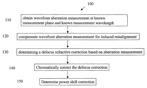
Figure 1 is a process flow diagram according to an embodiment of the
invention;
Figure 2 is a process flow diagram according to a preferred aspect embodiment
of
the invention; and
2
CA 02499978 2005-03-23
WO 2004/028356 PCT/US2003/028370
Figure 3 is a copy of a wavefront sensor eye measurement image illustrating
the
centration- of the wavefront measurement on the optical zone (OZ) of a lens;
and
Figure 4 is a line drawing of a hardware configuration embodiment of the
invention.
DETAILED DESCRIPTION OF A PREFERRED EMBODIMENT
The following detailed description is set forth in terms of data obtained from
a
Zywave wavefront analyzer (Bausch & Lomb Incorporated, Rochester, NY),
however, it
is to be appreciated that the invention is not limited in this manner; any
accurate
mathematical representation of a wavefront aberration would be suitable for
practicing
the invention. The Zywave incorporates a Harhnann-Shack wavefront sensor
(HSWFS )
to measure the wavefront aberration exiting a patient's eye at the eye's
entrance pupil
plane. The retinal illumination source in the Zywave is a diode laser emitting
light
having a wavelength of 780nm. The laser energy diffusely reflects off the
patient's fovea
and passes back tlirough the eye and into the HSWFS. The HSWFS samples the
wavefront at known intervals and a computer calculates a mathematical
description of the
wavefront aberration as a set of 18 Zernike coefficients (T3 through TZo)
measured in
microns. Other data provided by the Zywave include a normalization radius (RN)
measured in millimeters, identification of the patient's eye (left or right),
equivalent
sphere power (SE), and, when applicable, the rotation angle (S) measured in
degrees of a
trial lens worn by a patient during measurement. The equivalent sphere power
is defined
by the equation
SE [RN2 + (2*sqrt(3)*T3)z] - (2*2*sqrt(3)*T3),
3
CA 02499978 2005-03-23
WO 2004/028356 PCT/US2003/028370
where T3 represents the fourth Zernike term in Born & Wolf notation.
Figure 1 shows the process flow steps of an algoritliin 100 for determining a
surface parameter of an ophthalmic customized correcting surface from a
wavefront
aberration measurement of an eye. At step 110, a wavefront aberration
measurement of
the patient's eye is obtained at a known measurement plane location and at a
known
measurement wavelength. The preferred measurement plane location is the
entrance
pupil of the patient's eye, and the preferred measurement wavelength is 780nm
so as to
minimally affect patient fixation and pupil size. As mentioned above, the
Zywave
calculates the wavefront aberration in the form of Zernike polynomials per
Born & Wolf.
In a preferred embodiment, the Zernike polynomials are represented by the
"Fringe" or
University of Arizona notation (see Zemax User's Guide, Version 10.0, pp.124-
126).
Two differences between the Arizona notation, and the Born & Wolf notation are
that the
polynomial terms are ordered differently, and the B&W notation uses scalar
normalization terms in front of the polynomial terms. The first 11 terms of
each notation
are shown below:
Term Arizona Notation Born & Wolf Notation
Z1 1 1
Z2 p*cos(6) 2*p*cos(O)
Z3 p*sin(0) 2*p *sin(O)
Z4 2*p~2-1 sqrt(3)*(2*p~2 - 1)
Z5 p~2*cos(20) sqrt(6)*p~2*sin(20)
Z6 p~2*sin(20) sqrt(6)*p~2*cos(20)
Z7 (3*pA3 - 2*p)*cos(6) sqrt(8)*(3*p~3 - 2*p)*sin(O)
Z8 (3*pA3 - 2*p)*sin(8) sqrt(8)*(3*p~3 - 2*p)*cos(9)
Z9 p A3*cos(30) sqrt(8)*p~3*sin(30)
Z10 p~3*sin(30) sqrt(8)*p~3*cos(30)
Z11 6*p~4 - 6*p~2 + 1 sqrt(5)*(6*p~4 - 6*pA2 + 1)
4
CA 02499978 2007-05-29
=
At step 120, image misaligmnent is corrected. The wavefront aberration at the
patient's entrance pupil is rotated 180 degrees before it reaches the HSWFS.
Thus, the
Zemike coefficients must be modified in order to account for this rotation.
This is done
by multiplying all coefficients with odd-theta dependence by -1. Those
coefficients with
no theta dependence or even-theta dependence are not modified.
At step 130, the equivalent sphere power, SF,, is calculated by
SE = [RN2 + (2*s9rt(3)*T3)2) - (2*2*sqrt(3)*T3),
where T3 represents the fourth Zemike term in Born & Wolf notation. However,
the
measurement wavelength 780nm focuses deeper into the eye than does light at
555 nm,
which is the center of the wavelength region for normal human vision. Tbus,
the Zywave
erroneously measures a patient's necessary correction by +0.45 D different
from tfieir
actual necessary correction. This correction is made at step 140. The correct
power
adjustment is represented as B = SE - 0.45. Defocus is primarily defined by
the fourth
Zernike term, T3, therefore, T3 must be modified to account for this known
chromatic
aberration. If ocular biometry data are collected, then the 0.45 D defocus
shift to convert
from 780 to 555nm could be optimized for individual patients on a case-by-case
basis.
Ocular biometry includes comeal topography or keratometry, axial length of the
eye and,
optionally, thickness of the crystalline lens. From these measurements, a more
precise
calculation of longitudinal chromatic aberration can be perfonmed.
At step 150, the algorithm accounts for the power shift due to the measurement
being taken at the patient's entrance pupil but the correction being done at
the ophthalmic
correction surface, such as a modified corneal surface, a custom contact lens
surface, a
customized IOL surface, a customized inlay surface, or a spectacle surface.
CA 02499978 2005-03-23
WO 2004/028356 PCT/US2003/028370
In a preferred aspect of the embodiment described above, the method is
directed
to determining an anterior surface parameter of a dry-form CCL designed to
operate at
555nm, from a wavefront aberration measurement of an eye. The method 200 is
set forth
in Fig. 2. Process steps 110 through 150 remain unchanged but the correction
is done at
the anterior surface of the contact lens. The 3-D sag profile of the anterior
surface of a
dry custom-correction contact lens is described by the equation:
3-D Sag =(r2/Rd)/(1 + sqrt(1 - rZ/Rd2)) + E(Z;P;)
where r is the radial coordinate, Rd is the dry radius of the anterior
surface, Z; is a set of
Zernike coefficients, Pi is a set of Zernike polynomials, and i E[4, 27]. The
Zernike
coefficients and polynomials are of the Fringe or University of Arizona
notation.
The entrance pupil of a typical human eye is located 3.1 mm from the anterior
surface of the cornea into the eye. A typical custom contact lens has a center
thickness of
0.16 mm. Thus, the correction is located 3.26 mm away from the entrance pupil,
and this
distance causes a slight power shift between measured power error and the
correcting
power. This shift is explained by the following equation, where B is the
measured power
and C is the correcting power located 3.26 mm away from the measurement plane.
C = B - 0.00326*B2.
At step 160, the Zernike coefficients are converted from wavefront
deformations
to wet lens surface deformations. All coefficients are divided by (n - 1),
where n is the
index of refraction of the wet contact lens material at 555 nm.
At step 170, the algorithm modifies the surface parameter to mathematically
reverse hydration-induced expansion in going from wet lens parameters to dry
lens
parameters. All coefficients are divided by an empirically obtained sag
expansion
6
CA 02499978 2005-03-23
WO 2004/028356 PCT/US2003/028370
(sag_exp) factor for the lens material. The normalization radius is divided by
the
empirically obtained diameter expansion (dia exp) factor. Both of these
factors typically
range between about 4% to 35% depending upon the lens material.
A Summary of Modified Zemike Coefficients and Normalization Radius is
presented as follows:
NR = NR/dia exp
Z4 = T3'*sqrt(3)/(n - 1)/1000/sag_exp
Z5 = T5*sqrt(6)/(n - 1)/1000/sag_exp
Z6 = T4*sqrt(6)/(n - 1)/1000/sagexp
Z7 = T7*(-1)*sqrt(8)/(n - 1)/1000/sag_exp
Z8 = T6*(-1)*sqrt(8)/(n - 1)/1000/sag_exp
Z9 = Tl0*sqrt(5)/(n - 1)/1000/sag_exp
Z10 = T9*(-1)*sqrt(8)/(n - 1)/1000/sag_exp
Z11 = T8*(-1)sqrt(8)/(n - 1)/1000/sag_exp
Z12 = Tl 1*sqrt(10)/(n - 1)/1000/sag_exp
Z13 = T12*sqrt(10)/(n - 1)/1000/sag_exp
Z14 = T15*(-1)*sqrt(12)/(n - 1)/1000/sag_exp
Z15 = T16*(-1)*sqrt(12)/(n - 1)/1000/sag_exp
Z16 = (not currently used)
Z17 = T13*sqrt(10)/(n - 1)/1000/sag_exp
Z18 = T14*sqrt(10)/(n - 1)/1000/sag_exp
Z19 = T17*(-1)*sqrt(12)/(n - 1)/1000/sag_exp
Z20 = T18*(-1)*sqrt(12)/(n - 1)/1000/sag_exp
Z21 = T23*sqrt(14)/(n - 1)/1000/sag_exp
Z22 = T22*sqrt(14)/(n - 1)/1000/sagexp
Z23 = (not currently used)
Z24 = (not currently used)
Z25 = (not currently used)
Z26 = T19*(-1)*sqrt(12)/(n - 1)/1000/sag_exp
Z27 = T20*(-1)*sqrt(12)/(n - 1)/1000/sag_exp
In an exemplary aspect of the preferred embodiment, the patient is fitted with
a
prism- or peri-ballasted spherical trial lens of known geometry. The power of
the trial
lens may be plano or may match the patient's spherical-equivalent refraction.
The latter
is most preferred. The trial lens preferably will be made of the same material
as the
CCL. The base curve of the trial lens, possibly chosen to include a plurality
of base
7
CA 02499978 2005-03-23
WO 2004/028356 PCT/US2003/028370
curves, will be matched to the patient's needs, similar to the CCL. The
wavefront
aberration is measured while the patient wears the trial lens. The measurement
is
centered on the center of the OZ of the trial lens, which is where the custom
lens
modifications will be centered. This step is depicted at 105 in Figure 2. The
measurement thus takes into account effects such as, for example, lens tilt
and
decentration, lens deformation as it adheres to the cornea, tear film effects,
lens rotation,
etc. It is often difficult to see the OZ on a lens while the patient is
wearing it, and
likewise when viewing the lens with the aberrometer camera. Furthermore, in a
prism-
ballasted lens, the OZ is decentered relative to the geometric center of the
lens. Thus, it
is preferable to mark the OZ such that it will be visible when viewed through
the
wavefront sensor. In a preferred aspect, illustrated in Fig. 3, the trial lens
302 is indexed
with a lathe mark 304 in the form of a ring having a 7.3mm dry inner diameter,
that is
centered on the OZ of the lens. The ring was made with a Imm tip radius
cutting tool.
Other ring dimensions or index markings made by lathe cuts, laser inscription,
or other
means lcnown in the art may also provide suitable centering marks. Moveable
crosshairs
306 in the wavefront sensor device are centered on the wavefront measuring
beam, and a
variable circular indexing mark 308 is also centered in the crosshairs. The
diameter of
ring 308 is changed until it coincides with the latlie ring mark 304. The
wavefront
measurement is thus centered on the OZ of the trial lens. In Figure 3, small
circle 310
shows the measurement beam entry location on the eye, but is not relevant to
an
understanding of the instant invention. Likewise, bright spots 312 are
instrument LED
reflections from the cornea and are not relevant to the instant invention.
Alternatively,
the OZ of the trial lens can be encircled with a ring or dots of indelible,
FDA-approved
8
CA 02499978 2007-05-29
a
ink placed every 15 or 30 degrees on the lens in the dry state. Thereafter,
the amount and
direction (CW or CCW) of the lens rotation is measured and accounted for
during the
manufacturing of the lens. The conversions for the rotation adjusted Zernike
fringe
coefficients in dry lens units are:
(adjusted norraalization radius = normalization radius)
A4 = Z'4
A5 = Z'5*cos(2*d) - Z'6*sin(2*d)
A6 = Z'5*sin(2*d) + Z'6*cos(2*d)
A7 = Z'7*cos(d) - Z'8*sin(d)
A8 = Z'7*sin(d) + Z'8*cos(d)
A9 = Z'9
A10 = Z'10*cos(3*~d) - Z'11*sin(3*d)
Al l = Z'10*sin(3*d) + Z'11 *cos(3*d)
A12 = Z'12*cos(2*d) - Z'13*sin(2*d)
A13 = Z'12*sin(2*d) + Z'13*cos(2*d)
A14 = Z'14*cos(d) - Z'15*sin(d)
A15 = Z'14*sin(d) + Z'15*cos(d)
A16 = Z'16
A17 = Z'17*cos(4*d) - Z'18*sin(4*d)
A18 = Z'17*sin(4*d) t Z'18*cos(4*d)
A19 = Z'19*cos(3*d) - Z'20*sin(3*d)
A20 = Z'19*sin(3*d) + Z'20*cos(3*d)
A21 = Z'21 *cos(2*d) - Z'22*sin(2*d)
A22 = Z'21 *sin(2*d) + Z'22*cos(2*d)
A23 = Z23*cos(d) - Z'24*sin(d)
A24 = Z'23*sin(d) + Z'24*cos(d)
A25 = Z25
A26 = Z'26*cos(5*d) - Z'27*sin(5*d)
A27 = Z'26*sin(5*d) + Z27*cos(5*d)
The hardware configuration of a device embodiment 400 of the invention is
shown in the block diagram of Fig. 4. A device readable medium 410 includes an
algorithm 420 (i.e., a computable set of steps to achieve a desired result)
for determinilig
a surface parameter of an ophthalmic correcting surface from a wavefront
measurement
of an eye, as described in detail above. The device readable medium can take
any well
known form such as a disk or diskette, CD, DV'D, waveguide, etc. thatcan carry
the
9
CA 02499978 2007-05-29
-
algorithm 420. The device 430 is preferably a P.C. that is connected to a
surface
machining apparatus '440. For a CCI,, IOL, or in-vitro inlay (al1:450), the
apparatus 440
is preferably a numerically controlled, multi-axis lathe such as an Optoform
50/Varifore lathe (Precitech, Keene, N.H., USA), or an excimer laser system.
For
corneal refractive surgery or an in-vfvo inlay (all 450), the apparatus 440
preferably is an
excimer laser system.
While various advantageous embodiments have been chosen to illustrate the
invention, it will be understood by those skilled in the art that changes and
modifications
can be made therein without departing from the scope of the invention as
defined in the
appended claims.
