Note: Descriptions are shown in the official language in which they were submitted.
CA 02500160 2005-03-23
WO 2004/037066 PCT/US2003/033297
BLOOD PROCESSING SYSTEMS AND METHODS FOR COLLECTING
PLASMA FREE OR ESSENTIALLY FREE OF CELLULAR
BLOOD COMPONENTS
FIELD OF THE INVENTION
This invention relates to systems and methods
for processing and collecting blood, blood constituents,
or other..suspensions of cellular material..
BACKGROUND OF TH$ INVENTIONv
Today people routinely separate whole blood,
usually by centrifugation, into its various therapeutic
components, such as red blood cells, platelets, and
plasma.
Conventional blood processing methods use
durable centrifuge equipment in association with single
use, sterile processing systems, typically made of
plastic. The operator loads the disposable systems upon
the centrifuge. before processing- and removes them.
;afterwards . .
Conventional blood centrifuges are of a size
that does not permit easy transport between collection
sites. Furthermore, loading and unloading operations can
sometimes be time consuming and tedious.
In addition, a need exists for further
improved systems and methods for collecting blood
components in a way that lends itself to use in high
volume, on line blood collection environments, where
CA 02500160 2005-03-23
WO 2004/037066 PCT/US2003/033297
- 2 -
higher yields of critically needed cellular blood
components, like,.plasma, red blood cells, and platelets,
can be realized iri reasonable short processing times.
. The operational and performance demands upon
such~~fluid processing systems become more complex and
sophisticated, even as the demand for smaller and more
portable systems intensifies. The need therefore exists
for automated blood processing controllers that can
. gather and generate more detailed information and control
signals to aid the operator in maximizing processing and
separation efficiencies.
SUMMA12Y OF THE INVENTION
One aspect of the invention provides blood
separation systems and methods that introduce blood into
15. , an annular separation.. channel , between..a. low-.G wall and a.
high=G. cva~Il. .wh3le::zota,tix~g the separation ~~cha~el about
an axis, for separation of the blood into blood
components. The annular separation channel has an annular
boundary~wall. The systems and methods direct a first
blood component into a constricted channel along the low-
G wall. The systems and methods remove the first blood
component through a first path that communicates~with the
separation channel through an opening that adjoins the
constricted channel adjacent the low-G wall. The systems
. and methods direct a second blood component along.. a.
surface v that ~. extends : .genera7.'ly.~w.in~..an '.aa~ia~.~ direction
along the high-G wall toward the annular boundary wall.
The systems and methods collect the second blood
component through a second path that communicates with
the separation channel through an opening that adjoins
the surface adjacent the high-G wall axially spaced from
the annular boundary wall.
In one embodiment, the second passage includes
a ledge that extends radially within the second passage
at the second opening to constrict the second passage
CA 02500160 2005-03-23
WO 2004/037066 PCT/US2003/033297
- 3 -
along the high-G wall. In one arrangement, away from the
second opening, the ledge adjoins an axial surface that
is generally aligned with the low-G wall, along whicYi a
blood component entering the second passage is directed
away~from the annular boundary wall for removal from the
separation channel.
Another aspect of the invention provides blood
separation systems and methods that introduce blood into
an annular separation channel between a low-G wall and a
high-G wall while rotating the separation channel about
an axis, for separation of the blood into blood
components. The annular separation channel has an annular
boundary wall. The systems and methods direct a first
blood component into a constricted channel along the low-
:15 G. wall for removal through a first path that communicates.. ._
with ahe ~. separat.io~ . channel through a fizst ~opening~ °that.'
adjoins the~constricted channel.adjacent the.low-G wall.
The systems and methods direct a second blood component
for~removal through a second path that communicates with
the separation channel through a second opening that is
adjacent the high-G wall and faces the annular boundary
wall. The second passage includes a ledge that extends
radially within the second passage at the second opening
to define a constricted channel along the high-G wall,
through which, .the second , blood. component , .enters the
second path for'removal from the_;separation.channel~. ~.
According to either~aspect of the invention,
the first and second blood components can be collected,
at least for time, simultaneously.
Also according to either aspect of the
invention, the second blood component can include, e.g.,
red blood cells, and also desirably includes platelets,
and leukocytes. In this arrangement, the first blood
component includes plasma and, desirably, plasma that is
free or essentially free of cellular blood components
CA 02500160 2005-03-23
WO 2004/037066 PCT/US2003/033297
- 4 -
such as red blood cells, platelets, and leukocytes.
Other ~ features and advantages of the
inventions are set forth ~in the following specification
and attached drawings. .
DRIEF DESCRIPTION OF THE ARAi~TINGS
Fig. 2 is a perspective view of a fluid
processing system, ideally suited for blood processing,
comprising a blood processing device (shown in a closed
condition for transport and storage) and a disposable
liquid and blood flow set, which interacts with the blood
processing device to cause separation and collection of
one. or more blood components (shown packaged in a tray
for transport and storage before use).
Fig. 2 is a perspective view of the blood
processing,, device .shown in Fig... 1,. shown , in an ;.opened ..
condition for .opez-at-ion., . ~ . : . .. , , . .
Fig. 3 is a perspective view of~the blood
processing device shown in Fig. 2, with the centrifugal
station open to receive a blood processing chamber and
the pump and valve station open to receive a fluid
pressure-actuated cassette.
Fig. 4 is a perspective view of the blood
processing device shown in Fig. 3, with the tray
containing the disposable liquid and blood flow set
25. ..positioned ..for loading, the flow set ox~ the , device .
~;. Fig.s : 5 ~ and ~ 6 area . .~respectivel.ji, xigTit y ;and ,
left side perspective views' of the blood processing
device shown in Fig. 2 after the liquid and blood flow
set has been loaded onto the device for use.
Fig. 7 is a perspective view of the blood
processing chamber and attached umbilicus that forms a
part of the liquid and blood flow set shown in. Figs. 5
and 6.
Fig. 8 is a perspective view of the interior
of a representative embodiment of the blood processing
CA 02500160 2005-03-23
WO 2004/037066 PCT/US2003/033297
_ 5 _
chamber of a type shown in Fig. 7, the interior of the
chamber 'being configured to perform aced blood cell
separation and collection procedure using the device
shown in Figs. 5 and 6.
~ Fig. 9 is a perspective view of the interior
of the centrifuge station of the device shown in Figs. 5
and 6, with the station door opened to receive a blood
processing chamber of a type shown in Fig. 7.
Fig. 10 is a perspective view of the interior
of the centrifuge station shown in Fig. 9 after a blood
processing chamber Qf a type shown in Fig. 7 has been
loaded for use.
Fig. 11A is an enlarged perspective view of a
fixture that is carried by the umbilicus shown in Fig. 7,
1.5. showing. its. intended. association with an optical sensing
tation, .that . forrris ~ a part: .of. the device shown in ~ Figs . w5~.
and 6 . . .. . ~ : .
Fig. 11B is a side section view of the optical
sensing station shown in Fig. 11A.
Fig. 11C is an exploded perspective view of
the optical sensing station shown in Fig. 11A.
Fig. 11D is a top view of the optical sensing
station shown in Fig. 11A.
Figs. 11E and 11F are schematic views of a
25., circuit.., that can. be used in association with the,. optical
.erisi.ng station : shoc~iz:~ .in_ Fig.. .11A.
Fig. 12 ~is a diagrammatic view of the iiiterior~
of the blood processing chamber of a type shown in Fig.
7, showing the separation of whole blood into a red blood
cell layer, a plasma layer, and an intermediate buffy
coat layer, with the position of the layers shown in a
desired relationship.
Fig. 13 is a diagrammatic view of the interior
of the blood processing chamber of a type shown in Fig.
7, with the buffy coat layer having moved very close to
CA 02500160 2005-03-23
WO 2004/037066 PCT/US2003/033297
- 6 -
the low-G wall, creating an undesired over spill
condition that sweeps huffy coat components into the
plasma being collected.
Fig. 14 is a diagrammatic view of the interior
of the blood processing chamber of a type shown in Fig.
7, with the huffy coat layer having moved very close to
the high-G wall, creating an undesired under spill
condition that leads to a reduction of the hematocrit of
red blood being collected.
Fig. 15 is an exploded perspective view of the
fluid pressure-actuated cassette that forms a part of the
liquid and blood flow set shown in Figs. 5 and 6 and its
operative association with the pump and valve station on
the device, also shown in Figs. 5 and 6, which applies
Z5 positive and negative pneumatic"pressure.to the cassette.
to circulate. liquid ~and~'blood through. the. cassette:
Fig. 16 is a schematic view of.a fluid~circuit.
that can be implemented in the cassette shown in Fig. 15
to enable the performance of different blood processing
and collection procedures.
Fig. 17 is a plane view of a cassette in which
the fluid circuit shown in Fig. 17 is implemented.
Fig. 18 is a top perspective view of the
interior of a representative embodiment of the blood
25. processing ,.chamber of a type. shown. in Fig. 7, , the
interior , of :~tlie : chamber :being:; configured .:.to...perfori~. a..
plasma separation and collection procedure using the
device shown in Figs. 5 and 6.
Fig. 19 is a bottom perspective view of the
blood processing chamber shown in Fig. 18.
Fig. 20 is an enlarged side perspective view
of an interior region in the blood processing chamber
shown in Fig. 18, showing a barrier having a tapered
surface that directs red blood cells from the separation
zone in a path separate from plasma.
CA 02500160 2005-03-23
WO 2004/037066 PCT/US2003/033297
Fig. 21 is an enlarged bottom perspective view
of the region shown in Fig. 20, showing the path that red
blood cells take as they are directed from the separation
zone by the barrier.
~ Fig. 22 is an enlarged top perspective view of
the region shown in Fig. 20, showing the separate paths
that red blood cells and plasma take as they are directed
from the separation zorie by the.barrier,
Fig. 23 is a schematic view of a cassette of a
type shown in Figs. 16 and 17 coupled to a liquid and
blood flow set in a.configuration that can be used for a
plasma collection procedure.
Fig. 24 is a schematic view of a cassette of a
type shown in Figs. 16 and 17 coupled to a liquid and
15. blood flow set . in a .configuration' that can be used for a ., .. .
v double 'unit . red ~ blood w cell ~.-colleetion. .procedure,. the: ,
blood flow. set also being shown in .Figs. 5 and 6 after
being loaded on the blood processing device.
Figs. 25A and 25B are schematic views of the
fluid circuit~shown in Fig. 16 being conditioned by
application of positive and negative pneumatic pressures
to transport air in a controlled manner that verifies
that tubing intended to convey blood and liquids to and
from the donor has been properly installed on the device,
as..shown in Figs. 5. and 6.. , .._ . .
.; Figs. . 26A and. 2~~8 are schematic views.'.of, the
fluid circuitshown in ~ Fig. 16 ~ ~beirig conditioned by
application of positive and negative pneumatic pressures
to transport air in a controlled manner that verifies
that tubing intended to convey anticoagulant into blood
drawn from the donor has been properly installed on the
device, as shown in Figs. 5 and 6.
Figs. 27 to 29 are schematic views of the
fluid circuit shown in Fig. 16 being conditioned by
application of positive and negative pneumatic pressures
CA 02500160 2005-03-23
WO 2004/037066 PCT/US2003/033297
_ g _
to transport a liquid in a controlled manner that
verifies the physical integrity of the cassette prior to
use.
The invention may be embodied in several forms
S without departing from its spirit or essential
characteristics. The scope of the invention is defined
in the appended claims, rather than in the specific
description preceding them. All embodiments that fall
within the meaning and range of equivalency of the claims
are therefore intended to be embraced by the claims.
DESCRIPTION OF THE PREFERRED EMBODIMENTS
Fig. 1 shows a fluid processing system 10 that
embodies the features of the invention. The system 10
can be used for processing various fluids.
The._.system~.l0..is particularly well .suited, for..
processingy whole: ,blood, anc3v. other y si~sperisioiis, .'bf
biological cellular materials. Accordingly, the
illustrated embodiment shows the system 10 used for this
purpose.
I. System Overview
The system 10 includes two principal components.
These are: (i) a blood processing device 14 -- shown in
Fig. 1 in a closed condition for transport and storage,
and in Figs. 2 and 3 in an opened condition for
~ operation) ; . and...(:~.i) .a .liquid .:and .blood .flow set .12,
.which interacts: with. the;.blood processing; d~va.ce 14: t.a.
cause separation and collection of one or more blood
components -- the set 12 being shown in Figs , 1 and 4
packaged in a tray 48 for transport and storage before
use, and in Figs. 5 and 6 removed from the tray 48 and
mounted on the blood processing device 14 for use.
A. The Processing Device
The blood processing device 14 is intended to be a
durable item capable of long term use. In the
CA 02500160 2005-03-23
WO 2004/037066 PCT/US2003/033297
_ g _
illustrated and preferred embodiment, the blood
processing device 14_is mounted inside a portable housing
or case .36. The case 36 presents a compact footpririt~,
suited for set up and operation upon a table top or other
relatively small surface. The case 36 is also intended to
be transported easily to a collection site.
The case 36 includes a base 38 and w hinged lid 40,
which closes for transport' (as Fig. 1 shows) and which
opens for use (as Figs. 2 to 4 show). In use, the base 38
is intended to rest in a generally horizontal support
surface. The case 36 can be formed into a desired
configuration, e.g., by molding. The case 36 is
preferably made from a lightweight, yet durable, plastic
material.
A controller 16 is,carried onboard the device 14.
The controller~l6..~governs the, interaction,between~the.~
components of the device 14 and the components of the
flow set 12 to perform a blood processing and collection
procedure selected by the operator. In the illustrated
embodiment, the controller 16 comprises a main processing
unit (MPU), which can comprise, e.g., a Pentium' type
microprocessor made by Intel Corporation, although other
types of conventional microprocessors can be used. The
MPU can be mounted inside the lid 40 of the case 36. A
power supply . with...,power, cord 184 , supplies .electrical .
power :.to the MPU . and,other.. components of ' he, device :1.4 :~ .~ .
Preferably, ~ ~ the ~ controller ~ 16 also ~. includes an
interactive user interface 42, which allows the operator
to view and comprehend information regarding the
operation of the system 10. In the illustrated
embodiment, the interface 42 is implemented on an
interface screen carried in the lid 40, which displays
information for viewing by the operator in alpha-numeric
format and as graphical images.
Further details of the controller 16 can be found in
CA 02500160 2005-03-23
WO 2004/037066 PCT/US2003/033297
_
Nayak et al, United States Patent 6,261,065, which is
incorporated hexein by i:eference. Further details of the
interface can be found in Lyle et al, United States
Patent 5,581,687, which is also incorporated herein by
5 reference.
As Fig. 2 shows, the lid 40 can be used to support
other input/outputs to couple other external devices to
w
the controller 16 or other components of the device 14.
For example, an ethernet port 50, or an input 52 for a
10 bar code reader or the like (for scanning information
into the controller ~6), or a diagnostic port 54, or a
port 56 to be coupled to a pressure cuff 60 worn by a
donor to enhance blood flow rates during blood processing
(see, e.g., Figs. 23 and 24), or a system transducer
calibration port..58., can all be conveniently,mounted for
access on the .e.~te~ioi-. of. the .did 40, ~or e3sesvhere on the .
case 36 of~the device 14.
B. The Flow Set
The flow set 12, is intended to be a sterile, single
use, disposable item. Before beginning a given blood
processing and collection procedure, the operator Loads
various components of the flow set 12 in association with
the device 14 (as Figs. 4 and 5 show). The controller 16
implements the procedure based upon preset protocols,
taking into account other..input,.from the operator. Upon
completing. the: procedure,'. ther.operator~.removes .the'.fZow .
set l2.from association with.the device 14. The portion
of the set 12 holding the collected blood component or
components are removed from the device 14 and retained
for storage, transfusion, or further processing. The
remainder of the set 12 is removed from the device .14 and
discarded.
The flow set includes a blood processing chamber 18,
a fluid actuated pump and valve cassette 28, and an array
associated processing containers 64 and flow tubing
CA 02500160 2005-03-23
WO 2004/037066 PCT/US2003/033297
- 11 -
coupled to the chamber 18 and the cassette 28, as will be
identified in greater detail later.
1. ' The Blood Processing Chamber
In the illustrated embodiment (see Fig. 5), the flow
set 12 includes a blood processing chamber 18 designed
for use in association with a centrifuge. The processing
device 14 includes a centrifuge station 20 (see Figs. 2
and 3, which receives the processing chamber 18 for use
(see Fig. 5) .
As Figs . 2 and 3 show, the centrifuge station 20
comprises a compartment 24 formed in the base 38. The
centrifuge station 20 includes a door 22. The door 22
opens (as Figs. 3 and 5 show) to allow loading of the
processing chamber 18 into the compartment 24. The door
. . 22 closed . .(.as Figs . , .2 . and 6 ~ .show), tQ enclose: . the .
. . . . rproces.sing chamber. 18~ withzri , the . .cor~partmenty 24. during ~~
, . .
operation.
The centrifuge station 20 rotates the processing
chamber 18. When rotated, the processing chamber 18
2 0 centrifugally separates whole blood received from a donor
into component parts, principally, red blood cells,
plasma, and intermediate layer called the buffy coat,
which is populated by platelets and leukocytes. As will
be described later, the configuration of the chamber 18
~25 can .vary .according.:ta; the . intended. .blood 'separation..
objectives.
2. The Fluid Pressure-Actuated Cassettev
In the illustrated embodiment, the set 12 also
includes a fluid pressure-actuated cassette 28 (see Fig.
3 0 5). The cassette 28 provides a centralized, programmable,
integrated platform for aII the pumping and valuing
functions required for a given blood processing
procedure. In the illustrated embodiment, the fluid
pressure comprises positive and negative pneumatic
CA 02500160 2005-03-23
WO 2004/037066 PCT/US2003/033297
- 12 -
pressure, although other types of fluid pressure can be
used.
As Fig. S shows, the cassette 28 is mounted for use
in a pneumatic actuated pump and valve station 30, which
is located in the lid of the 40~of the case 36. The pump
and valve station 30 includes a door 32 that is hinged to
move between an opened position, exposing the pump and
valve station 30 (see Fig.s3) for loading and unloading
the cassette 28, and a closed position, enclosing the
cassette 28 within the pump and valve station 30 for use
(shown in Fig. 6). The pump and valve station 30 includes
a manifold assembly 34 (see Fig. 4) located behind a
valve face gasket 318. The manifold assembly 34 applies
positive and negative pneumatic pressure to the cassette
28. through .the gasket 318,. when the cassette 28 is. when.
mounted ..on the pump :and valve. station. 30;7 The:;pneumat.~;d~
pressures direct liquid flow through the cassette 28...
Further details of the cassette 28 and the operation
of the pump and valve station 3D will be described later.
Additional details can also be found in Nayak et al,
United States Patent 6,261,065, which has been
incorporated herein by reference.
3. B1~od Praaessing Containers and Tubing
Referred back to Figs. 5 and 6, the flow set Z6 also
:25 ~ inehudes., an -array: ~f tubes . ; and ., containers ' . in : f low
wcommunicat3on.~ with 'the cassettew28 ~, and .ahe: chamber :7.8 :,
The'"arrangement of tubes. and containers can vary
according to the processing objectives. Representative
blood processing procedures and the associated flow sets
accommodating such procedures will be described later.
An umbilicus 100 forms a part of the flow set 16.
When installed, the umbilicus 100 links the rotating
processing chamber I8 with the cassette 28 without need
for rotating seals. The umbilicus 100 can be made from
rotational-stress-resistant plastic materials, such as
CA 02500160 2005-03-23
WO 2004/037066 PCT/US2003/033297
- 13 -
Hytrel° copolyester elastomers (DuPont).
Referring now to Fig. 7, tubes 102, I04, and I06
extend from the proximal end of the umbilicus .100. The
tube 102 conveys whole blood into the processing chamber
18 for separation. The tubes 104 and 106 convey,
respectively, centrifugally separated red blood cells and
plasma from the processing chamber 18. The plasma can
either be rich or poor in platelets, depending upon the
processing objectives.
As Fig. 7 shows, a fixture 108 gathers the tubes
102, 104, and 106_ adjacent the umbilicus 100 in a
compact, organized, side-by-side array outside the
centrifuge station 20. The fixture 108 allows the tubes
102, 104, and 106 to be placed and removed as a group in
association with an optical, sensing. station 46, (see_ Figs.
10,.' : and .I1.) ; -~ which . . i,s ~ located: adj acent v. to , the
centrifuge station~20 outside the chamber.l8.
The optical sensing station 46 optically monitors
the presence or absence of targeted blood components
(e. g., red blood cells and platelets) in blood conveyed
by the tubes 104 and 106. The sensing station 46 provides
outputs reflecting the presence or absence of such blood
components. This output is conveyed to the controller 16.
The controller 16 processes the output and generates
signals.. to control processing events, based, in part, upon
the optical~:y .sensed events Further; -;details:yof:'~,the
operation of the controller~to contro7..processing events
based upon optical sensing will be described later.
Additional details can also be found in Nayalc et al,
United States Patent 6,261,065, which has been
incorporated herein by reference.
As shown.(see Figs. 5 and 6), the flow set 16
includes a phlebotomy needle 128, through which a donor
can~be coupled to the system 10 for blood processing. In
Figs. 5 and 6, the flaw set 16 also includes a blood
CA 02500160 2005-03-23
WO 2004/037066 PCT/US2003/033297
_ lg _
sampling assembly 110. The blood sampling assembly 110
allows for the collection of one or more samples of the
donor's blood at the commencement of a given blobd
processing procedure, through the phlebotomy needle 128.
A conventional manual clamp 114 (e. g., a Roberts Clamp)
is provided to control blood flow into the sampling
assembly 110.
As also shown in Figs. 5 and 6, the flow set 16 can
include an in-line injection site 112. The injection
site 112 allows a technician to introduce saline or
another physiologic ~.iquid or medication into the donor,
if necessary, using the phlebotomy needle 128, and
without requiring an additional needle stick.
An additional in-line manual clamp 116 is desirably
1,5 . included upstream of the blood sampling assemb7:y ,110 and
the..iiijectzbn site' 112. This. clamp, 116 makes it, possible
to. quickly isolate the donor ~ from the flow set 16, if
donor safety or comfort requires. Alternatively, a
separate hemostat device (not shown) can be applied for
the purpose. '
As Figs. 1 and 2 also show, the device 14 can
include other components compactly arranged to aid blood
processing. In addition to the centrifuge station 20 and
pump and valve station 30, already described, the device
.25 includes one. or more, weigh stations. 62 :and other forms of.
support... .for~.~,container~,...~,:Thev. ~ arrangement '~:ofv ~. these
components on the device 14 can,'or course, vary.
In the illustrated embodiment (see Fig. 3), the
weigh stations 62 comprise a series of container
hangers/weigh sensors arranged along the top of the lid
40. In the illustrated embodiment, additional swing-out
hangers/weigh sensors are also provided on the side of
the lid 40 and the base. In use (see Figs. 5 and 6) ,
containers are suspended on the weigh stations 62. As
Figs, 5 and 6 also show, pictorial icons 66 applied to
CA 02500160 2005-03-23
WO 2004/037066 PCT/US2003/033297
- 15 -
the lid 40 adjacent to the weigh stations 62 match
pictorial icons 66 applied on the contaa.ne~s. Bjr
matching the icons 66, the operator is visually guided to
place ,the proper containers on the intended weigh
stations 62. .
The weigh stations 62 can also comprise molded
recesses in the base 38 to rest containers. Pictorial
icons 66 on the base 38 adjacent the stations 62 match
pictorial icons 66 on the containers to guide the
operator in proper placement of containers during set up.
As blood or liquids are received into and/or
dispensed from the containers during processing, the
weigh stations 62 provide output reflecting weight
changes over time. This output is conveyed to the
15... . , controller 16 . The controller 16 processes . , the
incxemerital. -uieight , changes ~vw derive- .fluid' processing;.
volumes. The controller generates signals 'to control
processing events based, in part, upon the derived
processing volumes. Further details of the operation of
the controller 16 to control processing events will be
described later. Additional details can also be found in
Nayak et al, United States Patent 6,261,065, which has
been incorporated herein by reference.
4. Blood Processing Procedures
~Unde.r the control of the controller.,l6,;.the system
~10 can .:be v conditioned . ~to : pe.rform~, ~di.ffevent ~ blo.od ..
processing procedures. The MPU includes an application
control manager that administers the activation of a
library of control applications. Each control application
prescribes procedures for carrying out given functional
tasks using the centrifuge station 20 and the pump and
valve station 30 in a predetermined way. The applications
can, e.g., reside as process software in EPROM~s in the
MPU.
As will be described later, through selective
CA 02500160 2005-03-23
WO 2004/037066 PCT/US2003/033297
- 16 -
application of pressure to the cassette 28, it is
possible to use the same cassette 28 to carry out
different blood collection procedures.
For the sake of illustration, the implementation of
two clinical procedures will be described: (1) a plasma
collection procedure; and (2) a double unit red blood
cell collection procedure. During a plasma collection
procedure, whole blood from a donor is centrifugally
processed to yield up to 880 ml of plasma for collection.
All red blood cells are returned to the donor. During a
double unit red blood cell collection procedure, whole
blood from a donor is centrifugally processed to yield up
to two units (approximately 500 ml) of red blood cells
for collection. All plasma constituent is returned to the
donor.
AlthQUgh,.~ not described iri detail; other. , clinical,
procedures can ~be ~ conducted ~ by the' system . 10. For
example, a plasma / .red blood cell collection procedure
can be performed, during which whole blood from a donor
is centrifugally processed to collect up to about 550 ml
of plasma and up to about 250 ml of red blood cells. The
portion of the red blood cells not retained for
collection are periodically returned to the donor during
blood separation. Plasma collected in excess of the 550
ml target and red blood cells collected in excess of the
:250. iriltarget .are .alao ~etia~ned .to he donor at~.'the, ei~d~.,
~of the procedure. As~another~example,'duririg the~course
of a plasma collection and/or red blood cell collection
procedure, the buffy coat interface can be removed from
the chamber Z8 and collected. With subsequent processing
to remove leukocytes, the buffy coat serves as a source
of platelets.
Further details of the various blood collection
procedures that the system 10 can accomplish are
described in United States Patent 6,261,065, which has
CA 02500160 2005-03-23
WO 2004/037066 PCT/US2003/033297
_ 17 _
been incorporated herein by reference.
II. Other Technical Features of the'Blood Separation
Components of the System
The blood processing chamber 18 and the centrifuge
station 20 of the system 10 desirably possess other
technical features that support the implementation of
diverse blood processing protocols.
A. The Blood Processing Chaiaber
In the illustrated embodiment (see Figs. 7 and 8),
the processing chamber 18 is preformed in a desired shape
and configuration, e.g., by injection molding, from a
rigid, biocompatible plastic material, such as a non
plasticized medical grade acrilonitrile-butadiene-styrene
(ABS). In this arrangement, the chamber 18 includes two
principalycomponents . - e,a base, component ..2,OO..and. .a lid.:..
component ~ 2 0 2 ,.
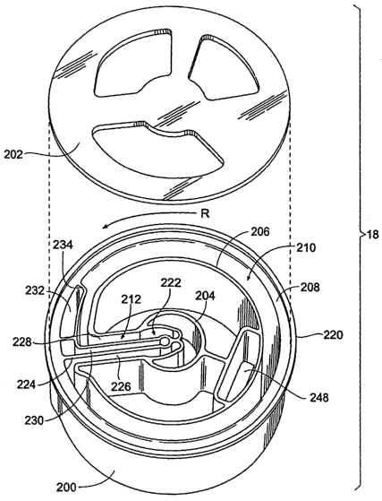
The base component 200 includes a center hub 204.
The hub 204 is surrounded by inside and outside annular
walls 206 and 208 that define a circumferential blood
separation channel 210. One or more radial passages 212
extend from the hub 204 and communicate with the channel
210. Blood and other fluids are directed from the hub 204
into and out of the channel 210 through these passages
212. A molded wall 214 forms an axial boundary of the
. ~ separation channel .210.., : The .li.d component 202, also,. forms ..
. ano~hex.~:axia~ boundary: ~of the separaticin channel'. 2~10.:~
While both axial boundaries are shown to be generally
flat (i.e., normal to the rotational axis), it should be
appreciated that the axial boundaries can be tapered,
rounded, V-shape, and the like.
The underside of the base component 200 includes a
shaped receptacle 216 that receives a shaped mount 218 on
the far end of the umbilicus 100. The mount 218 can be
secured to the receptacle 216 i~ various ways -- e.g., by
CA 02500160 2005-03-23
WO 2004/037066 PCT/US2003/033297
- 18 _
a tight, dry press fit or by solvent bonding or by
ultrasonic, welding' -- to-couple the umbilicus 100 in
fluid communication with the channel 210. The far~end of
the umbilicus 100 and the base component 200 rotate as a
unite.
.All contours, ports, channels, and walls that affect
the dynamics of the blood separation process are
preformed in the base component 200 in one or more
injection molding operations. The contours, ports,
channels, and walls that are preformed in the base
component 200 can vary, according to the particular
separation objectives desired. Representative examples
will be described in greater detail later.
B. The Centrifuge Station
1.5. The centrifuge, station .20 (see Fig.. 9).. includes, a.
centrifuge assembly- .68. :.The ~ centrifuge yassembly 68 :is
constructed to receive and support the molded~processing~
chamber 18 and umbilicus 100 for use.
As illustrated in Fig. 9, the centrifuge assembly 68
includes a frame or yoke 70 having bottom, top, and side
walls 72, 74, 76. The yoke 70 spins on a bearing element
78 (Fig. 9) attached to the bottom wall 72. An electric
drive motor 80 is coupled to the bottom wall 72 of the
yoke 70, to rotate the yoke 70 about an axis 82. In the
illustrated embodiment, ,the axis .82 is .essentially..
:..~hori zontal 'y ,v. ( see ~:. F'ig : . . 3 )., .-: ~~ although- w: vother-'
vartgular .
orientations can be used. ~ ~ The 'motor 80 is capable of
rotating the yoke 70 in either clockwise ar
counterclockwise directions, depending upon commands
issued by the controller 16.
A carrier or rotor plate 84 spins within the yoke 70
about its own bearing element 86, which is attached to
the top wall 74 of the yoke 70. The rotor plate 84 spins
about an axis that is generally aligned with the axis of
rotation 82 of the yoke 70.
CA 02500160 2005-03-23
WO 2004/037066 PCT/US2003/033297
- 19 -
As Fig. 7 shows, the top of the processing chamber
18 includes an annular lip 220, to which the lid
component 202 is secured. As Fig. 10 shows, the rotor
plate 84 includes a latching assembly 88 that removably
S grips the lip 220, to secure the processing chamber 18 on
the rotor plate 84 for rotation.
Details of the latching assembly 88 can be found in
co-pending United States Patent Application Serial No.
09/976, 829, filed October 13, 2001 and entitled ~~Blood
Separation Systems and Methods with Quick Attachment of a
Blood Separation Chamber to a Centrifuge Rotor," which
has been incorporated herein by reference.
As Fig. 10 best shows, a sheath 144 on the near end
of the umbilicus 100 fits into a preformed, recessed
pocket 90 .in, the centrifuge station. 20. The .pocket 90.
holds 'the, near .end.' of the~.uinbilicus X100: in : a non.-.rotating
stationary position aligned with the mutually aligned
rotational axes 82 of the yoke 70 and rotor plate 84.
The preformed pocket 90 is also shaped to
accommodate loading of the fixture 108 at the same time
the umbilicus sheath 144 is inserted. The tubes 102,
104, and 106 are thereby placed and removed as a group in
association with the sensing station 46, which is also
located within the pocket 90, as Fig. 11 shows.
2~ Umbilicus. drive or support. members 92. and 94 (see
. Figs .. 9 ~ aiid ' ~.0 )v are carried by: ~ a ~ side ..wallv 76 '. of . the
: yoke..
70. When the rotor plate~84 is located in~a prescribed
rotational position, the support members 92 and 94 are
presented on the left side of the processing chamber l8
to receive the umbilicus 100 at the same time that the
sheath 144 and fixture 108 are manipulated for fitting
into the pocket 90.
As Fig. 10 shows, one member 92 receives the mid
portion of the umbilicus 100. The member 92 includes a
surface against which the mid portion of the umbilicus
CA 02500160 2005-03-23
WO 2004/037066 PCT/US2003/033297
- 20 -
100 rests. The surface forms a channel 96, which faces
generally toward the yoke 70. The channel 96
accommodates passage of the mid portion of the umbilicus.
100, directing the upper portion of the umbilicus toward
the other member 94. The channel 96 inhibits travel of
the mid portion of the umbilicus 100 in radial directions
toward and away from the rotational axis 82. However, the
channel 96 permits rotatiori or twisting of the umbilicus
100 about its own axis. Before use, the surface of the
channel 96 is generally convex. The convex configuration
is intended to be sacrificial, in that the material of
the convex surface is intended to be worn away during use
by rotational contact with the umbilicus 200. The convex
configuration is dynamically changed by contact with the
umbilicus .during use, o., form ~.an .final . contact.
conf.igurat'ion ~~that is ~. dictated. by~ .they, mechanical. and'.
frictional interaction between thechannel 96 and ~the~
umbilicus 100 during use.
The other member 94 receives the upper portion of
the umbilicus 100, which the member 92 directs toward it.
The member 94 includes a surface against which the upper
portion of the umbilicus 100 rests. The surface forms a
channel 98 inclined toward the top wall 72 of the yoke
70. The channel 98 generally faces away from the yoke 70,
and is thereby in a reverse facing relationship with. the
. ,~ . clianiiel.: 96 . To provide ' a ': tzansitional . path ~- "for -the .
umbilicus between the two oppositely .facing channels~96
and 98, the channel 96 is offset slightly outward from
the channel 98. The channel 98 guides the upper portion
of the umbilicus 100 toward the recessed pocket 90, which
is located axially above the top wall 72 of the yoke 70,
where the umbilicus sheath 144 and fixture 108 are
fitted. Like the channel'96, the channel 98 inhibits
travel of the upper portion of the umbilicus 100 in
radial directions toward and away from the rotational
CA 02500160 2005-03-23
WO 2004/037066 PCT/US2003/033297
- 21 -
axis 82. However, like the channel 96, th.e channel 98
permits rotation or twisting of the umbilicus 100 about
its own axis. w
Because the support channels 96 and 98 are arranged
in a.reverse facing relationship, the channels 96 and 98
mutually engage the mid region of the umbilicus in a
complementary, ~~reverse grip" fashion regardless of the
direction of rotation of the yoke 70.
The inward facing orientation of the channel 96 best
captures the umbilicus during rotation of the yoke 70 in
the counterclockwise direction (when viewed from the top
of the rotor plate 84?. This, in turn, stabilizes the
remainder of the umbilicus for engagement with the
channel 98 during rotation in this direction. The
. , processing,,e. chamber: , 18 _ is intended, during , blood
~~processing~ : operations, ~ . to be. .~.ro~ated in ,. :.a.:_.
counterclockwise direction.
The member 94 includes opposed side edges 99 and 101
that taper inward toward the outward facing channel 98.
The tapered side edge 101 further guides the mid region
of the umbilicus into engagement with the outward facing
channel 98 in response to rotation of the yoke 70 in the
counterclockwise direction.
The outward facing guide edge 99 of the channel 98
defines an ..enlarg.ed . curved aurface .or ramp. that, extends
toward they rotati;onal~~ axis 82 . The xamp ':99 . is 'sized .:and ::
configured to accomplish self-loading the umbilicus into
the channel 98 when the yoke is rotated in this clockwise
direction (as viewed from the top of the rotor plate 84),
3 0 which is the direction opposite to the direction of
rotation intended for regular blood processing (i.e.,
counterclockwise).~The ramp 99 also thereafter keeps the
upper portion of the umbilicus 100 from slipping out of
the channel 98 when the yoke 70 is rotated in a
counterclockwise direction. This, in turn, stabilizes the
CA 02500160 2005-03-23
WO 2004/037066 PCT/US2003/033297
- 22 -
remainder of the umbilicus for engagement with the
channel 96 during rotation in this direction.
The configurations of the channels 96 and 98 thereby
complement each other, to keep the mid region of the
umbilicus in engagement with the channels 96 and 98 in
response to rotation of the yoke 70 and regardless of the
direction of rotation of the yoke 70
In the illustrated embodiment, the channel surfaces
96 and 98 of the support members 92 and 94 are preferably
fabricated from a low friction material, to thereby
eliminate the need for external lubrication or rotating
bearings on the umbilicus 100 itself. The material used
can, e.g., comprise Teflon° polytetrafluoroethylene
material (DuPont) or an ultra high molecular weight
polyethylene.. Made, from ,such materials, the channel
surfaces ~'96 and 98 .miri~:inize vmhilicus driue .friction ~~aiad
the~presence'of particulate matter due to umbilicus wear.
Further details of the support members 92 and 94 can
be found in co-pending United States Patent Application
Serial No. 09/976,830, filed October 13, 2001, and
entitled "Blood Separation Systems and Methods with
Umbilicus Driven Blood Separation Chambers," which is
incorporated herein by reference. .
Closing the centrifuge station door 20 positions a
holding bracket. 21. on the. underside .of the door 20 (see
yv Fig . ~ ,5 ) . in ~regist~iy~ with. the sheatli~ 7.44 . ~ .Another holdii~g
bracket 23 ~(as shown in lFig. ' S) on the underside of the
door 20 is positioned in registry with the fixture 108
when the door 20 is closed. A releasable latch 25
3 0 preferably holds the door 20 shut during operation of the
centrifuge assembly 68 (as Fig. 6 shows).
During operation of the centrifuge assembly 68, the
support members 92 and 94 carry the umbilicus 100 so that
rotation of the yoke 70 also rotates the umbilicus 100 in
tandem about the axis 82. Constrained within the pocket
CA 02500160 2005-03-23
WO 2004/037066 PCT/US2003/033297
- 23 -
90 at its near end (i.e., at the sheath 144) and coupled
to the chamber 16 at its far end ~(i.e., by the mount
218), the umbilicus 100 twists upon the channel surfaces
96 and 98 about its own axis as it rotates about the axis
82, even as the channel surfaces 96 and 98 inhibit radial
travel of the umbilicus relative to the rotation axis 82.
The twirling of the umbilicus 100 about its axis as it
rotates upon the channel surfaces 96 and 98 atone omega
with the yoke 70 (typically at a speed of. about 2250 RPM)
imparts a two omega rotation to the processing chamber 18
secured for rotatior~ on the rotor plate 84.
The relative rotation of the yoke 70 at a one omega
rotational speed and the rotor plate 84 at a two omega
rotational speed, keeps the umbilicus 100 untwisted,
avoiding. the need for rotating seals. The..illustrated
ar~angement~ a7ao. allows a.~s'ingle.; driwe~ motor 80 to: .impart '
rotation, through the umbilicus 100, to the mutually
rotating yoke 70 and processing chamber 18 carried on the
rotor plate 84. Further details of this arrangement are
disclosed in Brown et al U.S. Patent 4,120,449, which is
incorporated herein by reference.
As before described, the channel surfaces 96 and 98
are desirably formed and oriented in a complementary
fashion to accommodate rotation of the umbilicus 100 and
the driving of the . processing chamber 18. in either ,
clockwise.'ar ~couriter: clockwise: direct'ioris:v Thus,.1 the
chamber ~18 can'be rotated in'one direction conducive~to
one desired processing objective, e.g., to accommodate
priming and air venting prior to blood processing, and be
rotated in an opposite direction conducive to a different
processing objective, e.g., blood separation.
Furthermore, the close juxtaposition of the umbilicus
supports 92 and 94 to the umbilicus 100 when the rotor
plate 84 is in the prescribed rotational position to
accommodate mounting of the processing chamber 18, and
CA 02500160 2005-03-23
WO 2004/037066 PCT/US2003/033297
- 24 -
the complementary orientations of the channels 96 and 98
formed in the supports 92 and 94, which lead the near end
of the umbilicus toward the support pocket 90, make
possible an "easy-load" sequence of intuitive steps,
largely capable of being carried out in tandem, for
loading the processing chamber 18 fox use and unloading
the processing chamber 18 after use. The contours and
orientations of the channels 96 and 98 aid in "capturing"
the umbilicus 100 as a result of rotation of the yoke 70
in either direction, to thereby properly orient the
umbilicus 100 on the channel surfaces 96 and 98, even
should the operator fail to load the umbilicus 100
entirely correctly in the first instance.
More particularly, the complementary features of the
15. .channels 96, and 98 can be advantageously:used to: self
loadv the- umbilicu~~r 100 for .use._ 'Desirabhy, onde.'.the
processing chamber 18 i~s loaded vnto'the rotor plate'84,
and the umbilicus sheath 144 has been placed into the
pocket 90, while also initially placing the mid region of
the umbilicus 100 into the channels 96 and 98, the yoke
70 can then be initially rotated at a moderate speed
(e.g., 300 RPM) in the clockwise direction, which is the
direction in which the yoke 70 is rotated during blood
processing operations. Rotation in this direction makes
,25 use.of the elongated ramp 99 to assure that the umbilicus
100 .is.fully loaded. .into. the channel.. 98 : ~ (hereafter;,'. the
yoke 70 can berotated ~at ~ the ~ moderate speed in the
opposite (counterclockwise) direction, to assure that the
position of the umbilicus 100 has been stabilized in both
channels 96 and 98 for use. The yoke 70 can then be fully
ramped up to a rotational speed in the counterclockwise
direction conducive for blood processing.
C. Interface Control by Optical Sensing
In any of the above-described blood processing
procedures, the centrifugal forces present within the
CA 02500160 2005-03-23
WO 2004/037066 PCT/US2003/033297
- 25 -
processing chamber 18 separate whole. blood into a region
of packed red blood cells and a region of plasma (as
diagrammatically shown in Fig. 12. The centrifugal
forces cause the region of packed red blood cells to
congregate along the outside or high-G wall of the
chamber, while the region of plasma is transported to the
inside or low-G wall of the chamber.
An intermediate region forms an interface between
the red blood cell region and the plasma region.
Intermediate density cellular blood species like
platelets and leukocytes populate the interface, arranged
according to density, with the platelets closer to the
plasma layer than the leukocytes. The interface is also
called the "huffy coat," because of its cloudy color,
15. . .compared to the straw .co.lor of the plasma region and .the
red cblor of the redblood, cell .region. . , .. .. y
It is desirable to monitor the location of the huffy
coat, either to keep the huffy coat materials out of the
plasma or out of the red blood cells, depending on the
procedure, or to collect the cellular contents of the
huffy coat. The system includes the optical sensing
station 46 (also shown in Figs. 11A to I1D), which houses
two optical sensing assemblies 146 and 148 for this
purpose. This arrangement is also diagrammatically shown
2 5. in Figs . 12 , 13 ,. and Z4 . _,
v The"~f.irs.t-.smsing vasseWbly ~ T46, iw..~the stati~an: 46~
optically monitors, the passage of~ brood components
through the plasma collection tube 106. The second
sensing assembly 148 in the station 46 optically monitors
the passage of blood components through the red blood
cell collection tube 104.
The tubes 104 and 1.06 axe made from plastic (e. g.
polyvinylchloride) material that is transparent to the
optical energy used for sensing, at least in the region
where the tubes 104 and 106 are tv be placed into
CA 02500160 2005-03-23
WO 2004/037066 PCT/US2003/033297
- 26 -
association with the sensing station 46. The fixture 108
holds the tubes.104 and 106 in viewing alignment with its
' respective sensing assembly 148 and 146.~The~fixture 108
also holds the tube 102, which conveys whole blood into
the centrifuge station 20, even though no associated
sensor is provided. The fixture 108 serves to gather and
hold all tubes 102, 104, and 106 that are coupled to the
umbilicus 100 in a compactaand easily handled bundle.
The first sensing assembly 146 is capable of
detecting the presence of optically targeted cellular
species or components in the plasma collection tube 106.
The components that are optically targeted for detection
vary depending upon the procedure.
For a plasma collection procedure, the first sensing
assembly 146 detects the. presence .of platelets in the
. plasma ~collect.ion: ~:~.ube 106, 'so' chat control measures. can .
be initiated to move the interface between the plasma and
platelet cell layer back into the processing chamber.
This provides a plasma product that can be essentially
platelet-free or at least in which the number of
platelets is significantly minimized.
For a red blood cell-only collection procedure, the
first sensing assembly 146 detects the interface between
the huffy coat and the red blood cell layer, so that
25. cor~trol..measures, can.be. initiated. to move this,.interface.
back .~yn.to , the ~ processing, eh~mber. .'This:' maximizes ~ the 'redo''
blood cell yield.. ~ . . . . , , a
The presence of these cellular components in the
plasma, as detected by the first sensing assembly 146,
indicates that the interface is close enough to the low-G
wall of the processing chamber to allow all or some of
these components to be swept into the plasma collection
line (see Fig. 13). This condition will also be called an
"over spill . ~~
The second sensing assembly 148 is capable of
CA 02500160 2005-03-23
WO 2004/037066 PCT/US2003/033297
- 27 -
detecting the hematocrit of the red blood cells in the
red blood cell collection tube 104. The decrease of red
blood hematocrit below a set minimum level during
processing that the interface is close enough to the
high-G wall of the processing chamber to allow plasma to
enter the red blood cell collection tube 104 (see Fig.
14). This condition will also be called an~~~under spill."
The construction of tie sensing station 46 and the
first and second sensing assemblies 146 and 148 can vary.
In a desired implementation, the first sensing assembly
146 includes a light emitting diode (LED) 400 that can
selectively emit either red or green light, and an
oppositely facing photodiode 402, for measuring intensity
of light transmitted through the plasma tube 106 by the
LED.400. The different wavelengths (green~and.red) of he
. LED 400' are:.:selected _vto w have ' . generally ~tne wsame
~~attenuation for platelets but significantly different.
attenuation for red blood cells. The first sensing
assembly 146 can thereby differentiate between the
presence of platelets in the plasma flow (to detect an
over spill during a plasma collection procedure) and the
presence of red blood cells in the plasma flow (to detect
the huffy coat interface with red blood cells during a
huffy coat collection procedure).
In. a, desired implementation, .the second sensing
.assemb7;y ':1'48' , includes ari rinfra~ed LED 404 arid ~~ two v
~photodiodes 406 and ~ 408, one 406 ~ adjacent the infrared
LED 404 and the other 408 facing opposite to the infrared
LED 404. The photodiode 408 measures light intensity
transmitted through the red blood cell tube 104 by the
LED 404. The photodiode 406 measures reflected light
intensity.
The sensing station 46 and the fixture 108 locate
the red blood cell tube 104 in a desired distance
relationship to the infrared LED 404 and photodiode 406,
CA 02500160 2005-03-23
WO 2004/037066 PCT/US2003/033297
- 28 -
which has been observed to result in a linear correlation
between measured reflected light intensity and red~blood
cell hematocrit . As an example, the , iritens~ity of
reflected light measured at a predetermined radial
distance (e. g., 7.5 mm) from an incident light source
having a wavelength in the NIR spectrum (e.g., 805 nm)
(i.e., LED 404) varies as a linear function with
hematocrit for a hematocrit range of at least 10 and 90.
Thus, red blood cell hematocrit can be ascertained by
monitored reflected light intensity using the infrared
LED 404 and the photodiode 406.
The sensing station 46 can be constructed in various
ways. In one implementation, shown in Figs. 11A to 11D,
the station 46 includes a molded body 500 comprising two
facing . plates . 502. .and. 504 . The plates 502 .and 50.4 are
spaded apart ~to~ receive.:,,the ~fixtube~. 10.8arid ~to.~.hold: the
red.blood cell tube 104 and~plasma tube 106. in precise.
alignment with the first and second sensing assemblies
146 and 148.
Each plate 502 and 504 includes an array of light
pipes 506 A/B/C and 508 A/B/C that desirably comprise
integrally molded components of the body 500. The light
pipes 506 A/B/C and 508 A/B/C are in precise optical
alignment with the LED's and photodiodes comprising the
first and second sensing,..assemblies. 146. and 148. .These
.. :LED' s: ..and. photodiodes °are car-ried.::on ..circuit boards .510
,
.that are~mounted~on the exterior of the body 500 facing
the light pipes, e.g., using fasteners.
More particularly, the light pipe 506A of the plate
502 is in optical alignment with the photodiode 402 of
the first sensing assembly 146. Correspondingly, the
oppositely facing light pipe 508A of the plate 504 is in
optical alignment with the red/green LED 400 of the first
sensing assembly 146.
The light pipe 506B of the plate 502 is in optical
CA 02500160 2005-03-23
WO 2004/037066 PCT/US2003/033297
- 29 -
alignment with the infrared LED 404 of the second sensing
assembly 148. Correspondingly, the oppositely facing
light pipe 508B of the plate~504 is in optical alignment
with the transmitted light-detecting photodiode 408 of
the'second sensing assembly 148. The light pipe 506C of
the plate 502 is in optical alignment with the reflected
light-detecting photodiode 406 of the second sensing
assembly 148. In this arrangement, the light pipe 508C
of the plate 504 is empty.
The control circuitry supporting the first and
second sensing assemblies 146 and 148 can also vary. In
a representative embodiment, (schematically shown in
Figs. 11E and 11F), a CPLD controller 410 (see Fig. 11F)
receives a serial data stream (data stream B in Figs. 11E
and 11F) from a selected one of the.photodiodes 40,2, 406,,.
~w.andv408', wh'iCh a.s , indi:cative~ a: ,sensed light yintensity
(transmitted or reflected, as the case may be) sensed by
the selected photodiode. The CPLD controller 410
generates a photodiode selection signal (selection signal
C in Figs. 11E and 11F) to select the photodiode 402,
406, or 408) for data stream receipt.
The CPLD controller 410 controls the gain of gain
amplifiers 412 individually associated with each
photodiode 402, 406, and 408 (see Fig. 11E), via a
I2 5 digital, data stream (data stream C in Figs,. 11E and 11F),
,which is; : generated'. b~. ..a ~ seri:'al . output port ~. coritained~ .
within the controller 410~.~ Each gain amplifier 412
receives a voltage signal from a current-to-voltage
converter 414 individually associated with each
photodiode 402, 406, 408, which converts the current
output of each photodiode 404, 406, and 408 to a voltage.
The amplified analog voltage output of each gain
amplifier 412 is applied to individual analog-to-digital
converters, which converts the analog voltage into the
serial data stream for the selected photodiode (data
CA 02500160 2005-03-23
WO 2004/037066 PCT/US2003/033297
- 30 -
stream B), which the CPLD controller 410 receives for
further processing.
The serial data stream B received by the CPDL
controller 410 is applied to a serial to parallel port
418 .'to create a parallel data stream. The original
analog voltage from the selected gain amplifier 412 is
reconstructed by a digital to analog converter 420 and
applied to a bandpass filter 422. The bandpass filter
422 has a center frequency at the carrier frequency of
the modulated source Light (i.e., 2 KHz in the
illustrated embodiment). The output of the bandpass
filter 422 (which is sinusoidal) is. sent to a full wave
rectifier, which transforms the sinusoidal output to a DC
output voltage proportional to the sensed light
intensity. . ~ .
. A ,current. sourde ,428 i.s, .coupled to .the LED~.s:'400 and.
404.~The current source 428 uniformly supplies current to~
each LED 400 and 404, independent of temperature and the
power supply voltage levels. A modulator 430 modulates
the constant current at a prescribed frequency. The
modulation 430 removes the effects of ambient light and
electromagnetic interference (EMI) from the optically
sensed reading. In combination with the uniform current
source 428, the CPLD controller 410 also adjusts the
, magnitude of uniform current, and therefore the intensity.
of . each LED ~~4~OO. :and ~.404.~~... LED.~aurrent~.~cont~ol data~:~is~s
generated in serial form by the .~coritroller~ 410 (serial
data stream A in Figs. lIE and 11F). This serial data is
applied to digital-to-analog converters 426, individually
associated with each current source 428 for each LED 400
and 404.
The sensing assemblies 146 and 148 are operated by
the controller 16, which periodically actuates the
sensing assemblies 146 and 148 and samples the sensed
intensity outputs. Desirably, a given sensor output used
CA 02500160 2005-03-23
WO 2004/037066 PCT/US2003/033297
- 31 -
for control purposes comprises an average of multiple
samples taken during a~prescribed sampling period. For
example, during~a given sampling period (e.g., every 100
~.sec),.multiple samples (e. g., 64) are taken. An average
of these multiple samples is derived. The variance of the
sample average is also desirably determined by
conventional methodologies, and the sample average is
validated if the variance is less than a prescribed
maximum. If the variance of the sample average is equal
to or greater than the prescribed maximum, the sample
average is not used.for control purposes. Desirably, to
provide a more dependable output, a running average of
the last five validated sample averages is used as the
control value. As will be described in greater detail
later,.the magnitude of.the sample.variance can also be
used as .a means fay detecting,ahe.presence. of airvbtibm7;es
during an air purge conducted at the end of a given blood
processing procedure.
Further details of optical sensing arrangements are
disclosed in United States Patent No. 6,261,065, which
has been incorporated herein by reference.
III. Technical Features of the Pneumatically Actuated
Flow Control Components of the System
The cassette 28 and the pump and valve station 30 of
,25 the, system 10 desirably also..,possess..other .technical.
features .~vthat., support . diverse.:.blooa. 'processing 'protocols .
A. The Cassette
In a preferred embodiment (see Fig. 15), the
cassette 28 comprises an injection molded body 300 made
of a rigid medical. grade plastic material. Flexible
diaphragms 302 and 304, preferably made of flexible
sheets of medical grade plastic, overlay, respectively,
the front side and back sides of the cassette 28. The
diaphragms 302 and 304 are sealed about their peripheries
CA 02500160 2005-03-23
WO 2004/037066 PCT/US2003/033297
- 32 -
to the peripheral edges of the front and back sides of
the cassette 28.
As Fig, 15 ~sliows, the cassette 28 has an array of
interior cavities formed on both the front and back
side's. The interior cavities define pneumatic pump
stations (schematically designated PS in Fig. 15), which
are interconnected by a pattern of fluid flow paths
(schematically designated FP in Fig. 15) through an array
of in Line, pneumatic valve stations (schematically
designated VS in Fig. 1S).
The layout o~ the interior cavities can vary
according to the different objectives of different blood
processing procedures. Desirably, the interior cavities
of the cassette 28 define a programmable blood processing
15, circuit. 306 . (see , Figs. 16. and. 17) . The ..:programmable
circuit .306 can beconditioned by. tlie.~ cont~oller~. 16: .to ,
perform ~a variety of different blood processing
procedures in which, e.g., red blood cells are collected,
or plasma is collected, or both plasma and red blood
cells are collected, or the huffy coat is collected.
Fig. Z6 diagrammatically shows a programmable fluid
circuit 306 that can be implemented as an injection
molded, pneumatically controlled cassette 28 of the type
shown in Fag. 15. Fig. 17 shows the specific
implementation of the fluid circuit 306 in the cassette .
body '300. As. mill be described,: the, cass~et e,.28 .interacts ~.
with the pneumatic pump and valve station 30 to provide a
centralised, programmable, integrated platform, capable
of performing different blood processing functions.
The fluid circuit 306 includes dual pneumatic pump
chambers DP1 and DP2 , ( see Figs . 16 and 23 ) . The pump
chambers DPl and DP2 are desirably operated by the
controller 16 in tandem to serve as a general purpose,
donor interface pump. The dual donor interface pump
chambers DP1 and DP2 work in parallel. One pump chamber
CA 02500160 2005-03-23
WO 2004/037066 PCT/US2003/033297
- 33 -
draws fluid, while the other pump chamber expels fluid.
The~dual~pump chambers DP1 and DP2 thereby alternate draw
and expel functions to provide a uniform'outlet flow: The
donor tube 126 having the attached phlebotomy needle 128
is coupled to pump chambers DP1 and DP2.
The fluid circuit 306 also desirably includes a
pneumatic pump chamber ACP, which serves as a dedicated
anticoagulant pump, to Fdraw anticoagulant from an
external container 150 and meter the anticoagulant into
the blood drawn from the donor through an anticoagulant
tube 152, which is coupled to the donor tube 126.
A donor clamp 154 external to the fluid circuit 306
(see also Figs. 4 and 5? is operated by the controller 16
to close the donor tube 126 and anticoagulant tube 152
15. when. specified conditions occur during blood processing
that could affects . the. comfort, or. safety .of: t~e':aonor: .
The donor clamp 154 serves to isolate the~donor from the
fluid circuit 306 when these conditions occur. The
manually operated clamp 116 or a hemostat is also
desirably placed downstream of the donor tube
anticoagulant tube 152 junction for added donor safety.
The fluid circuit 306 shown in Fig. 16 also
desirably includes a pneumatic pump chamber IPP that
serves as a dedicated in-process whole blood pump, to
2_5 convey whole blood from a reservoir .158 into the
proeessfng ychamber ~yle .. The dedicated .function 'of ~.tl2e pump.:
chamber IPP frees the donor interface pump chambers DP1
and DP2 from the added function of supplying whole blood
to the processing chamber 18. Thus, the in-process whole
blood pump chamber IPP can maintain a continuous supgly
of blood to the processing chamber 18, while the donor
interface pump chambers DP1 and DP2 operate in tandem to
simultaneously draw and return blood to the donor through
the single phlebotomy needle. Processing time is thereby
minimized.
CA 02500160 2005-03-23
WO 2004/037066 PCT/US2003/033297
- 34 -
The fluid circuit 306 also desirably includes a
pneumatic pump chamber PP that serves- as a .plasma pump,
to convey~plasma from the processing chamber l8 into a
collection container 160. The ability to dedicate
S separate pumping functions provides a continuous flow of
blood into and out of the processing chamber I8, as welt
as to and from the donor.
The fluid circuit 306" includes an array of valves,
designated V1 to V26 in Fig. 16, that connect the pump
chambers DP1; DP2, IPP, PP, and ACP to an array of flow
paths that transport blood and blood components to and
from the donor and to and from the processing chamber.
The Functions of the valves V1 to V26 are summarized in
the following table:
CA 02500160 2005-03-23
WO 2004/037066 PCT/US2003/033297
- 35 -
Valve Valve
Function
V1 Controlsfluid flow through flow port 0 of
IPP
V2 Controlsisolation of an external collection
container
162
intended
to collect
red
blood
cells
during
processing
V3 Controls~conveyance of red blood cells to
the
externalcollectionscontainer 162
V4 Controlsconveyance of whole blood to the
externalin process container 158
V5 Controlsconveyance of red blood cells for
return
to the
donor
through
the
donor
tube
126
V6 Controlsfluid conveyance through one an end
of
DPi
V7 Controlsfluid conveyance through an end of
DP2
V8 Controls:conveyance of ..process~.ngy solution
. ~ (e g;
.
saline) through ends of
DPl and DP2 from an
externalsolution container 162
V9 Controlsisolation of the external collection
container 160 intended to collect plasma during
processing
V10 Controlsconveyance of plasma for return to
the
donor
through
the
donor
tube
126
V11 Controlsfluid conveyance through an end of
PP
V12 .Control...fluid. conveyance to :a,nd from donor
tube
12y:,~. y ..-. ..; y ....w . . ''. ~..:::....
~ : . .... .~.".. .y ,... : ....:..
'
V13 Controlsfluid conveyance through an end of~DPl
V14 Controlsfluid conveyance through an end of
DP2
V15 Controlsconveyance of processing solution
(e. g.,
saline) through ends of DP1 and DP2 from the
externalsolution container 164
V16 Controlsfluid conveyance through an end of
IPP
~V17 Controlsfluid conveyance through an end of
PP
CA 02500160 2005-03-23
WO 2004/037066 PCT/US2003/033297
- 36 -
Valve Valve Function
V18 Controls fluid conveyance through a chamber
.
housing a filtration medium, intended to
filter
blood being returned to the donor through
the
donor tube 126
V19 Controls isolation of an external collection
container 166 intended to collect buffy coat
during processing (if called for by the blood
processing protocol) '
V20 Controls isolation of the external container
164
holding processing fluid
V21 Controls fluid conveyance of red blood cells
through tube 104 from the processing chamber.
V22 Controls fluid conveyance through an end
of ACP
V23 Controls fluid. cbriveyance ~~through and
~ . end of ACP
V24 Controls isolation of an external container
~ 168
that holds a blood additive solution (if
called
for by the blood processing protocol)
V25 Controls isolation of the external container
164
holding processing fluid
V26 Controls fluid conveyance to addition external
blood collection containers) 172 (if called
for
by the blood processing protocol)
vTlie. flexible .diaphragms 302. and 304 : ovexlaying. the.
front and ~ back- sides ~~of ~ the cassette body v 300 ~ .rest
against upstanding peripheral edges surrounding the pump
chambers DP1, DP2, ZPP, PP, and ACP; the valves V1 to
V26, and array of connecting flow paths. The pre-molded
ports P1 to P13 (see Figs. 16 and 17) extend out along
two side edges of the cassette body 300 to couple the
fluid circuit 306 within the cassette body 300 to already
described external containers and to the donor.
The cassette 28 is vertically mounted for use in the
CA 02500160 2005-03-23
WO 2004/037066 PCT/US2003/033297
- 37 -
pump and valve station 30, as shown in Fig. 5. In this
orientation (see Fig; 15 as well), the diaphragm 302
faces outward toward the door 32. of the valve station ~30,
ports P8 to P13 face downward, and the ports P1 to P7 are
vertically stacked one above the other and face inward.
As will be described, localized application by the
pump and valve station 30 of positive and negative fluid
pressures upon the backside diaphragm 304 serves to flex
the diaphragm 304 to close and open the valve stations V1
to V26 and/or to expel and draw liquid out of the pump
chambers DP1, DP2, IPP, PP, and ACP.
As set forth in the above table, an additional
interior cavity 308 is provided in the cassette body 300.
The cavity 308 forms a station that holds a blood filter
. material. 174 (see .Fig. 17) ..to, remove clots and_cellular
aggrega.t3oiis that can form :~during~blood processing. _, As ~.
shown~schematically in Fig. 16., the cavity~308 is'.placed
in the circuit 306 between the port P8 and the donor
interface pump stations DP1 and DP2, so that blood
returned to the donor passes through the filter 174. The
cavity 308 also serves to trap air in the flow path to
and from the donor.
Another interior cavity 310 (see Fig. 16) is also
provided in the cassette body 300. The cavity 310 is
placed in the circuit ,306 between, the port PS and the
:va.lve Vl6 .v of the ~., in=process , .pumping , s.~atibny IPP ~.The~: .
cavity 310 serves as ~an.other air ~~rap~ within the cassette
body 300 ~in the whole blood flow path serving the
separation chamber 18. The cavity 310 also serves as a
capacitor to dampen the pulsatile pump strokes of the in
process pump IPP serving the separation chamber 18.
B. Pump and Valve Station
The cassette 28 interacts with a pneumatic actuated
pump and valve station 30, which is mounted in the lid of
the 40 of the case 36 (see Fig. 15).
CA 02500160 2005-03-23
WO 2004/037066 PCT/US2003/033297
- 38 -
The inside face 324 of the door 32 of the pump and
valve station 30 (which is~-desirably metal, as will be
explained later) carries an elastomeric gasket 312.: The
gasket,312 contacts the front side of the cassette body
300 when the door 32 is closed. An inflatable bladder 314
lays between the gasket 312 and the inside face 324 of
the door. With the door 32 opened (see Fig. 3), the
operator can place the cassette 28 into the pump and
valve station 30. Closing the door 32 and securing the
latch 316 (shown in Figs. 3 to 5) brings the gasket 312
into facing contact.with the diaphragm 302 on the front
side of the cassette body 300. Inflating the bladder 314
presses the gasket 312 into intimate, sealing engagement
against the diaphragm 302. The cassette body 300 is
thereby ..secured , in a tight,, ..sealing. _ . fit, within the pump
amd.:ualve station . 3p . ' ~ . ~ ~ .v . . : ~ ' ,
The pump~and valve station 30 includes~a pneumatic
manifold assembly 34, which is best shown in Fig. 15. In
use, the diaphragm 304 is held by the bladder 314 in
2 0 intimate engagement against the manifold assembly 34 when.
the door 32 of the pump station 20 is closed and the
bladder 314 is inflated. Desirably, a valve face gasket
318 overlies the pneumatic manifold assembly 34, to serve
as a spill shield. Fig. 3 shows the presence of the valve
, . face .gasket, 318, while, .in Figs. 4 and 15, the valve face
gasket. 318_ has been :par'tiaTly removed to~~ better :.show .the . :.,
manifold assembly 34.
The manifold assembly 34 includes an array of
actuator ports 320 arranged to mirror the array of pump
chambers and valves on the cassette 28. Under the
control of the controller 16, the manifold assembly 34
selectively distributes the different pressure and vacuum
levels to the actuator ports 320, which apply the levels
of pressure and vacuum systematically to the pump
chambers and valve of the cassette 28 through the
CA 02500160 2005-03-23
WO 2004/037066 PCT/US2003/033297
- 39 -
diaphragm 304, to route blood and processing liquids in
an intended fashion through the fluid circuit 306. Under w
the control of the controller 16, the manifold assembly
34 also distributes pressure levels to the door bladder
314 (already described), as well as to the donor pressure
cuff 60 (see Fig. 23) and to the donor clamp 154 (already
described).
The manifold assembly 34 generates Phard, or Hard
Pressure, and Pinpr, or In-Process Pressure, which are
high positive pressures (e.g., + 500 mmHg) applied for
closing the cassette valves V1 to V26 and to drive the
expression of liquid from the in-process pump IPP and the
plasma pump PP. The magnitude of Pinpr is sufficient to
overcome a minimum pressure of approximately 300 mm Hg,
which is typically present within..the, processing chamber
18 .. Pinpr ~ and Phard~ are operated at thev.liiglies 'pressure
to ensure~that upstream and. downstream valves used'in
conjunction with pumping are not forced opened by the
pressures applied to operate the pumps.
The manifold assembly 34 also generates Pgen, or
General Pressure (+ 300 mmHg), which is applied to drive
the expression of liquid from the donor interface pumps
DP1 and DP2 and the anticoagulant pump ACP.
The manifold assembly 34 also generates Vhard, or
Hard Vacuum, .. (-350 mmHg) , , which is the deepest vacuum
~app~lied ~iii~ they manifold : assemblyy,:34 ~ t:ov.open ..cassette
valves Vl to V26. The manifold assembly 34 also generates
Vgen, or General Vacuum (-300 mmHg), which is applied to
drive the draw function of each of the pumps DP1, DP2,
TPP, pp, and ACP. Vgen is required to be less extreme
than Vhard, to ensure that pumps DP1, DP2, IPP, pp, and
ACP do not overwhelm upstream and downstream cassette
valves V1 to V26.
Further details of the operation of the pump and
valve station 30 can be found in United States Patent
CA 02500160 2005-03-23
WO 2004/037066 PCT/US2003/033297
- 40 -
6,261,065, which has been incorporated herein by
reference.
C. CapaCitive Flow Sensing
The controller 16 desirably includes means for
monitoring fluid flow through the pump chambers of the
cassette 28. In the illustrated embodiment, the pump and
valve station 30 carries small printed circuit board
assemblies (PCBA's) 332. One PCRA 332 is associated with
each pneumatic actuator port 320 that applies negative
.and positive pressure to the diaphragm 304 to draw fluid
into and expel fluid from the cassette pump chambers DP1;
DP2; IPP; PP; and ACP. The PCBA's 332 are each coupled to
an electrical source and are each part of a capacitive
circuit that is in electrical conductive interaction or
. , contact . with f luids . within ..: their , , respective : . pump .
chambers .: .~Thev .~capacit~ive circuits ~ comprise capacitors
sandwiching each pump chamber. Each PCBA 332 forms one
capacitor plate, and the metallic inside face 324 of the
door 32 of the pump and valve station 30 forms the other
capacitor plate. Between the plates are the pump
chambers themselves. Fluid in the pump chambers are
shielded from actual physical contact with the circuits
by virtue of the cassette diaphragms 302 and 304, the
valve face gasket 318 overlying the pneumatic manifold
,25. assembly 3.4, ..and the gasket 312..overlying.the inside face ..
v w 324 of. the doom ~3.2 , ~ . The ~~.pass.age .: of ' electrical
energy through each PCBA 332 creates an electrical field
within the respective pump chamber. Cyclic deflection of
the diaphragm 304 associated with a given pump chamber to
draw fluid into and expel fluid from the pump chamber
changes the electrical field, resulting in a change in
total capacitance of the circuit through the PCBA 332.
Capacitance increases as fluid is draw into the pump
chamber, and capacitance decreases as fluid is expelled
from pump chamber.
CA 02500160 2005-03-23
WO 2004/037066 PCT/US2003/033297
- 41 -
In this arrangement, the PCBA's 332 each includes a
capacitive sensor (e.g~., a Qprox E2S). The capacitive
sensor registers changes in capac~aance for the circuit
332 for-each pump chamber. The capacitance signal for a
given circuit 332 has a high signal magnitude when the
pump chamber is filled with liquid, has a low signal
magnitude signal when the pump chamber is empty of fluid,
and has a range of intermediate signal magnitudes when
the diaphragm occupies intermediate positions.
At the outset of a blood processing procedure, the
controller 16 can calibrate the difference between the
high and low signal magnitudes for each sensor to the
maximum stroke volume of the respective pump chamber. The
controller 16 can then relate the difference between
sensed maximum and minimum signal. values during...
. ~ ~.:subsequenty draw and ~e~pel cycles vto. fluid.~voluriie= drawn
and expelled through the pump chamber. The controller 16
can sum the fluid volumes pumped over a sample time
period to yield an actual flow rate.
The controller 16 can compare the actual flow rate
to a desired flow rate. If a deviance exists, the
controller 16 can vary pneumatic pressure pulses
delivered to the actuators for the cassette pump chambers
to minimize the deviance.
, Fig... 15. shows..the .PCBA'.s 332 located entirely .
.outside :,the :casse to 28; ~ being. face.~inounted ;wi~~in. .the ~~ .
associated actuator port 320.. In one alternative
embodiment, a component of the circuit 332 (e.g., one of
the capacitor plates) can be placed inside the pump
chamber of the cassette 28, with the electrical
connection to the rest of the circuit routed outside the
pump chamber. In another alternative embodiment, the
circuit 332 and electrical connections can be implemented
on flexible electrode circuits face-mounted on the
manifold assembly 34 or as molded circuit board
CA 02500160 2005-03-23
WO 2004/037066 PCT/US2003/033297
- 42 -
components integrated with the body of the manifold
assembly 34. In the latter embodiment, electrical
'circuitry or routing is molded on thermoplastic parts,
e.g., by lithographic patterning, over-molding, or by
sealing a flexible circuit to a component part. The
thermoplastic parts, which perform electrical functions,
are integrated, e.g., by ultrasonic welding, to other
components that perform tie pneumatic functions of the
manifold assembly 34, forming compact, ~multi-layer,
mufti-functional assemblies. In this arrangement,
electrical connection with the external controller 16 and
other external sensors can be achieved, e.g., by female
electrical connectors soldered into place to receive
electrical pins from the controller 16 and related
, sensors.,. and/or, by use . of . consolidated ribbon cables .
vIV: v . - Use ~ of ~.~System - to ~ Perform. -a ~ P~as~a Co~lectioiz
Procedure
Use of a blood flow set 12 in association with the
device 14 and controller 16 to conduct a typical plasma
collection procedure will now be described.
The plasma collection procedure includes a pre-
collection cycle, a collection cycle, and a post-
collection cycle. During the pre-collection cycle, the
flow set 16 is primed with saline to vent air prior to
25. . venipuncture.. During the. collection: cycle,,. whole .blood ..
drawn from ~tlie donorw isprocessed vto.. collect plasma; ~.
while returning red blood cells to the donor. During the
post-collection cycle, excess plasma is returned to the
donor, and the set 16 is flushed with air, as will be
described in greater detail later.
A. The Blood Processing Chamber
Fig. 18 shows an embodiment of the centrifugal
processing chamber. l8, which can be used in association
with the system 10 shown in Fig. 1 to perform a plasma
CA 02500160 2005-03-23
WO 2004/037066 PCT/US2003/033297
- 43 -
collection procedure, yielding plasma that is free or
essentially free of platelets, red blood cells, and
leukocytes. The chamber 18 shown,in Fig. Z8 can also be
used to perform a plasma/red blood cell collection
procedure.
As previously described with respect to embodiment
of a chamber shown in Fig. 8 (with like parts being
assigned like reference numerals), the processing chamber
18 is desirably fabricated as separately molded base
component 200 and a lid component 202. The molded hub 204
is surrounded radially by inside and outside annular
walls 206 and 208 that define a circumferential blood
separation channel 210. A molded wall 214 (see Fig. 19)
forms an axial boundary of the channel 210. The lid
15. component,202 forms another axial. boundary of the channel
210 :.. .While ~ both axial ~boui~daries are, , shown. '.to,'. be.v.
generally flat (~i.e., normah to the rotational axis), i~t
should be appreciated that the axial boundaries can be
tapered, rounded, V-shape, and the like. When assembled,
the lid component 202 is secured to the top of the
chamber 18, e.g., by use of a cylindrical sonic welding
horn.
Tn the chamber 18 shown in Fig. 18, the inside. ,
annular wall 206 is open between one pair of stiffening
2,5 walls.. The , opposing,., .stiffening_. walls form an , open . .
~~interior :region 22~2.viii: tlie. .hub ~ ~:04;~:.which cortlmunicates:~~~
with the .~~~channel 210 : ~ Blood , anc3 ~ f luids are introduced
from the umbilicus 100 into and out of the separation
channel 210 through this region 222.
In the embodiment shown in Fig. 18, a molded
interior wall 224 is formed inside the region 222 that
extends entirely across the channel 210, joining the
outside annular wall 208. The wall 224 forms a terminus
in the separation channel 210, which interrupts flow
circumferentially along the channel 210 during
CA 02500160 2005-03-23
WO 2004/037066 PCT/US2003/033297
- 44 -
separation.
Additional molded interior walls divide the region
222 into~three passages 226, 228, and 230. The passages
226, 228, and 230 extend from the hub 204 and communicate
with~~the channel 210 on opposite sides of the terminus
wall 224. Blood and other fluids are directed from the
hub 204 into and out of the channel 210 through these
passages 226, 228, and 230.
As the processing chamber 18 is rotated (arrow R in
Fig. 18), an umbilicus 100 (not shown) conveys whole
blood into the channel 210 through the passage 226. The
whole blood flows in the channel 210 in the same
direction as rotation (which is counterclockwise in Fig.
18). Alternatively, the chamber 18 can be rotated in a
15. direction opposite to the circumferential flow of whole
blood.;. i . a : , ~ .clockwise; although whole blood flow: is r the
same direction as ratation~ is believed~.desirable~ for
optimal blood separation.
The whole blood separates within the chamber 18 as a
result of centrifugal forces in the manner shown in Fig.
12. Red blood cells are driven toward the high-G wall
208, while lighter plasma constituent is displaced toward
the low-G wall 206. The buffy coat layer resides between
the walls 206 and 208.
25_ Circumferentially spaced adjacent the terminus wall
224 riearly.,360=:degreesw~frQm~ the. whole -blood v.inlet passage..
226 are the plasma collection passage 228 and the red
blood cell collection passage 230. In an upstream flow
direction from these collection passages 228 and 230, a
3Q barrier 232 projects into the channel 210 from the high-G
wall 208. The barrier 232 forms a constriction in the
separation channel 210 along the low-G wall 206. In the
circumferential flow direction of the blood, the
constriction leads to the plasma collection passage 228.
35 As Figs. 20 and 21 show, the leading edge 234 of the
CA 02500160 2005-03-23
WO 2004/037066 PCT/US2003/033297
_ g5 _
barrier 232 is tapered toward an annular boundary of the
channel 210 (which; in the illustrated embodiment, is the
annular wall 214) in. the direction toward the terminus
wall 224. The tapered edge 234 of the barrier 232 leads
to an opening 236, which faces the annular boundary of
the separation channel 210. The opening 236 faces but is
spaced axially away from the annular boundary closely
adjacent to the high-G wall 208. The opening 236
communicates with the red blood cell collection passage
230.
A ledge 238 extends an axial distance within the
opening 236 radially from the low-G wall 206. The ledge
238 constricts the radial dimension of the opening 236
along the high-G wall 208. Due to the ledge 238, only
red. blood cells and other higher density components
adjacent to ~ he . high-G ~wal.l 208' cominumicate .with~...the'
opening 236. The ledge 238 keeps plasma, 'which is not
adjacent the high-G wall 208, away from communication
with the opening 236. Due to the radial restricted
opening 236 along the high-G wall 208, the plasma has
nowhere to flow except toward the plasma collection
passage 228. The plasma exiting the separation channel
210 is thereby free or essentially free of the higher
density materials, which exit the separation channel 210
through .the restricted, high-G opening 236,. . ..
The ledge. 238 .SoW svan., axialv surface :240, wh.~ich is~v
generallyaligned with the low-G wall 206. The axial
surface 240 extends axially along the axis of rotation to
the red blood cell collection passage 230. By virtue of
3 0 the barrier 232, the ledge 238, and other interior walls,
the red blood cell collection passage 230 is isolated
from the plasma collection passage 228 (as' Fig. 22
shows).
As Fig. 22 also best shows, plasma residing along
the low-G wall 206 is circumferentially directed by the
CA 02500160 2005-03-23
WO 2004/037066 PCT/US2003/033297
- 46 -
barrier 232 and ledge 238 to the plasma collection
passage 228 and into the umbilicus 100. The higher
density fluid containing red blood cells and the buffy
coat components (platelets and leukocytes), which reside
closer to the high-G wall 208, are directed axially along
the tapered edge 234 of the barrier 232 toward an annular
boundary and the restricted high-G opening 236. From the
high-G opening 236, the red blood cells and buffy coat
components comprising the higher density fluid are
directed aver the radial ledge 238 toward the low-G wall
206, and then axially into the red blood cell collection
passage 230 and into the umbilicus 100.
The tapered edge 234 that leads the higher density
materials axially toward an annular boundary of the
.separation channe1.21,0 for. collection, mitigates against
y , :abrupt changes. ~in flocN directions ~ruh2.l~ ~ .thehigher. and
'lower density materials are directed toward their
respective collection passages 230 and 228. Abrupt
changes in flow direction could induce undesired vortex
mixing of the buffy coat materials into the plasma. The
presence of the radial ledge 238 in the opening 236 also
promotes separation of the high density fluid from the
plasma, maintaining a desirably high red blood cell
hematocrit.
It should be appreciated that the barrier 232 could
~. be ~ configured .oppbsiaely relative ao the ~ direction . of .
blood flow, so that the tapered edge~234 directs blood
along the high-G wall 208 in an axial flow direction
upward from a bottom annular boundary wall toward an
upper boundary annular wall. In this arrangement, the
high-G opening 236 would be located adjacent and axially
spaced from the upper annular boundary wall, and the
removal of blood could occur from the opposite side of
the processing chamber, i.e., the bottom annular wall
side. In~a radial separation field established between
CA 02500160 2005-03-23
WO 2004/037066 PCT/US2003/033297
- 47 -
high-G and low-G surfaces, the axial flow direction
(either "up" or "down" along the axis of rotation) blood
takes along a high-G surf ace toward an. annular boundary
is not important to achieving the separation objective;
rather, it is the mitigation against abrupt changes in
the flow direction while higher and lower density
materials separated within the radial field are directed
toward their respectxive collection passages.
The contours, ports, channels, and walls that affect
the blood separation process may be preformed in the base
Component 200 in a .single, injection molded operation,
during which molding mandrels are inserted and removed
through the open end of the base component 200. The lid
component 202 comprises a simple flat.part that can be
easily., welded to. the ,open end of the base component 200
to. close, ~ ~it after. molding.. Because .a12 . features ~:vhat.,.
affect the separation process are incorporated into one
injection molded component, any tolerance differences
between the base 200 and the lid 202 will not affect the
separation efficiencies of the chamber 18.
If the contours, ports, channels, and walls that are
preformed in the base 200 create surfaces that do not
readily permit the insertion and removal of molding
mandrels through a single end of the base 200, the base
25, 20,0 can. be formed.. by . separate molded parts., either by
nestixig, cup.~shaped. subassemblies., or':two..synircietric; halves.. ~:
Alternatively, molding mandrels can be inserted and
removed from both ends of the base 200. In this
arrangement (see Fig. 19), the chamber Z8 can be molded
in three pieces; namely, the base 200, the lid 202 (which
closes one end of the base 200 through which top molding
mandrels are inserted and removed), and a separately
molded insert 242 (which closes the other end of the base
200 through which bottom molding mandrels are inserted
and removed, as shown in Fig. 19.
CA 02500160 2005-03-23
WO 2004/037066 PCT/US2003/033297
_ g8 _
The chamber 18 can be counterbalanced for rotation
in various ways. Interior structures can be molded .on
one side of the chamber 18 to counterbalance interior
structures on the opposite side of the chamber 18. Wall
thickness can be varied about the chamber 18 to achieve
counterbalancing. Alternatively, as shown in Fig. 18,
the chamber 18 can include a molded pocket 248 to carry a
suitable counterbalancing'weight.
B. The Cassette and Flow Set
Fig. 23 shows the cassette 28 previously described
coupled to external processing containers in a
configuration that can be used for a plasma collection
procedure. For a plasma collection procedure, the
containers include a plasma collection container 160, a
15. .red blood,cel:l collection container, or reservoir..162,.a
.whole ,blood. in;process .contair~ex 158, v.ari.,anticoagulant..
container 150, and a processing fluid (e~.g., saline)
container 164.
1. Plasma Collection Cycle
2 0 During a typical collection cycle of the plasma
collection procedure, whole blood drawn from the donor is
processed to collect plasma, while returning red blood
cells to the donor. The donor interface pumps DP1/DP2 in
the cassette, the anticoagulant pump ACP in the cassette,
. .2~ ~ the in-proce=ss :pump. IPP in the cassette,, and the .plasma
a . , pump ' PP in''the ~ cassette ware ,pneuinatical~l.y..wdrivezi.. by ~
.the .
controller 16, ~in conjunction with associated pneumatic
valves V1 to V26, to draw anticoagulated blood into the
in-process container 158, while conveying the blood from
30 the in-process container 158 into the processing chamber
18 at a controlled rate QWB for separation. This
arrangement also removes plasma from the processing
chamber 18 into the plasma container 160 at a controlled
rate QP, while removing red blood cells from the
CA 02500160 2005-03-23
WO 2004/037066 PCT/US2003/033297
- 49 -
processing chamber 18 into the red blood cell container
162 (at a rate QRBC = QWB - QP). This phase continues
until a targeted volume of plasma is collected in ~ the
plasma collection container 160 (as monitored by a weigh
S sensor) or until a targeted volume of red blood cells is
collected in the red blood cell collection container 162
(as also monitored by a weigh sensor),
If the volume of whole blood in the in-process
container 158 reaches a predetermined maximum threshold
before the targeted volume of either plasma or red blood
cells is collected, the controller 16 terminates
operation of the donor interface pumps DP1/DP2 to
terminate collection of whole blood in the in-process
container 158, while still continuing blood separation.
,15. If. the volume of whole. blood reaches,a predetermined.
minimmri threshold in the .in~process coritairiery 158 during . :,
blood separation, but before the targeted volume of
either plasma or red blood cells is collected, the
controller 16 returns to drawing whole blood to thereby
allow whole blood to enter the in-process container 158.
The controller toggles between these two conditions
according to the high and low volume thresholds for the
in-process container 158, until the targeted volume of
plasma has been collected, or until the target volume of
red blood : cells .has.. been collected, , whichever occurs
f first . ... .
2. .Red B7.ood Cell Return Cycle
During a typical return cycle (when the targeted
volume of plasma has not been collected), the controller
16 operates the donor interface pumps DP1/DP2 within the
cassette 28, the in-process pump IPP within the cassette,
and the plasma pump PP within the cassette, in
conjunction with associated pneumatic valves, to convey
anticoagulated whole blood from the in-process container
158 into the processing chamber 18 for separation, while
CA 02500160 2005-03-23
WO 2004/037066 PCT/US2003/033297
- 50 -
removing plasma into the plasma container 160 and red
blood cells into the red blood.cell container 162. This
arrangement also conveys red blood cells from the red
blood cell container 162 to the donor, while also mixing
saline from the container 164 in line with the returned
red blood cells. The in line mixing of saline with red
blood cells raises the saline temperature and improves
donor comfort. This phase continues until the red blood
cell container 162 is empty, as monitored by the weigh
sensor.
If the volume. of whole blood in the in-process
container 158 reaches a specified low threshold before
the red blood cell container 162 empties, the controller
16 terminates operation of the in-process pump IPP to
terminate blood. separation. The phase.continues.,:until
. the. ~xed blood cell container 162 enipti~es.:
Upon emptying the red blood cell container 162, the
controller 16 operates the donor interface pump station
DP1 to draw whole blood from the in process container 158
to fill the donor tube 126, thereby purge red blood cells
(mixed with saline) in preparation for another draw whole
blood cycle. The controller 16 then conducts another
collection cycle. The controller 16 operates in
successive collection and return cycles until the weigh
sensor indicates that a desired.volume of plasma has. been.
~collected.'.in: ahe...plasma.. collection v.container 160. The
~~~controller 1y6 terminates the supply and removal of blood
to and from the processing chamber, while operating the
donor interface pumps DP1/DP2 in the cassette 28 to
convey red blood cells remaining in the red blood cell
container 162 to the donor. The controller 16 next enters
an air purge cycle, the details of which will be
described later.
D. ~Cvntrol of the Interface
During a given plasma collection cycle, the
CA 02500160 2005-03-23
WO 2004/037066 PCT/US2003/033297
- 51 -
controller 16 desirably operates the sensing station 46,
to monitor the presence of targeted cellular blood
species component s (in particular, platelets or
leukocytes, or both) in the plasma collection tube 106.
The presence of these cellular components in the plasma,
which are detected by the first sensor 146, indicates an
over spill condition - i.e., indicating that the
interface is close enough to the low-G wall of the
processing chamber to allow all or some of these blood
species components to be swept into the plasma collection
tube 106 (see Fig. .13) . This is not desirable, as the
objective is to collect plasma free or substantially free
of cellular blood components (i.e., a platelet-poor
plasma product)
In response ..t.o: an over spill condition . (shown in.
Fig.: . ~13,).,. ahe.: controller ~,16 operates ~ the; in-processv;purrip
IPP to draw whole blood from the in-process container 158
into the processing chamber 18 at a predetermined flow
rate. Red blood cells continue to exit the chamber 18
through the tube 104 for collection in the collection
container 162. However, the controller 16 ceases
operation of the plasma pump PP for a preestablished time
period (e.g., 20 seconds). This action increases the
volume of plasma in the chamber 18 relative to the volume
of red blood cells, forcing"the interface, away,.from the.
.y,. low-G,. wall,:and back ~. toward_ the vtnidale of they separationy.
chamber (as.Fig. 12 shows). After the preestablished time
period, the controller 16 resumes operation of the plasma
pump PP for a short time period (e. g., 10 seconds), while
diverting the plasma to the red blood cell collection
container 162 for return to the donor. After this time
period, if the spill has been corrected, clean plasma
will be detected by the first sensor 146, and normal
plasma collection can be resumed. If clean plasma is not
sensed, indicating that the over spill has not been
CA 02500160 2005-03-23
WO 2004/037066 PCT/US2003/033297
- 52 -
corrected, the controller 16 repeats the above-described
sequence.
.The above-described sequence does not rely upon
ascertaining the actual physical position of the
interface within the separation chamber, but instead
relies upon the measurement resolution for the sensor 146
to discern the presence of cellular components should
they move too close to the high-G wall and exit the
chamber. When the prescribed maximum allowable platelet
contamination is set to a desired low threshold, the
platelet contamination threshold can lay below the
measurement resolution of the sensor 146. Therefore, a
control scheme that relies exclusively upon sensing an
over spill condition may not be optimal.
The difference between .the flow rate of whole,.blood
entering, tlie. . separation chamber.~:..(~QWB.) ._and .the flow:: rate.
ofplasma exiting theseparation chamber 18 (QP)
determines the flow rate of red blood cells exiting the
chamber (QRBC) (i.e., (QRBC - (QWB)- (QP)). (QWB) is
typically maintained at a fixed desired rate to optimize
processing time, which for a plasma collection procedure
generally about 70 ml/min. The ratio (QP)/(QWB) therefore
correlates to the physical position of the interface
within the separation chamber 18. At a given fixed (QWB),
increases in (QP), thereby increasing the ratio, removes
a ~~~:greater . volume ':of ~ ~ plasiria .~~ and ~ herefQremoves v the
interface toward the low-G wall (as Fig. ~ 13 shows).
Conversely, at a given fixed (QWB) , decreases in (QP) ,
thereby decreasing the ratio, removes a lesser volume of
plasma and therefore moves the interface toward the high-
G wall (as Fig. 14 shows).
The "ideal" ratio (QP)/(QWB)is one that keeps the
interface in a desired position within the chamber (as
Fig. L2 shows) to avoid an over spill condition in the
first instance. However, the "ideal" ratio (QP)/(QWB) is
CA 02500160 2005-03-23
WO 2004/037066 PCT/US2003/033297
- 53 -
a function of the hematocrit of the donor's whole blood,
which cannot be readily controlled or measured during the
course of a blood processing procedure..
It has been discovered that the magnitude of the
hematocrit of red blood cell exiting the chamber 18
(HCTRBC) can be used to control the physical position of
the interface within the separation chamber 18, and
thereby minimize or avoid aver spill conditions. More
particularly, the hematocrit of red blood cells exiting
the chamber 18 (HCTRBC) increases with increasing
distance between the_interface and the high-G wall. (i.e.,
with increases in the ratio (QP)/(QWB)). Conversely, the
hematocrit of red blood cell exiting the chamber 18
(HCTRBC) decreases with decreasing distance between the
interface and the high-G wall ( i . e.. , with decreases in
the.. patio ~ (QP) / (QWB) ~'y .~By ad~ust.ing the ratio : (Qp)y ~_Q~) ..
to achieve a targeted hematocrit of ~red~ blood~cells
exiting the chamber Z8 (HCTRBC), a targeted physical
position of the interface relative to the high-G wall can
be achieved, without inducing an under spi.ll.or over
spill condition.
As before described, the sensor 148 for the red
blood cell collection tube 104 is desirably adapted and
configured to optically detect hematocrit HCTRBC and
changes in the hematocrit of red blood cells exiting. the
grocess~ing: chamber. . l8. ~:~oVer.: time : , Alternatively,.:. 'various. .
conventional means for sensirig.red~blood cell hematocrit~.
can also be used.
An optimal set point for HCTRBC (SET_ HCTRBC) can be
selected based upon analysis of empirical clinical data
generated during system operation, which correlates
measured optimal plasma product quality (in terms of
platelet, red blood cell, and leukocyte contamination,
and, in particular, the absence thereof) and measured
optimal collection time. The data demonstrates that, at a
CA 02500160 2005-03-23
WO 2004/037066 PCT/US2003/033297
- 54 -
determinable high threshold HCTRBC, platelets will cease
exiting the chamber 58 with red blood cells. At thi s.
given high -threshold value HCTRBC, platelets tend to
remain with the plasma in the chamber 18, and thereby be
subject to mixing with the plasma. Based upon this
discovery, SET HCTRBC is set to approach, but not exceed,
this high threshold red blood cell hematocrit value. In a
representative implementation, SET HCTRBC equals about
80 ~ 5. Adjusting the ratio (QP)/(QWB) to achieve
(SET HCTRBC) during a given plasma collection procedure
serves to optimize plasma collection parameters for that
procedure, as well as mediate against or avoid over spill
conditions. Using SET HCTRBC as a control allows (QP) to
be maximized to optimize procedure time and maximize red
blood cell.hematocrit, while inducing platelets to leave
v 'the chamber ~ with the .red blood ~ wells to ~ 'avoid. .an. over. .
spill condition.
In this arrangement, the controller 16 periodically
compares sensed HCTRBC (sensed by the sensor 148) to
SET HCTRBC, and adjusts the ratio (QP)/(QWB) to minimize
the difference between sensed HCTRBC and SET HCTRBC.
Control based upon SET HCTRBC keeps the interface in a
location within the separation chamber that is
empirically determined to optimize plasma purity and
collection time, while avoiding or minimizing over spill
conditions r
In a representative implementation, 'the ratio (QP)./
(QWB) is desirably set at the outset of a given plasma
collection procedure to a value that is somewhat less
than an "ideal" (QP)/(QWB). In a representative
implementation, the "ideal" (QP)/ (QWB) is multiplied by
a decrement factor of about 95% to set the initial ratio
(QP)/ (QWB). In this implementation, the "ideal" (QP)/
(QWB) is set equal to (1 - Hi/Ho), where Hi is the
hematacrit of anticoagulated whole blood entering the
CA 02500160 2005-03-23
WO 2004/037066 PCT/US2003/033297
- 55 -
separation chamber 28, and Ho is SET HCTRBC. Hi is
derived based upon the actual or estimated hematocrit of
the donor (Donor HCT) aiid the dilution of the whole blood
as. a result of addition of anticoagulant. Hi can be
derived, e.g., by multiplying Donor~HCT by (1 minus the
anticoagulant-to-whole blood ratio/100).
As the procedure progresses, sensed HCTRBC is
periodically compared to~SET HCTRBC, and the initial
ratio (QP)/(QWB) is incremented or decremented to
minimize the difference. Preferably, to avoid an over
spill condition, the increments to the ratio (QP)/(QWB)
are determined taking into account the difference between
sensed HCTRBC and SET HCTRBC as well as the rate at which
the difference is changing. Conventional PID control
techniques can be used. Desirably, the ratio (QP)/,(QWB)
~. . . i.s vincremented or decremented ~witliin a~. set cniriinium and .
maximum range of values based upon the "ideal'P, ratio
(QP) / (Q~) .
Should an over spill be encountered, it is corrected
in the manner discussed above and processing then
proceeds.
As above described, "ideal" (QP)/ (QWB) is a
function, at least in part, of the anticoagulated whole
blood hematocrit of the donor(Hi). The donor's whole
blood hematocrit can be physically measured at the outset
of: ~ a:~., Processing procedure; :or :~~ be. ~; .based upon an
empirically . determined . default value ~~(e . g. , 0 . 4~1~ for ~ a
female donor and 0.43 for a male donor).
Since the system 10 includes a blood processing
chamber I8 of known maximum capacity, the controller 16
can empirically derive the anticoagulated whole blood
hematocrit of the donor on line at the outset of a given
blood processing procedure.
After venipuncture has been performed and the blood
inlet and return pathways primed with whole blood, the
CA 02500160 2005-03-23
WO 2004/037066 PCT/US2003/033297
- 56 -
controller 16 conditions the centrifuge station 20 to
undergo a ramp-up phase. 'During the ramp-up phase, the
processing chamber 18 is accelerated to blood collection
velocity. Whole blood is pumped into the separation
chamber 18. The red blood cell exit tube is closed,
while the plasma exit tube is opened. The controller 16
retains this state until the sensor on the plasma tube
detects the presence of red blood cells. This occurrence
indicates that the processing chamber 18 has been filled
with anticoagulated whole blood. With this occurrence,
the controller 16 registers the volume of whole blood
that has been conveyed into processing chamber 18. The
volume of whole blood required to fill the processing
chamber 18 will vary inversely with the donor's
.,15 anticoagula~ed whole. blood hematocrit. Since the, volume
of the' molded, processi:ng~ chamber '18 is.rfixed~~and knouin;..
the antiaoagulated whole blood hematocrit value~for the
donor can be directly derived from the measured volume of
anticoagulated whole blood required to fill it at the
outset of a given processing procedure.
V. Use of the System to Perform a Double Red Blood
Cell Collection Procedure
Use of the set 12 in association with the device 14
and controller 16 to conduct a typical double unit red
blood .cell collection procedure .will now be described for
~il:lust~rative'piirposes.~.
A. The Blood Processing Chamber
Fig. 8 shows an embodiment of the centrifugal
processing chamber 18, which can be used in association
with the system 10 shown in Fig. Z to perform the
intended red blood cell collection procedure. The chamber
18 shares many technical features of the chamber shown in
Fig. 18 and previously described, and common reference
numerals will be used for this reason. As previously
CA 02500160 2005-03-23
WO 2004/037066 PCT/US2003/033297
- 57 -
described, the processing chamber 18 is fabricated in two
separately molded pieces; namely, the base 200 and the
lid 202. The hub 204 is surrounded radially by inside and
outside annular walls 206 and 208 that define a
circumferential blood separation channel 210. A molded
annular wall 214 (see Fig. 7) closes the bottom of the
channel 210. The lid 202 closes the top of the channel
210. When assembled, the lid 202 is secured to the top
of the chamber 18, e.g., by use of a cylindrical sonic
welding horn.
As previously described, the inside annular wall 206
is open between one pair of stiffening walls. The
opposing stiffening walls form ~an open interior region
222 in the hub 204, which communicates with the channel
210. Blood and fluids are introduced from the umbilicus
100~~:irito aiid: ouE of the separation channel. .21,0. .ahrough .
this region 222..A molded interior wall 224 formedinside
the region 222 extends entirely across the channel 210,
joining the outside annular wall 208. The wall 224 forms,
a terminus in the separation channel 210, which
interrupts flow circumferentially along the channel 210
during separation.
Additional molded interior walls divide the region
222 into three passages 226, 228, and 230. The passages
226, 228, .and 230 extend from the hub 204 and communicate
. . ~. .~ ~.:~ .urith~ .the channel :2~0 ow.opposite sides ~,of~w..the .
terminus '
wall ~224.~Blood and other fluids are~~directed~from the
hub 204 into and out of the channel 210 through these
passages 226, 228, and 230.
As previously described, the chamber 18 can be
counterbalanced for rotation in various ways.
As the processing chamber 18 shown in Fig. 8 is
rotated (arrow R in Fig. 8), the umbilicus 100 conveys
whole blood into the channel 210 through the passage 226.
The whole blood flows in the channel 210 in the same
CA 02500160 2005-03-23
WO 2004/037066 PCT/US2003/033297
- 58 -
direction as rotation (which is counterclockwise in Fig.
8 ) . . Alternatively, the chamber 18 can be rotated in ;a
direction opposite to the circumferential flow of. whole
blood, i.e., clockwise, although a whole blood flow in
the same direction as rotation is believed to be
desirable for blood separation efficiencies.
The whole blood separates as a result of centrifugal
forces in the manner shown in Fig. 12. Red blood cells
are driven toward the high-G wall 208, while lighter
plasma constituent is displaced toward the low-G wall
206.
As Fig. 8 shows, a dam 244 projects into the channel
210 toward the high-G wall 208. The dam 244 prevents
passage of plasma, while allowing passage of red blood
15. . cells- into, a channel 246. .recessed, in the high-G wall 208 . '
The channel .246 directs the. red ~blood~ cells. into ~ tliev
umbilicus 100 through the.radial~passage 230. The plasma
constituent is conveyed from the channel 210 through the
radial passage 228 into umbilicus 100.
Because the red blood cell exit channel 246 extends
outside the high-g wall 208, being spaced further from
the rotational axis than the high-g wall, the red blood
cell exit channel 246 allows the positioning of the
interface between the red blood cells and the buffy coat
. 2,5 .. very close . ,to the high-g wall, . 208, during, .blood.
processing,:W i.thout' spilling tlie~ buffy~.coat ~~.~.ntor:t~.e yred
blood cell collection passage~230'.~(creating an over spill
condition). The recessed exit channel 246 thereby permits
red blood cell yields to be maximized (in a red blood
cell collection procedure) or an essentially platelet-
free plasma to be collected (in a plasma collection
procedure).
As before described, the contours, ports, channels,
and walls that affect the blood separation process may be
preformed in the base 200 in a single, injection molded
CA 02500160 2005-03-23
WO 2004/037066 PCT/US2003/033297
- 59 -
operation, during which molding mandrels are inserted and
removed through the open end of the base 200. If the
contours; ports, channels, and walls that are preformed
in the base 200 create surfaces that do not readily
permit the insertion and removal of molding mandrels
through a single end of the base 200, the base 200 can be
formed by separate molded parts, either by nesting cup
shaped subassemblies or two symmetric halves, or by
removal of molding materials through both ends of the
base 200 and use of inserts 242, as Fig. 19 shows.
B. The Cassette
The interior configuration of pump chambers, valves,
and fluid paths for cassette 28 used for the double unit
red blood cell procedure is the same as the cassette 28
_. . .used for . .the plasma ,procedure, and common , reference
numbers ware: ixsed fog 'this , reason.. Fig: 24v ,shows ~, they
cassette 28 previously described coupled to. external
processing containers in a configuration that can be used
for a double unit red blood cell collection procedure.
For a double unit red blood cell collection procedure,
the containers include the same array of containers used
for the plasma collection procedure; namely, a plasma
collection container 160, a red blood cell collection
container or reservoir 162, a whole blood in-process
25... container. 158,; an anticoagulant container 150,. and a.
:processi:ngv~fiu~:d (e:~g:., .~ saline), container 164... For. .a.;
doubleunit red blood cell collection ~procedure,~
additional containers are used; namely, a red blood cell
additive solution container 168 and a leukocyte reduction
collection assembly 176 comprising a leukocyte removal
filter 170 and one or more red blood cell storage
containers 172 and associated tubing 178. Figs. 5 and 6
show the mounting of cassette 28 and collection
containers shown in Fig. 24 on the device for a double
unit~red blood cell collection procedure.
CA 02500160 2005-03-23
WO 2004/037066 PCT/US2003/033297
- 60 -
1. Collection Cycle
During a typical collection cycle of the double unit
red. blood cell collection procedure, whole blood drawn
from the-donor is processed to collect two units of red
blood cells, whsle returning plasma to the donor. The
donor interface pumps DP1/DP2 in the cassette, the
anticoagulant pump ACP in the cassette, the in-process
pump IPP in the cassette, and the plasma pump PP in the
cassette are pneumatically driven by the controller 16,
in conjunction with associated pneumatic valves, to draw
anticoagulated blood into the in-process container 158,
while conveying the blood from the in-process container
158 into the processing chamber 18 for separation. This
arrangement also removes plasma from the processing
chamber into. the plasma . container ,160., while ,removing red.
blood eells~ frbm ~ the .processing . chamber into the red .
blood cell container 162..This phase continues until an
incremental volume of plasma is collected in~the plasma
collection container 160 (as monitored by a weigh sensor)
or until a targeted volume of red blood cells is
collected in the red blood cell collection container 162
(as monitored by a weigh sensor).
If the volume of whole blood in the in-process
container 158 reaches a predetermined maximum threshold
,25. before the targeted. volume of, either. plasma or red blood
. cells iscollected; y the. ..controller :. ..16: ~ terminates
.operation .of the donor interface' pumps DP1/DP2 to.
terminate collection of whole blood in the in-process
container 158, while still continuing blood separation.
If the volume of whole blood reaches a predetermined
minimum threshold in the in-process container 158 during
blood separation, but before the targeted volume of
either plasma or red blood cells is collected, the
controller 16 returns to drawing whole blood to thereby
allow whole blood to enter the in-process container 158.
CA 02500160 2005-03-23
WO 2004/037066 PCT/US2003/033297
- 61 -
The controller toggles between these two conditions
according to the high and low volume thresholds for the
~in-process container~158, until the requisite volume of
plasma has been collected, or until the target volume of
red blood cells has been collected, whichever occurs
first .
2. Return Cycle
During a typical return cycle (when the targeted
volume of red blood cells has not been collected), the
controller 16 operates the donor interface pumps DP1/DP2
within the cassette.28, the in-process pump IPP within
the cassette, and the plasma pump PP within the cassette,
in conjunction with associated pneumatic valves, to
convey anticoagulated whole blood from the in-process
container. 158 into the, processing, chamber 18. _for .
separation, . . yvhi-le , removing ~ : plasma : into ~. the ' plasma
container 160 and red blood cells into the red blood cell
container 162. This arrangement also conveys plasma from
the plasma container 160 to the donor, while also mixing
saline from the container 164 in line with the returned
plasma. The in line mixing of saline~with plasma raises
the saline temperature and improves donor comfort. This
phase continues until the plasma container 160 is empty,
as monitored by the weigh sensor. '
25., , . If the volume of whole., blood in the in-process
.~ .coritainer:.158._xeaches :a..specified-'low :thresliold~:.before :~,:
the plasma container 160 empties, the controller 16
terminates operation of the in-process pump IPP to
terminate blood separation. The phase continues until
the plasma container 160 empties.
Upon emptying the plasma container 160, the
controller 16 conducts another collection cycle. The
Controller 16 operates in successive collection and
return cycles until the weigh sensor indicates that a
desired volume of red blood cells have been collected in
CA 02500160 2005-03-23
WO 2004/037066 PCT/US2003/033297
- 62 -
the red blood cell collection container 162. The
controller 16 terminates, the supply and removal of blood
to and from the processing chambex, while operating the
donor interface pumps DP1/DP2 in the cassette 28 to
convey plasma remaining in the plasma container 160 to
the donor. The controller 16 next operates the donor
interface pumps DP1/DP2 in the cassette to convey the
blood contents remaining in~the in-process container 158
to the donor as well as convey saline to the, donor, until
a prescribed replacement volume amount is infused, as
monitored by a weigh sensor.
3. Forced Under Spill (Final Red Blood Cell
Pur a )
In an alternative embodiment, the controller 16
1.5 ~ .4 ~ : shortens the overall procedure .t ime by.. caus ing. : a forced
. , under ., spill of, '.red blood ycellsy~ from .the , separation
chamber into the red blood cell collection container near
the end of the procedure. The deliberately forced under
spill purges residual red blood cell volume from the
separation chamber at the end of a procedure, thereby
simplifying and shortening the time of collection and the
final return cycle.
In this embodiment, the controller 16 periodically
or constantly monitors the volume of red blood cells
25. remaining .to. be. collected, during .a given procedure. The
. ~ corit~ollex ~16 commei~ce.s the. forced .uridex spill~.~.cox~diyion
when the~wolume of~red blood cells .remaining to be
collected equals or approximates the volume of red blood
cells occupying the separation chamber 18. The volume of
red blood cells occupying the separation chamber can be
derived based upon (i) the area of the separation chamber
18 (KA) (which is a known quantity based upon geometry of
the chamber); (ii) the change in interface position
during a red blood cell purge (KI)(which is also a known
CA 02500160 2005-03-23
WO 2004/037066 PCT/US2003/033297
- 63 -
quantity based on geometry of the chamber); (iii) inlet
anticoagulated whole blood ~hematocrit (Hi), the .
derivation of which has been previously described, or
which can comprise a default value dependent upon gender;
(iv)~ outlet red blood cell hematocrit HCTRBC, the
derivation of which has also been previously described;
and (v) the absolute volume of red blood cells present in
the chamber l8 at the start of the red blood cell purge
sequence (KRBC) (which is a constant based upon the
geometry of the separation chamber 18). Representative
algorithms for deriving the volume of red blood cells
occupying the separation chamber based upon the above
factors (Forced Under SpiIIRBC) are:
Forced Under SpiIIRBC = (FCRBC) + DIP * HCTRBC
_ where: aIP is,the, in process blood volume .
.needed. to achieve .the under: spill ~- ~..(KI) / f (1-: yHi.).:)~/ .
HCTRBC% (KA) ~ ~ . . . . . .
During the forced under spill, the red blood cell
collection tube 104 is closed and the plasma collection
tube 106 is opened. In this state, the platelet and
leukocyte layer of the interface is conveyed from the
chamber 18 along with the plasma for return to the donor.
This reduces leukocyte contamination of the red blood
cells. When the controller 16 detects that red blood
cells have entered the plasma collection tube 106 (which
' :ythe 'sensor 146, vmll. .detect) ;;.the controllerycloses. ~the~ .
plasma ~coll.ection tube and opens the red blood cell
collection tube. This state allows the red blood cells
that have accumulated in the separation chamber to be
conveyed to the red blood cell collection container.
Typically, the blood cell collection target is achieved
during this state. If that target is not reached, the
controller 16 reverts to a normal red blood cell
collection state.
Upon completion of a red blood cell collection
CA 02500160 2005-03-23
WO 2004/037066 PCT/US2003/033297
- 64 -
procedure, the controller 16 enters an air purge cycle,
'the details of which will be. described later.
4. Leukofiltration
When the collection of red blood cells and the
return of plasma and residual blood components have been
completed, the controller 1~6 can switch, either
automatically or after prompting the operator, to an in
line leukofiltration cycle. During this cycle, red blood
cells are removed from the red blood cell collection
reservoir 162 and conveyed into the red blood cell
storage containers .172 through the leukocyte removal
filter 170. At the same time, a desired volume of red
blood cell storage solution from the container 168 is
mixed with the red blood cells.
. . The leukofilter 170 can be variously. constructed.,
T.he;: filter can, e:g..,s .:comprise a k~.ousing ~,~in.cla~ing:. a
filtration medium that can comprise a membrane or be made
from a fibrous material, such as melt blown or spun
bonded synthetic fibers (e.g., nylon or polyester or
polypropylene), semi-synthetic fibers, regenerated
fibers,~or inorganic fibers. If fibrous, the medium
removes leukocytes by depth filtration. If a membrane,
the medium removes leukocytes by exclusion. The housing
can comprise rigid plastic plates sealed about their
25,peripheries. Alternatively, the- housing. can comprise
. y °~ fl:eXib3e 'sheets of medicaZr:grade p~ast'~:c matexial:; ~
suck~~
as~polyvinyl chloride plasticized with~di-2-ethylhexyl
phthalate (PVC-DEHP), The filter 170 can be held duriz~g
use in a retaining fixture 182 on the base of the device.
In the first stage of the Ieukofiltration cycle, the
controller 16 operates donor interface pumps DP1/DP2 in
the cassette to draw air from the red blood cell storage
containers 172. the filter 170, and the tubing 178, and
to transfer this air into the red blood cell collection
reservoir 162. This stage minimizes the volume of air
CA 02500160 2005-03-23
WO 2004/037066 PCT/US2003/033297
- 65 -
residing in the red blood cell storage containers 172
,before the leukocyte removal process begins . The stage
also provides a volume of air in the- red blood cell
collection container 162 that can be used purge red blood
cells from the filter 170 into the red blood cell
collection containers 172 once the leukocyte removal
process is completed.
In the next stage, the controller 16 operates the
donor interface pumps DP1/DP2 in the cassette 28 to draw
a priming volume of storage solution from the solution
container 168 into the red blood cell collection
reservoir 162. This stage primes the tubing 180 between
the container 168 and the cassette 28, to minimize the
volume of air pumped into the final red blood cell
storage containers 172.
In .the ' next stage, :vtYi.e controller ~16 ~ opePates- the
donor interface .pumps DPl/DP2 .in the cassette 28~to
alternate pumping red blood cells from the red blood cell
collection reservoir 162 into the red blood cell
2 0 collection containers 172 (through the filter 170), with
pumping of red blood cell storage solution from the
container 168 into the red blood cell collection
containers 172 (also through the filter 170). This .
alternating process mixes the storage solution with the
red blood cells. The controller 16 counts the pneumatic
,pump atrokes far ~red:blood. cells, and.:~the storage v.soliiti:on .:
to obtain ~ a desired ~retio ~ ~of red cell. ~ volume to storage
solution volume (e. g., five pump strokes for red blood
cells, followed by two pump strokes for storage solution,
3 0 and repeating the alternating sequence). This
alternating supply of red blood cells and storage
solution continues until the weigh scale for the red
blood cell collection reservoir 162 indicates that the
reservoir 162 is empty.
When the red blood cell collection reservoir 162 is
CA 02500160 2005-03-23
WO 2004/037066 PCT/US2003/033297
- 66 -
empty, the controller 16 operates the donor interface
pumps DP1/DP2 to pump a predetermined volume of air from
'the~red blood cell collection reservoir 162 through'the
filter 170. The volume of air is predetermined based upon
the volume of air that was drawn into the red blood cell
collection reservoir 308 before the leukocyte removal
process began. The air serves to purge red blood cells
from the filter 170, to miri'.imize the presence of residual
red blood cells in the tubing, cassette 28, and filter
170. This step also assures that the red blood cell
collection reservoir 162 is completely empty.
The controller 16 next pumps additional storage
solution through the filter 170 and into the red blood
storage containers 172, as required to ensure that a
,,15 desired. ratio. between .storage solution volume and red..
bloodcellvolume exists': , ~T~hen; ~as, a ffinal ::st.ep,~ the
. controller. 16 pumps a~~.last, predetermined volume of
storage solution through.the filter 170 to rinse any
still-remnant red blood cells from the filter 170 and
into the storage containers 172. This final step
maximizes post-filtration percent red blood cell
recovery. The controller 26 desirably waits a
predetermined time period (e.g., 20 seconds) to allow the
filter 170 to complete draining.
25,., Further details of the leukofiltration cycle and the
~r~.7:eukof.iltratioii~: ~filteb 170 can be found, in Co=Pending ~,
United States Patent Application Serial No. 09/976,832,
filed October 13, 2001, and entitled "Blood Separation
Systems and Methods that Alternate Flow of Blood
Component and Additive Solution through an~ In-Line
Leukofilter," which is incorporated herein by reference.
VI. Air Purge
At the end of a given blood collection procedure,
the chamber 18 will contain residual volumes of red blood
cells and plasma. It is desirable to return these
CA 02500160 2005-03-23
WO 2004/037066 PCT/US2003/033297
residual volumes of blood components to the donor. This
is particularly true in the case of red blood cells. The.
ability to return as many red blood cells as possible
minimizes donor red blood cell loss and shortens the
subsequent deferral period, during which collection of
red blood cell from the donor is not permitted.
It has been discovered that the most efficient way
to flush red blood cells from the separation chamber for
return to the donor is by sending sterile air through the
separation chamber. The use of sterile air, instead of a
liquid, to flush red blood cells from the separation
chamber after blood processing also lessens the weight of
potentially bio-hazardous wastes that must be disposed of
after blood processing.
Ste,rile,air is purged from the system and parked in
.the in-prose-ss . wholeblood reservoir:. 'during vahe initial ~.
priming cycle, .prior to ~a given blood processing
procedure. This becomes the source of sterile air to
subsequently flush red blood cells from the separation
2 0 chamber after completion of the blood processing
procedure.
During a first phase of the air flush, the red blood
cell collection tube 104 is closed. Air is pumped
through the whole blood inlet tube 102 into the
2.5 separation chamber 18, while residual red blood cells are
drawn ;by operation ..of_ the plasma . pucrip PP . through ; the : ,
plasma outlet tube 106 from the chamber 18..Tfiis phase
continues until air is detected in the plasma tube 106.
A second phase of the air flush then commences.
3 0 During the second phase, the plasma outlet tube 106
is closed, and the red blood cell tube 104 is opened. The
separation chamber 18 is ramped into rotation to achieve
a relatively modest rotational rate (e. g., 300 RPM),
sufficient to displace red blood cells toward the high-G
35 wall of the chamber 18 for removal, and to displace air
CA 02500160 2005-03-23
WO 2004/037066 PCT/US2003/033297
- 68 -
residing in the separation chamber 18 toward the low-G
wall of the separation chamber 18. The. second phase
continues until air is detected iii the red blood cell
tube 104. At this point, the air flush is terminated.
~ The detection of air in the red blood cell tube 104
and the plasma tube 106 can be accomplished using a
conventional ultrasonic air detector. However, it has
been discovered that the same sensors 146 and 148 used
for optically detecting cellular components in the plasma
IO and red blood cell tubes 106,and 104 can also be used to
detect the presence of air in these tubes 106 and 104.
As previously described, the sensor 146 in the
plasma.tube 106 uses red and green light transmission to
determine the concentrations of platelets and/or red
.blood_cells in plasma exiting the.chamber,l8. The sensor..
148 in. the redvbloodcell v.tube 104 vuses infrared:: (805 nm)
reflectance and transmission to determine the hematocrit
of red blood cells exiting the separation chamber 18.
The sensors 146 and 148 are operated by the controller
16, which periodically actuates the sensors 146 and 148
and samples the outputs. A given sensor output is the
average of multiple samples.
It is been determined that the present of air
bubhles passing by either sensor 146 or 148 creates a
pronounced variance among the measurement samples taken
by the sensor, ~..ivhich'. significantly, exceeds ,the: ~uariance
used to~validate sample averages during normal operation.
A set threshold variance among samples taken during a
sample period can be correlated to the presence of air
during the air flush cycle. The variance of multiple
samples taken during a given sampling period can be
determined, e.g., by summing the difference between each
sample and the sample average, squaring the sum of the
differences, and dividing this quantity by the number of
samples minus one.
CA 02500160 2005-03-23
WO 2004/037066 PCT/US2003/033297
- 69 -
In the case of the plasma line sensor 146, if the
variance for either red or green transmittance
measurements exceeds a threshold variance of about 4000
(which is greater than the variance by which the validity
of samples are gauged for normal interface sensing
purposes), the controller 16 generates an air bubble
detection signal for the plasma tube 106. The controller
16 shifts from first phase to the second phase of the air
flush protocol.
In the case of the red blood cell line sensor 148,
if the variance of either infrared transmittance or
infrared reflectance measurements exceeds a threshold
variance of about 2000 (which is also greater than the
variance by which the validity of samples are gauged for
normal interface. sensing purposes)., the controller. l6
generates ~am. air bubble:vdetectib~~:.vsignal. for ~.tY~e veciv
blood~cell tube 104. The controller 16 terminates the
second phase of the air flush protocol.
YII. Cassette Integrity Checks
Installation of the blood flow set 12 involves
correct placement of the cassette 28 in the pump and
valve station 30, correct routing of the donor tube 126
and anticoagulant tube 152 through the donor clamp 154,
and the correct placement of a clamp 116 or a hemostat
downstream of the donor tube-anticoagulant tube 152
y junction. ~ Tlie ~:correct~vplacement 'of .the :cassette, '.correct
routing of Ythese 'tubes .126 .and' 152 through the donor
clamp 7.54, and the presence of a clamp 116 or a hemostat
is desirably checked in every procedure prior to
connecting the donor to the flow set.
A pneumatic seal between cassette diaphragm 304 and
the pneumatic manifold assembly 34 is necessary to ensure
proper functioning of fluid pressure-actuated valves and
pumps, as well as the integrity of the fluid flow
channels within the cassette. In addition to a pneumatic
CA 02500160 2005-03-23
WO 2004/037066 PCT/US2003/033297
- 70 -
seal, the amount of trapped air between cassette
diaphragm 304 and valve face gasket 318 of the pneumatic
manifold assembly 34 should be minimized for effective
operation of fluid valves and pumps. Inflation of the
door bladder 314 prior'to complete installation of
cassette 28 against the manifold assembly 34 can
compromise sealing. Defects in cassette sealing surfaces,
like knicks and dings, a~ well as improper loading of
cassette 28 in the cassette holder 26 can also compromise
sealing. These conditions also are desirably detected.
prior to connecting_the donor to the flow set 12.
Fox these reasons, the controller 16 desirably
undertakes a series of cassette installation and
integrity checks. In a representative implementation,
these installation and integrity checks include (1) a
w cassette presence .check,' ~~~vuhich verifies ~ the presence. of
cassette ~ 28~ in the pump and valve station 30 prior to
inflation of door bladder 314; (2) a burp routine to
minimize trapped air between cassette diaphragm 304 and
valve face gasket 318; (3) a valve cross-talk check,
which verifies proper seating of cassette 28 against the
manifold assembly 34 and the lack of leaks in the valve
face gasket 318; (4) a dry cassette integrity test, which
verifies -- using air -- the correct routing of the donor
~25 tube 126 and the .anticoagulant tube 152 through donor
y: clampv1.54: . arid...(5) ~ a.~ wet. cassette .i.ritegrity west,:. which..
~~verifies -- using a liquid (e. g., saline) -- the absence~~
of cassette defects which could compromise sealing of
valves and integrity of fluid channels.
A. Cassette Presence Check
This test verifies that a cassette 28 is installed
and the door 32 of the pump and valve station 30 is
closed prior to connecting a donor and starting a desired
blood processing session.
3 5 With reference to Fig. 15, the operator installs the
CA 02500160 2005-03-23
WO 2004/037066 PCT/US2003/033297
- 71 -
cassette 28 in the pump and valve station 30 and closes
the station door 32:, Tf the cassette 28 is present,. the
available volume fore expansion of 'door bladder 314 is'
reduced. Therefore, the time required to reach a given
pressure level is reduced. This property is used during
the cassette presence check to verify the presence of
cassette 28 in the pump and valve station 30.
The controller 26 directs the manifold assembly 34
to apply vacuum to open all cassette valves and pumps:
The controller 36 then directs the manifold assembly 34
to apply pneumatic pressure to the door bladder 314. The
controller 16 registers the build-up of pressure in the
bladder 314, while also tracking elapsed time. If the
pressure in the bladder 314 equals or exceeds a
prescribed-threshold pressure..(PBLAD) (e.g..., 800.mmHg).
Hiithin ~.a prescribed . time. period ( a yg :-, 3~O yseconds ) , ~ the ~y
controller 18 ~ deems that the cassette 28 ~is~ pxesent
within the station. Otherwise, the controller 16 alarms
and prompts the operator to load the cassette 28.
Once the presence of the cassette 28 is verified,
the controller 18 proceeds to the next integrity test,
which is the burp routine.
B. 8urb Routine
The burb routine minimizes the amount of air trapped
2$ . , between valve , face .gasket 3.18 and cassette diaphragm 304.,
afterytlie door ~32 has been:. closed.~(iri general, seewFig::.
15) . Trapped air can adverse7.y affect~~ the performance of
valves and pumps in the cassette 28.
The controller 16 invokes the burb routine after the
presence of the cassette 28 has been verified. During the
burb routine, the door bladder 314 is inflated to a
prescribed lesser pressure level (e. g., less than about
800 mmHg), which seats the cassette 28 against the
manifold assembly 34, but does not cause a pneumatic seal
with the valve face gasket 318. While the door bladder
CA 02500160 2005-03-23
WO 2004/037066 PCT/US2003/033297
- 72 -
314 is at this lesser pressure, the controller 16 then
directs the manifold assembly 34 to regulate PHARD and.
then PGEN for prescribed time periods. This regulation of
different pressures against the valve face gasket 33.8
causes the valve face gasket 318 to puff. This action
will expel residual air trapped between the cassette
diaphragm 304 and the valve face gasket 318. This action
is conducted for a predetermined time period, after which
the door bladder pressure is regulated to its full,
J.0 designated sealing pressure (e.g., about 900 mmHg). The
controller 18 proceeds to the next integrity test, which
is the valve cross-talk test.
C. Valve Cross-Talk Test
The objective of the valve cross-talk test is to
15. detect leaks in,the.valve.face gasket 318 prior to start
of a salziie prime. of. ..the~~~flotnJ~ set ~12: wThe controller .16 ~;
directs the manifold assemb7.y 34 to set the door bladder
314 to sealing pressure. Adjacent valves and pump
chambers are grouped by the Controller 16 into pressure
20 and vacuum categories, e.g., as follows (refer to Fig.
25A for a schematic overview view of the arrangement of
these valves):
Pressure V1; V3; V5; V7; V12; V14; V16; V17; V18;
V10;
V21; V24; V26; DP2; and ACP
DP1;
Vacuum V2;~ V4; .U6..vV8.;~:wVllV13; .V1S;. V19;
V9; :V20
. ... v..~~~.V22;. V23 ; ' .and ,PP..~ ' .
V~,5~;. IPP;~
~
The controller.l6 directs the manifold assembly 34
25 to sequentially apply PHARD, PGEN to the pressure regions
and to apply VHARD and VGEN to the vacuum regions. The
pressure leak rate for each region at each
pressure/vacuum level is determined and compared to an
acceptable specified level (e.g., less than about 2 to 3
30 mmHg/sec). The controller generates an alarm if any
CA 02500160 2005-03-23
WO 2004/037066 PCT/US2003/033297
- 73 -
region experiences a leak rate equal to or greater than
the specified acceptable level, which indicates leaks in
the valve face gasket 318.
If all regions experience a leak rate less than the
specified acceptable level, the controller 18 proceeds to
the next integrity test, which is the dry cassette
integrity test.
D. Dry Cassette Integrity Test
The dry cassette integrity check detects misload
conditions dealing with donor tube~126 and anticoagulant
tube 152 prior to performing a saline prime of the flow
set. The misload conditions can be any one or a
combination of (1) the donor tube 126 and/or
anticoagulant tube 152 bypassing the donor clamp 154; (2)
the donor tube,126. and/or the anticoagulant tube .152,
.being pinched; . (3') the. absence of ~ the ~clai~ip v 11:6. ~, dr' . ~a.
hemostat at the donor tube 126/anticoagulant tube 152
junction. In addition to misload conditions, the test can
also detect defects in the flow set, such as pin holes or
broken ports in the donor tube 126, anticoagulant tube
152, or anticoagulant container 150, which may have
occurred after quality assurance testing following
manufacture, e.g., during shipment and handling prior to
use.
The dry_cassette integrity test.pressurizes,selected.
.regions., of the .~~cassett.e 28 .using ° air: The :dry :cassette.
integrity test'uses air instead of liquid, so that proper
cassette installation can be ascertained before fluid is
introduced into the cassette 28. Thus, if a misload is
detected, the cassette 28 can be readily reinstalled in
an unused, sterile condition.
During a dry cassette integrity test (as
schematically depicted in Figs. 25A/25B and 26A/26B), the
controller 16 directs the manifold assembly 34 to actuate
designated pump chambers in the cassette 28 to draw air
CA 02500160 2005-03-23
WO 2004/037066 PCT/US2003/033297
- 74 -
from the umbilicus 100 into a selected region, and to
close designated valves to hold pressure within the
region. The initial pressure is sensed in a pump chamber
communicating with the region. The pump chamber is
coupled to the targeted tube through the donor clamp I54,
which is set to a closed condition. The manifold assembly
34 is directed to apply positive pressure to the pump
chamber serving the region; to try to expel air from the
pump chamber. A final pressure is sensed aftex a
specified time period. If the targeted donor tube 126 or
anticoagulant tube 152 is properly loaded in the donor
clamp 154, the donor clamp 154 should prevent air flow
and thereby prevent a pressure drop from occurring. If a
pressure drop ratio (final pressure/initial pressure) is
1S experienced that is greater . than . ;a . predetermined
..~ threshold, the _. donor clamp , 154: is not. p~everit.'ing .~ air
flow, and a misload is deemed to exist.
In a representative implementation, the dry cassette
integrity test comprises two phases. In the first phase,
misload conditions dealing with the donor tube 126 are
detected. In the second phase, misload conditions of the
anticoagulant tube 152 are detected.
1. Phase 7. (Misload Condition of Donor Tube)
The condition of the fluid circuit 306 ~at the outset
25. of Phase 1 is shown. in Fi,g.;..25A.. .
. . . DuringPhase. 7., the controller, 16~ regulates .PGEN,
~'PHARD, VGEN, and VHARD to. system pressure levers. ..The
donor clamp 154 is opened, and the entire cassette 28 is
vented to the blood processing chamber 18. All cassette
valves are then closed, except for the valves in a path
that allows air to be drawn from the umbilicus into the
donor pump chamber DP1 by operation of the plasma pump
PP. In the fluid circuit 306, this path can be created,
e.g., by opening V2/ V21 (opening the red blood cell tube
104 from the umbilicus 100 to the red blood cell
CA 02500160 2005-03-23
WO 2004/037066 PCT/US2003/033297
- 75 -
container 162); opening Vl/V16 (opening the whole blood
tube 102 into the wmbilicus 100 to' the in-process
container 158 .through the in-process pump' PPP; and'
opening V5/V6/V10/V11/V17 (opening the plasma tube 106
from'the umbilicus 100 to the donor pump DP1 through the
plasma pump PP). The donor clamp 154 is closed, as are
the other valves in the fluid circuit 306.
The controller 16 directs the manifold assembly 34
to~actuate the plasma pump PP for a designated number of
pump strokes. This draws air from the umbilicus 100 into
donor pump DP1 (as shown by the arrow ATR path in Fig.
25A) .
The controller 16 then directs the manifold assembly
34 to close V6, which closes the air path from the
umbilicus 100. The. controller then directs the. manifold
. assembly,~:34 vto., open valves :.V12/V13/V18;.~.whieh.,opens..a v.
path from thedonor pump PPl to the donor tube 126,
regulated only by the donor clamp 154, which remains
closed. The condition of the fluid circuit 306 at this
stage of Phase 1 is shown in Fig. 25B.
The. controller 16 next directs the manifold assembly
34 to hold PGEN and VHARD, vent VGEN, and, after a
prescribed delay period, record the initial PGEN1 in the
donor pump DP1.
2 S The controller 16 then directs the manifold assembly
3,4 . to apply , p.ressurew.to DPl.:. for .,a ~presc.ribedyperi.od of .
-'time. ~ This directs air from the donor pump DPI touiard
the donor clamp 154, as shown by the arrow AIR path in
Fig. 25B. The controller 16 records existing PGEN2.
If the ratio PGEN2/PGEN1 is less than a specified
value, the controller 16 deems that leakage of air has
occurred through the donor clamp 154, and that the donor
tube 126 is not properly installed in the donor clamp
154. The controller 16 prompts the operator to reinstall
the cassette 28. If the ratio PGEN2/PGEN1 is equal to or
CA 02500160 2005-03-23
WO 2004/037066 PCT/US2003/033297
- 76 -
greater than the specified value, the controller 16 deems
that leakage~of air through the donor~clamp 154 did not
occur', and that the donor. tube 126 is properly installed
in the donor clamp 154. In this instance, the controller
16 moves to Phase 2 of the dry cassette integrity test.
2. Phase 2 (Misload Conditions of Anti
coagulant Tube)
The condition of the~fluid circuit 306 at the outset
of Phase 2 is shown in Fig. 26A.
20. At the outset of Phase 2, the controller regulates
PGEN, PHAkD, VGEN, and VHARD to system pressure levels.
The donor clamp 154 is opened, and the entire cassette 28
is vented to the blood processing chamber 18. All
cassette valves are closed, except for the valves that
establish a .path that allows air. to be drawn from the ~.
w umbilicus 100; into the anticoagulant: .pump .~chatinber. ACP v.
through~the plasma pump PP,'donor pump PPl, and the donor
clamp 154. In the fluid circuit 306, this path can be
created, e.g., by opening V2/ V21 (opening the red blood
cell tube 104 from the umbilicus 100 to red blood cell
container 162); opening V1/V16 (opening the whole blood
tube 102 into the umbilicus 100 from in-process container
158 through the in-progress pump PPP; opening
V5/V6/V10/V11/V17 (opening the plasma tube 106 from the
.25 umbilicus 1.00 to donor.pump. DPl.through the, plasma pump
PP)~:j . and opening . V12/'V13/V22 .(operiirig ~'tlie ~donCir ~ tube 126 v
from the donor pump ~ ~PPl , through .. donor tube ~~ 12 s and
anticoagulant tube 152 into anticoagulant pump chamber
ACP). The clamp 116 or a hemostat is also clamped
closed. The controller 16 directs the manifold assembly
34 to actuate the plasma pump PP for a designated number
of pump strokes. This draws air from the umbilicus 100
into anticoagulant pump ACP, through the junction of the
donor tube 126 and anticoagulant tube 152 (as shown by
CA 02500160 2005-03-23
WO 2004/037066 PCT/US2003/033297
_ 77 _
the arrow AIR path in Fig. 26A). The controller 16 then
directs the manifold assembly 34 to close V22 and the
donor clamp 154, keeping the remainder of the path to~the
umbilicus 100 open.
' The controller 16 next directs the manifold assembly
34 to hold PGEN and VHARD, vent VGEN, and, after a
prescribed delay period, record the initial PGEN1. The
controller 16 then directs the manifold assembly 34 to
apply pressure to ACP while opening V22 for a prescribed
period of time. Air flow beyond V22 through the
anticoagulant tube 152 is regulated only by the donor
clamp 154, which remains closed. The controller 16
records existing PGEN2. The condition of the fluid
circuit 306 at this stage of Phase 2 is shown in Fig.
268, with the arrow AIR path from ACP to the donor clamp
154 indicated. .
.If the ratio~PGEN2/PGENl is less than a specified
value, the controller deems that leakage of air occurred
through the donor clamp 154, and that the anticoagulant
tube 152 is not properly installed in the donor clamp
154. The controller prompts the operator to reinstall
the cassette 28. If the ratio PGEN2/PGEN1 is equal to or
greater than the specified value, the controller deems
that leakage of air through the donor clamp 154 did not
occur., and that the anticoagulant . tube 152 is .properly ,
iristalled° in the dopor clamp :154 .~ In''this instance,' .the
controller moves~yto the final integrity check; ~'whicli i~s
the wet cassette integrity check.
E. Wet Cassette Integrity Check
3 0 The wet cassette integrity check is designed to
detect defects related to product quality and donor
safety that may occur in the cassette 28 itself. The
check is conducted after the fluid circuit has been
completely primed with a priming fluid, e.g., saline. The
check uses capacitive sensing to determine the ability of
CA 02500160 2005-03-23
WO 2004/037066 PCT/US2003/033297
_ 78 _
the fluid circuit to maintain a pneumatic seal in
selected test regions wYien the~fluid pathways are~filled
with the priming fluid.
During the wet cassette integrity tests, a selected
test' region that includes at least one pump chamber is
created. The test region is pneumatically sealed from the
remainder of the fluid circuit 306 by closing valves
about the boundary of the test region. During the test,
the pump chamber is filled with priming fluid. The
controller 16 conditions the manifold assembly 34 to
attempt to empty the priming fluid from the pump chamber
into the enclosed test region. Using capacitive sensing,
the controller 16 assesses the volume of fluid remaining
in the chamber after the emptying attempt is made. If
the volume of fluid remaining in the pump chamber after
the.. attempt. is ~ greatezv than. ~ 'a ~r.predetermined: miiiimuni; .
volume, the~controller l6.deems that~~the test region was
pneumatically sealed sufficiently to resist leakage of
fluid from the test region. If the volume of fluid
remaining in the pump chamber after the attempt is equal
to or less than the predetermined minimum volume, the
controller 16 deems that leakage of fluid out of the test
region has occurred, and a defect alarm is generated. The
testing desirably creates and tests a sequence of test
regions in succession. _ .
The boundary ~,-of ~ the, various. .test , regiozis .can be.y.
defined by evaluating thevarious possible sealing
failure modes that the circuit can experience.
In a representative implementation, the controller
16 opens the following valves to create a first targeted
test region: V3; V5; V6; V7; V15; V20; V25. Fig. 27 shows
the test region in bold solid lines. The test region
includes the donor pump DP1 and DP2, and the test region
includes a path through which blood and blood components
are conveyed to and from the donor.
CA 02500160 2005-03-23
WO 2004/037066 PCT/US2003/033297
_ 79 _
The controller 16 operates the donor pump DPl/DP2
and actuates the appropriate valves to draw saline from w
the saline container 164 into the test region, to
pressurize the test region with saline to a predetermined
~5 sensed pressure. The pump chambers DPI/DP2 are filled
with saline in the process.
Tn Fig. 27, the test region is pneumatically sealed
by boundary valves V2, V4; V10, VS, V13, and VI4. The
controller 16 desirably opens. additional valves
downstream of the boundary valves to provide leak paths
that fluid exiting the test region through the boundary
valves can follow, thereby creating a more sensitive test
of the specific boundary valves themselves. Tn Fig. 27,
the following valves downstream of the boundary valves
15, can be , opened, to. provide leak paths : V1; , V11;. v17 ;. V22 ;
V23; the antiGO~gulant pump;ACP; the~plasma pump.. PP;~.the
in-process pump IPP; and the donor clamp 154. The
possible fluid leak paths are shown in phantom lines in
Fig. 27, with the valves outside of the boundary valves
2 0 that can be opened in the leak paths marked with an
asterisk (*).
The controller 16 isolates the pump chambers DP1/DP2
by closing valves V6/V7/V13/V14 and, by capacitive
sensing, records the pump fill volumes for each chamber.
25 . The .controller 16. opens., the region under test to the
donor ..pump:vby.,:~opening~ valves ~V6~ and'~V7 .and:. close donor.
pump chambers ~DP1 and ~DP2 for ~a predetermined shortened
push time to move fluid into the test region. The
controller 16 then closes valves V6 and V7 and waits for
3 0 a sample delay period. The controller 16 then obtains
capacitance sensor readings. If the final values for
either pump chamber is less than a threshold minimum
value (which can, e,g., represents a baseline volume
above a completely empty chamber), fluid leakage fxom the
35 test region has occurred. An alarm is generated. If the
CA 02500160 2005-03-23
WO 2004/037066 PCT/US2003/033297
- 80 -
final values for both pump chambers are equal to or
greater than the-threshold minimum, fluid leakage has not
occurred, and the test proceeds.
The integrity of another test region can be tested
by opening the following valves: VS; V6; V7; V15; V20;
V25. Fig. 28 shows this test region in bold solid lines.
The controller 16 can open the following valves
downstream of the boundary~valves to provide fluid leak
paths to create a more sensitive test: V11; V17; V21;
V22; V23; the anticoagulant pump ACP; the plasma pump PP;
the in-process pur~p_IPP; and donor clamp 154. The fluid
leak paths are shown in phantom lines in Fig. 28, with
the valves outside of the boundary valves that can be
opened in the leak paths marked with an asterisk (*).
The donor pump Dpl/Dp2 is actuated for a
predetermined ~.numbe~. of .pumpy;strokes to:.pressurize ~. the
region under test with salirie from .the external salirie
container 164. During this time, the donor pump chambers
DP1 and DP2 are filled with saline from the saline
container 164. The controller 16 isolates the pump
chambers DP1/DP2 by closing valves V6/V7/V13/V14 and, by
capacitive sensing, records the pump fill volumes for
each chamber. The controller 16 opens the region under
test to the donor pump DP1/DP2 by opening valves V6 and
V7 and close donor pump chambers DP1 and DP2 for a
predeterminecM:shoxtened;.push tide to move fluid,vinto.. the
t~est~~regiori.~ The cbntroller 16 then closes valves V6~ and
V7 and waits for a sample delay period. The controller 16
then obtains capacitance sensor readings. If the final
values for either pump chamber is less than a threshold
(which represents a baseline volume above an empty
chamber), fluid leakage into the test region has
occurred. An alarm is generated. If the final values for
both pump chambers are equal to or greater than a
threshold (which represents a baseline volume above an
CA 02500160 2005-03-23
WO 2004/037066 PCT/US2003/033297
_ 81 _
empty chamber), fluid leakage has not occurred, and the,
test proceeds.
The integrity of another test region can be tested'
by opening the following valves, the following valves are
opened to create yet another test region: V4; V13; V14;
V15; and V20. Fig. 29 shows the test region in bold solid
lines. As in the preceding test regions, the following
valves downstream of the b'bund~ary valves can opened to
create leak paths: V3; V5; V10; V11; V21; V22; V23; the
anticoagulant pump ACP; the plasma pump PP;.the in-
process pump IPP; and the donor clamp 154. The fluid leak
paths are shown in bold phantom lines in Fig. 29, with
the valves outside of the boundary valves that can be
opened in the leak paths marked with an asterisk (*).
. The donor, pump DP1/DP2 is, actuated , for a
'~ predetei~rnihed .~riumber of-. pump. strokes . to pres.sur~ize the.
region under test ~"with saline from the in-process
container I58, by passing the umbilicus 100. During this
time, the donor pump chambers DP1 and DP2 are filled with
2 0 saline. The controller 16 isolates the pump chambers
DP1/DP2 by closing valves V6/V7/V13/V14 and, by
capacitive sensing, records the pump fill volumes for
each chamber. The controller 16 opens the region under
test to the donor pump by opening valves V13 and V14 and
. .close .donor, pump chambers DP1 and DP2 for a .predetermined
shortened .push time .ao .move fluid :into the test: region.
The controller 16 then close~valves Vl3~and Vl4~and waits
for a sample delay period. The controller 16 then obtains
capacitance sensor readings. If the final values for
either pump chamber is less than a threshold (which
represents a baseline volume above an empty chamber),
fluid leakage into the test region has occurred. An alarm
is generated. If the final values for both pump chambers
are equal to or greater than a threshold (which
represents a baseline volume above an empty chamber),
CA 02500160 2005-03-23
WO 2004/037066 PCT/US2003/033297
_ 82 _
fluid leakage has not occurred, and the three-phase test
of the representative implementation is concluded.
Of course other test regions could be established
and tested according to the above-described rationale.
Following the battery of cassette integrity tests,
venipuncture and blood processing using the system 10 can
proceed.
VIII. Conclusion
The many features of the invention have been
IO demonstrated by describing their use in separating whole
blood into component parts for storage and blood
component therapy. This is because the invention is well
adapted for use in carrying out these blood processing
procedures. It should be appreciated, however, that the
features of the invention equally lend themselves to use.
in other processing procedures.::, ' ~ . , .. .. ... . . . . . .
.For example, the systems~and methods described,
which make use of a programmable cassette in association
with a blood processing chamber, can be used for the
purpose of washing or salvaging blood cells during
surgery, or for the purpose of conducting therapeutic
plasma exchange, or~in any other procedure where blood is
circulated in an extracorporeal path for treatment.
Furthermore, the systems and methods described are not
limited to the processing of human or animal blood drawn
.from.. vascular .circulatory systems.;. but vcan also be .used:
toprocess ~or separate suspensions created outside
vascular circulatory systems and containing cellular
blood components or matter recombinantly produced or
collected from naturally occurring sources.
Features of the invention are set forth in the
following claims.