Note: Descriptions are shown in the official language in which they were submitted.
CA 02500711 2005-03-30
WO 2004/030578 PCT/US2003/030682
MEDICAL DEVICES AND METHODS OF MAKING THE SAME
TECHNICAL FIELD
[0001] The invention relates to medical devices, such as, for example, stents
and stent-
grafts, and methods of making the devices.
BACKGROUND
[0002] The body includes various passageways such as arteries, other blood
vessels, and
other body lumens. These passageways sometimes become occluded or weakened.
For
example, the passageways can be occluded by a tumor, restricted by plaque, or
weakened
by an aneurysm. When this occurs, the passageway can be reopened or
reinforced, or even
replaced, with a medical endoprosthesis. An endoprosthesis is typically a
tubular member
that is placed in a lumen in the body. Examples of endoprosthesis include
stents and
covered stents, sometimes called "stent-grafts".
[0003] Endoprostheses can be delivered inside the body by a catheter that
supports the
endoprosthesis in a compacted or reduced-size form as the endoprosthesis is
transported to
a desired site. Upon reaching the site, the endoprosthesis is expanded, for
example, so that
it can contact the walls of the lumen.
[0004] The expansion mechanism may include forcing the endoprosthesis to
expand
radially. For example, the expansion mechanism can include the catheter
carrying a
balloon, which carries a balloon-expandable endoprosthesis. The balloon can be
inflated to
deform and to fix the expanded endoprosthesis at a predetermined position in
contact with
the lumen wall. The balloon can then be deflated, and the catheter withdrawn.
[0005] In another delivery technique, the endoprosthesis is formed of an
elastic material
that can be reversibly compacted and expanded, e.g., elastically or through a
material
phase transition. During introduction into the body, the endoprosthesis is
restrained in a
compacted condition. Upon reaching the desired implantation site, the
restraint is
removed, for example, by retracting a restraining device such as an outer
sheath, enabling
the endoprosthesis to self-expand by its own internal elastic restoring force.
[0006] To support a passageway open, endoprostheses are sometimes made of
relatively
strong materials, such as stainless steel or Nitinol (a nickel-titanium
alloy), formed into
-1-
CA 02500711 2010-09-08
60412-3356
struts or wires. These materials, however, can be relatively radiolucent. That
is, the
materials may not be easily visible under X-ray fluoroscopy, which is a
technique used to
locate and to monitor the endoprostheses during and after delivery. To enhance
their
visibility (e.g., by increasing their radiopacity), the endoprostheses can be
coated with a
relatively radiopaque material, such as gold. Because the endoprostheses are
typically kept
in the body for a relatively long time, it is desirable that they have good
biocompatibility.
SUMMARY
[0007] Some embodiments of the invention relate to methods of making medical
devices, such as,
for example, stents and stent-grafts, and methods of making the devices. More
particularly, some
embodiments of the invention features an endoprosthesis, such as a stent,
having a layer that can
enhance the biocompatibility of the endoprosthesis.
[0008] In one aspect, the invention features a stent including a member having
a first
portion, and a second portion disposed outwardly of the first portion. The
second portion
is more radiopaque than the first portion and has a first layer including a
radiopaque
material, and a second layer defining an outer surface of the member and
including the
radiopaque material and a second material.
[0009] Embodiments may include one or more of the following features. The
second layer
includes an alloy of the radiopaque material and the second material. The
radiopaque
material is selected from the group consisting of gold, platinum, palladium,
and tantalum.
The second material is selected from the group consisting of titanium,
chromium,
palladium, niobium, and silicon- The first portion includes a material
selected from the
group consisting-of stainless steel and nickel-titanium alloy.
[0010] The first portion can be the innermost portion of the member, and/or
contact the
second portion.
[00111 The stent can further include a third portion between the first portion
and the
second portion, a polymeric layer on the member, and/or a drug-releasing layer
on the
member.
-2-
CA 02500711 2005-03-30
WO 2004/030578 PCT/US2003/030682
[0012] In another aspect, the invention features a stent including a member
having a first
portion having a first layer including a radiopaque material, and a second
layer defining an
outer surface of the member and including the radiopaque material and a second
material.
[0013] In another aspect, the invention features a stent including a member
having a first
portion, and a second portion disposed outwardly of the first portion. The
second portion
is more radiopaque than the first layer and includes a first layer having a
radiopaque
material, and a second layer including the radiopaque material and defining an
outer
surface of the member, the second layer having a lower oxidation potential
than an
oxidation potential of the first layer.
[0014] Embodiments may include one or more of the following features. The
radiopaque
material is selected from the group consisting of gold, platinum, palladium,
and tantalum.
The second layer includes an alloy of the radiopaque material and a second
material. The
second material is selected from the group consisting of titanium, niobium,
palladium,
chromium, and silicon.
[0015] The first portion can include a material selected from the group
consisting of
stainless steel and a nickel-titanium alloy. The first portion can be the
innermost portion of
the member. The first portion can contact the second portion.
[0016] The first and second portions can have different compositions.
[0017] The stent can further include a polymeric layer on the member and/or a
drug-
releasing layer on the member.
[0018] In another aspect, the invention features a stent having a member
having a first
portion including a first layer comprising a radiopaque material, and a second
layer
comprising the radiopaque material and defining an outer surface of the
member. The
second layer has a lower oxidation potential than an oxidation potential of
the first layer.
[0019] In another aspect, the invention features a stent having a member
including a first
portion having a concentration gradient of a radiopaque material, the first
portion defining
an outer surface of the member.
[0020] Embodiments may include one or more of the following features. The
concentration of the radiopaque material increases as a function of distance
from the outer
surface. The concentration gradient varies substantially linearly along a
thickness of the
- 3 -
CA 02500711 2010-09-08
6041'2-3356
first portion. The radiopaque material is selected from a group consisting of
gold,
platinum, palladium, and tantalum. The first portion is formed of an alloy
including
the radiopaque material and a second material. The member further includes a
second portion disposed inwardly of the first portion, the second portion
being
more radiolucent than the first portion.
[0021] In another aspect, the invention features a method of making a stent
including a member. The method includes forming an outer layer on the member
having a radiopaque material and a second material, and oxidizing a portion of
the
outer layer.
[0022] Embodiments may include one or more of the following features.
Oxidizing the portion includes forming an oxide or a nitride from the outer
layer.
The method further includes forming a radiopaque layer having the radiopaque
material. The outer layer is formed with a compositional gradient.
[0023] The outer layer is formed by a process selected from the group
consisting of physical vapor deposition, chemical vapor deposition, and
electrodeposition.
[0024] Oxidizing the portion of the outer layer can be performed by
electropolishing, by heating the outer layer in an oxidizing environment,
and/or by
ion implanting oxygen in the outer layer and heating the outer layer.
[0025] The method can further include forming a polymeric layer on the
outer layer, and/or forming a drug-releasing layer on the outer layer.
[0025a] There is also provided a stent, comprising: a member including a first
portion; and a second portion disposed outwardly of the first portion and
having a
first layer including a radiopaque material that is more radiopaque than the
first
portion, wherein the radiopaque material is selected from the group consisting
of
gold, platinum, and palladium, a second layer comprising an alloy comprising
the
radiopaque material and a second material, and a third layer comprising an
oxidized form of the alloy.
-4-
CA 02500711 2010-09-08
6041.2-3356
[0025b] Another aspect of the invention provides a stent, comprising: a
member including a first portion having a first layer including a radiopaque
material, wherein the radiopaque material is selected from the group
consisting of
gold, platinum, and palladium, and a second layer comprising an alloy
comprising
the radiopaque material and a second material, and a third layer comprising an
oxidized form of the alloy.
[0025c] A further aspect of the invention provides a stent, comprising: a
member including an innermost first portion; and a second portion disposed
outwardly of the first portion and having a first layer including a radiopaque
material that is more radiopaque than the first portion, a second layer
comprising
an alloy comprising the radiopaque material and a second material, wherein at
least one of the radiopaque material and the second material comprises
iridium,
and a third layer comprising an oxidized form of the alloy, the oxidized form
comprising an oxide.
[0026] Other aspects, features and advantages will be apparent from the
description of the preferred embodiments thereof and from the claims.
DESCRIPTION OF DRAWINGS
[0027] Fig. 1 is a perspective view of an embodiment of a stent.
[0028] Fig. 2 is a schematic, cross-sectional view of the stent of Fig. 1,
taken along line 2-2.
[0029] Fig. 3 is a schematic, cross-sectional view of a strut of an embodiment
of a stent.
[0030] Fig. 4 is a schematic, partial cross-sectional view of a strut of an
embodiment of a stent.
-4a-
CA 02500711 2005-03-30
WO 2004/030578 PCT/US2003/030682
[0031] Fig. 5 is a schematic diagram of an embodiment of an ion beam assisted
deposition
system.
[0032] Fig. 6 is a plot of material concentration as a function of time.
[0033] Fig. 7 is a table of parameters for an ion beam assisted deposition
process.
[0034] Fig. 8 is a table of parameters for an ion beam assisted deposition
process.
[0035] Fig. 9 is a table of parameters for an ion beam assisted deposition
process.
DETAILED DESCRIPTION
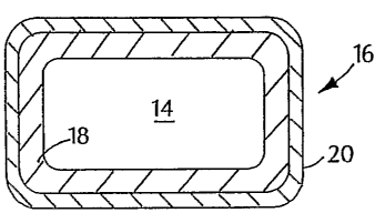
[0036) Fig. 1 shows a support 12 carrying a stent 10, which is in the form of
a tubular
member defined by struts 11 and openings 13. Depending on the type of stent 12
(e.g.,
balloon-expandable or self-expandable), support 12 can be a balloon catheter
or a catheter
shaft. Referring to Fig. 2, stent 10 includes multiple cross-sectional
portions. In particular,
struts 11 of stent 10 are formed of a relatively radiolucent core 14
surrounded by a
relatively radiopaque portion 16. Radiopaque portion 16 includes a radiopaque
layer 18,
e.g., made of gold, and a layer 20, e.g., made of a gold-titanium alloy, that
can enhance the
biocompatibility of stent 10. For example, layer 20 can be passivated to
provide stent 10
with a relatively inert outer surface.
[0037] In general, stent 10 can be formed by coating a relatively radiolucent
stent with a
radiopaque material, such as gold or platinum, to form layer 18. Layer 20 is
then formed
on the radiopaque material. Layer 20 can be formed on the pre-forined
radiopaque layer
18 and/or formed from a portion of the radiopaque layer. Layer 20 is then
passivated, e.g.,
by forming a layer of an oxide or nitride on layer 20 or by converting layer
20 to an oxide
or a nitride.
[0038] Core 14 is generally formed of one or more core material selected to
provide stent
with certain physical and mechanical properties. For example, the core
material is
selected to provide stent 10 with sufficient hoop strength and radial strength
so the scent
can maintain a body vessel open. Suitable core materials include stainless
steel (e.g., 316L
stainless steel), Nitinol (e.g., for self-expandable stents), other titanium
alloys, tantalum
alloys, zirconium alloys, and/or niobium alloys. At the same time, it is also
desirable to
reduce (e.g., minimize) differences or mismatch in mechanical properties
(e.g., stiffness)
- 5 -
CA 02500711 2005-03-30
WO 2004/030578 PCT/US2003/030682
between the stent and the body vessel. The mechanical mismatch can cause, for
example,
inflammation and/or re-occlusion of the vessel. One method of reducing
mechanical
mismatch is to form the stent with less material (e.g., by forming smaller
struts 11),
thereby approximating the compliancy or resiliency of the vessel. However,
reducing the
amount of core material in stent 10 can also reduce the radiopacity of the
stent.
[0039] To increase the radiopacity of stent 10, the stent includes radiopaque
portion 16
disposed over core portion 14. Portion 16 includes radiopaque layer 18, which
is formed
with a radiopaque material. The radiopaque material can be any material with a
density
and/or linear absorption coefficient sufficient to enhance the radiopacity of
stent 10. In
embodiments, the radiopaque material has a density and/or linear absorption
coefficient to
attenuate an incident X-ray beam. In some cases, the radiopaque material has a
density of
equal to or greater than about 10 g/cc. Examples of radiopaque materials
include gold,
platinum, palladium, tantalum, iridium, cobalt, titanium, tungsten, stainless
steel, Nitinol,
and metal alloys containing a sufficient percentage of heavy elements.
Radiopaque layer
18 can be, for example, up to about 8 microns thick, e.g., about 6-8 microns,
thick.
Methods of forming radiopaque layer 18 include, for example,
electrodeposition, physical
vapor deposition (e.g., sputtering), chemical vapor deposition, galvanizing,
and/or dipping
(e.g., in molten material).
[0040] In some cases, however, the radiopaque materials do not have a desired
level of
biocompatibility and/or the biocompatibility of the material is unknown (e.g.,
in the long
term). It is believed, for example, that gold may affect (e.g., catalyze)
electron transfer in
certain undesirable reactions in the body. Accordingly, radiopaque portion 16
includes a
relatively inert layer 20 disposed over radiopaque layer 18.
[0041] Layer 20 enhances the biocompatibility of stent 10 by providing the
stent with a
layer (as shown, an outer layer) that can be passivated, e.g., more easily
than radiopaque
layer 18. For example, layer 20 is, capable of reacting (e.g., oxidizing) and
forming
products, such as oxides, nitrides, and/or carbides, that are more inert, and
therefore, more
biocompatible, than the material(s) in radiopaque layer 18. Relative to
radiopaque layer
18, layer 20 has a lower oxidation potential, i.e., can be more easily
oxidized to form a
biocompatible product.
-6-
CA 02500711 2005-03-30
WO 2004/030578 PCT/US2003/030682
[0042] In some embodiments, layer 20 includes a mixture (here, an alloy) of
the
radiopaque material(s) in radiopaque layer 18 and one or more alloying
material. The
alloying material can be any material capable of forming a mixture with the
radiopaque
material(s), and forming a product that is more easily passivated than the
radiopaque
material(s). The alloying material can be, for example, tantalum, titanium,
niobium,
zirconium, chromium, silicon, rhodium, iridium, platinum, and/or palladium.
Any of the
alloying materials can be used with any of the radiopaque materials described
above.
[0043] As an example, for a gold radiopaque layer 18, the alloying material
can be
titanium. In this example, layer 20 includes an alloy of gold-titanium, such
as Auos0Tio.70,
which can be more easily passivated than gold. That is, relative to gold, the
gold-titanium
alloy can more easily form or be converted to a product, e.g., an oxide, that
is relatively
inert and biocompatible. In embodiments, for the alloy of gold-titanium
(AuTiy) x can
range from about 0-30%, and y can range from about 70-100%. For example, x can
be
equal to or greater than about 0%, 5%, 10%, 15%, 20%, or 25%, and/or equal to
or less
than about 30%,25%,20%,15%, 10%, or 5%. In embodiments, the concentration of
titanium, y, can be equal to or greater than about 70%, 75%, 80%, 85%, 90%, or
95%,
and/or less than or equal to 100%, 95%, 90%, 85%, 80%, or 75%. Layer 20 can be
up to
about 10 microns thick, e.g., about 0.1-10 microns thick. Ternary (e.g., Au-Ti-
Cr) or
higher mixtures or alloy systems can be formed.
[0044] In some embodiments, layer 20 can be formed on a pre-formed radiopaque
layer
18. For example, after radiopaque layer 18 is formed, modified layer 20 can be
applied on
the radiopaque layer by physical vapor deposition, including sputtering and
ion beam
assisted deposition, chemical vapor deposition, or electrodeposition. Layer 20
can also be
formed by forming layers, e.g., alternating layers, of the radiopaque material
and the
alloying material on layer 18 in a predetermined ratio, and heating the layers
(e.g., at
elevated, annealing temperatures) to form the alloy by diffusion.
[0045] Alternatively or in addition, layer 20 can be formed from a portion of
a formed
radiopaque layer 18. That is, a portion of the radiopaque layer 18 can be
converted to layer
20. For example, a gold-titanium layer 20 can be formed by implanting titanium
ions into
a formed gold radiopaque layer 18, and annealing the radiopaque layer. As a
result, a
-7-
CA 02500711 2005-03-30
WO 2004/030578 PCT/US2003/030682
certain thickness of the radiopaque layer (e.g., in the sub-micron range) is
converted to an
alloyed modified layer that can be passivated. In another example, a layer of
alloy
material, e.g., Ti, can be deposited on radiopaque layer 18, e.g., Au, and the
layers can be
heated, e.g., annealed, to form an alloy, e.g., Au-Ti.
[0046] It should be noted that while Fig. 2 shows radiopaque layer 18 and
layer 20 as two
discrete, well-defined layers, in some embodiments, the interface between the
layers is not
well defined. As a result, the endoprosthesis can be formed with good adhesion
and high
durability (e.g., reduced risk of flaking). Corrosion from contact of
dissimilar material can
also be reduced. The interface may not be well defined, for example, when
modified layer
20 is formed from a formed radiopaque layer 18.
[0047] In some embodiments, radiopaque portion 16 does not include an
interface between
two layers. Referring to Fig. 3, a strut 22 of a stent is formed of a
relatively radiolucent
core 24 surrounded by a relatively radiopaque layer 26. Core 24 is generally
the same as
core 14 described above. Radiopaque layer 26 includes one or more radiopaque
material
and one or more alloying material, as described above. In addition, radiopaque
layer 26 is
formed having a compositional gradient in which the concentration(s) of the
alloying
material(s) and/or the radiopaque material(s) varies along the thickness of
layer 26 (arrows
A and B). As an example, for a radiopaque layer 26 formed of a gold-titanium
alloy, layer
26 can be relatively gold-rich (or titanium-poor) at surface 28 adjacent to
core 24, and
relatively gold-poor (or titanium-rich) at outer surface 30. At surface 28,
the concentration
of the radiopaque material can be about 100%; and at outer surface 30, the
concentration of
the alloying material can be about 100%. The concentration(s) of the
radiopaque
material(s)l and/or the alloying material(s) can vary linearly or non-linearly
(e.g.,
exponentially) between surfaces 28 and 30. The concentration(s), e.g., of the
alloying
material, can increase or decrease from surface 28 to surface 30. In certain
embodiments,
layer 26 having the compositional gradient can be formed on a radiopaque
layer, such as
radiopaque layer 18.
[0048) Methods of forming compositionally-graded layer 26 include using
physical vapor
deposition while controlling the source of materials used for deposition. In
another
method, layer 26 can be formed by forming alternating layers of a radiopaque
material and
-8-
CA 02500711 2010-09-08
60412-3356
an alloying'material in a predetermined ratio, and annealing the layers. For
example,
referring to Fig. 4, to form a concentration gradient of titanium along layer
26, layers of
titanium 27a, 27b, and 27c can be formed alternating with layers of gold 29a,
29b, and 29c.
Titanium layer 27a is thicker than layer 27b, which is thicker than layer 27c.
Gold layers
29a-29c are of equal thickness. When the layers are subsequently annealed,
they can
diffuse together and form a gold-titanium alloy in which the concentration of
titanium
varies along the thickness of layer 26 (here, increasing with increasing
distance from core
24).
[0049] After layer 20 or 26 is formed, stent 10 can be passivated by exposing
the stent, to
an appropriate environment. For example, stent 10 can be oxidized by heating
the stent in
an oxidizing atmosphere, such as one containing oxygen and/or water, to form
an oxide
layer on layer 20 or 26. Nitrides can be formed by heating stent 10 in an
atmosphere
containing nitrogen, nitrogen-hydrogen, and/or ammonia. Carburizing, e.g.,
increasing the
surface concentration of carbon, can be performed by exposing stent 10, at an
elevated
temperature, to an atmosphere rich in a hydrocarbon gas, such as methane.
Alternatively
or in addition, passivation can be performed by electropolishing to produce an
oxide-rich
surface layer. In some cases, passivation can occur relatively spontaneously,
e.g., upon
exposure to air, when the oxidation potential is relatively low.
[0050] Stent 10 can then be finished, e.g., electropolished to a smooth
finish, according to
conventional methods. Stent 10 can be finished before passivation.
Alternatively, stent 10
can be formed textured.
[0051] Stent 10 can then be used, e.g., delivered and expanded, according to
conventional
methods.
[0052] Generally, stent 10 can be self-expandable, balloon-expandable, or a
combination
of both. Examples of stent 10 and support 12 are described in U.S. Patent Nos.
5,725,570
(Heath) and 5,234,457 (Andersen).
[0053] In other embodiments, stent 10 is a part of a stent-graft. The stent-
graft can be a
stent attached to a biocompatible, non-porous or semi porous polymer matrix
made of
polytetrafluoroethylene (PTFE), expanded PTFE, polyethylene, urethane, or
polypropylene. Stent 10 can include a releasable therapeutic agent or a
pharmaceutically
-9-
CA 02500711 2010-09-08
60412-3356
active compound, such as described in U.S. Patent No. 5,674,242, and commonly-
assigned
U.S. Patent Application Publication No. 2003/0003220, published January 2,
2003. The
therapeutic agents or pharmaceutically active compounds can include, for
example, anti-
thrombogenic agents, antioxidants, anti-inflammatory agents, anesthetic
agents, anti-
coagulants, and antibiotics.
[0054) The following examples are illustrative and not intended to be
limiting.
[0055] Example
[0056] The following example describes ion beam assisted deposition (IBAD) as
a method
for depositing thin films on a substrate, e.g., a stent.
[0057] Referring to Fig. 5, an IBAD system 50 generally includes a fixture
assembly 52
configured to support a stent 54, and a deposition assembly 56. System 50 is
used in-a
vacuum chamber 51 at pressures of about lx 10a-3x 10⁻⁴ Torr, provided in part
by a
diffusion pump 58.
[0058] Deposition assembly 56 includes two crucibles 60 and 62, their
respective shutters
64 and 66, two electron beam evaporators 63 and 70, and an ion beam gun 72.
Crucibles
60 and 62, e.g., made of graphite, contain materials to be deposited,'such as
gold and
titanium.: Electron beam evaporators 68 and 70 are configured to generate'a
flow of
electrons that can b.e focused (e.g., using magnetic fields) on the materials
in crucibles 60
and 62,, respectively, to melt and to evaporate' the materials to form
thermally evaporated
materials 76. Evaporators 68 and 70 can have water-cooled jackets that cool
crucibles 60
and 62, respectively. Ion beam gun 72 is configured to receive.a flow of argon
(e.g., 2-4
sccm) and to ionize the argon to form a plasma 74. Plasma 74 is accelerated
out of ion
beam gun 72 to stmt 54 using magnets (not shown). Shutters 64 and 66 can be
moved,
e.g., swiveled, to allow or to block the flow of evaporated material 76 from
crucibles 60
and 62, respectively.
[0059] - Fixture assembly 52 is generally configured to allow stent 54 to be
uniformly
coated with evaporated material 76. Typically, the thermal evaporation process
can
deposit a film of material 76 on a substrate that is in a line of sight of
crucible 60 or 62. To
provide uniform coverage on scent 54, the start is rotated during deposition.
In
-10-
CA 02500711 2005-03-30
WO 2004/030578 PCT/US2003/030682
embodiments, stent 54 is placed on a rotatable spindle. The friction between
the stent and
the spindle can hold the stent in place during rotation to provide a coated
stent without
contact points. Alternatively, stent 54 can be clipped to a rotatable shaft.
[0060] A quartz crystal 78 is used to determine the thickness of the deposited
material.
Crystal 78 is interfaced to a controller (not shown) and oscillated. The
controller is
calibrated such that the thickness of material deposited on crystal 78 (and
thus also stent
54) can be calculated by measuring the change in the oscillation frequency of
the crystal.
[0061] A method of coating using IBAD will now be described.
[0062] Stent 54, e.g., a Nitinol or stainless steel stent, is thoroughly
chemically cleaned.
For example, stent 54 can be cleaned in a solvent (such as isopropyl alcohol
or acetone)
and a degreaser, and rinsed with deionized water. Heat and/or agitation, e.g.,
using
ultrasonic energy, can be used to clean stent 54. Stent 54 is then placed on
fixture
assembly 52, which is then placed in vacuum chamber 51, with the stent about
two feet
from crucibles 60 and 62.
[0063] Stent 54 is then subjected to a sputter cleaning. Chamber 51 is
evacuated to a
pressure of about 1x10⁻⁵ Torr, and ion beam gun 72 is activated. Ion beam gun
72 ionizes
argon gas to form plasma 74, and the plasma is accelerated to stent 54 to
sputter clean/etch
the surface of the stent. The angle of incidence for plasma 74 can be about 45-
90 , e.g.,
about 70 . In embodiments, stent 54 is sputter cleaned for about 20-30
minutes. An
estimated 100-300 angstroms of material can be removed.
[0064] A first material, e.g., gold in crucible 60, is then deposited. During
the final ten
minutes of sputter cleaning, electron beam evaporators 68 and 70 are slowly
ramped up.
Shutters 64 and 66 are over their respective crucibles 60 and 62, so no
material can deposit
on stent 54. After sputter cleaning is complete and the material to be
deposited is molten,
shutter 64 moves, e.g., swivels, to allow evaporated material to coat stent
54. The surface
of stent 54 is simultaneously bombarded with plasma 74. It is believed that as
ions of the
first material deposit on stent 54, plasma 74 transfers energy to the ions,
freeing some ions
from the surface of the stent and allowing some ions to migrate on the stent
surface. As a
result, it is believed that a composite including the first material is formed
with enhanced
density.
-11-
CA 02500711 2005-03-30
WO 2004/030578 PCT/US2003/030682
[0065] A second material, e.g., titanium, tantalum, or platinum, is then
deposited. After
the thickness of the first material coated on stent 54 reaches, e.g., about
200-500
angstroms, shutter 66 is moved to allow the second material (in crucible 62)
to co-deposit
with the first material. The concentrations of each material can be controlled
by adjusting
the power to evaporators 68 and 70. For example, referring to Fig. 6,
initially the
concentration of the first material is relatively high, and the second
material is then slowly
introduced. In embodiments, at time t, shutter 64 is moved to prevent the
first material
from depositing on stent 54, and a pure layer of the second material is
deposited over the
alloy layer (i.e., the layer having the first and second materials). Then,
stent 54 is allowed
to cool, chamber 51 is returned to atmospheric pressure, and the stent is
removed from the
chamber.
[0066] In embodiments, stent 54 is then annealed. Annealing can promote
diffusion
between the layers of materials and/or the layers and the stent substrate, and
can strengthen
bonding or adhesion between the layers. In some cases, a Nitinol stent can be
annealed at
about 300-400 C, and a stainless steel stent can be annealed at about 500-
1000 C.
Annealing times can vary, e.g., from a few minutes to days, depending, for
example, on the
diffusion of the materials in stent 54, which can be temperature-dependent.
[0067] Fig. 7 shows ranges for some process parameters.
[0068] A stent was coated with titanium using the procedures described above.
The
process parameters are shown in Fig. 8.
[0069] A stent was coated with a platinum-gold using the procedures described
above.
The process parameters are shown in Fig. 9. The platinum-gold gradient was
similar to
that shown in Fig. 6.
[0070] Other Embodiments
[0071] In other embodiments, one or more intermediate layers can be formed
between core
14 or 24 and radiopaque layer 18 or 26, i.e., at least a portion of the core
and the
radiopaque layer do not contact. For example, in embodiments in which there is
lattice
mismatch between the core and the radiopaque layer, intermediate layer(s) can
be selected
to have intermediate lattice parameters to serve as buffer layer(s), thereby
reducing (e.g.,
-12-
CA 02500711 2010-09-08
60412-3356
minimizing) stress between the core and the radiopaque layer. The intermediate
layer(s)
can be, for example, a mixture of the core material and the radiopaque
material.
[0072] Layer 20 may not include the radiopaque material(s) in radiopaque layer
18. For
example, a radiopaque layer may include gold, while layer 20 includes a
material that can
be passivated, such as a platinum-titanium alloy.
[0073] Radiopaque layer 18, layer 20, and/or layer 26 can cover all or only
one or more
selected portions of a stent. For example, radiopaque layer 18, layer 20,
and/or layer 26
may be formed only on one or more end portions of the stent.
(0074] In some embodiments, other types of layers can be formed on layer 20 or
26. For
example, one or more selected portions of a stent may include a magnetopaque
(i.e., visible
by magnetic resonance imaging (MR1)) material on layer 20 or 26. Suitable
magnetopaque
materials include, for example, non-ferrous metal-alloys containing
paramagnetic elements
(e.g., dysprosium or gadolinium) such as terbium-dysprosium, dysprosium, and
gadolinium; non-ferrous metallic bands coated with an oxide or a carbide layer
of
dysprosium or gadolinium (e.g., Dy203 or Gd203); non-ferrous metals (e.g.,
copper, silver,
platinum, or gold) coated with a layer of superparamagnetic material, such as
nanocrystalline Fe3O4, CoFe2O4i MnFe2O4, or MgFe2O4; and nanocrystalline
particles of
the transition metal oxides (e.g., oxides of Fe, Co, Ni).
(0075] In other embodiments, radiopaque layer 18, layer 20, and/or layer 26
may be
formed on medical devices other than stents and stent-grails, for example,
those where
radiopacity is desired such as orthopedic implants.
[00761 Other embodiments are within the claims.
-13-