Note: Descriptions are shown in the official language in which they were submitted.
CA 02500726 2005-03-31
WO 2004/030753 PCT/US2003/031372
-1-
ACTIVE FLUID DELIVERY CATHETER
The present invention relates generally to implantable medical leads and more
specifically to an implantable medical lead and fluid delivery system for
treating a volume
of tissue in which the medical lead may remain implanted.
Electrical stimulation of excitable body tissue is used as a method for
treating
various pathological conditions. Therapeutic stimulation generally requires
making an
electrical contact between excitable tissue and an electrical pulse generator
through use of
one or more stimulation leads. Various lead systems and various techniques for
implanting
these lead systems in contact with excitable body tissue, and particularly the
heart, have
been developed.
In order to achieve cardiac pacing, sensing, cardioversion and/or
defibrillation at
different locations in the heart, various types of cardiac leads have been
developed
including epicardial leads, endocardial leads, and coronary vein leads. A
transvenous
endocardial lead establishes electrical contact between an electrical pulse
generator, such
as a pacemaker or implantable cardioverter defibrillator, and the endocardial
surface of the
heart, typically in a right heart chamber. Endocardial leads, and cardiac
leads in general,
may be held in place by passive fixation mechanisms, such as tines that
interact with the
ventricular trabeculae, or active fixation mechanisms, such as a helix. A
coronary vein
lead may be passed through' a venous pathway, into the right atrium, through
the coronary
sinus ostiurn and ultimately to a location deep in the cardiac veins. Contact
is made with
the epicardial surface of the left atrium or left ventricle for delivering
stimulation or
sensing cardiac signals in the left heart chambers. Epicardial leads are also
known in the
art and generally require a thoracotomy for placement on the epicardial
surface of a heart
chamber.
The safety, efficacy and longevity of an electrical pulse generator depends,
in part,
on the performance of the associated cardiac leads) used in conjunction with
the pulse
generator. Various properties of the lead, the electrodes and the tissue
interfacing with an
CA 02500726 2005-03-31
WO 2004/030753 PCT/US2003/031372
-2-
electrode will result in a characteristic impedance, stimulation threshold and
sensing
threshold.
Stimulation threshold is the energy required in a stimulation pulse to
depolarize, or
"capture," the heart tissue. A relatively high impedance and low threshold is
desired to
minimize the current drawn from a pulse generator battery in delivering a
stimulation
pulse. Maximizing the useful life of the pulse generator battery is important
since a
surgical procedure is required to replace the pulse generator once the battery
has reached
the end of its useful life.
One factor that can affect the stimulation threshold, particularly during the
ftrst
several weeks after implantation of a lead, is the natural imrnunological
response of the
body to the lead as a foreign object. The presence of the lead activates the
immunologic
response, which ultimately results in ftbrotic encapsulation of the lead and
its electrodes.
Since fibrotic tissue is not excitable tissue, an elevated stimulation
threshold can persist
due to the degraded electrical properties of the electrode-tissue interface.
To reduce the inflammatory response, medical leads that elute an anti-
inflammatory steroid have been developed. Steroid eluting leads are described
in U.S. Pat.
No. 4,506,680 issued to Stokes and related Medtronic U.S. Pat. Nos. 4,577,642,
and
4,606,118, all incorporated herein by reference. Steroid eluting leads may
require a
monolithic controlled release device (MCRD) to contain the steroid and to
thereafter
slowly leach out the water soluble steroid into the surrounding tissue. A
method for
applying a steroid directly to the surface of an electrode is disclosed in
U.S. Pat. No.
5,987,746 issued to Williams, incorporated herein by reference in its
entirety. Advantages
of this method include elimination of additional structures for carrying the
steroid and the
presentation of the steroid directly at the tissue-electrode interface.
One limitation of a steroid eluting electrode or MCRD, however, is that a
relatively
limited volume of tissue is treated by the eluting drug since the drug is
presented only at
the endocardial or epicardial surface. Other devices have been proposed which
allow the
delivery of a drug to a potentially larger volume of tissue by actually
penetrating the tissue
rather than relying on diffusion of the drug from the tissue surface. Drug
delivery
catheters may incorporate a drug dispensing needle or helix that penetrates a
targeted
tissue for delivering a drug or fluid. Catheters that may be used to deliver a
fluid or drug
CA 02500726 2005-03-31
WO 2004/030753 PCT/US2003/031372
-3-
into the myocardium are disclosed in U.S. Pat. No. 6,102,887 issued to Altman
and U.S.
Pat. No. 5,431,649 issued to Mulier et al.
Drug delivery catheters may include an electrode to allow sensing or
stimulation of
the myocardium. An implantable pacing lead having an active fixation electrode
with a
stylet introduced, anti-inflammatory drug delivery system is disclosed in U.S.
Pat. No.
5,447,533 issued to Vachon et al. A delivery system for delivering a
therapeutically
effective amount of a genetic material to an identified cardiac location
adjacent an atrial or
ventricular electrode is disclosed in PCT Patent Publication WO 98/02040
issued to
Stokes et al, incorporated herein by reference in its entirety. This delivery
system may
combine a pacing lead and a delivery catheter. Other implantable leads with
drug delivery
capabilities are disclosed in U.S. Pat. No. 4,360,031 to White, and U.S. Pat.
No. 5,496,360
to Hoffinan.
Advancements in gene therapies and cellular modifications through the delivery
of
proteins, peptides or even cell delivery, such as stem cell delivery, offer
opportunities to
alter the properties of tissue to further improve the benefit of a delivered
stimulation
therapy or improve the ability to sense cardiac signals. Genetic or biologic
agents may be
used to alter ion channel activity or protein expression at the cellular
level. Potential
benefits include decreased inflammatory response, increased tissue
conductivity for
reduction of stimulation thresholds or upregulation of ion channels for
increasing
membrane potentials to allow better sensing. For example, upregulation of ion
channels
could enhance cardiac P-waves or R-waves allowing them be more easily sensed
by a
pacemaker or other cardiac monitor. In particular, cardiac fast sodium
channels are
responsible for the fast upstroke of the action potential in myocardial cells
(Fozzard, et al.,
Circ. Res. 1995, 56:475-485). A human cardiac voltage-dependent sodium
channel,11H1,
has been cloned, sequenced, and functionally expressed (Gellens, et al., Proc.
Natl. Acad.
Sci. USA, 1992, 89:554-558). Alteration of myocardial conductivity may be
possible
through delivery of proteins that alter cellular electrical coupling. The gap
junction
protein Connexin43 has been found to play an important role in ventricular
conduction
(Guerrero PA et al., J. Clin. Invest. 1997, 99:1991-1998).
Because locally effective doses of a pharmacologic, genetic, or biologic agent
may
be toxic when given systemically, it is desirable to provide a method for
delivering an
CA 02500726 2005-03-31
WO 2004/030753 PCT/US2003/031372
-4-
agent locally at a targeted tissue site. Drug-eluting electrodes may be
limited to treating
only a relatively small volume of tissue at an electrode-tissue interface. The
pharmacological effect is in part limited by the kinetics of the drug leaving
the electrode or
lead. Furthermore, because biologic and genetic agents may have a limited
shelf life,
unique storage requirements such as requiring refrigeration, and may not
tolerate
sterilization procedures, it is not desirable to package a lead having drug
eluting
capabilities with the biologic or genetic agent already incorporated therein.
Other medical
leads having drug dispensing capabilities may require additional components
that increase
the size, stiffness or complexity of the lead.
To take advantage of various genetic or cellular modification therapies, it is
desirable to provide an implantable lead and fluid delivery system that allows
a
pharmaceutical, genetic, or biologic agent to be delivered to a targeted lead
implant site at
a depth within the myocardium to treat a volume of tissue. Once a fluid agent
has been
delivered, the fluid delivery components are no longer needed and may be
removed from
the patient's body. .An acutely implanted fluid delivery system eliminates the
need to
include dispensing components in the medical lead, reducing its complexity,
yet still offers
the benefit of treating a volume of tissue at a lead implant site, potentially
improving lead
performance. There is a need, therefore, for a system that allows an acutely
implanted
fluid delivery device to treat a volume of tissue during a lead implant
procedure, or at any
time post-operatively, and further allows a lead to be implanted and remain in
the location
of the treated tissue.
The present invention is directed toward providing a medical lead and fluid
delivery system for treating a volume of tissue with a pharmaceutical,
genetic, or biologic
agent at the time of the medical lead implant and/or at any time post-
operatively.
In one embodiment of the present invention, a guide catheter is provided with
a
fluid dispensing, fixation member at its distal end. The fixation member
communicates
with a lumen extending the length of the guide catheter body through which a
fluid may be
delivered. The fixation member, which may be provided as a hollow helix, is
provided
with one or more apertures for dispensing a drug into the surrounding tissue
in which the
helix is fixed. The fixation member may optionally function as an electrode in
addition to
CA 02500726 2005-03-31
WO 2004/030753 PCT/US2003/031372
-5-
being a fixation device. After treating a volume of tissue by dispensing a
fluid through the
fixation member, a medical lead, advanced through a lead-delivery lumen of the
guide
catheter, may be implanted in the treated tissue. The guide catheter may then
be removed
leaving the medical lead implanted at the treated tissue site.
In another embodiment, a system is provided including a guide catheter having
a
fixation member, a fluid delivery device, and an implantable lead. After
fixing the guide
catheter at a desired implant site, the fluid delivery device, advanced down a
lumen of the
guide catheter, may be used to treat a volume of tissue at that site. The
fluid delivery
device may then be removed and an implantable lead may be advanced through the
guide
catheter to the treated tissue site. The guide catheter may then be removed,
leaving the
lead implanted at the treated tissue site.
In alternative embodiments, a transvenous lead having a distal fixation member
is
provided with a center lumen through which a fluid delivery device may be
advanced to
treat a volume of tissue in which the lead is implanted. A seal is preferably
provided at the
distal end of the lead body to prevent fluid ingress. The distal fixation
member may be
provided as a passive or active fixation member. In one embodiment, a
retractable active
fixation member is provided. In another embodiment, the implantable lead may
be
pxovided as an epicardial lead.
The fluid delivery device may take the form of a hollow needle or stylet that
may
be advanced through a lumen of the lead body, penetrated through the seal and
into the
targeted tissue site. The fluid delivery device may be provided with a
conductive tip to
allow sensing of electrophysiological signals. Fluid delivery may be performed
once the
delivery device is inserted in the taxgeted tissue location as verified by
sensing
electrophysiological signals characteristic of the targeted tissue. After
delivery of a fluid,
which may be a pharmaceutical, genetic or biologic agent carried in a liquid
medium, the
fluid delivery device may be removed, leaving the transvenous lead implanted
at the
treated tissue site.
The medical lead may further include a reservoir that may be filled by the
fluid
delivery device with a pharmaceutical, genetic or biologic agent. The agent
will elute
from the reservoir into surrounding tissue over time. Treatment of a volume of
tissue with
CA 02500726 2005-03-31
WO 2004/030753 PCT/US2003/031372
-6-
a pharmaceutical, genetic or biologic agent may thus be treated by delivering
a bolus
injection directly into the tissue or slow elution of the agent over time, or
both.
A fluid may be delivered post-operatively by gaining access to the lumen of an
implanted lead through an access port on the implanted device to which the
lead is
connected. Multiple injections at the lead implant site at various time
intervals are
possible by inserting a fluid delivery device through the access port and
advancing it
through the lead lumen. Fluid may be delivered directly to the lead implant
site, or a fluid
reservoir may be refilled.
Thus, the present invention provides a medical lead and fluid delivery system
that
allows a lead to be implanted in a volume of tissue treated concurrently with
the lead
implant procedure, or at any time post-operatively, by an acutely delivered
fluid delivery
device.
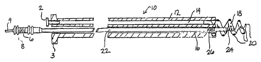
Figure 1 is a side, cut-away view of an implantable lead and fluid delivery
system
including a guide catheter having fluid dispensing capabilities and an
implantable medical
lead.
Figure 2 is a side, cut-away view of an alternative embodiment of the guide
catheter shovm in Figure 1 in which a fixation member on the guide catheter
may also
function as an electrode.
Figures 3A and 3B are side, cut-away views of the distal end of an implantable
medical lead and fluid delivery system that includes a guide catheter, a fluid
delivery
device and a medical lead.
Figure 4A is a plan view of an alternative embodiment of an implantable lead
and
fluid delivery system including a transvenous medical lead and a fluid
delivery device that
may be deployed through a lumen of the lead.
Figure 4B is a side cut-away, view of the distal end of the system of Figure
4A.
Figure 5 is an exploded, side, cut-away view of the distal end of an
implantable
lead and fluid delivery system in which the lead is provided with a
retractable fixation
member.
Figure 6 is an exploded, side, cut-away view of the distal end of an
implantable
medical lead and fluid delivery system for use on the epicardial surface of
the heart.
CA 02500726 2005-03-31
WO 2004/030753 PCT/US2003/031372
Figure 7 is a cut-away, side view of the distal end of an implantable medical
lead
and fluid delivery system wherein the medical lead is provided as a
transvenous lead
having a passive fixation mechanism.
Figures 8 is side, cut-away view of the distal end of an implantable medical
lead
and fluid delivery system wherein the medical lead is further provided with a
fluid
reservoir for holding a pharmaceutical, genetic or biologic agent and allowing
the agent to
elute into adjacent body tissue over time.
Figure 9 is a side, cut-away view of the distal end of an implantable medical
lead
and fluid delivery system wherein the medical lead is provided as a
transvenous lead
having a passive fixation mechanism and a fluid reservoir.
Figure I O is a plan view of an implantable lead and fluid delivery system
that may
be used to deliver a fluid agent to a lead implant site post-operatively.
As described above, the present invention is directed at providing an
implantable
lead and fluid delivery system in which a fluid delivery device may be used to
treat a
volume of tissue concurrently with a lead implantation procedure, or at any
tune post-
operatively. After delivering a fluid, the fluid delivery device may be
removed leaving the
lead implanted at the treated tissue site. Figure 1 is a side, cut-away view
of one
embodiment of an implantable lead and fluid delivery system in accordance with
the
present invention. The system includes a guide catheter 10 having fluid
dispensing
capabilities. Catheter 10 is provided with a proximal handle 3 and an
elongated catheter
body 12 having at least two lumens 14 and 16 and is preferably formed from a
biocompatible polymer such as polyurethane, silicone, TeflonC~, or other
acceptable
plastic. A fluid-delivery lumen 14 is in communication with an active
fixation, fluid
dispensing member shown as a hollow fixation helix 18 located at the distal
end of guide
catheter 10. An active fixation, fluid dispensing member may alternatively be
provided as
a hollow "fish hook" type member, stake-like member, or any other type of
active fixation
member that can be provided as a hollow structure having one or more
apertures. Hollow
fixation helix 18 is provided with one or moxe apertures 20 through which
fluid injected
through lumen 14 may exit into a tissue site. Fixation helix 18 is preferably
formed from a
biocompatible metal, such as stainless steel, in which apertures 20 may be
formed by Iaser
CA 02500726 2005-03-31
WO 2004/030753 PCT/US2003/031372
_g_
drilling. A hollow fixation helix that may be used for fluid delivery is
disclosed in the
'649 patent issued to Mulier et al., incorporated herein by reference in its
entirety, and the
WO 98/02040 patent issued to Stokes et al. A fluid fitting 2, such as a Luer
lock fitting,
may be inserted or mounted at the proximal end of fluid delivery lumen 14 to
allow
connection of a syringe for injecting fluid into lumen 14.
Catheter 10 rnay be provided as a steerable catheter having a manipulative
handle
and steering mechanism, such as a pull wire, to aid in maneuvering catheter 10
through
body vessels or organs. Steering mechanisms included in catheter 10 may be
embodied as
generally described in U.S. Pat. No. 5,396,902, issued to Brennen, et al., for
example, or
U.S. Pat. No. 5,807,249 issued to Qin, et al., both patents incorporated
herein by reference
in their entirety.
A lead-delivery lumen 16 is provided for delivering an implantable lead 22 to
a
desired implant site. The lead-delivery lumen 16 is sized to allow lead 22 to
easily pass
through guide catheter 10 without undue friction or resistance. Lead 22 is
shown as an
exemplary bipolar lead having a helical tip electrode 24 located at the distal
lead end and a
ring electrode 26 spaced proximally from tip electrode 24. In other
embodiments, lead 22
may be a unipolar, bipolar, or multipolar lead carrying any combination of
tip, ring and/or
coil electrodes or other sensors. Lead 22 is shown with an active fixation
helical electrode
24 but could also be provided with other types of active fixation electrodes
or
mechanisms, such as a "fish hook" electrode. Lead 22 may alternatively be
provided with
a generally spherical, hemispherical or ring-shaped tip electrode with passive
fixation
mechanisms, such as tines as generally known in the art.
A connector assembly 8 is provided at the proximal lead end with a pin
connector
4 and ring connector 6 which are electrically coupled to respective conductors
that extend
to tip electrode 24 and ring electrode 26. Conductors extending the length of
lead 22 may
be coiled conductors or cabled or stranded conductors as is known in the art.
During a lead implantation procedure, guide catheter 10 may be passed through
a
venous pathway into a desired heart chamber until a desired implantation site
is reached.
A guide wire or electrophysiological mapping catheter, passed through inner
lumen 16,
could be used for passage of the catheter through the venous and cardiac
anatomy to allow
access to the targeted tissue. This guide wire or electrophysiological
catheter could be
CA 02500726 2005-03-31
WO 2004/030753 PCT/US2003/031372
-9-
steerable and would provide the additional benefit of protecting helix 18 to
prevent
snagging or entanglement with anatomic structures. Fixation helix 18 is
advanced into the
myocardial wall by rotating catheter 10 at its proximal end. Catheter body 12
is therefore
provided with torsional stiffness adequate to translate rotational force to
the distal fixation
helix 18. A fluid, which may be a pharmacological, genetic, or biologic agent,
may then
be injected into drug-delivery lumen 14 such that it is dispersed out of
apertures 20 into
the tissue surrounding fixation helix 18. A relatively large volume of tissue
may be
treated by the relatively large helix 18 on guide catheter 10.
Lead 22 may then be passed through lead delivery Lumen 16 and implanted at the
treated tissue site by advancing helical tip electrode 24 into the tissue. The
position of
guide catheter I O is maintained by helix 18 such that lead 22 may be
implanted in the
same volume of tissue treated by the injection of fluid through helix 18.
After implanting
lead 22, guide catheter 10 may be removed by rotating catheter 10 in an
appropriate
direction to remove helix 18 from the tissue site and withdrawing catheter 10
over lead 22.
Catheter 10 may be provided as a splittable or slittable catheter such that it
may be
removed from lead 22 without passing it over connector assembly 8.
Alternatively,
connector assembly 8 may be provided as a low profile connector assembly sized
to allow
catheter 10 to be readily passed over assembly 8.
Figure 2 is a side, cut away plan view of an alternative embodiment of the
guide
catheter 10 shown in Figure 1 in which the distal fluid dispensing, fixation
member, helix
18, may function as an electrode. In Figure 2, all identically labeled
components
correspond to those illustrated in Figure 1. In Figure 2, however, fixation
helix 18 is
shown coupled to a conductor 1 S that extends the length of catheter body 12
to a proximal
terminal 17 enabling connection to a monitoring device, such as an
electrocardiogram
monitor. Helix 18 may thus serve as an electrode allowing electrophysiological
signals to
be sensed and monitored in order to verify that guide catheter I O is fixed in
a desired
location. Monitoring of electrophysiological signals may also aid in verifying
a short-term
pharmacological effect after delivering a fluid through lumen 14 and helix 18.
Figures 3A and 3B are cut-away plan views of the distal end of an implantable
medical lead and fluid delivery system that includes a guide catheter 200, a
fluid delivery
device 208, and a medical lead 212. Figure 3A shows a guide catheter 200
having an
CA 02500726 2005-03-31
WO 2004/030753 PCT/US2003/031372
-10-
elongated, tubular catheter body 202 with inner lumen 204. Guide catheter 200
is
provided with a fixation member 206, shown in this embodiment as a helix, that
allows
catheter 200 to be fixed at a targeted implant site. Fixation member 206 may
be a solid
helix and may function exclusively as a fixation device. Alternatively,
fixation member
206 may also function as an electrode as described above with reference to
Figuxe 2.
A separate fluid delivery device 208 may be advanced through catheter lumen
204
until device 208 exits the distal end of catheter 200. Fluid delivery device
208, which may
generally take the form of a hollow needle or stylet, may be tapered at its
distal end and is
preferably provided with a sharpened or beveled tip 210 such that it may
easily pierce the
tissue at the targeted implant site. The tip 210 may also take the form of a
helix or other
shape that may penetrate the tissue to a desired depth and dispense a fluid
through one or
more apertures to treat a volume of tissue. Once fluid delivery device 208 is
advanced
into the tissue, a fluid may be injected in the proximal end of fluid delivery
device 208 and
dispensed into a volume of tissue through tip 210.
Fluid delivery device 208 may also serve as an electrode, alternatively or in
addition to helix 206 of catheter 200. Fluid delivery device 208, which may be
formed
from a conductive metal such as stainless steel, may be provided with an
insulating
coating, such as a coating of ethylene tetrafluoroethylene (ETFE) or Parylene,
except for
at distal tip 210. The proximal end of device 208 may be coupled to a monitor
such that
electrophysiological signals sensed at uninsulated tip 210 may be monitored.
Verification
that tip 210 is in a desired tissue site, and not in blood or non-excitable
tissue, may be
made by monitoring electrophysiological signals sensed at tip 210.
After dispensing a fluid into the targeted implant site, the fluid delivery
device 208
may be withdrawn from lumen 204 of guide catheter 200 and replaced with an
implantable
medical lead 212 as shown in Figure 3B. Lead 212 is shown as an exemplary
bipolar lead
having an active fixation helical tip electrode 214 at its distal end and a
ring electrode 216
spaced proximally from tip electrode 214. Lead 212 may be advanced through
lumen 204
and implanted at the treated tissue site by advancing helical tip electrode
214 into the
tissue. Guide catheter 200 may then be removed, leaving the electrode 214
implanted in
the treated tissue.
CA 02500726 2005-03-31
WO 2004/030753 PCT/US2003/031372
-11-
Figure 4A is a plan view of an alternative embodiment of an implantable lead
and
fluid delivery system. This system includes a transvenous lead 30 and a fluid
delivery
device 44. The lead 30 has an elongated, tubular lead body 32. Lead body 32
may be
formed from a resilient, biocompatible polymer, such as silicone or
polyurethane. Lead 30
is shown as a unipolar lead having an active ftxation tip electrode 34 located
at its distal
end, shown as a helical electrode. Lead 30 may alternatively be a bipolar or
multipolar
lead having, in addition to active fixation tip electrode 32, one or more ring
electrodes
and/or one or more coil electrodes.
A connector assembly 62 is provided at the proximal lead end to allow
connection
of lead 30 to an implantable pulse generator or monitoring device. Connector
assembly 62
includes a pin terminal 64 that is electrically coupled to tip electrode 48
via a conductor
extending the length o~ lead body 32. Pin terminal 64 is provided as a hollow
pin that is in
communication with a central lumen of lead body 32. Sealing rings 63 form a
fluid-tight
seal with the inner surface of a connector port on an implantable pulse
generator or
monitoring device.
Fluid delivery device 44 is shown inserted into the proximal end of hollow pin
terminal 44. Fluid delivery device 44 may take the form of a hollow needle or
stylet as
described above in conjunction with Figure 3A. Fluid delivery device 44
includes a
hollow shaft 46 sized to pass easily thxough pin terminal 64 and the Iumen of
lead body 32
such that distal tip 48 of fluid delivery device 44 may exit the distal end of
lead 30. A fluid
fitting 60, which may take the form of a Luer lock fitting, is provided at the
proximal end
of device 44 to allow connection of a syringe for injecting fluid through
shaft 46 to be
dispensed from tip 48.
Figure 4B is a side cut-away view of the distal end of lead 30 and fluid
delivery
device 44. Helical tip electrode 34 is electrically coupled to a conductive
sleeve 50,
preferably by laser or resistance welding. Conductive sleeve 50 is
electrically coupled to a
conductor 36. Conductor 36 extends to connector assembly 62 at the proximal
end of lead
and is coupled to pin terminal 64. Conductive sleeve 50 may be coupled to
conductor
36 by crimping conductive sleeve 50 such that it is compressed against
conductor 36,
30 which is supported on its internal diameter by internal sleeve 40. In this
way, electrode 34
is electrically coupled to conductor 36 and pin terminal 64.
CA 02500726 2005-03-31
WO 2004/030753 PCT/US2003/031372
-12-
Conductor 36 is preferably a coiled conductor provided with insulation 37.
Insulation 37 may be provided as a coating formed from an appropriate
insulating material
such as polytetrafluoroethylene (PTFE) or ETFE, preferably surrounding each
individual
fllar included in conductor 36. Insulation 37 may alternatively be provided as
heat shrink
tubing fabricated from PTFE or ETFE as generally described in U.S. Pat. No.
6,052,625
issued to Marshall, incorporated herein by reference in its entirety.
Conductor 36 may
alternatively be provided as an insulated cabled or stranded conductor, such
as the
conductor generally disclosed in U.S. Pat. No. 5,246,014 issued to Williams.
Insulation
37 may also be provided as a material having a high Young's modulus, such as a
high
durometer polyurethane or polyimide, to impart additional lead body stiffness
to the small
diameter lead as generally described in U.S. Pat. No. 6,366,819 issued to
Stokes,
incorporated herein by reference in its entirety.
Insulation 37 electrically isolates conductor 36 from tip 48 and shaft 46 of
fluid
dispensing device 44 allowing distal tip 48 to function as a sensing electrode
for detecting
electrophysiological signals at a tissue site. When tip 48 is used as a
sensing electrode,
fluid delivery device 44 may also be insulated along the entire length of
shaft 46,
particularly if conductor 36 is not provided with insulation. Distal tip 48
remains
uninsulated. Insulation on shaft 46 may be provided by an adhesive coating,
such as
silicone adhesive, or as a tubular sleeve formed from an insulating material
such as PTFE,
ETFE or Parylene. A conductive clamp, connected to a monitor such as an ECG
monitor,
may be coupled to fitting 60 at the proximal end of fluid delivery device 44
for observing
electrophysiological signals at the site in which the uninsulated tip 48 is in
contact. Fox
example, cardiac P-waves or R-waves could be sensed by tip 48.
Lead 30 is preferably provided with a seal 38 to prevent the ingress of body
fluids.
Seal 38 is generally cup shaped and may be formed from a resilient,
biocompatible
polymer, such as molded silicone rubber. Seal 38 is shown in Figure 4B to be
molded
onto internal sleeve 40, which is preferably formed from a rigid, insulating
material such
as Delrin~, available from DuPont. Internal sleeve 40 is provided with an
annular,
laterally extending flange 52. Seal 38 is retained by the interaction of
flange 52 and
conductive sleeve 50. Seal 38 may be provided as generally described in U.S.
Pat. No.
6,192,280 issued to Sornmer et al., incorporated herein by reference in its
entirety.
CA 02500726 2005-03-31
WO 2004/030753 PCT/US2003/031372
-13-
Alternatively, the seal 38 can be fabricated such that it is entirely
contained within a
portion of conductor 36 at a point at the distal end of the lead 32 or at a
location more
proximal. Alternative embodiments of a seal at or near the distal end of a
medical lead or
medical device that may be adapted for use with the present invention are
disclosed in
U.S. Pat. Application 20020016622 to Janke et al., and U.S. Pat. Application
20020077685 to Sundquist et al., both of which are incorporated herein by
reference in
their entirety. Other types of seals for preventing fluid from entering a
tubular body may
also be used.
During an implantation procedure, Iead 30 may be deployed to a desired implant
site. Lead 30 deployment may be performed with the aid of a guide wire,
stylet, or guide
catheter. Helical tip electrode 34 may then be fixed in the tissue at the
implant site. If a
guide wire or stylet is used, it is removed from lumen 42 after lead 30 is
positioned so that
fluid delivery device 44 may be advanced through lumen 42. Fluid delivery
device tip 48
is preferably sharpened or beveled such that it can easily pierce through seal
38. The fluid
delivery device 46 might also be shapeable, allowing it to be used for
positioning of the
lead 32. Seal 38 may be pre-pierced at line 54 to define a path for the fluid
delivery
device 44 to pass through. Tip 48 is then further advanced into the implant
site.
Verification that tip 48 is in a desired implant site rnay be made by
monitoring
electrophysiological signals sensed by uninsulated tip 48. If no signal is
sensed, tip 48
may not be advanced completely through seal 3 8 or may not be fully inserted
into the
tissue site. Once tip 48 is adequately advanced into the implant site, a fluid
may be
injected through device 44 to treat a volume of tissue in which helical tip
electrode 34 is
implanted. Fluid delivery device 44 may then be withdrawn and removed, leaving
lead 30
implanted with helical tip electrode 34 fixed in the treated tissue.
Figure 5 is an exploded, cut-away plan view of the distal end of an
implantable
lead and fluid delivery system wherein the lead 70 is provided with a
retractable fixation
member. A lead 70 is provided with a helical tip electrode 76 that may be
retracted into an
electrode housing 74. Electrode housing 74 is preferably formed from a
relatively rigid
biocompatible polymer, such as polyurethane. Housing 74 is bonded to an
elongated,
tubular lead body 72, which may be formed of polyurethane, silicone rubber, or
another
biocompatible polymer.
CA 02500726 2005-03-31
WO 2004/030753 PCT/US2003/031372
-14-
Helical tip electrode 76 is mounted on a conductive sleeve 78, which is
electrically
coupled to a conductor 92. Conductive sleeve 78, which is preferably machined
from a
conductive metal such as stainless steel, includes a retraction mechanism
shown as a
threaded barrel 86 that is coaxial with sleeve 78 and located on the outer
diameter of
S sleeve 78. Thread 88, running along the outer surface of barrel 86, acts to
engage multiple
thread guides 90 mounted on the inner diameter of housing 74. Conductor 92 may
be
rotated relative to lead body 72 by rotating a connector pin to which
conductor 92 is
coupled at its proximal end. Rotation of a coiled conductor may be achieved as
generally
described in U.S. Pat. No. 4,106,SI2, issued to Bisping, incorporated herein
by reference
in its entirety. Rotation of conductor 92 causes rotation of sleeve 80
relative to electrode
housing 74. Rotation of sleeve 80 causes advancement of helical electrode 76
as threaded
barrel 86 is actuated on thread guides 90. A stop mechanism 89 may be provided
as a
ridge or peg near the proximal end of thread 88 that engages a thread guide 90
to prevent
over extension of helical electrode 76. During retraction, threaded barrel 86
will interact
1 S with housing 74 at lateral face 96 to pxevent over-retraction of helix 76.
Alternatively, a
stop mechanism may be provided near the distal end of thread 88 to prevent
over-
retraction of helix 76. A retraction stop mechanism that may be adapted for
use in the
present invention is disclosed in U.S. Pat. No. 5,837,006, issued to Ocel et
al.,
incorporated herein by reference in its entirety.
Lead 70 is provided with a seal 82, preferably formed of a resilient
biocompatible
polymer such as silicone rubber, molded to the distal end of the conductive
sleeve 78 to
prevent ingress of body fluids. Seal 82 may be generally cup shaped and may be
pre-
pierced at line 94 to guide a fluid delivery device 100 as it passes through
seal 82. Seal 82
further includes an annular sealing ring 84, coaxial with seal 82 and
extending laterally
2S from the outer diameter of seal 82. Sealing ring 84 interacts with the
inner surface of
housing 74 to complete a fluid-tight seal of the distal end of lead 70.
Sealing zing 84
further acts to center helix 76 within housing 74.
A fluid delivery device 100 is provided which may be generally in the form of
a
hollow stylet or needle having an elongated shaft 106 extending between a
proximal end
through which fluid may be injected and a distal tip 102 through which fluid
may be
dispensed. Distal tip 102 is sharpened or beveled such that it may easily
pierce through
CA 02500726 2005-03-31
WO 2004/030753 PCT/US2003/031372
-15-
seal 82 and enter a targeted tissue site. A distal segment 104 of fluid
delivery device 100
is provided with a reduced diameter allowing it to extend through conductive
sleeve 78
such that distal tip 102 may extend out of housing 74 when helix 76 is
extended into a
tissue site. Lateral face 108 may act as a mechanical stop by interacting with
the distal
end of sleeve 78 and thereby control the maximum depth that fluid delivery
device 100 is
inserted into the targeted tissue site. The outer dimensions of shaft 106 and
distal segment
104 and the spacing of lateral face 108 from distal tip 102 may alternatively
be
dimensioned to provide a stopping interface that interacts with a reduced
inner diameter of
sleeve 78 or helix 76. Alternatively, the tip of helix 76 may be bent to cross
the center
axis of helix 76 to act as a stop for fluid delivery device 100. Any of these
methods for
providing a mechanical stop for fluid delivery device 100 allows the tissue
depth at which
' the fluid is injected to be controlled.
Figure 6 is an exploded, cut-away side view of the distal end of an
implantable
medical lead and fluid delivery system for use on the epicardial surface of
the heart. A
~ lead 150 is provided with a lead body 152, an insulating electrode head 154
and an active
fixation electrode 158. Electrode 158 is shown as a helical electrode but may
also take the
form of a "fish hook" type electrode, or any other active fixation electrode.
Electrode
head 154 includes a tapered body 155 and flange 156, both of which may be
formed from
silicone rubber and provide a flexible structure for stabilizing the position
of lead 150 on
the epicardial surface. A tool may be used for, implanting lead 150 by
attaching to and
rotating the electrode head 154 to screw the helical electrode 158 into the
epicardium as is
generally known in the art. Epicardial leads and tools for implanting
epicardial leads are
disclosed in U.S. Pat. No. 3,737,539 issued to Bolduc, U.S. Pat. No. 5,143,090
issued to
butcher, and U.S. Pat. No. 6,010,526 issued to Sandstrom et al., all of which
patents are
incorporated herein by reference in their entirety. Flange 156 may be
reinforced with an
embedded netting or mesh material, such as polyester netting. Netting material
may
optionally be coated with an anti-inflammatory steroid to reduce the
inflammatory
response at the tissue-lead interface.
Helical electrode 158 is electrically coupled to a conductive sleeve I70,
which is
further coupled to a conductor 174, shown as a coiled conductor. Conductive
sleeve 170
is provided with an annular flange I72. A seal 160 is molded to flange 172 to
prevent the
CA 02500726 2005-03-31
WO 2004/030753 PCT/US2003/031372
-16-
ingress of bodily fluids into the lead body lumen 164. Seal 160 may be pre-
pierced at line
162 to define a path fox fluid delivery device I00 to pass through. Fluid
delivery device
100 may correspond to the fluid delivery device shown in Figure 5 and is shown
in Figure
6 with identically labeled components corresponding to those in Figure 5.
Lateral face
I08 may engage with the proximal end of conductive sleeve 170 to control the
depth that
fluid delivery device 100 is inserted into the tissue.
After implanting lead 150, fluid delivery device I00 may be extended through
lead
body lumen 164 and seal 160 to dispense a fluid into the tissue surrounding
helical
electrode 158. Fluid delivery device 100 may then be withdrawn from lumen 164
and
I O removed from the patient's body, leaving lead 150 implanted at the treated
tissue site.
Figure 7 is a cut-away, side view of the distal end of an implantable medical
lead
and fluid delivery system wherein the medical lead is provided as a
transvenous lead
having a passive fixation mechanism. In this embodiment, all identically
labeled
components correspond to those illustrated in Figure 4B, howevex, in this
case, in place of
15 an active fixation electrode at the tip of the lead 250, a ring electrode
252 is provided.
Ring electrode 252 is electrically coupled to conductive sleeve 50, which is
further
coupled to insulated conductor 36 as previously described with reference to
Figure 4B. To
stabilize the implanted position of lead 252, passive fixation members 254 are
provided,
which may take the form of tines as is generally known in the art. Seal 38 may
be molded
20 onto internal sleeve 40 as described previously and forms a fluid-tight
seal with the inner
diameter of ring electrode 252. Ring electrode 252 may be provided with an
annular lip
256 which may act to retain seal 38.
Figures 8 and 9 are side, cut-away views of the distal end of an implantable
medical lead and fluid delivery system wherein the medical Iead is further
provided with a
25 fluid reservoir for holding a pharmaceutical, genetic or biologic agent and
allowing the
agent to elute into adjacent body tissue over time. A body implantable lead
having a
cavity suitable for retaining a drug is disclosed in U.S. Pat. No. 4,506,680
issued to
Stokes, incorporated herein by reference in its entirety. A combined catheter
and
reservoir, useful for applications involving delivery of genetic material, is
disclosed in the
30 previously cited PCT Patent Publication WO 98/02040.
CA 02500726 2005-03-31
WO 2004/030753 PCT/US2003/031372
-17-
The lead shown in Figure 8 corresponds to the lead of Figure 4B having a
helical
tip electrode 34 electrically coupled to stem 50 which is further coupled to
an insulated
conductor 36. In addition to or in place of a seal at or near the distal end
of the lead, a
fluid reservoir 300 is located near the distal end of the lead. A fluid
delivery device in the
form of a hollow stylet or needle, having a shaft 46 and sharpened tip 48, may
be used to
fill reservoir 300 with a fluid. Reservoir 300 preferably includes a seal 304
covering a
proximal opening to reservoir 300 and a seal 302 covering a distal opening to
reservoir
300. Fluid delivery device tip 48 pierces through the proximal seal 304, which
may be
pre-pierced at line 308 and may be provided with a concave proximal surface to
guide tip
48 to reservoir 300 and through seal 302. Fluid may then be injected into
reservoir 300,
and the fluid delivery device may be removed. The pharmaceutical, genetic, or
biologic
agent will elute from reservoir 300, through distal seal 302, into the
adjacent tissue over
time.
Fluid reservoir 300 may be formed from silicone rubber or alternatively
polyurethane or another elastomer. 'The seals 302 and 304 are preferably
formed from
silicone rubber. Seal 304 may be provided as a less permeable material than
seal 302 to
prevent blood or bodily fluids from entering the lead body lumen 42 while
still allowing a
pharmaceutical, genetic or biologic material to elute through seal 304. The
reservoir 300
may be provided as a micro-osmotic pump. For example reservoir 300 may
optionally
contain a salt-loaded silicone material, which would swell over time as salt
is replaced by
water, or another polymeric material capable of swelling upon exposure to body
fluids.
Such swelling would aid in "pumping" a fluid agent out of reservoir 300.
Optionally, the fluid delivery device may be further advanced through distal
seal
302, which may be pre-pierced at line 306. The fluid delivery device may then
be inserted
into the tissue in which electrode 34 is implanted to deliver a bolus of fluid
directly to the
tissue site, at a desired depth within the tissue. 'The fluid delivery device
may then be
withdrawn into reservoir 300 and used to fill reservoir 300 to allow a
pharmaceutical,
genetic or biologic agent to elute slowly over time into the adjacent tissue.
In this way,
local treatment of a volume of tissue may be performed by delivering a bolus
of fluid
directly into the tissue, or allowing the agent to elute from reservoir 300
over time, or
both. Furthermore, one or more fluid agents may be delivered directly into the
tissue site,
CA 02500726 2005-03-31
WO 2004/030753 PCT/US2003/031372
-18-
and another fluid agent may be used to fill reservoir 300 and elute over time
allowing the
volume of tissue in which electrode 34 is implanted to be treated by at least
two different
pharmaceutical, genetic or biologic agents over different time courses.
A fluid reservoir for storing a fluid agent that will elute over time may also
be
included in other embodiments of medical lead and fluid delivery systems.
Figure 9 is a
cut-away, side view of the distal end of an implantable medical lead and fluid
delivery
system wherein the medical lead is provided as a transvenous lead having a
passive
fixation mechanism and a fluid reservoir. The system shown in Figure 9 is
similar to the
system shown in Figure 7, and identically labeled components correspond to
those shown
in Figure 7. However, in Figure 9, the transvenous lead is shown having a
fluid reservoir
300, similar to the reservoir described above in conjunction with Figure 8.
Ring tip
electrode 252 is provided with a central bore 310 that may be filled with a
porous material
through which a pharmaceutical, genetic or biologic agent eluting out of
reservoir 300
may pass to reach adjacent body tissue. A porous elution path may be formed
from
sintered metal structures as disclosed in the above incorporated '680 patent.
Alternatively
central bore 310 may be left open, as shown previously in Figure 7, to allow a
fluid
delivery device to be passed through tip electrode 252 to inject fluid
directly into the tissue
as well as providing an open elution pathway.
In some cases, it may be desirable to deliver a therapeutic fluid at a time
after the
lead implantation procedure. For example, pharmacological, genetic ox
biological
treatments may need to be repeated at certain intervals over time post-
operatively in order
to achieve a desired therapeutic effect. A situation may also arise requiring
a chronically
implanted Lead to be repositioned due to dislodgment or declining stimulation
or sensing
performance. It may be desirable to treat the tissue at the new implant site
at the time the
lead is repositioned. On the other hand, factors that may be causing poor lead
function,
such as poor tissue conductivity or low membrane potential signals, may be
improved by
treating the tissue at the chronic lead implant site with a fluid agent,
thereby avoiding the
need for Lead repositioning.
Figure 10 is a plan view of an implantable lead and fluid delivery system that
may
be used to deliver a fluid agent to a lead implant site post-operatively. In
this
embodiment, lead 30 corresponds generally to that shown in Figure 4A, and all
identically
CA 02500726 2005-03-31
WO 2004/030753 PCT/US2003/031372
-19-
labeled components correspond to those illustrated in Figure 4A. In Figure 10,
connector
assembly 62 at the proximal end of lead 30 is inserted into a connector bore
264 of a
connector block 262 provided on a medical device 260, which may be a pacemaker
or
implantable cardioverter defibrillator, or other type of implantable pulse
generator or
electrophysiological monitor. Pin terminal 64 is electrically coupled to
terminal 266 of
connector block 262 to provide electrical connection between lead 30 and
device 260. The
lumen 42 (indicated by dashed line) of lead body 32 that is continuous with
hollow pin 64
communicates with a lumen 268 within connector block 262. Lumen 218 may be
accessed through access port 272, which is preferably sealed against body
fluids by a
grommet 270. Fluid delivery device 44, which may generally correspond to the
fluid
delivery device described in conjunction with Figure 4A, may be inserted
through access
port 272 and grommet 270 such that it may be passed through lumen 268, hollow
pin
terminal 64 and lead body lumen 42. Fluid delivery device 44 may then exit the
distal end
of lead 30 until it penetrates the tissue at the lead 30 implant site, as
described previously.
Once penetrated to a desired depth, fluid may be delivered through fluid
delivery device
44. Fluid delivery device 44 may then be removed. Additionally or
alternatively, fluid
delivery device 44 may be used to refill a fluid reservoir that may be
provided near the
distal lead end as described in conjunction with Figures 8 and 9.
Access port 272 may be exposed during a minor surgical procedure by making a
small skin incision at the site that device 260 is implanted. In this way, a
volume of tissue
at the lead implant site may advantageously be treated using a fluid delivery
device at any
time post-operatively without performing major surgery or catheterization
procedures.
Thus, the present invention provides a system for treating a volume of tissue
concurrently with a lead implant procedure such that the lead may remain
implanted at the
treated tissue site. The present invention further allows tissue at a lead
implant site to be
treated at any time post-operatively through minimally invasive procedures.
The various
embodiments described herein include a medical lead and fluid delivery system
that allow
the fluid delivery components to be removed from the patient's body after
treating a
targeted tissue site so that only the lead remains implanted. However, the
inventive
system rnay also be used in procedures for treating a volume of tissue in
which chronic
implantation of a lead is not required. The lead may be used acutely with an
associated
CA 02500726 2005-03-31
WO 2004/030753 PCT/US2003/031372
-20-
fluid delivery device to deliver a fluid agent to a targeted tissue site and
then removed with
the fluid delivery device rather than remaining implanted or implanted at
another site. For
example, other therapy modalities that may benefit from the inventive system
and may ox
may not require chronic implantation of a lead may include treatment of
myocardial
infarction via cell delivery or treatment of coronary artery disease via drugs
or biologic
agents such as angiogenic factors. While the embodiments described herein have
been
described with regard to cardiac leads and the treatment of cardiac tissue,
aspects of the
inventive system may also be used in regard to other types of leads and other
types of
bodily tissue, such as kidney, brain, pancreas, or other organs or tissues.
The described
embodiments are therefore exemplary and should not be considered limiting with
regard to
the following claims.