Note: Descriptions are shown in the official language in which they were submitted.
CA 02500852 2010-09-03
WO 2004/032704 PCT/US2003/030600
PACKAGED ANTIMICROBIAL MEDICAL DEVICE
AND METHOD OF PREPARING SAME
10 FIELD OF THE INVENTION
The present invention relates to a packaged antimicrobial medical device and
its methods
of making.
BACKGROUND OF THE INVENTION
Each year, patients undergo a vast number of surgical procedures in the United
States.
Current data shows about twenty-seven million procedures are performed.per
year. Post-
operative or surgical site infections ("SSTs") occur in approximately two to
three percent of
all cases. This amounts to more than 675,000 SSIs each year.
The occurrence of SSIs is often associated with bacteria that can colonize on
implantable
medical devices used in surgery. During a surgical procedure, bacteria from
the
surrounding atmosphere may enter the surgical site and attach to the medical
device.
Specifically, bacteria can spread by using the implanted medical device as a
pathway to
surrounding tissue. Such bacterial colonization on the medical device may lead
to
1
CA 02500852 2005-04-01
WO 2004/032704 PCT/US2003/030600
infection and trauma to the patient: Accordingly, SSIs may significantly
increase the cost
of treatment to patients.
Implantable medical devices that contain antimicrobial agents applied to or
incorporated
within have been disclosed and/or exemplified in the art. Examples of such
devices are
disclosed in European Patent Application No. EP 0 761 243. Actual devices
exemplified
in the application include French Percuflex catheters. The catheters were dip-
coated in a
coating bath containing 2,4,4'-tricloro-2-hydroxydiphenyl ether (Ciba Geigy
Irgasan
(DP300)) and other additives. The catheters then were sterilized with ethylene
oxide and
stored for thirty days. Catheters coated with such solutions exhibited
antimicrobial
properties, i.e., they produced a zone of inhibition when placed in a growth
medium and
challenged with microorganism, for thirty days after being coated. It is not
apparent from
the application at what temperature the sterilized, coated catheters were
stored.
Most implantable medical devices are manufactured, sterilized and contained in
packages
until opened for use in a surgical procedure. During surgery, the opened
package
containing the medical device, packaging components contained therein, and the
medical
device, are exposed to the operating room atmosphere, where bacteria from the
air may be
introduced. Incorporating antimicrobial properties into the package and/or the
packaging
components contained therein substantially prevents bacterial colonization on
the package
and components once the package has been opened. The antimicrobial package
and/or
packaging components in combination with the incorporation of antimicrobial
properties
2
CA 02500852 2005-04-01
WO 2004/032704 PCT/US2003/030600
onto the medical device itself would substantially ensure an antimicrobial
environment
about the sterilized medical device.
SUMMARY OF THE INVENTION
The present invention relates to packaged antimicrobial medical devices and
methods for
preparing such packaged medical devices. In accordance with embodiments of the
present
invention, an antimicrobial agent is disposed on the surfaces of the medical
device. The
medical device is positioned within a package or within a packaging component
such as a
containment compartment within a package, and upon being subjected to
sufficient
conditions, a portion of the antimicrobial agent transfers from the medical
device to the
package and/or the containment compartment. The transfer of the antimicrobial
agent is in
an amount sufficient to inhibit bacterial growth on and about the medical
device, the
package and/or the containment compartment.
An embodiment of the packaged antimicrobial medical device includes at least
one
package having an inner surface with an antimicrobial agent disposed thereon,
the
antimicrobial agent being selected from halogenated hydroxyl ethers,
acyloxydiphenyl
ethers, and combinations thereof, in an amount sufficient to substantially
inhibit bacterial
colonization on the package; and at least one medical device positioned within
the
package, the medical device having one or more surfaces having an
antimicrobial agent
disposed thereon, the antimicrobial agent being selected from halogenated
hydroxyl ethers,
acyloxydiphenyl ethers, and combinations thereof, in an amount sufficient to
substantially
inhibit bacterial colonization on the medical device.
3
CA 02500852 2005-04-01
WO 2004/032704 PCT/US2003/030600
Another embodiment of the packaged antimicrobial medical device, includes a
package
having an inner surface and a containment compartment for securing the medical
device
and that resides within the package. In this embodiment, at least one surface
of the
containment compartment includes an antimicrobial agent disposed thereon,
present in an
amount sufficient to substantially inhibit bacterial colonization on the
containment
compartment. In an alternate embodiment, the inner surface of the package and
at least
one surface of the containment compartment include an antimicrobial agent
disposed
thereon, present in an amount sufficient to substantially inhibit bacterial
colonization on
the package and the containment compartment. The packaged medical device also
includes at least one medical device positioned within the containment
compartment. The
medical device also has one or more surfaces having an antimicrobial agent
disposed
thereon. The antimicrobial agent is present on the medical device in an amount
sufficient
to substantially inhibit bacterial colonization on the medical device. The
antimicrobial
agent disposed on the package, the containment compartment and medical device
may be
selected from antimicrobial compounds which include halogenated hydroxyl
ethers,
acyloxydiphenyl ethers, and combinations thereof.
Another embodiment is an antimicrobial suture assembly comprising a
containment
compartment comprising one or more surfaces having an antimicrobial agent
disposed
thereon, the antimicrobial agent being selected from the group consisting of
halogenated
hydroxyl ethers, acyloxydiphenyl ethers, and combinations thereof, in an
amount sufficient
to substantially inhibit bacterial colonization on the containment
compartment; and a
suture positioned within the containment compartment, the suture comprising
one or more
4
CA 02500852 2010-09-03
WO 2004/032704 PCT/US2003/030600
surfaces having an antimicrobial agent disposed thereon, the antimicrobial
agent being
selected from the group consisting of halogenated hydroxyl ethers,
acyloxydiphenyl ethers,
and combinations thereof, in an amount sufficient to substantially inhibit
bacterial
colonization on the suture.
The present invention is also directed to a method for preparing a packaged
antimicrobial
medical device, which includes the steps of providing a package and/or a
containment
compartment that is substantially free of an antimicrobial agent; positioning
a medical
device within the package or the containment compartment, the medical device
including
one or more surfaces having an antimicrobial agent disposed thereon, the
antimicrobial
agent being selected from the group consisting of halogenated hydroxyl ethers,
acyloxydiphenyl ethers, and combinations thereof; subjecting the package
and/or the
containment compartment and the medical device to conditions sufficient to
transfer a first
portion of the antimicrobial agent from the medical device to the package
and/or the
containment compartment, while retaining a second portion of the antimicrobial
agent on
the surface of the medical device, thereby substantially inhibiting bacterial
colonization on
the medical device, the package and/or the containment compartment.
5
CA 02500852 2010-09-03
In a further aspect, this is provided:
A packaged suture having antimicrobial properties comprising:
an outer package comprising an inner surface having an antimicrobial agent
disposed thereon, said antimicrobial agent being selected from the group
consisting of
halogenated hydroxyl ethers, acyloxydiphenyl ethers, and combinations thereof,
in an
amount sufficient to substantially inhibit bacterial colonization on said
inner surface of
said outer package; and
a suture assembly comprising:
a containment compartment comprising one or more surfaces having an
antimicrobial agent disposed thereon, said antimicrobial agent being selected
from the
group consisting of halogenated hydroxyl ethers, acyloxydiphenyl ethers, and
combinations thereof, in an amount sufficient to substantially inhibit
bacterial
colonization on said containment compartment; and
a suture positioned within the containment compartment, the suture comprising
one or more surfaces having an antimicrobial agent disposed thereon, said
antimicrobial
agent being selected from the group consisting of halogenated hydroxyl ethers,
acyloxydiphenyl ethers, and combinations thereof, in an amount sufficient to
substantially inhibit bacterial colonization on the suture.
In a further aspect, this is provided:
An antimicrobial suture assembly comprising:
a containment compartment comprising one or more surfaces having between
about 5 ppm and 5000 ppm of 2,4,4'-trichloro-2'-hydroxydiphenyl ether disposed
thereon, to substantially inhibit bacterial colonization on said containment
compartment;
and
an elongate braided suture positioned within the containment compartment, said
elongate braided suture formed from a plurality of filaments of a polymeric
material
comprising greater than about 70% polymerized glycolide and comprising one or
more
surfaces having a coating disposed thereon, said coating comprising a film-
forming
absorbable polymer, a substantially water-insoluble salt of a fatty acid and
between about
30 ppm and 5000 ppm of 2,4,4'-trichloro-2'-hydroxydiphenyl ether to
substantially
inhibit bacterial colonization on said braided suture.
I 5a
CA 02500852 2010-09-03
In a further aspect, this is provided:
A packaged antimicrobial suture produced according to the process of:
providing a containment compartment that is substantially free of an
antimicrobial
agent;
positioning a suture within the containment compartment, said suture
comprising
one or more surfaces having an antimicrobial agent disposed thereon, said
antimicrobial
agent being selected from the group consisting of halogenated hydroxyl ethers,
acyloxydiphenyl ethers, and combinations thereof;
placing the containment compartment having the suture in an outer package; and
subjecting the outer package, the containment compartment and the suture to
time,
temperature and pressure conditions sufficient to vapor transfer an effective
amount of
the antimicrobial agent from the suture to the containment compartment, while
retaining
an effective amount of said antimicrobial agent on the suture, thereby
substantially
inhibiting bacterial colonization on the suture and the containment
compartment.
In a further aspect, this is provided:
A method of making a packaged antimicrobial suture comprising the steps of
providing a containment compartment That is substantially free of an
antimicrobial
agent;
positioning a suture within the containment compartment, said suture
comprising
one or more surfaces having an antimicrobial agent disposed thereon, said
antimicrobial
agent being selected from the group consisting of halogenated hydroxyl ethers,
acyloxydiphenyl ethers, and combinations thereof;
placing the containment compartment having the suture in an outer package; and
subjecting the outer package, the containment compartment and the suture to
time, temperature and pressure conditions sufficient to vapor transfer an
effective amount
of the antimicrobial agent from the suture to the containment compartment,
while
retaining an effective amount of said antimicrobial agent on the suture,
thereby
substantially inhibiting bacterial colonization on the suture an the
containment
compartment.
In a further aspect, this is provided:
A packaged medical device having antimicrobial properties, comprising:
5b
CA 02500852 2010-09-03
at least one hermetically sealed package comprising an inner surface having an
antimicrobial agent disposed thereon, said antimicrobial agent being selected
from the
group consisting of halogenated hydroxyl ethers, acyloxydiphenyl ethers, and
combinations thereof, in an amount sufficient to substantially inhibit
bacterial
colonization on said package; and
at least one implantable medical device positioned within said at least one
hermetically sealed package, said medical device comprising one or more
surfaces having
an antimicrobial agent disposed thereon, said antimicrobial agent being
selected from the
group consisting of halogenated hydroxyl ethers, acyloxydiphenyl ethers, and
combinations thereof, in an amount sufficient to substantially inhibit
bacterial
colonization on said medical device.
In a further aspect, this is provided:
A packaged medical device having antimicrobial properties, comprising:
at least one package comprising an inner surface having an antimicrobial agent
disposed thereon, said antimicrobial agent being selected from the group
consisting of
halogenated hydroxyl ethers, acyloxydiphenyl ethers, and combinations thereof,
in an
amount sufficient to substantially inhibit bacterial colonization on said
package; and
at least one implantable medical device positioned within said at least one
package, said medical device being selected from the group consisting of
sutures, surgical
meshes, hernia plugs, brachy seed spacers, suture clips, suture anchors,
adhesion
prevention meshes and films, and suture knot clips; and said medical device
comprising
one or more surfaces having an antimicrobial agent disposed thereon, said
antimicrobial
agent being selected from the group consisting of halogenated hydroxyl ethers,
acyloxydiphenyl ethers, and combinations thereof, in an amount sufficient to
substantially inhibit bacterial colonization on said medical device.
In a further aspect, this is provided:
A method of making a packaged medical device comprising the steps of:
providing a package comprisng an inner surface that is substantially free of
an
antimicrobial agent;
positioning a medical device within the package, said medical device
comprising
one or more surfaces having an antimicrobial agent disposed thereon, said
antimicrobial
I 5c
CA 02500852 2010-09-03
agent being selected from the group consisting of halogenated hydroxyl ethers,
acyloxydiphenyl ethers, and combinations thereof; and
subjecting the package and the medical device to time, temperature and
pressure
conditions sufficient to vapor transfer an effective amount of the
antimicrobial agent from
the medical device to the inner surface of the package, while retaining an
effective
amount of said antimicrobial agent on the medical device, thereby
substantially inhibiting
bacterial colonization on the medical device and the inner surface of the
package.
In a further aspect, this is provided:
A braided suture having antimicrobial properties comprising:
an elongate braided structure formed from a plurality of polymeric filaments,
said
filaments being formed from a polymeric material that is absorbable under
physiological
conditions; and
a coating material disposed on said elongate braided structure, said coating
comprising a film forming absorbable polymer, a substantially water-insoluble
salt of a
fatty acid and an effective amount of an antimicrobial agent selected from the
group
consisting of halogenated hydroxyl ethers, halogen-o-hydroxy-diphenyl ethers,
acyloxydiphenyl ethers and combinations thereof, said effective amount being
sufficient
to substantially inhibit microbial growth on or adjacent said suture when said
suture is
implanted in a patient's body.
In a further aspect, this is provided:
A braided suture having antimicrobial properties comprising:
an elongate braided structure formed from a plurality of filaments of a
polymeric
material comprising greater than aboilt 70% polymerized glycolide;
a coating material disposed on said elongate braided structure, said coating
comprising a film forming absorbable polymer, a substantially water-insoluble
salt of a
fatty acid and between about 30 ppm and 5000 ppm of 2,4,4'-trichloro-2'-
hydroxydiphenyl ether for substantially inhibiting microbial growth on or
adjacent said
braided suture when said suture is implanted in a patient.
In a further aspect, this is provided:
An improved braided suture having antimicrobial properties, said braided
suture
being an elongate braided structure formed from a plurality of filaments
comprising
5d
CA 02500852 2010-09-03
greater than about 70% polymerized glycolide and having a coating material
disposed
thereon, said coating material comprising a film forming absorbable polymer
and a
substantially water-insoluble salt of a fatty acid, wherein the improvement
comprises said
coating having incorporated therein between about 30 ppm and 5000 ppm (wt./wt.
suture)
of 2,4,4'-trichloro-2'-hydroxydiphenyl ether, thereby providing a
concentration of more
than about 0.01 ppm of said 2,4,4'-trichloro-2'-hydroxydiphenyl ether on a
surface of
said braided suture after immersion of said braided suture in a physiological
buffer under
physiological conditions for seven days thereby substantially inhibiting
opportunistic
pathogenic microbial growth on or adjacent said braided suture when said
suture is
implanted in a patient
BRIEF DESCRIPTION OF THE DRAWINGS
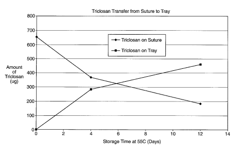
Fig. 1 is a graph illustrating the transfer of an antimicrobial agent from the
medical device to a containment compartment at 55C as a function of time.
Fig. 2 is a photographic representation of a containment compartment on a TSA
plate challenged Staphylococcus aureus.
5e
CA 02500852 2005-04-01
WO 2004/032704 PCT/US2003/030600
Fig. 3 is a photographic representation of a suture on a TSA plate challenged
Staphylococcus epidermidis.
Fig. 4 is a scanning electron microscope ("SEM") image of suture strands
coated
with an antimicrobial composition and exposed to methicillin-resistant
Staphylococcus
epidermidis.
Fig. 5 is a scanning electron microscope ("SEM") image of suture strands,
which
are not coated with an antimicrobial composition, exposed to methicillin-
resistant
Staphylococcus epidermidis.
DETAILED DESCRIPTION OF EMBODIMENTS OF THE INVENTION
Packaged Antimicrobial Medical Device
One embodiment of the packaged antimicrobial medical device includes at least
one
package having an inner surface. The inner surface includes an antimicrobial
agent
disposed thereon, present in an amount sufficient to substantially inhibit
bacterial
colonization on the package. The packaged medical device also includes at
least one
medical device positioned within the package. The medical device also has one
or more
surfaces having an antimicrobial agent disposed thereon. The antimicrobial
agent is
present on the medical device, in an amount sufficient to substantially
inhibit bacterial
colonization on the medical device. The antimicrobial agent disposed on the
package and
medical device may be selected from antimicrobial compounds which include
halogenated
hydroxyl ethers, acyloxydiphenyl ethers, and combinations thereof.
6
CA 02500852 2005-04-01
WO 2004/032704 PCT/US2003/030600
In another embodiment, the packaged medical device includes a package having
an inner
surface and a containment compartment for securing the medical device and that
resides
within the package. In this embodiment, at least one surface of the
containment
compartment includes an antimicrobial agent disposed thereon, present in an
amount
sufficient to substantially inhibit bacterial colonization on the containment
compartment.
In an alternate embodiment, the inner surface of the package and at least one
surface of the
containment compartment include an antimicrobial agent disposed thereon,
present in an
amount sufficient to substantially inhibit bacterial colonization on the
package and the
containment compartment. The packaged medical device also includes at least
one
medical device positioned within the containment compartment. The medical
device also
has one or more surfaces having an antimicrobial agent disposed thereon. The
antimicrobial agent is present on the medical device, in an amount sufficient
to
substantially inhibit bacterial colonization on the medical device. The
antimicrobial agent
disposed on the package, the containment compartment and medical device may be
selected from antimicrobial compounds which include halogenated hydroxyl
ethers,
acyloxydiphenyl ethers, and combinations thereof.
Another embodiment is an antimicrobial suture assembly comprising a
containment
compartment comprising one or more surfaces having an antimicrobial agent
disposed
thereon, the antimicrobial agent being selected from the group consisting of
halogenated
hydroxyl ethers, acyloxydiphenyl ethers, and combinations thereof, in an
amount sufficient
to substantially inhibit bacterial colonization on the containment
compartment; and a
suture positioned within the containment compartment, the suture comprising
one or more
7
CA 02500852 2005-04-01
WO 2004/032704 PCT/US2003/030600
surfaces having an antimicrobial agent disposed thereon, the antimicrobial
agent being
selected from the group consisting of halogenated hydroxyl ethers,
acyloxydiphenyl ethers,
and combinations thereof, in an amount sufficient to substantially inhibit
bacterial
colonization on the suture.
The medical devices described herein are generally implantable medical
devices, including
but not limited to mono and multifilament sutures, surgical meshes such as
hernia repair
mesh, hernia plugs, brachy seed spacers, suture clips, suture anchors,
adhesion prevention
meshes and films, and suture knot clips. Also included are implantable medical
devices
that are absorbable and non-absorbable. An absorbable polymer is defined as a
polymer
that, when exposed to physiological conditions, will degrade and be absorbed
by the body
over a period of time. Absorbable medical devices typically are formed from
generally
known, conventional absorbable polymers including, but not limited to,
glycolide, lactide,
co-polymers of glycolide, or mixtures of polymers, such as polydioxanone,
polycaprolactone and equivalents thereof. Preferably, the polymers include
polymeric
materials selected from the group consisting of greater than about 70%
polymerized
glycolide, greater than about 70% polymerized lactide, polymerized 1,4-dioxan-
2-one,
greater than about 70% polypeptide, copolymers of glycolide and lactide,
greater than
about 70% cellulosics and cellulosic derivatives. Examples of absorbable
medical device
include mono and multifilament sutures. The multifilament suture includes
sutures
wherein a plurality of filaments are formed into a braided structure. Examples
of non-
absorbable medical devices include mono and multifilament sutures, surgical
meshes such
8
CA 02500852 2005-04-01
WO 2004/032704 PCT/US2003/030600
as hernia repair mesh, hernia plugs and brachy seed spacers, which may be
polymeric or
nonpolymeric.
Suitable antimicrobial agents may be selected from, but are not limited to,
halogenated
hydroxyl ethers, acyloxydiphenyl ethers, or combinations thereof. In
particular, the
antimicrobial agent may be a halogenated 2-hydroxy diphenyl ether and/or a
halogenated
2-acyloxy diphenyl ether, as described in U.S. Patent No. 3,629,477, and
represented by
the following formula:
5' 6' 6 5 (Hal)al
4
4' 0-0 P23
3' 2' ZO
In the above formula, each Hal represents identical or different halogen
atoms, Z
represents hydrogen or an acyl group, and w represents a positive whole number
ranging
from 1 to 5, and each of the benzene rings, but preferably ring A can also
contain one or
several lower alkyl groups which may be halogenated, a lower alkoxy group, the
allyl
group, the cyano group, the amino group, or lower alkanoyl group. Preferably,
methyl or
methoxy groups are among the useful lower alkyl and lower alkoxy groups,
respectively,
as substituents in the benzene rings. A halogenated lower alkyl group,
trifluoromethyl
group is preferred.
9
CA 02500852 2010-09-03
WO 2004/032704 PCT/US2003/030600
Antimicrobial activity similar to that of the halogen-o-hydroxy-diphenyl
ethers of the
above formula is also attained using the O-acyl derivatives thereof which
partially or
completely hydrolyze under the conditions for use in practice. The esters of
acetic acid,
chloroacetic acid, methyl or dimethyl carbamic acid, benzoic acid,
chlorobenzoic acid,
methylsulfonic acid and chloromethylsulfonic acid are particularly suitable.
One particularly preferred antimicrobial agent within the scope of the above
formula is
2,4,4'-trichloro-2'-hydroxydiphenyl ether, commonly referred to as triclosan
(manufactured by Ciba Geigy under the trade name Irgasan DP300 or Irgacare
MP).
Triclosan is a broad-spectrum antimicrobial agent that has been used in a
variety of
products, and is effective against a number of organisms commonly associated
with SSIs.
Such microorganisms include, but are not limited to, genus Staphylococcus,
Staphylococcus epidermidis, Staphylococcus aureus, methicillin-resistant
Staphylococcus
epidermidis, methicillin-resistant Staphylococcus aureus, and combinations
thereof.
It is advantageous to use a coating composition as a vehicle for delivering
the
antimicrobial agent to the surface of the device where such coating already is
used
conventionally in the manufacture of the device, such as, for example,
absorbable and non-
absorbable multifilament sutures. Examples of medical devices, as well as
coatings that
may be applied thereto, may be found in U.S. Patent Nos. 4,201,216, 4,027,676,
4,105,034,
4,126,221, 4,185,637, 3,839,297, 6,260,699, 5,230,424, 5,555,976, 5,868,244,
and
5,972,008, As disclosed in U.S.
Patent No. 4,201,216, the coating composition may include a film-forming
polymer and a
CA 02500852 2005-04-01
WO 2004/032704 PCT/US2003/030600
substantially water-insoluble salt of a C6 or higher fatty acid. As another
example, an
absorbable coating composition that may be used for an absorbable medical
device may
include poly(alkylene oxylates) wherein the alkylene moieties are derived from
C6 or
mixtures of C4 to C12 diols, which is applied to a medical device from a
solvent solution, as
disclosed in U.S. Patent No. 4,105,034. The coating compositions of the
present invention
may include a polymer or co-polymer, which may include lactide and glycolide,
as a
binding agent. The compositions may also include calcium stearate, as a
lubricant, and an
antimicrobial agent. Medical devices not conventionally employing a coating in
the
manufacturing process, however, also may be coated with a composition
comprising an
antimicrobial agent. The coating may be applied to the device by, for example,
dip
coating, spray coating, suspended drop coating, or any other conventional
coating means.
Absorbable medical devices are moisture sensitive, that is, they are devices
that will
degrade if exposed to moisture in the atmosphere or in the body. It is known
by those of
ordinary skill in the art that medical devices made from absorbable polymers
may
deteriorate and lose their strength if they come into contact with water vapor
prior to use
during surgery. For instance, the desirable property of in vivo tensile
strength retention for
sutures will be rapidly lost if the sutures are exposed to moisture for any
significant period
of time prior to use. Therefore, it is desirable to use a hermetically sealed
package for
absorbable medical devices. A hermetically sealed package is defined herein to
mean a
package made of a material that serves as both a sterile barrier and a gas
barrier, i.e.,
prevents or substantially inhibits moisture and gas permeation.
11
CA 02500852 2010-09-03
WO 2004/032704 PCTIUS2003/030600
Materials useful for constructing the package for absorbable medical devices,
for example,
include single and multilayered conventional metal foil products, often
referred to as heat-
sealable foils. These types of foil products are disclosed in U.S. Patent No.
3,815,315,
Another type of foil product that
may be utilized is a foil laminate referred to in the field of art as a
peelable foil. Examples
of such peelable foil and substrates are disclosed in U.S. Patent No.
5,623,810,
If desired, conventional non-metallic
polymer films in addition to or in lieu of metal foil may be used to form the
package for
absorbable medical devices. Such films are polymeric and may include
conventional
polyolefins, polyesters, acrylics and the like, combinations thereof and
laminates. These
polymeric films substantially inhibit moisture and oxygen permeation and may
be coated
with conventional coatings, such as, for example, mineral coatings that
decrease or reduce
gas intrusion. The package may comprise a combination of polymer and metal
foils,
particularly a multi-layer polymer/metal-foil composite.
Nonabsorbable medical devices may be packaged in any of the materials
described above.
In addition, it is desirable to package nonabsorbable medical devices in a
package made of
a material that serves as a sterile barrier, such as a porous material, i.e.,
medical grade
paper, or a polymeric film that is permeable to moisture and gas, i.e., TYVEK
film,
manufactured by DuPont and made from high-density polyethylene fibers.
Packages for surgical needles, sutures and combinations including the suture
and a surgical
needle typically comprise a suture tray as the containment compartment, for
securely
12
CA 02500852 2010-09-03
WO 2004/032704 PCTIUS2003/030600
holding the suture and/or surgical needle in place. One type of containment
compartment
typically used for surgical needles and/or sutures is a folder package made
from a stiff,
medical grade paper. A folder package will typically have a plurality of
foldable panels
and cut-out tabs and tab pockets. Folder packages for surgical needles and
sutures are
illustrated and disclosed in the following patents:
U.S. Pat. Nos. 4,126,221, 4,120,395 and 5,555,976. Another
conventionally used containment compartment for surgical needles and/or
sutures is a
molded plastic tray having a central floor surrounded by an outer winding
channel for
receiving and retaining a suture, e.g., an oval channel. The containment
compartment may
further include a medical grade paper or plastic cover that may be mounted to
the top of
the winding channel, or the molded plastic tray may have molded retainer
elements, in
order to maintain the suture in the channel. The molded plastic tray may be
made from a
thermoplastic material selected from the group consisting of polyester,
polyvinyl chloride,
polypropylene, polystyrene, and polyethylene. Containment compartments having
winding channels are illustrated in the following:
U.S. Pat. Nos. 4,967,902, 5,213,210 and 5,230,424.
Microorganisms of the genus Staphylococcus are the most prevalent of all of
the organisms
associated with device-related surgical site infection. S.aureus and S.
epidermidis are
commonly present on patients' skin and as such are introduced easily into
wounds. One of
the most efficacious antimicrobial agents against Staphylococcus is 2,4,4'-
trichloro-2'-
hydroxydiphenyl ether. This compound has a minimum inhibitory concentration
(MIC)
against S. aureus of 0.01 ppm, as measured in a suitable growth medium and as
described
13
CA 02500852 2005-04-01
WO 2004/032704 PCT/US2003/030600
by Bhargava, H. et al in the American Journal of Infection Control, June 1996,
pages 209-
218. The MIC for a particular antimicrobial agent and a particular
microorganism is
defined as the minimum concentration of that antimicrobial agent that must be
present in
an otherwise suitable growth medium for that microorganism, in order to render
the growth
medium unsuitable for that microorganism, i.e., the minimum concentration to
inhibit
growth of that microorganism. The phrase "an amount sufficient to
substantially inhibit
bacterial colonization" as used herein is defined as the minimum inhibitory
concentration
for S. aureus or greater.
A demonstration of this MIC is seen in the disk diffusion method of
susceptibility. A filter
paper disk, or other object, impregnated with a particular antimicrobial agent
is applied to
an agar medium that is inoculated with the test organism. Where the anti-
microbial agent
diffuses through the medium, and as long as the concentration of the
antimicrobial agent is
above the minimum inhibitory concentration (MIC), none of the susceptible
organism will
grow on or around the disk for some distance. This distance is called a zone
of inhibition.
Assuming the antimicrobial agent has a diffusion rate in the medium, the
presence of a
zone of inhibition around a disk impregnated with an antimicrobial agent
indicates that the
organism is inhibited by the presence of the antimicrobial agent in the
otherwise
satisfactory growth medium. The diameter of the zone of inhibition is
inversely
proportional to the MIC.
Alternatively, the concentration of triclosan on the surface of a medical
device such as a
coated suture may be greater than about 0.01 ppm (wt./wt. coating) or between
about 30
ppm to 5,000 ppm (wt./wt. suture). The concentration of triclosan on the
surface of
14
CA 02500852 2005-04-01
WO 2004/032704 PCT/US2003/030600
package or containment compartment may be between about 5 ppm to 5,000 ppm
(wt./wt.
package or compartment). For other particular applications, however, higher
amounts of
antimicrobial agent may be useful and should be considered well within the
scope of the
present invention.
Method for Making a Packaged Antimicrobial Medical Device
In accordance with various methods of the present invention, a package and
containment
compartment that are initially substantially free of an antimicrobial agent,
i.e., no
antimicrobial agent is intended to be present on the package or containment
compartment
surfaces, may be provided. A medical device, which has an antimicrobial agent
disposed
thereon, is positioned within the package or containment compartment.
Subsequently, the
package, the containment compartment if utilized and the medical device are
subjected to
time, temperature and pressure conditions sufficient to vapor transfer a
portion of the
antimicrobial agent from the medical device to the package and/or the
containment
compartment.
The rate of transfer of an antimicrobial agent such as triclosan from the
medical device to
the package and/or containment compartment is substantially dependent upon the
time,
temperature and pressure conditions under which the package with the
containment
compartment and the medical device is processed, stored and handled. For
example,
Figure 1 illustrates that triclosan is capable of transferring from a suture
to a containment
compartment (in a closed vial at atmospheric pressure) when the temperature is
maintained
at 55C over a period of time. The conditions to effectively vapor transfer an
antimicrobial
CA 02500852 2010-09-03
WO 2004/032704 PCTIUS2003/030600
agent such as triclosan include a closed environment, atmospheric pressure, a
temperature
of greater than 40C, for a period of time ranging from 4 to 8 hours. Also
included are any
combinations of pressure and temperature to render a partial pressure for the
antimicrobial
agent that is the same as the partial pressure rendered under the conditions
described
above, in combination with a period of time sufficient to render an effective
amount or
concentration of the antimicrobial agent on the package and/or containment
compartment,
i.e., the minimum inhibitory concentration (MIC) or greater. Specifically, it
is known to
one of ordinary skill that if the pressure is reduced, the temperature may be
reduced to
effect the same partial pressure. Alternatively, if the pressure is reduced,
and the
temperature is held constant, the time required to render an effective amount
or
concentration of the antimicrobial agent on the package and/or containment
compartment
may be shortened. While a portion of the antimicrobial agent is transferred to
the
package and/or containment compartment during this process, a second portion
is retained
on the surface of the medical device. Accordingly, after the transfer, the
medical device
and the package and/or the contaiment compartment contain the antimicrobial
agent in an
amount effective to substantially inhibit bacterial colonization thereon and
thereabout.
Medical devices typically are sterilized to render microorganisms located
thereon non-
viable. In particular, sterile is understood in the field of art to mean a
minimum sterility
assurance level of 10-6. Examples of sterilization processes are described in
U.S. Patent
Nos. 3,815,315, 3,068,864, 3,767,362, 5,464,580, 5,128,101 and 5,868,244.
Specifically, absorbable medical devices may be
16
CA 02500852 2005-04-01
WO 2004/032704 PCT/US2003/030600
sensitive to radiation and heat. Accordingly, it may be desirable to sterilize
such devices
using conventional sterilant gases or agents, such as, for example, ethylene
oxide gas.
An ethylene oxide sterilization process is described below, since the time,
temperature and
pressure conditions sufficient to vapor transfer a portion of the
antimicrobial agent from
the medical device to the package and/or containment compartment, are present
in an
ethylene oxide sterilization process. However the time, temperature and
pressure
conditions sufficient to vapor transfer the antimicrobial agent from the
medical device to
the package and/or containment compartment may be effected alone or in other
types of
sterilization processes, and are not limited to an ethylene oxide
sterilization process or to
sterilization processes in general.
As discussed above, absorbable medical devices are sensitive to moisture and
are
therefore often packaged in hermetically sealed packages, such as sealed foil
packages.
However, sealed foil packages are also impervious to sterilant gas. In order
to compensate
for this and utilize foil packages in ethylene oxide gas sterilization
processes, processes
have been developed using foil packages having gas permeable or pervious vents
(e.g.,
TYVEK polymer). The gas permeable vents are mounted to an open end of the
package
and allow the passage of air, water vapor and ethylene oxide into the interior
of the
package. After the sterilization process is complete, the package is sealed
adjacent to the
vent, and the vent is cut away or otherwise removed, thereby producing a gas
impervious
hermetically sealed package. Another type of foil package having a vent is a'-
pouch-type
package having a vent mounted adjacent to an end of the package, wherein the
vent is
17
CA 02500852 2005-04-01
WO 2004/032704 PCT/US2003/030600
sealed to one side of the package creating a vented section. After the
sterilization process is
complete the package is sealed adjacent to the vent, and the package is cut
away for the
vented section.
The package and containment compartment are substantially free of, and
preferably
completely free of, antimicrobial agent prior to the transfer of the
antimicrobial agent from
the medical device to the package and/or the containment compartment. The
medical
device may first be placed within the containment compartment, if necessary,
and then
within the package. After the peripheral seal and side seals have been formed
in the
package, the packaged medical device may be placed into a conventional
ethylene oxide
sterilization unit. If the package is a foil package, the gas permeable vents
described above
may be used. Prior to the start of the cycle, the sterilization unit may be
heated to an
internal temperature of about 25 C. The sterilization unit is maintained about
22 to 37 C
throughout the humidification and sterilization cycles. Next, a vacuum may be
drawn on
the sterilization unit to achieve a vacuum of approximately 1.8 to 6.0 kPa. In
a
humidification cycle, steam then may be injected to provide a source of water
vapor for the
product to be sterilized. The packaged medical devices may be exposed to water
vapor in
the sterilization unit for a period of time of about 60 to 90 minutes. Times
may vary,
however, depending upon the medical device being sterilized.
Following this humidification portion of the cycle, the sterilization unit may
be pressurized
by the introduction of dry inert gas, such as nitrogen gas, to a pressure of
between about 42
and 48 kPa. Once the desired pressure is reached, pure ethylene oxide may be
introduced
18
CA 02500852 2005-04-01
WO 2004/032704 PCT/US2003/030600
into the sterilization unit until the pressure reaches about 95 kPa. The
ethylene oxide may
be maintained for a period of time effective to sterilize the packaged medical
device. For
example, the ethylene oxide may be maintained in the sterilization unit for
about 360 to
about 600 minutes for surgical sutures. The time required to sterilize other
medical
devices may vary depending upon the type of product and the packaging. The
ethylene
oxide then may be evacuated from the sterilization unit and the unit may be
maintained
under vacuum at a pressure of approximately 0.07 kPa for approximately 150 to
300
minutes in order to remove residual moisture and ethylene oxide from the
sterilized
packaged medical devices. The pressure in the sterilization unit may be
returned to
atmospheric pressure.
The following stage of the process is a drying cycle. The packaged medical
device may be
dried by exposure to dry nitrogen and vacuum over a number of cycles
sufficient to
effectively remove residual moisture and water vapor from the packaged medical
device to
a preselected level. During these cycles, the packaged medical device may be
subjected to
a number of pressure increases and decreases, at temperatures greater than
room
temperature. Specifically, the jacket temperature of the drying chamber may be
maintained at a temperature of between approximately 53 C to 57 C throughout
the drying
cycle. Higher temperatures, however, may be employed, such as about 65 C to 70
C for
sutures, and higher depending upon the medical device being sterilized. A
typical drying
cycle includes the steps of increasing the pressure with nitrogen to
approximately 100 kPa,
evacuating the chamber to a pressure of approximately 0.07kPa over a period of
180 to 240
minutes, reintroducing nitrogen to a pressure of 100 kPa and circulating the
nitrogen for
19
CA 02500852 2005-04-01
WO 2004/032704 PCT/US2003/030600
approximately 90 minutes, evacuating the chamber to a pressure of
approximately 0.01
kPa over a period of approximately 240 to 360 minutes and maintaining a
pressure of not
more than 0.005 kPa for an additional 4 to 96 hours. At the end of the
humidification,
sterilization and drying cycles, which takes typically about 24 hours, the
vessel is returned
to ambient pressure with dry nitrogen gas. Once drying to the preselected
moisture level is
complete, the packaged medical device may be removed from the drying chamber
and
stored in a humidity controlled storage area.
Upon completion of the sterilization process, the antimicrobial medical
device, the
package and/or the containment compartment have thereon an amount of the
antimicrobial
agent effective to substantially inhibit colonization of bacteria on or
adjacent the
antimicrobial device, the package and/or the containment compartment.
Example 1
A series of USP standard size 5-0 coated polyglactin 910 sutures were coated
with
a 2% triclosan coating composition so that each suture contained about a total
of 23.2 g
triclosan before sterilization. The coated sutures each were placed in a
package as
described herein above including a containment component, i.e., a tray, for
holding the
suture and a paper component for covering the suture in the tray. The suture
in the
containment component and packaging were sterilized as described herein above.
After
sterilization, it was determined that that suture contained about 5.5 gg
triclosan, the tray
about 0.2 g triclosan, the paper component about 2.3 g triclosan, and the
package heat
seal coating about 1.5 gg triclosan. Triclosan not recovered after
sterilization was about
CA 02500852 2005-04-01
WO 2004/032704 PCT/US2003/030600
13.7 g triclosan. Fig. 1 indicates triclosan transfer from the antimicrobial
suture to the
tray of the package as a function of time at 55 C.
After sterilization, the paper component and tray of the sterilized package
were
tested for antimicrobial properties utilizing a zone of inhibition test as
indicated herein
below. Zone of inhibition testing is a conventional method for estimating the
inhibitory
effects of antimicrobial substances against specific bacterial strains of
interest. Zone of
inhibition assays are useful for testing diffusible agents. As the agent
diffuses away from
the disk, the concentration decreases logarithmically. The sensitivity of the
organism to
the agent is judged by the appearance and size of a zone where no growth
occurs, i.e., the
zone of inhibition.
A comparative example of a package that contained a conventional commercially
available suture, i.e., not having triclosan applied thereto, also was
prepared and tested for
antimicrobial properties.
Fig. 2 is a photographic representation of the zone of inhibition with respect
to a
tray of the antimicrobial package on a TSA plate challenged with
Staphylococcus aureus.
The results of the zone of inhibition assays for the paper component and tray
are
listed in Table 1. The zones were measured for both treated and untreated tray
and paper
component. As shown in Table 1, zones of inhibition were present for all
treated c
21
CA 02500852 2005-04-01
WO 2004/032704 PCT/US2003/030600
omponents against both Staphylococcus aureus and Staphylococcus epidermidis.
The
untreated components exhibited no zones of inhibition.
Table 1. Zone of Inhibition Assay for Package Components
Staphylococcus epidermidis
Treated Package Zone size Untreated Package Zone size
Component Compone
Tray 18 mm Tray 0
Paper 13 mm Paper 0
Staphylococcus aureus
Treated Package Zone size Untreated Package Zone size
Component Component
Tray 12 mm Tra 0
Paper 13 mm Paper 0
Example 2
This example is a 24-hour aqueous immersion assay. The purpose of this assay
was to determine the effect of aqueous exposure on the antimicrobial
properties of suture
material for a range of suture diameters. Sterile sutures in USP sizes 2-0, 3-
0, 4-0, and 5-0,
with and without a 1% triclosan coating applied thereto, were aseptically cut
into 5-cm
pieces. One half of the cut pieces were stored in a sterile Petri dish and
kept under a dry
nitrogen atmosphere for 24 hours (dry suture). One half of the cut pieces were
aseptically
transferred to sterile 0.85% saline and incubated at 37 C for 24 hours (wet
sutures).
The dry and wet sutures were then aseptically placed in individual sterile
Petri
dishes and challenged with 100 microliters of inoculum containing 105 colony-
forming
units (CFU) of Staphylococcus aureus or Staphylococcus epidermidis. Ten
replicates of
each suture size were used for each organism and for both the dry and wet
sample groups.
TSA was poured into each dish and allowed to solidify. The plates were
incubated at 37 C
22
CA 02500852 2005-04-01
WO 2004/032704 PCT/US2003/030600
for 48 hours. After incubation, the plates were examined under a darkfield
colony counter
and the zones of inhibition were measured.
The results of the zone of inhibition assays are listed in Table 2. Zones of
inhibition were present for all sizes of coated polyglactin 910 suture having
triclosan
applied thereto. Both the dry and wet samples exhibited significant zones of
inhibition.
The coated polyglactin 910 suture controls had no zones of inhibition. A
typical zone of
inhibition is depicted in Fig. 3.
Table 2. 24 Hour Aqueous Immersion Assay: Zone of Inhibition Diameter
Zone Diameter Average (mm)
S aureus S epidermidis
Diy Wet Dry Wet
Suture Material
Size 2-0
+Triclosan 10 9 10 9
Control 0 0 0 0
Size 3-0
+ Triclosan 10 10 10 8
Control 0 0 0 0
Size 4-0
+ Triclosan 10 3 10 2
Control 0 0 0 0
23
CA 02500852 2005-04-01
WO 2004/032704 PCT/US2003/030600
Size 5-0
+ Triclosan 10 3 10 2
Control 0 0 0 0
All suture samples were from different lots. Average zone diameter is based on
triplicate plates.
As shown in Fig. 3, areas of inhibited bacterial growth were observed around
coated polyglactin 910 suture containing triclosan, while the control suture
without
triclosan had confluent bacterial growth. The response was similar for
Staphylococcus
epidermidis (shown), Staphylococcus aureus, MRSA, and MRSE, and was consistent
for a
variety of suture sizes.
Example 4
This example is directed to a 7-day aqueous immersion assay. The purpose of
this
assay was to determine if the antimicrobial effect of triclosan treatment
would endure for 7
days in a buffered aqueous environment.
Sterile USP size 2-0 coated polyglactin 910 suture coated with a 1%, 2%, and
3%
triclosan coating solution, respectively, and ethylene oxide sterilized USP
size 2-0 coated
polyglactin suture were aseptically cut into 5-cm pieces. Samples were tested
on each of 7
days in triplicate.
On day 1, 3 pieces of each suture material were placed into individual sterile
Petri
dishes and inoculated with 0.1 mL of challenge organism containing
approximately 104
CFU. TSA was poured into each dish and allowed to solidify. All remaining
pieces of
suture material were placed into 100 mL of sterile phosphate buffered 0.85%
saline (PBS).
24
CA 02500852 2005-04-01
WO 2004/032704 PCT/US2003/030600
Every 24 hours for the next 6 days, 3 pieces of each suture material were
removed from the
PBS, inoculated, and pour plated in tryptic/soy/agar (TSA). All plates were
incubated at
37 C for 48 hours and the plates examined for the presence or absence of a
zone of
inhibition.
The results for the 7-day assay are presented in Table 4. The coated
polyglactin
910 suture with triclosan produced zones of inhibition after every challenge.
The control
coated polyglactin 910 suture without triclosan produced no growth inhibition.
Table 4. 7-Day Aqueous Immersion Assay: Zone of Inhibition Diameter
Zone Diameter Average (mm)
1 2 3 4 5 6 7
Day
Triclosan coating
1% 20 18 20 20 19 21 20
2% 24 20 22 21 24 24 23
3% 27 25 15 25 27 30 27
Control (0%) 0 0 0 0 0 0 0
All suture samples were from different lots. Average diameter is based on
triplicate
plates.
This example is a demonstration of the efficacy of the antimicrobial suture
where
samples of the antimicrobial suture and a conventional suture were each
separately
exposed by immersion in aqueous buffer as a model of physiological conditions
for up to
seven days. On each day, samples of both the conventional and the
antimicrobial suture of
the invention were removed and placed on tryptic/soy/agar (TSA) plates that
had been
CA 02500852 2005-04-01
WO 2004/032704 PCT/US2003/030600
inoculated with a 104 colony forming unit (CFU) Staphylococcus challenge. As
is shown
in Table 4, the antimicrobial suture of the invention developed a zone of
inhibition around
it on the plate, even after seven days of immersion, providing evidence that
the
concentration of the antimicrobial agent on and around the antimicrobial
suture of the
invention was still above the MIC, while the conventional sutures, treated
similarly,
developed no zone of inhibition, i.e. the microorganisms freely grew on and
around the
conventional suture.
Example 6
This example relates to scanning electron microscopy. Scanning electron
microscope (SEM) images were prepared using sutures that had been exposed to
MRSE in
broth culture. Single 6-inch strands of USP size 2-0 coated polyglactin 910
suture coated
with 0.5% triclosan coating solution were placed in separate tubes containing
30 mL of
sterile TSB and inoculated with 0.1 mL of a 24-hour culture of the challenge
organism in
TSB. Single 6-inch strands of USP size 2-0 Polysorb (braided lactomer 9-1)
suture,
available from United States Surgical Corporation, and which did not contain
triclosan,
were also prepared in the same fashion. The tubes were incubated for 24 hours
at 37 C.
After incubation, the sutures were prepared for SEM as follows.
Each strand of the suture was removed from the broth and rinsed by vortexing
in
100 mL of sterile saline for 10 seconds. The rinsed strands were fixed in 10%
buffered
formalin for 5 minutes. The fixed strands were dehydrated in ethanol using
sequential 5-
minute exposures of 50%, 70%, 85%, 95%, and 100% ethanol. A final dehydration
was
performed using a 5-minute exposure in hexamethylenedisilazane. The samples
were air
26
CA 02500852 2005-04-01
WO 2004/032704 PCT/US2003/030600
dried prior to SEM. The SEM used for imaging the bacteria was a JEOL (Japan
Electronics and Optics Laboratory) JSM-5900LV scanning electron microscope.
Figs. 4 and 5 illustrate the differences between the triclosan-treated suture
(a) and
the untreated suture (b). The triclosan-treated suture had very few bacteria
associated with
it anywhere on the surface, while the untreated suture was uniformly and
heavily coated
with bacteria.
The data presented above indicate that coated polyglactin 910 suture with
triclosan
exhibits antimicrobial activity in vitro against Staphylococcus aureus and
Staphylococcus
epidermidis compared to untreated controls. This activity is evident on a
range of suture
diameters. The antimicrobial activity endures despite extended exposure to a
buffered
aqueous environment. Methicillin-resistant strains of Staphylococcus aureus
and
Staphylococcus epidermidis were inhibited after 24 hours of aqueous extraction
by
polyglactin 910 with triclosan at low triclosan concentrations. Low levels of
triclosan on
the suture are sufficient to greatly reduce colonization of the suture
compared to controls as
illustrated by scanning electron microscopy. These data support the conclusion
that coated
polyglactin 910 suture with triclosan provides an antimicrobial effect
sufficient to prevent
in vitro colonization of the suture by Staphylococcus aureus and
Staphylococcus
epidermidis.
Moreover, coated medical devices may be stable for extended periods of time.
During storage, coated devices may maintain a sufficient amount of triclosan
to exhibit
desired antimicrobial effects. Standard accelerated aging tests may be used to
estimate
antimicrobial properties after exposure to typical storage conditions.
27
CA 02500852 2005-04-01
WO 2004/032704 PCT/US2003/030600
Upon exposure to accelerated aging tests, triclosan coated sutures exhibited
zones
of inhibition against Staphylococcus aureus and Staphylococcus epidermidis. In
particular,
triclosan coated sutures were exposed to 50 C for 157 days. Table 6 indicates
triclosan
loss from various USP size 2-0 coated dyed polyglactin 910 sutures with
varying levels of
triclosan upon exposure of the sutures to 50 C for 157 days. The exposure took
place after
the sutures had been ethylene oxide sterilized and placed in a hot room for
three days.
Table 7 exhibits antimicrobial properties of those sutures after such
exposure. As
indicated in Table 7, zones of inhibition were exhibited against both
Staphyloccocus
aureus and Staphylococcus epidermidis after exposure. Although no zones of
inhibition
were exhibited against Streptococcus agalacticae under these testing
conditions, higher
concentrations of triclosan are known to inhibit growth of Streptococcus
agalacticae. It is
important to note that standard accelerated aging tests do not employ true
hospital storage
conditions, and thus, typically demonstrate worst-case scenarios. As such, the
stability of
triclosan coated sutures is believed to be significantly longer under normal
shelf-storage
conditions.
Table 6. Triclosan Loss at 50 C for 2-0 Dyed Vicryl Suture After Ethylene
Oxide
Sterilization and 3 Days in Hot Room
1% Solution 2% Solution 3% Solution
at 50 C Irgacare at 50 C Irgacare at 50 C Irgacare
Days ppm Days ppm Days ppm
0 200 0 295 0 333
3 127 3 216 3 266
3 132 3 235 3 291
3 156 3 230 3 291
11 94 11 163 11 227
11 91 11 163 11 213
18 89 18 140 18 189
32 69 32 120 32 155
58 58 58 108 58 164
28
CA 02500852 2005-04-01
WO 2004/032704 PCT/US2003/030600
157 59 157 118 157 130
157 39 157 79 157 101
Table 7: Zones of Inhibition for 2-0 Dyed Vicryl Suture After Exposure to 50 C
for 157
Days
Zone of Inhibition (Yes/No)
Triclosan Triclosan Stre
on Storage Conditions/ S. aureus p S. a idermidis
Coating Suture Sterilization Cycle agalacticae P
Conc.
( ~0 (PPM) 24 hr. 48 hr. 24 hr. 48 hr. 24 hr. 48 hr.
No No No No No No
1.0 39 50C for 157 days / N Yes No No No Yes Yes
cycle
2.0 79 50C for 157 days / N Yes Yes No No Yes Yes
cycle
3.0 101 50C for 157 days / N Yes Yes No No Yes Yes
cycle
1.0 59 50C for 157 days / N Yes No No No Yes Yes
cycle
2.0 118 50C for 157 days / N Yes Yes No No Yes Yes
cycle
3.0 130 50C for 157 days / N Yes Yes No No Yes Yes
cycle
29