Note: Descriptions are shown in the official language in which they were submitted.
CA 02500961 2005-04-O1
WO 2004/033040 PCT/US2003/031774
-1-
APPARATUS FOR PERFORMING PHOTOBIOSTIMULATION
PRIORITY
The present invention claims priority to U.S. Provisional Application No.
60/416,664, filed October 7, 2002 entitled "Methods and Apparatus for
Performing
Photobiostimulation."
BACKGROUND OF THE INVENTION
This invention is directed to methods and apparatus for performing
photobiostimulation of tissue, and more particularly to methods and apparatus
for
performing temperature controlled photobiostimulation of tissue.
Low-power emitting lasers (i.e., typically less than 100 mV~ have been used
worldwide over the past three decades to treat a variety of clinical
conditions. For
example, light has been reported to stimulate DNA synthesis, activate enzyme-
substrate
complexes, transform prostaglandins and produce microcirculatory effects.
There have
been numerous reports of such effects resulting from irradiating endogenous
chromophores (i.e., without application of exogenous photosensitizers) in
cells or
tissues.
The use of low-level light to achieve such photochemical responses is commonly
referred to as photobiostimulation. In addition to laser light,
photobiostimulation may
be achieved using other monochromatic or quasi-monochromatic light sources
(e.g.,
LEDs) or by suitably filtering broadband light sources (e.g., filtering
fluorescent lamps,
halogen lamps, incandescent lamps, discharge lamps, or natural sunlight).
Biostimulation achieved by laser sources is also referred to as low-level
laser therapy
(LLLT).
Low-level light or low-level laser therapy stimulates the tissues and promotes
healing by penetrating deep into the tissues initializing the process of
photobiostimulation. The light energy is absorbed in cytochromes and
porphyrins
within cell mitochondria and cell membranes producing a small amount of
singlet
oxygen. Healing results from such treatments as demonstrated in many thousands
of
clinical study cases. Typically, patients can expect to feel noticeable
improvement after
four to six sessions for acute conditions and after six to eight treatments
for chronic
CA 02500961 2005-04-O1
WO 2004/033040 PCT/US2003/031774
_2_
conditions. In many instances, photobiostimulation can be a viable alternative
to
surgery.
The photochemical process resulting from photobiostimulation is believed to
involve the integration of photons into the cellular machinery of biochemical
reactions.
Generally, the principle of light absorption and integration of the photon
energy into the
cellular respiratory cycle is a well-known natural phenomenon. Photosynthesis
and
vision are two examples of this phenomenon. In these processes, the
photoacceptor
molecules are chlorophyll and rodopsin, respectively.
In the case of photobiostimulation, several concurrent mechanisms of action
have
been demonstrated in vitro. One example of such a mechanism involves
cytochrome c
oxidase, which is a primary cellular photoacceptors of low level light.
Cytochrome c
oxidase is a respiratory chain enzyme residing within the cellular
mitochondria, and is
the terminal enzyme in the respiratory chain of eulcaryotic cells. In
particular,
cytochrome c oxidase mediates the transfer of electrons from cytochrome c to
molecular
oxygen. The involvement of cytochrome c is known to be central to the redox
chemistry
leading to generation of free energy that is then converted into an
electrochemical
potential across the inner membrane of the mitochondrion, and ultimately
drives the
production of adenosine triphosphate (ATP). Accordingly, it has been
postulated that
photobiostimulation has the potential of increasing the energy available for
metabolic
activity of cells.
It has been further demonstrated that photobiostimulation may be used to
enhance cellular proliferation to achieve therapeutic effects. ATP molecules
serve as a
substrate to cyclic AMP (CAMP) which, in conjunction with calcium ions (Ca2+),
stimulate the synthesis of DNA and RNA. CAMP is a pivotal secondary messenger
affecting a multitude of physiological processes such as signal transduction,
gene
expression, blood coagulation and muscle contraction. Accordingly, it has been
postulated that an increase in ATP production by photobiostimulation may
provide a
means to increase cell proliferation and protein production.
Light-stimulated ATP synthesis, such as that caused by photobiostimulation, is
wavelength dependent. Karu (Lasers in Medicine aid Dentistry. Ed. Z.
Simunovic,
Vitgraf Rijeka, 2000, pp.97-125.) demonstrated in vitro that prokaryotic and
eulcaryotic
cells are sensitive to two spectral ranges, one at 350-450 nm and another at
600-830 nm.
CA 02500961 2005-04-O1
WO 2004/033040 PCT/US2003/031774
-3-
Karu demonstrated that the light receptors of the red wavelengths are the
semichinon
type of the flavoproteins of the reductase (dehydrogenases) and the cytochrome
alai of
cytochrome c. Cytochrome c oxidase in its oxidation form is the specific
chromophore
of 800 nm through 830 nm wavelength range.
Another mechanism of biostimulation involves causing a very limited irritation
to the blood cells and walls in the vessels of the dermis. This results in a
low-grade
inflammatorylgrowth response. Inflammatory mediators are released through the
vessel
walls that stimulate fibroblast activity and eventually lead to a "healing"
effect.
While the above mechanisms and positive effects have been demonstrated in
numerous in vitro studies, results of clinical trials have been so far
inconclusive. While
some groups reported varying degree of success in the treatment of a range of
conditions, others observed no or minimal effect. U.S. Patent Nos. 5,514,168,
5,640,978, 5,989,245, 6,156,028, 6,214,035, 6,267,780, and 6,221,095, which
are hereby
incorporated by reference, provide examples of methods and devices for
biostimulation.
While various methods and devices of biostimulation exist in the art, more
efficient and
efficacious methods of treatment that yield quicker results with less
treatment sessions
are needed.
Photobiostimulation has been typically performed using relatively inexpensive
sources, such as diode lasers or LEDs such as Ga-As and Ga-Al-As (e.g.,
emitting in the
infrared spectrum (600- 980 nm)). Existing sources of low power laser light
and light
emitting diodes (LEDs) deliver power levels ranging from 1 to100 milliwatts;
accordingly power densities necessary to perform photobiostimulative
procedures are
achieved by concentrating the light beam output into a very small spot sizes
(typically
less than 10 mm). This results in a typical power density at the skin surface
in a range
between 1 and 100 mW/cm2. The small beam size makes a scanning device
necessary to
treat large areas. Treatment times used in most studies are in the range of 5
to 30 min
and multiple treatments are often required.
There exists a need in the art for improved methods and devices for
biostimulation that improve efficacy of treatment of disease andlor cosmetic
conditions
and, thus, will require less treatment sessions.
CA 02500961 2005-04-O1
WO 2004/033040 PCT/US2003/031774
-4-
BRIEF SUMMARY OF THE INVENTION
The present invention provides methods and devices for modulating the efficacy
and/or increasing the efficiency of treatment of disease and/or cosmetic
conditions
through photobiostimulation combined with heating and/or cooling of the
treatment
region. In one aspect, methods and devices of the present invention are
directed to
modulating the efficacy of photobiostimulation in a target region by
controlling the
temperature in the region and/or its surrounding volume. According to some
aspects of
the present invention, tissue is heated such'that biostimulation is applied to
tissue that is
hyperthermic. Alternatively, portions of the target region can be cooled to
selectively
target biostimulation to a specific region at a desired depth below the skin
surface. A
feedback mechanism is also provided so that the temperature of the target
region can be
selectively and accurately controlled.
The present invention is based in part on the discovery that heat enhances the
effects of biostimulation. Heat enhanced biostimulation can take various
forms. For
example, heat may slow the repair of radiation-induced DNA damage, leaving
more
damage unrepaired and increased amounts of free radicals in the target region
resulting
in increased effects of biostimulation. Heat may also induce the production or
activation
of heat shock proteins or modify the rates of enzymatic processes. Currently,
treatment'
sources and operating conditions used in conventional photobiostimulation
provide
negligible heating of treated tissue (e.g., less than 1°C above normal
body temperature).
In one aspect, the invention provides methods and devices for biostimulating a
target region of a subject comprising irradiating a target region with a
radiation,
generated by a radiation source which has at least one selected wavelength
component
suitable for biostimulation, for a selected time duration and controlling a
temperature of
the irradiated target region with a source independent of said biostimulating
radiation so
as to modulate efficacy of said biostimulation. The time duration is chosen so
as to
cause biostimulation of the target region. In some embodiments, the target
region is
disposed at a depth below a skin surface of the subject. Time duration can be
selected
based on the desired application. Preferably time durations are chosen to be
in a range
of about 10 seconds to about one hour or in the range of about 10 minutes to
about one
hour. The temperature can be controlled, for example, by placing the target
region in
thermal contact with a surface having a selected temperature, by generating a
flow of a
CA 02500961 2005-04-O1
WO 2004/033040 PCT/US2003/031774
-5-
fluid or air over the target region to be in thermal contact therewith, by
applying
electromagnetic or ultrasound radiation to the target region, or by applying a
vaporizing
cream, or a precooled and/or preheated cream or lotion to the target region.
Those
having ordinary skill in the art will appreciate that the other methods may
also be
utilized for controlling the temperature of the target region and/or its
surrounding
volume.
The wavelength component can be selected to be in a range of about 380 nm to
about 1250 nm, in a range of about 380 nm to about 600 nm, in a range of about
380 nm
to about 450 nm, in range of about 600 nm to about 700 nm, or in a range of
about 760
nm to 880 nm depending on the desired application. The radiation source can
preferably
generate radiation with a narrow bandwidth, for example, a bandwidth less than
about
100 nm.
The radiation can deliver a power flux in a range of about 1 to about 250
mWlcm2 to the target region, or more preferably in a range of about 10 to
about 100
mW/cm2. The radiation can deliver an energy flux in a range of about 1
Joule/cm' to
about 1000 Joules/cm2, or more preferably in the range of about 1 Joule/cm2 to
about
100 Joules/cm2, to the irradiated target region during irradiation time.
According to some aspects of the invention, the target region is irradiated by
exposing it to a beam of radiation having a cross-sectional area in a range of
about 1 cm2
to about 10 cm2. However, the beam's cross-section can be increased based on
the
application.
In some aspects, the step of controlling temperature includes heating the
irradiated target region, referred to as hyperthermia herein, so as to
increase efficacy of
the biostimulation. The heating step can be performed by contact heating,
convection,
or application of electromagnetic radiation, such as ultrasound, microwave, or
infrared
energy. Hyperthermia is defined herein to be a temperature greater than normal
body
temperature. Normal body temperature can range from 36.1°C to
37.2°C depending on
the time of day. Accordingly, the temperature of the surface area of the
target region to
which biostimulation is applied in practice of the invention can be increased
to 37-50°C
and preferably 37-45°C. In some embodiments, the temperature of the
target area can be
increased to be within a range of about 37-42°C or, alternatively, be
within a range of
about 38-42°C. In other embodiments, the temperature of the target area
is increased to
CA 02500961 2005-04-O1
WO 2004/033040 PCT/US2003/031774
-6-
be within a range of about 38-41°C. The temperature is preferably
elevated above
normal body temperature, but below a temperature at which pain and
denaturation of a
significant concentration of critical biomolecules occurs.
Further aspects of the present invention are directed to cooling a target
region to
which biostimulative radiation is applied. According to at least some aspects
of the
invention, a portion of the region of tissue is cooled such that the skin is
protected from
heat damage and/or the efficacy of biostimulation in the region is reduced to
control
depth of treatment. The target region can be cooled to a value in a range of
about 0°C to
about 36°C , or about 10-36 °C, or about 15-36 °C, or
about 20-36°C, or about 28-36°C.
In some embodiments, controlling the temperature comprises utilizing a
separate
radiation source to heat the target region irradiated with biostimulating
radiation.
The separate radiation source can include a narrowband source or broadband
source.
The separate radiation source can generate radiation having one or more
wavelength
components in a range of about 380 nm to about 2700 nm, preferably in a range
of about
1000 nm to about 1250 nm, or more preferably in a range of about 700 nm to
about 900
nm.
In one aspect of the invention, the step of controlling the temperature of the
irradiated target region comprises heating a first selected portion of the
target region and
cooling a second selected portion of the target region. Heating and cooling
can be either
simultaneous or sequential. Beneficial effects may result from rapidly
changing or
fluctuating the temperature of the target region before, during, or between
irradiation
sessions.
In another aspect of the invention, a method of biostimulating a target region
of a
patient disposed at a depth below the patient's skin is disclosed. The method
includes
exposing a portion of the patient's slcin for a selected time duration to a
radiation having
at least one selected wavelength component capable of penetrating to a depth
associated
with the target region so as to irradiate the target region. The temperature
of a volume
of the patient through at least a portion of which the radiation traverses to
reach the
target region is controlled so as to modulate biostimulation within that
volume relative to
the target region. The wavelength component and the time duration are chosen
to cause
biostimulation within the target region. The temperature can be controlled to
cool the
volume and decrease biostimulation therein. For example, the temperature of
the
CA 02500961 2005-04-O1
WO 2004/033040 PCT/US2003/031774
_7_
volume can be decreased to be within the range of about 0°C to about
36°C or preferably
in a range of about 15°C to about 36°C. The wavelength component
can be selected to
be in a range of about 380 nm to about 1250 nm or more specific ranges
described
herein. The radiation source can generate radiation with a narrow bandwidth
that can be
less than about 100 nm.
In yet another aspect, the invention discloses a device for biostimulating a
patient's target region that includes a first source for generating
electromagnetic
radiation having one or more wavelength components suitable for causing
biostimulation in the target region; a radiation guidance device optically
coupled to the
source for delivering the radiation to the target region; and a second source
in
communication with the target region for controlling a temperature of the
target region
in order to modulate efficacy of biostimulation caused by the electromagnetic
radiation.
The first source can generate radiation having a narrow bandwidth, for
example, less
than about 100 nm. The first source can generate radiation having one or more
wavelength components in a range of about 380 nm to about 1250 nm. The second
source can include a source of electromagnetic radiation generating radiation
suitable for
heating the target region so as to enhance the efficacy of biostimulation. For
example,
the second source can generate one or more wavelength components in a range of
about
380 nm to about 2700 nm.
In a related aspect, the device can further include an optical fiber coupled
at an
input thereof to the first radiation source and an output thereof to the
radiation guidance
device, for example, a lens system, so as to direct light generated by the
radiation source
to the lens system. The lens system can have at least one movable lens to
allow
adjusting a cross-sectional area of a radiation beam generated by the first
source for
irradiating the target region. The lens system can comprise a Fresnel lens.
In another aspect, the radiation guidance device may include a beam splitter
adapted to receive a radiation beam from the first source in order to generate
a plurality
of beam portions, and one or more reflective surfaces optically coupled to the
beam
sputter to direct one or more of the beam portions to a surface of the
patient's skin so as
to irradiate the target region. The reflective surfaces can allow a
substantially uniform
illumination of the skin surface. The beam sputter can be, for example, a
prism, and at
least one of the reflective surfaces can exhibit a curved profile.
CA 02500961 2005-04-O1
WO 2004/033040 PCT/US2003/031774
_g_
In another aspect, the invention provides a method of biostimulating a
subject's
target region that includes irradiating the target region with radiation
having one or more
wavelength components suitable for causing biostimulation within the target
region, and
actively controlling a temperature of at least a portion of the target region
to ensure it
remains within a pre-defined range of an operating temperature in order to
modulate
efficacy of biostimulation within the target region. The step of actively
controlling the
temperature can include measuring a temperature of a portion of the patient's
skin in
thermal contact with the target region and comparing the measured temperature
with at
least one pre-defined threshold. The amount of heat delivered to or extracted
from the
target region can be controlled in response to the comparison of the measured
temperature with the pre-defined threshold.
In yet another aspect, the invention provides a method for biostimulating a
plurality of target regions of a subject by moving a radiation source over a
portion of the
subject's skin so as to irradiate sequentially a plurality of target regions
with radiation
having at least one wavelength component suitable for causing biostimulation.
The
moving of radiation source can be performed at a rate selected to expose each
of the
regions to sufficient radiation for causing biostimulation therein. The
temperature of the
target regions can be controlled by a source independent of the biostimulating
radiation
so as to modulate efficacy of biostimulation within each of the target
regions. The
moving radiation source can expose each target region, once, or alternatively,
multiple
times, to biostimulative radiation.
BRIEF DESCRIPTION OF THE DRAWINGS
Figure 1 schematically illustrates an embodiment of the invention in which a
target region, which extends from the surface of the skin to a selected depth,
is heated
such that biostimulation is applied to a hyperthermic volume of tissue;
Figure 2 schematically illustrates another embodiment of the invention in
which
biostimulation is applied to a heated target region in proximity of the skin
surface while
biostimulation is applied simultaneously to an unheated volume below the
target region;
CA 02500961 2005-04-O1
WO 2004/033040 PCT/US2003/031774
-9-
Figure 3 schematically illustrates another embodiment of the invention in
which
photobiostimulation is generated in a volume of tissue at a depth region below
the
surface of skin while cooling is applied to the surface of skin;
Figure 4 schematically illustrates another embodiment of the invention in
which
biostimulation is applied to a hyperthermic volume of tissue that is at a
selected depth
below the surface of the skin, and unheated volumes are located above and
below the
hyperthermic volume of tissue;
Figure 5 schematically illustrates another embodiment of the invention in
which
enhanced biostimulation occurs in a first volume of tissue, which is both
hyperthermic
and located at a selected depth below the surface of the skin, and
biostimulation (without
hyperthermia) also occurs in a second volume of tissue that is located below
the first
volume of tissue;
Figures 6 is a graph of selected temperature profiles of type II skin using
exemplary wavelengths of monochromatic light without skin cooling;
Figures 7 is a graph of selected temperature profiles of type II skin using
exemplary wavelengths of monochromatic light with parallel skin cooling;
Figure 8 is a schematic diagram of a light projection system for
biostimulating a
target region, according to the teachings of the invention;
Figure 9A is an exemplary embodiment of a light projection system for forming
substantially uniform illumination of a non-flat surface;
Figure 9B is a schematic diagram of an exemplary beam splitter suitable for
use
in a device according to the teachings of the invention;
Figure 10 is a schematic diagram of another exemplary embodiment of a light
projection system for forming substantially uniform illumination over a non-
flat surface;
CA 02500961 2005-04-O1
WO 2004/033040 PCT/US2003/031774
- 10-
Figures 1 lA is a schematic diagram of another embodiment of a light
projection
system according to the teachings of the invention that utilizes a rotatable
head to
provide substantially uniform illumination to a non-flat surface, where the
rotatable head
is positioned to direct light onto a front portion of the non-flat surface
Figures 11B is a schematic diagram of another embodiment ofa light projection
system according to the teachings of the invention that utilizes a rotatable
head to
provide substantially uniform illumination to a non-flat surface, where the
rotatable head
is positioned such that light is directed onto a first side portion of non-
flat surface;
Figures 11C is a schematic diagram of another embodiment of a light projection
system according to the teachings of the invention that utilizes a rotatable
head to
provide substantially uniform illumination to a non-flat surface, where the
rotatable head
is positioned such that light is directed onto a second side portion of non-
flat surface;
Figure 12A is a graph of the temperature of type II skin surface as a function
of
time of exposure to a 800 nm radiation at a flux of 680 mW/cm2, wherein the
beam has a
diameter larger than 2.5 cm;
Figure 12B is a graph of temperature profiles in which the type II skin
surface is
cooled and kept at 36°C while being exposed to different wavelengths of
radiation
according the invention;
Figure 13A is an exemplary embodiment of a light projection system for use in
the invention;
Figure 13B depicts an exemplary set of lens parameters according to the
invention;
Figure 14 illustrates an exemplary embodiment of a device, according to the
invention, capable of irradiating a target region and controlling the
temperature of that
region through a feedback mechanism; and
CA 02500961 2005-04-O1
WO 2004/033040 PCT/US2003/031774
-11-
Figure 15 illustrates an exemplary embodiment of a device, according to the
invention, capable of irradiating a target region using a 2D matrix of
radiation sources.
DESCRIPTION OF THE INVENTION
In one aspect, the present invention is directed to controlling the efficacy
of
photobiostimulation in a target region by controlling the temperature of that
region. The
heating or cooling of the target region, i.e., patient's skin, hair, eye,
teeth, nails, or other
body tissue, can trigger biological processes within the body that can work
synergistically with photobiostimulation to yield better, more efficient
results. The
temperature of the target region is modulated during, prior to, or between
photobiostimulation. The synergy between irradiation and temperature
modulation can
vary depending on the order of application andlor the disease or cosmetic
condition to be
treated. In a preferred embodiment, modulation of the temperature and
irradiation
occurs simultaneously.
In one embodiment, the temperature of the target region is increased. Heating
of
tissue, hyperthermia, leads to increased local tissue perfusion and increased
blood and
lymph circulation. The increase in blood flow has multiple effects on
photobiostimulated tissues. The cellular biochemical reactions of
biostimulation are
accelerated since the rates of some enzymatic reactions increase at higher
temperatures.
Additionally, more oxygen is available for the increased cellular metabolism,
and the
toxic by-products of metabolism are removed more readily, through the blood
and
lymphatic circulation. In addition, heating of blood vessels can increase
vessel wall
and/or cell wall permeability, which may result in improved delivery of
therapeutic
additives (i.e., vitamins, antioxidants, lations, etc.) or drugs to the target
area. For
example, topical drugs may be enclosed in thermosensitive liposomes that
selectively
release their drug content when exposed to heat.
Hyperthermia in a tissue to be treated may be achieved by use of any suitable
technique, including but not limited to use of contact heating, convection
(i.e., by heated
air), or application of electromagnetic radiation. In some embodiments,
hyperthermia in
a tissue to be treated is achieved by absorption of a portion of the incident-
electromagnetic radiation from a biostimulative source used to biostimulate
the tissue.
For example, absorption of electromagnetic radiation may be by tissue
chromophores
CA 02500961 2005-04-O1
WO 2004/033040 PCT/US2003/031774
-12-
such as melanin, hemoglobin, water, lipids or other chromophores which cause a
photothermal interaction leading to an increase in tissue temperature.
Hyperthermia
generates a cascade of events, such as increasing vasodilation, increasing
blood
circulation, increasing production of heat shock proteins, which can act
synergistically
with photobiostimulation resulting in improved efficacy of treatment.
Additionally, local hyperthermia is known to activate the heat shock (HS)
response, the~motolerance and hormesis (P. Verbeke, et al. Cell Biol hrter.
2001;
25:845-857). The phenomenon of tlze~»zotole~ahce is defined as the capacity of
cells,
following a cycle of heat stress and recovery, to survive a second stress,
which would
otherwise be lethal. Mild heat shock treatment may prevent cell death from a
variety of
subsequent stresses. Similar to exposure of cells and organisms to stresses
such as
caloric restriction, exercise, oxidative and osmotic stress, heavy metals,
proteosome
inhibitors, amino acid analogues, ethanol, and metabolic poisons, heat shock
treatment
induces a cellular stress response leading to the preferential transcription
and translation
of heat shock proteins (HSPs). Numerous families of HSPs have been identified
(P.
Verbeke, et al. Cell Biol Ihtei°. 2001; 25:845-857).
When a cell encounters a stressor, modifications of the cytoslceleton,
cytoplasmic
structures, cell surface morphology, cellular redox status, DNA synthesis,
changes in
protein metabolism and protein stability occur. Such stress generates a
molecular
remodeling or damage, especially abnormal folded proteins, which can aggregate
and
initiate a sequence of stress responses. The induction of the HS response
occurs through
molecular links between the environmental stresses and the stress response.
When stress
alters protein folding, or proteins begin to unfold and denature, HSPs have
been shown
to assist in protein refolding, to protect cellular systems against protein
damage, to
solubilize aggregates to some extent, to sequester overloaded and damaged
proteins into
large aggregates, to target fatally damaged proteins for degradation, and to
interfere with
the apoptotic progression (P. Verbelee, et al. Cell Biol Inter. 2001; 25:845-
857).
HSPs that are involved in the renaturation of unfolded proteins are referred
to as
chaperones. Chaperones recognize and bind to other proteins when they are in
non-
native conformations and are exposing hydrophobic sequences. Such HSPs protect
many
different systems involved in maintenance of cellular functions. Some HSPs
induce an
increase in the cellular glutathione (GSH) level leading to the protection of
the
CA 02500961 2005-04-O1
WO 2004/033040 PCT/US2003/031774
-13-
mitochondria) membrane potential during stress. Members of the HSP70 and HSP90
families are associated with the centrosome. They are known to bind and
stabilize actin,
tubulin and the microtubules/microfilament network, playing a role in the
cellular
morphology and transduction pathways.
The~motole~~a~ce is believed to be mainly due to the orchestrated regulation
of
expression and accumulation of various HSPs in the endoplasmic reticulum and
in the
cytosol, leading to marcromolecular repair mechanisms as a defensive strategy
against
subsequent challenges. A further characteristic of responses to HS is that
various HSPs
are soluble and transfer across the cell membrane to other adjacent cells.
Consequently,
the protective stress response is transferable to neighboring cells that might
not be able
to mount such a reaction. Accordingly, a next treatment can be done with
higher
temperature. This mechanism can be used to increase the maximum tolerable
incident
power applied to the skin surface. Specifically, the power can be increased
gradually,
allowing the organism to adapt to the thermal stress and thus survive a higher
level of
hyperthermia than would be possible without such adaptation.
In addition to the HSP-dependent effects described above, HSP-independent
effects may arise from hyperthermia. Other mechanisms of stress tolerance
include the
synthesis of osmotic stress protectants, modifications of the saturation of
cell membrane
lipids, and expression of enzymes such as radical scavengers.
Similar to thermotolerance, ho~ynesis is a response to repeated mild stress,
which
enhances cellular defense processes. Hormesis is a process by which cells
adapt to
gradual changes in their environment so as to be able to survive subsequent
exposure to
otherwise lethal conditions. Such a phenomenon has been observed in relation
to
irradiation, toxins, heat shock and other stresses. Ratan et al observed anti-
aging
homnetic effects of repeated mild HS on human fibroblasts (Rattan et al.
Biochem Mol
Biol Iht 199;45:753-759). Kevelaitis et al showed that local and brief
application of
heat (42.5 °C for 15 minutes) to the myocardium improved cardiac
systolic and diastolic
functions (Kevelatis et al. A~32 To~ac Surg 2001;72:107-113).
The above indicates that systems according to aspects of the present invention
should improve the clinical utility and outcome of biostimulation therapy. It
further
appears that aspects of the present invention provide synergistic effects of
photochemical biostimulation of cells and mild tissue hyperthermia, which
stimulate
CA 02500961 2005-04-O1
WO 2004/033040 PCT/US2003/031774
- 14-
HSP-dependent and HSP-independent thermotolerance, and/or hormesis. This
synergism may lead to repair of cell damage and improved functionality of
compromised cells. Those effects may help in the treatment of conditions
associated
with infection, acute and chronic inflammation, micro circulatory stagnation,
and may
also stimulate regeneration and rejuvenation of tissues subjected to
degenerative
processes, for example, by stimulating fibroblast proliferation, or by
increases in growth
factors eventually leading to new synthesis of intracellular and extracellular
proteins,
glycoproteins and lipid soluble molecules. Additional aspects of the present
invention
control the effectiveness of biostimulation provided by selectively delivered
photobiostimulative light to deep structures through the use of temperature
control (e.g.,
via heating and/or cooling of a tissue surface) and/or through control of
radiation spot
size.
In another aspect of the invention, a means for controlling specific
mechanisms
of photobiostimulation in order to achieve a desired therapeutic effect is
provided. It is
known that the biological response to photobiostimulation can vary as a
function of the
state of the biological system. For example, human fibroblasts can display a
diversity of
responses when exposed to outside stimuli (Lasers isz Medicine ahd Dentistry.
Ed. Z.
Simunovic, Vitgraf Rijeka, 2000, pp.97-125). In particular, both stimulation
of
proliferation of fibroblasts and an increase in production of type I collagen
have been
reported. However, production of collagen was affected in a manner inverse to
the
effect on cell proliferation, i.e., when proliferation increased, production
of collagen
decreased. Therefore, one can manipulate the state of the target system in
order to
channel the action of biostimulation into a desired pathway. One factor
greatly
influencing the state of the biological system is the temperature. The present
invention
provides a way to fine-tune the resulting biological response through the
control of the
temperature of the biostimulated area.
The present invention provides methods and devices for modulating the efficacy
of biostimulation. The term "modulates efficacy" as used herein refers to a
change of
the resulting biostimulation effects of greater than 10°l°,
preferably greater than 20%,
more preferably greater than 30%, more preferably greater than 40%, more
preferably
greater than 50%, more preferably greater than 60%, more preferably greater
than 70%,
more preferably greater than 80%, more preferably greater than 90% and most
CA 02500961 2005-04-O1
WO 2004/033040 PCT/US2003/031774
-15-
preferably greater than 100%. The efficacy of biostimulation can be measured
in terms
ofthe time necessary to achieve a desired outward appearance, i.e., removal of
wrinkles
or scar tissue, or a~time needed for patient satisfaction, i.e., pain relief,
or the rate of the
underlying enzymatic mechanisms. For example, substantially increasing the
efficacy of
biostimulation of a target region can refer to an increase in the rate of
enzymatic
processes in that target region of more than 10% relative to unstimulated
steady-state
condition. The rate of the enzymatic processes can be determined using any of
the
methods known in the art (See, for example, T. Bugg, An Ihtroductiofz to
Enzyme and
CoenzyJZZe Chemistzy, Blackwell, 1997; Wright et al. Photochefza Photobiol.
2002
Ju1;76(1):35-46; Koekemoer et al. Comp Biochem Physiol B Biochei~z Mol Biol.
2001
Jul;129(4):797-807). For example, the enzymatic activity of cytochrome c
oxidase or
the rate of radical production, i.e., singlet oxygen, can be used as a measure
of
biostimulation in the target region. Free radical production can be determined
by
measuring superoxide dismutase (SOD) and catalase or glutathione peroxidase
levels in
the cytoplasm. In addition, indirect measures of free radical production can
be used
such as through consumption of antioxidants.
The mechanisms described above are illustrative, and are not exhaustive.
Accordingly, they should not be considered as limiting the scope of the
presented
invention. Additionally, because photobiostimulation is an emerging field, the
theories
regarding the mechanisms achieving a given result are in many instance
speculative.
Figures. 1-5 are schematic cross-sectional views of systems that illustrate
five
exemplary treatment scenarios for achieving photobiostimulation and
temperature
control (e.g., hyperthermia and/or hypothermia) of a volume of tissue
according to at
least some aspects of the present invention.
In each of the treatment scenarios, biostimulation is achieved by applying
electromagnetic radiation to the skin surface from a source suitable for
achieving
biostimulation. For example, a suitable source may comprise a narrow bandwidth
source, such as a monochromatic or quasi-monochromatic source. Appropriate
sources
can include lasers, LEDs or suitably filtered broadband sources (e.g.,
filtered lamps).
The invention can also utilize a 2D matrix of radiation sources. A suitable
narrow
bandwidth source preferably has a bandwidth (i.e., wavelength range) of less
than
approximately 100 nm, preferably below approximately 20 nm and more preferably
CA 02500961 2005-04-O1
WO 2004/033040 PCT/US2003/031774
- 16-
below approximately 10 nm. The wavelength may be selected to achieve any known
biostimulative effect. The wavelength of the radiation may be, for example, in
a range of
380-2700 nm. For example, radiation with a wavelength in a range of about 380-
600 nm
can be utilized for treating superficial tissues, while radiation with a
wavelength in a
range of about 600-1250 nm can be utilized for deep tissues. In an exemplary
embodiment, preferred wavelength ranges that can be utilized for
biostimulation are
380-450nm, 600-700nm, and 760-880nm. However, the choice of wavelength depends
on the specific application. Biostimulation has uses in cosmetics, dentistry,
dermatology, ENT (ear, nose, and throat), gynecology, and surgery.
With reference to Figure 15, in one exemplary embodiment, a 2D matrix of
radiation sources can be employed to irradiate a target region to cause
biostimulation
therein while simultaneously, or in separate time intervals, delivering heat
thereto. The
exemplary radiation matrix 1500 includes a plurality of radiation sources 1510
(depicted
as larger circles) that provide radiation with one or more wavelength
components
suitable for causing biostimulation in tissue, and a plurality of separate
radiation sources
1520 (depicted as smaller circles) that can generate radiation with spectra
suitable for
heating a target region. A variety of radiation sources, such as LED or
lasers, can be
utilized for forming the 2D radiation matrix 1500.
Examples of applications of aspects of the invention include, but are not
limited
to, skin texture improvement, scar removal or healing, wrinkle removal, skin
tightening,
skin elasticity improvement, skin thickening, skin rejuvenation, cellulite
treatment/fat
reduction, vascular and lymph regeneration, subcutaneous collagen structure
improvement, acne treatment, psoriasis treatment, fat reduction, hair growth
stimulation,
treatment of alopecia, treatment of lentigo senile, treatment of striae, pain
relief, wound
healing, healing of epidermis and dermatitis, treatment of eczema, treatment
of
decubitus ulcer, healing of haematoma, treatment after skin resurfacing, odor
reduction,
muscles contraction relaxation, reduction of gum inflammation, reduction of
pulpitis,
treatment of herpes, treatment of alveolities, aphtae and hyperemia, reduction
of
oedema, drum healing, treatment of tinnitus, reduction of microscars and
polyposis,
treatment of adnexitis, bartholinitis, cervicitis, epiziotomy, HPV,
menorrhagia, and
parametritis, and vulvitus. Non-limiting wavelength ranges that can be used to
treat a
variety of diseases and cosmetic conditions can be found in Table 1.
CA 02500961 2005-04-O1
WO 2004/033040 PCT/US2003/031774
- 17-
Table 1. Examples of wavelength ranges useful for the treatment of specific
diseases
and cosmetic conditions.
DermatologylCosrnetology
Acne 390-450 and 600-700 nm
Scars 380-420, 620-680 and 760-830
mn
(depending on scar nature)
Wrinkles 620-680 and 760-880 nm
Cellulite 760-880 nm
Striae 760-880 nm
Lentigo senile 600-700 nm
Alopecia 620-680 and 760-880 nm
Skin rejuvenation 600-700 and 760-880 nm
Hair growth stimulation 600-700 and 760-880 nm
Psoriasis 600-700 nm
Dentistry
Gingivitis 380-450 and 600-700 nm
Gum inflammation 380-450 and 600-700 nm
Other
Burns 760-880 nm
Pain relief 760-880 nm
Wound healing 380-1250 nm (depending
on wound
nature)
The treatment time is generally selected based on the time necessary to
achieve
hyperthermia of the tissue to be treated and the time necessary to irradiate
the volume of
hyperthermic skin with biostimulative radiation for a time sufficient to
achieve a desired
photobiochemical output.
According to some aspects of the invention, the time necessary to irradiate a
volume of hyperthermic skin with biostimulative radiation can be determined
using an
assumption that there are approximately 1023 molecules/ cm3 in human tissue,
and that a
minimum of one photon is to be delivered to each molecule during the course of
a single
CA 02500961 2005-04-O1
WO 2004/033040 PCT/US2003/031774
-18-
photobiostimulative treatment. For example, for a 1 cm3 treatment volume, 1023
photons
must be delivered. Assuming uniform distribution of the absorbed photons and
that light
is delivered through a 1 cm2 window, the light fluence at the skin surface is
equal to 1023
times the energy in one photon of the monochromatic light, and the fluence
divided by
the light power output of the source determines the typical minimum treatment
time.
Typical treatment times are 10 seconds to 60 minutes. In some embodiments, the
pulse
duration is between 1 min to 1 hour. In other embodiments, the pulse duration
is
between 10 min to 1 hour. Treatments can be performed as often as necessary.
For
example, treatment may occur 5 tol0 times, with 1 day interval between
treatments. The
typical amount of total energy delivered to the target area can range from 1
J/cmZ to 1
KJ/cm2 and preferably is between about 1 J/cm2 to 100 J/cm2.
According to the present invention, hyperthermia can be achieved by any known
means of achieving hyperthermia at the depth indicated in each of the
scenarios. In the
case of photohyperthermia, the source may be a broadband radiation source or a
narrowband radiation source, and may be pulsed or continuous wave (cw). In
some
embodiments, pulsed light may be synchronized to a biological period of a
patient (e.g.,
the patient's heart pulse, biological cycle). Further details regarding
photohyperthermia
are discussed below.
Exemplary ranges for parameters (e.g., wavelengths fluxes, temperatures,
areas)
described herein below for achieving temperature control and biostimulation
indicate
values which may be used to achieve a specified treatment; the values to be
utilized for a
specific treatment will depend on many factors including, but not limited to,
the patient's
skin type, the part of the patient's body being treated, the desired
treatment, the depth of
the treatment, the temperature of the treatment volume, etc. Additionally, it
is to be
appreciated that parameters are also interrelated. For example, energylfluence
and time
of application are inversely related, one increasing as the other decreases in
order to
provide a desired number of photons at a target volume. Examples of parameters
which
provide desired results are provided herein and parameters for other
treatments can be
determined by one of ordinary skill in the art from the information provided
herein
andlor empirically.
CA 02500961 2005-04-O1
WO 2004/033040 PCT/US2003/031774
-19-
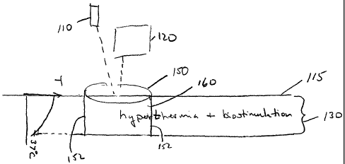
Figure 1 illustrates an exemplary embodiment of the invention in which a
volume of tissue 160 is heated such that biostimulation is applied to a
hyperthermic
volume of tissue, wherein volume of tissue 160 extends from the surface of
skin 115.
Volume of tissue 160 is defined by a depth region 130 and a skin surface area
150.
While the side 152 of volume of tissue 160 is illustrated as perpendicular to
the surface
of skin 115, it is to be understood that the area of treatment in Figure l, as
well as those
described below with reference to Figures 2-5, will typically increase with
depth below
the skin surface due to scattering of light by tissue. Additionally, while the
boundaries of
the volume of tissue 160 are illustrated with continuous lines, it is to be
understood that
the actual volume of treatment may be highly irregular, and regions of tissue
outside of
such bounds may receive both biostimulation and hyperthermia; however,
biostimulation and/or hyperthermia may be to a lesser degree than for tissue
in volume
of tissue 160.
Biostimulation may be achieved using radiation from a suitable
photobiostimulative source 110 as described above. For example,, source 110
delivers
radiation to the skin surface 115 with a flux in the range of about 1-250
mW/cm2, and
preferably in the range of about 10-100mW/cm2. Depth region 130 over which
biostimulation is achieved is determined by the flux, the wavelength of light
from source
110, and the size of area 150. For example, irradiation with radiation having
a
wavelength of 380-1250 nm at a flux 1-250 mW/cm2 will achieve biostimulation
to a
depth up to 10 mm for a beam having a diameter of greater than lcm. While area
150 is
illustrated as circular, it is to be understood that area 150 (and the other
skin surface
areas described below with reference to Figures 2-5) may be oval, square,
rectangular,
hexagonal or have any other suitable shape. Source 110 may be operated in
contact with
surface of skin 115 or project radiation onto surface of skin 115 from a
distance.
Hyperthermia, an increased temperature, in volume of skin 160 may be achieved
by any known source 120 capable of raising the temperature of volume 160 to a
value
within a range of about 37-50°C and preferably about 37-45°C.
Normal body
temperature can range from 36.1°C to 37.2°C depending on the
time of day. In some
embodiments, the temperature of the target area can be increased to be within
a range of
about 37-42°C. In some embodiments, the temperature of the target area
is increased to
be within a range of about 38-42°C. In other embodiments, the
temperature of the target
CA 02500961 2005-04-O1
WO 2004/033040 PCT/US2003/031774
-20-
region is increased to be within the range of about 38-41°C. In other
embodiments, the
temperature of the target region can be increased to about 38°C. In yet
other
embodiments, the temperature of the target region can be increased to about
39°C. In
yet another embodiment, the temperature of the target region can be increased
to about
40°C. For example, hyperthermia may be achieved by projecting hot air
onto area 150,
applying AC or DC electrical current, or using a conductive heat source (i.e.,
a device,
such as a heated plate or heating pad, in contact with surface 115). Further
examples of
heating a tissue include using ultrasound and microwave radiation, as
described in U.S.
Pat. No. 5,230,334, and U.S. Pat. No. 4,776,086, respectively, herein
incorporated by
reference. If contact heating is desired, the heating source may be
transparent to the
biostimulative radiation such that the biostimulation can be provided to
tissue through
the heating source. Heating can be applied before, during or between
photobiostimulation treatment sessions.
Optionally, source 120 may be a radiative source capable of achieving
hyperthermia. Hyperthermia achieved using radiation is also refe~Ted to as
photohyperthermia. A radiative source 120 may be any suitable radiative source
that
does not interfere with achieving biostimulation. To achieve hyperthermia,
heating can
be obtained using a broadband source or a narrowband source selected to
achieve a
desired temperature of tissue. Hyperthermia may be achieved using any suitable
wavelength or wavelengths of electromagnetic radiation; for example, the
radiation may
be in the wavelength range 380-2700 nm; or preferably in the range 500-1250
nm, and
more preferably in the ranges 650-900 nm and/or 1000-1250 nm. For example, the
sources included in Figure 6 may be combined in a weighted manner to provide a
suitable temperature profile. A radiative source 120 may be operated in
contact with
surface of skin 115 or project radiation onto surface of skin 115 from a
distance.
It is believed that a radiative source 120 will not interfere with achieving
biostimulation if the spectral density of the combined output of
biostimulative source
110 and source 120 is predominated by wavelengths that effect biostimulation.
For
example, the spectral density of the wavelengths in the band that effects
biostimulation
is 100 times greater than the spectral density of light in any other band, and
preferably
greater than 1,000 times. The phrase "spectral density" is herein defined to
refer to the
CA 02500961 2005-04-O1
WO 2004/033040 PCT/US2003/031774
-21 -
number photons in a specified bandwidth (e.g., the bandwidth at which
biostimulation is
achieved).
Biostimulation according to aspects of the invention may be achieved using
sources applied in a conventional small area of irradiation (e.g., a round
area having a
spot size less than 10 mm2 in diameter), or a larger area (e.g., a round area
having a spot
size 1 cm2-200 cm2 or more up to and including the entire human body).
Similarly,
photohyperthermia according to aspects of the invention may be achieved using
sources
applied using a conventional small area (e.g., a round area having a spot size
less than 10
mm in diameter), or a larger area (e.g., a round area having a spot size 1 cm2-
200 cm2 or
more). Large areas offer advantages, including but not limited to, reduced
treatment
time. For example, large areas may be used to treat large areas of tissue such
as a face,
neck, back or thigh. Methods of achieving a large area of irradiation are
described in
greater detail with reference to Figures 8-11 and 13 below.
The present invention recognizes that boundary effects diminish as the volume
to
be irradiated increases. As the volume of the target region increases, the
probability that
the scattered radiation will remain within the irradiated volume also
increases.
Therefore, radiation can penetrate the target tissue to a greater depth when a
larger beam
of irradiation andlor a larger target area is used. Accordingly, in some
embodiments,
where treatment is to be affected to a significant depth in the tissue, a
large area of
illumination is used to effect the treatment. In contrast, conventional
biostimulation
apparatuses have used narrow incident beams, which are strongly attenuated
such that
the photons comprising the beam do not reach deeply into the dermis and
subcutaneous
tissue (and/or into muscles and bones) in high enough concentration to achieve
the
desired biostimulation. Additionally, in a conventional biostimulation
apparatus, since
only small areas are treated at a given time, the beneficial effect arising
fiom the
treatment of large areas of tissue are nonexistent. In some embodiments,
photobiostimulative radiation is directed onto the skin surface using an area
of
illumination greater than approximately 0.8 cm' (e.g., a circular spot size
greater than 1
cm2) and preferably greater than 1.6 cm2 to provide biostimulation to tissue
at relatively
large depths below the skin surface, and to achieve time efficiencies
resulting from
treating a large area at one time. In one aspect, the present invention
provides devices
capable of providing such treatment.
CA 02500961 2005-04-O1
WO 2004/033040 PCT/US2003/031774
-22-
Figure 2 illustrates another embodiment of the invention in which a volume of
tissue 260 is heated such that biostimulation is applied to a hyperthermic
volume of
tissue 260, wherein volume of tissue 260 is adjacent to the surface of skin
115, and a
volume of tissue 270 receiving biostimulation (without hyperthermia) is
located below
volume 260. Volume of tissue 260 is defined by a depth region 230 and an area
250.
According to this aspect of the invention, the same light source 210 is used
to produce
both hyperthermia and biostimulation of volume of tissue 260. Light source 210
also
produces biostimulation in volume 270 in a depth 240.
An additional advantage of embodiments according to this aspect of the
invention is that the depth of the biostimulation zone is effectively
increased by
increasing the flux of source 210 relative to the flux provided in Figure 1.
For example,
an increase of flux incident on skin surface 115 from 100 mW/cm2 to 200 mW/cm2
is
sufficient to induce pronounced hyperthermia, and will also increase effective
biostimulation depth by up to 30% (i.e., an increase of the total
biostimulation depth
including depth regions 230 and 240 when compared to depth region 130 in
Figure 1).
Hyperthermia and biostimulation are achieved in volume of tissue 260 by
directing electromagnetic radiation from a narrowband source 210 onto an area
250.
The wavelength of source 210 is selected to achieve a desired
photobiostimulative result,
and flux of source 210 is chosen to achieve a selected temperature profile as
indicated by
Figures 6 and 7. Biostimulation in volume 270 (defined by depth region 240 and
area
250) is achieved where the intensity of light is sufficient to achieve
biostimulation, but
not sufficient to achieve a hyperthermic temperature (i.e., the temperature is
less than
38°C) as indicated in Figure 2. It is to be appreciated that the effect
of biostimulation is
weaker in depth region 230 than in depth region 240 due to the absence of
hyperthermia
in depth region 240.
Biostimulation and photohyperthermia according to the second aspect of the
invention, nnay be achieved using a conventional small area of irradiation
(e.g., a round
area having a spot size less than 10 mm in diameter), or a larger area (e.g.,
a round area
having a spot size larger than 1 cm2, up 200 cm2 or more). Generally, the
larger the
area, the deeper depth regions 230 and 240 extend below surface 115 due to a
reduction
in the effect of scattering. For example, irradiation with a wavelength of 600-
1250 nm
at a flux 0.1-1.0 W/cmz, and a spot size 1-200 cm after 80 seconds of exposure
will
CA 02500961 2005-04-O1
WO 2004/033040 PCT/US2003/031774
-23-
achieve heating and biostimulation to a depth up to 30 mm and biostimulation
(without
hyperthermia) from 30 mm - 50 mm.
Figures 6 and 7 present graphical data for achieving a selected temperature
profile using exemplary wavelengths of monochromatic light without skin
cooling
v
(Figure 6) and with parallel skin cooling (Figure 7). Specifically, the
numbered entries
in Tables 2 and 3 describe the flux at the skin surface and the time necessary
to achieve
a correspondingly-numbered steady-state temperature profile in Figures 6 and
7,
respectively. It is to be understood that the wavelengths in Figures 6 and 7
are
exemplary and light of any suitable wavelength may be used to achieve
hyperthermia.
Exemplary profile 7, in Figure 6, illustrates hyperthermia in a volume of
tissue (e.g.,
volume of tissue 260) which extends from the surface of skin (illustrated as
skin depth 0
in Figure 6). Sources coiTesponding to exemplary profiles 1-6 and 8-10 may
also be
used to achieve hyperthermia in a volume of tissue (e.g., volume of tissue
260) which
extends from the surface of skin by suitably increasing the power of source to
achieve a
greater flux.
Table 2. Flux and minimum exposure time to heat body up to +42°C
without active
cooling.
N Wavelength, Flux, Wlcmz Heating time,
nm s
1 800 0.683 209
2 925 0.573 193
3 960 0.466 206
4 1060 0.535 187
5 1208 0.383 189
6 1240 0.377 199
7 1440 0.491 208
8 1540 0.354 219
9 1730 0.359 212
10 2200 0.425 214
CA 02500961 2005-04-O1
WO 2004/033040 PCT/US2003/031774
-24-
Table 3. Flux and minimum exposure time to heat skin up to +42°C with
active cooling
of skin surface at the temperature +36°C.
N Wavelength, Flux, W/cmZ Heating time,
nm s
1 800 1.76 41
2 925 1.135 36
3 960 1.085 47
4 1060 0.967 35
1208 0.643 37
6 1240 0.685 41
7 1440 3.39 170
8 1540 1.21 132
9 1730 0.996 124
2200 2.335 170
5 Figure 12A illustrates the temperature at the skin surface as a function of
time of
exposure to a 800 nm radiation at a flux of 680 mW/cm2, wherein the beam has a
diameter larger than 2.5 cm. The data illustrated in Figure 12A was calculated
using a
computer model including the following assumption: a 3 mm skin thiclaiess, a 5
mm
subcutaneous fat thickness, muscle extending below the subcutaneous fat, and a
body
10 temperature of 37°C. Figure 12B illustrates temperature profiles
corresponding to an .
embodiment of Figure 2 in which the skin surface is cooled and kept to
36°C. The
temperature profiles of Figure 12B correspond to the data of Table 3. The data
illustrated in Figure 12B were calculated using a computer model including the
following assumption: a 3 mm skin thickness, a 5 mm subcutaneous fat
thickness,
muscle extending below the subcutaneous fat, and a body temperature of
36°C.
Figure 3 illustrates a third aspect of the invention to generate
photobiostimulation
in a volume of tissue 360 in a depth region 330 below the surface of skin 115
and
cooling is applied to the surface of skin 115. Photobiostimulation may be
suppressed or
reduced in efficacy in volume of tissue 380 in a depth region 320 by cooling
surface of
skin 115. Volume of tissue 360 is defined by depth region 330, and an area
350.
Hyperthermia does not occur in any portion of volume of tissue 360.
CA 02500961 2005-04-O1
WO 2004/033040 PCT/US2003/031774
-25-
To achieve photobiostimulation (without hyperthermia) in volume 360 with
suppressed biostimulation or biostimulation of reduced efficacy in volume 3
80, a source
310 projects radiation in a 1-10,000 mW/cm2 range and cooler 312 applies
cooling at the
skin surface to decrease temperature in a volume 380 defined by area 350 and
depth
region 320 to a hypothermic temperature (i.e., a temperature below normal body
temperature). Cooler 312 can be any suitable cooler, for example a fan, flow
of cold
(below 36 °C) fluid (i.e., liquid or gas), cryogenic spray, vaporizing
cream, cold plate or
window in contact with skin, or other contact or non-contact cooler.
The temperature of the target region may be reduced to approximately 0-36
°G,
or about 10-36 °C, or about 15-36 °C, or about 20-36 °C,
or about 28-36 °C.
Hypothermia may be used to protect the skin from damage caused by heat
generated by
irradiation. Additionally, by reducing the temperatures, the efficacy of
biostimulation
may be reduced or biostimulation may be suppressed. A reduction in efficacy
may be
due to a variety of factors, including reduced mirocirculation of blood, and
slowing
down of relevant biochemical reactions with lower temperature. Cooling of the
target
region can slow down metabolic and physiological processes and reduce the
oxygen
need of cells, particularly neurons. Care must be taken to prevent frostbite,
which can
occur at temperatures below 0 °C. In addition, the total body
temperature (i.e., rectal
temperature) should not be reduced below about 28 °C, the point at
which the ability to
regain normal temperature is lost. In some embodiments, temperatures below 0
°C can
be used on a small target area for short time periods.
In some aspects, hypothermia may result in increased biostimulation. Reducing
temperature leads to the generation of specific cold shock proteins, phase
transfer in
lipid structure of cell membrane or fat cells. These changes to the target
region can
increase the efficacy of biostimulation for the treatment of specific diseases
or cosmetic
conditions.
For example, to achieve biostimulation without hyperthermia, irradiation with
a
wavelength of 500-1200 nm at a flux 1-1,000 mW/cm2 and beam area of 0.8 cmz
(e.g., a
round area yielding a spot size at the target area of greater than 1 cm2), for
a time
interval greater than 60 seconds will achieve biostimulation to a depth of f5
mm. If the
skin surface 115 is kept at 0-32~°C, hypothermia will exist in a volume
380 above
CA 02500961 2005-04-O1
WO 2004/033040 PCT/US2003/031774
-26-
treatment region 360 resulting in reduced or suppressed biostimulation in this
volume.
In some embodiments, hypothermia can increase biostimulation.
Figure 4 illustrates another aspect of the invention in which a volume of
tissue
460 is heated such that biostimulation is applied to a hyperthermic volume of
tissue 460,
wherein volume of tissue 460 is at a selected depth below the surface of the
skin 115,
and volumes (without hyperthermia) 465, 470 are located above and below volume
460,
respectively. Hyperthermia is suppressed in volume 465 by a cooler 412 and
volume
470 is not heated sufficiently to achieve hyperthermia. Volume of tissue 460
is defined
by depth region 430, and an area 450.
To achieve photobiostimulation and hyperthermia in volume 460, a source 410
projects radiation in a 100-10,000 mW/cmz range and cooler 412 applies cooling
at the
skin surface (0-30 °C) to suppress hyperthermia at surface 115.
Treatments, such as the
treatment of Figure 4, may be achieved using a biostimulative source applied
using a
relatively large area of illumination (e.g., a round area having a spot size
with a diameter
larger than 1 cm-200 cm or more). Heating a volume of tissue wherein the
volume is a
selected depth below the surface of the skin is described in U.S. Provisional
Application
60/389,871, filed June 19, 2002, entitled "Method and Apparatus for
Photothermal
Treatment of Tissue at a Depth," the substance of which is incorporated by
reference
herein.
For example, to achieve photobiostimulation and hyperthermia according to the
present aspect of the invention, irradiation with a wavelength of 500-1250 nm
at a flux
100-10,000 mW/cm2 and a area of irradiation of 0.8 cm2 after 60 seconds of
exposure
will achieve biostimulation in a range of depths 0-50 mm below the skin
surface, and if
the skin surface is kept at 0-30 °C hyperthermia will be achieved in a
range of depths
0.2-30 mm below the skin surface. Treatments according to this aspect of the
invention
may be achieved using a relatively large area (e.g., a round area having a
spot size
diameter 1 cm-200 cm or more).
Figure 5 illustrates another aspect of the invention in which a volume of
tissue
560 is heated by source 510 such that enhanced biostimulation occurs in this
hyperthermic volume of tissue, volume 560 being located a selected depth below
the
surface of the slcin 115. The skin surface 550 can be cooled by the cooling
source 512
either simultaneously or sequentially to the heating. Biostimulation (without
CA 02500961 2005-04-O1
WO 2004/033040 PCT/US2003/031774
_27_
hyperthermia) occurs in a volume 540 located below volume 560. A volume of
tissue
560 is defined by depth region 530, and an area 550.
As described above with reference to Figure 4, the efficacy of biostimulation
is
suppressed in a volume 520 adjacent to skin surface. However, according to
this aspect
of the invention, hyperthermia occurs only in volume 560.
For example, to achieve photobiostimulation and hyperthermia according this
aspect of the invention, irradiation with a wavelength of 500-1250 nm at a
flux 100-
10,000W/cm2 and an area of irradiation greater than 0.8 cm2 after 60 seconds
of
exposure will achieve biostimulation in a range of depths 0.1-50 mm below the
skin
surface, and if the skin surface is kept at 0-30 °C, hypertheunia will
be achieved in a
range of depths 0.2-30 mm below the skin surface. Treatments according to this
aspect
of the invention may be achieved using a relatively large area (e.g., a round
area having
a spot size 1 cm-200 cm or more).
Figure 7 depicts graphical data and corresponding tabular data, for achieving
a
selected temperature profile using exemplary wavelengths of monochromatic
light, in
which the skin surface is cooled to a temperature of 10 °C and
photobiostimulation is
suppressed in a region of tissue adjacent the skin surface. Specifically, the
numbered
entries in Table 3 describe the flux at the skin surface and the time
necessary to achieve
a correspondingly-numbered steady-state temperature profile in Figure 7. It is
to be
understood that the wavelengths in Figures 6 and 7 are exemplary and light of
any
suitable wavelength may be used to achieve hyperthermia, and biostimulation.
Although the above discussion describes static (i.e., non-moving) radiation
sources, the desired combination of photobiostimulation and photohyperthermia
can be
achieved by moving an output head of a radiation source across the surface of
the skin
so as to achieve the desired tissue temperature and/or deliver the desired
amount of light
to achieve biostimulation. The head may be moved over each skin surface area a
single
time or multiple times as required to achieve the desired therapeutic effect.
Moving a
source across the surface of the skin can be used to achieve hyperthermia in a
volume of
tissue due to the relatively long thermal relaxation time of bulls tissue.
Further details
regarding moving sources and heating oftissue is given in U.S. Pat. No.
6,273,884 B1 ,
entitled "Method and Apparatus for Dermatology Treatment," to Altshuler et
al., issued
August 14, 2001, the substance of which is hereby incorporated by reference.
CA 02500961 2005-04-O1
WO 2004/033040 PCT/US2003/031774
_28_
Photobiostimulation can be achieved by moving the source output head across
the skin
at a rate and/or for a number of iterations such that the desired number of
photons are
delivered to the treatment volume of tissue.
The above aspects of the invention are directed to applying biostimulation to
a
hyperthermic and/ or a hypothermic volume of tissue. For these aspects, the
heating
source and biostimulative radiation source may be applied simultaneously, and
for some
embodiments may be the same source, or the heating source may be discontinued
during
application of the biostimulative radiation, or the heating source may be
applied in a
reduced amount to maintain the hyperthermic condition.
Figure 8 is a schematic diagram of a light projection system 800 appropriate
for
use with aspects of the present invention according to Figure 2 above. Light
projection
system 800 is composed of a radiation source 802 and a lens system 820. The
radiation
source may be any suitable narrowband source for generating hyperthermia and
biostimulation according to an embodiment of the invention described above
with
reference to Figure 2. For example, the source may be a laser (e.g., a
continuous-wave
diode laser, emitting at 805 nm with output power of 90 W) or an array of
lasers, an
LED (or an array ~of LEDs) or a lamp. The radiation from source 802 may be
coupled to
an optical fiber 803 (e.g., a 1 mm core quartz-polymer fiber) or a suitable
fiber bundle,
which is coupled on its proximal end to light source 802.
Lens system 820 may be any suitable lens system for transmitting light from
source 802 to a patient's skin surface with a flux and beam size as described
above with
reference to Figure 2. In one embodiment, lens system 820 includes a negative
lens 806,
and a positive lens 808 that forms a collimated output beam 810. In one
embodiment of
system 800, lens 806 is a refractive lens, and lens 808 is a Fresnel lens. A
Fresnel lens
' may provide safety effects (e.g., a more uniform illumination pattern due to
a reduction
of speckle). As an example of this embodiment, lens 806 is a negative lens
having a
focal length of 25 mm and a diameter of 25 mm, and lens 808 is a 152 mm
diameter
Fresnel lens with a focal length 152 mm; and the distance between radiation
source 802
and lens 806 is 20 mm, and the distance between the lenses 806 and 808 is 105
mm.
According to some aspects of the invention, output beams having larger
diameters are used to direct narrowband light (e.g., laser or monochromatic
filtered
light) more deeply into the dermis and subcutaneous tissue than conventional
low power
CA 02500961 2005-04-O1
WO 2004/033040 PCT/US2003/031774
-29-
laser sources emitting small beam sizes. For example, according to the above
exemplary
embodiment of lens system 820, for a 90W source, lens system 820 produces an
output
beam 810 having a diameter of 160 mm, and has an output flux of 200 mW/cm2 to
2000
mW/cm2 (at a distance of 23 cm from lens 808).
Figure 13A is an exemplary embodiment of a light projection system 1300
according to aspects of the present invention, enabling one to practice the
invention
according to the scenarios illustrated in Figures 1 and 3, 4, and 5. For
example,
projection system 1300 may be any system that provides an output beam having
suitable
diameter and flux at skin surface 1350. In one embodiment, projection system
1300
includes an optical source 1302, and optical elements 1304, 1306, 1312, 1314,
and 1308.
One exemplary set of lens parameters is given in Figure 13B.
Optical elements 1306 and 1314 may be movable along optical axis 1301 such
that output beam 1310 has a variable diameter. For example, lenses 1306 and
1314 may
be connected to a rigid fi~ame 1316 (e.g., a translation stage), allowing
synchronous
movement of the lenses 1306 and 1314 along optical axis 1301 of the system
1300.
Such movement provides variation in the beam width of the output beam 1310
(e.g., spot
size is changed) and provides a corresponding variation in flux on skin
surface 1350.
For example, the system 1300 can provide continuous variations of a spot size
between 4
cm and 8 cm, with the flux varying through a corresponding range of 7 W/cm2 to
2
W/cm2 (assuming source 1302 is a 90 W source). It is to be appreciated that by
suitable
selection of elements and source 1302, lens system 1300 may be designed to
achieve any
output beam 1310 as described herein, and any suitable output density as
described
herein.
System 1300 includes at least one air tube 1318, connected on its proximal
ends
to a cold or hot air source (not shown) and providing, at its distal end, an
airflow 1320
directed at patient's skin 1350. For example, a total air flow from the at
least one air
tube 1318 may be at least 50 m3/min to vary air temperature in accordance with
the
embodiments illustrated in Figures 3 -5 (e.g., the temperature will be between
0 °C and
45 °C at skin surface 1350); and in accordance with Figure l, a hot air
flow will be
provided to skin surface 1350. By varying the beam diameter and the air
temperature,
all regimens of Figures 1, 3, 4, and 5 can be realized using the system of
Figure 13A.
CA 02500961 2005-04-O1
WO 2004/033040 PCT/US2003/031774
-30-
While Figures 8 and 13A were described by specifying beam diameters, it is to
be
appreciated that by appropriate aperturing, any shape beam may be achieved.
Figure 9A is a first exemplary embodiment of a light projection system 900 for
forming substantially uniform illumination over a non-flat surface 950, such
as a
patient's head or thigh. A collimated beam from a source 902 is directed onto
a beam
sputter 904 to form a plurality of beam portions 905a-c. In the illustrated
embodiment,
beam sputter 904 forms three component beam portions 905a 905b,and 905c
however a
light projection system 900 having two or more beam portions may provide
advantages.
Beam portion 905b is directed directly on the surface 950, and beam portions
905a and
905c are directed onto miiTOrs 910a and 910b, respectively, and then
redirected to the
sides of the surface 950. The clear apertures of beam sputter 904, mirrors
910a, 910b or
additional apertures can be selected to achieve any desired area of
irradiation on surface
950 (e.g., 1-200 cm2). Light projection system 900 may be modified (e.g., to
treat one
side of a patient's face) by blocking one of beams 910a and 910b.
Figure 9B is a schematic of one example of a beam sputter 904. Beam sputter
904 is a prism having two flat surfaces 912a, 912b appropriately angled to
direct light
onto mirrors 910a, b, and a surface 913 having a negative power to expand
light onto the
front portion of surface 950.
Figure 10 is a schematic of a second exemplary embodiment of a light
projection
system 1000 for forming substantially uniform illumination over a non-flat
surface 950.
Light projection system 1000 has a head 1002 adapted to project light in two
directions.
A first portion of light 1006 is directed in a first direction onto a curved
reflector 1004
and then onto surface 950, and a second portion 1008 is directed in a second
direction
onto a surface 950. First portion of light 1006 is projected onto reflector
1004 directly
or through an optical element (lens 1005), and second portion 1008 projected
directly
onto surface 950 or through an optical element (e.g., lens 1009).
Reflector 1004 may have any suitable shape for achieving a selected treatment.
In some embodiments, reflector 1004 is designed such that center 1010 of
surface 950
(e.g., the center of a patient's head) is located substantially at the center
of curvature of
reflector 1004. Alternatively, reflector 1004 may have an elliptical curvature
and center
1010 of surface 950 (e.g., the center of a patient's head) is located
substantially at a
CA 02500961 2005-04-O1
WO 2004/033040 PCT/US2003/031774
-31-
focus of reflector 1004 and the center 1010 of surface 950 is located at a
second focus of
reflector 1004. In one embodiment, reflector 1004 can be a diffuse reflector.
Projection system 1000 may include a control module 1016 comprising an
electrical power source and control electronics. Additionally, a light source
(not shown)
may be mounted in head 1002; alternatively, a light source may be mounted in
module
1016 and delivered to head 1002 by an optical fiber or a bundle of fibers.
Light sources
can be narrow band (e.g., diode lasers, LEDs), or broadband (e.g., filtered
lamp).
Alternatively, light sources may be a combination of narrow band and broadband
sources. Optionally, in accordance with the embodiments described above, cold
or hot
air can be directed on the surface fi~om head 1002 onto surface 950.
Figures 1 lA, 11B, and 11C are schematics of a third example of an embodiment
of a light projection system 1100 for forming substantially uniform
illumination over a
non-flat surface 950 in which a rotatable head 1102 reflects light from a
surface 1110
onto surface 950. In Figure 11A, rotatable head 1102 is positioned such that
light is
directed onto the front portion of surface 950. In Figure 11B, rotatable head
1102 is
positioned such that light is directed onto a first side portion of surface
950. In Figure
11C, rotatable head 1102 is positioned such that light is directed onto a
second side
portion of surface 950.
Optionally, head 1102 may be omitted, and replaced with a source mounted on
surface 1110 such that the source is moved to various positions on surface
1110 to direct
light onto each of the portions indicated in Figures 1 lA-11C. Alternatively,
a plurality
of sources can be mounted on surface 1110 and selectively illuminated to
direct light
onto each of the portions.
In another aspect, the present invention provides a feedback mechanism for
controlling the temperature of a target region within a selected range while
causing
biostimulation within that target region andlor a volume above, below, or
adjacent to the
target region. The feedback mechanism can be used to control both heating and
cooling
of the target region. With reference to Figure 14, in an exemplary embodiment,
the
source of electromagnetic radiation 1410 generates radiation for illuminating
a portion
of the surface area of the patient' s skin 1450 so as to irradiate a volume of
the patient's
tissue 1460 that extends from the surface of the skin 1415 to a given depth
1430 below
the skin. The radiation includes one or more wavelength components that can
cause
CA 02500961 2005-04-O1
WO 2004/033040 PCT/US2003/031774
- 32 -
biostimulation of the irradiated tissue volume 1460. Another source 1420, for
example,
a separate source of electromagnetic radiation, controls the temperature of
the irradiated
volume, e.g., by illuminating the skin surface area 1450 with radiation having
wavelength components suitable for heating tissue. A sensor 1470, for example,
an
optical pyrometer, measures the temperature of the illuminated skin portion
1450, and
transmits the measured temperature to a feedback control circuitry 1480. The
feedback
circuitry 1480 compares the measured temperature with at least one threshold
temperature, and transmits feedback signals, if needed, to the source 1420
based on this
comparison. For example, if the measured temperature exceeds a pre-defined
upper
threshold, such as when the portion of the surface area of the patient's skin
1450 is
heated to cause hyperthermia, the feedback circuitry can transmit a signal to
the source
1420 to lower the amount of heat delivered to the skin portion 1450.
Alternatively, the
feedback circuitry can instruct the source 1420 to increase the amount of heat
delivered
to the skin portion 1450 if the measured temperature falls below a pre-defined
lower
threshold. In this manner, the temperature of the illuminated skin portion
1450, and
consequently that of the target region 1460, can be actively maintained within
a selected
range about an operating temperature. For example, the above feedback
mechanism can
ensure that the operating temperature remains within ~ 1 °C of 39
°C. A variety of
sensors and feedback circuitry suitable for use in the practice of the
invention are known
in the art.
Those skilled in the art will appreciate, or be able to ascertain using no
more than
routine experimentation, further features and advantages of the invention
based on the
above-described embodiments. Accordingly, the invention is not to be limited
by what
has been particularly shown and described, except as indicated by the appended
claims.
The contents of all references, patents and published patent applications
cited throughout
this application, are incorporated herein by reference.