Note: Descriptions are shown in the official language in which they were submitted.
CA 02501233 2005-04-05
WO 2004/033647 PCT/US2003/031822
TITLE OF THE INVENTION
ASSAY METHODS FOR STATE-DEPENDENT CALCIUM CHANNEL
AGONISTS/ANTAGONISTS
CROSS-REFERENCE TO RELATED APPLICATIONS
This application claims the benefit of U.S. Provisional Application No.
60/418,017, filed October 10, 2003, the contents of which are incorporated
herein by reference in
their entirety.
FIELD OF THE INVENTION
The present invention is directed to methods and cells for studying the effect
of
candidate compounds on the activity of calcium channels. The methods utilize
cells that express
a calcium channel of interest and which express a potassium channel. The
engineered cells
allow for fine control of the membrane potential of the cells, which, in turn,
provide a high
resolution assay for studying the effects of targeted compounds at various
states of the calcium
channel.
BACKGROUND OF THE INVENTION
Certain molecular events in eukaryotic cells depend on the existence or
magnitude
of an electric potential gradient across the plasma (i.e., outer) membrane of
the cells. Among the
more important of such events is the movement of ions across the plasma
membrane through
voltage-gated ion channels. Voltage-gated ion channels form transmembrane
pores that open in
response to changes in cell membrane potential and allow ions to pass through
the membrane.
Voltage-gated ion channels have many physiological roles. They have been shown
to be
involved in maintaining cell membrane potentials and controlling the
repolarization of action
potentials in many types of cells (Bennett et al., 1993, Cardiovascular Drugs
& Therapy 7:195-
202; Johnson et al., 1999, J. Gen. Physiol. 113:565-580; Bennett & Shin,
"Biophysics of voltage-
gated sodium channels," in Cardiac Electro~hysiolog_y: From Cell to Bedside,
3rd edition, D.
Zipes & J. Jalife, eds., 2000, W.B. Saunders Co., pp.67-86; Bennett & Johnson,
"Molecular
physiology of cardiac ion channels," Chapter 2 in Basic Cardiac
Electrophysiology and
Pharmacolo~y, 15' edition, A. Zasa & M. Rosen, eds., 2000, Harwood Academic
Press, pp. 29-
57). Moreover, mutations in sodium, calcium, or potassium voltage-gated ion
channel genes
leading to defective channel proteins have been implicated in a variety of
disorders including the
congenital long QT syndromes, ataxia, migraine, muscle paralysis, deafness,
seizures, and
cardiac conduction diseases, to name a few (Bennett et al., 1995, Nature
376:683-685; Roden et
-1-
CA 02501233 2005-04-05
WO 2004/033647 PCT/US2003/031822
al., 1995, J. Cardiovasc. Electrophysiol. 6:1023-1031; Kors et al., 1999,
Curr. Opin. Neurol.
12:249-254; Lehmann et al., 1999, Physiol. Rev. 79:1317-1372; Holbauer &
Heufelder, 1997,
Eur. J. Endocrinol. 136:588-589; Naccarelli & Antzelevitch, 2000, Am. J. Med.
110:573-581).
Several types of voltage-gated ion channels exist. Voltage-gated potassium
channels establish the resting membrane potential and modulate the frequency
and duration of
action potentials in neurons, muscle cells, and secretory cells. Following
depolarization of the
membrane potential, voltage-gated potassium channels open, allowing potassium
efflux and thus
membrane repolarization. This behavior has made voltage-gated potassium
channels important
targets for drug discovery in connection with a variety of diseases.
Dysfunctional voltage-gated
potassium channels have been implicated in a number of diseases and disorders.
Wang et al.,
1998, Science 282:1890-1893 have shown that the voltage-gated potassium
channels KCNQ2
and KCNQ3 form a heteromeric potassium ion channel known as the "M-channel."
Mutations in
KCNQ2 and KCNQ3 in the M-channel are responsible for causing epilepsy
(Biervert et al.,
1998, Science 279:403-406; Singh et al., 1998, Nature Genet. 18:25-29;
Schroeder et al., Nature
1998, 396:687-690).
Voltage-gated sodium channels are transmembrane proteins that are essential
for
the generation of action potentials in excitable cells (Catterall, 1993,
Trends Neurosci. 16:500-
506). In mammals, voltage-gated sodium channels consist of a macromolecular
assembly of a
and (3 subunits with the a subunit being the pore-forming component. a
subunits are encoded by
a large family of related genes, with some a subunits being present in the
central nervous system
(Noda et al., 1986, Nature 322:826-828; Auld et al., 1988, Neuron 1:449-461;
Kayano et al.,
1988, FEBS Lett. 228:187-194) and others in muscle (Rogart et al., 1989, Proc.
Natl. Acad. Sci.
USA 86:8170-8174; Trimmer et al., 1989, Neuron 3:33-49).
Voltage-gated calcium channels are transmembrane proteins that in the open
configuration allow the passive flux of Ca2+ ions across the plasma membrane,
down the
electrochemical gradient. They mediate various cell functions, including
excitation-contraction
coupling, signal transduction, and neurotransmitter release. Three major
classes of calcium
channel antagonists including the dihydropyridines, benzothiazepines and
phenylalkylamines
have been widely used clinically in the treatment of cardiovascular diseases.
These drugs
antagonize the L-type calcium channels found throughout the body, including
the cardiovascular
system. Calcium channels are allosteric proteins that undergo changes in
conformational state.
The distinct conformational states of these proteins have different affinities
for ligands, including
these antagonists. Membrane potential is an allosteric effector of these
conformational changes
in ion channel proteins. The potency of inhibition by these calcium channel
antagonists is
dependent on the state of the calcium channel. Previously studies on state-
dependent
-2-
CA 02501233 2005-04-05
WO 2004/033647 PCT/US2003/031822
interactions of these antagonists were identified through voltage clamp (1),
radioligand binding
(2) and cell based, e.g. smooth muscle contraction (3) studies. While each of
these methods
yields valuable information each has its drawbacks in terms of information
content or
throughput, respectively.
Calcium channels are membrane-spanning, mufti-subunit proteins that allow
controlled entry of Ca+2 ions into cells from the extracellular fluid. Cells
throughout the animal
kingdom, and at least some bacterial, fungal and plant cells, possess one or
more types of
calcium channel.
The most common type of calcium channel is voltage dependent. Most
"excitable" cells in animals, such as neurons of the central nervous system
(CNS), peripheral
nerve cells and muscle cells, including those of skeletal muscles, cardiac
muscles, and venous
and arterial smooth muscles, have voltage-dependent calcium channels.
"Opening" of a voltage-
dependent channel to allow an influx of Ca+2 ions into the cells requires a
depolarization to a
certain level of the potential difference between the inside of the cell
bearing the channel and the
extracellular environment bathing the cell. The rate of influx of Ca+2 into
the cell depends on
this potential difference.
Multiple types of calcium channels have been identified in mammalian cells
from
various tissues, including skeletal muscle, cardiac muscle, lung, smooth
muscle and brain, [see,
e.g., Bean, B. P. (1989) Ann. Rev. Physiol. 51:367-384 and Hess, P. (1990)
Ann. Rev. Neurosci.
56:337]. The different types of calcium channels have been broadly categorized
into five
classes, L-, T-, N-, P/Q and R-type, distinguished by current kinetics,
holding potential
sensitivity and sensitivity to calcium channel agonists and antagonists.
Current methods of drug discovery often involve assessing the biological
activity
(i.e., screening) of tens or hundreds of thousands of compounds in order to
identify a small
number of those compounds having a desired activity. In many high throughput
screening
programs, it is desirable to test as many as 50,000 to 100,000 compounds per
day.
Unfortunately, current methods of assaying the activity of voltage-gated ion
channels are ill
suited to the needs of a high throughput screening program. Current methods
often rely on
electrophysiological techniques. Standard electrophysiological techniques
involve "patching" or
sealing against the cell membrane with a glass pipette followed by suction on
the glass pipette,
leading to rupture of the membrane patch (Hamill et al., 1981, Pflugers Arch.
391:85-100). This
has limitations and disadvantages. Accessing the cell interior may alter the
cell's response
properties. The high precision optical apparatuses necessary for
micromanipulating the cells and
the pipettes make simultaneous recording from more than a few cells at a time
impossible.
-3-
CA 02501233 2005-04-05
WO 2004/033647 PCT/US2003/031822
Given these difficulties, the throughput that can be achieved with
electrophysiological
techniques falls far short of that necessary for high throughput screening.
Various techniques have been developed as alternatives to standard methods of
electrophysiology. For example, radioactive flux assays have been used in
which cells are
exposed with a radioactive tracer (e.g., 86Rb+, 22Na+~ ~14C~_guanidinium and
45Ca) and the
flux of the radio-labled ion is monitored. Cells loaded with the tracer are
exposed to compounds
and those compounds that either enhance or diminish the efflux of the tracer
are identified as
possible activators or inhibitors of ion channels in the cells' membranes.
Assays that measure the change in a cell's membrane potential due to the
change
in activity of an ion channel have been developed. Such assays often employ
voltage sensitive
dyes that redistribute between the extracellular environment and the cell's
interior based upon a
change in membrane potential and that have a different fluorescence spectrum
depending on
whether they are inside or outside the cell. A related assay method uses a
pair of fluorescent
dyes capable of fluorescence resonance energy transfer to sense changes in
membrane potential.
For a description of this technique, see Gonzalez & Tsien, 1997, Chemistry &
Biology 4:269-
277. See also Gonzalez & Tsien, 1995, Biophys. J. 69:1272-1280 and U.S. Patent
No.
5,661,035. Other methods employ ion selective indicators such as calcium
dependent
fluorescent dyes to monitor changes in Ca2+ influx during opening and closing
of calcium
channels.
Ideally, methods of screening against voltage-gated ion channels require that
the
transmembrane potential of the cells being assayed be controlled and/or that
the ion channels
studied be cycled between open and closed states. This has been done in
various ways. In
standard electrophysiological techniques, the experimental set-up allows for
direct manipulation
of membrane potential by the voltage clamp method (Hodgkin & Huxley, 1952, J.
Physiol.
(Lond.) 153:449-544), e.g., changing the applied voltage. In other methods,
changing the
extracellular K+ concentration from a low value (e.g., 5 mM) to a higher value
(e.g., 70-80 mM)
results in a change in the electrochemical potential for K+ due to the change
in the relative
proportion of intracellular and extracellular potassium. This results in a
change in the
transmembrane electrical potential towards a more depolarized state. This
depolarization can
activate many voltage-gated ion channels, e.g., voltage-gated calcium, sodium,
or potassium
channels. Alternatively, Na+ channels can be induced into an open conformation
by the use of
toxins such as veratridine or scorpion venom (Strichartz et al., 1987, Ann.
Rev. Neurosci.
10:237-267; Narahashi & Harman, 1992, Meth. Enzymol. 207:620-643). While
sometimes
effective, such experimental manipulations may alter the channel pharmacology,
can be
awkward to perform, and can lead to artifactual disturbances in the system
being studied.
-4-
CA 02501233 2005-04-05
WO 2004/033647 PCT/US2003/031822
Electrical field stimulation (EFS) has been used to activate ion channels. In
this
approach, membrane potential is altered but not controlled. The uncertainty
and lack of control
of membrane potential make EFS a less than optimal method for the study of ion
channels.
HEK293 cells have been grown on a silicon chip made up of an array of field-
effect transistors. Some of the cells were positioned over the gate region of
the transistors, thus
having portions of their plasma membranes overlying the source and the drain.
When a patch
pipette in such cells manipulated the intracellular voltage, Maxi-K potassium
channels in the
cells' plasma membranes were opened. This led to current flow in the region
between the cells'
membrane and the transistor. This current flow modulated the source-drain
current, which could
be detected by an appropriate device. The chip plus cells was said to have
potential as a sensor
and as a prototype for neuroprosthetic devices. See Straub et al., 2001,
Nature Biotechnol.
19:121-124; Neher, 2001, Nature Biotechnol. 19:114.
SUMMARY OF THE INVENTION
The present invention is directed to methods of identifying activators and
inhibitors of voltage-gated ion channels, and specifically calcium ion
channels. The methods
employ cells transformed to express a voltage-gated calcium ion channel of
interest and an
inward rectifier potassium channel. The addition of the potassium channel
allows for the fine
control of the membrane potential of the cells. Manipulation of the
extracellular potassium
concentration controls the membrane potential which in turn affects the
open/close state
transitions of the voltage-gated ion channels. This allows for more
convenient, more precise
manipulation of these transitions, and, coupled with efficient methods of
detecting ion flux,
results in methods that are especially suitable for high throughput screening
in order to identify
substances that are channel state dependent modulators of voltage-gated ion
channels.
According to a specific embodiment, the present invention describes the state-
dependent interactions of the calcium channel antagonists directly in a
functional cell-based
FLIPR (Fluorometric Imaging Plate Reader) assay, which measures calcium influx
through a
voltage-dependent calcium channel (VDCC). The cell line used in this
embodiment has a stably
transfected L-type calcium channel, the al C subunit. It also was transfected
with the Kir 2.3
inward rectifier K channel, which allows for control of cell membrane
potential through
alteration of extracellular [K+]o. Preincubation of the cells for 10 min in 30
mM [K+]o partially
depolarizes the cells. The inhibitory effect of calcium channel antagonists on
calcium influx in
response to a high [K+]o depolarization (final [K+]o 85.8 mM) was shifted to
the left compared
with that observed for cells in normal, physiological [K+]o (5.8 mM). The
ratio of IC50 values
between the potencies for the antagonists tested in the normally polarized and
depolarized cells
-5-
CA 02501233 2005-04-05
WO 2004/033647 PCT/US2003/031822
was 4 to 20-fold. The results suggest that the interaction of these calcium
channel antagonists
with the channel expressing cells is dependent upon the state of the channel,
which is modulated
by changes in membrane potential. The state dependent assay demonstrated in
these studies is
useful for evaluating state dependent inhibitory potency of a large number of
samples and can be
used to identify state-dependent calcium channel antagonists.
BRIEF DESCRIPTION OF T)= ZIE DRAWINGS
Figure 1 shows immunostaining of alpha 1C subunit in the wild type HEK 293
cells and cells stably transfected with the L-type a1C channel (C1-6-37-3).
Figure 2 shows immunostaining of Kir2.3 subunit in the wild type HEK293 cells
and the cells stably transfected with the L-type a1C channel (C1-6-37-3).
Figure 3 is a graph showing the relationship between extracellular potassium
([K]o) and cell membrane potential. Three situations are shown. One is the
prediction of the
Nernst equation for a perfectly K-selective membrane. The other curves show
the effects of
partial permeability by other ions, Na+ and/or Cl-. Membrane potential can be
set in a non-
voltage clamped cell by adjusting external potassium.
A cell line expressing an inward rectifier K channel (Kir2.3) to set the
resting
membrane potential will permit control of membrane resting potential by
extracellular
potassium.
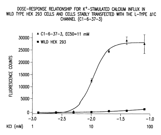
Figure 4 is a graph demonstrating the dose-response relationship for K+-
stimulated calcium influx in wild type HEK 293 cells and cells stably
transfected with the L-type
a1C channel (C1-6-37-3).
Figure 5 is a graph demonstrating a comparison of nimodipine and mibefradil
inhibition curves in K+-stimulated calcium influx in C1-6-37-3 cells under
resting condition (5.8
mM K =-65 mV).
Figure 6 is a graph representing the nimodipine inhibition curve stimulated by
K
(final 85.8 mM) either in 30 mM K+ (depolarized condition, -28 mV) or 5.8 mM
K+ (resting
condition, -65 mV).
Figure 7 is summary table of IC50 (nM) values for calcium channel antagonists
in 30 mM K+ (depolarized condition, -28 mV) and 5.8 mM K+ (resting condition, -
65 mV).
DETAILED DESCRIPTION OF THE INVENTION
Without intending to bound by any theory, voltage gated calcium channels open
as a function of membrane potential such that the probability of opening
increases with
membrane depolarization. Voltage gated calcium channels inactivate (close /
desensitize) as a
-6-
CA 02501233 2005-04-05
WO 2004/033647 PCT/US2003/031822
function of membrane potential such that the probability of inactivation
increases with
membrane depolarization. These steady state voltage dependent processes
overlap. Changes in
membrane potential populate different conformational states of these channels
(closed, open or
inactivated). Drug binding to voltage gated calcium channels is often channel
state dependent
such that more or less binding occurs depending upon the state occupied.
Control of membrane
potential, permits channels to be manipulated into various states. This
membrane potential
control is typically achieved by voltage clamp electrophysiology methods, but
this method is not
at present amenable to high throughput drug screening.
Specifically exemplified herein is an assay to determine state-dependent drug-
calcium channel interactions using a cell line that co-expresses a potassium
channel (Kir2.3) that
determines the resting membrane potential of the cells as a function of the
external potassium ion
concentration ([K]o) and a voltage gated calcium channel. Co-expressed in
these cells is the L-
type voltage gated calcium channel complex (alphalC, alpha2-delta, beta2a).
Potassium is used
in a two step manner in this assay. First it is used to set the resting
membrane potential (Vm)
during antagonist incubation. Two conditions were selected for illustration
purposes, polarized
and depolarized resting conditions. In the polarized resting condition, cells
are incubated in 5.8
mM [K]o to set the membrane potential to -65 mV (Vm as a function of [K]o).
Drugs exposed
to these cells will bind to calcium channels primarily in the closed, rested,
low affinity state. In
order to reveal higher affinity states of the calcium channels, the cells are
incubated in 30 mM
[K]o, in order to chronically and partially depolarize them to -28 mV during
drug exposure.
This change in the membrane potential, shifts the calcium channels into the
higher affinity
inactivated states and antagonist binding is enhanced. Upon establishing these
two different
conditions for drug exposure, channels are then forced to open by further
depolarization to near 0
mV by exposure to 85.8 mM [K]o. Opening of these channels normally under
control, non-
antagonist exposed conditions, allows calcium influx into the cells. This
calcium influx is
detected using a calcium sensitive dye (eg Fluo-3, Fluo-4, Fura2, etc.). If
the calcium influx is
diminished by exposure to antagonists, this will be detected when compared to
the control
condition. In some cases, antagonists will bind with greater affinity to the
channels in the
depolarized (30 mM [K]o) condition. In these cases, the same drug will appear
more potent
under these depolarized assay conditions. This approach creates a novel high
throughput
calcium channels assay system that is capable of detecting and measuring
calcium channel state
dependent drug interactions as have been described using low throughput
voltage clamp
measures on single cells.
This foregoing approach and the referenced cells have been tested using
conventional voltage- and current- clamp methods, and the membrane potential
changes as a
CA 02501233 2005-04-05
WO 2004/033647 PCT/US2003/031822
function [K]o and the state dependent calcium current and drug affinities have
been confirmed
experimentally. The foregoing approach can be modified as taught herein to
study state-
dependencies of agonists/antagonists for many different types of ion channels.
In one embodiment, the present invention involves providing a substrate upon
which living eukaryotic cells, preferably mammalian cells, are present where
the cells express
voltage-gated calcium ion channels in their plasma membranes. Upon application
of varying
concentrations of extracellular calcium, voltage-gated ion channels either
open or close, thereby
modulating the flow of at least one type of ion through the plasma membranes
of the cells. This
modulation of ion flow, or a change in membrane potential that results from
the modulation of
ion flow, is detected, either directly or indirectly, preferably by the use of
fluorescent indicator
compounds in the cells. Collections of substances, e.g., combinatorial
libraries of small organic
molecules, natural products, phage display peptide libraries, etc., are
brought into contact with
the voltage-gated ion channels in the plasma membranes of the cells and those
substances that
are able to affect the modulation of ion flow are identified. In this way, the
present invention
provides methods of screening for activators and inhibitors of voltage-gated
ion channels,
particularly calcium channels. Such activators and inhibitors are expected to
be useful as
pharmaceuticals or as lead compounds from which pharmaceuticals can be
developed by the
usual processes of drug development, e.g., medicinal chemistry.
Accordingly, the present invention provides a method for identifying
modulators
of the activity of a voltage-gated calcium ion channel comprising:
(a) providing cells expressing the voltage-gated calcium ion channel and
expressing an inward rectifying potassium channel;
(b) dividing the cells into group 1 and group 2;
(c) changing extracellular potassium concentration of the group 2;
(c) exposing the cells of groups 1 and 2 to a substance of interest;
(d) depolarizing the cells of groups 1 and 2 while monitoring ion flux through
the voltage-gated calcium ion channel;
(c) comparing the ion flow through the voltage-gated calcium ion channel in
groups 1 and 2;
where a difference in the ion flow through the voltage-gated calcium ion
channel
in groups 1 and 2 indicates that the substance is a modulator of the voltage-
gated channels, and
where the potency of the modulator is affected by the state of the voltage-
gated calcium ion
channel.
For the sake of simplicity, the above methods are described in terms of "a"
voltage-gated ion channel although those skilled in the art will understand
that in actual practice
_g_
CA 02501233 2005-04-05
WO 2004/033647 PCT/US2003/031822
the cells will express a plurality of the voltage-gated ion channels for which
modulators are
sought. Generally, each cell will express at least 102, 103, 104, 105, 106 or
more molecules of
the voltage-gated ion channel. Also, ion flow will be monitored through the
plurality of the
voltage-gated ion channels rather than through a single voltage-gated ion
channel. Similarly, the
methods will generally be practiced by employing a plurality of cells, even
though the methods
are described above in terms of "a" cell.
Generally, the methods of the present invention will be carried out on a
substrate
that is a modified version of a standard multiwell tissue culture plate or
microtiter plate.
The skilled person will recognize that it is generally beneficial to run
controls
together with the methods described herein. For example, it will usually be
helpful to have a
control in which the substances are tested in the methods against cells that
preferably are
essentially identical to the cells that are used in the methods except that
these cells would not
express the voltage-gated ion channels of interest. In this way it can be
determined that
substances which are identified by the methods are really exerting their
effects through the
voltage-gated ion channels of interest rather than through some unexpected non-
specific
mechanism. One possibility for such control cells would be to use non-
recombinant parent cells
where the cells of the actual experiment express the voltage-gated ion
channels of interest due to
the recombinant expression of those voltage-gated ion channels of interest.
Other types of controls would involve taking substances that are identified by
the
methods of the present invention as activators or inhibitors of voltage-gated
ion channels of
interest and testing those substances in the methods of the prior art in order
to confirm that those
substances are also activators and inhibitors when tested in those prior art
methods.
One skilled in the art would recognize that, where the present invention
involves
comparing control values for the flow of ions to test values for the flow of
ions and determining
whether the control values are greater or less than the test values, a non-
trivial difference is
sought. For example, if in the methods of identifying inhibitors, the control
value were found to
be 1% greater than the test value, this would not indicate that the substance
is an inhibitor.
Rather, one skilled in the art would attribute such a small difference to
normal experimental
variance. What is looked for is a significant difference between control and
test values. For the
purposes of this invention, a significant difference fulfills the usual
requirements for a
statistically valid measurement of a biological signal. For example, depending
upon the details
of the experimental arrangement, a significant difference might be a
difference of at least 10%,
preferably at least 20%, more preferably at least 50%, and most preferably at
least 100%.
One skilled in the art would understand that the cells that give rise to the
control
values need not be physically the same cells that give rise to the test
values, although that is
-9-
CA 02501233 2005-04-05
WO 2004/033647 PCT/US2003/031822
possible. What is necessary is that the cells that give rise to the control
values be substantially
the same type of cell as the cells that give rise to the test values. A cell
line that has been
transfected with and expresses a certain voltage-gated ion channel could be
used for both the
control and test cells. Large numbers of such cells could be grown and a
portion of those cells
could be exposed to the substance and thus serve as the cells giving rise to
the test value for ion
flow while a portion would not be exposed to the substance and would thus
serve as the cells
giving rise to the control value for ion flow. No individual cell itself would
be both control and
test cell but the virtual identity of all the cells in the cell line ensures
that the methods would
nevertheless be reliable.
"Substances" can be any substances that are generally screened in the
pharmaceutical industry during the drug development process. For example,
substances may be
low molecular weight organic compounds (e.g., having a molecular weight of
less than about
1,000 daltons); RNA, DNA, antibodies, peptides, or proteins.
The conditions under which cells are exposed to substances in the methods
described herein are conditions that are typically used in the art for the
study of protein-ligand
interactions: e.g., physiological pH; salt conditions such as those
represented by such commonly
used buffers as PBS or in tissue culture media; a temperature preferably of
about 18°C to about
45°C; incubation times of from several seconds to several hours.
Generally, the cells are present
in wells in the substrate and the substances are added directly to the wells,
optionally after first
washing away the media in the wells.
Determining the values of ion flux in the methods of the present invention can
be
accomplished through the use of fluorescent indicator compounds. One type of
fluorescent
indicator compound is sensitive to the level of intracellular calcium ions in
the cells used in the
present invention. This type of fluorescent indicator compound can be used
when the methods
are directed to those voltage-gated ion channels whose activity results in a
change in intracellular
calcium levels. Such voltage-gated ion channels include not only voltage-gated
calcium
channels but also other types of voltage-gated ion channels where the activity
of those channels
is naturally or can be coupled to changes in intracellular calcium levels.
Many types of voltage-
gated potassium channels can be so coupled. When using this approach to study
a voltage-gated
ion channel of interest that is not a voltage-gated calcium channel, it may be
desirable to
engineer the cells employed so as to recombinantly express voltage-gated
calcium channels that
are coupled to the voltage-gated ion channel of interest.
Fluorescent indicator compounds suitable for measuring intracellular calcium
levels include various calcium indicator dyes (e.g., fura-2, fluo-3, fluo-4,
indo-l, Calcium Green;
see Veli~elebi et al., 1999, Meth. Enzymol. 294:20-47).
-10-
CA 02501233 2005-04-05
WO 2004/033647 PCT/US2003/031822
Calcium indicator dyes are substances which show a change in a fluorescent
characteristic upon binding calcium, e.g., greatly increased intensity of
fluorescence and/or a
change in fluorescent spectra (i.e., a change in emission or excitation
maxima). Fluo-3, fura-2,
and indo-1 are commonly used calcium indicator dyes that were designed as
structural analogs of
the highly selective calcium chelators ethylene glycol-bis(~i-aminoethyl
ether) N,N,N',N'-
tetraacetic acid (EGTA) and 1,2-bis(2-aminophenoxy) ethane-N,N,N',N'-
tetraacetic acid
(BAPTA). The fluorescence intensity from fluo-3 increases by more than 100-
fold upon binding
of calcium. While the unbound dye exhibits very little fluorescence, calcium-
bound fluo-3
shows strong fluorescence emission at 526 nm. Fura-2 is an example of a dye
that exhibits a
change in its fluorescence spectrum upon calcium binding. In the unbound
state, furs-2 has an
excitation maximum of 362 nm. This excitation maximum shifts to 335 nm upon
calcium
binding, although there is no change in emission maximum. Binding of calcium
to fura-2 can be
monitored by excitation at the two excitation maxima and determining the ratio
of the amount of
fluorescence emission following excitation at 362 nm compared to the amount of
fluorescence
emission following excitation at 335 nm. A smaller ratio (i.e., less emission
following excitation
at 362 nm) indicates that more fura-2 is bound to calcium, and thus a higher
internal calcium
concentration in the cell.
The use of calcium indicator dyes entails loading cells with the dye, a
process
which can be accomplished by exposing cells to the membrane-permeable
acetoxymethyl esters
of the dyes. Once inside the plasma membrane of the cells, intracellular
esterases cleave the
esters, exposing negative charges in the free dyes. This prevents the free
dyes from crossing the
plasma membrane and thus leaves the free dyes trapped in the cells.
Measurements of
fluorescence from the dyes are then made, the cells are treated in such a way
that the internal
calcium concentration is changed (e.g., by exposing cells to an activator or
inhibitor of a voltage-
gated ion channel), and fluorescence measurements are again taken.
Fluorescence from the indicator dyes can be measured with a luminometer or a
fluorescence imager. One preferred detection instrument is the Fluorometric
Imaging Plate
Reader (FLIPR) (Molecular Devices, Sunnyvale, CA). The FLIPR is well suited to
high
throughput screening using the methods of the present invention as it
incorporates integrated
liquid handling capable of simultaneously pipetting to 96 or 384 wells of a
microtiter plate and
rapid kinetic detection using a argon laser coupled to a charge-coupled device
imaging camera.
A typical protocol for use of calcium indicator dyes would entail putting
cells
expressing a voltage-gated ion channel of interest into an appropriate
substrate (e.g., clear, flat
bottom, black-wall 96 well plates) and allowing the cells to grow overnight in
standard tissue
culture conditions (e.g., 5% C02, 37°C). The cells are generally plated
at a density of about
-11-
CA 02501233 2005-04-05
WO 2004/033647 PCT/US2003/031822
10,000 to 100,000 cells per well in appropriate growth medium. On the day of
the assay, growth
medium is removed and dye loading medium is added to the wells.
If the calcium indicator dye is fluo-3, e.g., dye loading medium could be
prepared
by solubilizing 50 wg of fluo-3-AM ester (Molecular Probes F-1242) in 22 pl
DMSO to give a 2
mM dye stock. Immediately before loading the cells, 22 p.l 20% pluronic acid
(Molecular Probes
P-3000) is added to the dye. The tube containing the dye is mixed with a
vortex mixer. For one
96-well plate, 44 ml of the dye/pluronic acid solution is added to 10.5 ml of
Hanks Balanced Salt
Solution (Gibco/BRL Cat # 14025-076) with 20 mM HEPES (Gibco/BRL Cat # 1560-
080), and
1% fetal bovine serum (Gibco/BRL Cat # 26140-087; not BSA)). The dye and the
loading
medium are mixed by repeated inversion (final dye concentration about 4 pM).
Growth medium can be removed from the cells by washing (wash medium is
Hanks Balanced Salt Solution (Gibco/BRL Cat # 14025-076) with 20 mM HEPES
(Gibco/BRL
Cat # 1560-080), and 0.1% bovine serum albumin (Sigma Cat # A-9647; not FBS)
two times,
leaving 100 pl residual medium in the wells after the second wash. Then 100
p,l of the dye in the
loading medium is added to each well. The cells are then incubated for 60
minutes at 37°C to
allow for dye loading.
Following dye loading, the cells in each well are washed for four times, then
fluorescent measurements of the cells are taken prior to exposure of the cells
to substances that
are to be tested. The cells are then exposed to the substances and those
substances that cause a
change in a fluorescent characteristic of the dye are identified. The
measuring instrument can be
a fluorescent plate reader such as the FLIPR (Molecular Devices). Substances
that cause a
change in a fluorescent characteristic in the test cells but not the control
cells are possible
activators or inhibitors of the voltage-gated ion channel.
The exact details of the procedure outlined above are meant to be
illustrative.
One skilled in the art would be able to optimize experimental parameters (cell
number, dye
concentration, dye loading time, temperature of incubations, cell washing
conditions, and
instrument settings, etc.) by routine experimentation depending on the
particular relevant
experimental variables (e.g., type of cell used, identity of dye used).
Several examples of
experimental protocols that can be used are described in Velirelebi et al.,
1999, Meth. Enzymol.
294:20-47. Other suitable instrumentation and methods for measuring
transmembrane potential
changes via optical methods includes microscopes, multiwell plate readers and
other
instrumentation that is capable of rapid, sensitive ratiometric fluorescence
detection. For
example, the VIPR (Aurora Biosciences, San Diego, CA) is an integrated liquid
handler and
kinetic fluorescence reader for 96-well and greater multiwell plates. The VIPR
reader integrates
an eight channel liquid handler, a multiwell positioning stage and a fiber-
optic illumination and
-12-
CA 02501233 2005-04-05
WO 2004/033647 PCT/US2003/031822
detection system. The system is designed to measure fluorescence from a column
of eight wells
simultaneously before, during and after the introduction of liquid sample
obtained from another
microtiter plate or trough. The VIPR reader excites and detects emission
signals from the
bottom of a multiwell plate by employing eight trifurcated optical bundles
(one bundle for each
well). One leg of the trifurcated fiber is used as an excitation source, the
other two legs of the
trifurcated fiber being used to detect fluorescence emission. A ball lens on
the end of the fiber
increases the efficiency of light excitation and collection. The bifurcated
emission fibers allow
the reader to detect two emission signals simultaneously and are compatible
with rapid signals
generated by the FRET-based voltage dyes. Photomultiplier tubes then detect
emission
fluorescence, enabling sub-second emission ratio detection.
In particular embodiments, the calcium indicator dye is selected from the
group
consisting of: fluo-3, fura-2, fluo-4, fluo-5, calcium green-1, Oregon green,
488 BAPTA,
SNARF-1, and indo-1.
In particular embodiments, the change in fluorescent characteristic is an
increase
in intensity of a fluorescence emission maximum. In other embodiments, the
change in
fluorescent characteristic is a shift in the wavelength of an absorption
maximum.
In particular embodiments, the cells naturally express the voltage-gated ion
channel of interest. In other embodiments, the cells do not naturally express
the voltage-gated
ion channel of interest but instead have been transfected with expression
vectors that encode the
voltage-gated ion channel of interest so that the cells recombinantly express
the voltage-gated
ion channel of interest. Transfection is meant to include any method known in
the art for
introducing expression vectors into the cells. For example, transfection
includes calcium
phosphate or calcium chloride mediated transfection, lipofection, infection
with a retroviral
construct, and electroporation.
An alternative to the use of calcium indicator dyes is the use of the aequorin
system. The aequorin system makes use of the protein apoaequorin, which binds
to the
lipophilic chromophore coelenterazine forming a combination of apoaequorin and
coelenterazine
that is known as aequorin. Apoaequorin has three calcium binding sites and,
upon calcium
binding, the apoaequorin portion of aequorin changes its conformation. This
change in
conformation causes coelenterazine to be oxidized into coelenteramide, C02,
and a photon of
blue light (466 nm). This photon can be detected with suitable
instrumentation.
Since the gene encoding apoaequorin has been cloned (U.S. Patent No.
5,541,309;
U.S. Patent No. 5,422,266; U.S. Patent No. 5,744,579; Inouye et al., 1985,
Proc. Natl. Acad. Sci.
USA 82:3154-3158; Prasher et al., 1985, Biochem. Biophys. Res. Comm. 126:1259-
1268),
apoaequorin can be recombinantly expressed in cells in which it is desired to
measure the
-13-
CA 02501233 2005-04-05
WO 2004/033647 PCT/US2003/031822
intracellular calcium concentration. Alternatively, existing cells that stably
express recombinant
apoaequorin can be used. Such cells derived from HEK293 cells and CHO-K1 cells
are
described in Button & Brownstein, 1993, Cell Calcium 14:663-671. For example,
the
HEK293/aeql7 cell line can be used as follows.
The HEK293/aeql7 cells are grown in Dulbecco's Modified Medium (DMEM,
GIBCO-BRL, Gaithersburg, MD, USA) with 10% fetal bovine serum (heat
inactivated), 1 mM
sodium pyruvate, 500 pg/ml Geneticin, 100 p.g/ml streptomycin, 100 units/ml
penicillin.
Expression vectors encoding the voltage-gated ion channel of interest as well
as, optionally, the
desired voltage-gated calcium channel subunits (alA, alB~ alC~ alD~ alE~ alG~
alH~ alh
a2S, X31, ~2, a3~ ~4, etc.) can be transfected into the HEK293/aeql7 cells by
standard methods in
order to express the desired voltage-gated ion channel subunits and voltage-
gated calcium
channel subunits in the HEK293/aeql7 cells. The cells are washed once with
DMEM plus 0.1 %
fetal bovine serum, and then charged for one hour at 37°C /5% C02 in
DMEM containing 8 ~M
coelenterazine cp (Molecular Probes, Eugene, OR, USA) and 30 N,M glutathione.
The cells are
then washed once with Versene (GIBCO-BRL, Gaithersburg, MD, USA), detached
using
Enzyme-free cellissociation buffer (GIBCO-BRL, Gaithersburg, MD, USA), diluted
into ECB
(Ham's F12 nutrient mixture (GIBCO-BRL) with 0.3 mM CaCl2, 25 mM HEPES, pH7.3,
0.1%
fetal bovine serum). The cell suspension is centrifuged at 500 x g for 5 min.
The supernatant is
removed, and the pellet is resuspended in 10 ml ECB. The cell density is
determined by
counting with a hemacytometer and adjusted to 500,000 cells/ml in ECB. The
substances to be
tested are diluted to the desired concentrations in ECB and aliquoted into the
assay plates,
preferably in triplicate, at 0.1 ml/well. The cell suspension is injected at
0.1 ml/well, read and
integrated for a total of 400 readings using a luminometer (Luminoskan Ascent,
Labsystems Oy,
Helsinki, Finland). Alternatively, the cells may first be placed into the
assay plates and then the
substances added. Data are analyzed using the software GraphPad Prism Version
3.0 (GraphPad
Software, Inc., San Diego, CA, USA).
It will be understood by those skilled in the art that the procedure outlined
above
is a general guide in which the various steps and variables can be modified
somewhat to take into
account the specific details of the particular assay that is desired to be
run. For example, one
could use semisynthetic coelenterazine (Shimomura, 1989, Biochem. J. 261:913-
920;
Shimomura et al., 1993, Cell Calcium 14:373-378); the time of incubation of
the cells with
coelenterazine can be varied somewhat; somewhat greater or lesser numbers of
cells per well can
be used; and so forth.
For reviews on the use of aequorin, see Creton et al., 1999, Microscopy
Research
and Technique 46:390-397; Brini et al., 1995, J. Biol. Chem. 270:9896-9903;
Knight & Knight,
-14-
CA 02501233 2005-04-05
WO 2004/033647 PCT/US2003/031822
1995, Meth. Cell. Biol. 49:201-216. Also of interest may be U.S. Patent No.
5,714,666 which
describes methods of measuring intracellular calcium in mammalian cells by the
addition of
coelenterazine co-factors to mammalian cells that express apoaequorin.
Another way to measure ion flow indirectly is to monitor changes in
transcription
that result from the activity of voltage-gated ion channels by the use of
transcription based
assays. Transcription-based assays involve the use of a reporter gene whose
transcription is
driven by an inducible promoter whose activity is regulated by a particular
intracellular event
such as, e.g., changes in intracellular calcium levels, that are caused by the
activity of a voltage-
gated ion channel. Transcription-based assays are reviewed in Rutter et al.,
1998, Chemistry &
Biology 5:8285-8290. Transcription-based assays of the present invention rely
on the
expression of reporter genes whose transcription is activated or repressed as
a result of
intracellular events that are caused by the interaction of a activator or
inhibitor with a voltage-
gated ion channel.
An extremely sensitive transcription-based assay is disclosed in Zlokarnik et
al.,
1998, Science 279:84-88 (Zlokarnik) and also in U.S. Patent No. 5,741,657. The
assay disclosed
in Zlokarnik and U.S. Patent No. 5,741,657 employs a plasmid encoding (3-
lactamase under the
control of an inducible promoter. This plasmid is transfected into cells
together with a plasmid
encoding a receptor for which it is desired to identify agonists. The
inducible promoter on the (3-
lactamase is chosen so that it responds to at least one intracellular signal
that is generated when
an agonist binds to the receptor. Thus, following such binding of agonist to
receptor, the level of
~3-lactamase in the transfected cells increases. This increase in ~3-lactamase
is measured by
treating the cells with a cell-permeable dye that is a substrate for cleavage
by ~i-lactamase. The
dye contains two fluorescent moieties. In the intact dye, the two fluorescent
moieties are
physically linked, and thus close enough to one another that fluorescence
resonance energy
transfer (FRET) can take place between them. Following cleavage of the dye
into two parts by
(3-lactamase, the two fluorescent moieties are located on different parts, and
thus can diffuse
apart. This increases the distance between the fluorescent moieties, thus
decreasing the amount
of FRET that can occur between them. It is this decrease in FRET that is
measured in the assay.
The assay described in Zlokarnik and U.S. Patent No. 5,741,657 can be modified
for use in the methods of the present invention by using an inducible promoter
to drive (3-
lactamase where the promoter is activated by an intracellular signal generated
by the opening or
closing of a voltage-gated ion channel. Cells expressing a voltage-gated ion
channel and the
inducible promoter-driven (3-lactamase are placed in the apparatus of the
present invention,
where the open or closed state of the voltage-gated ion channels can be
controlled. The cells are
exposed to the cell-permeable dye and then exposed to substances suspected of
being activators
-15-
CA 02501233 2005-04-05
WO 2004/033647 PCT/US2003/031822
or inhibitors of the voltage-gated ion channel. Those substances that cause a
change in the open
or closed state of the voltage-gated ion channel are identified by their
effect on the inducible
promoter-driven (3-lactamase and thus on FRET. The inducible promoter-driven
(3-lactamase is
engineered with a suitable promoter so that (3-lactamase is induced when the
substance is either
an activator or an inhibitor, depending upon the nature of the assay.
The flow of ions through voltage-gated ion channels can also be measured by
measuring changes in membrane potential via the use of fluorescent voltage
sensitive dyes. The
changes in membrane potential will depend on the ion channels in the cell
membrane. The
resultant membrane potential will depend on the net properties of all the
channels and the change
caused by inhibiting (through a substance that is an inhibitor or antagonist)
or activating (through
a substance that is an activator or an agonist) the voltage-gated ion channel
of interest. One
knowledgeable in cellular and membrane biophysics and electrophysiology will
understand the
directions of the changes in membrane potential since those changes depend on
the ion channels
present and the inhibition or activation of those channels by test substances.
In many cases when
using fluorescent voltage sensitive dyes, the experimental system can be
calibrated by using
known activators or inhibitors of the voltage-gated ion channel of interest.
The present invention therefore includes assays that monitor changes in ion
flow
caused by activators or inhibitors of voltage-gated ion channels based upon
FRET between a first
and a second fluorescent dye where the first dye is bound to one side of the
plasma membrane of
a cell expressing a voltage-gated ion channel of interest and the second dye
is free to move from
one face of the membrane to the other face in response to changes in membrane
potential. In
certain embodiments, the first dye is impenetrable to the plasma membrane of
the cells and is
bound predominately to the extracellular surface of the plasma membrane. The
second dye is
trapped within the plasma membrane but is free to diffuse within the membrane.
At normal (i.e.,
negative) resting potentials of the membrane, the second dye is bound
predominately to the inner
surface of the extracellular face of the plasma membrane, thus placing the
second dye in close
proximity to the first dye. This close proximity allows for the generation of
a large amount of
FRET between the two dyes. Following membrane depolarization, the second dye
moves from
the extracellular face of the membrane to the intracellular face, thus
increasing the distance
between the dyes. This increased distance results in a decrease in FRET, with
a corresponding
increase in fluorescent emission derived from the first dye and a
corresponding decrease in the
fluorescent emission from the second dye. See figure 1 of Gonzalez & Tsien,
1997, Chemistry
& Biology 4:269-277. See also Gonzalez & Tsien, 1995, Biophys. J. 69:1272-1280
and U.S.
Patent No. 5,661,035.
-16-
CA 02501233 2005-04-05
WO 2004/033647 PCT/US2003/031822
In certain embodiments, the first dye is a fluorescent lectin or a fluorescent
phospholipid that acts as the fluorescent donor. Examples of such a first dye
are: a coumarin-
labeled phosphatidylethanolamine (e.g., N-(6-chloro-7-hydroxy-2-oxo-2H--1-
benzopyran-3-
carboxamidoacetyl)-dimyristoylphosphatidyl-ethanolamine) or N-(7-nitrobenz-2-
oxa-1,3-diazol-
4-yl)-dipalmitoylphosphatidylethanolamine); a fluorescently-labeled lectin
(e.g., fluorescein-
labeled wheat germ agglutinin). In certain embodiments, the second dye is an
oxonol that acts as
the fluorescent acceptor. Examples of such a second dye are: bis(1,3-dialkyl-2-
thiobarbiturate)trimethineoxonols (e.g., bis(1,3-dihexyl-2-
thiobarbiturate)trimethineoxonol) or
pentamethineoxonol analogues (e.g., bis(1,3-dihexyl-2-
thiobarbiturate)pentamethineoxonol; or
bis(1,3-dibutyl-2-thiobarbiturate)pentamethineoxonol). See Gonzalez & Tsien,
1997, Chemistry
& Biology 4:269-277 for methods of synthesizing various dyes suitable for use
in the present
invention. In certain embodiments, the assay may comprise a natural
carotenoid, e.g.,
astaxanthin, in order to reduce photodynamic damage due to singlet oxygen.
The use of such fluorescent dyes capable of moving from one face of the plasma
membrane to the other is especially appropriate when the methods of the
present invention are
directed to inwardly rectifying potassium channels. Activation of inwardly
rectifying potassium
channels results in increased potassium current flow across the plasma
membrane. This
increased current flow results in a hyperpolarization of the cell membrane
that can be detected by
use of the technique described above since such hyperpolarization will result
in greater FRET.
In particular embodiments of the present invention, cells are utilized that
have
been transfected with expression vectors comprising DNA that encodes a voltage-
gated ion
channel. Preferably, the cells do not naturally express corresponding voltage-
gated ion channels.
For example, if the expression vectors direct the expression of a voltage-
gated calcium channel,
the cells will not naturally express voltage-gated calcium channels.
Alternatively, if the cells
naturally express corresponding voltage-gated ion channels, those
corresponding voltage-gated
ion channels can be distinguished from the transfected voltage-gated ion
channels in some
manner, e.g., by the use of appropriate inhibitors, by manipulation of
membrane potential. A
preferred cell line for use in the present invention is the HEK293 cell line
(ATCC 1573) since
this cell line naturally expresses endogenous potassium channels, which may be
beneficial for
electrical field stimulation experiments with channels that cause membrane
potential
depolarization (e.g., sodium or calcium channels).
In a specific embodiment, the subject invention relates to a C 1-6-37-3 cell
and
cell line. The C1-6-37-3 cell expresses the alphalC calcium ion channel
subunit and the Kir 2.3
inward rectifying potassium channel on its plasma membrane.
-17-
CA 02501233 2005-04-05
WO 2004/033647 PCT/US2003/031822
Cells are generally eukaryotic cells, preferably mammalian cells. The cells
may
be grown to the appropriate number on the substrates or they may be placed on
the substrate and
used without further growth. The cells may be attached to the substrate or, in
those
embodiments where the cells are placed or grown in wells, the cells may be
suspension cells that
are suspended in the fluid in the wells. Primary cells or established cell
lines may be used.
Suitable cells for transfection with expression vectors that direct the
expression of
voltage-gated ion channels include but are not limited to cell lines of human,
bovine, porcine,
monkey and rodent origin. The cells may be adherent or non-adherent. Cells and
cell lines
which are suitable and which are widely available, include but are not limited
to: L cells L-
M(TK-) (ATCC CCL 1.3), L cells L-M (ATCC CCL 1.2), HEK293 (ATCC CRL 1573),
Raji
(ATCC CCL 86), CV-1 (ATCC CCL 70), COS-1 (ATCC CRL 1650), COS-7 (ATCC CRL
1651), CHO-K1 (ATCC CCL 61), 3T3 (ATCC CCL 92), NIH/3T3 (ATCC CRL 1658), HeLa
(ATCC CCL 2), C127I (ATCC CRL 1616), BS-C-1 (ATCC CCL 26), MRC-5 (ATCC CCL
171), CPAE (ATCC CCL 209), Saos-2 (ATCC HTB-85), ARPE-19 human retinal pigment
epithelium (ATCC CRL-2302), GH3 cells, T-REx-293 cells (Invitrogen, 8710-07),
T-REx-CHO
cells (Invitrogen, 8718-07) and primary cardiac myocytes.
A variety of voltage-gated ion channels may be used in the present invention.
For
example, voltage-gated sodium channels, voltage-gated potassium channels, and
voltage-gated
calcium channels are suitable.
In certain embodiments of the present invention, the cells used do not
naturally
express the voltage-gated ion channel of interest. Instead, DNA encoding the
voltage-gated ion
channel is transfected into cells in order to express the voltage-gated ion
channel in the plasma
membrane of the cells. DNA encoding voltage-gated ion channels can be obtained
by methods
well known in the art. For example, a cDNA fragment encoding a voltage-gated
ion channel can
be isolated from a suitable cDNA library by using the polymerise chain
reaction (PCR)
employing suitable primer pairs. The cDNA fragment encoding the voltage-gated
ion channel
can then be cloned into a suitable expression vector. Primer pairs can be
selected based upon the
known DNA sequence of the voltage-gated ion channel it is desired to obtain.
Suitable cDNA
libraries can be made from cellular or tissue sources known to contain mRNA
encoding the
voltage-gated ion channel.
One skilled in the art would know that for certain voltage-gated ion channels,
it is
desirable to transfect, and thereby express, more than one subunit in order to
obtain a functional
voltage-gated ion channel. For example, N-type calcium channels are composed
of a
multisubunit complex containing at least an alB, an a28, and a (31 subunit. On
the other hand,
T-type calcium channels are functional with only a single subunit, e.g., alG,
alH, or alI.
-18-
CA 02501233 2005-04-05
WO 2004/033647 PCT/US2003/031822
Common knowledge in the art of the subunit composition of a voltage-gated ion
channel of
interest will lead the skilled artisan to express the correct subunits in the
transfected cells. U.S.
Patent No. 5,851,824 provides sequences for the alpha.-1C/alpha-1D, alpha-2,
(3-1, and
gamma.subunits
One skilled in the art could use published voltage-gated ion channel sequences
to
design PCR primers and published studies of voltage-gated ion channel
expression to select the
appropriate sources from which to make cDNA libraries in order to obtain DNA
encoding the
voltage-gated ion channels. The following publications may be of use in this
regard:
U.S. Patent No. 5,876,958;
U.S. Patent No. 6,096,514;
U.S. Patent No. 6,090,623
Hondeghem, L.M., Katzung, B.G. (1984) Antiarrhythmic agents: the modulated
receptor mechanism of action of sodium and calcium channel-blocking drugs.
Annu-Rev-
Pharmacol-Toxicol. 24:387-423.;
Zheng, W., Stoltefuss, J., Goldmann, S., and Triggle, D.J. (1992)
Pharmacologic
and radioligand binding studies of 1,4-dihydropyridines in rat cardiac and
vasculr preparations:
stereoselectivity and voltage dependence of antagonist and activator
interactions. Mol.
Pharmacol. 41(3):535-541.; and
Triggle, D.J., Hawthorn, M.H. and Zheng, W. (1988) Potential-dependent
interactions of nitrendipine and related 1,4-dihydropyridines in functional
smooth muscle
preparations. J. Cardiovasc. Pharmacol., 12(Suppl.4)a91-s93.
The following table provides a list of known ion channels and information
concerning each:
-19-
CA 02501233 2005-04-05
WO 2004/033647 PCT/US2003/031822
TABLE 1
Some ion
channel
enes
of interest
for ion
flux
ex eriments
Symbol Full Name CytogeneticMIM PubMed
Location Number ID
SCN1 s mbol withdrawn, see
SCN1A
SCN1A sodium channel, voltage-gated,2q24 182389 8062593
type I,
al ha of a tide
SCN1B sodium channel, voltage-gated,19 600235 8394762
type I, beta
of a tide
SCN2A1 sodium channel, voltage-gated,2q22-q23 182390 1317301
type II,
al ha 1 0l a tide
SCN2A2 sodium channel, voltage-gated,2q23-q24 601219 1317301
type II,
al ha 2 0l a tide
SCN2A s mbol withdrawn, see -
SCN2A1
SCN2B sodium channel, voltage-gated,l 1q22-qter601327 10198179
type II,
beta of a tide
SCN3A sodium channel, voltage-gated,2q24 182391 9589372
type III,
al ha of a tide
SCN4A sodium channel, voltage-gated,17q23-q25.3603967 1654742
type IV,
al ha of a tide
SCN4B sodium channel, voltage-gated,reserved
type IV,
beta of a tide
SCNSA sodium channel, voltage-gated,3p21 600163
type V,
alpha polypeptide (long
(electrocardio ra hic)
QT s ndrome 3)
SCN6A sodium channel, voltage-gated,2q21-q23 182392 10198179
type VI,
al ha of a tide
SCN7A s mbol withdrawn, see -
SCN6A
SCNBA sodium channel, voltage 12q13.1 600702 7670495
gated, type VIII,
al ha of a tide
SCN9A sodium channel, voltage-gated,2q24 603415 7720699
type IX,
al ha of a tide
SCN10A sodium channel, voltage-gated,3p21-p22 604427 9839820
type X,
al ha of a tide
-20-
CA 02501233 2005-04-05
WO 2004/033647 PCT/US2003/031822
TABLE 1 (Continued)
Some ion
channel
enes
of interest
for ion
flux
ex eriments
Symbol Full Name CytogeneticMIM PubMed
Location Number ID
SCN11A sodium channel, voltage-gated,3p21-p24 604385 10444332
type XI,
al ha of a tide
SCN12A sodium channel, voltage-gated,3p23-p21.3 10623608
type XII,
al ha of a tide
SCNN1 s mbol withdrawn, see -
SCNN1A
SCNN1A sodium channel, nonvolta 12 13 600228 7896277
e- ated 1 al ha
SCNN1B sodium channel, nonvoltage-gated16p12.2- 600760
1, beta
(Liddle s ndrome) 12.1
SCNN1D sodium channel, nonvoltage-gated1p36.3- 601328 8661065
1, delta
36.2
SCNN1G sodium channel, nonvoltage-gated16p12 600761 7490094
1,
anima
CACNAlA calcium channel, voltage-dependent,19p13 601011 8825650
P/Q
t e, al ha lA subunit
CACNA1B calcium channel, voltage-dependent,9q34 601012 8825650
L
t e, al ha 1B subunit
CACNA1C calcium channel, voltage-dependent,l2pter-p13.2114205 1650913
L
t e, al ha 1C subunit
CACNA1D calcium channel, voltage-dependent,3p14.3 114206 1664412
L
t e, al ha 1D subunit
CACNAlE calcium channel, voltage-dependent,1q25-q31 601013 8388125
alpha
lE subunit
CACNA1F calcium channel, voltage-dependent,Xp11.23- 300110 9344658
alpha
1F subunit 11.22
CACNA1G calcium channel, voltage-dependent,17q22 604065 9495342
alpha
1G subunit
CACNA1H calcium channel, voltage-dependent,16p13.3 9670923
alpha
1H subunit
CACNAlI calcium channel, voltage-dependent,22q12.3- 10454147
alpha
lI subunit 13.2
-21-
CA 02501233 2005-04-05
WO 2004/033647 PCT/US2003/031822
TABLE 1 (Continued)
Some
ion
channel
enes
of interest
for
ion
flux
ex eriments
Symbol Full Name CytogeneticMIM PubMed
Location Number ID
CACNA1S calcium channel, voltage-dependent,1q31-q32 114208 7916735
L
t e, al ha 1S subunit
CACNA2 s mbol withdrawn, see CACNA2D1-
CACNA2D1calcium channel, voltage-dependent,7q21-q22 114204 8188232
alpha
2/delta subunit 1
CACNA2D2calcium channel, voltage-dependent,reserved
alpha
2/delta subunit 2
CACNB1 calcium channel, voltage-dependent,17q21-q22114207 8381767
beta 1
subunit
CACNB2 calcium channel, voltage-dependent,1Op12 600003 9254841
beta 2
subunit
CACNB3 calcium channel, voltage-dependent,12q13 601958 8119293
beta 3
subunit
CACNB4 calcium channel, voltage-dependent,2q22-q31 601949 9628818
beta 4
subunit
CACNG1 calcium channel, voltage-dependent,17q24 114209 8395940
amma subunit 1
CACNG2 calcium channel, voltage-dependent,reserved 602911
aroma subunit 2
CACNG3 calcium channel, voltage-dependent,reserved
aroma subunit 3
CACNG4 calcium channel, voltage-dependent,17q24 10613843
aroma subunit 4
CACNGS calcium channel, voltage-dependent,17q24 10613843
aroma subunit 5
CACNG6 calcium channel, voltage-dependent,19q 13.4 11170751
aroma subunit 6
CACNG7 calcium channel, voltage-dependent,19q 13.4 11170751
aroma subunit 7
CACNG8 calcium channel, voltage-dependent,19q13.4 11170751
aroma subunit 8
-22-
CA 02501233 2005-04-05
WO 2004/033647 PCT/US2003/031822
TABLE 1 (Continued)
Some ion
channel
enes
of interest
for ion
flux
ex eriments
Symbol Full Name CytogeneticMIM PubMed
Location Number ID
KCNAl potassium voltage-gated 12p13 176260 1349297
channel, shaker-
related subfamily, member
1 (episodic
ataxia with m ok mia)
KCNA1B literature alias, see -
KCNAB1
KCNA2 potassium voltage-gated 12 176262
channel, shaker-
related subfamil , member
2
KCNA2B literature alias, see -
KCNAB2
KCNA3 potassium voltage-gated 1p13.3 176263 2251283
channel, shaker- or 13
related subfamil , member
3
KCNA3B literature alias, see -
KCNAB3
KCNA4 potassium voltage-gated l 1p14 176266 2263489
channel, shaker-
related subfamil , member
4
KCNA4L potassium voltage-gated l 1q14 8449523
channel, shaker-
related subfamil , member
4-like
KCNAS potassium voltage-gated 12 176267
channel, shaker-
related subfamil , member
5
KCNA6 potassium voltage-gated reserved 176257
channel, shaker-
related subfamil , member
6
KCNA7 potassium voltage-gated 19 176268
channel, shaker-
related subfamil , member
7
KCNA8 literature alias, see -
KCN 1
KCNA9 s mbol withdrawn, see -
KCNQ1
KCNA10 potassium voltage-gated reserved 602420
channel, shaker-
related subfamil , member
10
KCNAB potassium voltage-gated 3q26.1 601141 8838324
1 channel, shaker-
related subfamil , beta
member 1
KCNAB2 potassium voltage-gated 1p36.3 601142 8838324
channel, shaker-
related subfamil , beta
member 2
KCNAB3 potassium voltage-gated 17p13.1 604111 9857044
channel, shaker-
related subfamil , beta
member 3
-23-
CA 02501233 2005-04-05
WO 2004/033647 PCT/US2003/031822
TABLE 1 (Continued)
Some
ion
channel
enes
of interest
for
ion
flux
ex eriments
Symbol Full Name CytogeneticMIM PubMed
Location Number ID
KCNB potassium voltage-gated 20q13.2 600397 7774931
1 channel, Shab-
related subfamil , member
1
KCNB2 potassium voltage-gated 8 9612272
channel, Shab-
related subfamil , member
2
KCNC1 potassium voltage-gated 11p15 176258 8449507
channel, Shaw-
related subfamil , member
1
KCNC2 potassium voltage-gated 12 and 176256 8111118
channel, Shaw-
related subfamil , member 19 13.4
2
KCNC3 potassium voltage-gated 19 176264 1740329
channel, Shaw-
related subfamil , member
3
KCNC4 potassium voltage-gated 1p21 176265 1920536
channel, Shaw-
related subfamil , member
4
KCND1 potassium voltage-gated Xp11.23- 300281 10729221
channel, Shal-
related subfamil , member 11.3
1
KCND2 potassium voltage-gated 7q31-32 605410 10551270
channel, Shal-
related subfamil , member
2
KCND3 potassium voltage-gated 1p13.2 605411 10942109
channel, Shal-
related subfamil , member
3
KCNE1 potassium voltage-gated 21q22.1- 176261 8432548
channel, Isk-
related famil , member 22.2
1
KCNE1L potassium voltage-gated Xq22.3 300328 10493825
channel, Isk-
related famil , member
1-like
KCNE2 potassium voltage-gated 21q22.1 603796 10219239
channel, Isk-
related famil , member
2
KCNE3 potassium voltage-gated reserved 604433 10219239
channel, Isk-
related famil , member
3
KCNE4 potassium voltage-gated reserved 10219239
channel, Isk-
related famil , member
4
KCNF1 potassium voltage-gated 2p25 60378? 9434767
channel,
subfamil F, member 1
KCNF2 literature alias, see KCNG2-
I
-24-
CA 02501233 2005-04-05
WO 2004/033647 PCT/US2003/031822
TABLE 1 (Continued)
Some
ion
channel
enes
of interest
for
ion
flux
ex eriments
Symbol Full Name CytogeneticMIM PubMed
Location Number ID
KCNF s mbol withdrawn, see KCNF1
KCNG1 potassium voltage-gated 20q13 603?88 9434767
channel,
subfamil G, member 1
KCNG2 potassium voltage-gated 18q22- 605696 10551266
channel,
subfamil G, member 2 18 23
KCNG s mbol withdrawn, see KCNG1-
KCNH1 potassium voltage-gated 1q32-41 603305 9738473
channel,
subfamil H (ea -related),
member 1
KCNH2 potassium voltage-gated 7q35-q36 152427 7842012
channel,
subfamil H (ea -related
, member 2
KCNH3 potassium voltage-gated 12q13 604527 10455180
channel,
subfamil H (ea -related),
member 3
KCNH4 potassium voltage-gated reserved 604528 10455180
channel,
subfamil H (ea -related),
member 4
KCNHS potassium voltage-gated 14 605716 9738473
channel,
subfamil H (ea -related
, member 5
KCNIP1 Kv channel interactin roteinreserved 10676964
1
KCNIP2 Kv channel-interactin rotein10 10676964
2
KCNIP3 literature alias, see CSEN-
KCNJ1 potassium inwardly-rectifyingl 1q24 600359 7680431
channel,
subfamil J, member 1
KCNJ2 potassium inwardly-rectifying17q23.1- 600681 7696590
channel,
subfamil J, member 2 24.2
KCNJ3 potassium inwardly-rectifying2q24.1 601534 8088798
channel,
subfamil J, member 3
KCNJ4 potassium inwardly-rectifying22q13.1 600504 8016146
channel,
subfamil J, member 4
KCNJS potassium inwardly-rectifyingl 1q24 600734
channel,
subfamil J, member 5
KCNJ6 potassium inwardly-rectifying21q22.1 600877 7796919
channel,
subfamil J, member 6
-25-
CA 02501233 2005-04-05
WO 2004/033647 PCT/US2003/031822
TABLE 1 (Continued)
Some
ion
channel
enes
of interest
for
ion
flux
ex eriments
Symbol Full Name CytogeneticMIM PubMed
Location Number ID
KCNJ7 s mbol withdrawn, see KCNJ6-
KCNJB potassium inwardly-rectifying12p11.23 600935 8595887
chanhel,
subfamil J, member 8
KCNJ9 potassium inwardly-rectifying1q21-1q23600932 8575783
channel,
subfamil J, member 9
KCNJ10 potassium inwardly-rectifyinglq 602208 9367690
channel,
subfamil J, member 10
KCNJ11 potassium inwardly-rectifying11p15.1 600937 7502040
channel,
subfamil J, member 11
KCNJ12 potassium inwardly-rectifying17p11.1 602323 7859381
channel,
subfamil J, member 12
KCNJ13 potassium inwardly-rectifying2q37 603208 9878260
channel,
subfamil J, member 13
KCNJ potassium inwardly-rectifying19q 13 603953 9592090
14 channel,
subfamil J, member 14
KCNJ15 potassium inwardly-rectifying21q22.2 602106 9299242
channel,
subfamil J, member 15
KCNJ16 potassium inwardly-rectifying17q23.1- 605722 11240146
channel,
subfamil J, member 16 24.2
KCNJN1 channel, subfamily J, inhibitor17p11.2- 602604 8647284
1
11.1
KCNK1 potassium channel, subfamily1q42-q43 601745 8661042
K, member
1 (TWIK-1
KCNK2 potassium channel, subfamily1q41 603219 9721223
K, member
2 (TREK-1)
KCNK3 potassium channel, subfamily2p23 603220 9312005
K, member
3 (TASK-1)
KCNK4 potassium inwardly-rectifyingl 1q13 605720 10767409
channel,
subfamil K, member 4
KCNKS potassium channel, subfamily6p21 603493 9812978
K, member
5 (TASK-2
-26-
CA 02501233 2005-04-05
WO 2004/033647 PCT/US2003/031822
TABLE 1 (Continued)
Some ion
channel
enes
of interest
for ion
flux
ex eriments
Symbol Full Name CytogeneticMIM PubMed
Location Number ID
KCNK6 potassium channel, subfamily19q13.1 603939 10075682
K, member
6 (TWIK-2)
KCNK7 potassium channel, subfamilyl 1q13 603940 10206991
K, member
7
KCNK9 potassium channel, subfamily8 605874 10734076
K, member
9 (TASK-3)
KCNK10 potassium channel, subfamilyreserved 605873
K, member
10
KCNK12 potassium channel, subfamily2p22-2p21
K, member
12
KCNK13 potassium channel, subfamily14q24.1- 11060316
K, member
13 14 24.3
KCNK14 potassium channel, subfamily2p22-2p21 11060316
K, member
14
KCNK15 potassium channel, subfamilyreserved
K, member
15
KCNMA1 potassium large conductance10 600150 7987297
calcium-
activated channel, subfamily
M, alpha
member 1
KCNMB potassium large conductanceSq34 603951 8799178
1 calcium-
activated channel, subfamily
M, beta
member 1
KCNMB2 s mbol withdrawn, see
KCNMB3
KCNMB2 potassium large conductancereserved 605214 10097176
calcium-
activated channel, subfamily
M, beta
member 2
KCNMB2L s mbol withdrawn, see -
KCNMB3L
KCNMB3 potassium large conductance3q26.3-q27605222 10585773
calcium-
activated channel, subfamily
M beta
member 3
-27-
CA 02501233 2005-04-05
WO 2004/033647 PCT/US2003/031822
TABLE 1 (Continued)
Some ion
channel
eves
of interest
for ion
flux
ex eriments
Symbol Full Name CytogeneticMIM PubMed
Location Number ID
KCNMB3L potassium large conductance22q11 10585773
calcium-
activated channel, subfamily
M, beta
member 3-like
KCNMB4 potassium large conductancereserved 605223
calcium-
activated channel, subfamily
M, beta
member 4
KCNMBL s mbol withdrawn, see -
KCNMB3
KCNMBLP s mbol withdrawn, see -
KCNMB3L
KCNN1 potassium intermediate/small19p13.1 602982 8781233
conductance
calcium-activated channel,
subfamily N,
member 1
KCNN2 potassium intermediate/smallreserved 605879
conductance
calcium-activated channel,
subfamily N,
member 2
KCNN3 potassium intermediate/small22q11-q13.1602983 9491810
conductance
calcium-activated channel,
subfamily N,
member 3
KCNN4 potassium intermediate/small19q13.2 602754 9380751
conductance
calcium-activated channel,
subfamily N,
member 4
KCNQ1 potassium voltage-gated 11p15.5 192500 8528244
channel, KQT-
like subfamil , member
1
KCNQ 1 KCNQ 1 overla in transcri11 15.5 604115 10220444
OT 1 t 1
KCNQ2 potassium voltage-gated 20q13.3-2121200 9425895
channel, KQT-
like subfamil , member 20 13.3
2
KCNQ3 potassium voltage-gated 8q24 121201 9425900
channel, KQT-
like subfamil , member
3
KCNQ4 potassium voltage-gated 1p34 603537 10025409
channel, KQT-
like subfamil , member
4
KCNQS potassium voltage-gated 6q14 10787416
channel, KQT-
like subfamil , member
S
-28-
CA 02501233 2005-04-05
WO 2004/033647 PCT/US2003/031822
TABLE 1 (Continued)
Some ion
channel
enes
of interest
for ion
flux
ex eriments
Symbol Full Name CytogeneticMIM PubMed
Location Number ID
KCNS1 potassium voltage-gated reserved 602905 9305895
channel, delayed-
rectifier, subfamil S,
member 1
KCNS2 potassium voltage-gated 8q22 602906 9305895
channel, delayed-
rectifier, subfamil S,
member 2
KCNS3 potassium voltage-gated reserved 603888 10484328
channel, delayed-
rectifier, subfamil S,
member 3
PCR reactions can be carried out with a variety of thermostable enzymes
including but not limited to AmpliTaq, AmpliTaq Gold, or Vent polymerase. For
AmpliTaq,
reactions can be carried out in 10 mM Tris-Cl, pH 8.3, 2.0 mM MgCl2, 200 p,M
of each dNTP,
50 mM KCI, 0.2 p,M of each primer, 10 ng of DNA template, 0.05 units/p.l of
AmpliTaq. The
reactions are heated at 95°C for 3 minutes and then cycled 35 times
using suitable cycling
parameters, including, but not limited to, 95°C, 20 seconds,
62°C, 20 seconds, 72°C, 3 minutes.
In addition to these conditions, a variety of suitable PCR protocols can be
found in PCR Primer,
A Laboratory Manual, edited by C.W. Dieffenbach and G.S. Dveksler, 1995, Cold
Spring
Harbor Laboratory Press; or PCR Protocols: A Guide to Methods and
Applications, Michael et
al., eds., 1990, Academic Press.
It is desirable to sequence the DNA encoding voltage-gated ion channels
obtained
by the herein-described methods, in order to verify that the desired voltage-
gated ion channel has
in fact been obtained and that no unexpected changes have been introduced into
its sequence by
the PCR reactions. The DNA can be cloned into suitable cloning vectors or
expression vectors,
e.g., the mammalian expression vector pcDNA3.1 (Invitrogen, San Diego, CA) or
other
expression vectors known in the art or described herein.
A variety of expression vectors can be used to recombinantly express DNA
encoding voltage-gated ion channels for use in the present invention.
Commercially available
expression vectors which are suitable include, but are not limited to, pMClneo
(Stratagene),
pSGS (Stratagene), pcDNAI and pcDNAIamp, pcDNA3, pcDNA3.1, pCR3.1 (Invitrogen,
San
Diego, CA), EBO-pSV2-neo (ATCC 37593), pBPV-1(8-2) (ATCC 37110), pdBPV-
MMTneo(342-12) (ATCC 37224), pRSVgpt (ATCC 37199), pRSVneo (ATCC 37198),
pCLneo
(Promega), pTRE (Clontech, Palo Alto, CA), pV lJneo, pIRESneo (Clontech, Palo
Alto, CA),
pCEP4 (Invitrogen, San Diego, CA), pSCI l, and pSV2-dhfr (ATCC 37146). The
choice of
-29-
CA 02501233 2005-04-05
WO 2004/033647 PCT/US2003/031822
vector will depend upon cell type in which it is desired to express the
voltage-gated ion channels,
as well as on the level of expression desired, and the like.
The expression vectors can be used to transiently express or stably express
the
voltage-gated ion channels. The transient expression or stable expression of
transfected DNA is
well known in the art. See, e.g., Ausubel et al., 1995, "Introduction of DNA
into mammalian
cells," in Current Protocols in Molecular Biolo~y, sections 9.5.1-9.5.6 (John
Wiley & Sons,
Inc.).
As an alternative to the above-described PCR methods, cDNA clones encoding
ion channels can be isolated from cDNA libraries using as a probe
oligonucleotides specific for
the desired voltage-gated ion channels and methods well known in the art for
screening cDNA
libraries with oligonucleotide probes. Such methods are described in, e.g.,
Sambrook et al.,
1989, Molecular Cloning: A Laboratory Manual; Cold Spring Harbor Laboratory,
Cold Spring
Harbor, New York; Glover, D.M. (ed.), 1985, DNA Cloning: A Practical Approach,
MRL Press,
Ltd., Oxford, U.K., Vol. I, II. Oligonucleotides that are specific for
particular voltage-gated ion
channels and that can be used to screen cDNA libraries can be readily designed
based upon the
known DNA sequences of the voltage-gated ion channels and can be synthesized
by methods
well-known in the art.
EXAMPLE 1
Immunofluorescence staining was all performed at room temperature. Cells were
washed three times with Dulbecco's phosphate buffered saline (D-PBS) and then
fixed with 4%
paraformldehyde for 30 min. After three washes with D-PBS, the cells were
blocked and
permeabilized with TBS (10 mM Tris-HCI, pH 7.5, and 150 mM NaCI) containing 4%
nonfat
dry milk and 0.1 °Io Triton X-100 for 1 hr, and incubated with the
affinity purified polyclonal
antibodies against human alpha 1C or kir2.3 for 1 hr. Then the cells were
washed three times
with TBS and incubated with the secondary antibody (Cy3-conjugated anti-rabbit
IgG, at 1:250,
Jackson ImmunoResearch, PA) for 1 hr. The cells were finally washed with D-PBS
three times
and viewed under indirect immunofluorescence on a Zeiss Axioskop microscope.
Figures 1 and
2 show that cells were successfully transfected and expressing calcium and
potassium channels,
respectively, on their plasma membranes.
-30-
CA 02501233 2005-04-05
WO 2004/033647 PCT/US2003/031822
EXAMPLE 2
Hek 293 cells were stably transfected with the alpha 1C subunit of the L-type
Calcium ion channel and Kir 2.3 inward K+ rectifying channel (C1-6-37-3
cells). Calcium
influx into the cells was measured in a FLIPRTM (Molecular Devices, CA). The
C1-6-37-3 cells
were seeded into black 96 well plates with clear bottoms coated with poly-D-
lysine at density of
50000 cells/well and cultured overnight. Next day the cells were washed twice
with assay buffer
containing 137 mM NaCI; 0.34 mM Na2HP04; 4.2 mM NaHC03; 0.44 mM KH2P04; 0.41
mM
MgS04; 0.49 mM MgCl2; 20 mM HEPES; 5.5 mM D-glucose and 0.1% BSA and incubated
with Fluo-3AM (final concentration 4 ~M, Molecular probe) for 1 hr at
37°C, 5% C02 and 95%
02. After cells were washed four times either with resting condition (5.8 K+)
or depolarized
condition (30 mM K+), the cell plate was placed into the FLIPRTM to monitor
cell fluorescence
(SEX='188 nm, ~,EM=540 nm) before and after the addition of calcium Mockers
and agonists
(final 85.8 mM K+)
Cellular membrane potentials were measured using an Axopatch 200B patch
amplifier (Axon Instruments Inc., Foster City, CA) in current clamp more using
the "perforated
patch" clamp method (Horn and Korn). The patch pipette contained (in mM): 120
KMeS04, 20
KCI, 9 Mg2Cl, 10 HEPES, Nystatin 200 pg/ml, pH 7.3. The bath solution
contained (in mM):
140 NaCI, 1.2 Mg2Cl, 10 HEPES, 1.3 Ca2Cl, 21 D-glucose, pH 7.4. Standard
electrophysiological methods were employed. Changes in extracellular potassium
were made by
additions of a concentrated stock to the standard bath solution to the
appropriate dilution.
Results:
Table 2 shows the membrane potential of the C1-6-37-3 cells recorded at
various
extracellular potassium concentrations. This experiment confirms that changes
in potassium
alter the membrane potential of these cells approximately as predicted by the
Nernst equation.
Figure 4 shows that calcium influx into fluo-3 loaded cells in response to
increasing potassium concentration was concentration dependent and possessed
an ECSp of 11
mM K+. The potency of the inhibitory effect of nimodipine and other calcium
channel
antagonists on calcium influx through the a1C channel was shown to depend on
membrane
potential (table 3, Figures 5-7). Preincubation of cells with 30 mM K+ (Vm =-
28 mV) increased
the potency of nimodipine to block calcium influx compared to the
preincubation of these cells
with 5.8 mM K+ (Vm = -65 mV). This assay captures the state-dependent
interactions of 1,4-
dihydropyridines that have been identified previously.
-31-
CA 02501233 2005-04-05
WO 2004/033647 PCT/US2003/031822
Table 2. Membrane potential of C1-6-37-3 cell line recorded in various
potassium
concentrations using Nystatin perforated patch
[K]out ~ Restin otential n
0.4 -99.3 10.6 6
4.0 -73 0.7 6
5.8 -64.7 2.6 7
30 -27.6 2.4 7
80 7.57.1 ~7
Values are the mean ~
Table 3. IC50 (nM) values of calcium channel antagonists for inhibition of K+-
induced calcium
influx either in 30 mM K+ (depolarized condition) or 5.8 mM K+ (resting
condition).
Anta onists5.8 mM [K]on 30 mM [K]o n F
Nimodi ine 59 27 4 3 3 5 21
Nifedi ine 43 12 4 7 1 3 7
Nitrendi 51 18 4 6 3 2 8
ine
Mibefradil 3458 867 4 791 43 5 4
Values are the mean ~ S.D.
F indicates the ratio of the IC50 values of 5.8 mM K+ and 30 mM K+.
EXAMPLE 3
Cellular membrane potentials were measured using an Axopatch 200B patch
amplifier (Axon Instruments Inc., Foster City, CA) in current clamp more using
the 'perforated
patch' clamp method (Horn and Korn). The patch pipette contained (in mM): 120
KMeS04, 20
KCI, 9 Mg2Cl, 10 HEPES, Nystatin 200 ~g/ml, pH7.3. The bath solution contained
(in mM):
140 NaCI, 1.2 Mg2Cl, 10 HEPES, 1.3 Ca2Cl, 21 D-glucose, pH7.4. Standard
electrophysiological methods were employed. Changes in extracellular potassium
were made by
additions of a concentrated stock to the standard bath solution to the
appropriate dilution. Figure
3 shows the relationship between extracellular potassium ([K]o) and cell
membrane potential.
Three situations are shown. One is the prediction of the Nernst equation for a
perfectly K-
selective membrane. The other curves show the effects of partial permeability
by other ions,
-32-
CA 02501233 2005-04-05
WO 2004/033647 PCT/US2003/031822
Na+ and/or Cl-. Membrane potential can be set in a non-voltage clamped cell by
adjusting
external potassium. A cell line expressing an inward rectifier K channel
(Kir2.3) to set the
resting membrane potential will permit control of membrane resting potential
by extracellular
potassium.
Various publications are cited herein, the disclosures of which are
incorporated by
reference in their entireties to the extent not inconsistent with the
teachings herein. All patents,
patent applications, publications, texts and references discussed or cited
herein are understood to
be incorporated by reference to the same extent as if each individual
publication or patent
application was specifically and individually set forth in its entirety. In
addition, all references,
patents, applications, and other documents cited in an Invention Disclosure
Statement,
Examiner's Summary of Cited References, or otherwise entered into the file
history of this
application are taken to be incorporated by reference into this specification
for the benefit of later
applications claiming priority to this application. Finally, all terms not
specifically defined are
first taken to have the meaning given through usage in this disclosure, and if
no such meaning is
inferable, their normal meaning.
- 33 -