Note: Descriptions are shown in the official language in which they were submitted.
CA 02501301 2005-03-18
HUMAN ENDOGENOUS FOAMY RETROVIRUS AND USES THEREOF
FIELD OF THE INVENTION
The present invention relates to the preparation of novel vector systems.
BACKGROUND OF THE INVENTION
Endogenous retroviruses (ERVs) are integral parts in the genomes of many, if
not alt, species. ERVs likely resulted from infection of germ line cells with
exogenous
retroviruses 'and subsequent fixation of their genetic information in the host
genome.
Spumavirus, also known as foamy virus for the characteristic vacuolization the
virus induces in cell culture, belongs to a distinct group of retroviruses.
The simian
foamy viruses include isolates from Old World and New World monkeys.
Many types of human endogenous retroviruses (HERVs) have been
characterized previously and they have been classified into different groups
or
families partly on the basis of their sequence identity and partly according
to the
similarity of their primer binding sites to host tRNAs.
The notion of gene therapy to insert novel genes or corrected genes into cells
of humans as a form of medical therapy has been a dream since at least the
70's.
This dream was spurred on by the many advances made in molecular biology, with
the ability to analyze and change segmerits of DNA. One such advance arguably
has
to include the technique of polymerase chain reaction (PCR). This technique
involves
repeated amplification cycles of copying the sequence identified by primer
sets, each.
new round beginning with the dissociation of the newly transcribed double
stranded
cDNA, reannealing of primers, and 'primer extension'. Once this was adapted to
gene sequencing, it reduced the amount of time to sequence several hundred
based
pairs from about a week to a matter of hours or minutes. And so the Human
Genome
project was completed many years ahead of predictions:
With the completion of the sequencing of the genome, it was discovered the
human genome contains many forms of repetitive elements one of these being
endogenous retroviruses, or remnants of endogenous retroviruses. By far, mast
of
CA 02501301 2005-03-18
2
these aren't much more than a set of no longer related left and right LTRs.
Perhaps
as much as 5 to 8 % of the human genome contains bits and pieces of endogenous
retroviruses. The more complete forms are fewer in number and constitute about
0.2
% of the human genome. When one considers how big the genome is (3.4 trillion
base pairs), that is a lot of DNA taken up by invading "retroelements'.
This saturation of the genome by once mobile elements and the passing on
from generation to generation, has caused many to wonder what are they doing
there.
There is growing evidence that these endogenous retroviruses may play
important
biological roles. These roles include the formation of syncytiotrophoblast in
the
development of the human placenta. For human endogenous retroviruses of the L
type (HERV-L), interference with exogenous viral replication through
expression of
antisense mRNA, is another proposed role. Many postulate these once mobile
elements may have contributed to genomic diversity and thus, evolution of the
species. The expression of endogenous retroviruses (and partial forms) has
been
linked to disease, particularly chronic diseases, and is more frequent with
aging.
Many of these illnesses may be characterized by autoimmune activity (diabetes,
multiple sclerosis, arthritis etc.) and in others, neurodegeneration
(Alzheimer's,
Parkinson's, and dementia associated with aging).
To date no gene therapy has received marketing approval in Canada, the
United States or for that matter in the world despite the fact the first gene
therapy was
performed on September 14, 1990. There are many problems, not the least of
which
concerns immunogenicity issues. This term refers to the notion that vectors
used for
gene transfer are foreign to humans and this enables humans to mount
immunological responses both antibody based and cell mediated. This means
after
the first exposure there is a risk of an immunological reaction with each
subsequent
injection. Sometimes these reactions are manageable, other times they are not
and
can be deadly.
Other untoward side effects of gene therapy even when performed ex vivo
(cells are transfected in the laboratory and then re-injected back to the same
individual) concerns leukemia. In 2002, a first then second case of leukemia
occurred
CA 02501301 2005-03-18
3
in clinical trials using the murine leukemia virus as a vector, and gene
therapy clinical
trials were halted. Here the retrovirus vector, genetically devoid of
transforming
sequences, nevertheless led to cancer due to insertional mutation (first case)
and
insertional activation (second case) of a normal gene LMO-2, an oncogene
responsible for childhood leukemias. The safety and efficacy of gene therapies
is yet
to be shown particularly for retroviral vectors derived from retroviruses
affiliated with
leukemia induction.
Additional protective strategies are employed in the construction of gene
therapy vectors to ensure only one round of replication occurs. This is
because to
date, the vectors chosen have been derived from disease associated animal
viruses
and one would not want to start a new epidemic if fully functional infectious
vectors
were instead used. They are usually built in two or more pieces, so that the
functional
genes required for packaging the cDNA are provided on a separate element from
those genes needed for integration into the host genome. Because these various
parts are on different strips of cDNA, this only permits one round of
replication and
one chance for integration if derived from the "packaging cell" where both
elements
have been transfected.
Restricting replication to a single round helps to prevent the establishment
of a
viremia (which thereby decreases the chance of a leukemia for a leukemic
retrovirus,
or immune deficiency for, an AIDS like lentivirus). However, this has the
disadvantage
that most host cells will not be transfected by the vector and thus the gene
is not
delivered to sufficient number of cells in the host for the therapy to have
value. This
intrinsic limitation of retroviral vectors injected in vivo, is why for blood
related
disorders, usually bone marrow stem cells are isolated, transfected in vitro,
and tested
and enumerated before being re-implanted back into the host.
Human endogenous retroviruses (HERVs) constitute about 0.5 % of the human
genome, but the only HERV family known to express virus-like particles is HERV-
K
(1-3). None of the HERVs described so far has been shown to be infectious (3),
but
genetic evidence suggests some members of HERV-K, such as HERV-K113, might
be either infectious or at least recently active in reintegrating within the
genome (4).
CA 02501301 2005-03-18
4
Up to 50 different copies of HERV-K are present in the human genome, but few
of
these contain full-length genes encoding viral structural proteins (reviewed
in 1 ). The
prototype HERV-K10 was first identified in the human genome by virtue of its
homology to the exogenous mouse mammary tumor virus (5), although HERV-K10 is
itself defective (5). Subsequently 6 groups with homology to the mouse mammary
tumor virus were identified and were named HML-1 through HML-6 (where HML
refers to human mammary tumor virus like) (6, 7). More recently 25 HERV-K10-
like
elements related to HERV-K102 (belonging to the HML-2 subfamily) have been
described (7). Many HERV-K proviruses have been mapped and cloned (8,9)
through
the human genome project. These analyses have further revealed there are two
types of HERV-K proviral genomes differing by the presence (Type II) or
absence
(Type I) of a 292 by segment at the pol-env boundary (10). HERV-K102 (GenBank
AF164610), a member of the Type I family, has been mapped to chromosome 1 and
is closely related to K10, K107, K108, K109, K101, and K103 (10, 11) as well
as K113
(GenBank AY037928) at about 98 % homology at the nucleotide level. Thus, it
was of
major significance to determine if in fact HERV-K102 in analogy to HERV-K113
might
be infectious, as no HERV has yet been ascribed this capability. Indeed no
infectious
foamy retrovirus (spumavirus) originating from humans has yet been found.
Since infectious HERV-K 102 or K102-like particles are intrinsic to humans,
and probably expressed in the placenta, this means humans would be
immunologically tolerant to these particles and the vector would be unlikely
to cause
disease. This would provide a distinct advantage over current gene therapy
vectors
as there would be little risk of an immunological or other adverse reactions
using
HERV-BZU as the vector. Thus, HERV-BZU could be repeatedly injected for one
purpose, or could be subsequently used for a different purpose without the
risk of
anaphylactic shock or other immunological adverse reactions.
Current retroviral vectors such as Murine Leukemia Virus (MLV) vectors have
additional limitations in that cells must be replicating in order for
infection and
integration to occur. It is possible that a HERV-K type vector in analogy to
foamy
virus vectors may infect both non-replicating and replicating cells,
indicating a broader
CA 02501301 2005-03-18
usefulness to target many cell types. In this regard it would be particularly
suited to
stem cell gene therapy, as many stem cells exist in non-replicative phases. It
is
recognized that the combination of gene therapy with autologous stem cell
therapy is
one area of medicine expected to grow significantly over the next few years.
This is
5 expected to have the most potential for more immediate clinical applications
as the
transfection occurs in vitro, is more easily controlled, and can be tested for
any
unexpected alterations before injection back into the host.
SUMMARY OF THE INVENTION
BRIEF DESCRIPTION OF THE FIGURES
Drawing 1. Electron Microscopy showing Vacuolation in Cord Blood (CB)
and Adult Peripheral Blood Mononuclear Cells (PBMC).
A) CB at 10 days of culture under mixed lymphocyte reaction (MLR)
conditions.
B) PBMC at 5 days of culture.
C) Particles of uniform size (100 nm) are found within vacuoles in CB at
10 days of MLR culture.
D) Similar particles are found in day 5 cultured adult PBMCs.
E) Negative Staining of Particles from Cord Blood MLR - 100 nm size
Drawing 2. Hematoxylin and Eosin Stains of Cultured CB Cells.
I) Vacuolation is not present in freshly isolated cells but develops upon
culture and is independent of source of serum used for culture.
A) Uncultured freshly prepared CB cells.
B) Autologous Serum from one of the CB donors.
C) Medicorp Fetal Calf Serum (FCS).
D) Wisent Fetal Calf Serum (FCS).
E) Commercial Normal AB Serum (lipemic).
II) Vacuolation is blocked in Presence of IL-2 and PHA used for the
CA 02501301 2005-03-18
6
Culture of Herpes Viruses.
Drawing 3. PCR Primers and Sequencing
A) HERV-K poi PCR Method and Novel Primer Sets
B) Forward Primer Sets and Sequence Comparison for HERV-K Family
Members
C) Reverse Primer Sets and Sequence Comparison for HERV-K Family
Members
D) Sequence of HERV-K RT-PCR Product (Day 5 of Induction in
Cultured PBMC)
E) Sequence of HERV-K102
F) Homology of MERV poi to HERV-K Family Members
Drawing 4. Time Course of Induction of HERV-K102 mRNA Encoding the
Polymerase (Poi).
Drawing 5. Table 1. Time Course of Cell Number and Viability.
Drawing 6. Antigenic Peptides of HERV-K102 Used for Production of
Antiserum.
a) Amino acid sequences of peptides selected for immunization.
B) Location of peptides in the envelope gene of HERV-K102.
C) Table 2. Titration of Rabbit Antisera (ML4 and ML5) to Peptides ML4
and ML5 by ELISA
D) Blast (GenBank) of Peptide ML4. This comparison indicates identity
of HERV-K102 ML4 peptide to HERV K101, K103, -K10 and K, but not
other family members such as K107, K109, K113 or K115.
E) Blast (GenBank) of Peptide MLS. This comparison indicates identity
of HERV-K102 ML5 peptide to...
CA 02501301 2005-03-18
7
Drawing 7. Demonstration of Induction of HERV-K102 Envelope Protein
Expression
By Flow Cytometry on Permeabilized Cells with ML4 Rabbit Antisera on
Day 0 versus Day 4.
Drawing 8. Table 3. Summary of Demonstration of Infectivity of HERV-
K102
A) MRC-5 (human embryonic fibroblast cells),
B) Vero Cells (green monkey kidney cells)
C) HFL-1 Cells (human fetal lung fibroblast cells) .
Drawing 9. Demonstration of Infectivity of HERV-K102: Light Microscopy
for Cytopathic Effects
A) MRC-5 (human embryonic fibroblast cells),
B) Vero Cells (green monkey kidney firbroblast cells)
C) HFL-1 Cells (human fetal lung fibroblast cells) .
Media (i), day 0 (ii) or day 4 (iii) samples of either single donor cord
blood or mixed cord blood from two donors were tested for infectivity as
given in Drawing 8.
Drawing 10. Demonstration of Infectivity of HERV-K102 by RT-PCR for
HERV-K102 in Indicator Cells
Drawing 11. PCR of HERV-K poi DNA and mRNA In Plasma
A) Table 4. HERV-K poi DNA is Detectable in Plasma
B) Sequences of HERV-K Resembles HERV-K103 and MERV
C) PCR Gels HERV-K poi
D) PCR Gels B-actin Controls
CA 02501301 2005-03-18
Drawing 12. ELISAs on Peptides Using Sera from Normal Controls and
Individuals with High HIV or Herpes Viremia
Table 1 - Similarity of HERV-BZU to foamy viruses.
Table 2 - PCR Plasma testing for evidence of HERV-BZU activity in vivo.
Table 3 - Titration of B-actin and HERV-BZU pol for relative PCR
sensitivity.
Table 5. A) ML4 Peptide Example Showing Positive and Negative
Controls
B) ML5 Peptide Example Showing Positive and Negative
Controls
Table 6. Summary of All ELISA Screening Results
DESCRIPTION OF THE PREFERRED EMBODIMENTS
Unless defined otherwise, all technical and scientific terms used herein
have the same meaning as commonly understood by one of ordinary skill in the
art to
which the invention belongs. Although any methods and materials similar or
equivalent to those described herein can be used in the practice or testing of
the
present invention, the preferred methods and materials are now described. All
publications mentioned hereunder are incorporated herein by reference.
This invention pertains to the discovery of vacuolation associated HERV-K102
or HERV-K102 like provirus expression correlated with particle formation in
and
infectivity from normal Cord Blood (blood drawn from the expelled placenta)
and
Peripheral Blood Mononuclear Cells (purified from adult blood). This provirus
is
induced upon in vitro culture of these mononuclear cells in the notable
absence of
exogenous cytokines or activators. Critical to this invention of a new
provirus vector
CA 02501301 2005-03-18
9
and its usefulness to humans, are the notions of the natural infectivity of
the
endogenous human retrovirus(es), the enhanced safety and utility of such a
derived
vector bestowed by immunological tolerance within the human population, and
its
non-pathogenicity due in part to co-evolution with the human species.
In addition, because the described provirus appears to functionally resemble
spumaviruses or foamy retroviruses, there are additional advantages over
existing
gene therapy vectors including: inferred lack of capability for disease
causation in any
host species, wide host range, and wide range of cell types which may be
infected,
importantly including non-replicating and replicating cells.
As discussed herein, the novel retrovirus can be used in a number of
applications. For example, in some embodiments, HERV-BZU or an element derived
therefrom is used as a human foamy virus vector. In these embodiments, HERV-
BZU
and/or genetically modified derivatives thereof are used as a new and improved
gene
therapy vector in general, as described below, or for vaccinating individuals
against
intractable viral diseases such as HIV and CJD, particularly using a strategy
known as
intracellular immunization. Other examples include but are by no means limited
to
viral proteins of viruses such as HIV, HTLV, hepatitis B, hepatitis C, human
papilloma
virus, cytomegalovirus, HSV or influenza virus; bacterial proteins, such as
outer
membrane proteins of Campylobacter, E. coli, Salmonella and the like; or
bacterial
toxins.
As will be appreciated by one of skill in the art and as discussed below, the
HERV vector may include an inducible promoter for driving expression of the
HERV
construct under certain conditions, a cell-specific promoter for expression in
certain
types of cells or a constitutive promoter. It is of note that examples of such
promoters
are well known in the art.
As will be appreciated by one of skill in the art, the HERV-BZU vector may
include an intact copy of HERV-BZU or fragments thereof sufficient for
autonomous
insertion and replication of the retroelement. As discussed herein, the vector
may also
include suitable insertion sites for insertion of non-HERV sequences,
including
antisense RNA and mRNA encoding peptides, examples of which are provided
CA 02501301 2005-03-18
herein. In other embodiments, The HERV-BZU derived vector comprises a first
DNA
element arranged for integration into a target host genome which includes an
exogenous element and a second DNA element which includes the HERV elements
necessary for "particle" formation but is non-replicative, such that
"particles" are
5 formed and the "particles" are infectious for only one round.
In another embodiment of the invention, the construction of vectors suitable
for
gene replacement therapy or similar processes are herein described wherein
suitable
promoters are combined with sequences derived from HERV-BZU and therapeutic
inserts, examples of which are provided herein. As will be appreciated by one
of skill
10 in the art, these therapeutic inserts may include genetic sequences which
correct
genetic defects, act as antigens, act as protectants or induce apoptosis, as
discussed
herein and as known in the art.
In other embodiments, the detection of HERV-BZU in blood (or other bodily
fluids) is used as a means to screen individuals for a concomitant viremia.
For
example, presence of HERV-BZU may be used for: confirming a viral associated
disease in an individual such as chronic fatigue or Multiple Sclerosis; for
detecting the
exposure to a novel or unknown infectious agent such as during a new epidemic;
for
the screening of blood donors to eliminate those harboring a viremia or
potentially
incubating a prion disease or Transmissible Spongiform Encephalopathies
(TSEs); for
xenotransplantation, to monitor xenograft recipients for infectious episodes;
during
and after transplantation and transfusion, to monitor for the inadvertent
transmission
of infectious agents; and for determining if a therapy (such as antiviral
drugs) is
clearing the causative agent of disease (such as a virus).
Thus, in these embodiments, activation of HERV-BZU within a sample, that is,
endogenous activation of HERV-BZU within a sample is used as a marker for the
potential presence of a number of diseases, examples of which are given above.
As
will be appreciated by one of skill in the art, detection of HERV-BZU may be
carried
by a number of ways, for example, transcription may be detected using primers
or
probes based upon the HERV-BZU sequence; translation of HERV-BZU elements
may be detected using antibodies or other ligands that bind specifically to
HERV-BZU
CA 02501301 2005-03-18
11
peptides or regions thereof.
In other embodiments, HERV-BZU is cultured in vitro from human lymphocytes
to, for example, screen for new antiviral agents; and isolate and generate a
gene
therapy vector.
In other embodiments, HERV-BZU is isolated from plasma, for example, for
isolating and generating a gene therapy vector.
The following is a list of attributes of primate foamy viruses, some rendering
foamy viruses more suitable as vectors for gene therapies than other
retroviruses or
viruses (finial ML, 1999, J. Virol. 73: 1747-1755; Linial ML. Foamy virus
replication:
implications for interaction with other retroviruses and host cellular
sequences. In
Brown F, Lewis AM, Peden K, Krause P (eds), Evolving Scientific and Regulatory
Perspectives on Cell Substrates for Vaccine Development, Dev. Biol. Basel,
Karger
2001, Vol 106, pp 231-236).
1. Vacuolation or cytopathic effects in certain cells in vitro not often found
in vivo,
which is consistent with lack of pathogenicity.
2. Budding of infectious particles into endoplasmic reticulum vesicles rather
than
from cell surface.
3. Envelope contains endoplasmic reticulum sorting signal (ERS) and most
infectious particles are not released by cell surface budding.
4. Most infectious particles are cell associated which can be released by
multiple
rounds of freeze-thawing.
5. Highly cytopathic in many types of cells in tissue culture, leading to
rapid
vacuolization of cells and cell death.
6. Human diploid fibroblast cells and baby hamster kidney cells are
particularly
sensitive to FV-induced cytopathic effects.
7. Persistent infection occurs in human hematopoietic cells where fairly high
titers
of replication-competent virus are made. This reflects in part that multiple
insertions are made and this can then allow for high expression (also
replication). This is important in terms of i) making the vector for therapy,
ii)
efficacy of vector expression, and iii) that the cells are presumably not
killed
CA 02501301 2005-03-18
12
when vector is expressed in these type of cells. Also this is very relevant
for
the transfection of bone marrow cells a commonly used "stem cell" often
targeted for gene therapy purposes.
8. Infective genomes are DNA and not RNA. This is significant because it
denotes
a probable foamy virus rather than a regular retrovirus. Also because of this,
the particles appear less mature. Furthermore, integration occurs in non-
dividing cells as well as dividing as the reverse transcription has already
taken
place.
9. Polymerase sequences part of packaging signals, and pol is expressed from a
spliced mRNA lacking any gag determinants.
10. The Tas-defective form of HFV accumulates, prevents lysis and can lead to
persistent infection, like a defective interfering (DI) virus and has been
linked to
the activities of an unique FV Bet protein. Bet might be an important player
in
the maintenance of a persistent, low-level infection in vivo, which might help
explain the lack of overt pathogenesis in vivo.
11. There are two promoters: one in the LTR and one is internal. There are
temporal activities of these promoters whereby the internal promoter leads to
synthesis of Tas, and Tas then can activate the LTR promoter.
12. FV-infected cells have high levels of unintegrated DNA likely to be found
in
particles (hundreds to thousands of copy numbers). This is useful for making
the gene therapy vector.
13. Persistently infected cells contain large amounts of integrated DNA.
14. Cleavage of gag does not lead to separate matrix, capsid and nucleocapsid
proteins, consequently infectious foamy viruses have an immature phenotype
(referred to as infectious particles rather than virions).
15. The nucleocapsid region (which usually protects retroviral genomes from
degradation by histone like binding and preferentially binds RNA for most
retroviruses) has affinity for DNA and to a lesser extent RNA.
16. A feature of FV is that gag localizes to the nucleus of the cell.
17. Viral budding requires gag and env (whereas most retroviruses only require
CA 02501301 2005-03-18
13
9ag)~
18. Viral life cycle is a unique hybrid between complex RNA retroviruses and
DNA
retroviruses (Hepatitis B Virus) but where integration is an obligate event in
the
life cycle.
19. No evidence that FV's are pathogenic in either naturally or accidentally
infected
hosts which is why there is interest of foamy-type viruses as a vector for
gene
therapy.
20. Based on polymerase sequences, SFV-cpz (hu) is closely related to HERV-L,
but not to HERV-K. However, SFV-cpz (hu) uses a Lysine (K) tRNA primer (like
HERV-K) for reverse transcription.
21. Foamy viruses have a very broad host range. All vertebrate cells tested,
including all tested human cell types are infectable.
22. Natural infection is believed to be via saliva and/or biting.
As will be appreciated by one of skill in the art, it can be difficult to
comprehend
that a virus which kills the cells in which it replicates in vifro does not
have
pathogenicity in vivo. While not wishing to be bound to a particular theory or
hypothesis, for HERV-BZU this could be one way by which it is able to curtail
the
replication/invasiveness of incoming exogenous viruses. On the other hand, it
is
believed that if HERV-BZU was injected by a transfusion to a human, that this
would
be taken up by host cells (with integration) but expression would stop and the
HERV-
BZU would enter a latent state because human cells would have naturally
occurring
repressors. In a different host, potentially a different story because the new
species
host would not have co-evolved with HERV-BZU. When HERV-BZU replicates to
relatively high titre (say due to exogenous infection by some other agent),
the cell
death associated with vacuolation would release infectious particles which
then would
help protect adjacent cells.
It is believed that different conditions may induce HERV activity. These
suspected triggers include but are by no means limited to viruses, bacteria,
transformed states, stress and pregnancy hormone, to name a few.
CA 02501301 2005-03-18
14
One way to circumvent at least one of the problems which has emerged during
gene therapy clinical trials, namely the immunogenicity issue, is to use
vectors which
are based on infectious proviruses of the human genome, and which are
expressed in
the neonate and the adult, such as the HERV-K102-like HERV-BZU provirus
described here. Accordingly, humans would be expected to be immunologically,
tolerant of the proteins encoded by HERV-BZU. This means the risk of an
immunological reaction would be far less than current vectors, due to natural
tolerance. of the host to the vector.
While there are 3 HERVs so far implicated in MS, ours seems to be exclusively
HERV-K Type I and at least for the polymerase either K102 or K103. The
envelope
seems to be both K102 and possibly K103 or K101.
Another added advantage of HERV-K102 based vectors involves the issue of
lack of pathogenicity in the natural host. These HERVs even if expressed (or
in the
case of infectious HERV, viremic in the natural host), would not generally be
expected
to be associated with disease due to co-evolution and selection for non-
pathogenicity
in humans. Of course, if for some reason the infectious endogenous retrovirus
jumped from its natural host to a new species, disease may potentially ensue
in the
new species. This would be dependent upon whether the virus can infect and
replicate in the new host although this is not always inevitable. As an
example, think
of HIV which naturally occurs in chimpanzees and causes no harm despite
viremia
(detectable levels of infective virus in the blood). One of the cross-species
transfer to
humans lead to the current AIDS epidemic. It is tempting to speculate perhaps
ane
function of infectious HERVs is in fact to wipe out predators which on a
population
scale, would contribute to survival and evolution of the human species. In
this regard
it is interesting that the HERV-K family unlike other HERVs appears to have
emerged
more recently after the split of old world monkeys from new world monkeys. It
would
be also interesting to determine which HERV-K elements are specific to man and
absent in chimpanzees, and other closely related monkeys, if any. This would
also
need to be determined for the purpose of choosing an appropriate animal model
for
the safety evaluation of gene therapy using the HERV-K vector.
CA 02501301 2005-03-18
In addition there is the possibility for the development of novel
"intracellular
vaccines", particularly against infectious agents which may be notably
resistant to
clearance by the immune system due to latency. It is possible that in the same
or
similar manner proposed for HERV-L which is thought to express antisense and
which
5 abolishes the expression of exogenous retroviruses, the most useful aspect
of the
invention may be in the fight against exogenous retroviruses or in the battle
against
chronic diseases related to inappropriate HERV activation. The antiviral gene
delivered by the vector could be "antisense mRNA", ~smaal interfering RNAs",
or
encode a protein which interferes with viral replication, expression and/or
packaging,
10 such as anti-tat for HIV. This prevention or treatment approach has been
called
"intracellular immunization" to distinguish it from traditional vaccines.
Given recent evidence for the activation of HERV-K elements by exogenous
retroviruses and herpes viruses, this feature could be exploited in the
clearance (or
more correctly permanent inactivation) of the otherwise persistent infection.
Initially
15 the vector would be dormant once integrated. When a herpes or retrovirus
enters the
cell, this would transactivate the vector to express the antiviral gene or
gene product.
Anti-viral mechanisms would ensue, diminishing the levels of invading virus
produced.
In doing so, this would 'extinguish' the levels of invading virus, which in
turn no longer
can activate the integrated vector. Thus, the vector would be self-regulating
and is in
an off position when there is no co-infection by an exogenous or endemic
virus. This
possibility offers unique advantages over existing vaccines and gene therapy
strategies in that it naturally becomes active but only when needed.
Similarly, in cancer cells, an environment particularly favourable to HERV-K
Type I expression, the vector might be manipulated for a lytic infection
and/or for the
expression of apoptosis-inducing genes. As normal cells are not normally
permissive
for expression, this would provide a means to target gene expression
specifically to
tumor tissue. If the vector is purposefully made infectious by the design of
the
construct, local and distant infections would make it more likely to target
all the tumor.
To date a means to target all tumor cells has not been clinically
demonstrated.
However, one has to bear in mind this could also lead to transmission to third
parties,
CA 02501301 2005-03-18
16
and should not be undertaken without the co-development of control strategies
such
as an inducible cis element designed into the vector which can stop the
expression or
replication of the vector, if needed. It should be noted that certain viruses
such as
reovirus naturally causes a lytic infection in transformed tissues, and is
being pursued
in early clinical trials for the treatment of cancers.
And so the finding of an infectious HERV expressed in normal cells of
neonates (cord blood specimens) and in the adult (peripheral blood mononuclear
cells), is a significant milestone in the search for a suitable vector for
gene therapy as
well as new and more traditional types of vaccines.
Additional protective strategies are employed in the construction of the
vectors
to ensure only one round of replication occurs. This is because to date, the
vectors
chosen have been derived from disease associated animal viruses and one would
not
want to start a new epidemic if fully functional infectious vectors were
instead used.
They are usually built in two or more pieces, so that the functional genes
required for
packaging the cDNA are provided on a separate element from those genes needed
for integration into the host genome. Because these various parts are on
different
strands of cDNA, this only permits one round of replication and one chance for
integration if derived from the "packaging cell" where both elements have been
transfected. Genetists say that the functional genes are provided in "traps"
(on
another genetic element) not on a contiguous genetic element or not in "cis".
It is
presently not clear if such safety requirements would be absolutely needed for
a
vector based on HERV-K, given viremia may naturally and commonly occur in
normal
humans and appears generally not to be pathogenic. However one cannot
presently
discount the notion, should the transgene in the construct be "foreign" and
imrnunogenic in the host, and a low but persistent level of viremia occurs,
there may
be associated signs and symptoms of an ongoing infection in the host. This may
not
be desirable or acceptable if for example, this leads to chronic fatigue, or
through a
chronic wounding model of carcinogenesis, leads to cancer.
Summary of Advantages of New Vector
CA 02501301 2005-03-18
17
In summary the following is a list of distinct potential advantages of a
vector
based on an infectious HERV-K102 or K102-like provirus over those currently
available or in clinical use (listed in no particular order of significance):
1. Efficiencies in both replicating and non-replicating cells because
functionally it
appears to resemble foamy viruses (i.e. vacuolating retrovirus). HERV-L is the
most
homologous to known foamy viruses but genomes are incomplete. Genetically
there
is very little homology to PFV, except that it is a retrovirus and therefore
functionally
seems to resemble foamy viruses. Note that since infection and lysis occurred
in all
MRC-5 cells (i.e. 100 % by 24 hours) and perhaps only 5 to 10 % of these cells
would
be in S phase, this provides evidence that infection and expression does not
depend
on DNA replication which is consistent with a foamy virus.
2. Particular suitability for transfection of non-replicating stem cells.
Foamy
viruses can integrate into non-dividing cells and thus replication can be
independent
of phase of the cell. This is very important for transfections of stem cells,
the most
logical target for gene therapy.
3. Able to infect a wider range of cell types in analogy to foamy viruses.
4. Evidence suggests high efFiciency of transfection approaching 100 % in
vitro
within 24 hours (see data on MRC-5 cells). Most other vectors only transfect a
few percentage of the target population and often after considerable time (21
to
60 days). This enables quick transfections of autologous cells for almost same
day treatment of patients.
5. A means to target genes directly to tumor tissues or cells which are
virally
transformed associated with latency or infection of another virus, such as
HIV,
herpes, etc.
6. Due to the known preferential expression of the provirus in cancer cells
over
normal cells, which may naturally serve to limit activities of the vector
(such as
apoptosis induction) to tumor tissue, this provides a natural type of cellular
targetting of activities to abnormal cells (either virally transformed or
tumor
tissue) without the need for additional proteins which might prove to be
CA 02501301 2005-03-18
18
immunogenic in humans. Thus, exploiting these natural mechanisms may be a
safer vector with the possibility of longer continued use. Suitable inserted
factors include but are by no means limited to antisense or interfering
factors
for alpha-fetoprotein and any other protein commonly involved in apoptosis
resistance of cancer cells.
7. The vector could probably be manipulated to go to lytic infection (for
example
to kill cancer cells), integration mode (for viral silencing), replication
mode, or
inducible promoter mode depending on the desired outcome. For example, it
may be possible to target to specific cell types by enclosure in liposomes
(with
antibodies for targeting).
8. There may be an ability to infect multiple species as no species barrier
known
for foamy viruses (easy to model in terms of expression of the construct but
not
necessarily of safety or for profiling multiple use and immunogenicity for
humans).
9. There are probably decreased risks of immunologic clearance as humans
would be immunologically tolerant so vector has a better chance to survive in
the circulation and infect more and different cell types in vivo. This would
improve efficiency in vivo.
10. Much better safety profile due to almost non-existent immunogenicity
issues so
the risk of inflammatory adverse events are greatly diminished. Specifically,
vectors which are immunogenic may have such~a short half-life that they are
ineffective or could alternatively cause adverse events.
11. Vectors based on infectious HERVs are expected to be inherently non-
pathogenic for humans when compared to foreign vectors or vectors based on
viruses which have already been linked to diseases:
12. Vaccines delivered by a HERV-K102 vector may naturally target monocytes,
dendritic cells and other non-replicating antigen presentation cells improving
efficacy of immunization.
13. Immunization in situ would probably produce both cellular and humoral
immunity which generally is more desirable than just humoral which tends to
CA 02501301 2005-03-18
19
be the focus of traditional vaccines. An antiviral response is both antibody
(clears and inactivates free particles) and Tctl (kills cells producing
virus).
14. One might expect far less problems with insertional mutation or activation
of
oncogenes with vectors based on HERV-K probably since the provirus is
probably naturally down-regulated for the most part, in normal cells in the
immunocompetent adult.
15. If HERV-K102 and K102-like family members are functionally similar to
Simian
Foamy Virus SFV-cpz (hu) [previously known as Human Foamy Virus (HFV)],
there are two promoters where the LTR promoter remains totally inactive
without the expression of Tas from the internal promoter. Thus, without the
"intrinsic" activity of the LTR enhancer- promoter, this may decrease the
chance of oncogene activation by insertion near an oncogene. There is no
evidence to date for oncogene activation associated with primate foamy
viruses.
16. It is unknown if foamy-like viruses can package host mRNA, but it is
possible
foamy viral type vectors may be safer than other retroviral vectors in these
respects due to a preference for DNA packaging.
17. With less inflammatory reactions (by the immune system) there may be less
chance for an accidental activation or mutation. Attacking immune cells
produce reactive oxygen species which causes genetic mutations and which is
known to lead to cancer (through the chronic wounding model of
carcinogenesis).
18. For therapies requiring active, repetitive therapy and not just one shot
deals
such as for vaccines, it would be straightforward to overcome any natural
down-regulation in humans by the co-transfection of an inducible promoter if
needed or desired.
This invention also covers the detection of the expression of the provirus
(directly via PCR humans are unlikely to make antibodies to HERV-BZU which are
active at 37°C) or the detection of provirus particles, associated with
underlying
diseases inclusive of tumors (malignant or benign), with infectious agents
(particularly
CA 02501301 2005-03-18
exogenous retroviruses and endemic herpes viruses), and in some instances may
relate to vacuolation seen in situ associated with a number of
neurodegenerative
diseases. This detection may assist with the diagnosis and prognosis of
diseases. It
may also help in the selection of suitable treatments of underlying diseases
although
5 the provirus itself may not be the causative agents of such diseases. For
example,
you could monitor the patient for HERV-BZU levels even when you don't know
what
exogenous virus is causing the disease and then try various agents in vivo
(such as
natural products) and see which ones shut down HERV-BZU. You could then wait
for
resolution of the pathology with continued agent administration.
10 !n our efforts to identify potential xenozoonotic infectious agents
relevant to
clinical xenotransplantation, we employed human cord blood as indicator cells
for co-
cultures with pig spleen cells. We were quite surprised to observe a
distinctive
cytopathic effect consisting of vacuolation and particle formation in cells
not co-
cultured with pig cells (see Figure 1 ). Vacuolation was not found in freshly
isolated
15 cord blood cells nor in peripheral blood mononuclear cells (PBMC), but
developed by
day 3 in a few cells and appeared to spread to other cells such as by day 5
about 80
of the cells seemed vacuolated. By day 7 it seemed the cells were beginning to
die
in culture and this progressed during culture out to day 12. It is notable
that activation
schemes used for the culture of HIV or for herpes viruses abolished
vacuolation. This
20 is probably relevant to the fact that HERV-BZU activation might abolish
Herpes and
HIV activation so one has to block HERV-BZU to see the infection by Herpes and
other retroviruses (the orthoviridiae. The vacuolation was not related to the
serum
used in the culture media and we could rule out other non-specific mechanisms
such
as toxicity or inherited lipid storage diseases. Instead, this suggested to us
that an
endogenous human retrovirus might be implicated. Our work using novel PCR
polymerase primer sets combined with DNA sequencing of the PCR products (see
Figure 3), has identified Human Endogenous Retrovirus K?02 (HERV-K102)
expression correlated with onset and progression of vacuolation in cultured
human
cord blood and adult PBMC. Virus-like particles can be visualized by electron
microscopy, are formed within the vacuoles, are of uniform size (about 100 nm)
and
CA 02501301 2005-03-18
21
morphology, and are similar in these respects to particles previously
demonstrated for
HERV-K in teratocarcinoma cell lines (1 ) and HERV-K10-like particles in a
breast
cancer cell line (2).
The expression of HERV-K102 in health and in disease is complicated by
virtue of the fact that primers used in PCR may detect HERV-K Type I or Type
II or
both, but very limited information exists about HERV-K102 specific expression.
Type I
HERV-K can express an alternatively spliced product called np9 whereas Type II
expresses instead, cORF (12). Recent studies have shown np9 is expressed more
frequently than cORF in human cancer cell lines and tumor tissue, but is not
found in
normal tissues including freshly isolated lymphocytes and fibroblasts (12).
This is in
keeping with our finding that HERV-K102, a Type I HERV-K, is not expressed in
freshly isolated PBMC (M. Laderoute et al., see below). Thus, reports of HERV-
K
expression in freshly isolated PBMC (13, 14) most likely relates to Type II
HERV-K
transcripts.
Type II can be detected in freshly isolated PBMC, indicating it cannot be
specifically induced by the presence of an incoming infection. Thus, detection
of
HERV-K Type II is not likely to offer informative data for detecting the
presence of an
active infection with a bloodborne viremia, for example, to be used to screen
blood
donors for the presence of an unknown viremia. Similarly, for gene therapy,
one
prefers a construct which could be specifically induced under defined
conditions, not a
constitutive expression. Constitutive expression may interfere with the
process of
gene therapy (directed expression).
To date the only disease association specifically involving HERV-K102
concerns the expression of envelope protein mRNA in 45 % ofi breast cancers
(15)
but not in normal tissues. Of interest, Type II HERV K expression was not at
all
detected in these samples (15). More generally, activation of monocytes or
U937
monocytoid cells is known to increase the expression of HERV-K (16).
Additionally
HERV-K expression is increased in brain tissues from patients with multiple
sclerosis
or human immunodeficiency virus infection but not in Alzheimer's dementia
(16). By
serology, HERV-K expression has been linked to a number of other chronic
diseases
CA 02501301 2005-03-18
22
such as various forms of autoimmunity (17, 18) as well as germ cell tumors
(19)
although no causal connection has been established for pathogenesis. Thus,
HERV-
K102 expression potentially may be associated with a variety of chronic
diseases in
humans and should be investigated further.
Vacuolation is a common in vitro property of foamy retroviruses (20). However,
in general vacuolation, like apoptosis, is rarely observed in situ except for
neurodegenerative disorders (21 ). Of interest, neurodegenerative diseases as
studied
in animal models typically involve prions or retroviruses (22), although some
neurodegenerative diseases are known to be genetic. It is also known toxic
substances can induce vacuolation associated with cell death, albeit this
usually
occurs within a matter of hours (23) and thus is unlikely to play a role in
our cultures
or in chronic diseases. In some clinical cases such as ovarian cancers,
infective
particles associated with vacuolation in vitro have also been demonstrated
(24). A
recent entry to GenBank (November 23, 2002, AY186778 GenBank) purports to the
existence of a HERV expressed in human melanoma cells which can be passaged in
bovine cells (25) but no publication or patent application has yet surfaced.
The only
sequence information available at this time is for a pol and it is more
closely related to
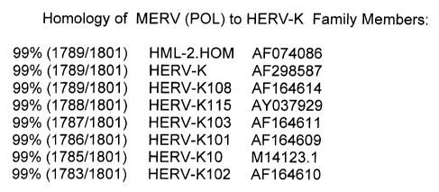
HML-2.HOM (AF074086) at 1789/1801 nucleotide pairs HERV-K103 (1787/1801 ) and
HERV-K102 (1783/1801 ). See homology listing in Drawing 3.
WO 01/70941 A2 has evidence for the expression of reverse transcriptase in
culture supernatants of human PBMC used as indicator cells, but oddly was only
detectable at 7 to 12 weeks after transfer. Supernatants from EBV transformed
B
cells from a few multiple myeloma patients were used as the sources of the
putative
HERV-H, but supernatants from EBV transformed B cells from normal controls
were
not tested or not reported. Without the specificity controls these results are
inconclusive.
There is an US patent (5,756,281 ) by John Martin wherein an electron
micrograph showing vacuolation of a PBMC is provided which is similar to our
Figure
1. In this patent it is purported that the vacuolation of the PBMC related to
a severe
encephalopathy in a single patient. However, this blood sample had been stored
at
CA 02501301 2005-03-18
23
room temperature for 48 hours before being examined by electron microscopy,
and
no control blood samples similarly stored were provided or analyzed,
indicating this
observation cannot be interpreted. Attempts to identify a virus resulted in
the provision
of a small piece of sequence apparently related to human CMV herpes virus. Our
HERV-BZU does not have homology to CMV.
US patent 5,882,912 claims an infectious Simian Foamy Virus (SFV) was
isolated from a human. Previous patents had not established any evidence for
primate
foamy viruses to productively infect humans, and the infectious ability is a
key
attribute of a vector for gene therapy. This foamy virus was derived from
monkeys
and thus, humans are not expected to be tolerant to its viral antigens. Thus,
as
argued below, the HERV-K102 provirus, which appears to share many attributes
with
primate foamy viruses, is a significant advance over the SFV as a vector, due
to its
origin within the human genome.
As mentioned above, we observed vacuolation in cultured single donor or
mixed donor blood cells whether derived from Cord Blood (CB) or adult
peripheral
blood mononuclear cell (PBMC) samples (see electron micrographs in Drawing 1
a, b).
Within these vacuoles, particles about 100 nm could be visualized
(Drawing1c,d, and
by negative staining 1e). This could not reflect an inborn error of metabolism
as it
developed in all CB and PBMC tested (now about 30 -35 samples) but was not
apparent in uncultured, freshly isolated cells (see Drawing 2.1.A). Thus, it
was not
rare, and was inducible. Furthermore activation schemes involving PHA and IL-2
commonly used in clinical virology laboratories to isolate herpes and HIV
viruses,
blocked vacuolation (Drawing 2.2), indicating not only was the vaculolation
inducible
but it was subject to regulation. This along with the visualization of
particles in the
vacuoles, indicated a foamy-type virus was likely involved.
The vacuolation initially started in few cells and appeared to spread with
time to
other cells in the cultures. As this was shown for both CB and PBMC, as well
as
single donor (non-proliferating cells) and for mixed lymphocyte reaction (a
mix of non-
proliferating and proliferating), these results suggest the infectious agent
can replicate
in non-proliferating and proliferating cells.
CA 02501301 2005-03-18
24
Given that all cultures used fetal calf serum (FCS), we had to determine if
the
FCS was the source of the apparent foamy-like virus. Upon testing various
sources of
serum, and showing vacuolation occurred in all cultures including normal human
AB
serum and autologous serum, this clearly indicated FCS was not the source of
the
vacuolation (Drawing 2.1 ). This indicated to us that an endogenous human
retrovirus
might be involved:
As reviewed in the background, only HERV-K has been associated with
particle formation so we devised a novel primer set based on the detection of
polymerase (pol) to allow us to detect members of the HERV-K family (see
folder
marked Design of Primer Sets). The sequence of the novel primer set is
provided in
Drawing 3. The size of the PCR product is 293 base pairs. We determined the
sequence of the RT-PCR product amplified with the primer set (encoding mRNA
induced in day 4 CB cells (single donor)) and surprising it gave a single
clear
sequence signal (Drawing 3). The sequence of the PCR product was identical to
HERV-K102, although this sequence was still 98% related to a number of close
family
members, such as K101, K103, K10, and others.
By using PCR for the pol, we were able to show the induction of mRNA
corresponding to the pol of HERV-K102 (Drawing 4) correlated with vacuolation
which
was induced by about 48 hours, peaked at about 5 days and seemed to decrease
thereafter due to cell death. Indeed a time course of cell number and
viability clearly
indicated (Table 1 in Drawing 5) that the cells were starting to die by day 7
of culture
We sought to provide further evidence of HERV-K102 expression and
translation, and thus, potential for particle formation. In foamy viruses, the
expression
of the envelope is required for particle formation and for infectivity: As
well, the
envelope protein of retroviruses tends to contain most of the heterogeneity of
retroviral sequences, moreso than gag which often shows cross-reactivity
between
similar types of retroviruses but for different species. Accordingly,
antibodies specific
to "antigenic" peptides of HERV-K102 envelope were made through a commercial
company, Washington Biotechnologies, Inc. The company was given the full
sequence of the envelope antigen of HERV-K102, and provided us with the most
CA 02501301 2005-03-18
antigenic/immunogenic short peptides based on an algorithim they had
developed.
We then selected two peptides for further study and immunization (see Drawing
6 a
and b for description and location of sequences in the envelope protein) based
on
blasting for identities in GenBank. Peptides were synthesized and rabbits were
5 immunized. We titrated the two antisera (ML4 and ML5) onto both peptides, 4
and 5.
As shown in the Table 2 in Drawing 6 C, only the ML4 antisera gave specific
reactions, in that it was not cross-reactive with peptide 5. Affinity purified
antibody
had the same properties (Drawing 6 C). A blast of Peptide 4 in GenBank showed
it
was identical not only to HERV-K102, but also K101, K103, K10 and K, but not
other
10 family members such as K107, K109, K113 or K115 (Drawing 6D).
Using the rabbit antisera, we looked for the induction of HERV-K102 envelope
protein expression in cultured versus non-cultured, . freshly isolated cells
(referred to
as Day 0) by flow cytometry on permeabilized cells. These results are shown in
Drawing 7. While no detection of HERV-K102 envelope was found on day 0 using
15 this very sensitive method (flow cytometry), day 4 cells in which
vacuolation and
HERV-K102 pol expression could also be demonstrated, had clearly detectable
expression when compared to the pre-immunization sera of the rabbits. We used
a
rabbit antihuman lymphocyte antisera (ALS) as a positive control for the human
lymphocytes which is positive as expected on day 0 and day 4 samples. We have
20 used a more sensitive flow cytometry, method, biotin-avidin flow cytometry
which is
expected to be at least one log more sensitive than the regular type flow
cytometry
indirect method, and have been unable to detect HERV-K102 expression within
freshly isolated uncultured human mononuclear cells, either CB or PBMC. As the
antisera seems to be very concentrated (according to the ELISA given in
Drawing 6,
25 C), this may account for the background noted in Drawing 7 by flow
cytometry.
The finding of the expression of envelope protein corresponding to HERV-K102
strongly suggested the likelihood that the particles seen by electron
microscopy and
which formed in the vesicles, might be infectious, since for foamy viruses,
the
envelope needs to be expressed for infectivity. So we tested infectivity of
frozen-
thawed CB cells where single verses mixed donors had been cultured and
compared
CA 02501301 2005-03-18
26
this to media and freshly isolated and non-cultured (Day 0) cells. These
results are-
summarized in Table 3 in Drawing 8. Since we expected the infectivity to be
analogous to foamy viruses, we froze-thaw the cells, and then placed the
cellular
material onto indicator cells overnight as given in Table 3. Samples were
tested in
duplicate for both single and mixed donors. At 16 hours we washed away non-
adherent materials from the adherent cells. In none of the cases did we notice
any
cytopathic effects at 16 hours. We then incubated the cells an additional 8
hours, and
then observed the monolayers. For MRC-5 cells, virtually all the cells had
apoptosed
and detached from the monolayers at 24 hours (Drawing 9, MRC-5 iii), when day
4
induced CB cells (frozen-thawed) had been laid on top. In contrast, media and
day 0
did not show this obvious and pronounced cytopathic effect (MRC-5 i and ii).
No
alterations in the HFL-1 cells were observed suggesting either infection did
not take
place, it was not a lytic or productive infection (Drawing 9, HFL-1 iii). In
the Vero cells
which are green monkey cells, it appeared that there may have been some
. vacuolation but no other obvious cytopathic occured (see Drawing 9 Vero).
Thus, the
controls indicated the cytopathic effects were not due to some nonspecific
toxic effect.
This was also suggested by the absence of cytopathic effects at 16 hours. We
concluded that the particles seen by EM associated with vacuolation were
indeed
infective.
By using RT-PCR for HERV-K102 pol in the indicator cells we were able to
confirm expression in the day 4 MRC-5 cells and not in the HFL-1 cells. Both
fibroblast cell lines are embryonic and derived from humans. As expected media
and
day 0 controls were negative (Drawing 10).
In order to substantiate that HERV-K102 might be expressed in vivo we
purified DNA and mRNA from plasma using the Tri-reagent protocol. An analysis
revealed DNA but not mRNA corresponding to HERV-K could be commonly detected
in 65 % of samples analysed (Drawing 11, Table 4). In those-samples showing a
positive DNA result, we also analysed the preparation for B-actin to
potentially rule out
contamination by cellular debris. In some cases this was positive and thus,
was not
informative for HERV-K (15 % of the total). Overall, 50 % of samples tested
could be
CA 02501301 2005-03-18
27
shown to have HERV-K pol DNA in plasma. Titration of the B-actin probe verses
HERV-K102 showed the B-actin to be twice as sensitive.
Nevertheless, six of the positive samples were sent for sequencing. Five of
the
6 sequences were identical to HERV-K103 and also MERV (but for the latter the
available sequence information does not include the first 16 nucleotides of
the forward
primer). The other sequence seems to be related to HLM-2.HOM (Chromosome
7p22).
Given the possibility as suggested by the literature for reactivation of HERV-
K
family members associated with some HIV and herpes infections, and the need to
show some evidence for tolerance widely in humans, we screened sera from
"normal"
individuals from our archived sample bank, and compared this to sera from
patients
documented to have high viral loads of HIV and herpes viruses. For this ELISA
screening, sera was diluted 1/150 on the two peptides corresponding to the
envelope
protein, peptide 4 (Table 5a) or peptide 5 (Table 5b). As shown in Drawing 12
Table
5, each ELISA plate has positive and negative controls. In repeat and extended
analyses (Table 6, Drawing 12 B), we found some evidence for low antibody
levels in
2 of 44 tested sera (4.5 %) and these were in the animal/pig occupational
exposure
group..Only one sample had reactivity for peptide 4 but not peptide 5 (a CMV
patient).
While positives might be found in 22 to 29 % of active herpes infections, we
found as
much as 70 % of HIV patients (with high viremia) might be positive. However
only 20
of HIV sera appeared to be strongly positive (see summary at the end of Table
6 in
Drawing 12). Since there is no significant homology of the HERV-K102 peptide
sequences with HIV or herpes viruses, it is expected that this reaction is not
a cross-
reaction but reflects reactivation of HERV-K102 with underlying disease. It
remains to
be determined if the reactivation is direct by co-expression in the same
cells, or
indirect due to immunosuppression of the host (not necessarily expressed in
the same
cells). These results appear to suggest that during active acute viremias, at
least
temporary production of antibodies to HERV-K102 envelope antigens can be
detected. However most normal individuals do not have antibodies to HERV-K102
or
K102 like envelope antigens. That antibodies to HERV-K can be temporarily
CA 02501301 2005-03-18
28
produced with underlying disease, has been previously demonstrated in cases of
germ cell tumors, where the antibodies disappeared upon complete removal of
the
tumor (19). Our work corroborates the notion that humans are generally
tolerant of
HERV-K102 antigens. This result was expected since Cord Blood and adult blood
can
naturally express HERV-K102 particles. On the other hand this work shows the
potential for reactivation when there is underlying infectious diseases.
Table II PCR Plasma Testin4 for Evidence of HERV-BZU Activit~in Vivo
These tests differ from previous work in cells in that we first optimized the
PCR
methods (based on DNA results) then we used the QIAamp Ultra-Sensitive Kits to
isolate °particles" or nucleotides from 1 ml of plasma rather than use
the Tri-Reagent.
For a control for genomic contamination we used B-actin. We looked for DNA and
mRNA (since for foamy retroviruses, the infective genome is DNA [15]). Our B
actin
PCR is more sensitive than our HERV-BZU pol (see attached titration), which
provides us with some assurance that when HERV-BZU is detected it is not due
to
genomic contamination. Intensity of PCR products on acrylamide gels were
scored
on a scale of 0 through 4 plus. +/- are weak but visible bands, while '
indicates a '/2
increment. Results need to be confirmed by quantitative real-time PCR.
Plasma DNA- mRNA- DNA- mRNA- Comments
Sample HERV-BZU HERV-BZU B B actin
pol pol actin
30 Normal - - - - No activity of
HERV-BZU
(N) in vivo in normals
N spiked + + + - + + - Genomic contamination
with +
DNA from
cells
CF* +/- - - - Current or past
in vivo
Anti-viral activity (gene
therapy amplification)?
(natural
products)
MS001 + + + ' - - Infectious particles?
Initial
CA 02501301 2005-03-18
29
Diagnosis
MS001 + + + + +' - - Levels of infectious
Active- particles increase
with
Progressing disease activity?
MS001 - - - - HERV-BZU or MS
1 month infectious agent
on is
INF a therapy downregulated by
INF ~
MS001 - - - - Loss of HERV-BZU
with
3 months clinical remission
on of MS
therapy symptoms
and
clinically
confirmed
remission
EBV + + +/- - - Cross-activation?
Viremia
CB1 + + + - + - Placental activity?
CB2 + - - - Placental activity?
Legend: CF = Chronic Fatigue, MS = Multiple Sclerosis, CB = Cord Blood, N =
normal
In summary, we have not only discovered an infectious HERV, HERV-K102 or
K102-like provirus, but have provided evidence this infectious HERV-K102 to
which
humans appear to be generally tolerant, appears to be functionally similar to
primate
foamy viruses by the following criteria:
1. Acute vacuolation in susceptible cells in vitro associated with high copy
numbers or particles.
2. Probably productive infection in both dividing and non-dividing cells.
3. Budding of infectious particles (immature virions) into endoplasmic
reticulum
vesicles.
4. Infectious particles (100 nm) of the size and morphology of foamy
retroviruses.
5. Highly cytopathic for human fibroblast cells in vitro.
6. Infectious particles are cell associated, liberated by freeze-thawing.
7. Virtually 100 % of cells infected (MRC-5 cells) and human diploid
fibroblasts
are particularly sensitive to FV-induced cytopathic effects.
8. Fairly high titres of replication-competent virus in human hematopoietic
cells.
CA 02501301 2005-03-18
9. Uses a lysine (K) tRNA primer for reverse transcription.
While the preferred embodiments of the invention have been described above,
it will be recognized and understood that various modifications may be made
therein,
and the appended claims are intended to cover all such modifications which may
fall
5 within the spirit and scope of the invention.
References:
1. Bieda K et al, J General Virology 82: 591-596, 2001.
2. Patience C et al, J Virology 70: 2654-2657, 1996.
10 3. Mayer J, Dev Biol (Basel) 106: 439-4.41, 2001.
4. Turner G et al, Curr Biol 11: 1531-1535, 2001.
5. Ono M et al, J Virology 60: 589-598, 1986.
6. Zsiros J et al, J General Virology 79: 61-70, 1998.
7. Medstrand P & Blomberg J, J Virology 73: 2463-2466, 1993.
15 8. Tonjes RR et al, J Virology 73: 9187-9195, 1999.
9. Costas J. J Mol Evol 53: 237-243, 2001.
10. Lower R et al, PNAS 90: 4480-4484, 1993.
11. Barbulescu M et al, Curr Biol 9: 861-868, 1999.
12.Armbruester V et al, Clinical Cancer Research 8: 1800-1807, 2002.
20 13. Megstrand P et al, J Gen Virology 73: 2463-2466, 1992.
14. Brodsky I et al, Blood 81: 2369-2374, 1993.
15. Wang-Johanning F et al, Clinical Cancer Research 7: 1553-1560, 2001.
16.Johnston JB et al, Ann Neurol 50: 434-442, 2001.
17. Herve CA et al, Clin Exp Immunol 128: 75-82, 2002.
25 18. Hishikawa T et al, Viral Immunology 10 : 137-147, 1997.
19. Boller K et al, J Virology 71: 4581-4588, 1997.
20. Yu SF et al, J Virology 73: 1565-1572, 1999.
21. Hur K et al, Mech Ageing Dev 123: 1637-1647, 2002.
22. Lynch WP & Sharpe AH, J Virology 74: 1558-1565, 2000.
30 23. Zeine R et al, J Neurosci Res 64: 380-391, 2001.
CA 02501301 2005-03-18
31
24. Rakowicz-Szulczynska EM et al, Clin l7iagn Lab Immunol 6: 115-126, 1999.
25. Hirschl S & Muster T. GenBank entry AY186778 "MERV polymerase",
2002.