Note: Descriptions are shown in the official language in which they were submitted.
CA 02501461 2005-04-06
WO 2004/045709 PCT/US2003/034729
TITLE OF "'fHE INVENTION
CARDIAC STIMU4ATION SYSTEM AND METHOD
CROSS-REFERENCE TO RELATED APPLICATIONS
This application is a continuation of U.S. application serial number
10/374,899
filed on February 24, 2003, which claims priority to U.S. provisional
application serial
number 60/426,840 filed on November 16, 2002, which is herein incorporated in
its
entirety by reference thereto.
1o STATEMENT REGARDING FEDERALLY SPONSORED RESEARCH
OR DEVELOPMENT
Not Applicable
REFERENCE TO A COMPUTER PROGRAM APPENDIX
Not Applicable
BACKGROUND OF THE INVENTION
1. Field of the Invention
This invention is a system and method for stimulating a heart of a patient.
2o More specifically, it is a stimulation device assembly and method using a
stimulator
coupled with an implantable emitter that is in turn coupled with a conductive
agent
injected into the region of the heart to be stimulated.
2. Description of the Background Art
Various medical device systems and methods have been disclosed for
coupling energy to cardiac tissue in order to influence heart function. A
great many
of such systems and methods have been disclosed for the particular purpose of
treating various types of cardiac arrhythmias, including for example
fibrillation,
tachycardia, bradycardia, or other arrhythmias.
Among the many different devices and methods previously disclosed for
so energy coupling to cardiac tissue, various different types of energy
coupling have
also been employed.
One type of energy coupling having significant impact over the years in
-1-
CA 02501461 2005-04-06
WO 2004/045709 PCT/US2003/034729
treating cardiac arrhythmias delivers energy into targeted regions of cardiac
tissue
using energy emitters in or around the heart. Various previously disclosed
examples
of energy delivery systems and methods of this type include devices using:
emitters
of electrical energy, e.g. electrodes for delivering direct or alternating
current, such
s as radiofrequency (RF) current; emitters such as crystals or transducers for
delivering sonic energy (e.g. ultrasound); emitters such as fiber optics,
lenses, or
other light discharge elements (e.g. laser diodes) for delivering light (e.g.
laser); or
energy emitters using microwave coupling (e.g. induction).
Another type of energy coupling that has been investigated for treating
certain
~o cardiac arrhythmias includes hypothermia or cryogenic devices intended to
reduce
tissue temperature to a level. In general, these devices include regions that
are
cooled to low temperatures (relative to surrounding body temperature) so as to
thereby pull heat from and reduce the temperature in surrounding tissue. By
achieving this heat transfer at sufficient levels, an intended change in the
affected
15 tissue structure or function, either temporarily or permanently. To the
extent that
such hypothermic coupling relates to pulling thermal energy from the
surrounding
tissue so as to cool it, such devices are considered energy coupling devices.
The designs and features of the various different energy coupling devices for
cardiac treatment also vary in order to adapt such devices to achieve
different
2o intended results.
For example, certain such devices and methods generally herein referred to
as "ablation devices" are specifically adapted to couple sufficient energy
with cardiac
tissue so as to ablate the tissue. This may be performed for example in order
to
terminate a focal origin of arrhythmia, or to form a conduction block to
terminate a
2s harmful conduction pathway within the cardiac tissue network causing
arrhythmia.
Other ablation devices have also been disclosed for the purpose of forming
passageways through tissue or other material located within a patient, such as
for
recanalization of occluded lumens and vessels.
Other previously disclosed examples of cardiac treatment devices using
so energy coupling have been specifically adapted to stimulate cardiac tissue,
rather
than to ablate it. Various types of these cardiac stimulation systems include:
devices adapted to couple energy to cardiac tissue in a manner so as to
trigger an
_2_
CA 02501461 2005-04-06
WO 2004/045709 PCT/US2003/034729
arrhythmia in order to diagnose cardiac conduction through the heart;
pacemaker
assemblies and methods adapted to provide artificial pacing of the cardiac
cycle in
order to cure an arrhythmia; and defibrillator assemblies and methods wherein
the
heart is "shocked" out of an arrhythmia and back into sinus rhythm.
Because the cardiac conduction cycle is directly and intimately related to
electrical conduction through cardiac tissue, the previously disclosed cardiac
stimulation devices for triggering, pacing, or defibrillating, are generally
electrical
coupling devices that deliver electrical energy from electrode leads or
catheters
secured to or placed against the tissue to be stimulated. Pacemaker and
defibrillator
o assemblies have each been adapted with varied (and in some regards mutually
exclusive) designs appropriate to suit one or the other of temporary or
permanent
use, depending upon a particular need for either acute or chronic rhythm
management, respectively.
In general, permanent pacemaker systems include a pacemaker assembly
~s with a pacemaker coupled to an electrical lead generally called a
"pacemaker lead".
The implantable or permanent pacemaker typically includes a power source, such
as
a source of electrical current energy (e.g. a battery). This power source is
electrically
coupled to one end of the electrical lead. The other end of the electrical
lead is in
turn coupled to the cardiac tissue to be stimulated, usually by use of an
anchor such
2o as a needle, screw, spline, grasper, etc., which anchor may be the
electrode current
emitter itself.
In recent years, an increasing amount of interest, and research and
development, has been directed toward modifying the cellular make-up of
cardiac
tissue structures in order to enhance cardiac conduction or function in such
modified
25 structures.
Certain such efforts have been directed toward delivering conductive,
contractile muscle cells into regions of the heart where contraction is
compromised,
such as areas of necrosis. These efforts have been intended to increase the
cardiac
function in such areas. Such cells delivered may be for example prepared from
so cultures of the patient's own cells, which may be cardiac cells for
example, but may
also be skeletal cells, fibroblasts or stem cells. The delivered cells may
also be
modified in a manner to enhance their contractile function or conductivity,
and
-3-
CA 02501461 2005-04-06
WO 2004/045709 PCT/US2003/034729
including to enhance their expression of certain factor(s), such as for
example to
enhance expression of connexin 43, a protein known to enhance cardiac signal
conduction.
Connexins are found in connexons of gap junctions. Gap junctions regulate
intercellular passage of molecules, including inorganic ions and second
messengers,
thus achieving electrical coupling of cells via gap junctions. Connexin
proteins are
the major gap junction protein involved in the electrical coupling of cells.
For
example Connexin 43 is the major gap junction protein in ventricular
myocardium
responsible_for gap junction intercellular communication. Connexin 43,
abbreviated
herein as "Cx43", is a protein having structural, regulatory, or biochemical
functions
associated with gap junctions and electromechanical coupling. Connexins are a
whole family of proteins. There are specific connexins for various parts of
the heart.
Examples of Cx43 useful in the aspects of the invention providing for agent
delivery
into specified cardiac tissue regions associated with cardiac activation are
~ 5 polypeptide sequences such as human Cx43 (Genbank Accession Nos. XP
027460,
XP 027459, XP 004121, P17302, AAD37802, A35853, NP 000156 and
AAA52131 ), mouse Cx43 (Genbank Accession Nos. P23242, P18246, A39802,
A36623, NP 034418, CAA44640) and exemplary sequences for Rat Cx43 are found
at Genbank Accession Nos. P08050, S00532, NP 036699, AAA75194 and
20 1404339A. Connexin family in the cardiovascular system includes Cx37, Cx40,
Cx43, Cx45.
Various references herein to cardiac conduction, signal conduction, or
otherwise "conduction" through cardiac tissue are generally intended to mean
this
propagation related to a resulting contractile wave through cellular tissue,
including
25 via gap junctions.
Other examples have been disclosed for locally delivering agents into cells
resident in the target cardiac tissue structure that modify the cellular
function in-vivo,
such as by altering the genetic material within cells to enhance
conduction/contraction, such as for example by enhancing cellular expression
of
so certain compounds or agents that cause the intended effect (e.g. DNA
material to
cause expression or over expression of Cx43 or other such compounds).
There is yet to be a system or treatment method developed that combines
-4-
CA 02501461 2005-04-06
WO 2004/045709 PCT/US2003/034729
cardiac stimulation systems, such as pacemakers or defibrillators, with
delivery of
conductive agents, such as cell therapy, gene therapy, other modes of tissue
engineering, or other conductive agents such as conducting polymers, metals,
or
combinations thereof (such as injectable solutions or suspensions, e.g. gold,
conducting "dust", etc. ), in a manner that substantially enhances the
artificial
stimulation such as pacing or defibrillating of cardiac tissue structures.
In recent years, biventricular septal pacing is an area that has received
increasing attention and interest for new product development and research in
recent
years, in particular as it is intended as a curative measure for the complex
and
o dangerous conditions of bundle block (e.g. bundle of His) and congestive
heart
failure. Normal electrical activation of the ventricles generally proceeds as
follows. Electrical impulse is initiated from the sinus node, leading to
atrial activity
passed through the AV node, followed by ventricular activation. The
ventricular
activation phase includes the following events (typically in the sequence
described):
A) Activation of the left septum due to branches of the bundle of His entering
the septum higher on the left side of the septum versus the right;
B) Apical depolarization follows early depolarization of the RV
(depolarization
of the RV occurs quickly due to the thinness of the RV);
C) Depolarization of the lateral wall of the left ventricle; and
2o D) Late LV depolarization of the base
Various different disease states or abnormal conditions can affect this
ventricular activation phase of the cardiac cycle. One such example of
particular
concern is called left bundle branch block ("LBBB"). LBBB alters the entire
ventricular depolarization pathway. Depolarization starts from the right side
of the
septum and progresses toward the left front of the LV. Apical depolarization
then
occurs.
Biventricular pacing devices and methods have been disclosed that are
generally intended to resynchronize LV contractility by activating the LV with
a pacing
lead, typically via a left-sided (e.g. left ventricle) pacing lead.
so Further examples of systems, devices, and methods providing additional
background related to the present invention are variously disclosed in the
following
U.S. Patent Application Publications: US 2002/0035388 to Lindemans et al.; and
US
-5-
CA 02501461 2005-04-06
WO 2004/045709 PCT/US2003/034729
2002/0087089 to Ben-Haim. Other such examples are variously disclosed in the
following U.S. Patents: 4,399,818 to Money; 5,103,821 to King; 5,683,447 to
Bush
et al.; 5,728,140 to Salo et al.; 6,059,726 to Lee et al.; 6,101,410 to
Panescu et al.;
6,128,535 to Maarse; 6,151,525 to Soykan et al.; and 6,238,429 to Markowitz et
al..
s Still other examples are disclosed in the following PCT Patent Application
Publications: WO 90/10471 to King; WO 98/02150 to Stokes et al.; WO 98/28039
to
Panescu et al.; WO 00/59375 to Sen; WO 01/68814 to Field; WO 02/22206 to Lee;
and WO 02/051495 to Ideker et al. The disclosures of all these references
listed in
this paragraph are herein incorporated in their entirety by reference thereto.
There is still a need for improved cardiac stimulation systems and methods.
There is also still a need to improve conduction within cardiac tissue
structures during cardiac stimulation.
There is in particular still a need for a biventricular septal stimulation
syster~i~
and method, such as for biventricular pacing, that provides for artificial
cardiac
~s stimulation in combination with delivery of conductive agents in order to
enhance the
stimulation effect.
There is also still a particular need for septal stimulation system and method
that can capture a substantial region of septal tissue, such as in order to
provide
biventricular pacing in the setting of multiple left bundle branch block.
2o BRIEF SUMMARY OF THE INVENTION
One object of the invention is to affect cardiac tissue response to
stimulation
from a cardiac stimulation device such as a pacemaker or defibrillator.
Another object of the invention is to affect contractility or conduction
within a
region of cardiac tissue in a heart of a patient.
25 Another object of the invention is to affect ventricular septal function in
a heart
of a patient.
Another object of the invention is to provide for improved delivery of cardiac
stimulation leads and/or agents affecting cardiac contraction or conduction
into a
ventricular septum.
3o Another object of the invention is to provide cardiac stimulation to a
heart in a
patient.
Another object of the invention is to provide cardiac stimulation to a
ventricular
-6-
CA 02501461 2005-04-06
WO 2004/045709 PCT/US2003/034729
septum of a patient.
Another object of the invention is to pace a heart of a patient.
Another object of the invention is to defibrillate a heart of a patient.
Another object of the invention is to achieve biventricular pacing or
s defibrillation of a heart in a patient.
Another object of the invention is to provide both energy delivery and
improved tissue response to the delivered energy during cardiac stimulation.
Another object of the invention is to deliver cells into a region of cardiac
tissue
in a heart of a patient.
1o One aspect of the invention therefore is a cardiac stimulation system with
a
cardiac stimulation device assembly and an agent delivery assembly. The
cardiac
stimulation device assembly comprises a cardiac stimulation device and an
energy
emitter. The cardiac stimulation device comprises an energy source and is
adapted
to couple energy from the energy source to the energy emitter. The energy
emitter is
~ s adapted to be positioned within the patient's body so as to emit energy
into a region
of cardiac tissue to be stimulated. The agent delivery assembly is adapted to
deliver
into the region of cardiac tissue an agent that affects a stimulated response
in the
region to the emitted energy.
One mode of this aspect the agent delivery assembly comprises a delivery
2o catheter. In one embodiment the delivery catheter is a cardiac delivery
catheter. In
another embodiment, the delivery catheter is a vascular delivery catheter that
is
adapted to deliver the agent into the region via a blood vessel extending
within the
septum.
In another mode the agent delivery assembly comprises a needle. In one
2s embodiment of this mode, the needle is a surgical needle. In another
embodiment,
the agent delivery assembly further comprises a delivery catheter, and the
needle is
located along the distal end portion of the delivery catheter. In one
variation of this
embodiment, the delivery catheter is a cardiac delivery catheter. In another
variation,
the delivery catheter is adapted to be positioned within a blood vessel
extending
so within the septum such that the needle is adapted to puncture through the
blood
vessel wall and into cardiac tissue of the septum.
Another aspect of the invention is a cardiac stimulation system with a cardiac
CA 02501461 2005-04-06
WO 2004/045709 PCT/US2003/034729
stimulation device assembly in combination with an agent. The cardiac
stimulation
device assembly comprises a cardiac stimulation device and an energy emitter.
The
cardiac stimulation device comprises an energy source and is adapted to couple
energy from the energy source to the energy emitter so as to activate the
energy
emitter. The energy emitter is adapted to be positioned within the patient's
body so
as to emit energy into a region of cardiac tissue to be stimulated in response
to
activation from the cardiac stimulation device. The agent is adapted to be
located
within the region of cardiac tissue and to affect a stimulated response in the
region to
the emitted energy.
o According to one mode, the cardiac stimulation device is a pacemaker device
assembly. According to another mode, the cardiac stimulation device is a
defibrillation device assembly.
Another aspect of the invention is a system having a cardiac pacemaker that
cooperates with a cell therapy system in order to provide cardiac stimulation.
According to one mode, the cell therapy system comprises a volume connexin
43 and a cell delivery catheter that is adapted to deliver the connexin 43
into a region
of cardiac tissue to be stimulated by the cardiac pacemaker.
Another aspect of the invention is a cardiac stimulation system with a cardiac
stimulation assembly that is adapted to deliver energy into a region of tissue
within a
2o heart of a patient so as to stimulate the region, and also with a volume of
conductive
agent that is adapted to be delivered into the region and to enhance
stimulation of
the region with the energy delivered by the cardiac stimulation assembly.
According to one mode of this aspect, the cardiac stimulation assembly further
includes a delivery member with a proximal and portion and a distal end
portion that
2s is adapted to be positioned at a location within a heart of a patient, and
also
includes an array of extendable electrode assemblies cooperating with the
delivery
member and that each includes a stimulation electrode that is adjustable to
extend
from the delivery member at the location and into a unique location relative
to the
other extendable electrode assemblies within the region of tissue.
so In one embodiment of this mode, each of the array of extendable electrode
assemblies includes an extendable needle that is adjustable to extend from the
distal
end portion of the delivery assembly and into cardiac tissue so as to position
the
_$_
CA 02501461 2005-04-06
WO 2004/045709 PCT/US2003/034729
respective stimulation electrode at the unique location.
According to one variation of this embodiment, the stimulation electrode of
each extendable electrode assembly is integrated with the needle. In another
variation, the stimulation electrode of each extendable electrode assembly is
adjustable to extend from the respective needle to the unique respective
location.
According to other variations, the needle has a curved shape, or may be
constructed from a superelastic or shape memory metal alloy such as a nickel-
titanium alloy.
In another embodiment, each of the array of extendable electrode assemblies
o has a relatively pliable tubular body with an inner lumen, and each
stimulation
electrode is located along the tubular body of the respective extendable
electrode
assembly. According to this embodiment, each relatively pliable tubular body
is
adapted to be deflected and steered through the region of tissue so as to
place the
respective stimulation electrode at the respective unique location.
~5 In one further variation of this embodiment, a moveable stylet is adapted
to be
moveably engaged within the inner lumen of at least one of the tubular bodies
so as
to adjust the tubular body to extend from the delivery member and advance
through
the region of tissue such that the respective stimulation electrode is
positioned at
the respective unique location. In a further feature, the moveable stylet may
be
2o provided with a proximal end portion, and a shaped distal end portion that
is
torquable within the inner lumen by torquing the proximal end portion
proximally and
externally of the tubular body so as to deflect and steer the tubular body in
order to
place the electrode.
According to another embodiment, an anchor is provided and is extendable
25 from the delivery member and adapted to anchor the distal end portion at
the
location such that the array of extendable electrode assemblies is adapted to
be
positioned at the respective unique locations along the region of tissue. In
one
beneficial variation of this embodiment, the anchor is provided also as a
stimulation
electrode.
3o According to another embodiment, another stimulation electrode is located
at
the distal tip of the delivery member.
In a further mode, the cardiac stimulation assembly is a cardiac pacemaker,
_g_
CA 02501461 2005-04-06
WO 2004/045709 PCT/US2003/034729
and in a further embodiment the cardiac pacemaker is a bi-ventricular cardiac
pacemaker.
According to another mode of the present aspect, the conductive agent
includes living cells, and in further embodiments the cells include at least
one of
myoblasts, fibroblasts, or stem cells. In another regard, the living cells are
of a type
that are adapted to express a connexin at gap junctions with cardiac cells in
the
region of tissue, and in more particular beneficial variation are adapted to
express
connexin 43 at the gap junctions, and still further are beneficially
genetically modified
to over-express production of connexin 43.
1o In another mode, the conductive agent includes an injectable preparation of
a
conductive non-living material that is adapted to enhance electrical
conduction in
the region, such as a conductive metal.
According to another mode, a septal perforator delivery assembly is included
in the system and is adapted to couple the cardiac stimulation assembly to the
15 region of tissue via at least one septal perforator vessel, and also to
deliver the
volume of conductive agent to the region via the at least one septal
perforator
vessel.
In another mode, a transcardiac delivery member is provided for delivery of
the stimulation assembly and conductive agent via a cardiac chamber; such as
the
2o right atrium.
Another aspect of the invention is a cardiac stimulation system with a cardiac
stimulator assembly with an energy source, and an energy emitter that is
adapted to
be coupled to the energy source and to be positioned within a region of a
heart of a
patient to be stimulated, and that also includes means for enhancing
stimulation of
2s the region with the energy from the energy emitter.
Another aspect of the invention is a cardiac stimulation system with a
conductive agent delivery system that includes a source of conductive agent
and a
delivery assembly that is adapted to deliver a volume of conductive agent from
the
source and into a region of a heart in a patient, and also with a means for
delivering
so energy to at least a portion of the region. The volume of conductive agent
is
adapted to enhance stimulation of the region with the energy from the means
for
delivering energy.
-10-
CA 02501461 2005-04-06
WO 2004/045709 PCT/US2003/034729
Another aspect of the invention is a cardiac stimulation assembly with an
elongate body having a proximal end portion, a distal end portion that is
adapted to
be positioned at least in part at a location within a heart of a patient, and
a delivery
lumen extending at least in part along the distal end portion. This assembly
further
includes a means for delivering energy into a region of tissue within the
heart of the
patient, as well as a means for enhancing stimulation of the region of tissue
from
energy delivered from the means for delivering energy. Furthermore, the means
for
delivering energy and means for enhancing stimulation are adapted to cooperate
with the elongate body.
1o Another aspect of the invention is a cardiac stimulation assembly with an
elongate body having a proximal end portion, a distal end portion that is
adapted to
be positioned at least in part at a location within a heart of a patient, and
a lumen
extending at least in part along the distal end portion, and also with an
energy
delivery assembly and a volume of conductive agent. The energy delivery
assembly
15 has an energy emitter that is coupled to the elongate body and is
adjustable to
extend from the distal end portion and into a region of tissue within the
heart when
the distal end portion is at the location, and also has an energy lead that is
adapted
to couple to the energy emitter and also to a source of energy along the
proximal
end portion when the energy emitter is in the second position. The volume of
2o conductive agent is coupled to the lumen and is adapted to be delivered
through the
lumen into the region of tissue when the distal end portion is at the location
and the
energy emitter is in the second position.
Another aspect of the invention is a cardiac stimulation system with a cardiac
stimulation device and a volume of cells that are adapted to over-express
connexin
25 43. The cardiac stimulation device has an elongate body with a proximal end
portion, a distal end portion, and a cardiac stimulation electrode along the
distal end
portion. The cardiac stimulation device and volume of cells are adapted to
cooperate sows to stimulate a region of tissue within a heart of a patient.
Another aspect of the invention is a cardiac stimulation assembly with an
3o elongate body, an energy delivery assembly, and a volume of conductive
agent.
The elongate body has a proximal end portion, a distal end portion that is
adapted
to be positioned at least in part at a location within a heart of a patient,
and a lumen
-11-
CA 02501461 2005-04-06
WO 2004/045709 PCT/US2003/034729
extending at least in part along the distal end portion. The energy delivery
assembly
has an energy emitter that is coupled to the elongate body and is adjustable
to
extend from the distal end portion and into a region of tissue within the
heart when
the distal end portion is at the location. This assembly also has an energy
lead that
is adapted to couple to the energy emitter and also to a source of energy
along the
proximal end portion when the energy emitter is extended into the region of
tissue.
The volume of conductive agent is coupled to the lumen and is adapted to be
delivered at least in part through the lumen into the region of tissue when
the distal
end portion is at the location and the energy emitter is extended into the
region of
tissue.
Another aspect of the invention is a cardiac stimulation assembly with an
elongate body, an energy delivery assembly, and an anchor. The elongate body
has a proximal end portion and a distal end portion that is adapted to be
positioned
at least in part at a location within a heart of a patient. The energy
delivery
1s assembly has a plurality of energy emitters. Each energy emitter is coupled
to the
elongate body and adjustable to extend from the distal end portion and into a
unique respective position relative to the other energy emitters within a
region of
tissue within the heart when the distal end portion is at the location. Each
energy
emitter is also adapted to couple to a source of energy when the respective
energy
2o emitter is in the second position within the region of tissue. The anchor
is adapted
to anchor the distal end portion at the location within the heart.
Another aspect of the invention is a cardiac stimulation assembly with an
elongate body, an energy delivery assembly, and a cardiac stimulation energy
source. The elongate body has a proximal end portion, a distal end portion
that is
25 adapted to be positioned at least in part at a location within a heart of a
patient, and
a delivery lumen extending at least in part along the distal end portion. The
energy
delivery assembly has a plurality of energy emitters. Each energy emitter is
coupled
to the elongate body and adjustable to extend from the distal end portion and
into a
unique respective position relative to the other energy emitters within a
region of
3o tissue within the heart when the distal end portion is at the location.
Each energy
emitter is also adapted to couple to a source of energy via the proximal end
portion
when the respective energy emitter is extended into the region of tissue. The
-12-
CA 02501461 2005-04-06
WO 2004/045709 PCT/US2003/034729
cardiac stimulation energy source is adapted to couple to each of the energy
emitters and to energize each energy emitter so as to emit energy into the
region of
tissue. Accordingly, the energy emitters at each unique respective position
within
the region of tissue are adapted to stimulate the region of tissue.
Another aspect of the invention is a cardiac stimulation device with an
elongate body, a needle, and an energy emitter. The elongate body has a
proximal
end portion, a distal end portion, and a passageway located at least in part
along
the distal end portion. The needle has a shank with a distal tip and an inner
lumen.
The energy emitter that is adapted to be coupled to an energy source. Further
to
1o this aspect, the distal end portion of the elongate body is adapted to be
positioned
at a location within a heart of a patient, and the needle is adapted to be
coupled to
the passageway and is adjustable between a first position located at least in
part
within the passageway and a second position that extends at least in part from
the
passageway and into a region of tissue within the heart when the distal end
portion
15 is at the location. Furthermore, the energy emitter is coupled to the inner
lumen of
the needle and is adjustable to extend from the needle into the region of
tissue.
Another aspect of the invention is a bi-ventricular cardiac stimulation system
that includes an array of stimulation electrodes. Each electrode of the array
is
adapted to be positioned at a unique location relative to the other
stimulation
2o electrodes within an intra-ventricular septum of a heart of a patient, such
that the
array of positioned electrodes is adapted to form a pattern that defines a
region of
tissue within the septum. Furthermore, the pattern and region defined thereby
comprises such sufficient area so as to stimulate at least one-quarter of the
septum,
and in further embodiments may be sufficient to provide artificial stimulation
to at
25 least one-third or even as much as one-half or more of the septum.
Another aspect of the invention is a cardiac stimulation system with a bi-
ventricular cardiac stimulation energy source and a septal stimulation device
with an
elongate body having a proximal end portion and a distal end portion with an
array
of extendable cardiac stimulation electrodes. The distal end portion is
adapted to
so be positioned at a location within a heart of a patient associated with a
ventricular
septum. Each of the extendable cardiac stimulation electrodes is adjustable to
extend from the distal end portion so as to be positioned at a unique
respective
-13-
CA 02501461 2005-04-06
WO 2004/045709 PCT/US2003/034729
location relative to the other extendable cardiac stimulation electrodes
within a
region of tissue within the ventricular septum. Each of the extendable
stimulation
electrodes is also adapted to be coupled to the bi-ventricular cardiac
stimulation
energy source. Accordingly, by positioning the distal end portion of the
elongate
s body at the location, positioning each of the extendable cardiac stimulation
electrodes at each respectively unique location within the region of tissue,
and
coupling each extendable cardiac stimulation electrode to the bi-ventricular
cardiac
stimulation energy source, the array of electrodes is adapted to substantially
stimulate the region of tissue be energizing each electrode of the array with
the
o energy source to emit current from its respectively unique location within
the region.
The invention further includes other aspects providing methods of treatment
as follows.
Another aspect of the invention is a method comprising: delivering a volume of
cells into a region of cardiac tissue; and stimulating the region of cardiac
tissue with
15 a cardiac stimulation device assembly.
One further mode of this aspect comprises: pacing the heart of the patient
from the region of cardiac tissue containing the volume of cells.
Another mode of this aspect comprises: providing the cells in a condition
wherein connexin 43 is expressed.
2o A further embodiment of this mode comprises: providing the cells in the
condition such that connexin 43 is over-expressed more than resident cardiac
cells in
the region.
Another aspect of the invention is a method comprising: delivering a volume of
cells within a ventricular septum of a patient sufficient to enhance response
of the
25 septum to stimulus from a pacemaker or defibrillator.
Another aspect is a method comprising: using the vasculature of a ventricular
septum in a patient to deliver a volume of an agent into the septum that
enhances
the response of the tissue in the septum to stimulus from a cardiac
stimulation
device.
so Another aspect of the invention is a method comprising: using the
vasculature
of a ventricular septum in a patient to deliver a volume of an agent into the
septum
that enhances the cardiac contraction or conduction along the septum.
-14-
CA 02501461 2005-04-06
WO 2004/045709 PCT/US2003/034729
Another aspect of the invention is a method comprising: using the vasculature
of a ventricular septum in a patient to deliver cardiac stimulator leads into
the septum
for use in either pacing or defibrillating the heart via the septum.
Another aspect of the invention is a method comprising: emitting stimulating
energy from an energy emitter over a substantial portion of the ventricular
septum.
Another aspect of the invention is a method comprising: implanting an array of
energy emitters at unique locations within a ventricular septum of a patient
such that
an area bound and by such energy emitters comprises a substantial portion of
the
septum.
1o One mode of this method further comprises: simultaneously emitting energy
from each of the implanted energy emitters.
Another mode of this method further comprises: implanting an anchor into the
septum; and extending from the anchor at least one of the energy emitters.
Another mode of this method further comprises: implanting an anchor into the
septum; and extending from the anchor each of the energy emitters.
Another mode of this method further comprises: coupling the implanted
energy emitters with a pacemaker.
Another mode of this method further comprises: coupling the implanted
energy emitters with a defibrillator.
2o Another mode of this method further comprises: emitting electrical current
from each of the energy emitters.
Another mode of this method further comprises: delivering each of the energy
emitters to the unique location through a needle.
Another mode of this method further comprises: advancing an array of
needles into the septum; and delivering each energy emitter to its respective
unique
location within the septum through a unique one of the needles
Another mode of this method further comprises: positioning multiple ones of
the array of energy emitters on a delivery member; advancing the delivery
member
into the septum such that each of the energy emitters positioned on the
delivery
3o member is located at its respective unique location.
Another aspect of the invention is a method for stimulating a region of a
heart
in a patient by providing a cardiac stimulation assembly, providing a
conductive
-15-
CA 02501461 2005-04-06
WO 2004/045709 PCT/US2003/034729
agent delivery system, delivering energy to a location associated with the
region of
the heart to be stimulated with the cardiac stimulation system, and delivering
a
volume of conductive agent from the conductive agent delivery system to the
region
of the heart. Further to this method aspect, the volume of conductive agent
enhances stimulation of the region of the heart with the energy being
delivered to
the location.
Another aspect of the invention is a method for providing bi-ventricular
stimulation to a heart of a patient by: providing' an array of cardiac
stimulation
electrodes, positioning a distal end portion of a delivery member against a
portion of
1o an intra-ventricular septum of the heart, and extending the array of
cardiac
stimulation electrodes from the distal end portion of the delivery member and
into
the intra-ventricular septum such that each cardiac stimulation electrode is
positioned at a unique location relative to the other cardiac stimulation
electrodes
within a region of tissue of the intra-ventricular septum.
15 Another aspect of the invention is a method for manufacturing a bi-
ventricular
cardiac stimulation system for providing bi-ventricular stimulation to a heart
of a
patient. This method is performed by: providing a bi-ventricular stimulation
device
having an elongate body with a proximal end portion, a distal end portion that
is
adapted to be positioned at a location within a ventricle, a stimulation
electrode
20 located at a position along the distal end portion so as to be electrically
coupled to a
region of tissue of a septum of the heart when the distal end portion is at
the
location, and a passageway extending at least along the distal end portion.
This
method further includes: loading a volume of cells within the passageway of
the bi-
ventricular stimulation device, wherein the cells are adapted to over-express
2s connexin-43 or an analog, derivative, or biological equivalent thereof.
Another aspect of the invention is also method for manufacturing a cardiac
stimulation system for providing cardiac stimulation to a heart of a patient.
This
method includes providing a cardiac stimulation device having an elongate body
with a proximal end portion, a distal end portion that is adapted to be
positioned at a
so location within the heart, an array of extendable cardiac stimulation
electrodes that
are each adapted to be extended from the distal end portion at the location so
as to
be positioned at a unique location relative to the other extendable cardiac
-16-
CA 02501461 2005-04-06
WO 2004/045709 PCT/US2003/034729
stimulation electrodes within a region of tissue within the heart, and a
passageway
extending at least along the distal end portion. This method further includes:
loading a volume of cells within the passageway, wherein the cells are adapted
to
enhance electrical stimulation of the region of tissue from the array of
extendable
electrodes.
Another aspect of the invention is a method for stimulating a region of a
septum in a heart of a patient by: delivering at least one cardiac stimulation
electrode into at least one septal perforator vessel within an intra-
ventricular septum
of the heart, delivering a volume of conductive agent to a region of tissue
within the
1 o septum associated with the septal perforator vessel, and, after the volume
of
conductive agent is delivered into the region, stimulating the region of
tissue with the
at least one cardiac stimulation electrode.
Another aspect of the invention is a method for providing bi-ventricular
stimulation to a heart in a patient by stimulating a region of tissue that
comprises at
15 least one-quarter of the inter-ventricular septum, and in further modes at
least one-
third, or even one-half, and up to as much as all of the septum.
Another aspect of the invention is a method for stimulating a region of a
heart
in a patient by delivering a volume of cells into the region that are adapted
to over-
express connexin-43 or an analog, derivative, or biologic equivalent thereof,
and
2o providing an electrical stimulus to the region.
The various aspects, modes, embodiments, variations, and features just
described are considered to be independently beneficial without requiring
combination with the others, though the invention further contemplates the
benefits
from their various combinations as may be made by one of ordinary skill based
upon
2s the totality of this disclosure.
Further objects and advantages of the invention will be brought out in the
following portions of the specification, wherein the detailed description is
for the
purpose of fully disclosing preferred embodiments of the invention without
placing
limitations thereon.
3o BRIEF DESCRIPTION OF THE DRAWINGS
The invention will be more fully understood by reference to the following
drawings which are for illustrative purposes only:
-17-
CA 02501461 2005-04-06
WO 2004/045709 PCT/US2003/034729
FIG. 1 shows across-section of a heart with multiple regions of left bundle
block indicated by arrows before treatment according to the systems and
methods of
the present invention.
FIG. 2 shows a similar cross-section of the heart with multiple regions of
left
s bundle block shown in FIG. 1, and shows one mode of the invention with a
delivery
catheter positioned against the septum in the right ventricle.
FIG. 3 shows a similar cross-section of the heart shown in FIGS. 1 and 2, and
shows a subsequent mode of using the invention wherein an agent affecting
cardiac
conduction is delivered into a cardiac tissue region related to the multiple
left bundle
1 o blocks.
FIG. 4 shows a similar cross-section of the heart shown in FIGS. 1-3, except
showing another mode implanting an array of energy emitters into a cardiac
tissue
region related to the multiple left bundle blocks.
FIG. 5 shows a cross-section of a heart with a left bundle block similar to
that
1s shown in FIG. 1, except showing the heart with a localized, single region
of left
bundle block as indicated by an arrow.
FIG. 6 shows a similar cross-section of the heart with the localized region of
left bundle block shown in FIG. 5, and shows one mode of the invention with a
delivery catheter positioned against the septum in the right ventricle.
2o FIG. 7 shows a similar cross-section of the heart shown in FIGS. 5 and 6,
and
shows a subsequent mode of using the invention wherein an agent affecting
cardiac
conduction is delivered into a cardiac tissue region related to the left
bundle block.
FIG. 8A shows a side view, including some internal structures, of a distal end
portion of an extendable electrode array assembly according to the invention.
25 FIG. 8B shows a transverse cross-section taken along lines B-B in FIG. 8A.
FIG. 8C shows a schematic partially cross-sectioned view of a particular
embodiment for delivering an electrode such as according to the array assembly
shown in FIGS. 8A-B.
FIG. 9 shows a partially segmented view of another extendable electrode
3o assembly according to the invention.
FIG. 10 shows a cross-sectioned view of a heart with another extendable
electrode assembly of the invention using a screw anchor and multiple
electrode
-18-
CA 02501461 2005-04-06
WO 2004/045709 PCT/US2003/034729
leads deployed within a ventricular septum and coupled to a pacemaker (shown
schematically) for biventricular septal pacing.
FIGS. 11A-B show transverse cross-sectioned views taken along lines A and
B, respectively, in FIG. 10.
s FIG. 12A shows a cross-sectioned view of a heart with another extendable
electrode assembly of the invention using a screw anchor and a single lead
with
multiple electrodes deployed within a ventricular septum and coupled to a
pacemaker (shown schematically) for biventricular septal pacing.
FIG. 12B shows a partially cross-sectioned view of an extendable electrode
1o assembly adapted for use according to the embodiment shown in FIG. 12A.
FIG. 13A shows a cross-sectioned view of a heart with another electrode
assembly using a single intracardiac lead deployed within the ventricular
septum in a
first position.
FIG. 13B shows a similar view to FIG. 13A but with multiple electrode leads
15 implanted at first and second positions within the ventricular septum.
FIG. 13C shows a further mode stitching an electrical lead along a region of
inter-ventricular septum.
FIG. 14A shows a perspective view of a pacing electrode system using a
coronary sinus delivery system for delivering pacing electrodes into
myocardial
2o septum of the ventricles through vessel walls of septal perforator blood
vessels.
FIG. 14B shows a perspective view of an agent delivery system using the
coronary sinus route to deliver agent that enhances cardiac conduction into
myocardial tissue of the ventricular septal wall through the vessel wall of a
septal
perforator blood vessel.
2s FIG. 15 shows a schematic view of a combined cardiac stimulation system
according to an embodiment of the invention.
DETAILED DESCRIPTION OF THE INVENTION
Referring more specifically to the drawings, for illustrative purposes the
present invention is embodied in the embodiments generally shown in FIGS.1
3o through 15. It will be appreciated that the apparatus may vary as to
configuration
and as to details of the parts, and that the method may vary as to the
specific steps
and sequence, without departing from the basic concepts as disclosed herein.
-19-
CA 02501461 2005-04-06
WO 2004/045709 PCT/US2003/034729
In patients with impaired conduction via the bundle branches (especially left
bundle branch block or "LBBB"), capturing more of the septum with multiple
leads
coupled with an injectable conductor will enable more of the septum to be
activated
therefore enabling depolarization of the LV and improved synchronization.
The present invention contemplates various approaches to achieving the
objects of the invention, in particular as they relate to delivering agents
into regions
of cardiac tissue in order to affect the conduction and enhance response to
electrical
stimulation there.
In one regard, an agent that is adapted to enhance conduction of a cardiac
~o signal (i.e. biological conductor, conducting metals, conducting polymers,
etc) is
delivered into the myocardial septum, such as for example being injected via a
percutaneous, transcardiac approach such as shown via a transcardiac delivery
catheter in FIGS. 3 and 7. Various conventional or other delivery systems may
be
suitable for such delivery, such as for example those delivery catheters and
systems:
~5 the "Noga" system developed by Johnson & Johnson, the "Myocath" device and
system developed by BioHeart, Inc.; the "Stilleto" catheter device and system
developed by Boston Scientific Corporation; and at least one other catheter
device
and system commercially developed by "BioCardia".
In another regard, a pacemaker lead system may include delivery features,
2o e.g. lumens, through which such agent may be delivered. In this regard,
while such
pacemaker lead may require increased profile to accommodate an agent delivery
lumen, benefits include the substantial benefit of knowing that the conductor
agent is
delivered in the proximity of the source of energy emission for
depolarization.
In addition to the specific embodiments herein shown or described, other
2s agent deliver devices or methods may be suitable to accomplish the intended
objects
of the invention as herein contemplated, as is apparent to one of ordinary
skill based
at least in part upon this disclosure.
Notwithstanding other embodiments elsewhere herein shown and described,
and variations and modifications as would be apparent to one of ordinary
skill, the
so following embodiments are considered highly beneficial in particular
respect, though
not limited to, biventricular pacing: delivering a substance or agent
affecting cardiac
conduction through a delivery system; delivering a substance or agent
affecting
-20-
CA 02501461 2005-04-06
WO 2004/045709 PCT/US2003/034729
cardiac conduction through a pacemaker lead; and providing extendable
electrode
structures with multiple leads that are adapted to span a substantial area of
the
ventricular septum for more efficient pacing there.
With respect to the extendable electrode aspect of the invention, according to
s one beneficial embodiment, microfilaments are generally used to extend and
increase the electrode surface area/volume in which to electrically stimulate
the
myocardium. The use of the extended electrode will allow for a greater amount
of
the myocardium to be stimulated thus synchronizing electro-mechanical
contraction
of the heart. The novel electrode array and related cardiac stimulation system
is
1o considered particularly useful in patients with bundle branch block, such
as illustrated
in the Figures, as well as patients suffering from congestive heart failure.
Accordingly, it will be seen that this invention may be used for particular
benefit in stimulating the ventricular septum, such as is illustrated by
reference to
certain particular, illustrative embodiments in the Figures as follows.
1s By general reference to the Figures, a heart 1 is shown in various cross-
sectioned views to include a right ventricle 2, bi-cuspid valve 3, left
ventricle 4, inter-
ventricular septum 6 that includes right and left bundles 8,10, respectively,
and an
apex 9.
As shown by reference to FIGS. 1-4, patients having hearts with multiple
2o regions of left bundle block are particularly well treated by use of the
present
invention.
As indicated by arrows in FIG. 1, the multiple areas of block 12, 14, 16 are
shown
and create a particular challenge for pacing from a conventional, single
electrode
approach. Particular modes of using the present embodiment of this invention
in this
2s multiple left bundle block setting are illustrated in FIGS. 2-4.
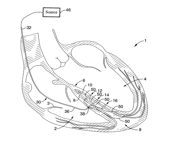
As shown in FIG. 2, an agent delivery catheter 30 is shown with a proximal
end portion 32 and distal end portion 36 that includes a distal tip 38. Distal
end
portion 36 is delivered into the right ventricle 2 by manipulating proximal
end portion
32 externally of the body via a percutaneous, translumenal approach through
the
3o venous system, and then into the ventricle across the right atrium via the
bicuspid
valve 3. The distal tip 38 of the distal end portion 36 of the delivery
catheter 30 is
then positioned within the right ventricle 2 against the septum 6. A source of
agent
-21-
CA 02501461 2005-04-06
WO 2004/045709 PCT/US2003/034729
40 is coupled to a proximal end portion of the delivery catheter, as shown
schematically in FIG. 3. A volume of the conductive agent 18 from the source
is then
delivered through a delivery lumen (not shown) within the delivery catheter
30,
through a distal port located on the distal tip 38 of the delivery catheter
30; and into a
s region of cardiac tissue associated with the multiple left bundle blocks, as
shown in
FIG. 3. This may be accomplished using pressure alone, though in certain
beneficial
embodiments (e.g. described below) a needle tip, either integral with the
delivery
catheter or slideably disposed therein, is used to inject the agent into the
tissue.
Where such a separate cooperating needle is used, the internal bore of the
needle
will be coupled proximally with the source of agent, as will be further
developed
below.
With respect to septal electrode aspects of the invention, a percutaneous,
translumenal delivery catheter 30 may also be used for delivery of such
cardiac
stimulation assembly to the region of septum to be stimulated, as shown in
FIG. 4.
15 Also shown in that FIG. 4, a plurality of electroded members 50 are
delivered through
the delivery catheter 30, which electroded members 50 may be splines,
filaments, or
other types of leads coupled to electrodes, which are further coupled to an
electrical
energy source 46 proximally for electrical current emission from members 50
into the
septal tissue. By providing these leads 50 in an array with electrodes
positioned to
2o span a sufficiently wide area of the septum as shown, their coordinated
current
emission allows for improved stimulation over a region of tissue spanning a
wide
area well suited for providing proper cardiac conduction from such an area of
multiple blocks, such as blocks 12, 14, 16 shown in the illustrative
embodiment in
FIG. 4.
2s The electrode leads 50 may include filaments, splines or other types of
suitable structures to provide requisite mechanical support for delivery and
also
conduct signals to the electrodes. The leads 50 may be advanced through the
cardiac tissue according to various techniques and tools as would be apparent
to
one of ordinary skill. In one example; the distal tips of the leads are
sharpened
so allowing the leads to be advanced simply as needles mechanically pushing
through
tissue. In another example, a separate extendable delivery device such as
deployable needle is extended from the anchor point to the location to deliver
the
-22-
CA 02501461 2005-04-06
WO 2004/045709 PCT/US2003/034729
lead, and the lead is delivered to that location through that needle. In other
alternative or combined examples (not shown), an ablative energy source may be
coupled between the leads and the tissue such that the leads or splines ablate
their
way through the cardiac tissue for deployment.
Such stimulation is provided by coupling the electroded lead members 50 with
a source of stimulating energy 46, as also shown in FIG. 4. This may be for
example
a pacemaker or defibrillator, which may be of particular type or style to meet
the
particular needs for stimulation at the location in the heart chosen for the
therapy
according to the invention. In the specific beneficial setting of the present
~o embodiments for stimulating a septum, the energy source 46 may be a bi-
ventricular
pacemaker assembly with appropriate software and hardware incorporated therein
to
provide the appropriate stimulus there. Further more detailed examples of such
energy sources contemplated for use with the present invention include without
limitation: a dual chamber pacer under the product name "Kapp 900" (model
~5 #KDR901 ), intracardiac defibrillator (ICD) under the product name "Marquez
DR"
(model # 7274), and bi-ventricular ICD under the product name "Insync ICD"
(model
#7272), all commercially available from Medtronic Device Corporation; and
Guidant
DDD under the product name "Insignia" (model #1298), an ICD under the product
name "Prism II" (e.g. model #1861); and Guidant BiV (Renewal, model #H135),
all
2o commercially available from Guidant Corporation.
As elsewhere herein described, an array of electroded members 50 may also
be delivered subsequent to, before, or simultaneous with delivery of agent 18
for
enhancing conduction of the stimulated septal region. For example, the
embodiment
of FIGS. 3 and 4 would be combined. In this highly beneficial setting, the
wide area
2s stimulation from the array of electroded members 50 is further combined
with the
enhanced conduction due to the agent 18 in the area to give optimal results.
In another regard, the embodiments elsewhere herein described are also
useful for other arrhythmic conditions in the septum or elsewhere, such as for
example in the setting of a more focal region of local left bundle block as
shown by
3o reference to block 12 for illustration according to the agent 18 delivery
embodiment
in FIGS. 5-7.
A further highly beneficial embodiment for a cardiac stimulation assembly to
-23-
CA 02501461 2005-04-06
WO 2004/045709 PCT/US2003/034729
be used according to the invention is shown in FIGS. 8A-C. More specifically,
delivery catheter 30 includes an array of lumens or passageways 34, including
respective ones that are circumferentially spaced around a central one. The
circumferentially spaced lumens 34 each houses a lead of a respective
electroded
member 50, whereas the central lumen 34 houses another electroded lead 60 that
forms a screw-shaped anchor adjustable in and out of that central lumen 34 for
delivery to and then anchoring into the septum, respectively. Furthermore, the
circumferentially spaced electroded members 50 are shown according to a still
more
detailed embodiment in FIG. 8C to include a pre-shaped needle member 52, which
o may be made of nickel-titanium alloy or other superelastic, shape memory, or
other
suitable material, that is adapted to be housed within its respective lumen 34
during
delivery of tip 38 to abut a septal wall, and then extendable from lumen 34 to
advance into the septal wall. Further shown is an extendable electrode member
56
that is further adjustable in and out of needle member 52. Yet a further ring
~5 electrode is shown at tip 38 of delivery catheter 30, which may be used to
assist in
mapping to find the optimal place for placement of the stimulation electrodes,
and/or
for additional surface area for stimulation as a stimulation electrode.
Though the specific configurations shown in FIGS. 8A-C are considered
beneficial, the various features such as number, placement, or specific types
of
2o elements are illustrative and other suitable substitutes may be made. For
example,
other numbers and corresponding placements for the circumferentially spaced
electroded members 50 may be used, generally desiring 2 or more electroded
members 50 according to the present embodiment, and generally between 2 to 4
electroded members 50 may be optimal for many circumstances. In another
25 example shown in FIG. 9, a moveable stylet 58 is moveable within a
passageway of
an electroded member 50 that includes a pliable shank 52 with an electrode 54
at its
tip. The moveable stylet 58 is adapted to assist shank 52 during advancement
through septal wall tissue to the desired location for positioning electrode
54 for
stimulation. Such features may be provided instead of use of the needle
assembly
so shown and described by reference to FIG. 8C, or various modifications may
be made
to combine various aspects between those two approaches, including for example
for a particular electroded assembly 50, or by providing one such assembly
with one
-24-
CA 02501461 2005-04-06
WO 2004/045709 PCT/US2003/034729
design and one or more according to the other design.
In any case, a further schematic view of the broad aspects for an arrayed
electrode septal stimulation assembly during use is shown in FIG. 10. The
array of
electroded members 50 is shown in angular arrangement within a transversely
cross-
sectioned heart for illustration, but they may share a planar orientation,
such as in a
plane transverse to the plane of cross-section shown for heart 1. Accordingly,
anchor element 60 is located within a region of septal wall tissue that is
bound by
electroded members 50 that have been positioned at unique respective locations
around such central anchor 60 across the region. By providing members 50, 60,
and
~o tip 38 as stimulation electrodes coupled to a source of stimulation energy
(not
shown), the tissue bounded by electroded members 50 may be substantially
stimulated, such as for biventricular pacing.
For further illustration of the orientation of such electroded elements are
shown in different planes in FIGS. 11A-B, whereas FIG. 11 B is further
provided with
~5 a shadowed reference to the region 18 corresponding to the tissue being
stimulated.
However, the circumferential arrangement shown such as in FIG. 11 B
corresponding to region 18 may be modified, with different shapes than
circular, with
different lengths of members 50, for example, or with the central area such as
at
anchor 60 offset within the bound region 18. In one regard, the view of FIG.
11 B
2o shows a particular view of a planar array of members 50 in two dimensions.
However, they may be of modified orientation to lie in different planes such
that a
three dimensional volume of septal tissue is defined as the region. Still
further, the
array of members 50 may be further modified such that the resulting stimulated
region 18 is instead two or more discrete regions, as further herein
described.
25 It is to be appreciated that despite the benefits of stimulating such
region by
elements 50, 60, and a ring electrode 38 at the septal wall surface, it is not
necessary to provide all such elements as stimulation electrodes, and removal
of any
one or more of them and such resulting combination arrays are further
contemplated
embodiments hereof. For example, central screw electrode 60 may instead merely
so be provided as an anchor without electrical stimulation capability. Or, it
may instead
be a simple electrical lead and not necessary of the screw anchor
configuration. In
further examples of modifications that are contemplated, discrete electrodes
may be
-25-
CA 02501461 2005-04-06
WO 2004/045709 PCT/US2003/034729
positioned at various locations along the members 50 and within region 18, as
shown
at electrodes 55 in FIGS. 12A-B. Or the, the electroded elements 50 may be
continuous segments with stimulation capability along their lengths out to the
boundaries of region 18.
s In one particular further embodiment shown in FIGS. 13A-B, an electroded
member 50 is threaded into a region of tissue in septal wall 6, such as
according to
the needle or stylet embodiments of FIGS. 8C or 9, respectively. Multiple such
elements may be placed in this manner, as shown in FIG. 13B, in an arrangement
such that the combination of multiple electroded elements 50 and 57 correspond
to
1o the overall region of tissue 18 that is stimulated thereby. These elements
50,57 may
be placed separately into the septal wall 6, with leads extending separately
therefrom, such as shown in FIG. 13B, and such may be done with the same
intracardiac delivery catheter 30 or separate delivery devices. After
placement as
shown in FIG. 13B, the leads for elements 50,57 are coupled to an energy
source,
15 such as a pacemaker or defibrillator. It is further contemplated that the
delivery
catheter 30 is removed after placement of leads 50,57 and before or after
coupling to
the energy source, or may remain indwelling if provided with sufficiently low
profile
and adapted for such long-term use (or if for temporary pacing).
A further modification is shown in FIG. 13C, wherein a remote mechanical
2o stitching mechanism (not shown) is provided at the distal end 36 of
delivery catheter
30 and adapted to stitch a single lead member 50 over a length or region of
septal
wall 6 tissue. Examples of such mechanism providing for such stitched lead
placement over an extended length are provided in the following reference:
"Flexible
Microelectrode Arrays With Integrated Insertion Devices," by O'Brien, David
P.,
2s Nichols, T. Richard, and Allen, Mark G., variously of the School of
Electrical and
Computer Engineering at Georgia Institute of Technology and the School of
Medicine
at Emory University, both in Atlanta, Georgia. The disclosures of this
reference is
herein incorporated in its entirety by reference thereto.
Various modifications of the preceding embodiments may be made without
so departing from the scope of the invention, in particular in so far as
modified in order
to achieve certain particular desired results consistent with the objects of
the
invention.
-26-
CA 02501461 2005-04-06
WO 2004/045709 PCT/US2003/034729
For example, one desired result of the agent delivery and extendable
electrode delivery embodiments is to pace the heart 1 over a large region of
the
septum 6, e.g. electrodes or agent spanning sufficient area of the septum 6 to
void
effects of bundle block there. Therefore, "substantial" area of the septum 6
generally
means at least one-fifth of the septal wall, and may be even more beneficially
one-
fourth, and still more beneficially more than one-third or even one-half of
the septum
(ideally capturing the entire septum). In this regard, such "stimulation" is
herein
intended to mean the region that experiences artificial stimulation, such as
either by
the electrical discharge directly from an excitation electrode, or by enhanced
~o propagation thereof via artificially delivered conductive agent. Moreover,
deploying
such agent or extendable electrodes may reach to the apex 9 or beyond. In any
event, though stimulating such substantial regions is highly beneficial in
many
applications of the invention, it is not required in order to still achieve
many of the
other benefits afforded by the invention according to its various embodiments
15 described herein.
The invention is particularly described herein for use with a pacemaker as an
energy source 46 to be coupled to the electroded arrays 50 herein described,
which
can be implantable or temporary, and may be of the type commercially
available. Or,
such pacemaker may be modified for use with the electrode array assemblies 50
2o andlor agent or "bioelectrodes" 18 herein described. For example, for a
given
number of joules normally used to pace a heart 1 via the septum 6 using
conventional electrode leads, such energy dose may be divided among the many
electrodes of the array of the invention, thereby reducing the current density
at the
electrodes themselves. In another regard, more joules may be delivered in an
2s impulse from the pacemaker over the span of electrodes in the array before
reaching
the same level at any one electrode otherwise delivered using conventional
systems.
Other areas of the heart 1 may also be stimulated using the embodiments
herein described, which may be modified as appropriate for such use according
to
one of ordinary skill.
so In addition, other stimulation energy modalities may be used, e.g,
ultrasound
or microwave, though electrical stimulus is considered highly beneficial and
efficacious according to prior experience in the industry. Moreover, to the
extent
-27-
CA 02501461 2005-04-06
WO 2004/045709 PCT/US2003/034729
"stimulation" is described with respect to the embodiments, it is generally
intended
that such stimulation is done to excite conductive activity and, therefore,
done
according to energy delivery modes that are generally non-ablative.
Cell cultures and in particular expression or overexpression of Cx43 or
otherwise connexin have been specified herein as highly beneficial agent
according
to embodiments using agents to enhance cellular conduction in the heart. In
particular association with stimulation devices, such agents are therefore
considered
"bioelectrodes", effectively extending the reach of the energy emitter (e.g.
electrode)
by virtue of the locally enhanced conduction - thus stimulating greater areas
of the
~o heart. The electrode and bioelectrode stimulate larger areas, mitigating
deficits for
synchronized conduction between the ventricles.
Other agents than "bioelectrodes" also having beneficial effects in similar
uses
are contemplated. For example, other substances may be injected or otherwise
applied to the target tissue. Examples of such other substances include:
polymers,
which may be conducting in one regard (e.g. poly-parol), or non-conducting
(e.g.
PLGA) in which case they may be coated such as with conductive metal;
hydrogels,
e.g. of the type carrying an ionic charge; or other solutions or suspensions
such as
carrying gold or other conductive metal particles or ions.
Still further, the invention further contemplates combinations or blends of
the
2o foregoing, such as according to one highly beneficial example combining
cells, e.g.
overexpressing Cx43, with a polymer delivery matrix (that may also be
conductive, or
may be non-conductive).
Various combinations between the electrode assemblies and conductive
agent delivery are also described above by reference to the illustrative
embodiments,
25 but further combinations and subcombinations, and modifications thereto,
may be
made. For example, screw electrodes may be adapted with a hollow lumen and
used for the agent delivery. In another example, whereas FIGS. 14A-B show
highly
beneficial transvascular delivery of electrodes and conductive agent,
respectively,
into the ventricular septum, these may be combined. Alternatively, each may
also be
so accomplished in combination with a transcardiac approach of the other.
Still further,
whereas some agent and/or electrodes may be delivered via a transcardiac
delivery
modality, other agent and/or electrodes may also be delivered via the
transvascular
-28-
CA 02501461 2005-04-06
WO 2004/045709 PCT/US2003/034729
septal perforator approach - each approach may provide for enhanced
stimulation at
different areas of the septum, whereas their combination may provide a
complete
and still more beneficial biventricular pacing result. To this end, the
transcardiac
approach is generally herein shown and described as the right heart system is
often
s preferred for access. However, left ventricular transcardiac delivery of
either or both
of the agent or stimulus devices) is also contemplated, instead of or in
combination
with the right ventricular approach (or transvascular approach). Any
combination or
sub-combination of these are contemplated, as should be apparent to one of
ordinary skill based upon this disclosure.
o Different volumes of agent, and different numbers, sizes, patterns, andlor
lengths of stimulation leads may be used to suit a particular need. In one
regard, a
prior diagnostic analysis may be used to determine the extent of the
condition,
location of the condition, or various anatomical considerations of the patient
which
parameters set forth the volume of agent or electrode array to use. Or, a real
time
15 diagnostic approach may allow for stimulus effects to be monitored, such
that the
amount of agent, or distance, direction, or number of electrode deployment, is
modified until the correct result is achieved. Therefore, for example, the
electrodes
of such embodiments may be retractable and advanceable through tissue so that
different arrangements may be tried until synchronization is achieved.
2o It is further contemplated that the agent delivery and electrode
embodiments,
though highly beneficial in combination with each other, are independently
beneficial
and may be used to provide beneficial results without requiring the other.
Notwithstanding the foregoing, a particular beneficial overall assembly is
shown in FIG. 15. More specifically, cardiac stimulation system 100 is shown
to
2s include a delivery catheter 110 that cooperates to provide for both
delivery of a
bioelectrode 150 as well as stimulation electrodes 130 and an anchor 140 as
follows.
Delivery catheter 110 has a proximal end portion 112 with a proximal coupler
114,
distal end portion 116, and distal tip 118, and is an intracardiac delivery
catheter
adapted to deliver its contents toward the inter-ventricular septum from the
right
3o ventricle. Extendable from delivery catheter 110 is an inner catheter 120
with an
extendable screw anchor 140, and multiple spaced extendable electroded members
130 spaced about screw anchor 140. All or only some of central anchor 140,
-29-
CA 02501461 2005-04-06
WO 2004/045709 PCT/US2003/034729
extendable electroded members 130, and the tip of member 120 may be provided
as
stimulation electrodes to be coupled to energy source 160, such as via shaft
120.
Moreover, all or only some of central screw 140, extendable electroded members
130, or tip of member 220, may be further adapted to deliver a volume of
bioelectrode agent into the region also coupled by the stimulation electrode
sections,
as shown at regions 150, such as via ports coupled to passageways (not shown)
that
are further coupled to a source of such agent 170 (shown schematically).
This combination device is considered highly beneficial for stimulating
substantial portions of the inter-ventricular septum, such as for
biventricular pacing
~o and in particular treating LBBB. As further shown in FIG. 15 and
illustrative of other
embodiments providing extendable elements to be driven into tissue such as in
the
septal wall, a further device 180 may be coupled to such assembly that is an
actuator
that either allows for automated or manual extension of the respective
extendable
elements. Further elements that may be provided in an overall system such as
that
5 shown in FIG. 15 at 100, or other embodiments herein, include monitoring
sensors
and related hardware and/or software, such as incorporated into or otherwise
cooperating with an energy source such as a pacemaker/defibrillator, including
for
example: to map electrical heart signals for diagnostic use in determining the
stimulation mode; and/or feedback control related to the stimulation signal
itself,
2o such as set points, etc.
Connexin-Enhancing Conductive Agents
The present invention, to the extent using connexin-expressing cellular agents
for injection as bioelectrodes, is related to co-pending U.S. patent
application Serial
No. 10/291,202, filed November 7, 2002, by the same inventor hereof, the
disclosure
2s of which is herein incorporated in its entirety by reference herein. Such
aspect of the
invention is based upon the experimental observation, based upon on man-made
materials and methods, that contacting a myocardial cell with a recombinant
cell,
such as an adult skeletal muscle cell, which is modified to express a
recombinant
connexin 43 (e.g., in the presence or absence of endogenous connexin 43
so expression) allows for electrical coupling of the modified skeletal muscle
cell to the
myocardial cell. The present invention according to the present embodiments
thus
provides methods for using a recombinant cell genetically modified to produce
a
-30-
CA 02501461 2005-04-06
WO 2004/045709 PCT/US2003/034729
connexin protein to produce persistent functional gap junctions between the
recombinant cell and cardiomyocyte to obtain electrical communication between
these cells. The use of recombinant cells that express recombinant Cx43 (or
other
connexin protein) increases and maintains the communication between the
s recombinant cells and myocardial cells, thus providing improved and
coordinated
electrical coupling with increased efficacy of myocardial contractility. The
present
invention provides methods of treatment of cardiac disease by transplanting or
grafting recombinant cells modified to express a connexin into cardiac tissue
to effect
the ability to artificially stimulate regions of myocardial dysfunction, in
particular in the
1o setting of intra-ventricular septal stimulation, such as with pacemakers or
defibrillators, such as for example for bi-ventricular pacing as a curative
measure for
heart block.
Definitions Related to Connexin Embodiments
The following definitions are intended to apply to embodiments of the present
1s invention using biological or cellular material as bioelectrode agents, and
in particular
with respect to embodiments related to connexin-43 and/or such agents related
to
the production or expression thereof. To the extent such terms are elsewhere
herein
used with respect to other embodiments provided herein, such terms shall be
given
their ordinary, customary meaning according to one of ordinary skill in the
art related
2o to such embodiment, unless such terms are otherwise specifically defined
with .
respect to such respective embodiment.
"Polynucleotide" as used herein refers to an oligonucleotide, nucleotide, and
fragments or portions thereof, as well as to peptide nucleic acids (PNA),
fragments,
portions or antisense molecules thereof, and to DNA or RNA of genomic or
synthetic
25 origin which can be single- or double-stranded, and represent the sense or
antisense
strand. Where "polynucleotide" is used to refer to a specific polynucleotide
sequence
(e.g, a connexin 43 polypeptide-encoding polynucleotide), "polynucleotide" is
meant
to encompass polynucleotides that encode a polypeptide that is functionally
equivalent to the recited polypeptide, e.g., polynucleotides that are
degenerate
so variants (i.e., polynucleotides that encode the same amino acid sequence
but differ
in polynucleotide sequence due to the degeneracy of the genetic code), or
polynucleotides that encode biologically active variants or fragments of the
recited
-31-
CA 02501461 2005-04-06
WO 2004/045709 PCT/US2003/034729
polypeptide, including polynucleotides having substantial sequence similarity
or
sequence identity relative to the sequences provided herein. Similarly,
"polypeptide"
as used herein refers to an oligopeptide, peptide, or protein. Where
"polypeptide" is
recited herein to refer to an amino acid sequence of a naturally-occurring
protein
s molecule, "polypeptide" and like terms are not meant to limit the amino acid
sequence to the complete, native amino acid sequence associated with the
recited
protein molecule, but instead is meant to also encompass biologically active
variants
or fragments, including polypeptides having substantial sequence similarity or
sequence identify relative to the amino acid sequences provided herein.
o As used herein, " polypeptide" refers to an amino acid sequence of a
recombinant or non-recombinant polypeptide having an amino acid sequence of i)
a
native polypeptide, ii) a biologically active fragment of an polypeptide, iii)
biologically
active polypeptide analogs of an polypeptide, or iv) a biologically active
variant of an
polypeptide. Polypeptides useful in the invention can be obtained from any
species,
e.g., mammalian or non-mammalian (e.g., reptiles, amphibians, avian (e.g.,
chicken)), particularly mammalian, including human, rodenti (e.g., murine or
rat),
bovine, ovine, porcine, murine, or equine, preferably rat or human, from any
source
whether natural, synthetic, semi-synthetic or recombinant. For example, "Human
connexin 43 polypeptide" refers to the amino acid sequences of isolated human
2o Cx43 polypeptide obtained from a human, and is meant to include all
naturally-
occurring allelic variants, and is not meant to limit the amino acid sequence
to the
complete, native amino acid sequence associated with the recited protein
molecule.
A "variant" of a polypeptide is defined as an amino acid sequence that is
altered by one or more amino acids (e.g., by deletion, addition, insertion
and/or
2s substitution). Generally, "addition" refers to nucleotide or amino acid
residues added
to an end of the molecule, while "insertion" refers to nucleotide or amino
acid
residues between residues of a naturally-occurring molecule. The variant can
have
"conservative" changes, wherein a substituted amino acid has similar
structural or
chemical properties, e.g., replacement of leucine with isoleucine. More
rarely, a
so variant can have "nonconservative" changes, e.g., replacement of a glycine
with a
tryptophan. Similar minor variations can also include amino acid deletions or
insertions, or both. Guidance in determining which and how many amino acid
-32-
CA 02501461 2005-04-06
WO 2004/045709 PCT/US2003/034729
residues may be substituted, added, inserted or deleted without abolishing
biological
or immunological activity can be found using computer programs well known in
the
art, for example, DNAStar software.
By "nucleic acid of interest" is meant any nucleic acid (e.g., DNA) which
s encodes a protein or other molecule which is desirable for inducing or
maintaining
electrical coupling between cells. In general, the nucleic acid is operatively
linked to
other sequences which are needed for its regulation and expression, such as a
promoter and regulatory elements.
The term "biologically active" where herein used in particular respect to
~o connexin, or biological or cellular material effecting production thereof,
refers to, for
example, a compound having structural, regulatory, or biochemical functions of
a _
naturally occurring connexin polypeptide, particularly with respect to
facilitating the
establishment of an electrochemical connection between a cell modified to
express a
connexin polypeptide and a myocardial cell, and includes in particular such
human
~s connexin polypeptides. Likewise, "immunologically active" defines the
capability of
the natural, recombinant or synthetic human connexin polypeptide, or any
oligopeptide thereof, to induce a specific immune response in appropriate
animals or
cells and to bind with a connexin specific antibody.
The term "derivative" as used herein refers to the chemical modification of a
2o nucleic acid encoding a polypeptide or the encoded polypeptide.
Illustrative of such
modifications would be replacement of hydrogen by an alkyl, acyl, or amino
group. A
nucleic acid derivative would encode a polypeptide which retains essential
biological
characteristics of a natural polypeptide.
As used herein the term "isolated" is meant to describe a compound of
2s interest (e.g., either a polynucleotide or a polypeptide) that is in an
environment
different from that in which the compound naturally occurs. "Isolated" is
meant to
include compounds that are within samples that are substantially enriched for
the
compound of interest and/or in which the compound of interest is partially or
substantially purified.
3o As used herein, the term "substantially purified" refers to a compound
(e.g.,
either a polynucleotide or a polypeptide) that is removed from its natural
environment
and is at least 60% free, preferably 75% free, and most preferably 90% free
from
-33-
CA 02501461 2005-04-06
WO 2004/045709 PCT/US2003/034729
other components with which it is naturally associated.
By "transformation", "transduction" or "transfection" is meant a permanent or
transient genetic change, preferably a permanent genetic change, induced in a
cell
following incorporation of new nucleic acid (e.g., DNA or RNA exogenous to the
cell).
Genetic change can be accomplished either by incorporation of the new nucleic
acid
into the genome of the host cell, or by transient or stable maintenance of the
new
DNA as an episomal element.
By "transformed cell", "transfected cell" or "transduced cell" is meant a cell
into
which (or into an ancestor of which) has been introduced, by means of
recombinant
o DNA techniques, a DNA molecule encoding a protein of interest.
By "construct" is meant a recombinant nucleic acid, generally recombinant
DNA, that has been generated for the purpose of the expression of a specific
nucleotide sequence(s), or is to be used in the construction of other
recombinant
nucleotide sequences. Constructs useful in the invention are those which
comprise
connexin-encoding gene sequence operably linked to a promoter which will allow
for
the expression of the connexin protein in a transformed cell. Exemplary
constructs
useful for the expression of human and rat Cx43 in accordance with the
invention are
described in Shinoura, N, et al., J Neurosurg. 1996 May;84(5):839-45 and
Suzuki et
al, Ann. Thorac. Surg., 2001, 71:1724-33, respectively.
2o By "promoter" is meant a minimal sequence sufficient to direct
transcription in
a recombinant cell. "Promoter" is also meant to encompass those elements
sufficient for promoter-dependent gene expression controllable for cell-type
specific,
tissue-specific or inducible by external signals or agents; such elements may
be
located in the 5' or 3' regions of the native gene (e.g., enhancer elements).
25 By "operably linked" or "operatively linked" is meant that a DNA sequence
and
a regulatory sequences) are connected in such a way as to permit expression
when
the appropriate molecules (e.g., transcriptional activator proteins) are bound
to the
regulatory sequence(s).
By "connexin gene" is meant the open reading frame encoding a connexin
so polypeptide, or introns, or biologically active fragment thereof. "Connexin
gene"
includes adjacent 5' and 3' non-coding nucleotide sequences involved in the
regulation of expression, up to about 10 kb beyond the coding region, but
possibly
-34-
CA 02501461 2005-04-06
WO 2004/045709 PCT/US2003/034729
further in either direction. The DNA sequences encoding a connexin may be cDNA
or genomic DNA or a fragment thereof. The gene may be introduced into an
appropriate vector for extrachromosomal maintenance or for integration into
the host.
The term "cDNA" as used herein is intended to include all nucleic acids that
share the arrangement of sequence elements found in native mature mRNA
species,
where sequence elements are exons (e.g., sequences encoding open reading
frames of the encoded polypeptide) and 3' and 5' non-coding regions. Normally
mRNA species have contiguous exons, with the intervening introns removed by
nuclear RNA splicing, to create a continuous open reading frame encoding the
~ o polypeptide of interest.
By "cardiomyocyte" is meant a cardiac contractile cell, which is a cardiac
muscle cell. The cardiomyocyte cell may be isolated and cultured in vitro or
be part
of the myocardium of a host.
By "skeletal muscle cell" is meant a cell found in skeletal muscle which
~5 includes but not limited to myoblasts, myotubes and mature skeletal muscle
cells.
By "recombinant cell" is meant a cell comprising nucleic acid not normally
associated with the cell (e. g. a cell transformed, transduced or transfected
with a
construct encoding a specific protein, e.g., a connexin protein).
By "transplanted cell" is meant a cell which has been introduced into a host
so
2o as to be in contact with a cell within a host. For example, a recombinant
cell or cells
maybe grafted and/or implanted into the cardiac tissue of a host.
By "therapeutically effective amount" in the context of the present
embodiments for treatment of cardiac conduction disturbances is meant an
amount
effective to decrease a symptom of cardiac conduction disturbance and/or to
25 improve cardiac conductance (a measure of conduction).
By "overexpressing" or "overexpression" of a gene product (such as a Cx43
protein) is meant an increased level of protein expression over a normal level
of
protein expression for a particular cell or cell type at, for example, a
particular
developmental stage or stage of differentiation. In certain instances,
overexpressing
so can be a cumulative effect of protein expression from endogenous and
recombinant
genes or essentially protein expression from a recombinant gene.
Overexpression of
a connexin (e.g., Cx43) is meant to refer to the expression of connexin
protein within
-35-
CA 02501461 2005-04-06
WO 2004/045709 PCT/US2003/034729
a particular cell which is above the connexin expression level normally
associated
with a normal or wild-type cell at a particular stage of differentiation. For
cells which
normally do not express significant or detectable amounts of the connexin
(e.g. as
with Cx43 in adult skeletal muscle cells or myotubes), overexpression of
connexin
protein would mean any detectable expression of connexin, and particularly a
level of
expression sufficient to promote establishment of an electrochemical
connection
between the recombinant cell in which connexin expression is elevated and a
cardiomyocyte. In certain embodiments overexpression of connexin is meant an
increase in expression by a factor of at least about 2 fold, in other
embodiments at
o least about 5 fold and yet in still other embodiments at least about 10
fold.
The terms "subject", "patient", "host" and "individual" are used
interchangeably
herein to refer to any mammalian subject for whom diagnosis or therapy is
desired,
particularly humans. Other subjects may include cattle, dogs, cats, guinea
pigs,
rabbits, rats, mice, horses, and so on. Of particular interest are subjects
having a
s myocardial associated disorder that is amenable to treatment (e.g., to
mitigate
symptoms associated with the disorder) by the transplantation of cells which
express
a recombinant connexin (e.g., Cx43) into the subject (e.g., by introduction of
a
recombinant connexin expressing cell into the subject in vivo, or by grafting
cells
expressing a connexin (e.g., adult skeletal myoblasts, stem cells (e.g.,
mesenchymal,
2o hematopoietic), fibroblasts, cardiac cells, etc.) into the subject. In many
embodiments the hosts will be humans.
The terms "electrical coupling" with respect to inter-cellular junctions are
intended to mean the interaction between cells which allows for intracellular
communication between cells so as to provide for electrical conduction between
the
25 cells. Electrical coupling in vivo provides the basis for, and is generally
accompanied
by, electromechanical coupling, in which electrical excitation of cells
through gap
junctions in the muscle leads to muscle contraction.
By "cardiac conduction disturbance" is meant a disturbance in the normal
generation and transmission of the electrical activity that initiates
myocardial
3o contraction. Cardiac arrhythmias resulting from electrical conduction
disturbances
can lead to life threatening ventricular tachyarrhythmias, hemodynamically
-36-
CA 02501461 2005-04-06
WO 2004/045709 PCT/US2003/034729
compromising bradycardias, and heart block.
By "condition related to a cardiac conduction disturbance" is meant a
condition, symptom or disorder associated with cardiac conduction disturbance.
Examples of conditions related to cardiac conduction disturbance are irregular
heart
beat, fatigue, shortness of breath, and lack of synchronized heart muscle
contraction.
By "treatment", "treating", or "treat" is meant that at least an amelioraton
of the
symptoms associated with the condition afflicting the host is achieved, where
amelioration is used in a broad sense to refer to at least a reduction in the
magnitude
0 of a parameter, e.g. symptom (such as irregular heart beat, fatigue,
shortness of
breath, syncope can be symptoms associated with conduction disturbances as
heart
block, ventricular tachycardias or associated with congestive heart failure
(i.e, lack of
synchronized contraction)) associated with the condition being treated. As
such,
treatment also includes situations where the pathological condition, or at
least
symptoms associated therewith, are completely inhibited, e.g. prevented from
happening, or stopped, e.g. terminated, such that the host no longer suffers
from the
condition, or at least the symptoms that characterize the condition. In
another
regard, such terms include enhancing the ability to stimulate at least a
region of the
heart.
2o Methods of Establishing Electrical Connection Between A Connexin-
Overexpressing Cell and a Myocardial Cell
The present embodiments of the invention provide methods for establishing
an electrical connection between a recombinant cell expressing a connexin, and
a
myocardial cell, and including in additional modes a further coupling with an
artificial
25 cardiac stimulation energy source. The methods generally involve contacting
a
connexin recombinant cell (e.g., a skeletal muscle cell, stem cell (e.g.,
mesenchymal,
hematopoetic), fibroblast, cardiac cell, etc.) with a myocardial cell in a
manner that
provides for production of an electrical connection between the myocardial
cell and
the recombinant cell. The cell is recombinant, e.g., it is genetically
modified to
so produce a biologically active connexin protein, e.g., connexin 43 (Cx43)
protein.
Production of connexin in the recombinant cell provides for an electrical
connection,
and thus an electromechanical connection, between the recombinant cell and the
-37-
CA 02501461 2005-04-06
WO 2004/045709 PCT/US2003/034729
myocardial cell.
Connexin-Encoding Nucleic Acids
As summarized above, the methods of the present embodiments of the
invention utilize nucleic acid compositions, including genomic and cDNA
nucleic acid
compositions, that encode biologically active connexin 43 proteins, or
biologically
active fragments, homologs, or analogues thereof suitable for expression in a
recombinant cell which cell can subsequently form a electrochemical connection
with
a cardiac cell.
By "connexin protein" is meant a protein from the family of homologous
proteins found in connexins of gap junctions as homo- or heterohexameric
arrays.
Connexin proteins are the major gap junction protein involved in the
electrical
coupling of cells. Gap junctions regulate intercellular passage of molecules,
including inorganic ions and second messengers, thus achieving electrical
coupling
of cells. Over 15 connexin subunit isoforms are known, varying in size between
~5 about 25 kDa and 60 kDa and generally having four putative transmembrane 0-
helical spanners. Different connexins are specific for various parts of the
heart.
Connexin family proteins found in the cardiovascular system includes Cx37,
Cx40,
Cx43, and Cx45 (van Veen, AA; van Rijen, HV; Opthof, T., Cardiovascular
Research
2001 Aug 1, 51(2):217-29.; Severs, NJ; Rothery, S; Dupont, E; Coppen, SR; Yeh,
HI;
2o Ko, YS; Matsushita, T; Kaba, R; Halliday, D., Microscopy Research and
Technique
2001 Feb 1, 52(3):301-22; Kwong, KF; Schuessler, RB; Green, KG; Laing, JG;
Beyer, EC; Boineau, JP; Saffitz, JE.,Circulation Research 1998 Mar 23,
82(5):604-
12).
As used interchangeably herein, "Connexin 43"and "Cx43" refer to the amino
25 acid sequences of an isolated Cx43 polypeptide, having structural,
regulatory, or
biochemical functions associated with gap junctions and electromechanical
coupling,
obtained from any species, particularly mammalian, including human, rodenti
(e.g.,
murine or rat), bovine, ovine, porcine, murine, or equine, preferably human,
and may
be natural, synthetic, semi-synthetic or recombinant, and is meant to include
all
so naturally-occurring allelic variants, and is not meant to limit the amino
acid sequence
to the complete, native amino acid sequence associated with the recited
protein
molecule. Cx43 encompasses biologically active Cx43 fragments. Examples of
-38-
CA 02501461 2005-04-06
WO 2004/045709 PCT/US2003/034729
Cx43 include human Cx43 (Genbank Accession Nos. XP 027460, XP 027459,
XP 004121, P17302, AAD37802, A35853, NP 000156, AF151980, M65188, and
AAA52131 ), mouse Cx43 (Genbank Accession Nos. P23242, P18246, A39802,
A36623, NP 034418, NM 012567, NM 010288, CAA44640) and rat Cx43 are found
at Genbank Accession Nos. P08050, S00532, NP 036699, AAA75194 and
1404339A.
A connexin genomic sequence of interest comprises the nucleic acid present
between the initiation codon and the stop codon, with a connexin 43 gene being
of
particular interest, including all of the introns that are normally present in
a native
~o chromosome. It may further include the 3' and 5' untranslated regions found
in the
mature mRNA. It may further include specific transcriptional and translational
regulatory sequences, such as promoters, enhancers, etc., including about 10
kb,
but possibly more, of flanking genomic DNA at either the 5' or 3' end of the
transcribed region. The genomic DNA may be isolated as a large fragment of 100
kbp or more, or as a smaller fragment substantially free of flanking
chromosomal
sequence. In another embodiment, the connexin DNA is a cDNA, which lacks
intronic sequences that may be found in the genomic DNA. The cDNA may be
operably linked to a promoter that is normally associated with the connexin
sequence
(e.g., a promoter endogenous to the connexin gene) or that is heterologous to
the
2o connexin sequence (i.e., a promoter from a source other than the connexin
sequence).
The sequence of this 5' region, and further 5' upstream sequences and 3'
downstream sequences, may be utilized for promoter elements, including
enhancer
binding sites, that provide for expression in tissues where the connexin
polypeptide is
2s normally expressed. The connexin sequence used can be based on the
nucleotide
sequences of any species (e.g., mammalian or non-mammalian (e.g., reptiles,
amphibians, avian (e.g., chicken)), particularly mammalian, including human,
rodent
(e.g., murine or rat), bovine, ovine, porcine, murine, or equine, preferably
rat or
human) and can be isolated or produced from any source whether natural,
synthetic,
so semi-synthetic or recombinant. Where the recombinant cell is a human cell,
or
where the cardiac tissue into which the cell is to be implanted is human, the
connexin
-39-
CA 02501461 2005-04-06
WO 2004/045709 PCT/US2003/034729
is preferably a human connexin or derived from a human connexin.
The nucleic acid compositions used in the present embodiments of the
invention may encode all or a part, usually at least substantially all, of the
connexin
polypeptide as appropriate. Fragments may be obtained of the DNA sequence by
chemically synthesizing oligonucleotides in accordance with conventional
methods,
by restriction enzyme digestion, by PCR amplification, etc. For the most part,
DNA
fragments will be of at least about 100 contiguous nucleotides, usually at
least about
200 nt, more usually at least about 250 nt to about 500 nt.
The connexin genes are isolated and obtained in substantial purity, generally
o as other than an intact mammalian chromosome. Usually, the DNA will be
obtained
substantially free of other nucleic acid sequences that do not include a
sequence
encoding a Cx43 or fragment thereof, generally being at least about 50%,
usually at
least about 90% pure and are typically "recombinant", i.e. flanked by one or
more
nucleotides with which it is not normally associated on a naturally occurring
chromosome.
The sequence of the connexin protein, including flanking promoter regions
and coding regions, may be mutated in various ways known in the art to
generate
targeted changes in promoter strength, sequence of the encoded protein, etc.
The
DNA sequence or product of such a mutation will be substantially similar to
one or
2o more of the sequences provided herein, i.e. will differ by at least one
nucleotide or
amino acid, respectively, and may differ by at least two, or by at least about
ten or
more nucleotides or amino acids. In general, the sequence changes may be
additions, substitutions, insertions or deletions. Deletions may further
include larger
changes, such as deletions of a domain or exon. Such modified connexins
sequences can be used, for example, to generate constructs for introduction
into
cells for the purpose of promoting production of electrochemical connections.
It should be noted that preferably the connexin gene is selected according to
the genus and species of the host (e.g., where a human is to receive Cx43-
modifed
cells, then the Cx43 gene sequence is a human Cx43).
so The encoded connexin is biologically active, e.g., when produced in a
skeletal
muscle cell, a biologically active Cx43 polypeptide facilitates establishment
of a
-40-
CA 02501461 2005-04-06
WO 2004/045709 PCT/US2003/034729
connection between the skeletal muscle cell and a myocardial cell. Without
being
held to theory, the connexin protein (e.g., Cx43) is expressed at the cell
surface and
is inserted into the plasma membrane as part of gap junctions. To establish
electrical coupling between cells, connexin must be functional gap junctions
to form
gap functional intercellular communication (GJIC). The identification of an
electrical
connection between two cells (e. g. such as an adult skeletal muscle cell and
a
myocardial cell)can be readily determined by those skilled in the art. Gap
junctions
can be evaluated by microinjecting cells with a gap junction permeable dye,
e.g.,
Lucifer yellow (Molecular Probes, Or.), which is transferred from one cell to
another
1o when functional gap junctions are present. A micro injection protocol for
detecting
functional gap junctions (i.e. functional expression of Cx43) is given in the
Examples
section below.
The recombinant cells can optionally be genetically modified to express other
proteins, such as N-cadherin protein. However, the cells are preferably are
not so
modified so as to avoid additional genetic manipulation of the cell to be
transplanted.
Furthermore, the recombinant cell need not be modified to express or
overexpress
N-cadherin, as the inventors here have shown that expression of an exogenous
(e.g.,
introduced or recombinant ) connexin (either in the presence or absence of
expression of any endogenous connexin) is sufficient. .
2o Constructs For Connexin Nucleic Acids
Constructs comprising connexin nucleic acids are well known in the art. For
example, constructs containing the ~connexin 43 gene are described by EI
Oakley, et
al, Ann. Thorac. Surg., 2001, 71:1724-33. Constructs comprising connexin-
encoding
nucleic acids are utilized to transform, transfect or transduce specific cells
of interest
2s to allow for the expression of an introduced connexin-encoding nucleic acid
molecule
in the modified cell.
Where the nucleic acid to be expressed is DNA, any construct having a
promoter (e.g., a promoter that is functional in a eukaryotic cell) operably
linked to a
DNA of interest can be used in the invention. The constructs containing the
DNA
so sequence (or the corresponding RNA sequence) which may be used in
accordance
with the invention may be any expression construct suitable for use in a
mammalian
cell, and containing the DNA or the RNA sequence of interest. Such constructs
can
-41-
CA 02501461 2005-04-06
WO 2004/045709 PCT/US2003/034729
include nucleic acid of a plasmid or viral construct (e.g. adeno associated
virus,
adenovirus, and the liked) and can be circular or linear. Preferably the
construct is
capable of replication in eukaryotic and/or prokaryotic hosts. Suitable
constructs are
known in the art and are commercially available. The constructs can be
prepared
using techniques well known in the art. Likewise, techniques for obtaining
expression of exogenous DNA or RNA sequences in a genetically altered host
cell
are known in the art.
In one embodiment, the DNA construct contains a promoter to facilitate
expression of the DNA of interest within a mammalian cell. The promoter may be
a
o strong promoter that functions in mammalian cells, such as a promoter from
cytomegalovirus (CMV), mouse mammary tumor virus (MMTV), Rous sarcoma virus
(RSV), lenti-virus or adenovirus. More specifically, exemplary promoters
include the
promoter from the immediate early gene of human CMV (Boshart et al., Cell
41:521-
530, 1985) and the promoter from the long terminal repeat (LTR) of RSV (Gorman
et
al., Proc. Natl. Acad. Sci. USA 79:6777-6781, 1982). Alternatively, the
promoter
used may be a strong general eukaryotic promoter such as the actin gene
promoter.
In one embodiment, the promoter used may be a tissue-specific promoter. For
example, the promoter used in the construct may be a cardiac cell specific
promoter,
a myoblast specific promoter or an adult skeletal muscle cell specific
promoter (Luo,
2o et. al., Development 2001 Feb, 128(4):459-69; Lee, et. al. , J. Thor. Card.
Sur. 1999
Jul, 118(1 ):26-4, discussion 34-5). Primary cardiac myocytes from neonatal
rats
have been transfected with a reporter construct driven by the C promoter of
rat acyl-
coenzyme synthetase gene (Kanda, et al. Heart Vessels 2000, 15(4):191-6) as
well
as alpha- and beta-cardiac myosin heavy chain gene promoters(James, et. al.,
Circulation 2000 Apr 11, 101 (14):1715-21 ).
The constructs of the present embodiments of the invention may also include
sequences in addition to promoters which enhance and regulate connexin
expression in modified cells. For example the serum response factor (SRF) gene
has been shown to regulate transcription of numerous muscle and growth factor-
3o inducible genes. Because SRF is not muscle specific, it has been postulated
to
activate muscle genes by recruiting myogenic accessory factors. Myocardin is a
member of a class of muscle transcription factors, provides a mechanism
whereby
-42-
CA 02501461 2005-04-06
WO 2004/045709 PCT/US2003/034729
SRF can convey myogenic activity to muscle genes. (Wang, et. al., Cell. 2001
Jun
29;105(7):851-62).
In another embodiment, the promoter is a regulated promoter (e.g., inducible
promoter), such as a tetracycline-regulated promoter, expression from which
can be
s regulated by exposure to an exogenous substance (e.g., tetracycline).
Another
example of regulated promoter system useful in the present invention is the
lac
operator-repressor gene regulatory system to regulate mammalian promoters
(Cronin, et. al., Genes Dev. 2001 Jun 15, 15(12):1506-17).
For eukaryotic expression, the construct should contain at a minimum a
~o eukaryotic promoter operably linked to a DNA of interest, which is in turn
operably
linked to a polyadenylation signal sequence. The polyadenylation signal
sequence
may be selected from any of a variety of polyadenylation signal sequences
known in
the art. An exemplary polyadenylation signal sequence is the SV40 early
polyadenylation signal sequence. The construct may also include one or more
15 introns, where appropriate, which can increase levels of expression of the
DNA of
interest, particularly where the DNA of interest is a cDNA (e.g., contains no
introns of
the naturally-occurring sequence). Any of a variety of introns known in the
art may
be used (e.g., the human, -globin intron, which is inserted in the construct
at a
position 5' to the DNA of interest).
2o In an alternative embodiment, the nucleic acid delivered to the cell is an
RNA
encoding a connexin protein. In this embodiment, the RNA is adapted for
expression
(i.e., translation of the RNA) in a target cell. Methods for production of RNA
(e.g.,
mRNA) encoding a protein of interest are well known in the art, and can be
readily
applied to the product of RNA encoding connexin useful in the present
invention.
25 Production Of Recombinant Connexin Cells
Cells to be modified to express a recombinant connexin include any cell
capable of coupling with a cardiomyocyte via connexin-mediated gap junctions,
including skeletal muscle cells, stem cells (e.g., mesenchymal,
hematopoietic),
fibroblasts, cardiac cells, and the like, following genetic modification to
provide for
3o expression of a recombinant connexin (e.g., Cx43) in the cell. In one
embodiment of
particular interest, the cells are skeletal muscle cells.
Cells may be obtained from the host (e.g., endogenous cells) or from
-43-
CA 02501461 2005-04-06
WO 2004/045709 PCT/US2003/034729
appropriate cultured cell lines. Cells may be autologous, allogeneic, or
xenogeneic
(e.g., primate, pig, etc.) with respect to the host. In certain embodiments,
the cells
are collected from the subject or patient via biopsy (e.g., muscle biopsy).
This latter
embodiment allows for autologous transplantation of recombinant connexin-
expressing cells into host myocardium.
Cells suitable for use to produce recombinant connexin-expressing cells
include skeletal muscle cells, particularly adult skeletal muscle cells, stem
cells (e.g.,
mesenchymal, hematopoietic), fibroblasts, cardiac cells, and the like. An
expression construct that provides for production of connexin (e.g., Cx43) is
then
~o introduced into the cells which may be propagated and cultured in vitro
before and/or
after transformation to increase the number of recombinant connexin-expressing
cells available for transplantation into myocardial tissue.
In one more specific embodiment, the cell is a skeletal cell muscle cell or
cell
line, propagated and transformed with an appropriate vector for the expression
of a
connexin (e.g., Cx43). These recombinant connexin expressing cells are
cultured in
vitro and utilized for transplantation into myocardium. In another embodiment,
the
cells are cells of a fresh primary culture or a frozen culture.
Methods for introducing connexin constructs into a mammalian cell include
standard protocols known to those skilled in the art.
2o The regulation of connexin expression can be accomplished using regulatory
elements operably inserted into the construct comprising the connexin gene
used to
transduce the modified cells. Other methods of regulating connexin expression
may
include genomic regulatory elements endogenous to the recombinant cells or by
the
addition of compounds that modulate connexin expression (e.g., either at the
time of
2s or following implanting the recombinant cells.)
Connexin expression in the modified cells can be detected by such techniques
as western blotting, utilizing antibodies specific for the recombinant
connexin. Other
methods for confirming the expression of a recombinant connexin in transformed
cells may involve RT-PCR utilizing primers specific for connexin mRNA or
3o immunofluorescence techniques on transformed cells in culture. The ability
of a
connexin polypeptide, to facilitate production of an electrical connection
between a
recombinant cell and a cardiomyocyte can be tested in an in vivo model.
-44-
CA 02501461 2005-04-06
WO 2004/045709 PCT/US2003/034729
Production Of Functional Gap Junctions Bet~nreen Recombinant
Connexin Cells And Cardiomyocytes
The recombinant connexin-expressing cells can be cultured to expand the
number of cells in vitro. After a desired number of recombinant cells are
obtained,
the cells are introduced into myocardial tissue. Alternatively or in addition,
recombinant connexin cells and myocardial cells are co-cultured in vitro and
then
transplanted.
Production of a connexin allows the modified cells to induce an electrical
connection with myocardial cells via gap junctions. Due to the difference in
the
cellular and electrophysiological properties of myocardial cells and non-
myocardial
cells, tight coupling of myocardial and non-myocardial cells is required for
synchronized electrical communication. The present invention demonstrates a
unique and novel interaction between two different cell types which allows for
the
treatment and therapy of myocardial diseases and disorders.
~5 Methods of Treating Cardiac Conditions
The present embodiments of the invention provide methods for correction of
cardiac conduction disturbances and methods for treating cardiac conditions
related
to a cardiac conduction disturbance. These aspects of the present invention
incorporates an advancement over standard cellular transplantation by
increasing
2o cell to cell communication, thus allowing for more synchronized
contraction. The
methods generally involve contacting a cardiac tissue of a host with a
recombinant
cell that expresses a connexin protein (e.g., Cx43), such that the connexin
protein
facilitates production of an electrical connection between the recombinant
cell and
the cardiomyocyte. The connection facilitates correction of a cardiac
conduction
25 disturbance by improving conduction in the heart. In embodiments of
particular
interest, the recombinant cell is a skeletal muscle cell.
The various therapeutic uses of connexin and related biologically active
compounds or cells include uses in the treatment of a variety of different
conditions
in which an increase coordinated conduction of cardiomyocytes is desired.
so Exemplary diseases amenable to treatment by such methods include, but are
not
limited to, complete heart block, reentrant arrhythmias (e.g., ventricular
tachycardia)
congestive heart failure, and the like. Any cardiac disease or disorder that
would
-45-
CA 02501461 2005-04-06
WO 2004/045709 PCT/US2003/034729
benefit from improved synchronized contraction is amenable to treatment with
these
methods of local enhancement or delivery of connexins, or biological
equivalents
thereof, to affect cellular gap junctions.
Implantation Of Recombinant Connexin Cells
s The following is a description of exemplary implantation techniques for
connexin expressing cells, which description is not intended to be limiting,
in
particular in the context of other delivery methods provided according to
other
embodiments herein, and such other delivery techniques are to be considered in
combination with the description of the present embodiments, as well as
~o independently viable, as would be apparent to one of ordinary skill.
The transplantation of recombinant connexin cells into the myocardium of a
subject can use well known surgical techniques for grafting tissue and/or
isolated
cells into a heart. In general, there are two methods for introducing the
recombinant
cells into the subject's heart tissue:1 ) surgical, direct injection; or 2)
percutaneous
15 techniques as describe in US Patent No. 6,059,726 (Lee and Lesh, "Method
for
locating the AV junction of the heart and injecting active substances
therein").
The recombinant connexin cells can be implanted into any area of the heart
where conduction disturbances have occurred. The amount of recombinant cells
to
be transplanted is determined by the type of heart disease being treated, the
overall
2o damage of myocardial tissue and the level of connexin expression in the
cells to be
transplanted. Of particular interest with respect to cardiac stimulation
aspects of the
invention, the cells are delivered into a region of heart tissue to be
stimulated, or to
enhance propagation of such stimulation, including in particular in the inter-
ventricular septum such as for use in septal pacing. Accordingly, where
"damaged"
25 heart tissue is referenced, such description is herein intended to further
apply to such
regions of tissue to be stiimulated, though those particular regions may not
themselves be damaged (but the stimulation thereof will be effective in
treating
conditions related to other damage).
In certain embodiments, the recombinant connexin-expressing cells are
so transplanted by percutaneous methods. If the site of the damaged heart
tissue can
be accurately determined in a subject by non-invasive diagnostic techniques,
the
recombinant connexin cells can be injected directly into the damaged
myocardial
-46-
CA 02501461 2005-04-06
WO 2004/045709 PCT/US2003/034729
tissue using general methods for percutaneous injections into cardiac muscle
well
known in the art, and further with respect to the novel, beneficial delivery
embodiments provided herein. The amount of recombinant cells necessary to be
therapeutically effective will vary with the type of disorder being treated as
well as the
extent of heart damage that has occurred.
Immunosuppressants may be used in conjunction of transplantation of Cx43-
overexpressing cells not derived from the host to minimize the possibility of
graft
rejection, e.g., allogeneic or xenogeneic cells.
Combination IIVith Other Therapies
o The methods of the subject invention may also be utilized in combination
with
other cardiac therapies when appropriate. In certain embodiments, drugs used
to
treat certain types of conduction defects can be administered in combination
with
implanting recombinant connexin cells into the damaged myocardium (e.g., prior
to,
during and/or after implantation). Cardiac drugs that are suitable for use in
combination therapy with connexin or other gap junction enhancement methods
include, but are not limited to, growth factors, polynucleotides encoding
growth
factors, angiogenic agents, calcium channel blockers, antihypertensive agents,
antimitotic agents, inotropic agents, antiatherogenic agents, anti-coagulants,
beta-
blockers, anti-arrhythmic agents, antiinflammatory agents, vasodilators,
thrombolytic
2o agents, cardiac glycosides, antibiotics, antiviral agents, antifungal
agents, agents that
inhibit protozoans, antiarrhythmic agents (used for treatment of ventricular
tachycardia), nitrates, angiotensin converting enzyme (ACE) inhibitors; brain
natriuretic peptide (BNP); antineoplastic agents, steroids, and the like.
Connexin or other gap junction enhancement may also be a supplemental
25 procedure to coronary artery bypass grafting (CABG). Replacement of a non-
functioning myocardial scar with functioning muscle together with
revascularization
improves myocardial performance more than revascularization (bypass surgery)
alone. Transplantation of recombinant connexin cells in conjunction with CABG
provides for additive treatment during surgery by preventing the continued
so myocardial remodeling by reducing wall stress and ischemic burden.
Additional
surgical procedures to deliver the recombinant cells into the myocardium can
be
avoided by implanting the recombinant cells at the time of CABG surgery.
-47-
CA 02501461 2005-04-06
WO 2004/045709 PCT/US2003/034729
Assessmenf of Therapy
The effects of therapy according to the connexin or other gap junction
enhancement methods can be monitored in a variety of ways. Generally for heart
block disorders, such as related to various of the embodiments of the present
s invention, an electrocardiogram (ECG) or hotter monitor is utilized to
determine the
efficacy of treatment. The contraction of the heart occurs due to electrical
impulses
that are generated within the heart; an ECG is a measure of the heart rhythms
and
electrical impulses. Thus ECG is a very effective and non-invasive way to
determine
if therapy has improved or maintained, prevented, or slowed degradation of the
electrical conduction in a subject's heart. The use of a hotter monitor, a
portable
ECG that can be worn for long periods of time to monitor heart abnormalities,
arrhythmia disorders, and the like, is also a reliable method to assess the
effectiveness of therapy.
Electrophysiology tests which involve percutaneous placement of catheters
15 within the heart to assess the conduction properties of the heart, can also
be used to
assess therapy.
Where the condition to be treated with connexin or related bioactive agent
delivery is congestive heart failure, an echocardiogram or nuclear study can
be used
to determine improvement in ventricular function. Comparison of
echocardiograms
2o prior to and after the grafting of recombinant connexin cells into
myocardial tissue
allows for reliable assessment of treatment.
The above methods for assessing the efficacy of therapy are only exemplary
and are not meant to be limiting. Many appropriate assays for detecting
synchronized coupling, (e.g., by monitoring cardiac function) are well known
in the art
2s and can be adapted for use.
EXAMPLES
The following examples are put forth so as to provide those of ordinary skill
in
the art with a complete disclosure and description of how to make and use the
present embodiments of the invention, and are not intended to limit the scope
of
so what is regarded as the invention nor are they intended to represent that
the
experiments below are all or the only experiments performed, or that they are
the
only methods or treatments that are capable of being performed based upon this
-48-
CA 02501461 2005-04-06
WO 2004/045709 PCT/US2003/034729
disclosure and without undue experimentation according to one of ordinary
skill in
the art. Efforts have been made to ensure accuracy with respect to numbers
used
(e.g. amounts, temperature, etc.) but some experimental errors and deviations
should be accounted for. Unless indicated otherwise, parts are parts by
weight,
s molecular weight is weight average molecular weight, temperature is in
degrees
Centigrade, and pressure is at or near atmospheric.
While the present invention has been described with reference to the specific
embodiments thereof, it should be understood by those skilled in the art that
various
changes may be made and equivalents may be substituted without departing from
~o the true spirit and scope of the invention. In addition, many modifications
may be
made to adapt a particular situation, material, composition of matter,
process,
process step or steps, to the objective, spirit and scope of the present
invention. All
such modifications are intended to be within the scope of the claims appended
h a reto.
1s Example 1: Characterization of skeletal myoblasts/myotubes ability to
electrically excite cardiac tissue.
Tissue engineering techniques are attractive alternatives to conventional
therapies for the treatment of end stage heart disease and conduction
abnormalities.
Cell transplantation offers the promise of restoring function to patients.
2o Biopsied skeletal muscle have satellite cells, skeletal myoblasts, which
are
able to divide and multiply. Skeletal myoblasts initially express Cx43.
However, as
the cells mature and differentiate into myotubes (the basic unit which leads
to the
contractile muscle fiber), Cx43 expression is the least in the skeletal
myotubes.
Skeletal myoblasts and myotubes have different cellular electrophysiological
2s characteristics. Characterization of the action potential parameters during
different
periods of myoblasts differentiation to myotubes were determined. Skeletal
myoblasts were isolated by enzymatic dispersion from the hind limb muscle of 2-
5
day old neonatal rats. Myoblasts were differentiated into multinucleated
myotubes in
culture by replacing the growth medium with differential medium (DM). (98%
DMEM,
so 2% horse serum (HyClone), penicillin G 100U/ml and streptomycin 100p,g/ml).
Myoblasts and myotubes incubated in DM 2-14 days were studied. Whole cell
configuration of patch clamp technique was used to record action potentials.
The
-49-
CA 02501461 2005-04-06
WO 2004/045709 PCT/US2003/034729
following measurements were obtained: resting membrane potential (RMP), action
potential amplitude (APA), action potential duration at 50 %(APD5o)
repolarization
(Table 1 ).
Myoblasts began to differentiate into multinucleated myotubes in 4 days and
form a network of spontaneously contractile fibers by 10-14 days.
Table 1. Change of action potential parameters during different days in DM
Group RMP (mV) APA (mV) Vmax (V/s)ThresholdAPDSO
(nA) (ms)
DM 2 (n=10)-27.42.9 60.15.8 27.44.2 31.16.5 15.11.7
DM 4 (n=4) -38.33.6 94.46.5 72.26.7 23.82.39 8.10.1
DM 6 (n=8) -50.63.0 113.93.4 102.99.0 18.10.9 7.40.5
DM 8 (n=11 -52.81.8 123.74.3 123.56.4 17.21.9 7.40.4
)
DM 10 (n=7)-53.12.5 133.12.7 153.48.9 29.25.3 7.40.5
DM 11 (n=10)-531.7 133.53.1 146.42.9 332.8 5.10.5
DM 12 (n=9)-52.43.2 127.42.9 142.76.4 30.76.8 5.40.7
DM 13 (n=9)-48.83.1 120.63.0 129.36.8 304.9 7.50.6
DM 14 (n=9)-46.80.9 120.25.4 114.63.9 43.34.3 6.40.6
1o Freshly isolated skeletal myoblasts did not have measurable action
potentials
and were unable to be electrically stimulated.
RMP: There was no significant difference between days 8 and days 10-14.
APA: With the RMP of myotube becoming more negative during
development, the amplitude of action potential also increased and reached to a
peak
value at 10-11 days. Then, APA decreased in parallel until 13-14 days. No
significant difference was found between day 10 and day 8, day 11-14.
Vmax: similar changes were noticed as that of APA.
APD5o: The minium value of APD5o occurred at 11-12 days and then
increased. There was no significant among each group except day 2.
2o Thus, action potential parameters change during different periods of
myoblasts differentiation to myotubes.
The patch clamping data highlights the relative electrical inexcitability of
-50-
CA 02501461 2005-04-06
WO 2004/045709 PCT/US2003/034729
myoblasts in DM less than 7 days. According to these results, transplanted
skeletal
myoblasts/myotubes generally will not propagate sufficient electrical impulses
to
enhance inter-cellular conduction unless there is enhanced cellular coupling
via gap
junctions.
Computer modeling was used to assess cell to cell electrical excitation
between skeletal myoblasts and myofibers with cardiac myocardial cells (Lee R
et
al., Annals of Biomedical Engineering 28-1:S54, 2000). The modeling was
performed by incorportating measured cellular parameters of each cell's type.
The
computer modeling results determined that the action potential duration (APD)
of
1o skeletal cells is short (1.6 ms and 2.8 ms for myoblast and myofiber,
respectively), as
compared to the cardiac cell, and is the major limitation of skeletal-to-
skeletal and
skeletal-to-cardiac excitation. A high degree of intercellular coupling was
required for
skeletal cells to excite their downstream neighbors quickly enough, within 2.5
ms,
prior to their own repolarization. The cardiac APD is long (178 ms) and there
was a
long length of time for cardiac cells to charge their downstream neighbor,
before the
charging cell repolarizes. Decreasing intercellular coupling increased the
time
necessary to charge adjoining cells. The ratio of intercellular coupling
reduction to
still allow cell-to-cell excitation in homogeneous strands was 45:5:1 for the
ventricular, skeletal myoblast, and skeletal myofiber cell types,
respectively. In
2o mixed strands, the limiting factor in excitation was any instance that the
skeletal cell
was the source cell.
These results demonstrate that: 1 ) the short skeletal action potential limits
skeletal to cardiac conduction by limiting the capacity to provide a
sufficient
excitation charge to cardiac cells; 2) skeletal myoblast differentiation into
myofibers
further limits excitation capacity; 3) very high levels of gap junction
coupling are
needed for successful skeletal to cardiac conduction.
Thus, conditions which decrease intercellular coupling will markedly decrease
electrical transmission between transplanted skeletal cells and the adjoining
myocardium. Electrical conduction slowing or block can lead to potential life
so threatening arrhythmias.
-51-
CA 02501461 2005-04-06
WO 2004/045709 PCT/US2003/034729
Example 2 : Electrophysiologic consequences of skeletal muscle
transplantation
To assess the electrophysiologic consequences of skeletal muscle
transplantation into the myocardium, an in vivo model to assess cardiac
conduction
was used. The feasibility of gene transfer to specific areas of the cardiac
conduction
system has been previously demonstrated (Lee et al. 1198 PACE 21-II: 606;
Gallinghouse et al. November 1996 Am Heart Assoc.; U.S. Patent No. 6,059,726).
For example, the highly efficient and specifically localized expression of
recombinant
beta galactosidase in the AV node of rats and pigs has been described. The
accuracy and reproducibility of AV nodal injections has been validated by the
production of AV block in rats (Lee et al. 1998 J Appl Physiol. 85(2): 758-
763). As an
electrically insulated conduit for electrical transmission between the atrium
and the
ventricle, the AV conduction axis is in a strategic position for the study of
cardiac
electrophysiology.
To determine whether skeletal muscle transplantation alters conduction on AV
nodal electrophysiologic properties, a rat model for AV node injections was
utilized
(Lee et al. 1998 J Appl Physiol. 85(2): 758-763). Animals were chemically
denervated (using atropine and propranolol to inhibit the influence of
autonomic
nervous system) and studied with right atrial overdrive pacing and atrial
programmed
2o extrastimulation, both pre-injection and at the time of sacrifice. Surface
ECG PR
intervals were measured, together with AV nodal block cycle length (AVBCL)
(the
rate at which AV conduction becomes sequentially longer, then fails to
conduct) and
effective refractory period (ERP) (the coupling interval at which an atrial
extrastimulus fails to conduct through the AV node). A single injection of
skeletal
myoblasts (1 x 10⁵, 15 ul) or vehicle was injected into the AVN of rats (n=8).
Electrophysiologic properties of the AV junction were significantly altered in
animals with transplantation of skeletal myoblasts. Significant alterations in
the
Wenkebach cycle length (70.0 ~ 4.4 vs 57.0 ~ 5.0 msec;p < 0.01 ) and AV nodal
refractory period (113.8 ~ 5.6 vs 87.0 ~ 6.2 msec; p < 0.005) were recorded in
the
so skeletal myoblast injected rats as compared to control animals.
Histological
examination of the AVN revealed that approximately 10% of the AVN was involved
with minimal to no inflammation. Histologically the AV conduction axis
appeared
-52-
CA 02501461 2005-04-06
WO 2004/045709 PCT/US2003/034729
normal in control vehicle injections, Interestingly, the PR interval did not
significantly
change, reflecting the insensitivity of surface EKG markers for cardiac
conduction
properties.
These results add further evidence that transplanted skeletal myoblasts (even
when involving a small portion of the AVN) alters cardiac conduction and may
lead to
areas of slow conduction or conduction block. Therefore, as the skeletal
myoblasts
differentiate into myotubes and lose their ability to form gap junctions, the
ability to
propagate electrical impulses decrease.
EXAMPLES 3-7: Methods and Materials
The following materials and methods were utilized for Examples 3-7.
Skeletal myoblast isolation and culfure.
This protocol was approved by the Committee on Animal Research, University
of California at San Francisco and conducted in accordance with federal
guidelines.
Neonatal skeletal myobfasts were isolated as previously described by enzymatic
~5 dispersion from 2-5 days old C3H neonatal mice and cultured as previously
described (Rando, T., and Blau, H. M. (1994), J. Cell Biol. 725, 1275-1287).
After
isolation, cells were cultured with growth medium (GM) (80% F-10 medium (GIBCO
BRL), 20% FBS (HyClone Laboratories, Inc.), penicillin G 100U/ml and
streptomycin
100ug/mI,bFGF 2.5ng/ml(human, Promega Corp)). Skeletal myoblasts were
zo maintained in GM medium in humidified 95% air and 5% C02. Once the cultures
achieved 75% confluency (day 0), the myoblasts were cultured in either GM
medium
or changed to differential medium (DM) (98% DMEM, 2% horse serum (HyClone),
penicillin G 100U/ml and streptomycin 100ug/ml). Myoblasts cultured in DM were
incubated in humidified 95% air and 10% CO2. Myoblasts were collected on day
0,
25 day 2, day 4, day 7, respectively for extraction of RNA and protein.
Production of Connexin 43
The rat connexin 43 (Cx43) cDNA was cloned into the MFG retroviral vector;
and transduced into murine myoblasts as previously described (Springer ML,
Chen
AS, Kraft PE, Bednarski M, Blau HM., Molecular Cell. 1998, 2:549-558). This
vector
3o has been shown to be stably expressed in muscle (Dhawan J, Pan LC, Pavlath
GK,
Travis MA, Lanctot AM, Blau HM, Science 1991:254,1509-1512). Primary myoblasts
already expressing the E. coli ~i-galactosidase (~i-gal) gene (TR/Z) was used
as
-53-
CA 02501461 2005-04-06
WO 2004/045709 PCT/US2003/034729
control myoblasts (Springer, M. L., and Blau, H. M., Som. Cell Mol. Genet.
1997:23,
203-209).
Determination of mRNA levels using RT PCR
RNA from the cultured cells was prepared using the Qiagen Kit, Qiagen, Inc.
CA, and quantified by spectrophotometry (A260 and A280 measurements). RNA
(1 ug) of each sample was reverse-transcribed for 1 hour at 37°C using
Olig-dT and
the same amount of cDNA was amplified for connexin 43, myogenin, myoD, desmin
and GAPDH, respectively. The different primers used in this study were
described in
Table 2. After denaturing at 94°C for 5 minutes, amplification was
performed for
o certain cycles (94°C for 30", 55°C for 30" and 72°C for
30"), followed by 72 cycles for
another 5 minutes. The optimal cycles to semi-quantify the product for GAPDH
and
connexin 43 were 25; and for myogenin, myoD and desmin were 22. The PCR
products were resolved by electrophoresis on 2% agrose gel and analyzed by
densitometry with NIH software. The levels of connexin 43, myogenin, myoD, and
desmin expression were normalized to the level of GAPDH; and the level of day
0
was set as 1.
Table 2: Summary of Primers utilized in Experimental studies:
Genes Primer Forward Primer(Reverse)
Connexin 5'- 5'-
43
TACCACGCCACGACCGG GGCATTTTGGCTGTCGTCAG
CCCA-3' GGAA-3'
Myogenin 5'- 5'-
CCTTAAAGCAGAGAGCA GGAATTCGAGGCATAATATG
TCC-3' A-3'
MyoD 5'- 5'-
TTCTTCACCACACCTCTG GCCGTGAGAGTCGTCTTAAC
ACA-3' TT-3'
Desmin 5'- 5'-
CCGGAGGCTTGGGGTC CTGTTCCTGAAGCTGGGCCT
G CT-3' GG-3'
GAPDH 5'- 5'-
AAAGTGGAGATTGTTGC TTGACTGTGCCGTTGAATT-3'
CAT-3'
2o Detection of protein expression with Vl/estern blotting
The total soluble protein was extracted from the cultured cells and was
quantified by Bradford method. The soluble proteins (40p,g) were separated via
SDS-PAGE using a 10-20% resolving gel for connexin 43, MHC, P21 detection.
-54-
CA 02501461 2005-04-06
WO 2004/045709 PCT/US2003/034729
Proteins were electroblotted to HYBONDT""-ECL nitrocellulose membrane and
immunoreactions were carried out as described using the ECL detection kit.
Connexin 43 was detected using as anti-connexin 43 rabbit polyclonal antibody
(Zymed Laboratories,lnc. Ca.) (1:1000). Myosin heavy chain protein was
detected
with Mf 20 antibody (Developmental studies hybridoma bank, University of Iowa)
(1:2000 dilution). P21 protein was detected with P21 antibody (Chemicon
international, Inc. Cz.) (1:500 dilution).
Immunofluoresence analysis
Immunofluorescence method for connexin 43, MHC, Desmin were performed
~o as described by Tomakidi P, Cheng H, Kohl A, Komposch G, Alonso A, Cell
Tissue
Res, 2000;301 (2):323-327. Briefly, myoblasts were plated on chamber slides
with
GM medium. At 70-80% confluence, the medium was either maintained in GM or
switched to DM. Cells were collected on day 0, day 2, day 4 and day 7. After
fixation with 4% paraformaldehyde in PBS and post fixative permeabilization
with
0.2% triton X-100/PBS, cells were blocked with 3% BSA for 1 hour and incubated
with primary antibody at room temperature for 1 hour. After washing with PBS
three
times, FITC-conjugated secondary antibody were used for incubation 1 hour. The
dilution for Desmin antibody (Sigma, St. Louis, Mo), connexin 43 (Zymed
Laboratories,lnc. Ca.) and MF-20 (Developmental studies hybridoma bank,
2o University of Iowa) were 1:100, 1:100 and 1: 50, respectively.
Microinjection technique
Gap junctions were evaluated by microinjecting cells with the gap junction
permeable dye, Lucifer yellow (Molecular Probes, Or.). Microinjection was
performed in: 1 ) control(TR/Z) and CX43 myoblasts at 70-80% confluency, 2)
TR/Z
and CX43 myotubes and 3) co-cultured adult rat cardiomyocytes (ARC) and adult
skeletal myoblasts or myotubes. The dye solution was composed of 2% Lucifer
yellow (gap junction permeable) and 1 % tetramethylrhodamine-dextran (gap
junction
impermeable; Molecular Probes) in sterile distilled water. Microinjection was
performed with Micromanipulator 5171, FemtoJet, Eppendorf by a pulse pressure
of
so 80hpa of 0.3 second of duration through a 0.5~0.2p.m tip micropipette
(Femtotips,
Eppendorf). Cultured cells were washed and the medium was replaced with
phosphate-buffered saline (PBS) containing 10% FBS. Injections were done with
-55-
CA 02501461 2005-04-06
WO 2004/045709 PCT/US2003/034729
Nikon TE300 Microscope with phase and fluorescence optics.
Example 3: Expression of Gap Junction Proteins
Connexin 43-encoding nucleic acid was introduced into skeletal muscle cells
as described above. The formation of functional gap junctions between
recombinant
Cx43-expressing myoblasts or recombinant Cx43-expressing myoblasts which have
differentiated into myotubes with other types of myoblasts or myotubes was
evaluated. A control (TR/Z) myoblast cell, which expresses Cx43 initially and
then
down regulates Cx43 expression during differentiation into myotubes was
utilized as
a control for functional gap junctions and dye transfer in control myoblast
but not in
~o control myotubes.
The Cx43 mRNA (FIGS. 2A and B) and protein changes (FIGs. 2C and D) in
control cells and Cx43 cells were observed. An electrophoresis agarose gel of
RT-
PCR experiments indicated the mRNA Cx43 levels of control cells (TR/Z) and
recombinant Cx43-expressing cells at day 0, 2, 4 and 7. The average level of
Cx43
~ 5 mRNA was determined by RT-PCR for three control samples and three
recombinant
Cx43-expressing cell samples at day 0, 2, 4 and 7. The connexin 43 mRNA levels
were significantly down-regulated by day 7 in TR/Z control (untransformed)
skeletal
myotubes while in contrast, the Cx43-modified cells exhibited no significant
difference in Cx43 mRNA expression between day 0 and day 7, indicating that
2o retroviral transduction with the connexin 43 gene was accomplished and Cx43
was
expressed in mature myotubes unlike control myotubes (Day 7).
The Cx43 protein levels associated with the same cells analyzed for Cx43
mRNA were also observed. A western blot for Cx43 protein indicated the
relative
amounts of Cx43 protein present in control cells and recombinant Cx43-
expressing
25 cells at day 0, 2, 4 and 7. Cx43 western blotting experiments were used to
determine the relative amount of Cx43 protein in three control cell samples
and three
Cx43 expressing cell samples at day 0, 2, 4 and 7. Protein expression results
were
consistent with the RT-PCR results confirming that expression of recombinant
Cx43
can rescue connexin 43 loss in control cells at day 7. The RT-PCR results
so demonstrate Cx43 mRNA levels as expected, in control cells were gradually
down
and almost absent at day 7 while the level of Cx43 mRNA for recombinant CX43
expressing cells was unchanged through day 0 to day 7. GAPDH was utilized as
an
-56-
CA 02501461 2005-04-06
WO 2004/045709 PCT/US2003/034729
internal control in these RT-PCR studies. Western blotting with antibodies for
Cx43,
in control cells showed that CX43 expression was downregulated at day 2 and
almost absent after day 4 (during myotube formation) while recombinant Cx43-
expressing cells did not show any downregulation, and even upregulation could
be
detected at day 7. No differences in N-cadherin mRNA and protein expression
levels
were found in skeletal myoblasts before or after differentiation.
Microinjection studies to investigate the formation of functional gap
junctions
were completed on control cells (myoblast and myotubes) and recombinant Cx43-
expressing cells (myoblasts and myotubes). Injected cells were labeled with
~o rhodamine dextron and Lucifer Yellow, Lucifer yellow being capable of
transfer from
one cell to another through functional gap junctions. Phase contrast panels
indicated the injected cell in each set of experiments. A microinjection study
between skeletal myoblasts or myotubes indicated the relative transfer of
Rhodamine
or Lucifer yellow dyes. The cells of interest were observed under phase
contrast
microscopy and appropriate fluorescence illumination for either Rhodamine or
Lucifer
yellow fluorescent dyes. Observations were made regarding control myoblasts
which
express Cx43, contacting other control myoblasts; Cx43 myoblasts coupled to
Cx43
myoblasts; and control myotube (no Cx43 expression) to control myotube; and
Cx43
myotube to Cx43 myotubes.
2o These microinjection studies revealed that in skeletal myoblasts, dye
transfer
(Lucifer yellow) could be observed in both control (TRIZ) and Cx43 myoblasts.
After
7 days in culture with DM media, no dye transfer could be observed in myotubes
formed from control myoblasts. Dye transfer persisted in Cx 43 transduced
skeletal
cells placed in differentiation media for 7 days. In summary, these
microinjection
experiments showed that dye transfer occurred in Cx43 transduced skeletal
myoblasts placed in differentiation media and not in control myotubes.
Example 4: Gap Junction Function and co-culture experiments
To evaluate gap junction formation between myoblasts and cultured adult rat
cardiomyocytes (ARC), single adult rat cardiac ventricular myocytes were
3o enzymatically isolated from female Sprague-Dawley rats weighing 200-250g by
standard methods. Briefly, following intraperitoneal anesthesia (pentobarbtal
100mglkg), the rat heart was rapidly excised and perfused retrogradedly via
the aorta
-57-
CA 02501461 2005-04-06
WO 2004/045709 PCT/US2003/034729
using the Langendorff technique. The perfusion was performed at 37° C
using
solution A (norminal Ca2+ free solution, NaCI 134 mM, KCI 5.4 mM, Hepes 10 mM,
glucose 10mM, MgCl2 1 mM, NaH2P04 0.33 mM, titrated to pH 7.4 with NaOH.) for
min, solution A, 0.1 mM CaCl2 with 1 mg/ml collagenase (Type B, Boehringer
5 Mannheim, Germany) for about 15 min consequently, then washout with solution
A
and CaCl2 0.2 mM for 5 min. Afterwards the left ventricle was removed and
chopped into small pieces, which were incubated with 20 ml solution A and 0.1
mM
CaCl2 with shaking at 37°C for 10 min in a glass conical flask. The
cell suspension
was filtered (200 micron mesh) and the filtrate was sedimented for 5 min. The
Ca2+
concentration of the supernatant was gradually increased with 1 mM Ca2+-
containing
solution till 0.5 mM final concentration. ARC were grown in HAM-F-12/M199
(1:1)
supplemented with 10% FBS, penicillin G 100U/ml and streptomycin 100pg/ml in
laminin-coated dishes at densities of 104 rod-shaped cells
cm-2.
In serum-containing medium, ARC undergo a morphological change
described as dedifferentiation/redifferentiation, hallmarked by the loss of
the rod
shape and myofibrillar disintegration and subsequent spreading, and
reorganization
of the contractile apparatus. On day 3, cytosine arabinouranoside (5 ~,M) was
added
to prevent fibroblasts overgrowth. Most of the ARC were redifferentiated by
day 7
2o and contractile activity was observed. After completion of
differentiation/redifferentiation, skeletal myoblasts (104/cm2) were added to
the ARC
cultures. They were kept in the HAM-F-12/M199 medium for overnight and
microinjection was performed next day to evaluate dye transfer between
myoblast
and ARC. To induce myotubes formation, the medium was changed to DM and
microinjection was performed after myotubes formation (7 days).
Microinjection studies to investigate the formation of functional gap
junctions
between cardiomyocte cells and control cells (skeletal myoblast and myotubes)
or
with recombinant Cx43-expressing cells (skeletal myoblasts and myotubes) were
completed. Injected cells were labeled with rhodamine dextron and Lucifer
Yellow,
3o Lucifer yellow being capable of transfer from one cell to another through
functional
gap junctions. The injected cell in each set of experiments were observed. In
co-
culture experiments, dye transfer could be observed between adult rat
-58-
CA 02501461 2005-04-06
WO 2004/045709 PCT/US2003/034729
cardiomyocytes (ARC) and control myoblast which express Cx43 or with Cx43
myoblasts. Even after 7 days in differentiation culture, Cx43 cells were
capable of
dye transfer with ARC, indicating functional gap junctions. In contrast, there
was no
dye transfer between control skeletal myotubes and ARC. In summary, these
experiments indicate the unique and novel features of the present invention by
demonstrating that it is possible to form functional gap junctions between two
different cell types by expressing a recombinant connexin in one of the cells.
In
particular, that functional gap junctions can be formed between adult skeletal
muscle
cells modified to over express Cx43 and cardiomyocytes.
Example 5: Effects of Connexin 43 expression on skeletal myoblasts
differentiation
To determine the effect of Cx 43 expression on the differentiation of skeletal
myoblasts, expression levels of other proteins were analyzed. An
Immunofluorescence study was performed analyzing the expression levels of MHC
~5 and Desmin, two strong markers for myoblast differentiation into myotubes,
in control
and Cx43 cells. Control skeletal myoblasts differentiated into multinucleated
myotubes after incubation with DM for 7 days. In the Connexin 43 group,
myotubes
did not form even after 14 days in DM. Clearly, expression of recombinant Cx43
prevented myoblasts from forming myotubes. Immunofluorescence studies
2o demonstrated that MF-20 (MHC) and Desmin, two strong markers for myoblast
differentiation into myotubes, were present at day 7 in control samples and
absent in
the CX 43 expressing samples. MF-20 expression from western blotting study was
consistent with the immunofluorescent study. P21 expression, marker of cell
mitosis
arrest, had consistent changes among these groups and was up-regulated
gradually
25 from day 0 to day 7, which reflects that both TR/Z and Cx43 cells withdraw
from
dividing when medium was switched to DM.
To determine whether the expression of recombinant connexin 43 is harmful
to myotubes or is only deleterious during differentiation from myoblasts to
myotubes,
skeletal myoblasts and myotubes were transfected with a replication-deficient
so adenovirus with the Cx43 gene (Ad Cx43). Myoblasts transfected with Ad Cx43
and
transferred to differentiation media had impaired myotube formation. In
contrast,
fully differentiated myotubes transfected with Ad Cx43 remained normal
appearing
-59-
CA 02501461 2005-04-06
WO 2004/045709 PCT/US2003/034729
and aligned themselves in an orderly array analogous to control myotubes.
Transfection with control adenovirus without Cx43 developed normally.
Example 6: Cx 43 expression in skeletal muscle improves electrical
conduction in the AV node.
To determine whether the forced expression of connexins improve cardiac
conduction, skeletal muscle cells were transduced with Cx 43 (same cells as
used in
in vitro experiments) and injected into the AV node of immunodeficient rats
(Lee et
al. 1998 J Appl Physiol. 85(2): 758-763). Animals injected with Cx 43
transduced
skeletal myoblasts (2.5 x 10⁶ cells/ 25 p,l; n= 8) were compared to animals
injected
~o with control skeletal myoblasts (2.5 x 10⁶ cells/ 25 p,l; n= 5). Surface
ECG PR
intervals were measured, together with AV nodal block cycle length (AVBCL)
(the
rate at which AV conduction becomes sequentially longer, then fails to
conduct) and
AVN effective refractory period (AVN ERP) (the coupling interval at which an
atrial
extrastimulus fails to conduct through the AV node).
15 Significant shortening of the PR interval was observed in the animals
injected
with Cx43 transduced skeletal myoblasts as compared to the control skeletal
muscle
cell injected animals (40.6 ~ 1.9 ms vs 47.6 ~ 2.5 ms; p < 0.0001, paired T-
test).
The AVBCL (96.7 ~ 10 ms vs 112.0 ~ 11.0 ms; p < 0.03, paired T-test) and AVN
ERP (80.0 ~ 9.2 ms vs 100.0 ~ 16.0 ms; p<0.001, paired T-test) were
significantly
2o improved in animals injected with Cx43 transduced skeletal myoblasts as
compared
to animals injected with control skeletal myoblast
These results demonstrate that the electrical conduction through the AV
junction was significantly improved in animals injected with Cx43 transduced
skeletal
myoblasts as compared to control skeletal myoblasts. Thus connexin production
in
2s the recombinant cells provided for electrical connection between the
recombinant
cells and adjoining myocardial cells, which in turn would provide for better
electromechanical synchrony between the atria and the ventricle.
Example 7 : Autologous Transplantation of Cx43-Expressing Cells in
Patients with a Previous Myocardial Infarction
so The treatment of cardiomyopathy in humans is carried out as follows. A
muscle biopsy is obtained from patients who have experienced anterior, lateral
or
inferior wall myocardial infarction and may or may not be a patient that
requires
-60-
CA 02501461 2005-04-06
WO 2004/045709 PCT/US2003/034729
coronary artery bypass graft (CABG) surgery. The skeletal muscle cells
gathered
from the biopsy are cultured ex vivo and genetically modified to express a
human
connexin (such as Cx43) by the methods described above. The modified skeletal
muscles are analyzed for recombinant connexin expression by immunofluorescence
assay for connexin protein. In certain instances, the cells are analyzed for
the ability
to form functional gap junctions with cardiomyocyte cells by the in vitro
Lucifer dye
assays described above.
After analysis of the modified muscle cells, a therapeutically effective
amount
of the modified muscle cells are implanted into the patients heart tissue. In
certain
1o instances when the patients own skeletal muscle cells cannot be used for
cardiac
treatment, a recombinant muscle cell line which expresses recombinant human
Cx43
is utilized in conjunction with the appropriate use of immunosuprression drugs
known
to those skilled in the art. The Cx43 expressing muscle cells are then
implanted
endovascularly with a injection catheter, which catheters can be obtained from
a
1s variety of sources (e.g., injectable catheters such as Johnson & Johnson's
NOGA
system, BioHeart's Myocath, Biocardia, Boston Scientific's stilleto,
Transvascular
catheter, and the like) or with a hypodermic syringe for a CABG procedure. The
patient is monitored after surgery to evaluate the efficacy of treatment.
Patients
2o The patients are males and females generally between 18 and 75 years of
age with the diagnosis of previous myocardial infarction or non-ischemic
cardiomyopathy.
Biopsy
The skeletal muscle biopsy is obtained within a few weeks (e.g., 3-4 weeks) of
25 anticipated coronary artery bypass for patient where the procedure is
warranted.
Autologous skeletal muscle cells (myoblasts and myotubes) are isolated from
the
skeletal muscle biopsy. Under sterile surgical conditions, an open biopsy
technique
is utilized to excise skeletal muscle from the muscle belly. The biopsy is
obtained
from the thigh (Quadriceps-vastus lateralis) or the mid-calf (Gastrocnemius)
of the
so patient. An attempt is made to exclude contaminating fascia from the
biopsy.
Quadriceps-vastus lateralis- An incision is made longitudinally along the
anterolateral aspect of the thigh in the lower third of the thigh. Dissection
is carried
-61-
CA 02501461 2005-04-06
WO 2004/045709 PCT/US2003/034729
through the soft tissue and fascia and the quadriceps vastus lateralis will be
identified and exposed. A segment of muscle is resected longitudinally along
the
long axis of the muscle fiber and placed into a container of transport medium.
Gastrocnemius- An incision is longitudinally in the posterolateral
s gastrocnemius area in the mid calf. Dissection is made through to the deep
fascia to
expose the gastrocnemius muscle. A segment of muscle is resected
longitudinally
along the long axis of the muscle fiber and placed into a container of
transport
medium.
Ex vivo propagation and genetic modification of autologous cells
o The methods and protocols used for the isolation, expansion and transduction
of the autologous skeletal muscle cells with a human connexin construct ex
vivo are
as described above. For example, human connexin (e.g., Cx43) cDNA is cloned
into
the MFG retroviral construct and transduced into the autologous skeletal
mucslce
cells in a similar manner as described by Springer ML et. al., Molecular Cell.
1998,
15 2:549-558. This construct is generally stably expressed in the autologous
muscle
cells.
The genetically modified cells are cultured so as to provide for a
concentration
of about 106-109cells/ml. The modified cells may be stored under refrigeration
(usually around 0°C) prior to transplantation into the patient. Cell
viability via Trypan
2o Blue Dye Exclusion can be used as a cell viability assay. Potency is
confirmed via
the detection of Cx43 expression by immunofluorescence and/or by the
functional
gap junction assays described above.
Implantation of recombinant connexin expressing cells via a percutaneous
approach
25 Implanting the recombinant connexin expressing cells into the myocardium
involves administering the recombinant cells by using a catheter delivery
system.
The recombinant cells are injected into the akinetic myocardial scar at the
site of a
previous infarct. Depending on the size of the targeted infarct zone, between
400
million and 1 billion cells are injected as a suspension. Multiple injections
can be
3o used to deliver the recombinant cells.
The injections are carried out by advancing the needle through the end hole of
the catheter to a predetermined depth. The proximal end of the needle lumen is
-62-
CA 02501461 2005-04-06
WO 2004/045709 PCT/US2003/034729
attached to a calibrated syringe that contains the recombinant cell
suspension. After
adequate positioning against the endocardial surface by fluoroscopic,
intracardiac
echocardiography or magnetic resonance imaging guidance, the needle is
advanced
into the myocardium and the cell suspension is injected. Upon completion of
the
s injection, the needle is withdrawn into the catheter. This method is
repeated in the
target region until transfer of the cells is complete. An attempt is made to
cover the
entire area of the scar, including its periphery. If the cellular therapy is
delivered
during a CABG, then a needle and syringe are used to epicardially deliver the
cells to
the akinetic region as described above.
Monitoring and Evaluation of Treatment
Clinical status, adverse events, 12-lead electrocardiogram, 24 hour
ambulatory electrocardiogram, and routine clinical laboratory tests are
carried out by
methods and techniques known to those skilled in the art for the evaluation of
regional left ventricular wall function. Follow-up can be performed and
compared to
~s ~ baseline (i.e., prior to treatment) at selected periods post-implantation
(e.g,. 1, 2, 3,
4, 6, and 12 months). In certain instances, evaluation of treatment may
include
Dobutamine stress echocardiographic evaluation of regional wall motion and
wall
thickness in region of implantation (infracted region), contrast
ventriculography or
magnetic resonance imaging. The monitoring and evaluation post treatment can
be
2o used to determine the level of regeneration of functional muscle and
synchronized
electromechanical conduction within the infarct.
Example 8: Autologous Transplantation of Recombinant Cx43-
Expressing Cells in Patients with cardiac conduction disease.
Patients
2s The patients are males and females between 1 and 90 years of age with the
diagnosis of cardiac conduction disease (i.e., heart block). The heart block
can be
congenital, acquired, iatrogenic (e.g., as a complication of valve surgery or
catheter
ablation) or part of the normal aging process. Utilizing the methods described
in
Example 7, 1 -100 million modified cells can be injected in the AV node region
in a
3o volume of 0.2-0.5 ml. The recombinant connexin cells can be delivered
surgically via
a 25 gauge syringe, via the AV nodal artery or via a percutaneous delivery
system
(see, e.g., U.S. Patent No. 6,059,726.
-63-
CA 02501461 2005-04-06
WO 2004/045709 PCT/US2003/034729
Monitoring and Evaluation of Treatment
The detection of heart block (and its treatment) can be readily detected by
surface ECG. Exercise stress testing, hotter monitoring or an
electrophysiology
study are alternative supplemental tests to assess therapy.
Although the description above contains many specificities, these should not
be construed as limiting the scope of the invention but as merely providing
illustrations of some of the presently preferred embodiments of this
invention. Thus
the scope of this invention should be determined by the appended claims and
their
legal equivalents. Therefore, it will be appreciated that the scope of the
present
~o invention fully encompasses other embodiments which may become obvious to
those skilled in the art, and that the scope of the present invention is
accordingly to
be limited by nothing other than the appended claims, in which reference to an
element in the singular is not intended to mean "one and only one" unless
explicitly
so stated, but rather "one or more." All structural, chemical, and functional
~s equivalents to the elements of the above-described preferred embodiment
that are
known to those of ordinary skill in the art are expressly incorporated herein
by
reference and are intended to be encompassed by the present claims. Moreover,
it
is not necessary for a device or method to address each and every problem
sought
to be solved by the present invention, for it to be encompassed by the present
2o claims. Furthermore, no element, component, or method step in the present
disclosure is intended to be dedicated to the public regardless of whether the
element, component, or method step is explicitly recited in the claims. No
claim
element herein is to be construed under the provisions of 35 U.S.C. 112, sixth
paragraph, unless the element is expressly recited using the phrase "means
for."
-64-