Note: Descriptions are shown in the official language in which they were submitted.
CA 02501712 2005-04-07
WO 2004/034933 PCT/US2003/015323
FRAME BASED UNIDIRECTIONAL FLOW PROSTHETIC IMPLANT
FIELD OF THE INVENTION
The present invention relates to a medical device,
and more particularly to a frame based unidirectional flow
prosthetic valve, and the method for fabricating such
valve.
BACKGROUND OF RELATED ART
The human body has numerous biological valves that
control fluid flow through body lumens and vessels. For
example the circulatory system has various heart valves
that allow the heart to act as a pump by controlling the
flow of blood through the heart chambers, veins,,.. and
aorta. In addition, the venous system has numerous venous
valves that help control the flow of blood back to the
heart, particularly from the lower extremities.
These valves can become incompetent or damaged by
disease, for example, phlebitis, injury, or the result of
an inherited malformation. Heart valves are subject to
disorders, such as mitral stenosis, mitral regurgitation,
aortic stenosis, aortic regurgitation, mural valve
prolapse and tricuspid stenosis. These disorder are
potentially life threatening. Similarly, incompetent or
damaged venous valves usually leak, allowing the blood to
improperly flow back down through veins away from the
CA 02501712 2005-04-07
WO 2004/034933 PCT/US2003/015323
2
heart (regurgitation reflux or retrograde blood flow).
Blood can then stagnate in sections of certain veins, and
in particular, the veins in the lower extremities. This
stagnation of blood raises blood pressure and dilates the
veins and venous valves . The dilation of one vein may in
turn disrupt the proper function of other venous valves in
a cascading manner, leading to chronic venous
insufficiency.
Numerous therapies have been advanced to treat
symptoms and to correct incompetent valves. Less invasive
procedures include compression, elevation and wound care.
However, these treatments tend to be somewhat expensive
and are not curative. Other procedures involve surgical
intervention to repair, reconstruct or replace the
IS incompetent or damaged valves, particularly heart valves.
Surgical procedures for incompetent or damaged venous
valves include valvuloplasty, transplantation, and
transposition of veins. However, these surgical
procedures provide somewhat limited results. The leaflets
of some venous valves are generally thin, and once the
valve becomes incompetent or destroyed, any repair
provides only marginal relief.
As an alternative to surgical intervention, drug
therapy to correct valvular incompetence has been
CA 02501712 2005-04-07
WO 2004/034933 PCT/US2003/015323
3
utilized. Currently, however, there are no effective drug
therapies available.
Other means and methods for treating and/or
correcting damaged or incompetent valves include utilizing
xenograft valve transplantation (monocusp bovine
pericardium), prosthetic/bioprosthetic heart valves and
vascular grafts, and artificial venous valves. These
means have all had somewhat limited results.
What is needed is an artificial endovascular valve
for the replacement of incompetent biological human
valves, particularly heart and venous valves. These
valves may also find use in artificial hearts and
artificial heart assist pumps used in conjunction with
heart transplants.
SUMMARY OF THE INVENTION
The present invention relates to a medical device,
and in particular, to a stmt-based valve. One embodiment
of the invention comprises a radially expandable anchor
formed from a lattice of interconnected elements. The
anchor has a substantially cylindrical configuration with
first and second open ends and' a longitudinal axis
defining a longitudinal direction extending there between.
A connecting member is attached to the second end of the
proximal anchor. A tubular membrane is coaxially disposed
CA 02501712 2005-04-07
WO 2004/034933 PCT/US2003/015323
4
over and attached to the anchor and extends along the
connecting member in the longitudinal direction. The
prosthetic valve also comprises a cantilever valve strut
having first and second ends. The first end of the
cantilever valve strut is attached to the first end of the
proximal anchor, and the second end of the cantilever
valve strut~is attached to the tubular membrane.
Another embodiment of the present invention comprises
a radially expandable anchor formed from a lattice of
interconnected elements. The anchor has a substantially
cylindrical configuration with first and second open ends
and a longitudinal axis defining a longitudinal direction
extending there between. A connecting member is attached
to the second end of the anchor and a flex segment is.
attached to the first end of the anchor. A tubular
membrane coaxially disposed over and attached to the
anchor extends along the connecting member in the
longitudinal direction. The prosthetic valve further
comprises a cantilever valve strut having first and second
ends. The first end of the cantilever valve strut is
attached to the flex segment and the second end of the
cantilever valve is attached to the tubular membrane.
Another embodiment of the prosthetic valve comprises
a radially expandable anchor formed from a lattice of
interconnected elements. The anchor has a substantially
CA 02501712 2005-04-07
WO 2004/034933 PCT/US2003/015323
cylindrical configuration with a first and a second open
end and a longitudinal axis defining a longitudinal
direction extending there between. A connecting member
and a flex segment are attached to the second end of the
5 anchor. A tubular membrane is coaxially disposed over and
attached to the anchor, and extends along the connecting
member in the longitudinal direction. The prosthetic
valve also includes a cantilever valve strut having first
and second ends. The first end of the cantilever valve
strut is attached to the flex segment and the second end
of the cantilever valve strut is attached to the tubular
membrane.
In still another embodiment of the invention, a
prosthetic valve comprises a radially expandable
structural frame having a substantially cylindrical
configuration with first and a second open ends and a
longitudinal axis defining a longitudinal direction
extending there between. The structural frame includes a
proximal anchor, a distal anchor, one or more connecting
members connected between the proximal anchor and distal
anchor, and a cantilever valve strut having first and
second ends. The first end of the cantilever valve strut .
is attached to the proximal anchor. The prosthetic valve
also includes a tubular membrane coaxially disposed over
the structural frame assembly. The tubular membrane is
CA 02501712 2005-04-07
WO 2004/034933 PCT/US2003/015323
6
attached to the proximal anchor and extends in the
longitudinal direction along the one or more connecting
members. .
BRIEF DESCRIPTION OF THE DRAWINGS
Figure 1A shows a perspective view of a prosthetic
venous valve in the deployed state according to one
embodiment of the present invention.
Figure 1B shows a perspective view of the prosthetic
venous valve structural frame in the deployed state
according to one embodiment of the present invention.
Figure 1C shows a perspective view of the prosthetic
venous valve structural frame having helical connecting
members according to one embodiment of the present
invention.
Figure 1D shows a perspective view of the prosthetic
venous valve structural frame having an hourglass shape
according to one embodiment of the present invention.
Figure 2A shows a perspective view of the proximal
stmt-based anchor in the expanded deployed state
according to one embodiment of the present invention.
Figure 2B shows a close-up perspective view of a loop
having inner and outer radii according to one embodiment
of the present invention.
CA 02501712 2005-04-07
WO 2004/034933 PCT/US2003/015323
7
Figure 2C shows a perspective view of the prosthetic
venous valve structural frame having connecting members
connected between the proximal and distal anchors in a
peak-to-peak configuration according to one embodiment of
the present invention.
Figure 2D shows a perspective view of the prosthetic
venous valve structural frame having connecting members
connected between the distal and proximal anchors in a
peak-to-valley configuration according to one embodiment
of the present invention.
Figure 2E shows a perspective view of the prosthetic
venous valve structural frame having connecting members
connected between the distal and proximal anchors in a
valley-to-valley configuration according to one embodiment
of the present invention.
Figure 2F shows a perspective view of the prosthetic
venous valve structural frame having connecting members
connected between the distal and proximal anchors along
the strut members according to one embodiment of the
present invention.
Figure 3 shows a perspective view of the distal stmt
anchor having a plurality of hoop structures according to
one embodiment of the present invention.
CA 02501712 2005-04-07
WO 2004/034933 PCT/US2003/015323
8
Figure 4A is a perspective view illustrating one
embodiment of the expanded (deployed) prosthetic venous
valve assembly in the open position.
Figure 4B is a section view illustrating one
embodiment of the expanded (deployed) prosthetic venous
valve assembly in the open position.
Figure 5A is a perspective view illustrating one
embodiment of the expanded (deployed) prosthetic venous
valve assembly in the closed position.
Figure 5B is a section view illustrating one
embodiment of the expanded (deployed) prosthetic venous
valve assembly in the closed position.
Figure 6A is a perspective view illustrating a
membrane limiting means according to one embodiment of the
present invention.
Figure 6B is a perspective view illustrating a
membrane limiting means according to one embodiment of the
present invention.
Figure 6C is a perspective view illustrating a
membrane limiting means~according to one embodiment of the
present invention.
Figure 7 is a flow diagram illustrating the steps to
electro-statically spin a tubular membrane on a structural
frame according to one embodiment of the present
invention.
CA 02501712 2005-04-07
WO 2004/034933 PCT/US2003/015323
9
Figure 8A is section view illustrating the expanded
(deployed) prosthetic venous valve assembly in the open
position after some post processing according to one
embodiment of the present invention.
Figure 8B shows a close-up section view illustrating
a portion of the valve assembly after some post processing
according to one embodiment of the present invention.
Figure 9 is a flow diagram illustrating the steps to
electro-statically spin a tubular membrane on a structural
frame according to one embodiment of the present
invention.
Figure 10 is a flow diagram illustrating the steps to
place a tubular membrane over a structural frame according
to one embodiment of the present invention.
Figure 11A is a perspective view illustrating one
embodiment of the expanded (deployed) prosthetic venous
valve assembly having cantilever valve struts.
Figure 11B is a side view illustrating one embodiment
of the expanded (deployed) prosthetic venous valve
assembly having cantilever valve struts.
Figure 11C is an end view illustrating one embodiment
of the expanded (deployed) prosthetic venous valve
assembly having cantilever valve struts.
CA 02501712 2005-04-07
WO 2004/034933 PCT/US2003/015323
Figure 11D shows a close-up perspective view of a
segment loop member having inner and outer radii according
to one embodiment of the present invention.
Figure 11E is a perspective view of a Cantilever
5 valve strut having an undulating section according to one
embodiment of the present invention.
Figure 12A is a perspective view illustrating one
embodiment of the expanded (deployed) prosthetic venous
valve assembly having cantilever valve struts.
10 Figure 12B is a perspective view illustrating one
embodiment of the expanded (deployed) prosthetic venous
valve assembly having cantilever valve struts.
Figure 12C is a side view illustrating one embodiment
of the expanded (deployed) prosthetic venous valve
assembly having cantilever valve struts.
Figure 12D is an end view illustrating one embodiment
of the expanded (deployed) prosthetic venous valve
assembly having cantilever valve struts.
Figure 12E is a side view illustrating one embodiment
of the expanded (deployed) prosthetic venous valve
assembly having cantilever valve struts.
Figure 12F is an end view illustrating one embodiment
of the expanded (deployed) prosthetic venous valve
assembly having cantilever valve struts.
CA 02501712 2005-04-07
WO 2004/034933 PCT/US2003/015323
11
Figure 12G shows a close-up perspective view of a
segment loop member having inner and outer radii according
to one embodiment of the present invention.
Figure 12H is a perspective view of a cantilever
valve strut having an undulating section according to one
embodiment of the present invention.
DETAILED DESCRIPTION OF THE PREFERRED EMBODIMENTS
The stmt-based valves of the present invention
l0 provide a method for overcoming the difficulties
associated with the treatment of valve insufficiency.
Although stmt based venous valves are disclosed to
illustrate one embodiment of the present invention, one of
ordinary skill in the art would understand that the
disclosed invention can be equally applied to other
locations and lumens in the body, such as, for example,
coronary, vascular, non-vascular and peripheral vessels,
ducts, and the like, including but not limited to cardiac
valves , venous valves , valves in the esophagus and at the
stomach, valves in the ureter and/or the vesica, valves in
the biliary passages, valves in the lymphatic system and
valves in the intestines.
In accordance with one aspect of the present
invention, the prosthetic valve is designed to be
CA 02501712 2005-04-07
WO 2004/034933 PCT/US2003/015323
12
percutaneously delivered through a body lumen to a target
site by a delivery catheter. The target site may be, for
example, a location in the venous system adjacent to an
insufficient venous valve. Once deployed the prosthetic
venous valve functions to assist or replace the
incompetent or damaged natural valve by allowing normal
blood flow (antegrade blood flow) and preventing or
reducing backflow (retrograde blood flow).
A perspective view of a prosthetic venous valve in
the expanded (deployed) state according to one embodiment
of the present invention is shown in Figure 1A. The
prosthetic venous valve 100 comprises a structural frame
101 and a biocompatible membrane assembly 102. In one
embodiment, the membrane assembly 102 is comprised of a
tubular membrane, valve flaps and valve cusps. The flaps
and cusps may be independent components attached to the
tubular membrane to form the membrane assembly 102, but
are preferably part of, and integrated into, the tubular
membrane. In a preferred embodiment, the valve flaps and
valve cusps are formed into the tubular membrane by
processing techniques as will be discussed in greater
detail below.
For clarity, a perspective view of the prosthetic
venous valve 100 structural frame 101 is shown in Figure
1B. The structural frame 101 consists of proximal and
CA 02501712 2005-04-07
WO 2004/034933 PCT/US2003/015323
13
distal anchor structures 103, 104 connected by at. least
one connecting member 105. In a preferred embodiment, at
least three connecting members 105 are utilized.
It should be noted that the terms proximal and distal
are typically used to connote a direction or position
relative to~a human body. For example, the proximal end
of a bone may be used to reference the end of the bone
that is closer to the center of the body. Conversely, the
term distal can be used to refer to the end of the bone
farthest from the body. In the vasculature, proximal and
distal are sometimes used to refer to the flow of blood to
the heart; or away from the heart, respectively. Since
the prosthetic valves described in this invention can be
used in many different body lumens, including both the
arterial and venous system, the use of the terms proximal
and distal in this application are used to describe
relative position in relation to the direction of fluid
flow. For example, the use of the term proximal anchor in
the present application describes the upstream anchor of
structural frame 101 regardless of its orientation
relative to the body. Conversely, the use of the term
distal is used to describe the down stream anchor on
structural frame 101 regardless of its orientation
relative to the body. Similarly, the use of the terms
proximal and distal to connote a direction describe
CA 02501712 2005-04-07
WO 2004/034933 PCT/US2003/015323
14
upstream (retrograde) or downstream (antegrade)
respectively.
The connecting members 105 are attached between the
proximal and distal anchors 103, 104 to further support
the biocompatible membrane assembly 102 (not shown in
Figure 1B). In one embodiment, the connecting members 105
are substantially straight members, connecting the stmt
based proximal and distal anchors 103, 104 in a direction
substantially parallel to the longitudinal axis 106.
Although three connecting members 105 are shown in the
illustrated embodiment, this configuration should not be
construed to limit the scope of the invention.
Alternatively, the connecting members 105 may be
twisted in a helical fashion as they extend from the
proximal to distal anchors 103, 104. This alternate
embodiment is illustrated in Figure 1C. Specifically, the
connection points between the connecting members 105 and
the distal anchor 104, and the connecting members 105 and
the proximal anchor 103, are rotationally phased 180
degrees from each other to provide the helical design.
Each connecting member 105 may also be biased inward
slightly toward the longitudinal centerline 106 of the
stmt-based anchors 103, 104, creating a structural frame
101 having an hour-glass shape with the minimum radius
located substantially at the longitudinal midpoint along
CA 02501712 2005-04-07
WO 2004/034933 PCT/US2003/015323
the connecting member 105 length. An hourglass shaped
structural frame 101 is illustrated in Figure 1D.
The materials for the structural frame 101 should
exhibit excellent corrosion resistance and
5 biocompatibility. In addition, the material comprising
the structural frame 101 should be sufficiently radiopaque
and create minimal artifacts during MRI.
The present invention contemplates deployment of the
prosthetic venous valve 100 by both assisted (mechanical)
10 expansion, i.e. balloon expansion, and self-expansion
means. In embodiments where the prosthetic venous valve
100 is deployed by mechanical (balloon) expansion, the
structural frames 101 is made from materials that can be
plastically deformed through the expansion of a mechanical
15 assist device, such as by the inflation of a catheter
based balloon. When the balloon is deflated, the frame
101 remains substantially in the expanded shape.
Accordingly, the ideal material has a low yield stress (to
make the frame 101 deformable at manageable balloon
pressures), high elastic modulus (for minimal recoil), and
is work hardened through expansion for high strength. The
most widely used material for balloon expandable
structures 101 is stainless steel, particularly 316L
stainless steel. This material is particularly corrosion
resistant with a low carbon content and additions of
CA 02501712 2005-04-07
WO 2004/034933 PCT/US2003/015323
16
molybdenum and niobium. Fully annealed, stainless steel
is easily deformable.
Alternative materials for mechanically expandable
structural frames 101 that maintain similar
characteristics to stainless steel include tantalum,
platinum alloys, niobium alloys, and cobalt alloys. In
addition other materials, such as polymers and
bioabsorbable polymers may be used for the structural
frames 101.
~ Where the prosthetic venous valve 100 is self-
expanding, the materials comprising the structural frame
101 should exhibit large elastic strains. A suitable
material possessing this characteristic is Nitinol, a
Nickel-Titanium alloy that can recover elastic
deformations of up to 10 percent. This unusually large
elastic range is commonly known as superelasticity.
The disclosure of various materials Comprising the
structural frame should not be construed as limiting the
scope of the invention. One of ordinary skill in the art
would understand that other material possessing similar
characteristics may also be used in the construction of
the prosthetic venous valve 100. For example,
bioabsorbable polymers, such as polydioxanone may also be
used. Bioabsorbable materials absorb into the body after
a period of time, leaving only the biocompatible membrane
CA 02501712 2005-04-07
WO 2004/034933 PCT/US2003/015323
17
102 in place. The period of time for the structural frame
101 to absorb may vary, but is typically sufficient to
allow adequate tissue growth at the implant location to
adhere to and anchor the biocompatible membrane 102.
The structural frame 101 may be fabricated using
several different methods. Typically, the structural
frame 101 is constructed from sheet, wire (round or flat)
or tubing, but the method of fabrication generally depends
on the raw material form used.
The structural frame 101 can be formed from wire
using convention wire forming techniques, such as coiling,
braiding, or knitting. By welding the wire at specific
locations a closed-cell structure may be created. This
allows for continuous production, i.e. the components of
the structural frame 101, such as proximal and distal
anchors 103, 104, may be cut to length from a long wire
mesh tube. The connecting member 105 may then be attached
to the proximal and distal anchors 103 , 104 by welding or
other suitable connecting means.
In addition, the complete frame structure may be cut
from a solid tube or sheet of material, and thus the
structural frame 101 would be considered a monolithic
unit. Laser cutting, water-jet cutting and photochemical
etching are all methods that can be employed to form the
structural frame 101 from sheet and tube stock.
CA 02501712 2005-04-07
WO 2004/034933 PCT/US2003/015323
18
As discussed above, the disclosure of various methods
for constructing the structural frame 101 should not be
construed as limiting the scope of the invention. One of
ordinary skill in the art would understand that other
construction methods may be employed to form the
structural frame 101 of the prosthetic venous valve 100.
In one embodiment of the invention, the anchors 103,
104 are stmt-based structures. This configuration
facilitates the percutaneous delivery of the prosthetic
venous valve 100 through the vascular system in a
compressed state. Once properly located, the stmt-based
venous valve 100 may be deployed to the expanded state.
A perspective views of a typical st mt-based anchor
in the expanded (deployed) state is shown in Figures 2A.
Although a Z or S shaped pattern scent anchor is shown for'
the purpose of example, the illustration is not to be
construed as limiting the scope of the invention. One of
ordinary skill in the art would understand that other
stmt geometries may be used.
The stmt anchors (proximal and distal anchors 103,
104 respectively) each comprise a tubular configuration of
structural elements having proximal and distal open ends
and defining a longitudinal axis 106 extending
therebetween. The scent anchors 103, 104 have a first
diameter (not shown) for insertion into a patient and
CA 02501712 2005-04-07
WO 2004/034933 PCT/US2003/015323
19
navigation through the vessels, and a second diameter D2
for deployment into the target area of a vessel, with the
second diameter being greater than the first diameter.
The stent anchors 103, 104, and thus the stmt based
venous valve 100, may be either a mechanical (balloon) or
self-expanding stent based structure.
Each stent anchor 103, 104 comprises at least one
hoop structure 206 extending between the proximal and
distal ends . The hoop structure 206 includes a plurality
of longitudinally arranged strut members 208 and a
plurality of loop members 210 connecting adjacent struts
208. Adjacent struts 208 are connected at opposite ends
in a substantially S or Z shaped pattern so as to form a
plurality of cells. As previously discussed, one of
ordinary skill in the art would recognize that the pattern
shaped by the struts is not a limiting factor, and other
shaped patterns may be used. The plurality of loops 210
have a substantially semi-circular configuration, having
an inter radii 212 and outer radii 214, and are
substantially symmetric about their centers. The inner
and outer radii 212, 214 respectively, are shown in a
close-up perspective view illustrated in Figure 2B.
The stmt anchors may also have spurs or barbs (not shown)
protruding from their proximal or distal to further assist
anchoring the prosthetic valve.
CA 02501712 2005-04-07
WO 2004/034933 PCT/US2003/015323
The connecting member 105 may be connected to the
proximal and distal anchors 103, 104 at various points
along the structure. As illustrated in Figure 2C, the
connecting members 105 are connected between the proximal
5 end of the distal anchor 104 and the distal end of the
proximal anchor 103 at the inflection point of the loop
members X10. This configuration creates a "Peak-to-Peak"
connection bridging the outer radii 214 of the inflection
point of loop members 210 on the proximal anchor 103 with
10 the outer radii 214 of the inflection point of the loop
member 210 on the distal anchor 104.
Preferably the connecting members 105 are connected
to the inflection point of loop members 210 oriented
directly opposite one another, and are evenly spaced along
15 the circumference of the tubular anchors 103, 104. This
configuration facilitates the radial expansion of the
prosthetic valve from the collapsed (delivered) state to
the expanded (deployed) state, and provides a
substantially symmetrical valve configuration.
20 Alternatively, the connecting members 105 may be
connected between the distal and proximal anchors 104, 103
to create a "Peak-to-Valley" connection between the loop
members 210. In this configuration, illustrated in Figure
2D, the connecting members 105 are connected to the
proximal end of the distal anchor 104 at the outer radii
CA 02501712 2005-04-07
WO 2004/034933 PCT/US2003/015323
21
214 of the inflection point of loop member 210, and the
inner radii 212 of the inflection point of loop member 210
on the proximal end of the proximal anchor 103.
In a further embodiment, the connecting members 105
may be connected between the distal end of the distal
anchor 104 and the proximal end of the proximal anchor 103
at the inflection point of the loop members 210 as shown
in Figure 2E. This configuration creates a "Valley-to-
Valley" connection bridging the inner radii 212 of the
inflection point of loop members 210 on the proximal
anchor 103 with the inner radii 212 of the inflection
point of the loop member 210 on the distal anchor 104.
In still a further embodiment, the connecting members
105 may be connected between the strut members 208 of the
distal anchor 104 and the strut members 208 of the
proximal anchor 103 as shown in Figure 2F.
In any of the above described configurations, the
connections between the connecting members 105 and the
anchors 103, 104 may be made at every inflection point
around the circumference of the structure; or
alternatively, at a subset of the inflection points around
the circumference of the structure. In other words,
connected inflection points alternate with unconnected
inflection points in some defined pattern.
CA 02501712 2005-04-07
WO 2004/034933 PCT/US2003/015323
22
Although stmt anchors 103, 104 incorporating a
singular hoop structure are shown in the embodiment
illustrated in Figures 2A though 2F, each stmt anchor may
utilize a plurality of hoop structures.
Figures 3 shows a distal anchor having a plurality of
hoop structures 306A through 306D according to another
embodiment of the present invention. In the illustrated
embodiment, the distal stmt anchor 104 may further
comprise a plurality of bridge members 314 that connect
adjacent hoops 306A through 306D. Each bridge member 314
comprises two ends 316A, 316B. One end 316A, 316B of each
bridge 314 is attached to one loop on one hoop. Using
hoop sections 306C and 306D for example, each bridge
member 314 is connected at end 316A to loop 310 on hoop
section 306C at a point 320. Similarly, the opposite end
316B of each bridge member 314 is connected to loop 310 on
hoop sections 306D at a point 321.
The proximal and distal anchors 103, 104 secure the
prosthetic valve 100 to the inside wall of a body vessel
such as a vein, and provide anchor points for the
connecting members 105. Once deployed in the desired
location, the anchors 103, 104 will expand to an outside
diameter slightly larger that the inside diameter of the
native vessel (not shown) and remain substantially rigid
in place, anchoring the valve assembly to the vessel. The
CA 02501712 2005-04-07
WO 2004/034933 PCT/US2003/015323
23
connecting members 105 preferably have an inferior radial
stiffness, and will .conform much more closely to the
native diameter of the vessel, facilitating the operation
of the biocompatible membrane assembly 102.
The membrane assembly is formed from a flexible
membrane-like biocompatible material that is affixed to
the frame structure 101. The membrane must be strong
enough to resist tearing under normal use, yet thin enough
to provide the necessary flexibility that allows the
biocompatible membrane assembly 102 to open and close
satisfactorily.
Figure 4A and 4B are perspective and section views,
respectively, illustrating one embodiment of the expanded
(deployed) prosthetic venous valve assembly 100 in the
open position. The membrane material may be a biological
material, such as a vein or small intestine submucosa
(SIS), but is preferably a synthetic material such as a
polymer, for example an elastic or elastomeric polymer,
including a fluoropolymer, fluoroelastomer, or a
bioabsorbable material, such as a bioabsorbable polymer ar
bioabsorbable elastomer. Bioabsorbable materials may
allow cells to grow and form a tissue membrane (or valve
flaps) over the bioabsorbable membrane. The bioabsorbable
membrane then absorbs into the body, leaving the tissue
CA 02501712 2005-04-07
WO 2004/034933 PCT/US2003/015323
24
membrane and/or flaps in place to act as a new natural
tissue valve.
The membrane material may also be made from other
synthetics, such as thin metallic materials or membranes.
To achieve the necessary flexibility and strength of
the membrane assembly 102, the synthetic material may be
reinforced with a fiber, such as an electro-statically
spun (ESS) fiber, porous foam, such as ePTFE, or mesh.
The flexible membrane like biocompatible material is
formed into a tube (membrane tubular structure 400) and
placed over and around the structural frame 101. The
membrane tubular structure 400 has a first (distal) and
second (proximal) ends 401, 402 respectively, and
preferably also has integrated valve flaps 403 and valve
cusps 404. These components together comprise the
membrane assembly 102.
The first end 401 of the membrane tubular structure
400 is located between the proximal and distal anchors
103, 104, and is preferably located at the approximate
longitudinal midpoint of the connecting members 105
between the two anchors 103, 104. The second end 402 of
the membrane tubular structure 400 extends proximally from
the longitudinal midpoint, and is preferably located
proximal to at least one half of the proximal anchor 103.
In one embodiment of the invention, the membrane structure
CA 02501712 2005-04-07
WO 2004/034933 PCT/US2003/015323
400 completely covers the proximal anchor 103. This
configuration allows the proximal anchor 103 to expand the
membrane tubular structure 400 into the native vessel
wall, anchoring the membrane tubular structure 400 in
5 place, and providing adequate sealing against retrograde
blood flow.
The distal end 401 of the membrane tubular structure
400 terminates with the valve flaps 403. The number of
valve flaps 403 is directly proportional to the number of
10 connecting members 105 supporting the membrane tubular
assembly 102. The valve flaps 403 are sufficiently
pliable and supple to easily open and close as the blood
flow changes from antegrade to retrograde. When the valve
flaps 403 close (during retrograde flow) the interior
15 surfaces of the flaps 403 and/or membrane tubular
structure 400 come into contact to prevent or adequately
reduce retrograde blood flow.
To facilitate closing the valve flaps 403 during
retrograde blood flow, valve cusps 404 are formed into the
20 membrane tubular structure 400. The valve cusps 404 are
defined generally by the intersection of the connecting
members 105 and membrane tubular structure 400.
The use of the term "cusps" is not meant to limit the
scope of this invention. Although the term "cusps'° is
25 often more aptly used to describe the valve members in
CA 02501712 2005-04-07
WO 2004/034933 PCT/US2003/015323
26
semilunar valves, such as the aortic and pulmonary valves,
this discussion refers to both the cusps of semilunar
valves and the "leaflets" of venous and atrioventricular
valves. Accordingly, it should be understood that the
aspects discussed in relation to these valves could be
applied to any type of mammalian valve, including heart
valves, venous valves, peripheral valves, etc.
During retrograde flow, blood passes the leading edge
of valve flaps 403 and enters the valve cusps 404. Since
the membrane tubular structure 400 (and membrane assembly
102) are substantially sealed against the inner vessel
wall by proximal anchor 103, the valve cusps 404 form a
substantially fluid tight chamber. As the valve cusps 404
fill, the membrane tubular structure 400 is directed
inward until the interior surfaces of the membrane tubular
structure 400 contact each other, particularly along the
leading edges of valve flaps 403, closing the membrane
assembly 102. Figure 5A and 5B show perspective and
section views, respectively, illustrating one embodiment
of the expanded (deployed) prosthetic venous valve
assembly 100 in the closed position.
In a preferred embodiment of the invention, the
membrane assembly 102 is normally configured in the open
position, and only moves to the closed position upon
retrograde blood flow. This configuration minimises
CA 02501712 2005-04-07
WO 2004/034933 PCT/US2003/015323
27
interference with blood flow (minimized blocking) and
reduces turbulence at and through the valve. The
connecting members 105 in this embodiment have an inferior
radial stiffness, and provide a natural bias against the
movement of the membrane assembly 102 to the closed
position. This bias assists the valve flaps 403 and
valve cusps 404 when returning to the open position.
Depending on the application, it may also be desired
that the bias towards opening the membrane assembly I02
(against closing) be sufficiently high to commence opening
the valve before antegrade blood flow begins, i.e. during
a point in time when the blood flow is stagnant (there is
neither antegrade nor retrograde blood flow), or when
minimal retrograde flow is experienced.
In other applications, it may be desirable to have
the valve assembly normally configured in the closed
position, biased closed, and only open upon antegrade
flow.
As earlier described, the membrane assembly 1.02 is
made from a flexible membrane-like bioCOmpatible material
formed into the membrane tubular structure 400. The
membrane 400 can be woven, non-woven (such as
electrostatic spinning), mesh, knitted, film or porous
film (such as foam) .
CA 02501712 2005-04-07
WO 2004/034933 PCT/US2003/015323
28
The membrane assembly 102 may be fixedly attached to
the structural frame by many different methods, including
attachment resulting from radial pressure of the
structural frame 101 against the membrane assembly 102,
attachment by means of a binder, heat, or chemical bond,
and/or attachment by mechanical means, such as welding or
suturing. Preferably some of the membrane assembly 102,
such as distal end 402 of tubular membrane 400, is
slideably attached to the structural frame 101,
particularly along connecting members 105. Allowing the
distal end 402 to slide along the connecting members 105
may allow or improve the opening and closing of the flaps
403. The sliding movement may also assist the cusps 404
when filling and emptying.
In some applications, excessive sliding movement of
the membrane assembly 102 is undesirable. In these
embodiments, a limiting means may be integrated into the
prosthetic valve 100 to limit the sliding movement of the
membrane assembly 102. Examples of limiting means are
shown in Figures 6A to 6C. In each embodiment a stop 600
(illustrated as stop 600A, 600B, and 600C in Figures 6A to
6C respectively) is integrated into the connecting member
105. The membrane assembly 102 is wrapped around the
connecting member 105 and bonded to itself to form a loop
collar 605. The loop collar 605 must be sized to inhibit
CA 02501712 2005-04-07
WO 2004/034933 PCT/US2003/015323
29
the distal end 402 of the membrane assembly 102 from
sliding past the stop 600. In Figure 6A, the connecting
member 105 has a thickened or "bulbous" section forming
stop 600A. Figure 6B illustrates an undulating stop 600B
configuration. Similarly, Figure 6C shows the stop 600C
configured as a double bulbous section. It should be
noted that the various configurations illustrated in
Figures 6A through 6C are exemplary. One of ordinary
skill in the art would understand that other
configurations of stops may used.
In one embodiment of the invention the tubular
membrane 400 is manufactured from a fiber reinforced
elastomer, such as an elastomeric fluoropolymer. The
elastomer allows the tubular membrane 400 to be extremely
thin and elastic, while the fiber provides the necessary
strength. One method used to produce this type of
reinforced membrane valve is an Electro-Static Spinning
(ESS) process.
The ESS process can be used to form a tubular
membrane on many different types of structural frames,
including frames associated with stem s, stmt grafts,
valves, including percutaneously delivered venous valve,
AAA (Abdominal Aortic Aneurysm) devices, local drug
delivery devices, and the like. The disclosure of the ESS
process for forming the tubular membrane 400 on the
CA 02501712 2005-04-07
WO 2004/034933 PCT/US2003/015323
structural frame of a stmt-based venous valve is
exemplary, and thus not meant to limit the scope of this
invention.
Figure 7 shows the steps for electro-statically
5 spinning a reinforced tubular membrane onto a structural
frame according to one embodiment of the present
invention. The ESS process comprises first placing a
transfer sheath over a spinning mandrel as shown in step
700. The transfer sheath is a thin material that is used
10 to prevent the ESS spun fiber from adhering to the
mandrel. In instances where the mandrel itself is not
electrically conducting, the transfer sheet may also
provide the necessary electrical conductivity to attract
the ESS spun fiber.
15 In one embodiment of the invention, the transfer
sheath comprises a thin polymer tube, preferably
fluoropolymer, of such a thickness that it can be easily
deformed, and preferably collapsed, so that it is capable
of being withdrawn conveniently from the lumen of the
20 structural frame 101 and/or membrane tubular structure
400. The use of a transfer sheath made of other fibrous
or sheet materials, such as other polymer, polymeric or
metallic materials is not excluded. Most preferably, the
transfer sheath will be made of an ePTFE tube.
CA 02501712 2005-04-07
WO 2004/034933 PCT/US2003/015323
31
To enhance electrical Conductivity and reduce the
time it takes to build up the ESS layer, the ePTFE tube
may be first coated with gold on at least a portion of the
interior surface before placing the tube on the mandrel.
This process may be completed by coating the inside of the
tube, but is preferably done by coating the exterior of
the ePTFE tube and then inverting the tube so that the
gold coating is on the interior surface. The process may
also be completed by inverting the tube so that the
interior surface to be coated is exposed on exterior of
the tube, coating the now exposed interior surface, and
the inverting the tube so that the interior coated surface
is back on the inside of the tube.
It should be noted that under certain circumstances
it may not be necessary to use the transfer sheath. Such
circumstances may include, for example, where the spinning
mandrel is eleCtro-statically conducting and has a surface
or surface treatment that will prevent the ESS spun fiber
from adhering to the mandrel.
In a preferred embodiment, the spinning mandrel is
electrically conducting, and more preferably, is a metal
coated with Teflon ~. However, electrical conduction may
not be essential. In such embodiments the spinning
mandrel may be of any suitable material, including plastic
material. Non-conductors may be used so long as the
CA 02501712 2005-04-07
WO 2004/034933 PCT/US2003/015323
32
charge is capable of being transferred (i.e. bleed off)
onto the transfer sheet or through the material itself.
The spinning mandrel may be hollow or solid, and
preferably has a smooth surface to facilitate sliding
between the transfer sheath and mandrel during removal.
However, it may be desirable to maintain some degree of
frictional resistance between the transfer sheath and
mandrel to reduce slippage between the two components
during the ESS process.
The valve structural frame 101 is then placed on the
transfer sheath, step 710, and the ESS fiber is spun
directly onto the valve structural frame 101 as shown in
step 720. Preferably, the structural frame 101 is
configured in the expanded or deployed state prior to
placing the structural frame 101 on the spinning mandrel.
This is generally the case when the structural frame 101
is of the self-expanding design. In other embodiments,
such as balloon-expandable designs, the expansion
mechanism may be integrated within the spinning mandrel to
expand the structural frame during the spinning process.
The expandable mandrel may also be used for electro-
statically spinning a fiber onto a self-expanding
structural frame 101. In such instances, the self-
expanding structural frame 101 is placed on the spinning
mandrel in the expanded state, and the expansion mechanism
CA 02501712 2005-04-07
WO 2004/034933 PCT/US2003/015323
33
on the expandable mandrel is mandrel activated to further
radially expand the structural frame to a "super-expanded"
state. ESS fiber is then spun directly onto the super-
expanded structural frame 101. The larger diameter of the
super-expanded structural frame 101 allows more material
to be deposited on the structural frame, which may result
in less post processing procedures. Post processing is
described in step 760.
Electro-static spinning of a fiber is generally known
in the art, and typically involves creating an electrical
potential between a source component, i.e. the fiber or
preferably a fiber forming liquid, and a downstream
component, i.e. the spinning mandrel, transfer sheath or
structural frame. The electrical potential causes the
source component, typically the fiber forming liquid, to
be attracted to, and thus move towards, the downstream
component.
The electrical potential is created by providing an
electrical charge to either the source or downstream
component, and grounding the other component. Preferably,
the source component will receive an electrical charge,
while the downstream component is grounded.
Many different methods are known in the art for
producing an electrical charge on a source component . In
one embodiment, a fiber forming liquid is introduced into
CA 02501712 2005-04-07
WO 2004/034933 PCT/US2003/015323
34
an electric field, whereby the fiber forming liquid is
caused to produce a charged fiber. In another, more
preferred embodiment, a device (introducer device)
introducing the fiber forming liquid into the process is
electrically charged, thus causing the fiber forming
liquid to assume a like charge.
Several methods may be used to introduce the fiber
forming liquid into the process, including spraying the
fiber forming liquid from a nozzle, or injecting the fiber
forming liquid from a needle, orifice or drip tube. In a
preferred embodiment, the fiber forming liquid is
sufficiently viscous to be extruded into the process with
an extrusion device.
Once the fiber forming liquid is introduced into the
process, it is hardened to form the ESS fiber. Hardening
of the liquid into an ESS fiber may be accomplished, for
example, by cooling the liquid until the fiber forming
liquid will not lose its fibrous shape. Other methods for
hardening the fiber may also include hardening by
introducing a chemical hardener into the fiber forming
liquid, or directing an air stream over the electrically
drawn fiber forming liquid stream. In a preferred
embodiment, a polymer is put into solution with a solvent
to form a viscous fiber forming liquid. As the fiber
CA 02501712 2005-04-07
WO 2004/034933 PCT/US2003/015323
forming liquid is drawn from the introducer device, the
solvent comes out of solution forming the polymer fiber.
Various drying techniques may be applied. to evaporate
the solvent and bring the polymer out of solutions.
5 Drying techniques may include, for example, applying heat
or airflow to or over the coated fiber spun frame
assembly. In addition, the solvent may dry naturally
without applying artificial drying techniques.
The viscosity of the fiber forming liquid may be
10 adjusted based on the material used for the source
component, and the percent solids desired as the source
component reaches the downstream component. Typical
concentrations range from 2 to 100 percent. The choice of
concentration depends on the material, its molecular
15 weight, the solvent efficiency, and temperature. The
concentration and temperature also control the diameter of
the fiber. These visCOSities will typically produce a
fiber at the downstream component having percent solids in
the range of about 95 percent to about 100 percent, and
20 preferably over 99 percent. This is desirable in order to
produce structures that contain entangled or point bonded
fibers. Concentrations lower than 95 percent can be used
if it is desired to allow filaments to fuse together into
a sheet-like barrier structure.
CA 02501712 2005-04-07
WO 2004/034933 PCT/US2003/015323
36
The hardened fiber is then collected onto the
structural frame. Collecting of the fiber involves
attracting the ESS fiber to the downstream component (i.e.
spinning mandrel, transfer sheath or structural frame) of
the ESS system, while spinning the downstream component.
In a preferred embodiment, where the source component is
electrically charged, a downstream component is grounded
to complete the electric potential between the source and
downstream component, and thus attract the ESS fiber. In
other embodiments, a downstream component may be
electrically charged to attract the ESS fiber where the
source component is grounded. In still other embodiments,
various combinations of downstream components may be
electrically charged to enhance electrical conductivity
and reduce the time it takes to build up the ESS layer.
Particular ESS fibers suitable for this spinning
process include fluoropolymers, such as a crystalline
fluoropolymer with an 85/150 (weight/weight ratio) of
vinylidene fluoridejhexafluoropropylene (VDF/HFP). Solvay
Solef~ 21508 and Kynarflex 2750-O1 are two such examples.
However, one of skill in the art would understand that any
material possessing the desired characteristics may be
used, including, for example: bioabsorbable polymers, such
as polyglycolic acid, polylactic acid, poly
(paradioxanone), polycaprolactone, poly
CA 02501712 2005-04-07
WO 2004/034933 PCT/US2003/015323
37
(trimethylenecarbonate) and their copolymers; and
semicrystalline bioelastomers, such as
60/40o(weight/weight ratio) of polylactic acid /
polycaprolactone (PLA/PCL), 65/35 (weight/weight ratio) of
polyglycolic acid/polycaprolactone (PGA/PCL), or
nonabsorbable siliconized polyurethane, non-siliconized
polyurethanes, siliconized polyureaurethane, including
siliconized polyureaurethane end capped with silicone or
fluorine end groups, or natural polymers in combination
thereof. It should be noted that
poly(trimethylenecarbonate) can not be spun as a
homopolymer.
The spinning process should be continued until an ESS
fiber tube, or fabric, is formed having a wall thickness
of between 5~Zm and 100um or more, preferably,
approximately 20um. The ESS fiber spun structural frame
101 is then removed from the spinning mandrel, step 730,
before the transfer sheath is removed from the fiber spun
frame, step 740. Once this step is completed, the fiber
spun structural frame is coated in a solution of polymer,
such as fluoroelastomer, as shown in step 750.
Several different methods may be utilized to perform
the coating process on the fiber spun structural frame,
including spray coating with an air or airless sprayer,
dip coating, chemical vapor deposition, plasma coating,
CA 02501712 2005-04-07
WO 2004/034933 PCT/US2003/015323
38
co-extrusion coating, spin coating and insert molding. In
still another preferred embodiment, the fiber spun
structural frame is first dip coated in a polymer
solution, and then spun about its longitudinal axis to
more evenly distribute the coating. In this embodiment,
the fiber spun structural frame is not first removed from
the spinning mandrel. Instead, the frame/mandrel.assembly
is dip coated and spun before removing the fiber spun
structural frame from the spinning mandrel. Still other
methods for coating the fiber spun structural frame would
be obvious to one of skill in the art.
The coating process may act to encapsulate and
attach at least a portion of the spun ESS reinforcement
fiber to the structural frame 101. It should be noted
that it in some embodiments of the invention, some
movement between the membrane assembly 102 and the
structural frame 101 is desired. Accordingly, not all of
the ESS fiber spun structural frame may be coated.
The coating process may also remove some porosity of
the membrane material. However, it may be desirable to
maintain some porosity in particular embodiments to
promote biological cell grown on and within the membrane
tubular structure.
The coating solution preferably comprises a polymer
put into solution with a solvent. As the solvent
CA 02501712 2005-04-07
WO 2004/034933 PCT/US2003/015323
39
evaporates, the polymer comes out of solution forming the
coating layer. Accordingly, for the process to work
properly, the solvent used in the coating solution should
not dissolve or alter the ESS fibers being coated. By way
of example, a coating solution of 60/400 VDF/HFP in
methanol (methanol being the solvent) has been found to be
a suitable solution for coating an ESS fiber comprised of
85/150 VDF/HFP.
In one embodiment of the invention, the polymer
comprising the coating is Daikin's Dai-El G701BP, which is
a 60/400 VDF/HFP. In addition, Daikin's Dai-El T630, a
thermoplastic elastomer based on vinylidene
fluoride/hexafluoropropylene/tetrafluoroethylene
(VDF/HFP/TFE) can also be used. Again, one of ordinary
skill in the art would understand that other materials
having suitable characteristics may be used for the
coating, for example, other polymers, such as siliconized
polyurethane, including Polymer Technology Group's Pursil,
Carbosil, Purspan and Purspan F.
The coating process may be repeated until the desired
characteristics and thickness are achieved. For venous
valves a thickness of between 12~m and 100um and
preferably between 25um and 50um has been found to be
acceptable.
CA 02501712 2005-04-07
WO 2004/034933 PCT/US2003/015323
Once the coating process is complete some post
processing of the membrane tubular structure 400 may take
place to achieve particular desired characteristics or
configurations. This may include creating the final form
5 of the membrane assembly 102. The post processing step is
shown as optional step 760 in Figure 7.
The post processing step 760 may be used to form or
shape, for example, a valve cusp, similar to cusp 404, in
the membrane tubular structure 400. In addition, post
10 processing may change the characteristics of the membrane
tubular structure 400 by thickening or thinning the
membrane in particular locations. Thickening the membrane
may add rigidity and reinforcement to a particular area.
Thinning the membrane may make the membrane more pliable,
15 which is a desirable characteristic for the valve flaps
403. Still other post processing procedures may change
the physical shape of the membrane tubular structure 400,
for example, by forming the loop collar 605 along the
distal edge of membrane tubular structure 400. The loop
20 collar 605 may assist in controlling the movement
(translational and circumferential) of the membrane
assembly 102 along the connecting members 105. The loop
collars 605 may also reduce fatigue and tear stresses in
the membrane.
CA 02501712 2005-04-07
WO 2004/034933 PCT/US2003/015323
41
Figures 8A and 8B show an example of the result of a
post processing step that forms a loop collar 605
according to one embodiment of the present invention. To
achieve this result, the membrane tubular structure 400 is
wrapped around at least one element of structural frame
101 (connecting member 105) and bonded to itself at bond
point 800.
Another method for electro-statically spinning a
tubular membrane onto a radially expandable structural
frame according to another embodiment of the present
invention is shown in Figure 9. Although similar to the
process described above, this alternative method provides
an ESS spun membrane on the inside, as well as the outside
of the structural frame. The inner and outer ESS spun
membranes may mechanically adhere to each other, and in a
sense encapsulated the structural frame. This
configuration provides some additional features, including
having a smoother interior surface that reduces
turbulence, improves flow dynamics and lowers the chance
'20 of thrombosis formation.
Similar to the embodiment described earlier, the ESS
process comprises first placing a transfer sheath over a
spinning mandrel as shown in step 900. It should be noted
that under certain circumstances it may not be necessary
to use the transfer sheath. Such circumstances may
CA 02501712 2005-04-07
WO 2004/034933 PCT/US2003/015323
42
include, for example, where the spinning mandrel is
electro-statically conducting and has a surface or surface
treatment that will prevent the ESS spun fiber from
adhering to the mandrel.
An ESS fiber is then spun directly onto the transfer
sheath creating an inner coat membrane as shown in step
910. The ESS process should continue until an ESS tube is
formed having a wall thickness of between 2~m and 50um or
more, and preferably, approximately 20~m. As previously
stated, the inner coat membrane covers some or all of the
interior surface of structural frame 101. The structural
frame 101 is then radially expanded and placed over the
inner coat membrane on the spinning mandrel as shown in
step 920. Expansion of the structural frame 101 may be
achieved by several different methods. One method
includes taking advantage of the thermal and shape memory
characteristics of particular materials. For example,
shape memory materials, such as Nitinol, possess little or
no recoil ability when cooled, but exhibit a high degree
of memory, i.e. the ability to return to a configured
shape, when heated. Cooling the Nitinol structural frame
101 before expansion allows the structural frame to remain
in the expanded configuration until being heated.
Accordingly, the Nitinol structural frame 101 can be
cooled, expanded, and then placed over the inner coat
CA 02501712 2005-04-07
WO 2004/034933 PCT/US2003/015323
43
membrane. Once in place, the structural frame can be
heated to activate the Nitinol memory characteristics,
causing the Nitinol structural frame 101 to contract to
the pre-expansion size and configuration.
The structural frame 101 is sized such that when
configured in the expanded or deployed state, it will fit
tightly over the inner coat membrane on the spinning
mandrel. To fit the structural frame 101 over the inner
coat membrane, the structural frame 101 may have to be
radially expanded (~~super-expanded") to a diameter
slightly larger than the expanded deployed state to allow
the structural frame 101 to fit over the inner coat
membrane.
Once the structural frame 101 is placed over the
inner coat membrane, another ESS fiber is spun directly
onto the structural frame, as shown in step 930, to form a
top-coat membrane. The ESS process should continue until
the top-coat membrane tube is formed having a wall
thickness of between 2um and 50~.m or more, and preferably,
approximately ~O~m. The top-coat membrane may cover and
adhere to the inner coat membrane through the interstitial
spaces between the elements that comprise the structural
frame 101.
As stated in an earlier described embodiment of the
invention, the structural frame 101 is configured on the
CA 02501712 2005-04-07
WO 2004/034933 PCT/US2003/015323
44
mandrel in the expanded deployed state prior to spinning
the top-coat membrane. In other embodiments, it may be
desirable to expand (super expand) the structural frame
101 on the spinning mandrel during or prior to the
spinning process. This procedure may alter the
configuration and properties of the spun membrane,
resulting in less post processing of the membrane. Post
processing is described in step 960.
The structural frame 101, with the inner coat and top
coat membranes, is then removed from the spinning mandrel,
as shown in step 940, and coated with a solution of highly
elastic polymer as shown in step 950. As stated
previously, the coating process may be achieved using
several different coating methods, including spin coating,
spray coating, dip coating, chemical vapor deposition,
plasma coating, co-extrusion coating and insert molding.
As previously described, a representative elastomeric
polymer is a fluoroelastomer. The coating process may be
repeated until the desired characteristics and thickness
are achieved. For a venous valve application, a thickness
of between l2um and 100um, and preferably between 25um and
50um, has been found to be acceptable.
Once the coating process is complete, some post
processing of the tubular membrane may take place, as
shown as an optional step 960 in Figure 9.
CA 02501712 2005-04-07
WO 2004/034933 PCT/US2003/015323
Although each of the above described EMS methods spin
the fiber directly on to the structural frame, one of
ordinary skill in the art would understand that a tubular
membrane may also be spun separately, and then placed over
5 the structural frame 101 by known methods.
Another, more preferred method for forming the
membrane material over and around the structural frame 101
is shown in Figure 10. As described earlier, this method
zs presented in the context of a prosthetic valve
10 application. However, the method may be applied generally
to any application where a micro-cellular foam or porous
material, particularly an ePTFE membrane, needs to be
placed over and around a radially expandable structural
frame. Exemplary structural frames may include stem s,
15 scents grafts, valves (including percutaneously delivered
venous valves), AAA (Abdominal Aortic Aneurysm) devices,
local drug delivery devices, and the like. Accordingly,
the disclosed device is not meant to limit the scope of
the inventive method.
20 In this embodiment, a tubular structure is fabricated
from a polymer material that can be processed such that it
exhibits an expanded cellular structure, preferably
expanded Polytetrafluoroethylene (ePTFE). The ePTFE
tubing is made by expanding Polytetrafluoroethylene (PTFE)
25 tubing, under controlled conditions, as is well known in
CA 02501712 2005-04-07
WO 2004/034933 PCT/US2003/015323
46
the art. This process alters the physical properties that
make it satisfactory for use in medical devices. However,
one of ordinary skill in the art would understand that
other materials that possess the necessary
characteristics could also be used.
The method comprises first placing a transfer sheath
over a mandrel as shown in step 1000. As described
earlier, the transfer sheath is a thin material that is
used to prevent the tubing and coating from adhering to
the mandrel. The transfer sheath may be made of sheet
metal, metal foil, or, polymer sheet, such as for example
Polytetrafluoroethylene (PTFE). Preferably, the transfer
sheath will be made of a material that can be easily
deformed, and preferably collapsed so that it can be
withdrawn conveniently from the lumen of the tube once the
process is complete.
The transfer sheath/mandrel combination are then
coated in a solution of highly elastic polymer, such as
fluoroelastomer, as shown in step 1010, to form an inner
membrane. As stated previously, the coating may be
applied using various methods, including, for example,
spin coating, spray coating, dip coating, chemical vapor
deposition, plasma Coating, co-extrusion coating and
insert molding.
CA 02501712 2005-04-07
WO 2004/034933 PCT/US2003/015323
47
In one embodiment of the invention, the coating
solution comprises a polymer put into solution with a
solvent, such as methanol. In addition, most solvents can
be used with expanded Polytetrafluoroethylene (ePTFE).
In a preferred embodiment of the invention, the
polymer comprising the coating includes Daikin's Dai-El
T630, a thermoplastic elastomer based on vinylidene
fluoride/hexafluoropropylenejtetrafluoroethylene
(VDF/HFP/TFE) and blends thereof. Again, one of ordinary
skill in the art would understand that other materials
having suitable characteristics may be used for the
coating, for example, other polymers, such as siliconized
polyurethanes and blends thereof, including Polymer
Technology Group's Pursil, Carbosil, Purspan and Purspan.
F.
The coating process should continue until the inner
membrane achieves a wall thickness of between 6~m and
100um or more, preferably between l2pm to 25~m.
In an alternate embodiment, a polymer tube,
preferably an ePTFE tube, may be expanded and placed over
the sheath/mandrel combination (step 1015), before being
contracted (step 10201. Expansion may be by any suitable
expansion means known in the art, including mechanical
expansion, such as by means of a balloon expansion device
or expandable cage, expansion by utilizing a tapered
CA 02501712 2005-04-07
WO 2004/034933 PCT/US2003/015323
48
mandrel (i.e. sliding the polymer tube over a tapered
mandrel of increasing diameter), etc. In addition other
means may be used in conjunction with the expansion means
to assist placing the tube over the sheath mandrel
combination. These assist means may include, for example,
thermally expanding the tube with heat, or chemically
expanding the tube with a solvent. These methods are
known in the art.
Contraction of the tube is typically done by
l0 reversing the method used to expand the tube. For
example, ePTFE is naturally elastic. If the ePTFE tube
was expanded by a mechanical expansion means, removing the
expansion means would allow the ePTFE tube to contract
towards it pre-expansion configuration. In addition the
contraction of the tube may be enhanced by applying heat
or chemicals (solvents).
Once the tube is expanded over the sheath/mandrel,
the whole assembly may be coated with a solution of highly
elastic polymer, such as fluoroelastomer as shown in step
1025 to form the inner membrane. The coating process is
similar to that shown. in step 1010 above, and may be
achieved by any method known in the art capable of
achieving the desired result, including spin coating,
spray coating, dip coating, chemical vapor deposition,
plasma coating, co-extrusion coating and insert molding.
CA 02501712 2005-04-07
WO 2004/034933 PCT/US2003/015323
49
The coating process described in step 1025 should
continue until the inner membrane described in the
alternate embodiment is coated with a polymer base having
a wall thickness of between 6~m and 100~m or more,
preferably between 12~m to 25~m.
The structural frame 101 is then radially expanded
and positioned over the inner membrane as shown in step
1030. The structural frame 101 may be radially expanded
using any know expansion means, including a balloon
expansion device or frame expansion device. Tn one
embodiment of the invention, the structural frame 101 is
constructed from a shape memory alloy, such as Nitinol.
As previously described, Nitinol characteristically holds
a deformed shaped when cooled, and returns to its original
shape when heated. Accordingly, it is possible to hold a
Nitinol structural frame 101 in the radially expanded
state by cooling the frame before the expansion means is
removed. This will facilitate placement of the Nitinol
structural frame over the inner membrane.
The structural frame 101 may then be radially
contracted over the inner membrane, as shown in step 1040.
It is desirable to maintain a slight interference fit
between the structural frame 101 and 'the inner membrane.
The method to radially contract the structural frame 101
may depend on the material and type of construction of the
CA 02501712 2005-04-07
WO 2004/034933 PCT/US2003/015323
structural frame 101, and is not meant to limit the scope
of the invention. As described above, a structural frame
101 constructed from a shape memory alloy, such as
Nitinol, can be radially contracted (to the pre-expanded
5 and cooled size) by heating. Depending on the material
used, other methods that may also be employed to radially
contract the structural frame include, simply removing the
expansion means providing the radial expansion force, or
applying a compressive force about the structural frame
10 101. Still other methods to radially contract the
structural frame 101 would be obvious to one of skill in
the art.
Once the structural frame 101 is contracted over the
inner membrane, a second polymer tube, preferably an ePTFE
15 tube, is expanded and placed over the structural frame, as
shown in step 1050, forming an outer membrane. The tube
is then contracted into position as shown in step 1060.
As described earlier, the tube may be expanded by several
different means, including mechanical, thermal, or
20 chemical (solvents) expansion. Similarly, contraction of
the tube may be accomplished by the methods described in
step 1020.
In embodiments where two separate ePTFE tubes are
used for the inner and outer membranes, as described in
25 steps 1015 and 1050 respectively, each tube should have a
CA 02501712 2005-04-07
WO 2004/034933 PCT/US2003/015323
51
wall thickness of between 25um and 50~m before expansion;
yielding a wall thickness of between 6~m and 1011m after
expansion and placement. It should be noted that these
membranes may or may not be bonded together. If only a
single ePTFE tube is used for the outer membrane only, as
described in step 1050 (not following alternate steps 1015
through 1025), the tube should have a wall thickness
before expansion of between 5011m and 100~m; yielding a
wall thickness after expansion of between 12~.m and 20um.
The inner and outer membranes combine to for a
membrane structure. In the valve example described above,
the membrane structure would represent membrane tubular
structure 400, while the structural frame would represent
the structural frame 101.
Once the membrane structure is formed, some or all of
the assembly may be optionally coated with a solution of a
highly elastic polymer, such as a elastomeric polymer, as
shown in step 1070. The coating may be applied by any
method known in the art, including spin coating, spray
coating, dip coating, chemical vapor deposition, plasma
coating, co-extrusion coating and insert molding.
As described earlier (see step 1010) the coating
solution may be a fluoroelastomer. In one embodiment of
the invention, the coating is Daikin G701BP, which is a
60/400 VDF/HFP. Again, one of ordinary skill in the art
CA 02501712 2005-04-07
WO 2004/034933 PCT/US2003/015323
52
would understand that other materials having suitable
characteristics might be used for the coating, for
example, other polymers, such as siliconized polyurethane.
The coating process should continue until the coating
achieves a wall thickness of between Gum and 100~m or
more, preferably between 12~m to ~5um.
Once the coating process is complete, some post
processing of the membrane structure may take place to
achieve particular desired characteristics or
configurations. This post processing step is shown as
optional step 1080 in Figure 10.
By way of example, for valve applications, the post
processing step 1080 may be used to form or shape valve
cusps, similar to cusps 404, or valve flaps, such as flaps
403, in the membrane structure. In addition, post
processing may change the characteristics of the membrane
structure by thickening or thinning the membrane in
particular locations. Thickening the membrane may add
rigidity and reinforcement to a particular area. Thinning
the membrane may make the membrane more pliable. Still
other post processing procedures may change the physical
shape of the membrane structure, for example, by forming
the loop collar 605 along the distal edge of membrane
assembly 102. The loop collar 605 may assist in
controlling the translational and circumferential movement
CA 02501712 2005-04-07
WO 2004/034933 PCT/US2003/015323
53
of the membrane assembly 102 along the connecting members
105. The loop collars 605 may also reduce fatigue and
tear stresses in the membrane.
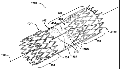
Another exemplary embodiment of a frame based valve
is shown in Figures 11A through 11C. Figure 11A is a
perspective view of an open prosthetic venous valve having
cantilever valve struts in the expanded (deployed) state.
Figures 11B and 11C show side and section views
respectively of the open prosthetic valve.
The prosthetic venous valve 1100 shown in Figures 11A
through 11C share many of the same components with the
prosthetic venous valve 100 previously described. In
addition, prosthetic valve 1100 may be constructed using
the methods described above for the prosthetic venous
valve 100. Accordingly, for ease of illustration, shared
components between prosthetic venous valve 100 and
prosthetic venous valve 1101 are given the same reference
numerals.
The prosthetic venous valve 1100 comprises a
structural frame 101 and a biocompatible membrane assembly
102. In one embodiment, the membrane assembly 102 is
comprised of a tubular membrane 400, valve flaps 403 and
valve cusps 404. The flaps 403 and cusps 404 may be
independent components attached to the tubular membrane to
form the membrane assembly 102, but are preferably part of
CA 02501712 2005-04-07
WO 2004/034933 PCT/US2003/015323
54
and integrated into, the tubular membrane 400. In a
preferred embodiment, the valve flaps 403 and valve cusps
404 are formed into the tubular membrane 400 by processing
techniques as previously disclosed.
The structural frame 101 consists of proximal and
distal anchor structures 103, 104 connected by at least
one connecting member 105. In a preferred embodiment, at
least two connecting members 105 are utilized. The
connecting members 7.05 are attached between the proximal
and distal anchors 103, 104 to further support the
biocompatible membrane assembly 102.
In one embodiment, the connecting members 105 are
substantially straight members, connecting the stent based
proximal and distal anchors 103, 104 in a direction
substantially parallel to the longitudinal axis 106.
Although two connecting members 105 are shown in the
illustrated embodiment, this configuration should not be
construed to limit the scope of the invention.
Alternatively, the- connecting members 105 may be
twisted in a helical fashion as they extend from the
proximal to distal anchors 103, 104. Specifically, the
connection points between the connecting members 105 and
the distal anchor 104, and the connecting members 105 and
the proximal anchor 103, are rotationally phased 180
degrees from each other to provide the helical design.
CA 02501712 2005-04-07
WO 2004/034933 PCT/US2003/015323
Each connecting member 105 may also be biased inward
slightly toward the longitudinal centerline 106 of the
stmt-based anchors 103, 104. This configuration creates
a structural frame 101 having an hour--glass shape with the
5 minimum radius located substantially at the longitudinal
midpoint along the length of connecting member 105.
The proximal and/or distal anchors 103, 104 may also
include spurs or barbs, such as barbs 1150 in Figure 11A,
protruding from the proximal or distal ends.
10 One or more cantilevered valve struts 1101 having
proximal and distal ends are attached to the proximal
anchor 203 to facilitate the opening and closing of the
membrane assembly 102. The proximal end of the valve
struts 1101 may be attached directly or indirectly, to the
15 distal end of the proximal anchor 103. The distal end of
the cantilever valve struts 1101 are attached to the
membrane assembly 102.
The cantilever valve struts 1101 illustrated in
Figures 11A through 11C are substantially straight members
20 between the connection point to the proximal anchor 103
(i.e. between their attachment point to loop ends 210 or
undulating flex segment 1102) and the distal end.
Alternatively, the valve struts 1101 may assume other
configurations, including struts 1101 having, for example,
25 an inward or outward bias, an hour-glass shape or an
CA 02501712 2005-04-07
WO 2004/034933 PCT/US2003/015323
56
undulating S or Z shaped pattern. Figure 11E is a
perspective view of a cantilever valve strut 1101 having
an undulating configuration. The undulating configuration
may add flexibility to the valve strut 1101 and reduce the
strains imposed on the structural frame 101 and membrane
assembly 102 by significant dynamic motion.
Although the exemplary embodiment illustrated in
Figures 11A and 11B shows two connecting members 105 and
two cantilever valve struts 1101, this should not be
construed as limiting the scope of the invention. In
fact, the use of cantilever valve struts 1101 will allow
the prosthetic valve 100 to incorporate a mono-cusp design
with a single connecting member 105 spanning between the
proximal and distal anchors 103, 104 respectively. Still
one of ordinary skill in the art would understand that
other designs may incorporate more or fewer valve struts
1101 and connecting members 105 than those shown.
The proximal end of the cantilevered valve struts
1101 may be connected directly to the proximal anchor 103,
or may be joined to a flexible segment 1102 attached to
the proximal anchor. Figures 11A and IlB illustrate one
embodiment of the invention having undulating flexible ,
segments 1102 and cantilevered valve strut 1101 attached
to the distal end of the proximal anchor 103.
CA 02501712 2005-04-07
WO 2004/034933 PCT/US2003/015323
57
The undulating flexible segment 1102 has a
semicircular configuration and includes a plurality of
longitudinally arranged segment strut members 1108 and a
plurality of segment loop members 1110 connecting adjacent
struts 1108. Adjacent segment strut members 1108 are
connected at opposite ends in a substantially S or Z
shaped pattern, however, one of ordinary skill in the art
would recognize that the pattern shaped by the segment
struts 1108 is not a limiting factor in this invention,
and other shaped patterns may be used.
The plurality of segment loop members 1110 comprise a
substantially semi-circular configuration, having an inner
radii 1112 and outer radii 1114, and are substantially
symmetric about their centers. The inner and outer radii
1112, 1114 respectively, are shown in a close-up
perspective view illustrated in Figure 11D.
Each end of the undulating segment 1102 is attached
to the proximal anchor 103 at attachment points 1115. In
a preferred embodiment, the attachment points 1115 are
located at the outer radii 214 of loop member 210 on the
distal end of the proximal anchor 103. Alternatively the
undulating segment 1102 may be attached to the proximal
anchor 103 at the inner radii 212 of loop members 210, or
at points along strut members 208.
CA 02501712 2005-04-07
WO 2004/034933 PCT/US2003/015323
58
The proximal end of the cantilevered valve strut 1101
is similarly attached to the undulating segment 1102. In
a preferred embodiment, the cantilevered valve strut 1101
is attached to the undulating segment 1102 at the distal
apex, i.e. outer radii 1114, of segment loop member 1110.
Preferably, the attachment point between the cantilevered
valve strut 1101 and the segment loop member 1110 is
centered between the attachment points 1115.
Alternatively, the cantilevered valve strut 1101 may
be attached to the undulating segment 1102 at other distal
or inner or outer radii on the undulating segment 1102; or
along any one of the segment strut members 1108 comprising
undulating segment 1102.
The connection between the cantilevered valve strut
member 1101 and the undulating segment 1102, as well as
the connection between the undulating segment 1102 and
proximal anchor 103, may be by welding or other suitable
connecting means. In addition, the complete frame
structure may be cut from a solid tube of material, and
thus the frame structure would constitute a monolithic
unit. Still other methods for fabricating the complete
frame structure as previously disclosed would be
understood by one of skill in the art . The distal end of
the cantilever valve strut 1101 may be attached to the
membrane assembly 102 by any suitable connection means,
CA 02501712 2005-04-07
WO 2004/034933 PCT/US2003/015323
59
including the use of a binder, heat, or chemical bond,
and/or attachment by mechanical means, such as welding or
suturing. In addition, the cantilever valve strut 1101
may be attached to the membrane assembly 102 during the
coating process as previously disclosed.
The cantilevered valve strut 2101 and undulating
segment 1102 work in tandem to close and open the valve
membrane assembly 102, particularly the valve flaps 403
and valve cusps 404. The cantilevered strut 1101 and
undulating segment 1102 are oriented to maintain the valve
membrane assembly 102 in a normally open position when no
stress is applied, i.e. during antegrade blood flow or
blood stagnation. During retrograde blood flow, the valve
membrane assembly 102, particularly the valve flaps 403
and cusps 404, displace the undulating segment 1102 and
cantilever strut 1101 from the at-rest normally open
position. This displacement generates a reactive force in
the undulating segment 1102, and to some extent in the
cantilever strut member 1101. This reactive force is
transmitted to the valve membrane assembly 102 by the
cantilevered strut 1101, improving the dynamic range of
the valve 100 and ensuring that the stagnation of any
blood that is in the artificial sinus or cusp 404 is
minimized.
CA 02501712 2005-04-07
WO 2004/034933 PCT/US2003/015323
In other embodiments, it may be desirable for the
cantilevered strut 1101 and undulating segment 1102 to
maintain the valve membrane assembly 102 in a normally
closed position when no stress is applied, i.e. during
5 retrograde blood flow or blood stagnation. During
antegrade blood flow, the valve membrane assembly 102,
particularly the valve flaps 403 and cusps 404, displace
the undulating segment 11D2 and cantilever strut 1101 from
the at-rest normally closed position to open the valve.
10 Tn addition, the valve struts 1101 and undulating segments
1102 may be configured in such a way that the valve
membrane 102 will start to close as antegrade flow
decreases, but before retrograde flow begins.
The addition of the cantilever strut member 1101 also
15 provides some rigidity or radial stiffness to the valve
assembly 100. This rigidity facilitates the valve coating
process by inhibiting the valve membrane 102 from
collapsing or closing under the added weight of the
polymer-doped solvent, thus preventing the membrane
20 assembly 102, particularly the valve flaps 403 and valve
cusps 404 from adhering to each other.
The radial stiffness may also allow the wall
thickness of the structural frame between the proximal and
distal anchors 103, 104 respectively (or diameter of the
25 individual connecting members 105) to be thinner than the
CA 02501712 2005-04-07
WO 2004/034933 PCT/US2003/015323
61
remainder of the structural frame. This feature enables
the radial stiffness of the intermediate portion of the
structural frame to be tailored separately from the ends,
and helps reduce the strains imposed on the intermediate
portion by significant dynamic motion.
In addition, the flexible segment 1102 also relieves
stress and distributes the material stress as it
transitions from the proximal anchor 103 to the membrane
assembly 102.
Additional exemplary embodiments of a frame based
valves having one or more cantilever valve struts are
shown in Figures 12A through 12F . Figure 12A and 12B are
perspective views of an open prosthetic venous valve in
the expanded (deployed) state. Figure 12C and 12E show
side views, and Figure 12D and 12F show section views of
these various embodiments.
The prosthetic venous valve 1200 shown in Figures 12A
through 12F is similar in construction to valve 1100,
except that the cantilever valve strut 1201 extends from
the proximal end of the proximal anchor 103 and not from
the distal end of the proximal anchor 103. This
configuration allows the valve struts 1201 to be longer,
increasing the flexibility of the struts 1201 and helps
reduce the strains imposed on the structural frame 101 or
membrane assembly 102 by significant dynamic motion. It
CA 02501712 2005-04-07
WO 2004/034933 PCT/US2003/015323
62
may also allow the cantilever valve strut 1201 to be
thicker, which facilitates the manufacturing processes for
making the structural frame.
As is the case with prosthetic valve 1100, prosthetic
valve 1200 shares many of the same components with the
prosthetic venous valve 100 previously described.
Similarly, the prosthetic valve 1200 may be constructed
using the methods described above for the prosthetic
venous valve 100. Accordingly, for ease of illustration,
shared components between prosthetic venous valve 100 and
prosthetic venous valve 1200 are given the same reference
numerals.
The prosthetic venous valve 1200 comprises a
structural frame 101 and a biocompatible membrane assembly
102, having a tubular membrane 400, valve flaps 403 and
valve cusps 404.
The structural frame 101 consists of proximal and
distal anchor structures 103, I04 respectively connected
by at least one connecting member 105. 2n a preferred
embodiment, at least two connecting members 105 are
utilized, however this configuration should not be
construed to limit the scope of the invention.
In one embodiment, the connecting members 105 are
substantially straight members, connecting the stmt based
proximal and distal anchors 103, 104 in a direction
CA 02501712 2005-04-07
WO 2004/034933 PCT/US2003/015323
63
substantially parallel to the longitudinal axis 106.
Alternatively, the connecting members 105 may be twisted
in a helical fashion or biased inward as they extend from
the proximal to distal anchors 103, 104 as previously
disclosed.
One or more cantilevered valve struts 1201 having
proximal and distal ends are attached to the proximal end
of the proximal anchor 103 to facilitate the opening and
closing of the membrane assembly 102. The proximal end of
the valve struts 1201 may be attached directly or
indirectly, to the proximal end of the proximal anchor 103
and loop back in a distal direction before being attached
to the interior surface of the tubular membrane assembly
102. The connection point between the membrane assembly
102 and the valve strut 1201 is at a point distal to the
proximal anchor 103.
As previously disclosed, the number of valve struts
1201 and number of connecting members 105 in the
accompanying figures should not be construed as limiting
the scope of the invention. One of ordinary skill in the
art would understand that other designs may incorporate
more or fewer valve struts 1201 and connecting members 105
than those shown.
The proximal end of each cantilevered valve strut
1201 may be connected directly to the proximal anchor 103,
CA 02501712 2005-04-07
WO 2004/034933 PCT/US2003/015323
64
or may be joined to a flexible segment 1202 attached to
the proximal anchor.
Figures 12A illustrates one embodiment of the
invention where the proximal end of the valve strut 1201
is. connected directly to the proximal anchor 103. The
valve strut 1201 in Figure 12A has a loop end 1211
incorporated into the proximal end. In addition, each
valve strut 1201 is shown with a bifurcated distal end
1213 having two branches 2214 attached to the membrane
assembly 102. The bifurcated end 1213 may further assist
the valve 1200 by distributing the stress created in the
membrane 102 when the valve 1200 opens and closes.
However, valve strut 1201 having distal end incorporating
a single branch (no bifurcation) or a valve strut 1201
having more than two branches 1214 are also contemplated
by the present invention.
The distal end of the cantilever valve strut 1201 may
be attached to the membrane assembly 102 by any suitable
connection means, including the use of a binder, heat, or
chemical bond, and/or attachment by mechanical means, such
as welding or suturing. In addition, each cantilever
valve strut 1201 may be attached to the membrane assembly
102 during the coating process as previously disclosed.
The loop end 1211 of the valve strut 1201 is attached
directly to the proximal anchor 103 at attachment point
CA 02501712 2005-04-07
WO 2004/034933 PCT/US2003/015323
1215. In a preferred embodiment, the attachment point
1215 is located at the outer radii 214 of loop member 210
on the proximal end of the proximal anchor 103.
Alternatively the proximal end of valve strut 1201 may be
5 attached to the proximal anchor 103 at the inner radii 212
of loop member 210, or at points along strut members 208.
The loop end 1211 has a semi-circular configuration,
substantially symmetric about its center. This
configuration allows the loop end, 1211 to effectively
10 reverse the direction of the cantilever valve strut 1201
from a proximal direction, where it attaches to the outer
radii 214 of loop member 210, to a distal direction.
The proximal end of the cantilever valve strut 1201
may also be indirectly connected to the proximal end of
15 the proximal anchor 103 through a flex segment. Figures
12B through 12D illustrate one such example of a
prosthetic venous valve 1200 according to an embodiment of
the present invention. Figure 12B is a perspective view
of an open prosthetic venous valve in the expanded
20 (deployed) state. Figure 12C and 12D show side and
section views respectively of an open prosthetic valve.
In this embodiment, each of the cantilever valve
struts 1201 are connected to an undulating flex segment
1202, which is in turn attached to the proximal end of the
25 proximal anchor 103.
CA 02501712 2005-04-07
WO 2004/034933 PCT/US2003/015323
66
The undulating flexible segment 1202 is similar in
construction to flex segment 1102 described in Figure 11A,
in that it has a semicircular configuration and includes a
plurality of longitudinally arranged segment strut members
1208 and a plurality of segment loop members 1210
connecting adjacent struts 1208. Adjacent segment strut
members 1208 are connected at opposite ends in a
substantially S or Z shaped pattern, however, the size
(number of loop members 1220 and segment strut members
1208) and pattern shaped by the flexible segment 1202 is
not a limiting factor in this invention, and other shaped
patterns may be used.
The plurality of segment loop members 1210 comprise a
substantially semi-circular configuration, having an inner
radii 1212 and outer radii 1214, and are substantially
symmetric about their centers. An enlarged perspective
view of a loop member 1210, showing the inner and outer
radii 1212 and 1218 respectively is shown in Figure 12G.
Each end of the undulating segment 1202 is attached
to the proximal anchor 103 at attachment points 1215. In
a preferred embodiment, the attachment points 1215 are
located at the outer radii 214 of loop member 210 on the
proximal end of the proximal anchor 103. Alternatively
the undulating segment 1202 may be attached to the
CA 02501712 2005-04-07
WO 2004/034933 PCT/US2003/015323
67
proximal anchor 103 at the inner radii 212 of loop members
210, or at points along strut members 208.
The cantilevered valve strut 1201 is attached to the
undulating segment 1202 at the outer radii 1214, of a
distal segment loop member 1210. Preferably, the
attachment point between. the cantilevered valve strut 1201
and the undulating flex segment 1202 is centered between
the attachment points 7.215.
Alternatively, the cantilevered valve strut 1201 may
be attached to the undulating segment 1202 at other
proximal inner or outer radii on the undulating segment
1202; or along any one of the segment strut members 1208
comprising undulating segment 1202.
The connection between the cantilevered valve strut
member 1201 and the undulating segment 1202, as well as
the connection between the undulating segment 1202 and
proximal anchor 103, may be any of the methods previously
disclosed. In addition, the complete frame structure may
be cut from a solid tube of material, and thus the frame
structure would constitute a monolithic unit. Still other
methods for fabricating the complete frame structure as
previously disclosed would be understood by one of skill
on the art.
The cantilever valve struts 1201 illustrated in
Figures 12A through 12D are substantially straight members
CA 02501712 2005-04-07
WO 2004/034933 PCT/US2003/015323
68
between the Connection point to the proximal anchor 103
(i.e. between their attachment point to loop ends 1211 or
undulating flex segment 1202) and the bifurcated end 1213.
Alternatively, the valve struts 1201 may assume other
configurations, including, for example, an inward or
outward bias, an hour-glass shape, or an undulating s or z
shape. Figure 12H is a perspective view of a cantilever
valve strut 1201 having an undulating configuration. The
undulating configuration may add flexibility to the valve
strut 1201 and reduce the strains imposed on the
structural frame 101 and membrane assembly 102 by
significant dynamic motion. In each of these described
embodiments, the cantilever valve struts 1201 remain
substantially aligned with their respective attachment
points 1215 to the proximal anchor 103. That is to say,
the connection point between a particular valve strut 1201
and the proximal anchor 103 is on the same diametric side
of the valve 1200 as the connection point between the
distal end of the valve strut 1201 and the membrane
assembly 102, regardless of the shape or orientation of
the cantilever valve strut 1201.
In other embodiments, the valve struts 1201 may not
be substantially straight members, but instead may assume
other configurations such twisting in a helical fashion as
they extend distally from the proximal anchor. In
CA 02501712 2005-04-07
WO 2004/034933 PCT/US2003/015323
69
addition the proximal and distal connection paints on the
valve struts 1201 between the proximal anchor 103 (loop
end 1211 or flex segment 1202) and membrane assembly 102
may not be substantially aligned. That is to say, the
connection point between a particular valve strut 1201 and
the proximal anchor 103 is on the opposite diametric side
of the valve 1200 as the connection point between the
distal end of the valve strut 1201 and the membrane
assembly 102.
Figures 12E and 12F show another embodiment of the
invention wherein the valve strut 1201 connection point to
the proximal anchor 103 (i.e. between the attachment point
to loop ends 1211 or undulating flex segment 1202) and the
membrane assembly 102 are substantially diametrically
opposed. As each valve strut 1201 extends distally from
their Corresponding undulating flex segment 1202, they
traverse or cross from one diametric side of the valve to
the other. To avoid interference between the two valve
struts 1201, each are off-set slightly.
As described above, the cantilevered valve strut 1201
and undulating segment 1202 work in tandem to close and
open the valve membrane assembly 102, particularly the
valve flaps 403 and valve cusps 404. In a preferred
embodiment, the cantilevered strut 1201 and undulating
segment 1202 are oriented to maintain the valve membrane
CA 02501712 2005-04-07
WO 2004/034933 PCT/US2003/015323
assembly 102 in a normally open position when no stress is
applied, i.e. during antegrade blood flow or blood
stagnation. During retrograde blood flow, the valve
membrane assembly 102, particularly the valve flaps 403
5 and cusps 404, displace the undulating segment 1202 and
cantilever strut 1201 from the at-rest normally open
position. This displacement generates a reactive force in
the undulating segment 1202, and to some extent in the
cantilever strut member 1201. This reactive force is
10 transmitted to the valve membrane assembly 102 by the
cantilevered strut 1201, improving the dynamic range of
the valve 100 and ensuring that the stagnation of any
blood that is in the artificial sinus or cusp 404 is
minimized.
15 In other embodiments, it may be desirable for the
cantilevered strut 1201 and undulating segment 1202 to
maintain the valve membrane assembly 102 in a normally
closed position when no stress is applied, i.e. during
retrograge blood flow or blood stagnation. During
20 antegrade blood flow, the valve membrane assembly 102,
particularly the valve flaps 403 and cusps 404, displace
the undulating segment 1202 and cantilever strut 1201 from
the at-rest normally closed position to open the valve.
In addition, the valve struts 1201 and undulating segments
25 1202 may be configured in such a way that the valve
CA 02501712 2005-04-07
WO 2004/034933 PCT/US2003/015323
71
membrane 102 will start to close as antegrade flow
decreases, but before retrograde flow begins.
The addition of the cantilever strut members 1201
also provides some rigidity or radial stiffness to the
valve assembly 100. This rigidity facilitates the valve
coating process by inhibiting the valve membrane 102 from
collapsing or closing under the added weight of the
polymer-doped solvent, thus preventing the membrane
assembly 102, particularly the valve flaps 403 and valve
cusps 404 from adhering to each other.
The radial stiffness may also allow the wall
thickness of the structural frame between the proximal and
distal anchors 103, 104 respectively (or diameter of the
individual connecting members 105) to be thinner than the
remainder of the structural frame. This feature enables
the radial stiffness of the intermediate portion of the
structural frame to be tailored separately from the ends,
and helps reduce the strains imposed on the intermediate
portion by significant dynamic motion.
In addition, the flexible segment 1202 also relieves
stress and distributes the material stress as it
transitions from the proximal anchor 103 to the membrane
assembly 102.
It is important to note that the local delivery of
drug/drug combinations may be utilized to treat a wide
CA 02501712 2005-04-07
WO 2004/034933 PCT/US2003/015323
72
variety of conditions utilizing any number of medical
devices, or to enhance the function and/or life of the
device. Medical devices that may benefit from this
treatment include, for example, the frame based
unidirectional flow prosthetic implant subject of the
present invention.
Accordingly, in addition to the embodiments described
above, therapeutic or pharmaceutic agents may be added to
any component of the device during fabrication, including,
for example, the ESS fiber, polymer or coating solution,
membrane tube, structural frame or inner and outer
membrane, to treat any number of conditions. In addition,
therapeutic or pharmaceutic agents may be applied to the
device, such as in the form of a drug or drug eluting
layer, or surface treatment after the device has been
formed. In a preferred embodiment, the therapeutic and
pharmaceutic agents may include any one or more of the
following: antiproliferative/antimitotic agents including
natural products such as vinca alkaloids (i.e.
vinblastine, vincristine, and vinorelbine), paclitaxel,
epidipodophyllotoxins (i.e. etoposide, teniposide),
antibiotics (dactinomycin (actinomycin D) daunorubicin,
doxorubicin and idarubicin), anthracyclines, mitoxantrone,
bleomycins, plicamycin (mithramycin) and mitomycin,
enzymes (L-asparaginase which systemically metabolizes L-
CA 02501712 2005-04-07
WO 2004/034933 PCT/US2003/015323
73
asparagine and deprives cells which do not have the
capacity to synthesize their own asparagine); antiplatelet
agents such as G(GP) llb/llla inhibitors and vitronectin
receptor antagonists; antiproliferative/antimitotic
alkylating agents such as nitrogen mustards
(mechlorethamine, cyclophosphamide and analogs, melphalan,
chlorambucil), ethylenimines and methylmelamines
(hexamethylmelamine and thiotepa), alkyl sulfonates-
busulfan, nirtosoureas (carmustine (BCNU) and analogs,
streptozocin), trazenes - dacarbazinine (DTIC);
antiproliferative/antimitotic antimetabolites such as
folic acid analogs (methotrexate), pyrimidine analogs
(fluorouracil, floxuridine, and cytarabine), purine
analogs and related inhibitors (mercaptopurine,
thioguanine, pentostatin and 2-chlorodeoxyadenosine
~cladribine}); platinum coordination complexes (cisplatin,
carboplatin), procarbazine, hydroxyurea, mitotane,
aminoglutethimide; hormones (i.e. estrogen);
anticoagulants (heparin, synthetic heparin salts and other
inhibitors of thrombin); fibrinolytic agents (such as
tissue plasminogen activator, streptokinase and
urokinase), aspirin, ~ dipyridamole, ticlopidine,
clopidogrel, abciximab; antimigratory; antisecretory
(breveldin); anti-inflammatory: such as adrenocortical
steroids (cortisol, cortisone, fludrocortisone,
CA 02501712 2005-04-07
WO 2004/034933 PCT/US2003/015323
74
prednisone, prednisolone, 6a-methylprednisolone,
triamcinolone, betamethasone, and dexamethasone), ndn-
steroidal agents (salicylic acid derivatives i.e.
aspirin; para-aminophenol derivatives i.e. acetominophen;
indole and indene acetic acids (indomethacin, sulindac,
and etodalac), heteroaryl acetic acids (tolmetin,
diclofenac, and ketorolac), arylpropionic acids (ibuprofen
and derivatives), anthranilic~ acids (mefenamic acid, and
meclofenamic acid), enolic acids (piroxicam, tenoxicam,
phenylbutazone, and oxyphenthatrazone), nabumetone, gold
compounds (auranofin, aurothioglucose, gold sodium
thiomalate); immunosuppressives: (cyclosporine, tacrolimus
(FK-506), sirolimus (rapamycin), azathioprine,
mycophenolate mofetil); angiogenic agents: vascular
endothelial growth factor (VEGF), fibroblast growth factor
(FGF); angiotensin receptor blockers; nitric oxide donors;
anti-sense oligionucleotides and combinations thereof;
cell cycle inhibitors, mTOR inhibitors, and growth factor
receptor signal transduction kinase inhibitors; retenoids;
cyclin/CDK inhibitors; HMG co-enzyme reductase inhibitors
(statins); and protease inhibitors.
While a number of variations of the invention have
been shown and described in detail, other modifications
and methods of use contemplated within the scope of this
invention will be readily apparent to those of skill in
CA 02501712 2005-04-07
WO 2004/034933 PCT/US2003/015323
the art based upon this disclosure. It is contemplated
that various combinations or subcombinations of the
specific embodiments may be made and still fall within the
scope of the invention. For example, the embodiments
5 ~rariously shown to be prosthetic "venous valves" may be
modified to instead incorporate prosthetic "heart valves"
and are also contemplated. Moreover, all assemblies
described are believed useful when modified to treat other
vessels or lumens in the body, in particular other regions
10 of the body where fluid flow in a body vessel or lumen
needs to be controlled or regulated. This may include,
for example, the coronary, vascular, non-vascular and
peripheral vessels and ducts. Accordingly, it should be
understood that various applications, modifications and
15 substitutions may be made of equivalents without departing
from the spirit of the invention or the scope of the
following claims.
The following claims are provided to illustrate
examples of some beneficial aspects of the subject matter
20 disclosed herein which are within the scope of the present
invention.