Note: Descriptions are shown in the official language in which they were submitted.
CA 02501946 2012-02-09
1
COMPOSITIONS AND METHODS FOR DETECTING
WEST NILE VIRUS
Field of the Invention
The present invention relates to the field of biotechnology. More
specifically, the
invention relates to diagnostic assays for detecting the nucleic acids of
flaviviruses, such as
West Nile virus.
Government Interest in Invention
Certain aspects of the invention disclosed herein were made with government
support
under contract N01-HB-07148 with the National Heart, Lung and Blood Institute
of the
National Institutes of Health. The United States government has certain rights
in these aspects
of the invention.
Background of the Invention
West Nile virus (WNV) is an RNA virus that primarily infects birds and culex
mosquitos, with humans and horses serving as incidental hosts. Amplification
of virus in a
bird-mosquito-bird cycle begins when adult mosquitos emerge in early spring
and continues
until fall. This timing coincides with the incidence of disease in humans,
which peaks in late
summer and early fall. Since it was first detected in New York during 1999,
the virus has
spread rapidly throughout most of the United States.
Indeed, during the first nine months of 2002, a total of more than 2,500 human
cases
with laboratory evidence of recent WNV infection were reported in 32 states
and the District of
Columbia. A total of more than 120 human deaths were reported, with the median
age of
decedents being 79 years. Additionally, there were reports of nearly 5,000
dead crows and
nearly 4,000 other dead birds with WNV infection in the United States. Of more
than 3,000
mammals detected with WNV infection, greater than 99% were horses. There were
also nearly
3,400 WNV-positive mosquito pools reported.
Most human infections with the virus are not clinically apparent. Overall,
only 1 in 150
CA 02501946 2005-04-11
WO 2004/036190
infections results in severe neurologic illness =such as
(111fliaatiteitC-TA5_29PIPJ3639
cord) or encephalitis (inflammation of the brain). Milder symptoms, which
generally last 3 to 6
days and are more commonly reported in connection with WNV infection, include
a fever of
sudden onset, often accompanied by malaise, anorexia, nausea, vomiting, eye
pain, headache,
myalgia, rash, and lymphadenopathy. The incubation period of WNV, although not
precisely
known, probably ranges from 3 to 14 days. An analysis of attack rates per
million persons
during the 1999 New York City outbreak showed that the incidence of severe
neurologic
disease was more than 40 times higher in those at least 80 years of age when
compared with
persons up to 19 years of age. Thus, advanced age is an important risk factor
for more severe
neurologic disease.
In addition to transmission from mosquitoes, transmission has been linked to
blood
transfusion and organ transplantation. For example, four recipients of
transplanted organs from
single donor in the U.S. became infected with West Nile virus in mid-2002.
Three of the
recipients developed encephalitis, with one of the three dying as a result.
The fourth recipient
developed mild symptoms of viral infection without encephalitis, but also
tested positive for
the virus. The organ donor, who was injured in an automobile accident,
received numerous
transfusions of blood products before dying. She was not known to have been
ill before the
accident, and a sample of her blood taken before any of the transfusions
showed no evidence of
West Nile virus. In a separate instance, a nursing mother whose breast milk
contained WNV
,0 and a male liver transplant patient both received transfused blood from
a common donor, and
both developed West Nile virus infections. A stored blood sample from that
donor tested
positive for the WNV, again suggesting a common source of the infectious
virus.
West Nile virus is a single-stranded plus-sense RNA virus taxonomically
classified in
the family Flaviviridae, under the genus Flavivirus. Accordingly, the virus is
a member of the
Japanese encephalitis virus serocomplex, which contains several medically
important viruses
associated with human encephalitis: Japanese encephalitis, St. Louis
encephalitis, Murray
Valley encephalitis, and Kunjin virus (an Australian subtype of West Nile
virus). The viral
genome size is approximately 11 kb.
Nucleic acid-based tests for WNV have been described. For example, Shi et al.,
in J.
30 Clin. Microbiol. 39:1264 (2001) have described a real-time polymerase
chain reaction (PCR)
assay for WNV nucleic acids. Lanciotti et al., in J. Clin. Microbiol. 38:4066
(2001) have
described a TaqMan-based assay for the detection of WNV RNA in human
specimens,
2
CA 02501946 2005-04-11
moscia2 liT0443Envian tissue specimens. Despite the
i'availabilitorifiest,cTa...S-M3J.9,.3..4-92,
there remains a need for a WNV screening assay that is specifically adapted
for the needs of
clinical testing laboratories. The method should particularly lend itself to
high throughput
screening which may be required for testing large numbers of clinical and
donated blood or
tissue samples.
Summary of the Invention
A first aspect of the invention relates to a hybridization assay probe for
detecting a
nucleic acid. This hybridization assay probe includes a probe sequence that
has a target-
complementary sequence of bases, and optionally one or more base sequences
that are not
complementary to the nucleic acid that is to be detected. The target-
complementary sequence
of bases consists of 12-87 contiguous bases contained within the sequence of
SEQ ID NO:101
or the complement thereof, allowing for the presence of RNA and DNA
equivalents, nucleotide
analogs and up to 10%, or even up to 20% base differences. In general, the
invented
hybridization assay probe can have a length of up to 100 bases. In a preferred
embodiment, the
target-complementary sequence of bases consists of 12-69 contiguous bases
contained within
the sequence of SEQ ID NO:102 or the complement thereof, allowing for the
presence of RNA
and DNA equivalents, nucleotide analogs and up to 10%, or even up to 20% base
differences.
Still more preferably, the hybridization assay probe includes the optional
base sequences that
are not complementary to the nucleic acid that is to be detected. Even still
more preferably, the
hybridization assay probe includes a detectable label. For example, the probe
may include a
= fluorophore moiety and a quencher moiety. In such an instance the
hybridization assay probe
can be a molecular beacon. An exemplary molecular beacon can include a target-
complementary sequence of bases that consists of any one of SEQ ID NO:179, SEQ
ID
NO:180 or SEQ ID NO:181. In accordance with another preferred embodiment of
the invented
hybridization assay probe, when the target-complementary sequence of bases
consists of 12-69
contiguous bases contained within the sequence of SEQ ID NO:102 or the
complement thereof,
allowing for the presence of RNA and DNA equivalents, nucleotide analogs and
up to 10%, or
even up to 20% base differences, the probe sequence does not include the
optional base
sequences that are not complementary to the nucleic acid that is to be
detected. Still more
preferably, the invented hybridization assay probe has a length of up to 69
bases, and yet still
more preferably includes a detectable label. In accordance with another
preferred embodiment,=
the target-complementary sequence of bases consists of 18-52 contiguous bases
contained
3
CA 02501946 2005-04-11
WO 2004/036190
PCT/US2003/033639
within the sequence of SEQ ID NO:103 or the complement thereof, allowing 'or
me presence
of RNA and DNA equivalents, nucleotide analogs and up to 10%, or even up to
20% base
differences. Still more preferably, the probe sequence does not include the
optional base
sequences that are not complementary to the nucleic acid that is to be
detected, but may further
include a detectable label. This detectable label can be either a
chemiluminescent label or a
fluorescent label. In accordance with an alternative embodiment, the
hybridization assay probe
consists of 18-52 contiguous bases contained within the sequence of SEQ ID
NO:103 or the
complement thereof, allowing for the presence of RNA and DNA equivalents,
nucleotide
analogs and up to 10%, or even up to 20% base differences, and has a length of
up to 52 bases.
LO When this is the case, the target-complementary sequence of bases can
consist of 18-22
contiguous bases contained within the sequence of SEQ ID NO:103 or the
complement thereof,
allowing for the presence of RNA and DNA equivalents, nucleotide analogs and
up to 10%, or
even up to 20% base differences, and the hybridization assay probe can have a
length of up to
22 bases. In one embodiment, the invented probe can have the sequence of SEQ
ID NO:116.
In another embodiment, the probe sequence can be any of SEQ ID NO:114, SEQ ID
NO:111,
SEQ ID NO:110, SEQ ID NO:109, SEQ ID NO:108, SEQ ID NO:107 or SEQ ID NO:106.
Another aspect of the invention relates to a kit for amplifying a target
nucleic acid
sequence that may be present in a biological sample. The invented kit contains
a first primer
that has a 3' terminal target-complementary sequence and optionally a first
primer upstream
sequence that is not complementary to the target nucleic acid sequence that is
to be amplified.
The 3' terminal target-complementary sequence of this first primer includes 22
contiguous
bases contained within SEQ ID NO:73, allowing for the presence of RNA and DNA
equivalents, nucleotide analogs and up to 10%, or even up to 20% base
differences. Also
included in the kit is a second primer that has a 3' terminal target-
complementary sequence and
optionally a second primer upstream sequence that is not complementary to the
target nucleic
acid sequence that is to be amplified. The 3' terminal target-complementary
sequence of the
second primer includes 18 contiguous bases contained within SEQ ID NO:59,
allowing for the
presence of RNA and DNA equivalents, nucleotide analogs and up to 10%, or even
up to 20%
base differences. In a preferred embodiment of the invented kit, the first
primer and the second
primer are each up to 60 bases in length. In another preferred embodiment, the
3' terminal
target-complementary sequence of the first primer and the 3' terminal target-
complementary
sequence of the second primer are each up to 35 bases in length. When this is
the case, it is
=
4
CA 02501946 2005-04-11
WO 2004/036190
preferable tor me .5. terminal target-complementary sequeiVee'Ofttlie Al:sr
p¨finr.C.VP.5,?9,3/.9.3fi.39
bases in length. Alternatively, and in accordance with yet another preferred
embodiment, the 3'
terminal target-complementary sequence of the first primer can be up to 35
bases in length and
the 3' terminal target-complementary sequence of the second primer can be up
to 22 bases in
length. When the first primer is up to 24 bases in length, it is highly
preferred for the 3'
terminal target-complementary sequence of the second primer to be up to 22
bases in length.
Still more preferably, the first primer includes a first primer upstream
sequence, such as a
promoter sequence for T7 RNA polymerase. In accordance with another preferred
embodiment
of the invented kit, when the 3' terminal target-complementary sequence of the
first primer is
0 up to 24 bases in length, and when the 3' terminal target-complementary
sequence of the
second primer is up to 22 bases in length, the 3' terminal target-
complementary sequence of the
first primer is preferably any of SEQ ID NO:75, SEQ ID.NO:76 and SEQ ID NO:77,
and the 3'
teiminal target-complementary sequence of the second primer is preferably any
of SEQ ID
NO:60, SEQ ID NO:62, SEQ ID NO:63, SEQ ID NO:64, SEQ ID NO:65, SEQ ID NO:66,
5 SEQ ID NO:67, SEQ ID NO:68, SEQ ID NO:69, SEQ ID NO:70 and SEQ ID NO:71.
In
accordance with another preferred embodiment, when the 3' terminal target-
complementary
sequence of the first primer is up to 24 bases in length, and when the 3'
terminal target-
complementary sequence of the second primer is up to 35 bases in length, the
3' terminal
target-complementary sequence of the first primer includes 22 contiguous bases
contained
within SEQ ID NO:74, allowing for the presence of RNA and DNA equivalents,
nucleotide
analogs and up to 10%, or even up to 20% base differences. When this is the
case, the 3'
terminal target-complementary sequence of the second primer can be up to 22
bases in length.
Alternatively, the first primer may include a first primer upstream sequence,
such as a promoter
sequence for T7 RNA polymerase.
25 Another aspect of the invention relates to a hybridization assay probe
for detecting a
nucleic acid. The invented hybridization assay probe includes a probe sequence
that has a
target-complementary sequence of bases, and optionally one or more base
sequences that are
not complementary to the nucleic acid that is to be detected. The target-
complementary
sequence of bases consists of 10-20 contiguous bases contained within the
sequence of SEQ ID
30 NO:99 or the complement thereof, allowing for the presence of RNA and
DNA equivalents,
nucleotide analogs and up to 10%, or even up to 20% base differences. Finally,
the invented
hybridization assay probe can have a length of up to 100 bases. In a preferred
embodiment, the
5
CA 02501946 2005-04-11
WO 2004/036190 " - " "µ"
TCT/US2003/033639
length of the hybridization assay probe is up to 30 bases. Still more
prererauly, LUG pitiuu
sequence includes the optional base sequences that are not complementary to
the nucleic acid
that is to be detected. In accordance with a first version of this embodiment,
there is further
included a detectable label. In accordance with a second version of this
embodiment, there is
further included a fluorophore moiety and a quencher moiety, and the
hybridization assay
probe is a molecular beacon. In a different embodiment, wherein the length of
the
hybridization assay probe is up to 30 bases, the probe sequence consists of 10-
20 contiguous
bases contained within the sequence of SEQ ID NO:99 or the complement thereof,
allowing for
the presence of RNA and DNA equivalents, nucleotide analogs and up to 10%, or
even up to
0 20% base differences, and does not include the optional base sequences
that are not
complementary to the WNV nucleic acids. More preferably, the hybridization
assay probe has
a length of up to 20 bases. In certain embodiments wherein the length of the
hybridization
assay probe is up to 30 bases, the target-complementary sequence of bases
consists of 19-20
contiguous bases contained within the sequence of SEQ ID NO:99 or the
complement thereof,
[5 allowing for the presence of RNA and DNA equivalents, nucleotide analogs
and up to 10%, or
even up to 20% base differences. In accordance with a first preferred version
of this
embodiment, the probe sequence consists of 19-20 contiguous bases contained
within the
sequence of SEQ ID NO:99 or the complement thereof, allowing for the presence
of RNA and
DNA equivalents, nucleotide analogs and up to 10%, or even up to 20% base
differences, and
20 does not include the optional base sequences that are not complementary
to the nucleic acid
that is to be detected. In accordance with a second preferred version of this
embodiment, the
hybridization assay probe further includes a detectable label, such as a
chemiluminescent label
or a fluorescent label. In accordance with a third preferred version of this
embodiment, the
target-complementary sequence of bases consists of 19-20 contiguous bases
contained within
25 the sequence of SEQ ID NO:99 or the complement thereof, allowing for the
presence of RNA
and DNA equivalents, nucleotide analogs and up to 10%, or even up to 20% base
differences,
and the hybridization assay probe has a length of up to 20 bases. For example,
the probe
sequence can be SEQ ID NO:100. In accordance with a different embodiment, when
the length
of the hybridization assay probe is up to 30 bases, and when the probe
sequence includes the
30 optional base sequences that are not complementary to the nucleic acid
that is to be detected,
the target-complementary sequence of bases may be any of SEQ ID NO:164, SEQ ID
NO:165,
SEQ ID NO:166, SEQ ID NO:167, SEQ ID NO:168, SEQ ID NO:169 or SEQ ID NO:170.
6
CA 02501946 2005-04-11
WO 2004/036190 -
- - PCT/US2003/033639
Another aspect of the invention relates to a kit for amplifying a target
iluts,icii.; duiu
sequence that may be present in a biological sample. This kit contains a first
primer that
includes a 3' terminal target-complementary sequence and optionally a first
primer upstream
sequence that is not complementary to the target nucleic acid sequence that is
to be amplified.
The 3' terminal target-complementary sequence of the first primer includes 22
contiguous bases
contained within SEQ ID NO:52, allowing for the presence of RNA and DNA
equivalents,
nucleotide analogs and up to 10%, or even up to 20% base differences. The kit
further contains
a second primer that includes a 3' terminal target-complementary sequence and
optionally a
second primer upstream sequence that is not complementary to the target
nucleic acid sequence
0 that is to be amplified. The 3' terminal target-complementary sequence of
the second primer
includes 22 contiguous bases contained within SEQ ID NO:41, allowing for the
presence of
RNA and DNA equivalents, nucleotide analogs and up to 10%, or even up to 20%
base
differences. In a preferred embodiment, the first primer and the second primer
each are up to
60 bases in length. In a different preferred embodiment, the 3' terminal
target-complementary
5 sequence of the first primer and the 3' terminal target-complementary
sequence of the second
primer are each up to 35 bases in length. When this is the case, the 3'
terminal target-
complementary sequence of the first primer is preferably up to 26 bases in
length. In
accordance with a different preferred embodiment, when the 3' terminal target-
complementary
sequence of the first primer and the 3' terminal target-complementary sequence
of the second
primer are each up to 35 bases in length, the 3' teiminal target-complementary
sequence of the
second primer can be up to 23 bases in length. In yet another preferred
embodiment, the 3'
terminal target-complementary sequence of the first primer is preferably up to
26 bases in
length, and the 3' terminal target-complementary sequence of the second primer
is up to 23
bases in length. In accordance with a first preferred version of this
embodiment, the 3' terminal
25 target-complementary sequence of the first primer may be selected from
the group consisting of
SEQ ID NO:53, SEQ NO:54 and SEQ ID NO:55, and the 3' terminal target-
complementary
sequence of the second primer may be selected from the group consisting of SEQ
ID NO:42,
SEQ ID NO:43, SEQ ID NO:44, SEQ ID NO:45, SEQ ID NO:46, SEQ ID NO:47, SEQ ID
NO:48, SEQ ID NO:49, SEQ ID NO:50, and SEQ ID NO:51. In accordance with a
second
30 preferred version of this embodiment, the 3' terminal target-
complementary sequence of the
second primer is up to 23 bases in length. When this is the case, the first
primer may include a
first primer upstream sequence, such as a promoter sequence for T7 RNA
polymerase.
7
CA 02501946 2005-04-11
WO 2004/036190
PCT/US2003/033639
Another aspect of the invention relates to a hybridization assay probe ror
clam Ling a
nucleic acid. The invented hybridization assay probe has a probe sequence that
includes a
target-complementary sequence of bases, and optionally one or more base
sequences that are
not complementary to the nucleic acid that is to be detected. The target-
complementary
sequence of bases consists of 13-37 contiguous bases contained within the
sequence of SEQ ID
NO:95 or the complement thereof, allowing for the presence of RNA and DNA
equivalents,
nucleotide analogs and up to 10%, or even up to 20% base differences.
Generally speaking, the
hybridization assay probe can have a length of up to 100 bases. In a preferred
embodiment, the
length of the hybridization assay probe is up to 37 bases. More preferably,
the hybridization
0 assay probe includes the optional base sequences that are not
complementary to the nucleic
acid that is to be detected. Still more preferably, the hybridization assay
probe further includes
a detectable label. For example, the hybridization assay probe may further
include a
fluorophore moiety and a quencher moiety. In this instance the hybridization
assay probe can
be a molecular beacon. In a different embodiment of the invented hybridization
assay probe,
.5 the probe sequence consists of 13-20 contiguous bases contained within
the sequence of SEQ
ID NO:95 or the complement thereof, allowing for the presence of RNA and DNA
equivalents,
nucleotide analogs and up to 10%, or even up to 20% base differences, and does
not include the
optional base sequences that are not complementary to the nucleic acid that is
to be detected.
In accordance with still another embodiment, when the length of the
hybridization assay probe
20 is up to 37 bases, the target-complementary sequence of bases consists
of 13-20 contiguous
bases contained within the sequence of SEQ ID NO:95 or the complement thereof,
allowing for
the presence of RNA and DNA equivalents, nucleotide analogs and up to 10%, or
even up to
20% base differences. More preferably, the probe sequence consists of 20
contiguous bases
contained within the sequence of SEQ ID NO:95 or the complement thereof,
allowing for the
25 presence of RNA and DNA equivalents, nucleotide analogs and up to 10%,
or even up to 20%
base differences, and does not include the optional base sequences that are
not complementary
to the nucleic acid that is to be detected. Still more preferably, the
hybridization assay probe
further includes a detectable label, such as a chemiluminescent label or a
fluorescent label. In
accordance with still yet another preferred embodiment, when the length of the
hybridization
30 assay probe is up to 37 bases, and when the target-complementary
sequence of bases consists
of 13-20 contiguous bases contained within the sequence' of SEQ ID NO:95 or
the complement
thereof, allowing for the presence of RNA and DNA equivalents, nucleotide
analogs and up to
8
CA 02501946 2005-04-11
WO 2004/036190-
It
5 PCT/US2003/033639
10%, or even up to 20% base differences, the probe sequence does not include
the optional
base sequences that are not complementary to the nucleic acid that is to be
detected, and the
hybridization assay probe has a length of up to 20 bases. For example, the
probe sequence may
be SEQ ID NO:98. Generally speaking, when the length of the hybridization
assay probe is up
to 37 bases, the target-complementary sequence of bases can, for example, be
any one of SEQ
ID NO:154, SEQ ID NO:155, SEQ ID NO:156, SEQ ID NO:157, or SEQ ID NO:158.
Another aspect of the invention relates to a kit for amplifying a target
nucleic acid
sequence that may be present in a biological sample. The invented kit contains
a first primer
that includes a 3' terminal target-complementary sequence, and optionally a
first primer
upstream sequence that is not complementary to the target nucleic acid
sequence that is to be
amplified. The 3' terminal target-complementary sequence of the first primer
includes 20
contiguous bases contained within SEQ ID NO:16, allowing for the presence of
RNA and
DNA equivalents, nucleotide analogs and up to 10%, or even up to 20% base
differences. The
kit further contains a second primer that includes a 3' terminal target-
complementary sequence
5 up to 30 bases in length, and optionally a second primer upstream
sequence that is not
complementary to the target nucleic acid sequence that is to be amplified. The
3' terminal
target-complementary sequence of the second primer includes 20 contiguous
bases contained
within SEQ ID NO:1, allowing for the presence of RNA and DNA equivalents,
nucleotide
analogs and up to 10%, or even up to 20% base differences. In a preferred
embodiment, the
!O first primer and the second primer are each up to 60 bases in length. In
a different preferred
embodiment, the 3' terminal target-complementary sequence of the first primer
is up to 35
bases in length. In accordance with a first preferred version of this
embodiment, the 3' terminal
target-complementary sequence of the first primer is up to 24 bases in length.
In accordance
with a second preferred version of this embodiment, the 3' terminal target-
complementary
?.5 sequence of the second primer is up to 24 bases in length. In yet
another preferred
embodiment, when the 3' terminal target-complementary sequence of the first
primer is up to
24 bases in length, the 3' terminal target-complementary sequence of the
second primer is
preferably up to 24 bases in length. In an alternative embodiment, the 3'
terminal target-
complementary sequence of the second primer is up to 26 bases in length and
includes 20
30 contiguous bases contained within SEQ ID NO:2, allowing for the presence
of RNA and DNA
equivalents, nucleotide analogs and up to 10%, or even up to 20% base
differences. In
accordance with a first preferred version of this embodiment, the 3' terminal
target-
9
CA 02501946 2005-04-11
WO 2004/036190i"" tt 1.4` `µ"`' PC
T/US 2 0 0 3/0 336 39
complementary sequence of the first primer is up to 24 bases in length. In
accordance wan a
second preferred version of this embodiment, the 3' terminal target-
complementary sequence of
the second primer is up to 24 bases in length. Preferably, the 3' terminal
target-complementary
sequence of the second primer is up to 24 bases in length. Still more
preferably, the 3' terminal
target-complementary sequence of the first primer is any one of SEQ ID NO:24,
SEQ ID
NO:25, SEQ ID NO:26 or SEQ ID NO:28. In certain embodiments wherein the 3'
terminal
target-complementary sequence of the first primer is up to 24 bases in length,
and the 3'
terminal target-complementary sequence of the second primer is up to 26 bases
in length and
includes 20 contiguous bases contained within SEQ ID NO:2, allowing for the
presence of
RNA and DNA equivalents, nucleotide analogs and up to 10%, or even up to 20%
base
differences, the 3' terminal target-complementary sequence of the second
primer is any one of
SEQ ID NO:10, SEQ ID NO:11, SEQ ID NO:12, SEQ ID NO:13, SEQ ID NO:14 or SEQ ID
NO:15. Alternatively, when the 3' terminal target-complementary sequence of
the first primer
is any one of SEQ ID NO:24, SEQ ID NO:25, SEQ ID NO:26 or SEQ ID NO:28, the 3'
terminal target-complementary sequence of the second primer may be any of SEQ
ID NO:10,
SEQ ID NO:11, SEQ ID NO:12, SEQ ID NO:13, SEQ ID NO:14 or SEQ ID NO:15. In
accordance with a highly preferred embodiment, the first primer includes the
first primer
upstream sequence, such as a promoter sequence for T7 RNA polymerase.
Definitions
:0 The following terms have the following meanings for the purpose of
this disclosure,
unless expressly stated to the contrary herein.
As used herein, a "biological sample" is any tissue or polynucleotide-
containing
material obtained from a human, animal or environmental sample. Biological
samples in
accordance with the invention include peripheral blood, plasma, serum or other
body fluid,
bone marrow or other organ, biopsy tissues or other materials of biological
origin. A biological
sample may be treated to disrupt tissue or cell structure, thereby releasing
intracellular
components into a solution which may contain enzymes, buffers, salts,
detergents and the like.
As used herein, "polynucleotide" means either RNA or DNA, along with any
synthetic
nucleotide analogs or other molecules that may be present in the sequence and
that do not
30 prevent hybridization of the polynucleotide with a second molecule
having a complementary
sequence.
As used herein, a "detectable label" is a chemical species that can be
detected or can
CA 02501946 2005-04-11
WO 2004/036190
PCT/US2003/033639
lead to a detectable response. Detectable labels in accordance with the
invenuon can De 1111.K.GD
to polynucleotide probes either directly or indirectly, and include
radioisotopes, enzymes,
haptens, chromophores such as dyes or particles that impart a detectable color
(e.g., latex beads
or metal particles), luminescent compounds (e.g., bioluminescent,
phosphorescent or
chemiluminescent moieties) and fluorescent compounds.
A "homogeneous detectable label" refers to a label that can be detected in a
homogeneous fashion by determining whether the label is on a probe hybridized
to a target
sequence. That is, homogeneous detectable labels can be detected without
physically removing
hybridized from unhybridized forms of the label or labeled probe. Homogeneous
detectable
0 labels are preferred when using labeled probes for detecting WNV nucleic
acids. Examples of
homogeneous labels have been described in detail by Arnold et al., U.S. Patent
No. 5,283,174;
Woodhead et al., U.S. Patent No. 5,656,207; and Nelson et al., U.S. Patent No.
5,658,737.
Preferred labels for use in homogenous assays include chemiluminescent
compounds (e.g., see
Woodhead et al., U.S. Patent No. 5,656,207; Nelson et al., U.S. Patent No.
5,658,737; and
[5 Arnold, Jr., et al., U.S. Patent No. 5,639,604). Preferred
chemiluminescent labels are
acridinium ester ("AE") compounds, such as standard AE or derivatives thereof
(e.g., naphthyl-
AE, ortho-AE, 1- or 3-methyl-AE, 2,7-dimethyl-AE, 4,5-dimethyl-AE, ortho-
dibromo-AE,
ortho-dimethyl-AE, meta-dimethyl-AE, ortho-methoxy-AE, ortho-methoxy(cinnamy1)-
AE,
ortho-methyl-AE, ortho-fluoro-AE, 1- or 3-methyl-ortho-fluoro-AE, 1- or 3-
methyl-meta-
a0 difluoro-AE, and 2-methyl-AE).
A "homogeneous assay" refers to a detection procedure that does not require
physical
separation of hybridized probe from non-hybridized probe prior to determining
the extent of
specific probe hybridization. Exemplary homogeneous assays, such as those
described herein,
can employ molecular beacons or other self-reporting probes which emit
fluorescent signals
25 when hybridized to an appropriate target, chemiluminescent acridinium
ester labels which can
be selectively destroyed by chemical means unless present in a hybrid duplex,
and other
homogeneously detectable labels that will be familiar to those having an
ordinary level of skill
in the art.
As used herein, "amplification" refers to an in vitro procedure for obtaining
multiple
30 copies of a target nucleic acid sequence, its complement or fragments
thereof.
By "target nucleic acid" or "target" is meant a nucleic acid containing a
target nucleic
acid sequence. In general, a target nucleic acid sequence that is to be
amplified will be
11
CA 02501946 2005-04-11
WO 2004/036190
PCT/US2003/033639
positioned between two oppositely disposed primers, and will include the poi
UULL Ul LIle turgui
nucleic acid that is fully complementary to each of the primers.
By "target nucleic acid sequence" or "target sequence" or "target region" is
meant a
specific deoxyribonucleotide or ribonucleotide sequence comprising all or part
of the
nucleotide sequence of a single-stranded nucleic acid molecule, and the
deoxyribonucleotide or
ribonucleotide sequence complementary thereto.
By "transcription associated amplification" is meant any type of nucleic acid
amplification that uses an RNA polymerase to produce multiple RNA transcripts
from a
nucleic acid template. One example of a transcription associated amplification
method, called
0 "Transcription Mediated Amplification" (TMA), generally employs an RNA
polymerase, a
DNA polymerase, deoxyribonucleoside triphosphates, ribonucleoside
triphosphates, and a
promoter-template complementary oligonucleotide, and optionally may include
one or more
analogous oligonucleotides. Variations of TMA are well known in the art as
disclosed in detail
in Burg et al., U.S. Patent No. 5,437,990; Kacian et al., U.S. Patent Nos.
5,399,491 and
5 5,554,516; Kacian et al., PCT No. WO 93/22461; Gingeras et al., PCT No.
WO 88/01302;
Gingeras et al., PCT No. WO 88/10315; Malek et al., U.S. Patent No. 5,130,238;
Urdea et al.,
U.S. Patent Nos. 4,868,105 and 5,124,246; McDonough et al., PCT No. WO
94/03472; and
Ryder et al., PCT No. WO 95/03430. The methods of Kacian et al. are preferred
for
conducting nucleic acid amplification procedures of the type disclosed herein.
!,0 As used herein, an "oligonucleotide" or "oligomer" is a polymeric
chain of at least two,
generally between about five and about 100, chemical subunits, each subunit
comprising a
nucleotide base moiety, a sugar moiety, and a linking moiety that joins the
subunits in a linear
spacialconfiguration. Common nucleotide base moieties are guanine (G), adenine
(A),
cytosine (C), thymine (T) and uracil (U), although other rare or modified
nucleotide bases able
Z5 to hydrogen bond are well known to those skilled in the art.
Oligonucleotides may optionally
include analogs of any of the sugar moieties, the base moieties, and the
backbone constituents.
Preferred oligonucleotides of the present invention fall in a size range of
about 10 to about 100
residues. Oligonucleotides may be purified from naturally occurring sources,
but preferably
are synthesized using any of a variety of well known enzymatic or chemical
methods.
30 As used herein, a `µ`probe" is an oligonucleotide that hybridizes
specifically to a target
sequence in a nucleic acid, preferably in an amplified nucleic acid, under
conditions that
promote hybridization, to form a detectable hybrid. A probe optionally may
contain a
12
CA 02501946 2005-04-11
WO 2004/036190
PCT/US2003/033639
detectable moiety which either may be attached to the end(s) of the probe or
may De 111Leillal.
The nucleotides of the probe which combine with the target polynucleotide need
not be strictly
contiguous, as may be the case with a detectable moiety internal to the
sequence of the probe.
Detection may either be direct (i.e., resulting from a probe hybridizing
directly to the target
sequence or amplified nucleic acid) or indirect (i.e., resulting from a probe
hybridizing to an
intermediate molecular structure that links the probe to the target sequence
or amplified nucleic
acid). The "target" of a probe generally refers to a sequence contained within
an amplified
nucleic acid sequence which hybridizes specifically to at least a portion of a
probe
oligonucleotide using standard hydrogen bonding (i.e., base pairing). A probe
may comprise
0 target-specific sequences and optionally other sequences that are non-
complementary to the
target sequence that is to be detected. These non-complementary sequences may
comprise a
promoter sequence, a restriction endonuclease recognition site, or sequences
that contribute to
three-dimensional conformation of the probe (e.g., as described in Lizardi et
al., U.S. Patent
Nos. 5,118,801 and 5,312,728). Sequences that are "sufficiently complementary"
allow stable
5 hybridization of a probe oligonucleotide to a target sequence that is not
completely
complementary to the probe's target-specific sequence.
As used herein, an "amplification primer" is an oligonucleotide that
hybridizes to a
target nucleic acid, or its complement, and participates in a nucleic acid
amplification reaction.
For example, amplification primers, or more simply "primers," may be
optionally modified
oligonucleotides which are capable of hybridizing to a template nucleic acid
and which have a
3' end that can be extended by a DNA polymerase activity. In general, a primer
will have a
downstream WNV-complementary sequence, and optionally an upstream sequence
that is not
complementary to WNV nucleic acids. The optional upstream sequence may, for
example,
serve as an RNA polymerase promoter or contain restriction endonuclease
cleavage sites.
By "substantially homologous," "substantially corresponding" or "substantially
corresponds" is meant that the subject oligonucleotide has a base sequence
containing an at
least 10 contiguous base region that is at least 70% homologous, preferably at
least 80%
homologous, more preferably at least 90% homologous, and most preferably 100%
homologous to an at least 10 contiguous base region present in a reference
base sequence
30 (excluding RNA and DNA equivalents). Those skilled in the art will
readily appreciate
modifications that could be made to the hybridization assay conditions at
various percentages
of homology to permit hybridization of the oligonucleotide to the target
sequence while
13
CA 02501946 2005-04-11
WO 2004/036190
PCT/US2003/033639
preventing unacceptable levels of non-specific hybridization. The degree of
similarity is
determined by comparing the order of nucleobases making up the two sequences
and does not
take into consideration other structural differences which may exist between
the two sequences,
provided the structural differences do not prevent hydrogen bonding with
complementary
bases. The degree of homology between two sequences can also be expressed in
terms of the
number of base mismatches present in each set of at least 10 contiguous bases
being compared,
which may range from 0-2 base differences.
By "substantially complementary" is meant that the subject oligonucleotide has
a base
sequence containing an at least 10 contiguous base region that is at least 70%
complementary,
0 preferably at least 80% complementary, more preferably at least 90%
complementary, and
most preferably 100% complementary to an at least 10 contiguous base region
present in a
target nucleic acid sequence (excluding RNA and DNA equivalents). (Those
skilled in the art
will readily appreciate modifications that could be made to the hybridization
assay conditions
at various percentages of complementarity to permit hybridization of the
oligonucleotide to the
.5 , target sequence while preventing unacceptable levels of non-specific
hybridization.) The
degree of complementarity is determined by comparing the order of nucleobases
making up the
two sequences and does not take into consideration other structural
differences which may exist ,
between the two sequences, provided the structural differences do not prevent
hydrogen
bonding with complementary bases. The degree of complementarily between two
sequences
20 can also be expressed in terms of the number of base mismatches present
in each set of at least
contiguous bases being compared, which may range from 0-2 base mismatches.
By "sufficiently complementary" is meant a contiguous nucleic acid base
sequence that
is capable of hybridizing to another base sequence by hydrogen bonding between
a series of
complementary bases. Complementary base sequences may be complementary at each
position
25 in the base sequence of an oligonucleotide using standard base pairing
(e.g., G:C, A:T or A:U
pairing) or may contain one or more residues that are not complementary using
standard
hydrogen bonding (including abasic "nucleotides"), but in which the entire
complementary
base sequence is capable of specifically hybridizing with another base
sequence under
appropriate hybridization conditions. Contiguous bases are preferably at least
about 80%, more
30 preferably at least about 90%, and most preferably about 100%
complementary to a sequence
to which an oligonucleotide is intended to specifically hybridize. Appropriate
hybridization
conditions are well known to those skilled in the art, can be predicted
readily based on base
14
CA 02501946 2005-04-11
WO 2004/036190
sequence composition, or can be determined empirically Fyns*'in`i'roni"iin-e'
Sambrook et al., Molecular Cloning, A Laboratory Manual, 2nd ed. (Cold Spring
Harbor
Laboratory Press, Cold Spring Harbor, NY, 1989) at 1.90-1.91, 7.37-7.57,
9.47-9.51 and
11.47-11.57 particularly at 9.50-9.51, 11.12-11.13, 11.45-11.47 and 11.55-
11.57).
By "capture oligonucleotide" is meant at least one nucleic acid
oligonucleotide that
provides means for specifically joining a target sequence and an immobilized
oligonucleotide
due to base pair hybridization. A capture oligonucleotide preferably includes
two binding
regions: a target sequence-binding region and an immobilized probe-binding
region, usually
contiguous on the same oligonucleotide, although the capture oligonucleotide
may include a
target sequence-binding region and an immobilized probe-binding region which
are present on
two different oligonucleotides joined together by one or more linkers. For
example, an
immobilized probe-binding region may be present on a first oligonucleotide,
the target
sequence-binding region may be present on a second oligonucleotide, and the
two different
oligonucleotides are joined by hydrogen bonding with a linker that is a third
oligonucleotide
containing sequences that hybridize specifically to the sequences of the first
and second
oligonucleotides.
By "immobilized probe" or "immobilized nucleic acid" is meant a nucleic acid
that
joins, directly or indirectly, a capture oligonucleotide to an immobilized
support. An
immobilized probe is an oligonucleotide joined to a solid support that
facilitates separation of
bound target sequence from unbound material in a sample.
By "separating" or "purifying" is meant that one or more components of the
biological
sample are removed from one or more other components of the sample. Sample
components
include nucleic acids in a generally aqueous solution phase which may also
include materials
such as proteins, carbohydrates, lipids and labeled probes. Preferably, the
separating or
purifying step removes at least about 70%, more preferably at least about 90%
and, even more
preferably, at least about 95% of the other components present in the sample.
By "RNA and DNA equivalents" or "RNA and DNA equivalent bases" is meant
molecules, such as RNA and DNA, having the same complementary base pair
hybridization
properties. RNA and DNA equivalents have different sugar moieties (i.e.,
ribose versus
deoxyribose) and may differ by the presence of uracil in RNA and thymine in
DNA. The
differences between RNA and DNA equivalents do not contribute to differences
in homology
because the equivalents have the same degree of complementarity to a
particular sequence.
CA 02501946 2005-04-11
WO 2004/036190
................................................................. -
PCT/US2003/033639
By "consisting essentially of" is meant that additional component(s),
composition(s) or
method step(s) that do not materially change the basic and novel
characteristics of the present
invention may be included in the compositions or kits or methods of the
present invention.
Such characteristics include the ability to selectively detect WNV nucleic
acids in biological
samples such as whole blood or plasma. Any component(s), composition(s), or
method step(s)
that have a material effect on the basic and novel characteristics of the
present invention would
fall outside of this term.
Brief Description of the Drawings
Figure 1 is a schematic diagram illustrating the various polynucleotides that
can be used
0 for detecting a target region within the WNV nucleic acid (represented by
a thick horizontal
line). Positions of the following nucleic acids are shown relative to the
target region: "Capture
Oligonucleotide" refers to the nucleic acid used to hybridize to and capture
the target nucleic
acid prior to amplification, where "T" refers to a tail sequence used to
hybridize an
immobilized oligonucleotide having a complementary sequence (not shown); "Non-
T7 Primer"
5 and "T7 Promoter-Primer" represent two amplification primers used for
conducting TMA,
where "P" indicates the promoter sequence of the T7 promoter-primer; and
"Probe" refers to
the probe used for detecting amplified nucleic acid.
Figure 2 shows a series of line graphs representing specific probe
hybridization signals,
measured in relative light units (y-axis) versus increasing levels of input
target (x-axis). The
target oligonucleotides used in the procedure had the sequences of SEQ ID
NO:148 (0), SEQ
ID NO:150 (4), SEQ ID NO:151 (II), and SEQ ID NO:152 (A).
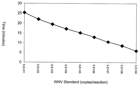
Figure 3 is a line graph relating the amount of WNV standard input into a real-
time
nucleic acid amplification reaction (x-axis) and the time-of-emergence of the
measured
fluorescent signal above a background threshold (y-axis).
25 Detailed Description of the Invention
Disclosed herein are compositions, methods and kits for selectively detecting
the
nucleic acids of flaviviruses, such as West Nile virus (WNV), in biological
samples such as
blood, serum, plasma or other body fluid or tissue. The probes, primers and
methods of the
invention can be used either in diagnostic applications or for screening
donated blood and
30 blood products or other tissues that may contain infectious particles.
Introduction and Overview
The present invention includes compositions (nucleic acid capture
oligonucleotides,
16
CA 02501946 2005-04-11
WO 2004/036190 - -
PCT/US2003/033639
amplification oligonucleotides and probes), methods and kits that are
particularly usemi for
detecting WNV nucleic acids in a biological sample. To design oligonucleotide
sequences
appropriate for such uses, known WNV nucleic acid sequences were first
compared to identify
candidate regions of the viral genome that could serve as reagents in a
diagnostic assay. As a
result of these comparisons, three different regions of the WNV genome were
selected as
targets for detection using the capture oligonucleotides, primers and probes
shown
schematically in Figure 1. Portions of sequences containing relatively few
variants between the
compared sequences were chosen as starting points for designing synthetic
oligonucleotides
suitable for use in capture, amplification and detection of amplified
sequences.
0 Based on these analyses, the capture oligonucleotide, amplification
primer and probe
sequences presented below were designed. Those having an ordinary level of
skill in the art
will appreciate that any primer sequences specific for WNV or other flavivirus
target, with or
without a T7 promoter sequence, may be used as primers in the various primer-
based in vitro
amplification methods described below. It is also contemplated that
oligonucleotides having
5 the sequences disclosed herein could serve alternative functions in
assays for detecting WNV
nucleic acids. For example, the capture oligonucleotides disclosed herein
could serve as
hybridization probes, the hybridization probes disclosed herein could be used
as amplification
primers, and the amplification primers disclosed herein could be used as
hybridization probes
in alternative detection assays.
!O The amplification primers disclosed herein are particularly
contemplated as components
of multiplex amplification reactions wherein several amplicon species can be
produced from an
assortment of target-specific primers. For example, it is contemplated that
certain preferred
WNV-specific primers disclosed herein can be used in multiplex amplification
reactions that
are capable of amplifying polynucleotides of unrelated viruses without
substantially
compromising the sensitivities of those assays. Particular examples of these
unrelated viruses
include HIV-1, HIV-2, HCV and HBV.
Useful Amplification Methods
Amplification methods useful in connection with the present invention include:
Transcription Mediated Amplification (TMA), Nucleic Acid Sequence-Based
Amplification
30 (NASBA), the Polymerase Chain Reaction (PCR), Strand Displacement
Amplification (SDA),
and amplification methods using self-replicating polynucleotide molecules and
replication
enzymes such as MDV-1 RNA and Q-beta enzyme. Methods for carrying out these
various
17
CA 02501946 2012-02-09
18
amplification techniques respectively can be found in U.S.-Patent No.
5,399,491, published
European patent application EP 0 525 882, U.S. Patent No. 4,965,188, U.S.
Patent No.
5,455,166, U.S. Patent No. 5,472,840 and Lizardi et al., BioTechnology 6:1197
(1988).
In a highly preferred embodiment of the invention, WNV nucleic acid sequences
are
amplified using a TMA protocol. According to this protocol, the reverse
transcriptase which
provides the DNA polymerase activity also possesses an endogenous RNase H
activity. One of
the primers used in this procedure contains a promoter sequence positioned
upstream of a
sequence that is complementary to one strand of a target nucleic acid that is
to be amplified. In
the first step of the amplification, a promoter-primer hybridizes to the WNV
target RNA at a
defined site. Reverse transcriptase creates a complementary DNA copy of the
target RNA by
extension from the 3' end of the promoter-primer. Following interaction of an
opposite strand
primer with the newly synthesized DNA strand, a second strand of DNA is
synthesized from
the end of the primer by reverse transcriptase, thereby creating a double-
stranded DNA
molecule. RNA polymerase recognizes the promoter sequence in this double-
stranded DNA
template and initiates transcription. Each of the newly synthesized RNA
amplicons re-enters
the TMA process and serves as a template for a new round of replication,
thereby leading to an
exponential expansion of the RNA amplicon. Since each of the DNA templates can
make 100- ,
1000 copies of RNA amplicon, this expansion can result in the production of 10
billion =
amplicons in less than one hour. The entire process is autocatalytic and is
performed at a
constant temperature.
Structural Features of Primers
As indicated above, a "primer" refers to an optionally modified
oligonucleotide which
is capable of participating in a nucleic acid amplification reaction.
Preferred primers are
capable of hybridizing to a template nucleic acid and which has a 3' end that
can be extended
by a DNA polymerase activity. The 5' region of the primer may be non-
complementary to the
target nucleic acid. If the 5' non-complementary region includes a promoter
sequence, it is
referred to as a "promoter-primer." Those skilled in the art will appreciate
that any
oligonucleotide that can function as a primer (i.e., an oligonucleotide that
hybridizes
specifically to a target sequence and has a 3' end capable of extension by a
DNA polymerase
activity) can be modified to include a 5' promoter sequence, and thus could
function as a
CA 02501946 2005-04-11
-
.............................................................................
-PCT/US2003/033639-
pronY,T.Z0p0SP2V9gimilarly, any promoter-primer can be modified by removal or,
or syntnesis
without, a promoter sequence and still function as a primer.
Nucleotide base moieties of primers may be modified (e.g., by the addition of
propyne
groups), as long as the modified base moiety retains the ability to form a non-
covalent
association with G, A, C, T or U, and as long as an oligonucleotide comprising
at least one
modified nucleotide base moiety or analog is not sterically prevented from
hybridizing with a
single-stranded nucleic acid. As indicated below in connection with the
chemical composition
of useful probes, the nitrogenous bases of primers in accordance with the
invention may be
conventional bases (A, G, C, T, U), known analogs thereof (e.g., inosine or
"I" having
hypoxanthine as its base moiety; see The Biochemistry of the Nucleic Acids 5-
36, Adams et
al., ed., 11th ed., 1992), known derivatives of purine or pyrimidine bases
(e.g., N4-methyl
deoxygaunosine, deaza- or aza-purines and deaza- or aza-pyrimidines,
pyrimidine bases having
substituent groups at the 5 or 6 position, purine bases having an altered or a
replacement
substituent at the 2, 6 or 8 positions, 2-amino-6-methylaminopurine, 06-
methylguanine, 4-thio-
pyrimidines, 4-amino-pyrimidines, 4-dimethylhydrazine-pyrimidines, and 04-
alkyl-
pyrimidines (see, Cook, PCT Intl Pub. No, WO 93/13121) and "abasic" residues
where the
backbone includes no nitrogenous base for one or more residues of the polymer
(see Arnold et
al., U.S. Patent No. 5,585,481). Common sugar moieties that comprise the
primer backbone
include ribose and deoxyribose, although 2'-0-methyl ribose (0Me), halogenated
sugars, and
other modified sugar moieties may also be used. Usually, the linking group of
the primer
backbone is a phosphorus-containing moiety, most commonly a phosphodiester
linkage,
although other linkages, such as, for example, phosphorothioates,
methylphosphonates, and
non-phosphorus-containing linkages such as peptide-like linkages found in
"peptide nucleic
acids" (PNA) also are intended for use in the assay disclosed herein.
Useful Probe Labeling Systems and Detectable Moieties
Essentially any labeling and detection system that can be used for monitoring
specific
nucleic acid hybridization can be used in conjunction with the present
invention. Included
among the collection of useful labels are radiolabels, enzymes, haptens,
linked
oligonucleotides, chemiluminescent molecules, fluorescent moieties (either
alone or in
SO combination with "quencher" moieties), and redox-active moieties that
are amenable to
electronic detection methods. Preferred chemiluminescent molecules include
acridinium esters
of the type disclosed by Arnold et al., in U.S. Patent No. 5,283,174 for use
in connection with
19
CA 02501946 2012-02-09
homogenous protection assays, and of the type disclosed by WoOdhead et
il.iñUTS.¨PaTent
No. 5,656,207 for use in connection with assays that quantify multiple targets
in a single
reaction. The disclosures contained in these patent documents are hereby
incorporated by
reference. Preferred electronic labeling and detection approaches are
disclosed in U.S. Patent
5 Nos. 5,591,578 and 5,770,369, and the published international patent
application WO
98/57158 Redox active
moieties useful as labels in the present invention include transition metals
such as Cd, Mg, Cu,
Co, Pd, Zn, Fe and Ru.
Particularly preferred detectable labels for probes in accordance with the
present
10 invention are detectable in homogeneous assay systems (i.e., where, in a
mixture, bound
labeled probe exhibits a detectable change, such as stability or differential
degradation,
compared to unbound labeled probe). While other homogeneously detectable
labels, such as
fluorescent labels and electronically detectable labels, are intended for use
in the practice of the
present invention, a preferred label for use in homogenous assays is a
chemiluminescent
= 15 compound (e.g., as described by Woodhead et al., in U.S. Patent No.
5,656,207; by Nelson et
al., in U.S. Patent No. 5,658,737; or by Arnold efal., in U.S. Patent No.
5,639,604).
Particularly preferred chemiluminescent labels include acridinium ester ("AE")
compounds,
such as standard AE or derivatives thereof, such as naphthyl-AE, ortho-AE, 1-
or 3-methyl-AE,
2,7-dimethyl-AE, 4,5-dimethyl-AE, ortho-dibromo-AE, ortho-dimethyl-AE, meta-
dimethyl-
20 AE, ortho-methoxy-AE, ortho-methoxy(cinnamy1)-AE, ortho-methyl-AE, ortho-
fluoro-AE, 1-
or 3-methyl-ortho-fluoro-AE, 1- or 3-methyl-meta-difluoro-AE, and 2-methyl-AE.
In some applications, probes exhibiting at least some degree of self-
complementarity
are desirable to facilitate detection of probe:target duplexes in a test
sample without first
requiring the removal of unhybridized probe prior to detection. By way of
example, structures
referred to as "Molecular Torches" are designed to include distinct regions of
self-
complementarily (coined "the target binding domain" and "the target closing
domain") which
are connected by a joining region and which hybridize to one another under
predetermined
hybridization assay conditions. When exposed to denaturing conditions, the two
complementary regions (which may be fully or partially complementary). of the
Molecular
Torch melt, leaving the target binding domain available for hybridization to a
target sequence
when the predetermined hybridization assay conditions are restored. Molecular
Torches are
designed so that the target binding domain favors hybridization to the target
sequence over the
CA 02501946 2012-02-09
21
target closing domain. The target binding domain and the target closing domain
of a Molecular
Torch include interacting labels (e.g., fluorescent/quencher) positioned so
that a different signal
is produced when the Molecular Torch is self-hybridized as opposed to when the
Molecular
Torch is hybridized to a target nucleic acid, thereby permitting detection of
probe:target
duplexes in a test sample in the presence of unhybridized probe having a
viable label associated
therewith. Molecular Torches are fully describedin U.S. Patent No. 6,361,945.
Another example of a self-complementary hybridization assay probe that may be
used
in conjunction with the invention is a structure commonly referred to as a
"Molecular Beacon."
Molecular Beacons comprise nucleic acid molecules having a target
complementary sequence,
an affinity pair (or nucleic acid arms) holding the probe in a closed
conformation in the absence
of a target nucleic acid sequence, and a label pair that interacts when the
probe is in a closed
conformation. Hybridization of the target nucleic acid and the target
complementary sequence
separates the members of the affinity pair, thereby shifting the probe to an
open conformation.
The shift to the open conformation is detectable due to reduced interaction of
the label pair,
which may be, for example, a fluorophore and a quencher (e.g., DABCYL and
EDANS).
Molecular Beacons are fully described in U.S. Patent No. 5,925,517.
Molecular beacons useful for detecting WNV-specific
nucleic acid sequences may be created by appending to either end of one of the
probe
sequences disclosed herein, a first nucleic acid arm comprising a fluorophore
and a second
nucleic acid arm comprising a quencher moiety. In this configuration, the WNV-
specific probe
sequence disclosed herein serves as the target-complementary "loop" portion of
the resulting
molecular beacon.
Molecular beacons preferably are labeled with an interactive pair of
detectable labels.
Examples of detectable labels that are preferred as members of an interactive
pair of labels
interact with each other by FRET or non-FRET energy transfer mechanisms.
Fluorescence
resonance energy transfer (FRET) involves the radiationless transmission of
energy quanta
from the site of absorption to the site of its utilization in the molecule, or
system of molecules,
by resonance interaction between chromophores, over distances considerably
greater than
interatomic distances, without conversion to thermal energy, and without the
donor and
acceptor coming into kinetic collision. The "donor" is the moiety that
initially absorbs the
energy, and the "acceptor" is the moiety to which the energy is subsequently
transferred. In
CA 02501946 2005-04-11
.WO 2004/036190
addition to PRET, there are at least three other "non-FRET" energy transfer
excitation energy can be transferred from a donor to an acceptor molecule.
When two labels are held sufficiently close that energy emitted by one label
can be
received or absorbed by the second label, whether by a FRET or non-FRET
mechanism, the
two labels are said to be in "energy transfer relationship" with each other.
This is the case, for
example, when a molecular beacon is maintained in the closed state by
formation of a stem
duplex, and fluorescent emission from a fluorophore attached to one arm of the
probe is
quenched by a quencher moiety on the opposite arm.
Highly preferred label moieties for the invented molecular beacons include a
.0 fluorophore and a second moiety having fluorescence quenching properties
(i.e., a "quencher").
In this embodiment, the characteristic signal is likely fluorescence of a
particular wavelength,
but alternatively could be a visible light signal. When fluorescence is
involved, changes in
emission are preferably due to FRET, or to radiative energy transfer or non-
FRET modes.
When a molecular beacon having a pair of interactive labels in the closed
state is stimulated by
[5 an appropriate frequency of light, a fluorescent signal is generated at
a first level, which may be
very low. When this same probe is in the open state and is stimulated by an
appropriate
frequency of light, the fluorophore and the quencher moieties are sufficiently
separated from
each other that energy transfer between them is substantially precluded. Under
that condition,
the quencher moiety is unable to quench the fluorescence from the fluorophore
moiety. If the
20 fluorophore is stimulated by light energy of an appropriate wavelength,
a fluorescent signal of a
second level, higher than the first level, will be generated. The difference
between the two
levels of fluorescence is detectable and measurable. Using fluorophore and
quencher moieties
in this manner, the molecular beacon is only "on" in the "open" conformation
and indicates that
the probe is bound to the target by emanating an easily detectable signal. The
conformational
25 state of the probe alters the signal generated from the probe by
regulating the interaction
between the label moieties.
Examples of donor/acceptor label pairs that may be used in connection with the
invention, making no attempt to distinguish FRET from non-FRET pairs, include
fluorescein/tetramethylrhodamine, IAEDANS/fluororescein, EDANS/DABCYL,
30 coumarin/DABCYL, fluorescein/fluorescein, BODIPY FL/BODIPY FL,
fluorescein/DABCYL, lucifer yellow/DABCYL, BODIPY/DABCYL, eosine/DABCYL,
erythrosine/DABCYL, tetramethylrhodamine/DABCYL, Texas Red/DABCYL, CY5/BH1,
22
CA 02501946 2005-04-11
WO 2004/036190tr 1" ""4t -
PCT/US2003/033639
CY5/t51-12, u r CY3/BH2 and fluorescein/QSY7 dye. Those having an
oramary ievei of
skill in the art will understand that when donor and acceptor dyes are
different, energy transfer
can be detected by the appearance of sensitized fluorescence of the acceptor
or by quenching of
donor fluorescence. When the donor and acceptor species are the same, energy
can be detected
by the resulting fluorescence depolarization. Non-fluorescent acceptors such
as DABCYL and
the QSY 7 dyes advantageously eliminate the potential problem of background
fluorescence
resulting from direct (i.e., non-sensitized) acceptor excitation. Preferred
fluorophore moieties
that can be used as one member of a donor-acceptor pair include fluorescein,
ROX, and the CY
dyes (such as CY5). Highly preferred quencher moieties that can be used as
another member
of a donor-acceptor pair include DABCYL and the BLACK HOLE QUENCHER moieties
which are available from Biosearch Technologies, Inc., (Novato, CA).
Synthetic techniques and methods of bonding labels to nucleic acids and
detecting
labels are well known in the art (e.g., see Sambrook et al., Molecular
Cloning, A Laboratory
Manual, 2nd ed. (Cold Spring Harbor Laboratory Press, Cold Spring Harbor, NY,
1989),
Chapter 10; Nelson et al., U.S. Patent No. 5,658,737; Woodhead et al., U.S.
Patent No.
5,656,207; Hogan et al., .U.S. Patent No. 5,547,842; Arnold et al., U.S.
Patent No. 5,283,174;
Kourilsky et al., U.S. Patent No. 4,581,333), and Becker et al., European
Patent App. No. 0 747
706.
Chemical Composition of Probes
Probes in accordance with the invention comprise polynucleotides or
polynucleotide
analogs and optionally may carry a detectable label covalently bonded thereto.
Nucleosides or
nucleoside analogs of the probe comprise nitrogenous heterocyclic bases, or
base analogs,
where the nucleosides are linked together, for example by phospohdiester bonds
to form a
polynucleotide. Accordingly, a probe may comprise conventional ribonucleic
acid (RNA)
and/or deoxyribonucleic acid (DNA), but also may comprise chemical analogs of
these
molecules. The "backbone" of a probe may be made up of a variety of linkages
known in the
art, including one or more sugar-phosphodiester linkages, peptide-nucleic acid
bonds
(sometimes referred to as "peptide nucleic acids" as described by Hyldig-
Nielsen et al., PCT
Intl Pub. No. WO 95/32305), phosphorothioate linkages, methylphosphonate
linkages or
combinations thereof. Sugar moieties of the probe may be either ribose or
deoxyribose, or
similar compounds having known substitutions, such as, for example, 2'-0-
methyl ribose and 2'
halide substitutions (e.g., 2'-F). The nitrogenous bases may be conventional
bases (A, G, C, T,
23
CA 02501946 2013-10-31
24
U), known analogs thereof (e.g., inosine or "I"; see The Biochemistry of the
Nucleic Acids 5-36,
Adams et al., ed., nth ed., 1992), known derivatives of purine or pyrimidine
bases (e.g., N4-methyl
deoxygaunosine, deaza- or aza-purines and deaza- or aza-pyrimidines,
pyrimidine bases having
substituent groups at the 5 or 6 position, purine bases having an altered or a
replacement substituent
at the 2, 6 or 8 positions, 2-amino-6-methylaminopurine, 06-methylguanine, 4-
thio-pyrimidines, 4-
amino-pyrimidines, 4-dimethylhydrazine-pyrimidines, and 04-alkyl-pyrimidines
(see, Cook, PCT
Int'l Pub. No. WO 93/13121) and "abasic" residues where the backbone includes
no nitrogenous
base for one or more residues of the polymer (see Arnold etal., U.S. Patent
No. 5,585,481). A
probe may comprise only conventional sugars, bases and linkages found in RNA
and DNA, or may
include both conventional components and substitutions (e.g., conventional
bases linked via a
methoxy backbone, or a nucleic acid including conventional bases and one or
more base analogs).
While oligonucleotide probes of different lengths and base composition may be
used for
detecting WNV nucleic acids, preferred probes in this invention have lengths
of up to 100
nucleotides, and more preferably have lengths of up to 60 nucleotides.
Preferred length ranges for
the invented oligonucleotides are from 10 to 100 bases in length, or more
preferably between 15
and 50 bases in length, or still more preferably between 15 and 30 bases in
length. However, the
specific probe sequences described below also may be provided in a nucleic
acid cloning vector or
transcript or other longer nucleic acid and still can be used for detecting
WNV nucleic acids.
Selection of Amplification Primers and Detection Probes Specific for WNV
Useful guidelines for designing amplification primers and probes with desired
characteristics are described herein. The optimal sites for amplifying and
probing WNV nucleic
acids contain two, and preferably three, conserved regions each greater than
about 15 bases in
length, within about 200 bases of contiguous sequence. The degree of
amplification observed with
a set of primers or promoter-primers depends on several factors, including the
ability of the
oligonucleotides to hybridize to their complementary sequences and their
ability to be extended
enzymatically. Because the extent and specificity of hybridization reactions
are affected by a
number of factors, manipulation of those factors will determine the exact
sensitivity and specificity
of a particular oligonucleotide, whether perfectly complementary to its target
or not. The effects of
varying assay conditions are known to those skilled in the art, and are
described by Hogan et al., in
U.S. Patent No. 5,840,488.
CA 02501946 2013-10-31
=
The length of the target nucleic acid sequence and, accordingly, the length of
the primer
sequence or probe sequence can be important. In some cases, there may be
several sequences from
a particular target region, varying in location and length, which will yield
primers or probes having
the desired hybridization characteristics. While it is possible for nucleic
acids that are not perfectly
5 complementary to hybridize, the longest stretch of perfectly homologous
base sequence will
normally primarily determine hybrid stability.
Amplification primers and probes should be positioned to minimize the
stability of the
oligonucleotide:nontarget (i.e., nucleic acid with similar sequence to target
nucleic acid) nucleic
acid hybrid. It is preferred that the amplification primers and detection
probes are able to
10 distinguish between target and non-target sequences. In designing
primers and probes, the
differences in these Tm values should be as large as possible (e.g., at least
it and preferably 5aC.).
The degree of non-specific extension (primer-dimer or non-target copying) can
also affect
amplification efficiency. For this reason, primers are selected to have low
self- or cross-
complementarity, particularly at the 3' ends of the sequence. Long homopolymer
tracts and high
15 GC content are avoided to reduce spurious primer extension. Commercially
available computer
software can aid in this aspect of the design. Available computer programs
include MacDNASISTM
2.0 (Hitachi Software Engineering American Ltd.) and OLIGO ver. 6.6 (Molecular
Biology
Insights; Cascade, CO).
Those having an ordinary level of skill in the art will appreciate that
hybridization involves
20 the association of two single strands of complementary nucleic acid to
form a hydrogen bonded
double strand. It is implicit that if one of the two strands is wholly or
partially involved in a hybrid,
then that strand will be less able to participate in formation of a new
hybrid. By designing primers
and probes so that substantial portions of the sequences of interest are
single stranded, the rate and
extent of hybridization may be greatly increased. If the target is an
integrated genomic sequence,
25 then it will naturally occur in a double stranded form (as is the case
with the product of the
polymerase chain reaction). These double-stranded targets are naturally
inhibitory to hybridization
with a probe and require denaturation prior to the hybridization step.
The rate at which a polynucleotide hybridizes to its target is a measure of
the thermal
stability of the target secondary structure in the target binding region. The
standard
CA 02501946 2005-04-11
measWQ_.?0.94/03 .1,90..-idization rate is the Cotin which is mdsiiredas
MbreSTEM93,3139'
liter multiplied by seconds. Thus, it is the concentration of probe multiplied
by the time at
which 50% of maximal hybridization occurs at that concentration. This value is
determined by
hybridizing various amounts of polynucleotide to a constant amount of target
for a fixed time.
The Cotin is found graphically by standard procedures familiar to those having
an ordinary
level of skill in the art.
Preferred Amplification Primers
Primers useful for conducting amplification reactions can have different
lengths to
accommodate the presence of extraneous sequences that do not participate in
target binding,
and that may not substantially affect amplification or detection procedures.
For example,
promoter-primers useful for performing amplification reactions in accordance
with the
invention have at least a minimal sequence that hybridizes to the WNV target
nucleic acid, and
a promoter sequence positioned upstream of that minimal sequence. However,
insertion of
sequences between the target binding sequence and the promoter sequence could
change the
length of the primer without compromising its utility in the amplification
reaction.
Additionally, the lengths of the amplification primers and detection probes
are matters of
choice as long as the sequences of these oligonucleotides conform to the
minimal essential
requirements for hybridizing the desired complementary sequence.
Tables 1 and 2 present specific examples of oligonucleotide sequences that
were used as
primers for amplifying WNV nucleic acids in the 5' non-coding region. Table 1
presents the
sequences of primers that were complementary to WNV sequences on one strand of
nucleic
acid. All of the illustrative primers presented in Table 1 have target-
complementary sequences
contained within the sequence of SEQ ID NO: 1. Different subsets of preferred
primers have
target-complementary sequences contained within the sequence of SEQ ID NO:2 or
SEQ ID
NO:3. It is preferred for one of the primers used in the amplification
procedure to have a
target-complementary sequence falling within one of these domains. Table 2
presents the
sequences of both the WNV target-complementary primers and the full sequences
for
promoter-primers that were used during development of the invention. All of
the illustrative
primers presented in Table 2 have target-complementary sequences contained
within the
sequence of SEQ ID NO:16, a feature which is presently preferred for one of
the primers used
in the amplification procedure. Notably, the oligonucleotide sequences in
Table 1 and Table 2
are complementary to opposite strands of the WNV nucleic acid.
26
CA 02501946 2005-04-11
WO 2004/036190
PCT/US2003/033639
Primers useful for amplifying the 5' non-coding region of WNV can include
nucieotkie
analogs. For example, the primers of SEQ ID NO:4 and SEQ ID NO:5 differ from
each other
by the substitution of a hypoxanthine base analog for an adenine base at
position 11. Similarly,
the primers of SEQ ID NO:6 and SEQ ID NO:7 also differ by the presence of this
base analog,
as do the opposite strand primers identified by SEQ ID NO:21 and SEQ ID NO:22.
This
illustrates how nucleobases in the primers may be substituted by modified
bases or nucleobase
analogs.
Table 1
Polynucleotide Sequences of Amplification Primers
Sequence SEQ ID NO:
CAATTAACACAGTGCGAGCTGTTT SEQ ID NO:4
CAATTAACACIGTGCGAGCTGTTT SEQ ID NO:5
TAACACAGTGCGAGCTG CFI SEQ ID NO:6
TAACACIGTGCGAGCTGTTTCYT SEQ ID NO:7
CGAGCTGTTTCTTAGCACGA SEQ ID NO:8
CGAGCTGTTTCTTAGCACGAA SEQ ID NO:9
GAAGATCTCGATGTCTAAGAAACC SEQ ID NO:10
AAGATCTCGATGTCTAAGAAACC SEQ ID NO:11
AGATCTCGATGTCTAAGAAACC SEQ ID NO:12
GATCTCGATGTCTAAGAAACCA SEQ ID NO:13
GATCTCGATGTCTAAGAAACC SEQ ID NO:14
ATCTCGATGTCTAAGAAACCAG SEQ ID NO:15
Table 2 presents WNV target-complementary oligonucleotide sequences and the
corresponding promoter-primer sequences that were used for amplifying WNV
nucleic acid
sequences in the 5' non-coding region of the viral genome. As indicated above,
all promoter-
primers included sequences complementary to a WNV target sequence at their 3'
ends, and a
T7 promoter sequence at their 5' ends. Primers identified by SEQ ID Nos:29-40
in Table 2 are
promoter-primers corresponding to the WNV-complementary primers identified as
SEQ ID
Nos:17-28, respectively.
27
CA 02501946 2005-04-11
WO 2004/036190
PCT/US2003/033639
Table 2 =
Polynucleotide Sequences of Amplification Primers
Sequence
SEQ ID NO:
GTTTTAGCATATTGACAGCCC
SEQ ID NO:17
GTTTTAGCATATTGACAGCC SEQ ID
NO:18
TTCCGCGTTTTAGCATATTGA
SEQ ID NO:19
ATTCCGCGTITTAGCATATTG
SEQ ID NO:20
ATCAAGGACAACACGCGGGGCAT
SEQ ID NO:21
ATCAAGGACAAIACGCGGGGCAT
SEQ ID NO:22
CCTCTTCAGTCCAATCAAGGACAA SEQ ID
NO:23
AGCCCTCTTCAGTCCAATCAAGGA
SEQ ID NO:24
TAGCCCTCT'TCAGTCCAATCAAGG
SEQ ID NO:25
=
ATAGCCCTC'T'TCAGTCCAATCAAG
SEQ ID NO:26
TAGCCCTCTTCAGTCCAATCAA
SEQ ID NO:27
ACATAGCCCTCTTCAGTCCAATCA SEQ ID
NO:28
AATTTAATACGACTCACTATAGGGAGAGTTTTAGCATATTGACAGCCC SEQ ID NO:29
AATTTAATACGACTCACTATAGGGAGAGTTITAGCATATTGACAGCC
SEQ ID NO:30
AATTTAATACGACTCACTATAGGGAGATTCCGCGTTTTAGCATATTGA SEQ ID NO:31
AATTTAATACGACTCACTATAGGGAGAA'TTCCGCGTTITAGCATATTG SEQ ID NO:32
AATTTAATACGACTCACTATAGGGAGAATCAAGGACAACACGCGGGGC SEQ ID NO:33
AT
AATTTAATACGACTCACTATAGGGAGAATCAAGGACAAIACGCGGGGC SEQ ID NO:34
AT
AATTTAATACGACTCACTATAGGGAGACCTCTTCAGTCCAATCAAGGA SEQ ID NO:35
CAA
AA'ITIAATACGACTCACTATAGGGAGAAGCCCTCYFCAGTCCAATCAA SEQ ID NO:36
GGA
AATTTAATACGACTCACTATAGGGAGATAGCCCTCTTCAGTCCAATCAA SEQ ID NO:37
GG
AA rn AATACGACTCACTATAGGGAGAATAGCCCTCTTCAGTCCAATCA SEQ ID NO:38
AG
AATTTAATACGACTCACTATAGGGAGATAGCCCTCTTCAGTCCAATCAA SEQ ID NO:39
AATTTAATACGACTCACTATAGGGAGAACATAGCCCTCTTCAGTCCAAT SEQ ID NO:40
CA
28
CA 02501946 2005-04-11
WO 2004/036190PCT/US2003/033639
.rrererrea sets of primers for amplifying WNV sequences in the 5'non-counig
region
include a first primer that hybridizes a WNV target sequence (such as one of
the primers listed
in Table 2) and a second primer that is complementary to the sequence of an
extension product
of the first primer (such as one of the primer sequences listed in Table 1).
In a highly preferred
embodiment, the first primer is a promoter-primer that includes a T7 promoter
sequence at its
5' end.
Tables 3 and 4 present specific examples of oligonucleotide sequences that
were used as
primers for amplifying WNV nucleic acids in the 3000 region of the viral
genome. Table 3
presents the sequences of primers that were complementary to WNV sequences on
one strand
of nucleic acid. All of the illustrative primers presented in Table 3 have
target-complementary
sequences contained within the sequence of SEQ ID NO:41, a feature which is
presently
preferred for one of the primers used in the amplification procedure. Table 4
presents the
sequences of both the WNV target-complementary primers and the full sequences
for
promoter-primers that were used during development of the invention. All of
the illustrative
primers presented in Table 4 have target-complementary sequences contained
within the
sequence of SEQ ID NO:52, a feature which is presently preferred for one of
the primers used
in the amplification procedure. Notably, the oligonucleotide sequences in
Table 3 and Table 4
are complementary to opposite strands of the WNV nucleic acid.
Primers useful for amplifying the 3000 region of WNV can include nucleotide
analogs.
For example, the primers of SEQ ID NO:42, SEQ ID NO:47 and SEQ ID NO:48 differ
from
each other by the substitution of a hypoxanthine base analog for the existing
base at different
individual positions within the primer sequence. Similarly, the primers of SEQ
ID NO:43 and
SEQ ID NO:49 also differ by the presence of this base analog, as do the
primers identified by
SEQ ID NO:45 and SEQ ID NO:50 and SEQ ID NO:51. This illustrates that
nucleobases in
the primers may be substituted by modified bases or nucleobase analogs.
29
CA 02501946 2005-04-11
WO 2004/036190
PCT/US2003/033639
Table 3
Po13/nucleotide Sequences of Amplification Primers
Sequence SEQ ID NO:
TTGACCCTMCAGTTGGGCCTT SEQ ID NO:42
CMTCAGTTGGGCCTTCTGGT SEQ ID NO:43
AGTTGGGCCTTCTGGTCGTGTT = SEQ ID
NO:44
TGGTCGTGTTCTTGGCCACCCA SEQ ID NO:45
TCGTGTTCTTGGCCACCCAGGA SEQ ID NO:46
'TTGAICCTTTTCAGTTGGGCCTT SEQ ID NO:47
TTGACCC=CAGITGGGCCTT SEQ ID NO:48
CTITTCAGITGGGCCTTCTGGT SEQ ID NO:49
TGGTCGTGTTITTGGCCACCCA SEQ ID NO:50
TGGTCGTGTTCITGGCCACCCA SEQ ID NO:51
Table 4 presents WNV target-complementary oligonucleotide sequences and the
corresponding promoter-primer sequences that were used for amplifying WNV
nucleic acid
sequences in the 3000 region of the viral genome. As indicated above, all
promoter-primers
included sequences complementary to a WNV target sequence at their 3' ends,
and a T7
promoter sequence at their 5' ends.
CA 02501946 2005-04-11
WO 2004/036190 PCT/US2003/033639
Table 4
Polynucleotide Sequences of Amplification Primers
Identifier Sequence SEQ ID NO:
WNV complementary primer CTGGCATGCTGATCTTGGCTGT
SEQ ID NO:53
WNV complementary. primer ATAGCTGGCATGCTGATCTTGGC SEQ ID
NO:54
WNV complementary primer ATAGCTGGCATGCTGATCTTGG
SEQ ID NO:55
T7 promoter-primer AA1T1AATACGACTCACTATAGGGA SEQ ID NO:56 '
GACTGGCATGCTGATCTTGGCTGT
T7 promoter-primer
AATTTAATACGACTCACTATAGGGA SEQ ID NO:57
GAATAGCTGGCATGCTGATCTTGGC
T7 promoter-primer
AATTTAATACGACTCACTATAGGGA SEQ ID NO:58
GAATAGCTGGCATGCTGATCTTGG
Preferred sets of primers for amplifying WNV sequences in the 3000 region of
the viral
genome include a first primer that hybridizes a WNV target sequence (such as
one of the
primers listed in Table 4) and a second primer that is complementary to the
sequence of an
extension product of the first primer (such as one of the primer sequences
listed in Table 3). In
a highly preferred embodiment, the first primer is a promoter-primer that
includes a T7
promoter sequence at its 5' end.
Tables 5 and 6 present specific examples of oligonucleotide sequences that
were used as
primers for amplifying WNV nucleic acids in the 3' non-coding region of the
viral genome.
Table 5 presents the sequences of primers that were complementary to WNV
sequences on one
strand of nucleic acid. All of the illustrative primers presented in Table 5
have target-
complementary sequences contained within the sequence of SEQ ID NO:59, a
feature which is
presently preferred for one of the primers used in the amplification
procedure. Table 6 presents
the sequences of both the WNV target-complementary primers and the full
sequences for
promoter-primers that were used during development of the invention. All of
the illustrative
primers presented in Table 6 have target-complementary sequences contained
within the
sequence of SEQ ID NO:72. Different subsets of preferred primers have target-
complementary
sequences contained within the sequence of SEQ ID NO:73 or SEQ ID NO:74. It is
preferred
for one of the primers used in the amplification procedure to have a target-
complementary
sequence falling within one of these domains. Notably, the oligonucleotide
sequences in Table
31
CA 02501946 2005-04-11
WO 2004/036190
PCT/US2003/033639
and Table 6 are complementary to opposite strands of the WNV nucleic acid.
Primers useful for amplifying the 3' non-coding region of WNV can include
nucleotide
analogs. For example, the primers of SEQ ID NO:60 and SEQ ID NO:61 differ from
each
other by the substitution of a hypoxanthine base analog for a thymine base at
position 1.
5 Similarly, the primers of SEQ ID NO:64 and SEQ ID NO:65 also differ by
the presence of this
base analog, in this instance substituting for cytosine. Likewise, the WNV-
complementary
primer sequences of SEQ ID NO:83 and SEQ ID NO:82, together with the
corresponding
promoter-primer sequences, also contain hypoxanthine base analog
substitutions. This further
illustrates that nucleobases in the primers may be substituted by modified
bases or nucleobase
0 analogs.
Table 5
Polynucleotide Sequences of Amplification Primers
Sequence SEQ ID NO:
[5 TCCGCCACCGGAAGTTGAG SEQ ID NO:60
ICCGCCACCGGAAGTTGAG SEQ ID NO:61
TCCGCCACCGGAAGTTGAGT SEQ ID NO:62
TCCGCCACCGGAAGTTGAGTA SEQ ID NO:63
CGCCACCGGAAGTTGAGT SEQ ID NO:64
20 IGCCACCGGAAGTTGAGT SEQ ID NO:65
CGCCACCGGAAGTTGAGTA SEQ ID NO:66
GGAAGTTGAGTAGACGGTGCT SEQ ID NO:67
GGAAGTTGAGTAGACGGTGCTG SEQ ID NO:68
GAAGTTGAGTAGACGGTGCT SEQ ID NO:69
25 GAAGTTGAGTAGACGGTGCTG SEQ ID NO:70
AAGTTGAGTAGACGGTGCTG SEQ ID NO:71
Table 6 presents WNV target-complementary oligonucleotide sequences and the
corresponding promoter-primer sequences that were used for amplifying WNV
nucleic acid
30 sequences in the 3' non-coding region of the viral genome. As indicated
above, all promoter-
primers included sequences complementary to a WNV target sequence at their 3'
ends, and a
T7 promoter sequence at their 5' ends.
32
CA 02501946 2005-04-11
WO 2004/036190
PCT/US2003/033639
Table 6
Polynucleotide Sequences of Amplification Primers
Sequence SEQ ID
NO:
TCCGAGACGGTTCTGAGGGCTTAC SEQ ID
NO:75
TCCGAGACGGTTCTGAGGGCTTA SEQ ID NO:76
TCCGAGACGGTTCTGAGGGCTT SEQ ID
NO:77
CCAGTCCTCCTGGGGITGAG SEQ ID
NO:78
ACCCAGTCCTCCTGGGGTTGAG SEQ ID
NO:79
ACCCAGTCCTCCTGGGGTTGA SEQ ID
NO:80
0 ACCCAGTCCTCCTGGGGTTG SEQ ID NO:81
GCTACAICAIGTGGGGTCCT SEQ ID
NO:82
GCTACAACAIGTGGGGTCCT SEQ ID
NO:83
AATTTAATACGACTCACTATAGGGAGATCCGAGACGGTTCTGAGGGCT SEQ ID NO: 84
TAC
5 AATTTAATACGACTCACTATAGGGAGATCCGAGACGGTTCTGAGGGCT SEQ ID NO: 85
TA
AATTTAATACGACTCACTATAGGGAGATCCGAGACGGTTCTGAGGGCT SEQ ID NO: 86
AATTTAATACGACTCACTATAGGGAGACCAGTCCTCCTGGGGTTGAG SEQ ID NO: 87
20 AATTTAATACGACTCACTATAGGGAGAACCCAGTCCTCCTGGGGTTGA SEQ ID NO:88
AATTTAATACGACTCACTATAGGGAGAACCCAGTCCTCCTGGGGTTGA SEQ ID NO:89
AATTTAATACGACTCACTATAGGGAGAACCCAGTCCTCCTGGGGTTG SEQ ID NO:90
AATTTAATACGACTCACTATAGGGAGAGCTACAICAIGTGGGGTCCT SEQ ID
NO:91
25 AATTTAATACGACTCACTATAGGGAGAGCTACAACAIGTGGGGTCCT SEQ ID NO:92
Preferred sets of primers for amplifying WNV sequences in the 3' non-coding
region
include a first primer that hybridizes a WNV target sequence (such as one of
the primers listed
in Table 6) arid a second primer that is complementary to the sequence of an
extension product
30
of the first primer (such as one of the primer sequences listed in Table 5).
In a highly preferred
embodiment, the first primer is a promoter-primer that includes a T7 promoter
sequence at its
5' end. Primers identified by SEQ ID Nos:84-92 in Table 6 are promoter-primers
33
CA 02501946 2005-04-11
WO 2004/036190
PCT/US2003/033639
corresponding to the WNV-complementary primers identified as SEQ ID Nos: /D-o-
i,
respectively.
Preferred Detection Probes
Another aspect of the invention relates to oligonucleotides that can be used
as
hybridization probes for detecting WNV nucleic acids. Methods for amplifying a
target nucleic
acid sequence present in the nucleic acid of WNV can include an optional
further step for
detecting amplicons. This procedure for detecting WNV nucleic acids includes a
step for
contacting a test sample with a hybridization assay probe that preferentially
hybridizes to the
target nucleic acid sequence, or the complement thereof, under stringent
hybridization
conditions, thereby forming a probe:target duplex that is stable for
detection. Next there is a
step for determining whether the hybrid is present in the test sample as an
indication of the
presence or absence of WNV nucleic acids in the test sample. This may involve
detecting the
probe:target duplex, and preferably involve homogeneous assay systems.
Hybridization assay probes useful for detecting WNV nucleic acid sequences
include a
sequence of bases substantially complementary to a WNV target nucleic acid
sequence. Thus,
probes of the invention hybridize one strand of a WNV target nucleic acid
sequence, or the
complement thereof. These probes may optionally have additional bases outside
of the targeted
nucleic acid .region which may or may not be complementary to WNV nucleic
acid.
Preferred probes are sufficiently homologous to the target nucleic acid to
hybridize
under stringent hybridization conditions corresponding to about 60 C when the
salt
concentration is in the range of 0.6-0.9 M. Preferred salts include lithium
chloride, but other
salts such as sodium chloride and sodium citrate also can be used in the
hybridization solution.
Example high stringency hybridization conditions are alternatively provided by
0.48 M sodium
phosphate buffer, 0.1% sodium dodecyl sulfate, and 1 mM each of EDTA and EGTA,
or by 0.6
M LiC1, 1% lithium lauryl sulfate, 60 mM lithium succinate and 10 mM each of
EDTA and
EGTA.
Probes in accordance with the invention have sequences complementary to, or
corresponding to one of three different domains of the WNV genome. As
reiterated below,
these domains were: (1) the 5' non-coding region/capsid region, (2) the 3000
region, and (3)
the 3' non-coding region. Certain probes that are preferred for detecting WNV
nucleic acid
sequences have a probe sequence, which includes the target-complementary
sequence of bases
together with any base sequences that are not complementary to the nucleic
acid that is to be
34
CA 02501946 2005-04-11
WO 2004/036190-
PCT/US2003/033639
detected, in the length range of from 10-100 nucleotides. Certain specific pro
D CS
preferred for detecting WNV nucleic acid sequences have target-complementary
sequences in
the length range of from 12-87, from 10-20, from 13-37 or from 17-23
nucleotides. Of course,
these target-complementary sequences may be' linear sequences, or may be
contained in the
structure of a molecular beacon or other construct having one or more optional
nucleic acid
sequences that are non-complementary to the WNV target sequence that is to be
detected. As
indicated above, probes may be made of DNA, RNA, a combination DNA and RNA, a
nucleic
acid analog, or contain one or more modified nucleosides (e.g., a
ribonucleoside having a 2'-0-
methyl substitution to the ribofuranosyl moiety).
Simply stated, preferred probes for detecting target nucleic acids of interest
in
connection with the present invention include sequences that are contained
within one or more
of several defined probe domains or the complements thereof, allowing for the
presence of
RNA and DNA equivalents, nucleotide analogs, up to 10% mismatched bases, and
even up to
20% mismatched bases. For example, preferred hybridization assay probes for
detecting
flaviviral nucleic acids, such as the nucleic acids of WNV, in the 5' non-
coding region can
include target-complementary sequences of bases contained within the sequence
of SEQ ID
NO:93, or within one of the subdomains defined by SEQ ID NO:94 or SEQ ID
NO:95.
Preferred hybridization assay probes for detecting flaviviral nucleic acids,
such as the nucleic
acids of WNV, in the 3000 region include target-complementary sequences of
bases contained
within the sequence of SEQ ID NO:99. Preferred hybridization assay probes
useful for
detecting flaviviral nucleic acids, such as the nucleic acids of WNV, in the
3' non-coding
region include target-complementary sequences of bases contained within the
sequence of SEQ
ID NO:101, or within the subdomains defmed by SEQ ID NO:102 or SEQ ID NO:103.
Optional sequences which are not complementary to the nucleic acid sequence
that is to be
detected may be linked to the target-complementary sequence of the probe.
Referring particularly to the allowability of base differences which can
distinguish
useful probe sequences from a defined target region which contains the target-
complementary
complementary sequence of bases of that probe, it should be noted that base
position 12 of the
probe having the sequence of SEQ ID NO:114 (occupied by a T) differs from the
corresponding position in the domain sequences defmed by SEQ ID NO:101, SEQ ID
NO:102
and SEQ ID NO:103 (all of which have a C at the corresponding position).
Certain preferred probes in accordance with the present invention include a
detectable
'
CA 02501946 2012-02-09
36
label. In one embodiment this label is an acridinium ester joined to the probe
by means of a
non-nucleotide linker. For example, detection probes can be labeled with
chemiluminescent
acridinium ester compounds that are attached via a linker substantially as
described in U.S.
Patent No. 5,585,481; and in U.S. Patent No. 5,639,604, particularly as
described at column 10,
line 6 to column 11, line 3, and in Example 8.
Table 7 presents the base sequences of some of the hybridization probes that
were used
for detecting WNV ampiicons from each of the three WNV target regions. Since
alternative
probes for detecting WNV nucleic acid sequences can hybridize to the opposite-
sense strand of
WNV, the present invention also includes oligonucleotides that are
complementary to the
sequences presented in the table.
CA 02501946 2005-04-11
WO 2004/036190
Table 7
PCT/US2003/033639
Polynucleotide Sequences of WNV Detection Probes
ITarget Sequence SEQ ID NO:
TGTCTAAGAAACCAGGAGGGC SEQ ID NO:96
P
GAAACCAGGAGGGCCCGG SEQ ID NO:97
-to
GCTGTCAATATGCTAAAACG SEQ ID NO:98
GGTCCTTCGCAAGAGGTGG SEQ ID NO:100
cr
GAGTAGACGGTGCTGCCTGCG SEQ ID NO:104
GTAGACGGTGCTGCCTGCG = SEQ ID NO:105
'to TGCGACTCAACCCCAGGAGGAC SEQ ID NO:106
ri4)
tv) TGCGACTCAACCCCAGGA SEQ ID NO:107
-o
0 CGACTCAACCCCAGGAGGAC SEQ ID NO:108
0 GACTCAACCCCAGGAGGAC SEQ ID NO:109
µ`µI' GACTCAACCCCAGGAGGA SEQ ID NO:110
ACTCAACCCCAGGAGGAC SEQ ID NO:111
CAGGAGGACUGGGUGAACA SEQ ID NO:112
GAGGACUGGGUGAACAAAG SEQ ID NO:113
GTGAACAAAGCTGCGAAGTG SEQ ID NO:114
AAGCCGCGAAGTGATCCATG SEQ ID NO:115
GTAAGCCCTCAGAACCGTC SEQ ID NO:116
As indicated above, any number of different backbone structures can be used as
a
scaffold for the nucleobase sequences of the invented hybridization probes. In
certain highly
preferred embodiments, the probe sequence used for detecting WNV amplicons
includes a
methoxy backbone, or at least one methoxy linkage in the nucleic acid
backbone.
37
CA 02501946 2005-04-11
WO 2004/036190PCT/US2003/033639
Selection and Use of Capture Oligonucleotides
Preferred capture oligonucleotides include a first sequence that is
complementary to a
WNV sequence (i.e., a "WNV target sequence") covalently attached to a second
sequence (i.e.,
a "tail" sequence) that serves as a target for immobilization on a solid
support. Any backbone
to link the base sequence of a capture oligonucleotide may be used. In certain
preferred
embodiments the capture oligonucleotide includes at least one rnethoxy linkage
in the
backbone. The tail sequence, which is preferably at the 3' end of a capture
oligonucleotide, is
used to hybridize to a complementary base sequence to provide a means for
capturing the
hybridized target WNV nucleic acid in preference to other components in the
biological
0 sample.
Although any base sequence that hybridizes to a complementary base sequence
may be
used in the tail sequence, it is preferred that the hybridizing sequence span
a length of about 5-
50 nucleotide residues. Particularly preferred tail sequences are
substantially homopolymeric,
containing about 10 to about 40 nucleotide residues, or more preferably about
14 to about 30
5 residues. A capture oligonucleotide according to the present invention
may include a first
sequence that specifically binds a WNV target polynucleotide, and a second
sequence that
specifically binds an oligo(dT) stretch immobilized to a solid support.
Using the components illustrated in Figure 1, one assay for detecting WNV
sequences
in a biological sample includes the steps of capturing the target nucleic acid
using the capture
20 oligonucleotide, amplifying the captured target region using at least
two primers, and detecting
the amplified nucleic acid by first hybridizing the labeled probe to a
sequence contained in the
amplified nucleic acid and then detecting a signal resulting from the bound
labeled probe.
The capturing step preferably uses a capture oligonucleotide where, under
hybridizing
conditions, one portion of the capture oligonucleotide specifically hybridizes
to a sequence in
25 the target nucleic acid and a tail portion serves as one component of a
binding pair, such as a
ligand (e.g., a biotin-avidin binding pair) that allows the target region to
be separated from
other components of the sample. Preferably, the tail portion of the capture
oligonucleotide is a
sequence that hybridizes to a complementary sequence immobilized to a solid
support particle.
Preferably, first, the capture oligonucleotide and the target nucleic acid are
in solution to take
30 advantage of solution phase hybridization kinetics. Hybridization
produces a capture
oligonucleotide:target nucleic acid complex which can bind an immobilized
probe through
hybridization of the tail portion of the capture oligonucleotide with a
complementary
38
CA 02501946 2005-04-11
WO 2004/036190
PCT/US2003/033639
immobilized sequence. Thus, a complex comprising a target nucleic acid,
capture
oligonucleotide and immobilized probe is formed under hybridization
conditions. Preferably,
the immobilized probe is a repetitious sequence, and more preferably a
homopolymeric
sequence (e.g., poly-A, poly-T, poly-C or poly-G), which is complementary to
the tail
sequence and attached to a solid support. For example, if the tail portion of
the capture
oligonucleotide contains a poly-A sequence, then the immobilized probe would
contain a poly-
T sequence, although any combination of complementary sequences may be used.
The capture
oligonucleotide may also contain "spacer" residues, which are one or more
bases located
between the, base sequence that hybridizes to the target and the base sequence
of the tail that
hybridizes to the immobilized probe. Any solid support may be used for binding
the target
nucleic acid:capture oligonucleotide complex. Useful supports may be either
matrices or
particles free in solution (e.g., nitrocellulose, nylon, glass, polyacrylate,
mixed polymers,
polystyrene, silane polypropylene and, preferably, magnetically attractable
particles). Methods
of attaching an immobilized probe to the solid support are well known. The
support is
preferably a particle which can be retrieved from solution using standard
methods (e.g.,
centrifugation, magnetic attraction of magnetic particles, and the like).
Preferred supports are
paramagnetic monodisperse particles (i.e., uniform in size d about 5%).
Retrieving the target nucleic acid:capture oligonucleotide:immobilized probe
complex
effectively concentrates the target nucleic acid (relative to its
concentration in the biological
sample) and purifies the target nucleic acid from amplification inhibitors
which may be present
in the biological sample. The captured target nucleic acid may be washed one
or more times,
further purifying the target, for example, by resuspending the particles with
the attached target
nucleic acid:capture oligonucleotide:immobilized probe complex in a washing
solution and
then retrieving the particles with the attached complex from the washing
solution as described
above. In a preferred embodiment, the capturing step takes place by
sequentially hybridizing
the capture oligonucleotide with the target nucleic acid and then adjusting
the hybridization
conditions to allow hybridization of the tail portion of the capture
oligonucleotide with an
immobilized complementary sequence (e.g., as described in PCT No. WO
98/50583). After the
capturing step and any optional washing steps have been completed, the target
nucleic acid can
then be amplified. To limit the number of handling steps, the target nucleic
acid optionally can
be amplified without releasing it from the capture oligonucleotide.
Useful capture oligonucleotides may contain mismatches to the above-indicated
39
CA 02501946 2005-04-11
WO 2004/036190 =- PCT/US2003/033639
sequences, as long as the mismatched sequences hybridize to the WNV nucleic
acia containing
the sequence that is to be amplified. Each capture oligonucleotide described
herein included
one of the WNV-complementary sequences presented in Table 8 linked to a poly-
(dA) tail at its
3' end. All of the capture oligonucleotides also included three optional
thymidine nucleotides
interposed between the WNV-complementary sequence and the poly-(dA) tail. The
presence
of these thymidine nucleotides is not believed to be essential for success of
the capture
procedure. The three thymidine nucleotides and the poly-(dA) tail were
synthesized using
DNA precursors, while the WNV-complementary portions of the oligonucleotides
were
synthesized using 2'-0Me nucleotide analogs.
=
CA 02501946 2005-04-11
WO 2004/036190
Table 8
PCT/US2003/033639
WNV-Complementary Portions of Capture Oligonucleotides
Sequence SEQ ID
NO:
AAUCCUCACAAACACUACUAAGU SEQ ID
NO:117
AAGAACGCCAAGAGAGCCAACAC SEQ ID NO:118
CCUCUUUUCUUULTGUUUUGAGCUCCG SEQ ID
NO:119
AATCCTCACAAACACTACTAAGT SEQ ID
NO:120
CCTCCTCYMCITTTGTTTTG SEQ ID
NO:121
CCUCCUCUUUUCUUUUGUUUUGAGC SEQ ID
NO:122
0 CCTCCTCTTITCTTTTGTITTGAGC SEQ ID
NO:123
UUCAUCGCUGUUUGUUUGUUCAC SEQ ID
NO:124
TGTGTCTGCACTGTCAGTGACCT SEQ ID
NO:125
UGUGUCUGCACUGUCAGUGACCU SEQ ID
NO:126
GULTUUGUCUUCCAUCCAUUCA SEQ ID
NO:127
5 GUUUUGUCUUCCAUCCAUUCAU SEQ ID
NO:128
UCUCUCUCUUUCCCAUCAUGUUGUA SEQ ID
NO:129
CCUCCUCUUUUCUUUUGULTUUG SEQ ID
NO:130
CCAACUGAUCCAAAGUCCCAAGC SEQ ID
NO:131
ACCCCUCCAACUGAUCCAAAGUCC SEQ ID
NO:132
?.0 GAACACCCCUCCAACUGAUCCAAA SEQ ID
NO:133
GCAGGUCCACGGUGUCCGCA SEQ ID
NO:134
UUCAUCGCUGLTUUGUUUGUUCAC SEQ ID
NO:135
CCTCCTCTTITCTITTGTMG SEQ ID
NO:136
GCAGGTCCACGGTGTCCGCA SEQ ID
NO:137
25 CUUCCAUCCALTUCALTUCUCCUC SEQ ID
NO:138
GUUUUGUCUUCCAUCCAUUCAUUC SEQ ID
NO:139
G=GTCTTCCATCCATTCAT SEQ ID
NO:140
CTGGGGTT1TGTCTTCCATCCAT SEQ ID
NO:141
CUGGGGLTUUUGUCUUCCAUCCAU SEQ ID
NO:142
41
CA 02501946 2005-04-11
WO 2004/036190-
PCT/US2003/033639
Preferred Methods for Amplifying and Detecting WNV Polynucleotide Sequences
Preferred methods of the present invention are described and illustrated by
the
Examples presented below. Figure 1 schematically illustrates one system that
may be used for
detecting a target region of the WNV genome (shown by a thick solid horizontal
line). This
system includes four oligonucleotides (shown by the shorter solid lines): one
capture
oligonucleotide that includes a sequence that hybridizes specifically to a WNV
sequence in the
target region and a tail ("T") that hybridizes to a complementary sequence
immobilized on a
solid support to capture the target region present in a biological sample; one
T7 promoter-
primer which includes a sequence that hybridizes specifically to a WNV
sequence in the target
region and a T7 promoter sequence ("P") which, when double-stranded, serves as
a functional
promoter for T7 RNA polymerase; one non-T7 primer which includes a sequence
that
hybridizes specifically to a first strand cDNA made from the target region
sequence using the
T7 promoter-primer; and one labeled probe which includes a sequence that
hybridizes
specifically to a portion of the target region that is amplified using the two
primers.
As indicated above, amplifying the captured target region using the two
primers can be
accomplished by any of a variety of known nucleic acid amplification reactions
that will be
familiar to those having an ordinary level of skill in the art. In a preferred
embodiment, a
transcription-associated amplification reaction, such as TMA, is employed. In
such an
embodiment, many strands of nucleic acid are produced from a single copy of
target nucleic
:0 acid, thus permitting detection of the target by detecting probes that
are bound to the amplified
sequences. Preferably, transcription-associated amplification uses two types
of primers (one
being referred to as a promoter-primer because it contains a promoter
sequence, labeled "P" in
Figure 1, for an RNA polymerase) two enzymes (a reverse transcriptase and an
RNA
polymerase), and substrates (deoxyribonucleoside triphosphates, ribonucleoside
triphosphates)
25 with appropriate salts and buffers in solution to produce multiple RNA
transcripts from a
nucleic acid template. 2
Referring to Figure 1, during transcription-mediated amplification, the
captured target
nucleic acid is hybridized to a first primer shown as a T7 promoter-primer.
Using reverse
transcriptase, a complementary DNA strand is synthesized from the T7 promoter-
primer using
30 the target DNA as a template. A second primer, shown as a non-T7 primer,
hybridizes to the
newly synthesized DNA strand and is extended by the action of a reverse
transcriptase to form
a DNA duplex, thereby forming a double-stranded T7 promoter region. T7 RNA
polymerase
42
CA 02501946 2005-04-11
WO 2004/036190.
PCT/US2003/033639
then generates multiple RNA transcripts by using this functional T7 promote'.
autocatalytic mechanism of TMA employs repetitive hybridization and
polymerization steps
following a cDNA synthesis step using the RNA transcripts as templates to
produce additional
transcripts, thereby amplifying target region-specific nucleic acid sequences.
The detecting step uses at least one detection probe that binds specifically
to the
amplified RNA transcripts or amplicons described above. Preferably, the
detection probe is
labeled with a label that can be detected using a homogeneous detection
system. For example,
the labeled probe can be labeled with an acridinium ester compound from which
a
chemiluminescent signal may be produced and detected, as described above.
Alternatively, the
0 labeled probe may comprise a fluorophore or fluorophore and quencher
moieties. A molecular
beacon is one embodiment of such a labeled probe that may be used in a
homogeneous
detection system.
Kits for Detecting WNV Nucleic Acids
The present invention also embraces kits for performing polynucleotide
amplification
5 reactions using viral nucleic acid templates. Certain preferred kits will
contain a hybridization
assay probe that includes a target-complementary sequence of bases, and
optionally including
primers or other ancilary oligonucleotides for amplifying the target that is
to be detected. Other
preferred kits will contain a pair of oligonucleotide primers that may be used
for amplifying
target nucleic acids in an in vitro amplification reaction. Exemplary kits
include first and
20 second amplification oligonucleotides that are complementary to opposite
strands of a WNV
nucleic acid sequence that is to be amplified. The kits may further contain
one or more
oligonucleotide detection probes. Still other kits in accordance with the
invention may
additionally include capture oligonucleotides for purifying WNV template
nucleic acids away
from other species prior to amplification.
25 The general principles of the present invention may be more fully
appreciated by
reference to the following non-limiting Examples.
Example 1 describes procedures that identified some of the hybridization
probes which
subsequently were used in assays for detecting WNV nucleic acids. More
particularly, the
following procedures identified probes that were 'capable of hybridizing to
nucleic acids
30 corresponding to one of three different WNV domains. These domains were:
(1) the 5' non-
coding region/capsid region (5' NC/C), (2) the 3000 region (NS1/NS2a region),
and (3) the 3'
non-coding region (3' NC). Six synthetic oligonucleotides served as targets
for binding the
43
CA 02501946 2012-02-09
44
probes.
Example 1
Oligonucleotide Probes for Detecting WNV
Synthetic WNV target oligonucleotides having the sequences presented in Table
9 were
prepared according to standard laboratory procedures using 2'-0Me nucleotide
analogs to
mimic an RNA structure. Probes for hybridizing these synthetic WNV targets had
the
sequences given in Table 7, and were also prepared using 2'-0Me nucleotide
analogs.
Table 9
_y_nthetic Target Sequences
Target Target Sequence SEQ ID NO:
GCCCUCCUGGUUUCUUAGACAUC SEQ ID NO:143
'bp
r14)
UUGCCGGGCCCUCCUGGUUUCUUAGACA SEQ ID NO:144
o U
c. C)
CGCGUUUUAGCAUAUTJGACAGCCC SEQ ID NO:145
rr)
210 TJCCACCUCUUGCGAAGGACCUCC SEQ ID NO:146
c)
GUCGCAGGCAGCACCGUCUACUCAAC SEQ ID NO:147
P'4 CAGUCCUCCUGGGGUUGAGUCGCA SEQ ID NO:148 ,p
b.0
GAGACGGUUCUGAGGGCUUACAU SEQ ID NO:149
CAGUCCCCCUGGGGUUGAGUCGCA SEQ ID NO:150
CAGUCCUCCUGGGGUUGAGCCGCA SEQ ID NO:151
CAGUCAUCCUGGGGUUGAGUCGCA SEQ ID NO:152
Hybridization reactions included about 1 x 10⁶ RLUs of AE-labeled probe having
a
specific activity of about 2 x 10⁸ RLU/pmole, and about 0.5 pmoles of
synthetic WNV target
oligonucleotide. Negative control reactions omitted the WNV target
oligonucleotide. The
probes listed in Table 7. were each labeled with an AE moiety joined to the
oligonucleotide
structure by an internally disposed non-nucleotide linker according to
procedures described in
U.S. Patent Nos. 5,585,481 and 5,639,604.
CA 02501946 2012-02-09
The linker on the probe of SEQ 10-N-096 Was
alternatively located between positions 5 and 6, between positions 9 and 10,
between positions
13 and 14, or between positions 16 and 17. The linker on the probe of SEQ ID
NO:97 was
located between positions 9 and 10. The linker on the probe of SEQ ID NO:98
was
5 alternatively located between positions 6 and 7, between positions 9 and
10, or between
positions 11 and 12. The linker on the probe of SEQ ID NO:100 was
alternatively located
between positions 6 and 7, between positions 9 and 10, between positions 11
and 12, or
between positions 13 and 14. The linker on the probe of SEQ ID NO:105 was
alternatively
located between positions 12 and 13, or between positions 13 and 14. The
linker on the probe
10 of SEQ ID NO:106 was located between positions 14 and 15. The linker on
the probe of SEQ
ID NO:107 was alternatively located between positions 6 and 7, or between
positions 7 and 8.
The linker on the probe of SEQ ID NO:108 was alternatively located between
positions 6 and
7, or between positions 12 and 13. The linker on the probe of SEQ ID NO:109
was
alternatively located between positions 5 and 6, or between positions 11 and
12. The linker on
15 the probe of SEQ ID NO:110 was located between positions 11 and 12. The
linker on the
probe of SEQ ID NO:111 was alternatively located between positions 9 and 10,
between
positions 10 and 11, between positions 12 and 13, or between positions 13 and
14. The linker
on the probe of SEQ ID NO:112 was alternatively located between positions 7
and 8, between
positions 8 and 9, between positions 10 and 11, or between positions 11 and
12. The linker on
20 the probe of SEQ ID NO:113 was alternatively located between positions 7
and 8, between
positions 8 and 9, or between positions 9 and 10. The linker on the probe of
SEQ ID NO:114
was located between positions 6 and 7. The linker on the probe of SEQ ID
NO:115 was
alternatively located between positions 12 and 13, between positions 13 and
14, or between
positions 15 and 16. The linker on the probe of SEQ ID NO:116 was
alternatively located
25 between positions 5 and 6, between positions 10 and 11, or between
positions 12 and 13. Use
of all of these different linker positions confirmed the versatility of this
labeling technique.
Probe hybridizations were carried out at 62 C for 15 minutes in 50 I volumes
of a succinate-
buffered solution that included about 300 mM LiC1 and about 0.75% (w/v)
lithium lauryl
sulfate. Hybridization reactions were followed by addition of 63 1 of 0.15 M
sodium
30 tetraborate (pH 8.5), and 1% TRITON X-100 (Union Carbide Corporation;
Danbury, CT).
These mixtures were first incubated at 62 C for 10 minutes to inactivate the
chemiluminescent
label joined to unhybridized probe, and cooled briefly to 4 C prior to reading
the hybridization
CA 02501946 2005-04-11
WO 2004/036190
-PCT/US2003/033639
signal. Chemiluminescence due to hybridized probe in each sample was assayea
using
LUMISTAR GALAXY luminescence microplate reader (BMG Labtechnologies Inc.;
Durham,
NC) configured for automatic injection of 1 mM nitric acid and 0.1% (v/v)
hydrogen peroxide,
followed by injection of a solution containing 1 N sodium hydroxide. Results
for the
chemiluminescent reactions were measured in relative light units (RLU).
Representative
results from this procedure are summarized in Table 10 for each of the three
different target
regions. Numerical values shown in the table indicate the average signal/noise
ratio (S/N Avg.)
calculated from either one or two trials, where each trial included four
replicates.
Table 10
0 Probe Hybridization Results
Target Region Probe Identifier Synthetic Target
S/N Avg.t
Identifier
SEQ ID NO:96 SEQ ID NO:143
814
5' Non-Coding Region SEQ ID NO:144
590 (n=1)
SEQ ID NO:97 SEQ ID NO:144 45
SEQ ID NO:98 SEQ ID NO:145
504
5 3000 Region SEQ ID NO:100 SEQ ID NO:146 1312
SEQ ID NO:104 SEQ ID NO:147
426
SEQ ID NO:105 SEQ ID NO:147
720
SEQ ID NO:106
"108
=
3' Non-Coding Region SEQ ID N0:108 99
SEQ ID NO:109 SEQ ID NO:148 79
SEQ ID NO:110 88
SEQ ID NO:111
103
SEQ ID NO:116 SEQ ID NO:149
609
t Unless indicated, all values represent the average of two trials (n=2) of
four replicates
20 each.
The results presented in Table 10 showed that each probe tested in the
procedure gave a
strong hybridization signal following interaction with the WNV target
sequence. Numerical
46
CA 02501946 2005-04-11
WO 2004/036190
values presentea me table are for the probes of SEQ IDLIN6:95 'kit&I'S'EQqtrc
7L1.1.3.9(q/03_40,49
their labels joined between nucleobase positions 5 and 6, for the probes of
SEQ ID NO:104 and
SEQ ID NO:108 having their labels joined between nucleobase positions 6 and 7,
for the
probes of SEQ ID NO:97, and SEQ ID NO:100 having their labels joined between
nucleobase
positions 9 and 10, for the probes of SEQ ID NO:111 and SEQ ID NO:116 having
their labels
joined between nucleobase positions 10 and 11, for the probes of SEQ ID NO:98
and SEQ ID
NO:110 having their labels between nucleobase positions 11 and 12, for the
probe of SEQ ID
NO:105 having its label joined between nucleobase positions 12 and 13, and for
the probe of
SEQ ID NO:106 having its label joined between positions 14 and 15. However,
all of the
0 probes used in the procedure gave S/N values substantially greater than
10 when hybridized
with at least one of the synthetic targets. Indeed, the positioning of any
detectable label joined
to any of the probes described herein can be varied and still fall within the
scope of the
invention. Each of the probes having one of the alternatively positioned
labels particularly
described above represents a preferred embodiment of the invented probe.
[5 Although numerical results are not presented in Table 10, additional
probes also were
tested and shown to hybridize synthetic WNV target nucleic acids with very
good results.
More specifically, hybridization of the probes having the sequence of SEQ ID
NO:112swith a
synthetic WNV target having the sequence of CUUUGUUCACCCAGUCCUCCUG (SEQ ID
NO:194) gave signal/noise ratios as high as about 1100. Hybridization of the
probes having the
20 sequence of SEQ ID NO:113 with the same synthetic target sequence gave
signal/noise ratios
as high as about 1050. Hybridization of the probes having the sequence of 115
with a synthetic
target having the sequence of ACAUGGAUCACUUCGCGGCUUUG (SEQ ID NO:196) gave
signal/noise ratios as high as about 1480. A probe having the sequence of SEQ
ID NO:107,
having its linker located between positions 7 and 8, was hybridized to a
synthetic WNV target
25 having the sequence of CCAGUCCUCCUGGGGUUGAGUCGCAGGGCA (SEQ ID NO:193)
and gave a signal/noise ratio of about 1000. Accordingly, each of the
foregoing probe
sequences represents a preferred embodiment of the invention, and falls within
at least one of
the extended probe domains defined herein.
Still other hybridization probes were tested by the same procedure and found
to give
30 good results. Again, all probes and targets were synthesized using 2'-
0Me nucleotide analogs.
Probes were labeled with chemiluminescent acridinium ester labels joined to
the probes by
non-nucleotide linkers, as described above. More particularly, a probe having
the sequence of
47
CA 02501946 2005-04-11
WO 2004/036190
CCCTGCGACTCAACCCC (SEQ ID NO:189), having its linker located
betgenaTeg316.1.39
and 12, was hybridized to a synthetic WNV target having the sequence of SEQ ID
NO:193 and
gave a signal/noise ratio of about 180. A probe having the sequence of
CCTGCGACTCAACCCC (SEQ ID NO:190), having its linker located between positions
13
and 14, was hybridized to a synthetic WNV target having the sequence of SEQ ID
NO:193 and
gave a signal/noise ratio of about 160. A probe having the sequence of
CCTGCGACTCAACCC (SEQ ID NO:191), having its linker located between positions
11 and
12, was hybridized to a synthetic WNV target having the sequence of SEQ ID
NO:193 and
gave a signal/noise ratio of about 190. Probes having the sequence of
AGGAGGACTGGGTGAACAA (SEQ ID NO:192), with labels alternatively located between
positions 7 and 8 or positions 8 and 9, were hybridized to a synthetic WNV
target having the
sequence of GAUCACUUCGCAGCUUUGUUCACCCAGUCCUCCUGG (SEQ ID NO:195)
and gave signal/noise ratios of about 200. Again, each of the foregoing probe
sequences
represents a preferred embodiment of the invention, and falls within at least
one of the
extended probe domains defined herein.
The allowability of mismatches between a WNV target sequence and a
substantially
complementary hybridization probe was illustrated using one of the above-
described probes ?
and a collection of synthetic targets representing naturally occurring variant
sequences. More
specifically, samples containing the above-described AE-labeled probe of SEQ
ID NO:111 =
!,0 were hybridized with synthetic target oligonucleotides containing
substantially complementary
portions of the viral sequences identified by GenBank accession numbers
AF297856 (SEQ ID
NO:150), AF260969 (SEQ ID NO:151) and AF297847 (SEQ ID NO:152). Each target
was
mismatched to the labeled hybridization probe at a different position, meaning
that the probe
and target were not complementary at the position of the mismatch. The
standard target of
25 SEQ ID NO:148 was fully complementary to the probe, and so was used as a
positive control.
The ability of the probe to hybridize each of the targets was assessed by the
above-described
procedure as a function of the input level of target.
As illustrated in Figure 2, the hybridization probe clearly detected the
targets which
were not fully complementary to the probe sequence. This stringent test proved
that
30 mismatches between the hybridization probe and its target could be
tolerated without
compromising the ability of the probe to detect the target. Indeed, probes of
the invention
allowably may contain up to 10%, and even up to 20% base mismatches to the
target without
=
48
CA 02501946 2005-04-11
WO 2004/036190
substantially compromising the ability of the probe to detea the aigk '8--
thanTlitatt4.!, ,3,49 9
target-complementary sequence of bases included in the invented hybridization
probes
allowably can differ from the extended domain sequence from which it was
derived at up to
10%, or even up to 20% of the base positions. Thus, hybridization probes and
primers that are
useful for detecting WNV will have target-complementary sequences of bases
having a
specified length range, and allowably may contain RNA and DNA equivalents,
nucleotide
analogs, and up to about 10%, or even up to 20% base differences when compared
with a
specified sequence which otherwise contains the probe or primer sequence. For
example, a
hybridization probe useful for detecting one of the above-described variant
flavivirus sequences
0 can have a target-complementary sequence of bases consisting of 18
contiguous bases
contained within the sequence of SEQ ID NO:103 or SEQ ID NO:111 or the
complements
thereof, allowing for the presence of RNA and DNA equivalents, nucleotide
analogs and up to
10% base differences, or even up to 20% base differences.
Hybridization assay probes having the sequences presented in Table 7 were
[5 subsequently used for demonstrating that a range of amplification
primers and capture
oligonucleotides could detect WNV nucleic acids in biological samples. Probes
having these
sequences or their complements, allowing for the presence of RNA and DNA
equivalents and
nucleotide analog substitutions, each represent particularly preferred
embodiments of the
invention.
20 Primers useful in accordance with the invention also exhibit
flexibility with respect to
the presence of base mismatches to an otherwise complementary target. This is
because the
amplification mechanism of nucleic acid amplification requires only transient
primer binding to
produce a first amplicon that will contain an exact match for complementary
primer binding in
a subsequent amplification cycle. Accordingly, and similar to the allowability
of base
25 differences or mismatches in hybridization probe sequences, primers that
are useful for
amplifying the nucleic acid sequences of flaviviruses, such as WNV, will have
a 3' terminal
target-complementary sequence of bases within a specified length range,
allowing for the
presence of RNA and DNA equivalents, nucleotide analogs and up to 10% base
differences, or
even up to 20% base differences when compared with a specified sequence that
otherwise
30 contains the primer sequence.
Preferred primer combinations for amplifying WNV nucleic acids were identified
in a
series of procedures that employed WNV virions as the source of nucleic acid
templates.
49
CA 02501946 2012-02-09
Promoter-primers and opposite strand primers were screened
iircomnitiatitienSingtfie-fridthei&"
described below. Although these procedures were particularly carried out using
a Transcription
Mediated Amplification (TMA) protocol, the primers disclosed herein may be
used to produce
amplicons by alternative in vitro nucleic acid amplification methods that will
be familiar to
5 those having an ordinary level of skill in the art.
Example 2 describes the methods that identified useful amplification primers
for the
West Nile virus 5' non-coding region.
Example 2
Identification of Amplification Primers
10 A viral lysate served as the source of WNV template sequences in
amplification
reactions that employed paired sets of primers. TMA reactions were carried out
essentially as
described by Kacian et al., in U.S. Patent No. 5,399,491.
Each promoter-primer included a T7
promoter sequence AATTTAATACGACTCACTATAGGGAGA (SEQ ID NO:153) upstream
15 of a WNV-complementary sequence. Amplification reactions were conducted
for various
primer combinations using either 5 1 or 1.41.11 of a 1:10,000 dilution of a
viral lysate of the
NY99 WNV strain as a source of the WNV template (each reaction contained less
than 1,PFU
viral equivalents), and 10 pmoles of each primer in 100 ,1 of reaction
buffer. The viral lysate
was obtained from the Centers for Disease Control, National Center for
Infectious Disease,
20 Division of Vector-Borne Infectious Disease, Fort Collins, CO. Nucleic
acids underwent
specimen processing and target capture prior to amplification essentially
according to the
procedures disclosed in published International Patent Application No.
PCT/US2000/18685,
except that the template was captured using WNV-specific oligonucleotides
rather than HIV-
specific oligonucleotides. Sets of capture oligonucleotides having the
sequences of SEQ ID
25 NO:117, SEQ ID NO:118 and SEQ ID NO:119 or the sequences of SEQ ID
NO:120, SEQ ID
NO:126 and SEQ ID NO:130 were used in combination, each at a level of 2-5
pmoles/reaction
for trials conducted using 5 1.1.1 of viral lysate as the source of template
nucleic acids. In a slight
variation of this procedure, capture oligonucleotides having the sequences of
SEQ ID NO:120,
SEQ ID NO:118 and SEQ ID NO:119 were used in combination, each at a level of 2-
5
30 pmoles/reaction for trials conducted using 1.4 .1 of viral lysate as
the source of template
nucleic acids. Target nucleic acids and primers were heated to 60 C for 10
minutes and then
cooled to 42 C to facilitate primer annealing. Moloney Murine Leukemia Virus
(M/VILV)
CA 02501946 2005-04-11
WO 2004/036190
reverse transcriptase (5,600 units/reaction) and T7 RNA.p6lAtiell'Se
T3,3tiniM.V.T33639
were then added to the mixtures. The final amplification reactions contained
50 mM Tris HC1
(pH 8.2 to 8.5), 35 mM KC1, 4 mM GTP, 4 mM ATP, 4 mM UTP, 4 mM CTP, 1 mM dATP,
1
mM dTTP, 1 mM dCTP, 1 mM dGTP, 20 mM MgC12, 20 mM N-Acetyl-L-Cysteine, and 5%
(w/v) glycerol. After a one hour incubation at 42 C, the entire 100 1
amplification reaction
was subjected to a hybridization assay essentially as described in Example 1
using the probe of
SEQ ID NO:98 (see Table 10). More particularly, the probe was labeled with
acridinium ester
to a specific activity of about 2 x 10⁸ RLU/pmol and then used in an amount
equivalent to 2 x
106 RLU for each hybridization reaction. Trials were conducted using
replicates of 10. To be
judged as a positive result, either the chemiluminescent signal indicating
probe hybridization
must have exceeded 50,000 RLU in an assay, or the signal-to-noise ratio (where
background
noise was measured in a negative amplification control reaction) must have
been at least 10.
Tables 11 and 12 present results from amplification procedures that were
respectively
conducted using amounts of WNV templates contained in 5 1.11 and 1.4 1 of
viral lysate and
different combinations of amplification primers. Results in the last columns
of the tables show
the number of positive trials and the number of replicate trials used in the
procedures.
=
51
CA 02501946 2005-04-11
WO 2004/036190
Table 11
PCT/US2003/033639
Amplification of 'WNV Polynucleotide Sequences Using
Various Primer Combinations
WNV-Complementary Opposite Strand Primer # Positive/
Sequence of the # Tested
Promoter-Primer
SEQ ID NO:21 SEQ ID NO:4 2/10 f
SEQ ID NO:5 10/10 f
SEQ ID NO:6 0/10 f
SEQ ID NO:7 8/10
SEQ ID NO:8 0/10 f
SEQ ID NO:9 0/10 f
SEQ ID NO:13 4/10 f
SEQ ID NO:14 10/10 f
SEQ ID NO:10 3/10 $
SEQ ID NO:11 1/10 $
SEQ ID NO:12 0/10 $
SEQ ID NO:15 0/10 $
.0 SEQ ID NO:22 SEQ ID NO:4 0/10 $
SEQ ID NO:5 0/10 $
SEQ ID NO:6 1/10 $
SEQ ID NO:7. 0/10 $
SEQ ID NO:8 0/10 $
SEQ ID NO:9 0/10 $
SEQ ID NO:13 0/10 f
SEQ ID NO:14 0/10 f
SEQ ID NO:10 0/10 $
SEQ ID NO:11 0/10.t
SEQ ID NO:12 1/10 $
SEQ ID NO:15 0/10 $
SEQ ID NO:23 SEQ ID NO:4 1/10 t
SEQ ID NO:5 10/10 f
SEQ ID NO:6 0/10 t
SEQ ID NO:7 10/10 f
SEQ ID NO:8 0/10 f
SEQ ID NO:9 0/10 t
SEQ ID NO:13 10/10 t
SEQ ID NO:14 10/10 f
SEQ ID NO:10 10/10 $
SEQ ID NO:11 10/10 $
SEQ ID NO:12 10/10 $
SEQ ID NO:15 9/10 $
52
CA 02501946 2005-04-11
WO 2004/036190 ...
"PCT/US2003/033639
WNV-Complementary Opposite Strand Primer # Positive/
Sequence of the # Tested
Promoter-Primer
SEQ ID NO:27 SEQ ID NO:4 2/10 f
' SEQ ID NO:5 0/10 f
SEQ ID NO:6 0/10 t
SEQ ID NO:7 0/10 f
SEQ ID NO:8 0/lot
SEQ ID NO:9 0/10 f
SEQ ID NO:13 10/10 f
SEQ ID NO:14 10/10 f
SEQ ID NO:10 10/10 t
SEQ ID NO:11 10/10 t
SEQ ID NO:12 10/10 t
SEQ ID NO:15 10/10 t
Capture oligonucleotides included the target-complementary sequences of SEQ ID
NO:117, SEQ ID NO:118 and SEQ ID NO:119.
Capture oligonucleotides included the target-complementary sequences of SEQ ID
NO:120,SEQ ID NO:126 and SEQ ID NO:130.
The results presented in Table 11 showed that many of the primer combinations
gave .
very high levels of WNV detectability, even at template levels lower than 1
PFU of viral
equivalents per reaction. Even primer combinations that gave low, but
measurable levels of
0 WNV detectability in the results presented herein indicated successful
amplification of WNV
templates and established the combination as a useful component of a WNV
nucleic acid
amplification assay. Importantly, the results from these procedures showed
that each of the
primers complementary to one strand of the WNV nucleic acid could be paired
with at least
one of the primers complementary to the opposite strand of WNV nucleic acid to
result in a
[5 highly sensitive amplification-based assay.
53
CA 02501946 2005-04-11
WO 2004/036190
PCT/US2003/033639
Table 12
Amplification of WNV Polynucleotide Sequences Using
Various Primer Combinations
WNV-Complementary Opposite Strand Primer # Positive/
Sequence of the # Tested
Promoter-Primer
SEQ ID NO:24 SEQ ID NO:10 9/10
SEQ ID NO:11 10/10
SEQ ID NO:12 9/10
SEQ ID NO:15 9/10
SEQ ID NO:25 SEQ ID NO:10 10/10
SEQ ID NO:11 10/10
SEQ ID NO:12 10/10
SEQ ID NO:15 10/10
SEQ ID NO:26 SEQ ID NO:10 10/10
SEQ ID NO:11 10/10
= =
SEQ ID NO:12 10/10
SEQ ID NO:15 9/10
0 SEQ ID NO:28 SEQ ID NO:10 10/10
SEQ ID NO:11 10/10
SEQ ID NO:12 10/10
SEQ ID NO:15 10/10
The results presented in Table 12 further illustrate how the above-described
capture
oligonucleotides, probes and primers could be used in a highly sensitive assay
for detecting
WNV nucleic acids at very low levels of input template.
5 Example 3 describes the methods that identified primers useful for
amplifying nucleic
acids of the West Nile virus 3000 region.
Example 3
Identification of Amplification Primers
Amplification reactions employing paired sets of primers specific for the 3000
region of
?,0 WNV were carried out essentially as described under Example 2, except
that promoter-primers
having the WNV-complementary sequences presented in Table 4 were used in
combination
with opposite strand primers having the sequences presented in Table 3.
Amplification
reactions were conducted for the various primer combinations using 5 111 or
1.4111 of a 1:10,000
dilution of the above-described viral lysate (each reaction contained less
than 1 PFU viral
2.5 equivalents). Nucleic acids underwent specimen processing in accordance
with Example 2,
using combinations of capture oligonucleotides that included the target-
complementary
54
CA 02501946 2005-04-11
h 91feritiecya,S2003/033639
WO 0
SeqUenecs20 ()I04/03619
,3.nk 11J NO:117, SEQ ID NO:118 and SEQ 115
complementary sequences of SEQ ID NO:120, SEQ ID NO:126, and SEQ ID NO:130.
Each
capture oligonucleotide was used at a level of 2-5 pmoles/reaction in the
target capture step.
Target nucleic acids and primers were heated to 60 C for 10 minutes and then
cooled to 42 C
and amplification reactions conducted as described above. At the conclusion of
the
amplification reactions, the entire reaction volumes were subjected to a
hybridization assay
using a probe having the sequence of SEQ ID NO:100 (see Table 10). More
particularly, the
probe was labeled with acridinium ester to a specific activity of about 2 x
108 RLU/pmol and
then used in an amount equivalent to about 1 x 10⁶ to 1 x io RLU for each
hybridization
reaction. Trials were conducted using replicates of 10. To be judged as a
positive result, the
chemiluminescent signal indicating probe hybridization must have exceeded
50,000 RLU in an
assay.
Table 13 presents results from amplification procedures that were conducted
using
different combinations of primers to amplify nucleic acids of the 3000 region
of WNV. Results
in the last column of the table show the number of positive trials and the
number of replicate
trials used in the procedure. Unless indicated to the contrary, all reactions
were carried out
using 5 .1 of WNV lysate as the source of viral nucleic acids.
CA 02501946 2005-04-11
WO 2004/036190
Table 13
PCT/US2003/033639
Amplification of WNV Polynucleotide Sequences Using
Various Primer Combinations
WNV-Complementary
Opposite Strand Primer # Positive/
Sequence of the # Tested
Promoter-Primer
SEQ ID NO:53 SEQ ID
NO:42 10/10 f
SEQ ID NO:43 8/10 f
SEQ ID NO:44 0/10 f
SEQ ID NO:45 6/10 f
SEQ ID NO:46 0/10 f
SEQ ID NO:47 10/10 f
SEQ ID NO:48 9/10 f
SEQ ID NO:49 10/10
SEQ ID NO:50 10/10 $
SEQ ID NO:51 10/10 $
SEQ ID NO:54 SEQ ID NO:42 4/10 t
.9 SEQ ID NO:43 1/10 f
SEQ ID NO:44 0/10 f
SEQ ID NO:45 0/10 f
SEQ ID NO:46 0/10 f
SEQ ID NO:47 10/10 f
SEQ ID NO:48 8/10 f
SEQ ID NO:49 8/10 $
SEQ ID NO:50 5/10
SEQ ID NO:51 2/10 $
SEQ ID NO:55 SEQ ID NO:42 0/10 f
SEQ ID NO:43 0/10 f
SEQ ID NO:44 0/10 f
SEQ ID NO:45 0/10 f
SEQ ID NO:46 0/10 f
SEQ ID NO:47 9/10 f
SEQ ID NO:48 0/10 f
SEQ ID NO:49 0/10 f
SEQ ID NO:50 9/10 f
SEQ ID NO:51 10/10 f
WNV template was provided in 1.4 1 of lysate.
30 f Capture oligonucleotides included the target-complementary
sequences of SEQ ID
NO:117, SEQ ID NO:118 and SEQ ID NO:119.
$ Capture oligonucleotides included the target-complementary sequences
of SEQ ID
NO:120, SEQ ID NO:126 and SEQ ID NO:130.
The results presented in Table 13 showed that many of the listed primer
combinations
56
CA 02501946 2005-04-11
WO 2004/036190Aoci2
were usefulM rrilifidhialirc T/US003/033639 tor creating highly sensitive
assays that involv
acids. Indeed, it is contemplated that any of the listed primers complementary
to one strand
can be used in combination with any of the listed primers complementary to the
opposite strand
for amplifying WNV nucleic acids at some level of input template.
Example 4 describes the methods that identified primers useful for amplifying
nucleic
acids of the West Nile virus 3' non-coding region.
Example 4
Identification of Amplification Primers
Amplification reactions employing paired sets of primers specific for the 3'
non-coding
region of WNV were carried out essentially as described under the preceding
Example, except
that promoter-primers having the WNV-complementary sequences presented in
Table 6 were
used in combination with opposite strand primers having the sequences
presented in Table 5.
Amplification reactions were conducted for the various primer combinations
using either 1.4 1
or 0.14 j.il of a 1:10,000 dilution of the above-described viral lysate (each
reaction contained
less than 1 PFU viral equivalents). Nucleic acids underwent specimen
processing using capture
oligonucleotides having the target-complementary sequences of SEQ ID NO:118,
SEQ ID
NO:119 and SEQ ID NO:120 in combination, each at a level of 2-5
pmoles/reaction. Target
nucleic acids and primers were heated to 60 C for 10 minutes and then cooled
to 42 C and
amplification reactions conducted as described above. At the conclusion of the
amplification
.0 reactions, the entire reaction volumes were subjected to a hybridization
assay using a probe
having the sequence of SEQ ID NO:104 (see Table 10). More particularly, the
probe was
labeled with acridinium ester to a specific activity of about 2 x 10⁸RLU/pmol
and then used in
an amount equivalent to about 1 x 10⁶ to 1 x 10⁷ RLU for each hybridization
reaction. Trials
were conducted using replicates of 10. To be judged as a positive result, the
chemiluminescent
signal indicating probe hybridization must have exceeded 50,000 RLU in an
assay.
Table 14 presents results from amplification procedures that were conducted
using
different combinations of primers. Results in the last columns of the table
show the number of
positive trials and the number of replicate trials used in the procedures.
57
CA 02501946 2005-04-11
WO 2004/036190
Table 14
PCT/US2003/033639
Amplification of 'WNV Polynucleotide Sequences Using
Various Primer Combinations
WNV-Complementary Opposite Strand Primer # Positive/
Sequence of the # Tested
Promoter-Primer
SEQ ID NO:60 6/10 t
SEQ ID NO:75 SEQ ID NO:62 4/10 t
SEQ ID NO:63 3/10 t
SEQ ID NO:64 5/10 t
SEQ ID NO:65 1/10 t
SEQ ID NO:66 2/10 t
SEQ ID NO:60 5/10 t
0 SEQ ID NO:76 SEQ ID NO:62 4/10 t
SEQ ID NO:63 7/10 t
SEQ ID NO:64 10/10 $
SEQ ID NO:65 2/10 $
SEQ ID NO:66 10/10 $
SEQ ID NO:60 4/10 t
SEQ ID NO:77 SEQ ID NO:62 7/10 t
SEQ ID NO:63 4/10 t
SEQ ID NO:64 10/10 $
SEQ ID NO:65 1/10 $
SEQ ID NO:66 10/10 $
5 t: tested with 0.14 Ill of a 1:10,000 dilution of WNV lysate
$ : tested with 1.4 p.1 of a 1:10,000 dilution of WNV lysate
The results presented in Table 14 showed that the listed primer combinations
were
useful for creating highly sensitive assays that involved amplification of WNV
nucleic acids.
Indeed, it is contemplated that any of the listed primers complementary to one
strand can be
ZO used in combination with any of the listed primers complementary to the
opposite strand for
amplifying WNV nucleic acids at some level of input template.
Notably, in all but a single instance, other promoter-primers that were used
in
combination with the opposite strand primers listed in Table 14 gave 0
positive/10 tests at the
indicated target input level. More particularly, T7 promoter-primers that
included WNV-
25 complementary sequences given by the following oligonucleotides did not
give good results
when tested using the conditions given above when tested in combination with
each of the
opposite strand primers listed in Table 14: SEQ ID NO:82, SEQ ID NO:83, SEQ ID
NO:80
and SEQ ID NO:81. If all primers were equivalent, then measurable results
would have been
58
CA 02501946 2005-04-11
expectwA3P8Mtirthese primers, and that was not the mid:
tiPrititaiRCT/US2003/03363911
amplification procedures with extraordinary sensitivity were contained within
the highly
preferred domains of SEQ ID NO:73 and SEQ ID NO:74.
To further demonstrate flexibility in the assay design, additional procedures
were
conducted to show how more than one same-sense primer could be used in the
amplification
reaction without compromising assay sensitivity. Indeed, the ability to employ
more than one
same-sense primer in the assay represents one strategy for detecting WNV
genetic variants.
Reactions for amplifying and detecting WNV in the 3' non-coding region were
carried
out substantially as described above, with the following minor modifications.
Capture
0 oligonucleotides used in the procedure included the target-complementary
sequences of SEQ
ID NO:134, SEQ ID NO:131 and SEQ ID NO:127. A promoter-primer that included
the
target-complementary sequence of SEQ ID NO:76 was present in all amplification
reactions.
Except as indicated in the table of results, all reactions included a primer
having the sequence
of SEQ ID NO:64 in combination with a second primer of the same sense that
hybridized to the
same strand of WNV nucleic acid. In one instance the primer having the
sequence of SEQ ID
NO:68 and the promoter-primer of SEQ ID NO:85, which included the target-
complementary
sequence of SEQ ID NO:76, were used for amplifying WNV nucleic acids in the
absence of a
third primer. A probe having the sequence of SEQ ID NO:111 was used in all
instances for
detecting amplicon production. Reactions were carried out in replicates of 10,
and were primed,
20 using 0.33 1 of the 1:10,000 dilution of the above-described viral
lysate (approximately 10-20
copies of the viral target per reaction). To be judged as a positive result,
the chemiluminescent
signal indicating probe hybridization must have exceeded 50,000 RLU in an
assay. Notably,
these trials additionally included an HIV-1 internal control template and
primers that did not
substantially affect amplification or detection of the WNV target.
25 The results presented in Table 15 showed that each of the tested
primer combinations
facilitated amplification and detection of WNV nucleic acids.
59
CA 02501946 2005-04-11
WO 2004/036190 Table 15
PCT/US2003/033639
Amplification of WNV Polynucleotide Sequences Using Combinations of
Same-Sense Primers
Promoter-Primer Opposite Strand Opposite Strand Testing
Results
Primer No. 1 Primer No. 2
None 9/10
SEQ ID NO:67 10/10
SEQ ID NO:85 SEQ ID NO:64 SEQ ID NO:69 10/10
SEQ ID NO:68 10/10
SEQ ID NO:70 10/10
SEQ ID NO:71 10/10
None SEQ ID NO:68 10/10
To illustrate still further the flexibility in the assay design, another
procedure was
conducted using the same set of capture oligonucleotides, and using multiple
primers for
generating WNV amplicons, but using different hybridization probes in the
detection step.
More specifically, capture oligonucleotides used in the procedure included the
target-
complementary sequences of SEQ ID NO:134, SEQ ID NO:131 and SEQ ID NO:127. A
promoter-primer having the WNV-complementary sequence of SEQ ID NO:76 was used
in
combination with opposite strand primers having the sequences of SEQ ID NO:64
and SEQ ID
NO :68. Amplification reactions were conducted using 1.4 p.1 of the above-
described viral
lysate as a template source. Detection reactions were carried out as described
above using
either of two different probes, one having the sequence of SEQ ID NO:107, and
the other
having the sequence of SEQ ID NO:114, each of these probes having been
described above.
Results from these procedures gave 10/10 positives using the probe of SEQ ID
NO:107, and
18/18 positives using the probe of SEQ ID NO:114. This confirmed the utility
of the
hybridization probes and further demonstrated how elements of the
amplification and detection
procedure could be combined to result in sensitive assays. Notably, the probe
of SEQ ID
NO:114 was found in other procedures to give exceptionally reproducible
results, even when
biological samples undergoing testing were prepared by slightly different
procedures. As noted
above, the probe sequence of SEQ ID NO:114 differs slightly from the
corresponding sequence
contained in the probe domains defined by SEQ ID NO:101, SEQ ID NO:102 and SEQ
ID
CA 02501946 2005-04-11
WO 2004/036190
NO:103.
PCT/US2003/033639
The following Example describes the methods used for testing candidate WNV
capture
oligonucleotides. In addition to the WNV-specific target capture,
amplification primer, and
probes described in this procedure, the reactions also tested the effect of
including capture
oligonucleotides specific for HIV-1, HCV and HBV analytes.
Example 5
Detection of WNV Target Sequences Using Different
Capture Oligonucleotides
Aliquots of the WNV lysate used in the above-described procedures were
dispersed in
0 400111 of lysis/capture reagent containing about 4 pmoles of each capture
oligonucleotide and
about 401.tg of 0.7-1.05 paramagnetic particles (Seradyn, Indianapolis, IN)
covalently linked
to poly-(dT14). Capture oligonucleotides used in the procedure had the
sequences given in
Table 8. The lysis/capture reagent further included an HIV-1 internal
amplification control
template, HIV-1, HCV and HBV--specific capture oligonucleotides, and a 100 mM
HEPES-
5 buffered solution containing 294 mM lithium lauryl sulfate, 730 mM
lithium chloride, and 50
mM lithium hydroxide. As stated above, a 5'.-TTT-3' spacer sequence was
interposed between
the WNV-complementary sequence and the oligo-(dA) tail region for each of the
capture
oligonucleotides shown in Table 8. The mixtures were heated to 55-60 C for
about 15-30
minutes, and then cooled to room temperature to allow hybridization. A
magnetic field was
20 applied to collect the particle complexes containing the immobilized
capture oligonucleotide
and WNV DNA using procedures such as those described by Wang in U.S. Patent
No.
4,895,650. The particles were washed twice with 1 ml of a washing buffer (10
mM HEPES,
6.5 mM NaOH, 1 mM EDTA, 0.3% (v/v) ethanol, 0.02% (w/v) methyl-paraben, 0.01%
(w/v)
propyl-paraben, 150 mM NaCl, 0.1% (w/v) sodium lauryl sulfate). Washed
particles were then
25 resuspended in 75 1 of the amplification reagent described under
Example 2. This reagent
included salts, nucleotides, ribonucleotides, WNV-specific primers. Some
trials additionally
included primers capable of amplifying an HIV-1 internal control template. The
WNV target
nucleic acid was then amplified, and the amplification products detected using
a homogenous
protection assay, essentially as described under Example 1 using the
hybridization probe of
30 SEQ ID NO:98 (see Table 10). Reactions that gave positive signals when
hybridized with a
probe specific for the internal control amplicon, or with a probe specific for
the WNV
amplicon, were scored as valid reactions. In order for a valid run to be
considered positive for
61
CA 02501946 2005-04-11
WO 2004/036190
the pre smAx VV IN V amplicons, the chemiluminescent
signar1ridiegitirfaRyCr4U,s2003/033639
must have exceeded 50,000 RLU in an assay.
Table 16 presents sample results correlating the identity of the WNV-specific
capture
oligonucleotide(s) and the ability of the system to amplify and detect WNV
sequences
efficiently. To achieve a positive result in the amplification reactions, the
WNV capture
oligonucleotide must have been able to act cooperatively with the
amplification primers and
probe(s) to capture WNV template nucleic acids, amplify the WNV template
nucleic acids, and
then detect the amplified nucleic acids.
Notably, promoter-primers used in this procedure and listed in Table 16 are
identified
0 by the complete sequence that included the T7 promoter. It is to be
understood however, that
the WNV-complementary portions of the promoter-primers represent essential
sequences for
performing amplification reactions by alternative protocols, such as the
polymerase chain
reaction, with the promoter sequence being optional. Thus, it is to be
understood that some of
the primers listed in Table 16 possessed optional promoter sequences, and that
the
5 corresponding primers which do not include the optional promoter
represent the essential
WNV-complementary sequences. These latter WNV-complementary sequences are
useful in
conjunction with opposite strand primers for amplifying WNV nucleic acids.
62
CA 02501946 2005-04-11
WO 2004/036190
Table 16 PCT/US2003/033639
Efficiency of WNV Detection Using Different Combinations of
Capture Oligonucleotides
Capture Oligonucleotide(s) Amplification Primers # Positive/#
Tested
None 9/20
SEQ ID NO:118, 17/20
SEQ ID NO:119 16/20
SEQ ID NO:117 14/20
SEQ ID NO:118 SEQ ID NO:35
0 SEQ ID NO:119 SEQ ID NO:14
SEQ ID NO:117 20/20
SEQ ID NO:120 20/20
SEQ ID NO:130 20/20
SEQ ID NO:121 20/20
5 SEQ ID NO:122 19/20
SEQ ID NO:123 20/20
The results presented in Table 16 confirmed that substantially all of the
capture
oligonucleotides that were tested, either alone or in combination, were useful
in the WNV
detection assay.
Like the NY99 strain that was used in the foregoing Examples, the Ugandan
strain of
West Nile virus is one of more than 100 known strains of the West Nile virus.
Based on
sequencing and phylogenetic analysis, the known viruses have been divided into
two lineages
¨ Lineage 1 and Lineage 2. Epidemiological data indicates that Lineage 2
strains, which have
a5
been isolated from either asymptomatic or mild febrile human cases, are
somewhat less virulent
than Lineage 1 strains. Lineage 1 strains have been associated with epidemics
in which there
have been human encephalitis cases and fatalities.
A key advantage of the above-described amplification systems was demonstrated
using
a "proficiency panel" of the West Nile virus Ugandan strain (an example of a
Lineage 2 strain).
30
Those having an ordinary level of skill in the art will appreciate that this
strain is only distantly
related at the nucleic acid level to the NY99 strain (an example of a Lineage
1 strain) that
predominates in the United States. Thus, whether an assay can detect both the
NY99 and
63
CA 02501946 2005-04-11
WO 2004/036190
Ugandan strains of wNV represents a highly stringent test feT ItildrUirkif,
0pliegONM:942,P,
1:01
Example 6 describes the methods used for demonstrating that the Ugandan strain
of
West Nile virus was detected by the same assays that detected the NY99 strain.
Example 6
Amplification and Detection of the West Nile Virus Ugandan Strain
A proficiency panel of samples containing known amounts of the Ugandan strain
of
WNV in 800 to 1,000 ill volumes of a human serum derivative was obtained from
Boston
Biomedica Inc. (MA). This panel consisted of a plurality of members, each
containing either 0,
30, 100, 1,000 or 10,000 copies/ml of the WNV Ugandan strain RNA. Target-
capture,
0 amplification and detection procedures were performed essentially as
described in the
preceding Examples. In this Example the capture oligonucleotides having the
target-
complementary sequences of SEQ ID NO:120, SEQ ID NO:130 and SEQ ID NO:126 were
used in combination with each other. In a preliminary procedure one of the
panel members
containing 10,000 copies/nil of the viral RNA was used to create a series of
dilutions that
5 contained either 10, 30, 100 or 300 copies of the viral nucleic acid.
Amplification and
detection reactions were performed using the oligonucleotide reagents listed
in Table 17. In the
present case the label on the probe having the sequence of SEQ ID NO:98 was
located between,
positions 11 and 12; the label on the probe having the sequence of SEQ ID
NO:100 was located
between positions 9 and 10; and the label on the probe having the sequence of
SEQ ID NO:104 ,
;0 was located between positions 6 and 7. When panel members were used for
testing without
prior dilution, a 500 ill aliquot was used for a reaction that amplified the
3000 region of the
target and the remaining volume of the panel member (amounting to less than
5000) was used
in a reaction that amplified the 3' non-coding region. Testing was restricted
to these two
regions because the volumes of undiluted panel members were limiting. Positive
results were
as scored when the signal-to-noise ratio was at least 10.
64
CA 02501946 2005-04-11
WO 2004/036190 Table 17 PCT/US2003/033639
Oligonucleotides Used for Amplifying and Detecting
the Ugandan Strain of WNV
Target Region Reagent Oligonucleotide
Identifier
5' Non-Coding Region Promoter-Primer SEQ ID NO:40
Opposite-Strand Primer SEQ ID NO:10
Probe SEQ ID NO:98
3000 Region Promoter-Primer SEQ ID NO:56
Opposite-Strand Primer SEQ ID NO:47
Probe SEQ ID NO:100
3' Non-Coding Region Promoter-Primer SEQ ID NO:85
Opposite-Strand Primer SEQ ID NO:64
Probe SEQ ID NO:104
Table 18 presents numerical results from this procedure. Precision among the
results
for each series of reactions conducted at a single target level was determined
by calculating a
coefficient of variability (%CV).
Table 18
Sensitivity Testing for Three Amplification Systems
using the Ugandan Strain of WNV
5' Non-Coding Region 3000 Region 3' Non-Coding Region
C/ml
Avg. %CV % Avg. %CV % Avg. %CV %
RLU Pos RLU Pos RLU
Pos
300 1,780,609 41 100 900,886 1 100 1,587,825 2 100
(N=5) (N=5) (N=5)
100 1,991,413 12 100 907,638 2 100 1,514,945 8 100
(N=10) (N=10) (N=10)
30 1,392,727 29 100 910,241 2 100 1,171,792 26 100
(N=10) (N=10) (N=10)
!O 10 896,831 . 56 90 893,069 3 100
636,116 40 100
(N=10) (N=10) (N=10)
0 1,445 37 0 1,179 8 0 1,383 8 0
(N=5) (N=5) (N=5)
CA 02501946 2005-04-11
WO 20Q4L93.,,VP,resented in Table 18 confirmed that eaffi 1bf theigrUetTS-
E0329.639
regions in the Ugandan strain of WNV could be amplified and detected in highly
sensitive
manner using the same oligonucleotide reagents that had been used for
amplifying and
detecting the NY99 strain. Each of the different amplification systems
detected viral nucleic
acids in 100% of the samples down to 30 copies/ml. The systems for detecting
targets in the
3000 region and 3' non-coding region detected viral nucleic acids in 100% of
the samples
down to 10 copies/ml. Because 0.5 ml samples of the various dilutions were
used in the
detection procedures, the number of viral RNA copies/reaction was one half of
the number of
viral RNA copies/ml. Positive results indicating the viral target was detected
when source
samples contained 10 copies/ml of the viral RNA meant that the assay detected
5 copies of the
viral RNA. Notably, the reactions that amplified nucleic acids in the 3000
region and 3' non-
coding region advantageously gave low %CV readings, thereby indicating high
levels of
precision in the amplification reactions.
66
CA 02501946 2005-04-11
WO 2004/036190 Table 19
PCT/US2003/033639
Proficiency Testing Verifies Sensitive Detection
of WNV Ugandan Strain
Panel Member WNV RNA Stock Results
Results
(copies/mD 3000 Region 3' Non-Coding
Region
QWN701.01 100 Positive Positive
QWN701.02 0 Negative Negative
_.
QWN701.03 10,000 Not Tested Not Tested
QWN701.04 30 Positive Positive
QWN701.05 1,000 Positive Positive
QWN701.06 300 Positive Positive
QWN701.07 100 Positive Positive
QWN701.08 0 Negative Negative
QWN701.09 30 Positive Positive
QWN701.10 1,000 Positive Positive
QWN701.11 100 Positive Positive
QWN701.12 10,000 Not Tested Not Tested
QWN701.13 0 Negative Negative
QWN701.14 30 Positive Positive
QWN701.15 300 Positive Positive
Negative Control 0 Negative Negative
Negative Control 0 Negative Negative
Negative Control 0 Negative Negative
Negative Control 0 Negative Negative
Negative Control 0 Negative Negative
The results in Table 19 showed that the assays for amplifying and detecting
sequences
in the 3000 and 3' non-coding regions of West Nile virus detected the Ugandan
strain of the
viral RNA target down to 15 copies/reaction, or lower, Without giving any
false-positive
67
CA 02501946 2005-04-11
resuIWO 2004/0361904s were consistent with the results preserited
thThJAffe1)c.T/US2003/033639
To illustrate further how the oligonucleotides described herein could be
combined to
produce highly sensitive assays, different combinations of capture
oligonucleotides, primers
and a probe were used for amplifying and detecting the West Nile virus Ugandan
strain.
Procedures similar to those described above were used, except that capture
oligonucleotides
that included the target-complementary sequences of SEQ ID NO:117, SEQ ID
NO:134 and
SEQ ID NO:128 were used, the promoter primer of SEQ ID NO:85 included the
target-
complementary sequence of SEQ ID NO:76, the opposite-strand primer had the
sequence of
SEQ ID NO:64, and the above-described oligonucleotide having the sequence of
SEQ ID
NO:111 was used as the probe. Additionally, samples containing 3, 1, 0.3 and
0.1 copies/ml of
the virus were tested in replicates of 20 to generate data for accurately
quantifying assay
sensitivity. Regression analysis using the Probit function in SAS System
software (version
8.02) (Cary, NC) was used to calculate the 95% and 50% detection levels.
Invalid reactions
were not re-tested and were not included in the analysis of analytical
sensitivity.
68
"
CA 02501946 2005-04-11
WO 2004/036190 PCT/US2003/033639
Table 20
Quantitative Sensitivity Testing using the Ugandan Strain of WNV
Analysis
C/ml % CV % Positive
95 % Detection Level 50 % Detection Level
in copies/ml in copies/ml
(95% Confidence) (95% Confidence)
300 5.9 100
100 33.9 100
30 60.7 100
98.4 95 9.0 4.5
10 3 118 45 (7.1 to 12.8) (3.5 to 6.1)
1 NA 5
0.3 128 0
0.1 178 0
0 129 0
The results presented in Table 20 again showed that the Ugandan strain of West
Nile
virus was detected with excellent sensitivity in the amplified assay. More
specifically, analysis ,
of the results predicted 95% detection, using the nucleic acid amplification
assay and end-point
detection system, down to about 7-13 viral copies/ml, an amount corresponding
to about 4-7
copies/reaction.
To further illustrate the versatility of the above-described analyte detection
systems,
amplicon production was monitored as a function of time in "real-time"
amplification
procedures. Amplicon-specific molecular beacons that were included in the
amplification
reactions provided a means for continuous monitoring of amplicon synthesis.
Fluorescent
emissions that increased with time indicated the production of amplicons that
hybridized to the
molecular beacon and caused a detectable transition to the "open" conformation
of the probe.
Molecular beacons comprise nucleic acid molecules having a target-
complementary
sequence, an affinity pair (or nucleic acid "arms") that interact to form a
"stem" structure by
complementary base pairing in the absence of a target (i.e., the "closed"
conformation), and a
paired set of labels that interact when the probe is in the closed
conformation. Those having an
69
CA 02501946 2012-02-09
ordinary level of skill in the art will understand that the taiget-
compiementary sequence
contained within the structure of a molecular beacon is generally in the form
of a single-
stranded "loop" region of the probe. Hybridization of the target nucleic acid
and the target-
complementary sequence of the probe causes the members of the affinity pair to
separate,
5 thereby shifting the probe to the open conformation. This shift is
detectable by virtue of
reduced interaction between the members of the label pair, which may be, for
example, a
fluorophore and a quencher. Molecular beacons are fully described in U.S.
Patent No.
5,925,517
Commercially available software was used to analyze time-dependent results
obtained
10 using molecular beacons that were specific for amplicons derived from:
(1) the 5' non-coding
region, (2) the 3000 region, and (3) the 3' non-coding region. Results from
these analyses
indicated a substantially linear relationship between the number of target
copies included in an
amplification reaction and the time at which the fluorescent signal exceeded a
background
threshold (i.e., "time-of-emergence" above background), as illustrated in
Figure 3. As
15 confirmed by the results presented below, these procedures were useful
for quantifying analyte
target amounts over a very broad range. More particularly, when known amounts
of analyte
polynucleotides are used as calibration standards, it is possible to determine
the amount of
analyte present in a test sample by comparing the measured time-of-emergence
with the
standard plot.
20 The fact that the amplification reaction used in the below-described
procedures operated
at constant temperature and without interruption for a separate detection
step, so that
amplification and detection took place simultaneously, imposed strict
requirements on the
molecular beacons. More specifically, success in the procedure required that
the molecular
beacon bind amplicon without inhibiting subsequent use of the amplicon as a
template in the
25 exponential amplification mechanism. Indeed, the finding that an
amplification reaction could
proceed efficiently in the presence of a molecular beacon indicated that
interaction of the probe
with its target did not irreversibly inhibit or poison the amplification
reaction.
Example 7 describes procedures wherein molecular beacon probes, each labeled
with an
interactive fluorophore/quencher pair, were used for monitoring time-dependent
amplicon
30 production in TMA reactions. Although the molecular beacons described in
this Example
hybridized to only one strand of the amplified nucleic acid product,
complementary probe
sequences also would be expected to hybridize to the opposite nucleic acid
strand, and so fall
CA 02501946 2005-04-11
Withinnie invention.
PCT/US2003/033639
Example 7
Real-Time Monitoring of Amplicon Production
Molecular beacons having binding specificity for. the different WNV amplicons
were
synthesized by standard solid-phase phosphite triester chemistry using 3'
quencher-linked
controlled pore glass (CPG) and 5' fluorophore-labeled phosphoramidite on a
Perkin-Elmer
(Foster City, CA) EXPEDITE model 8909 automated synthesizer. Fluorescein was
used as the
fluorophore, and DABCYL was used as the quencher for construction of the
molecular
beacons. All of the molecular beacons were constructed using 2'-methoxy
nucleotide analogs.
The CPG and phosphoramidite reagents were purchased from Glen Research
Corporation
(Sterling, VA). Following synthesis, the probes were deprotected and cleaved
from the solid
support matrix by treatment with concentrated ammonium hydroxide (30%) for two
hours at
60 C. Next, the probes were purified using polyacrylamide gel electrophoresis
followed by
HPLC using standard procedures that will be familiar to those having an
ordinary level of skill
in the art.
The nucleic acid targets used in the real-time amplification procedures were
in vitro
synthesized RNA transcripts of known concentration. The three in vitro
synthesized WNV
targets (a 5' non-coding region target, a 3000 region target, and a 3' non-
coding region target)
contained portions of the WNV genome that included sequences corresponding to,
or
complementary to each of the primers. Molecular beacons were used at a level
of about 0.2
pmoles/p.1 (4 pmoles/reaction). Reactions for amplifying WNV nucleic acids
were conducted
using from as low as 1.5x101 template copies/reaction up to as high as 5x10⁹
template
copies/reaction.
Tubes containing 15 ill of a buffered solution that included salts and
reagents
essentially as described under Example 2, a target polynucleotide, and a
molecular beacon were
first overlaid with 15 p.1 of inert oil to prevent evaporation. The tubes were
then incubated in a
dry heat block for 10 minutes at 60 C to facilitate primer annealing. Primers
for amplifying
the 5' non-coding region of the WNV target had the target-complementary
sequences of SEQ
ID NO:28 (which was contained within the sequence of the SEQ ID NO:40 promoter-
primer)
and SEQ ID NO:10. Primers for amplifying the 3000 region of the WNV target had
the target-
complementary sequences of SEQ ID NO:53 (which was contained within the
sequence of the
promoter-primer of SEQ ID NO:56) and SEQ ID NO:47. Primers for amplifying the
3' non-
71
CA 02501946 2005-04-11
codii119,21,M1612 WNV target had the target-complemeni-
ary¨seqtienceaCW2,q93./9
(which was contained within the sequence of the promoter-primer of SEQ ID
NO:85) and both
SEQ ID NO:64 and SEQ ID NO:68. Following the 60 C incubation step, tubes were
transferred to a 42 C heat block and then incubated for 10 minutes. Five-
microliter aliquots of
an enzyme reagent that included both MMLV reverse transcriptase and T7 RNA
polymerase
enzymes were added to each of the tubes using a repeat pipettor. Tubes were
vortexed briefly
and then transferred to a ROTORGENE-2000 (Corbett Research; Sydney, Australia)
rotor that
had been pre-warmed to 42 C. Amplification reactions were carried out at 42 C,
fluorescence
readings were taken every 30 seconds, and the results analyzed in real-time
using standard
software that was bundled with the ROTORGENE-2000 instrument.
Amplification in the 5' Non-Complementary Region
Table 21 presents the WNV target-complementary sequences contained in the loop
portions of molecular beacons that were used for monitoring production of
amplicons
corresponding to the 5' non-coding region. Notably, the ninth position
(occupied by an A
residue) of the target-complementary loop sequence of SEQ ID NO:158 was
mismatched to the
amplicon product of the Ugandan strain of West Nile virus. All of the WNV-
specific
molecular beacons used in the procedure had target-complementary sequences
that included
13-15 contiguous nucleotides contained within the sequences of SEQ ID NO:93
and SEQ ID
NO:95, allowing for the presence of RNA and DNA equivalents. The target-
complementary
sequences presented in Table 21 were independently incorporated into the loop
regions of
molecular beacons.
Table 21
Target-Complementary Sequences of WNV-Specific Molecular Beacons
(5' Non-Coding Region)
Sequence SEQ ID NO:
GUCAAUAUGCUAAAA SEQ ID NO:154
GUCAAUAUGCUAAA SEQ ID NO:155
UGUCAAUAUGCUAAA SEQ ID NO:156
GAGCCGGGCUGUC SEQ ID NO:157
AAACGCGGAAUGCCC SEQ ID NO:158
The complete sequences of molecular beacons that contained the WNV-
complementary
72
CA 02501946 2005-04-11
WO 2004/036190.'"" u
`""' -PCT/US2003/033639
sequences presentea in Table 21 appear in Table 22. With the exception of
0.1::y 11/ IN V.
each of the molecular beacons included a 5' CCGAG arm sequence, and a 3' CUCGG
arm
sequence appended to a WNV target-complementary sequence that appears in Table
21. The
molecular beacon having the sequence of SEQ ID NO:162 included a 5' GGCAC arm
sequence
and a 3' GUGCC arm sequence appended to the loop portion of the probe.
Notably, the loop
portion of the molecular beacon having the sequence of SEQ ID NO:161 included
a WNV
target-complementary sequence and a single C residue appended to the 3'
thereof, which was
not complementary to the WNV target. In all instances the WNV-complementary
sequences
were positioned as loop regions within the molecular beacon structures. Thus,
the last position
(occupied by a C residue) of the loop sequence of the molecular beacon having
the sequence of
SEQ ID NO:161 was mismatched to the amplicon target sequence, and the ninth
position of the
loop sequence (occupied by an A residue) in the molecular beacon having the
sequence of SEQ
ID NO:163 was mismatched to the amplicon product of the Ugandan strain of West
Nile virus.
Each of the molecular beacons used in the procedure included a fluorescein
fluorophore at its
5'-end, and a DABCYL quencher moiety at its 3'-end. Sequences corresponding to
complementary arm structures are represented in Table 22 by underlining.
Table 22
Complete Sequences of WNV-Specific Molecular Beacons
(5' Non-Coding Region)
Sequence SEQ ID NO:
CCGAG-GUCAAUAUGCUAAAA-CUCGG SEQ ID
NO:159
CCGAG-GUCAAUAUGCUAAA-CUCGG SEQ ID
NO:160
CCGAG-UGUCAAUAUGCUAAAC-CUCGG SEQ ID
NO:161
GGCAC-GAGCCGGGCUGUC-GUGCC SEQ ID
NO:162
CCGAG-AAACGCGGAAUGCCC-CUCGG SEQ ID NO:163
The results presented in Tables 23-25 confirmed that amplification reactions
which
included one of the WNV-specific molecular beacons desirably produced a
fluorescent signal
that increased with time until reaching a threshold level of detectability.
Because different
amounts of WNV template were used for testing the various probes in different
procedures, the
results of these procedures are presented in separate tables under which
similar target amounts
are grouped. All results were based on reactions that were conducted in
duplicate or triplicate.
73
CA 02501946 2005-04-11
With NT lt44,Taruf a single molecular beacon tested in-thi's¨pr`o'C eau' r-
e¨(11.1T1Isan 43, 91,11.
of the probes gave at least some level of time-dependent analyte detection.
There was no
attempt made to verify the integrity of the fluorescent labeling or probe
synthesis in the case of
the nonfunctional probe, and so the reason this probe did not give good
results was not
determined.
Significantly, the different molecular beacons tested in the procedure behaved
somewhat differently in the real-time assay format. For example, reactions
that included a
molecular beacon having the target-complementary sequence of SEQ ID NO:155
gave
exceedingly rapid detection of high target numbers and a strong linear
relationship between the
fluorescent signal and target amount on a logarithmic plot over the full range
of input target
levels tested (see Figure 3). Coefficients of variation (CVs) for the time-of-
emergence readings
obtained using this probe (see Table 23) were 3.3 % or less, thereby
indicating very high levels
of precision among the data points. Reactions that included a molecular beacon
having the
target-complementary sequence of SEQ ID NO:158 exhibited somewhat slower
detection
kinetics, but advantageously were capable of distinguishing low target levels
from each other
(see Table 24). These characteristics of the probes were reproduced when side-
by-side
reactions were conducted using molecular beacons containing the target-
complementary
sequences of SEQ ID NO:155 and SEQ ID NO:158. As indicated in Table 24, 32.2
minutes
distinguished the time-of-emergence for reactions that included 15 and 1.5x10⁷
copies of the
WNV template, and fully 10.7 minutes distinguished the time-of-emergence for
reactions that
included 15 and 150 copies of the WNV template when using the molecular beacon
containing
the WNV-complementary sequence of SEQ ID NO:158. It should be apparent that
the slope of
the line relating target copy number and time-of-emergence using this probe
was particularly
advantageous at the low target level range. Reactions that included a
molecular beacon having
the target-complementary sequence of SEQ ID NO:157 yielded a substantially
linear
relationship between input target copy number and time-of-emergence, but
exhibited a slope
that was somewhat more shallow when compared with the probe that included the
target-
complementary sequence of SEQ ID NO:158. Reactions that included a molecular
beacon
having the target-complementary sequence of SEQ ID NO:156 were carried out
using levels of
WNV target as low as about 4 copies/ reaction (see Table 25), and exhibited a
strong, although
again somewhat shallow linear relationship between measured time-of- emergence
and the
target copy number in the range of 4 - 4.23x10³ on a plot such as the one
illustrated in Figure 3.
74
CA 02501946 2005-04-11
WO 2004/036190
Indeea, a anrerence of nearly 6 minutes distinguished thetirn"e-a-µem-e7g"s'en-
aP.,C,ILTSµr.2Q,S9.42.q.2r
reactions conducted at these extremes when using a probe comprising the target-
complementary sequence of SEQ ID NO:156.
Table 23
Measured Time-of-Emergence During Real-Time Amplification
Time-of-Emergence Measured Using Molecular
Beacons Containing Different Target-
WNV Target
Complementary Sequences (minutes)
copies/rxn
[p SEQ ID NO:154 SEQ ID NO:155
5x10⁹ 9.0 5.9
5x10⁸ 10.9 8.5
5x10⁷ 12.2 10.5
5x10⁶ NT 13.0
5x10⁵ NT. 15.2
5x10⁴ NT 17.2
5x10³ NT 19.5
5x10² NT 22.0
5x10' NT 25.3
"NT" = not tested
"ND" = not detected =
CA 02501946 2005-04-11
WO 2004/036190
Table 24
PCT/US2003/033639
Measured Time-of-Emergence During Real-Time Amplification
Time-of-Emergence Measured Using
Molecular Beacons Containing Different
WNV Target
Target-Complementary Sequences (minutes)
copies/rxn
SEQ ID NO:157 SEQ ID NO:158
1.5x10⁷ 27.67 28.73
1.5x10⁶ 33.43 34.61
1.5x10⁵ 38.3 40.54
0 1.5x10⁴ 41.27 45.45
1.5x10³ 44.59 47.96
1.5x10² 46.26 50.3
1.5x101 52.34 60.97
"NT" = not tested
"ND" = not detected
Table 25
Measured Time-of-Emergence During Real-Time Amplification
Time-of-Emergence Measured Using Molecular
Beacon Containing the Target-Complementary
WNV Target
Sequence of SEQ ID NO:156 (minutes)
copies/i-xn
4.23x10⁴ 37.65
4.23x10³ 43.02
4.23x10² 45.38,
4.2x101 46.68
4x10 48.9
"NT" = not tested
"ND" = not detected
Amplification in the 3000 Region
76
=
CA 02501946 2005-04-11
WO 2004/036190 -
PCT/US2003/033639
Table 26 presents the WNV target-complementary sequences containeu 111 LUG
Wig)
portions of molecular beacons that were used for monitoring production of
amplicons
corresponding to the 3000 region. All ,of the WNV-specific molecular beacons
had target-
complementary sequences that included 10-20 contiguous bases contained in SEQ
ID NO:99,
allowing for the presence of nucleotide analogs and RNA and DNA equivalents.
The target-
complementary sequences presented in Table 26 were independently incorporated
into the loop
regions of molecular beacons.
Table 26
Target-Complementary Sequences of NVNV-Specific Molecular Beacons
(3000 Region)
Sequence SEQ ID NO:
GGUCCUUCGCAAGAGG SEQ ID NO:164
GGUCCUUCGCAAGAGGU SEQ ID NO:165
GGUCCUUCGC SEQ ID NO:166
AGGUCCUUCGCAAGAGGU SEQ ID NO:167
GGUCCUUCGCAAGAGGUG SEQ ID NO:168
GGUCCLTUCGCAAGAGGUGG SEQ ID NO:169
AGGUCCUUCGCAAGAGGUGG SEQ ID NO:170
The complete sequences of molecular beacons that contained the WNV-
complementary
sequences presented in Table 26 appear in Table 27. Each of the molecular
beacons included a
5' CCGAG arm sequence, and a 3' CUCGG arm sequence appended to its WNV target-
complementary sequence. Additionally, each of the molecular beacons used in
the procedure
included a fluorescein fluorophore at its 5'-end, and a DABCYL quencher moiety
at its 3'-end.
Sequences corresponding to complementary arm structures are represented in
Table 27 by
underlining.
77
CA 02501946 2005-04-11
WO 2004/036190
Table 27
PCT/US2003/033639
Complete Sequences of WNV-Specific Molecular Beacons
(3000 Region)
Sequence SEQ ID NO:
CCGAG-GGUCCUUCGCAAGAGG-CUCGG SEQ ID NO:171
CCGAG-GGUCCUUCGCAAGAGGU-cUCGG SEQ ID NO:172
CCGAG-GGUCCUUCGC-CUCGG" SEQ ID NO:173
CCGAG-AGGUCCUUCGCAAGAGGU-CUCGG SEQ ID NO:174
CCGAG-GGUCCUUCGCAAGAGGUG-CUCGG SEQ ID NO:175
0 CCGAG-GGUCCUUCGCAAGAGGUGG-CUCGG SEQ ID NO:176
CCGAG-AGGUCCUUCGCAAGAGGUGG-CUCGG SEQ ID NO:177
The results presented in Table 28 confirmed that amplification reactions which
included
one of the WNV-specific molecular beacons desirably produced a fluorescent
signal that
5 increased with time until reaching a threshold level of delectability.
Again, the different
molecular beacons behaved somewhat differently in the real-time assay format.
For example,
reactions that included a molecular beacon having the target-complementary
sequence of SEQ.
ID NO:167 gave extraordinarily rapid detection kinetics and a strong linear
relationship
between the fluorescent signal and target amount on a logarithmic plot over
the full range of
input target levels tested. Coefficients of variation (CVs) for the time-of-
emergence readings
obtained using this probe were 1.8 % or less, thereby indicating very high
levels of precision
among the data points. Reactions that included a molecular beacon having the
target-
complementary sequence of SEQ ID NO:165 exhibited different response
characteristics that
were somewhat less linear over the full range of target input levels tested.
However, it was
found that conventional curve-fitting of the numerical results obtained using
this probe yielded
a curve having an R.2 value of greater than 0.99 with a slope that wa
substantially greater at
low levels of input target when compared with high levels of input target.
This advantageously
permits more accurate quantitation of small differences between low target
copy numbers.
78
CA 02501946 2005-04-11
WO 2004/036190
PCT/US2003/033639
Table 28
Measured Time-of-Emergence During Real-Time Amplification
Time-of-Emergence Measured Using Molecular Beacons Containing
WNV Target Different Target-Complementary Sequences
(minutes)
copies/rxn SEQ ID NOs:
164 165 166 167 168 169
170
5x10⁹ NT NT NT NT NT NT
NT
5x10⁸ 10.9 NT 6.4 NT NT NT
NT
5x10⁷ NT NT NT NT NT NT NT
0 5x10⁶ NT 7.8 NT 3.6 7.4 6.7 7.2
5x10⁵ NT 9.3 NT 4.9 8.8 8.1
8.5
._
5x10⁴ NT = 11.0 NT 6.3 10.7 9.7
10.4
5x10³ NT 13.2 NT 7.9 12.2 11.7
12.3
5x10² NT 16.7 NT 9.6 14.2 13.6
14.0
5 5x10' NT ND NT 11.1 17.7 16.3 16.6
"NT" = not tested
"ND" = not detected
Amplification in the 3' Non-Coding Region,
ZO portions of molecular beacons that were used for monitoring production
of amplicons
corresponding to the 3' non-coding region. The target-complementary sequences
contained
within the molecular beacons tested in this procedure included 12-18
contiguous nucleotides
contained within the sequence of SEQ ID NO:101, more preferably within the
sequence of
SEQ ID NO:102, or still more preferably within the sequence of SEQ ID NO:103
or within the
25 sequence of TAGACGGTGCTGCCTGCG (SEQ ID NO:178), allowing for the
presence of
nucleotide analogs and RNA and DNA equivalents.
The target-complementary sequences presented in Table 29 were independently
incorporated
into the loop regions of molecular beacons.
79
CA 02501946 2005-04-11
WO 2004/036190 Table 29
PCT/US2003/033639
Target-Complementary Sequences of WNW-Specific Molecular Beacons
(3' Non-Coding Region)
Sequence SEQ ID NO:
CGGUGCUGCCUGCG SEQ ID NO:179
UAGACGGUGCUG SEQ ID NO:180
UAGACGGUGCUGCCUGCG SEQ ID NO:181
UGAACAAAGCCGCGAAGU SEQ ID NO:182
CUCAACCCCAGGAGGAC SEQ ID NO:183
The complete sequences of molecular beacons that contained the WNV-
complementary
loop sequences presented in Table 29 appear in Table 30. Each of the molecular
beacons
included a 5' CCGAG arm sequence, and a 3' CUCGG arm sequence appended to its
WNV
target-complementary sequence. Additionally, each of the molecular beacons
used in the
procedure included a fluorescein fluorophore at its 5'-end, and a DAB CYL
quencher moiety at
its 3'-end.
Table 30
Complete Sequences of WNV-Specific Molecular Beacons
(3' Non-Coding Region)
Sequence SEQ ID NO:
CCGAG-CGGUGCUGCCUGCG-CUCGG SEQ ID NO:184
CCGAG-UAGACGGUGCUG-CUCGG SEQ ID NO:185
CCGAG-UAGACGGUGCUGCCUGCG-CUCGG SEQ ID NO:186
CCGAG-UGAACAAAGCCGCGAAGU-CUCGG SEQ ID NO:187
CCGAG-CUCAACCCCAGGAGGAC-CUCGG SEQ ID NO:188
The results presented in Table 31 again confirmed that amplification reactions
which
included one of the WNV-specific molecular beacons desirably produced a
fluorescent signal
that increased with time until reaching a threshold level of detectability.
Notably, some of the
results presented in Table 31 were obtained in different experiments.
Nonetheless, it should be
clear that some of the probes, such as the one that included the target-
complementary sequence
CA 02501946 2005-04-11
WO 2004/036190
PCT/US2003/033639
of SEQ ID NO:182, advantageously detected the WNV target with rapid kinetics,
while other
probes, such as the the one that included the target-complementary sequence of
SEQ ID
NO:179, exhibited slower detection kinetics. Each species of probe will be
useful in a different
particular application.
Table 31
Measured Time-of-Emergence During Real-Time Amplification
Time-of-Emergence Measured Using Molecular Beacons
WNV Target Containing Different Target-Complementary Sequences
(minutes)
copies/rxn SEQ ID NOs:
0
179 180 181 182 183
5x10⁹ 18.7 NT NT 3.1 7.5
5x10⁸ 27.8 15.4 15.4 5.0
14.3
5x10⁷ 42.2 NT NT 10.7 ND
5x10⁶ ND NT NT ND ND
5 5x10⁵ ND NT NT NT NT
5x10⁴ ND NT NT NT NT
"NT" = not tested
"ND" = not detected
81
CA 02501946 2012-02-09
82
SEQUENCE LISTING
<110> GEN-PROBE INCORPORATED
<120> Compositions and Methods for Detecting
West Nile Virus
<130> 82022-51
<150> US 60/418,891
<151> 2002-10-16
<150> US 60/429,006
<151> 2002-11-25
<150> US 60/449,810
<151> 2003-02-24
<160> 196
<170> FastSEQ for Windows Version 3.0
<210> 1
<211> 58
<212> DNA
<213> West Nile Virus
<400> 1
caattaacac agtgcgagct gtttcttagc acgaagatct cgatgtctaa gaaaccag 58
<210> 2
<211> 26
<212> DNA
<213> West Nile Virus
<400> 2
gaagatctcg atgtctaaga aaccag 26
<210> 3
<211> 27
<212> DNA
<213> West Nile Virus
<400> 3
caattaacac agtgcgagct gtttctt 27
<210> 4
<211> 24
<212> DNA
<213> West Nile Virus
<400> 4
caattaacac agtgcgagct gttt 24
<210> 5
<211> 24
<212> DNA
<213> West Nile Virus
<220>
CA 02501946 2012-02-09
83
<221> modified base
<222> (11)...(1-.1)
<223> I
<400> 5
caattaacac ngtgcgagct gttt 24
<210> 6
<211> 23
<212> DNA
<213> West Nile Virus
<400> 6
taacacagtg cgagctgttt ctt 23
<210> 7
<211> 23
<212> DNA
<213> West Nile Virus
<220>
<221> modified base
<222> (7)...(7)
<223> I
<400> 7
taacacngtg cgagctgttt ctt 23
<210> 8
<211> 20
<212> DNA
<213> West Nile Virus
<400> 8
cgagctgttt cttagcacga 20
<210> 9
<211> 21
<212> DNA
<213> West Nile Virus
<400> 9
cgagctgttt cttagcacga a 21
<210> 10
<211> 24
<212> DNA
<213> West Nile Virus
<400> 10
gaagatctcg atgtctaaga aacc 24
<210> 11
<211> 23
<212> DNA
<213> West Nile Virus
<400> 11
aagatctcga tgtctaagaa acc 23
CA 02501946 2012-02-09
84
<210> 12
<211> 22
<212> DNA
<213> West Nile Virus
<400> 12
agatctcgat gtctaagaaa cc 22
<210> 13
<211> 22
<212> DNA
<213> West Nile Virus
<400> 13
gatctcgatg tctaagaaac ca 22
<210> 14
<211> 21
<212> DNA
<213> West Nile Virus
<400> 14
gatctcgatg tctaagaaac c 21
<210> 15
<211> 22
<212> DNA
<213> West Nile Virus
<400> 15
atctcgatgt ctaagaaacc ag 22
<210> 16
<211> 69
<212> DNA
<213> West Nile Virus
<400> 16
acatagccct cttcagtcca atcaaggaca acacgcgggg cattccgcgt tttagcatat 60
tgacagccc 69
<210> 17
<211> 21
<212> DNA
<213> West Nile Virus
<400> 17
gttttagcat attgacagcc c 21
<210> 18
<211> 20
<212> DNA
<213> West Nile Virus
<400> 18
gttttagcat attgacagcc 20
<210> 19
<211> 21
<212> DNA
CA 02501946 2012-02-09
<213> West Nile Virus
<400> 19
ttccgcgttt tagcatattg a 21
<210> 20
<211> 21
<212> DNA
<213> West Nile Virus
<400> 20
attccgcgtt ttagcatatt g 21
<210> 21
<211> 23
<212> DNA
<213> West Nile Virus
<400> 21
atcaaggaca acacgcgggg cat 23
<210> 22
<211> 23
<212> DNA
<213> West Nile Virus
<220>
<221> modified base
<222> (12)...(12)
<223> I
<400> 22
atcaaggaca anacgcgggg cat 23
<210> 23
<211> 24
<212> DNA
<213> West Nile Virus
<400> 23
cctcttcagt ccaatcaagg acaa 24
<210> 24
<211> 24
<212> DNA
<213> West Nile Virus
<400> 24
agccctcttc agtccaatca agga 24
<210> 25
<211> 24
<212> DNA
<213> West Nile Virus
<400> 25
tagccctctt cagtccaatc aagg 24
<210> 26
<211> 24
CA 02501946 2012-02-09
86
<212> DNA
<213> West Nile Virus
<400> 26
atagccctct tcagtccaat caag 24
<210> 27
<211> 22
<212> DNA
<213> West Nile Virus
<400> 27
tagccctctt cagtccaatc aa 22
<210> 28
<211> 24
<212> DNA
<213> West Nile Virus
<400> 28
acatagccct cttcagtcca atca 24
<210> 29
<211> 48
<212> DNA
<213> Artificial Sequence
<220>
<221> misc_feature
<222> (1)...(27)
<223> T7 promoter sequence
<220>
<221> misc_feature
<222> (28)...(48)
<223> WNV-complementary sequence
<400> 29
aatttaatac gactcactat agggagagtt ttagcatatt gacagccc 48
<210> 30
<211> 47
<212> DNA
<213> Artificial Sequence
<220>
<221> misc_feature
<222> (1)...(27)
<223> T7 promoter sequence
<220>
<221> misc_feature
<222> (28)...(47)
<223> WNV-complementary sequence
<400> 30
aatttaatac gactcactat agggagagtt ttagcatatt gacagcc 47
<210> 31
<211> 48
CA 02501946 2012-02-09
87
<212> DNA
<213> Artificial Sequence
<220>
<221> misc_feature
<222> (1)...(27)
<223> T7 promoter-primer
<220>
<221> misc_feature
<222> (28)...(48)
<223> WNV-complementary sequence
<400> 31
aatttaatac gactcactat agggagattc cgcgttttag catattga 48
<210> 32
<211> 48
<212> DNA
<213> Artificial Sequence
<220>
<221> misc_feature
<222> (1)...(27)
<223> T7 promoter sequence
<220>
<221> misc_feature
<222> (28)...(48)
<223> WNV-complementary sequence
<400> 32
aatttaatac gactcactat agggagaatt ccgcgtttta gcatattg 48
<210> 33
<211> 50
<212> DNA
<213> Artificial Sequence
<220>
<221> misc_feature
<222> (1)...(27)
<223> T7 promoter sequence
<220>
<221> misc_feature
<222> (28)...(50)
<223> WNV-complementary sequence
<400> 33
aatttaatac gactcactat agggagaatc aaggacaaca cgcggggcat 50
<210> 34
<211> 50
<212> DNA
<213> Artificial Sequence
<220>
<221> misc_feature
<222> (1)...(27)
CA 02501946 2012-02-09
88
<223> T7 promoter sequence
<220>
<221> misc_feature
<222> (28)...(50)
<223> WNV-complementary sequence
<220>
<221> modified base
<222> (39)...(9)
<223> I
<400> 34
aatttaatac gactcactat agggagaatc aaggacaana cgcggggcat 50
<210> 35
<211> 51
<212> DNA
<213> Artificial Sequence
<220>
<221> misc_feature
<222> (1)...(27)
<223> T7 promoter sequence
<220>
<221> misc_feature
<222> (28)...(51)
<223> WNV-complementary sequence
<400> 35
aatttaatac gactcactat agggagacct cttcagtcca atcaaggaca a 51
<210> 36
<211> 51
<212> DNA
<213> Artificial Sequence
<220>
<221> misc_feature
<222> (1)...(27)
<223> T7 promoter sequence
<220>
<221> misc_feature
<222> (28)...(51)
<223> WNV-complementary sequence
<400> 36
aatttaatac gactcactat agggagaagc cctcttcagt ccaatcaagg a 51
<210> 37
<211> 51
<212> DNA
<213> Artificial Sequence
<220>
<221> misc_feature
<222> (1)...(27)
<223> T7 promoter sequence
CA 02501946 2012-02-09
89
<220>
<221> misc_feature
<222> (28)...(51)
<223> WNV-complementary sequence
<400> 37
aatttaatac gactcactat agggagatag ccctcttcag tccaatcaag g 51
<210> 38
<211> 51
<212> DNA
<213> Artificial Sequence
<220>
<221> misc_feature
<222> (1)...(27)
<223> T7 promoter sequence
<220>
<221> misc_feature
<222> (28)...(51)
<223> WNV-complementary sequence
<400> 38
aatttaatac gactcactat agggagaata gccctcttca gtccaatcaa g 51
<210> 39
<211> 49
<212> DNA
<213> Artificial Sequence
<220>
<221> misc_feature
<222> (1)...(27)
<223> T7 promoter sequence
<220>
<221> misc_feature
<222> (28)...(49)
<223> WNV-complementary sequence
<400> 39
aatttaatac gactcactat agggagatag ccctcttcag tccaatcaa 49
<210> 40
<211> 51
<212> DNA
<213> Artificial Sequence
<220>
<221> misc_feature
<222> (1)...(27)
<223> T7 promoter sequence
<220>
<221> misc_feature
<222> (28)...(51)
<223> WNV-complementary sequence
<400> 40
CA 02501946 2012-02-09
aatttaatac gactcactat agggagaaca tagccctctt cagtccaatc a 51
<210> 41
<211> 49
<212> DNA
<213> West Nile Virus
<400> 41
ttgacccttt tcagttgggc cttctggtcg tgttcttggc cacccagga 49
<210> 42
<211> 23
<212> DNA
<213> West Nile Virus
<400> 42
ttgacccttt tcagttgggc ctt 23
<210> 43
<211> 22
<212> DNA
<213> West Nile Virus
<400> 43
cttttcagtt gggccttctg gt 22
<210> 44
<211> 22
<212> DNA
<213> West Nile Virus
<400> 44
agttgggcct tctggtcgtg tt 22
<210> 45
<211> 22
<212> DNA
<213> West Nile Virus
<400> 45
tggtcgtgtt cttggccacc ca 22
<210> 46
<211> 22
<212> DNA
<213> West Nile Virus
<400> 46
tcgtgttctt ggccacccag ga 22
<210> 47
<211> 23
<212> DNA
<213> West Nile Virus
<220>
<221> modified_base
<222> (5)...(5)
<223> I
CA 02501946 2012-02-09
91
<400> 47
ttganccttt tcagttgggc ctt 23
<210> 48
<211> 23
<212> DNA
<213> West Nile Virus
<220>
<221> modified base
<222> (15)...(15)
<223> I
<400> 48
ttgacccttt tcagntgggc ctt 23
<210> 49
<211> 22
<212> DNA
<213> West Nile Virus
<220>
<221> modified_base
<222> (9)...(9)
<223> I
<400> 49
cttttcagnt gggccttctg gt 22
<210> 50
<211> 22
<212> DNA
<213> West Nile Virus
<220>
<221> modified base
<222> (11)...(11)
<223> I
<400> 50
tggtcgtgtt nttggccacc ca 22
<210> 51
<211> 22
<212> DNA
<213> West Nile Virus
<220>
<221> modified base
<222> (12)...(7.2)
<223> I
<400> 51
tggtcgtgtt cntggccacc ca 22
<210> 52
<211> 26
<212> DNA
<213> West Nile Virus
CA 02501946 2012-02-09
92
<400> 52
atagctggca tgctgatctt ggctgt 26
<210> 53
<211> 22
<212> DNA
<213> West Nile Virus
<400> 53
ctggcatgct gatcttggct gt 22
<210> 54
<211> 23
<212> DNA
<213> West Nile Virus
<400> 54
atagctggca tgctgatctt ggc 23
<210> 55
<211> 22
<212> DNA
<213> West Nile Virus
<400> 55
atagctggca tgctgatctt gg 22
<210> 56
<211> 49
<212> DNA
<213> Artificial Sequence
<220>
<221> misc_feature
<222> (1)...(27)
<223> T7 promoter sequence
<220>
<221> misc_feature
<222> (28)...(49)
<223> WNV-complementary sequence
<400> 56
aatttaatac gactcactat agggagactg gcatgctgat cttggctgt 49
<210> 57
<211> 50
<212> DNA
<213> Artificial Sequence
<220>
<221> misc_feature
<222> (1)...(27)
<223> T7 promoter sequence
<220>
<221> misc_feature
<222> (28)...(50)
<223> WNV-complementary sequence
CA 02501946 2012-02-09
93
<400> 57
aatttaatac gactcactat agggagaata gctggcatgc tgatcttggc 50
<210> 58
<211> 49
<212> DNA
<213> Artificial Sequence
<220>
<221> misc_feature
<222> (1)...(27)
<223> T7 promoter sequence
<220>
<221> misc_feature
<222> (28)...(49)
<223> WNV-complementary sequence
<400> 58
aatttaatac gactcactat agggagaata gctggcatgc tgatcttgg 49
<210> 59
<211> 31
<212> DNA
<213> West Nile Virus
<400> 59
tccgccaccg gaagttgagt agacggtgct g 31
<210> 60
<211> 19
<212> DNA
<213> West Nile Virus
<400> 60
tccgccaccg gaagttgag 19
<210> 61
<211> 19
<212> DNA
<213> West Nile Virus
<220>
<221> modified base
<222> (1)...(1)
<223> I
<400> 61
nccgccaccg gaagttgag 19
<210> 62
<211> 20
<212> DNA
<213> West Nile Virus
<400> 62
tccgccaccg gaagttgagt 20
<210> 63
<211> 21
CA 02501946 2012-02-09
94
<212> DNA
<213> West Nile Virus
<400> 63
tccgccaccg gaagttgagt a 21
<210> 64
<211> 18
<212> DNA
<213> West Nile Virus
<400> 64
cgccaccgga agttgagt 18
<210> 65
<211> 18
<212> DNA
<213> West Nile Virus
<220>
<221> modified base
<222> (1)...(1)
<223> I
<400> 65
ngccaccgga agttgagt 18
<210> 66
<211> 19
<212> DNA
<213> West Nile Virus
<400> 66
cgccaccgga agttgagta 19
<210> 67
<211> 21
<212> DNA
<213> West Nile Virus
<400> 67
ggaagttgag tagacggtgc t 21
<210> 68
<211> 22
<212> DNA
<213> West Nile Virus
<400> 68
ggaagttgag tagacggtgc tg 22
<210> 69
<211> 20
<212> DNA
<213> West Nile Virus
<400> 69
gaagttgagt agacggtgct 20
<210> 70
CA 02501946 2012-02-09
<211> 21
<212> DNA
<213> West Nile Virus
<400> 70
gaagttgagt agacggtgct g 21
<210> 71
<211> 20
<212> DNA
<213> West Nile Virus
<400> 71
aagttgagta gacggtgctg 20
<210> 72
<211> 70
<212> DNA
<213> West Nile Virus
<400> 72
tccgagacgg ttctgagggc ttacatggat cacttcgcag ctttgttcac ccagtcctcc 60
tggggttgag 70
<210> 73
<211> 48
<212> DNA
<213> West Nile Virus
<400> 73
tccgagacgg ttctgagggc ttacatggat cacttcgcag ctttgttc 48
<210> 74
<211> 24
<212> DNA
<213> West Nile Virus
<400> 74
tccgagacgg ttctgagggc ttac 24
<210> 75
<211> 24
<212> DNA
<213> West Nile Virus
<400> 75
tccgagacgg ttctgagggc ttac 24
<210> 76
<211> 23
<212> DNA
<213> West Nile Virus
<400> 76
tccgagacgg ttctgagggc tta 23
<210> 77
<211> 22
<212> DNA
<213> West Nile Virus
CA 02501946 2012-02-09
96
<400> 77
tccgagacgg ttctgagggc tt 22
<210> 78
<211> 20
<212> DNA
<213> West Nile Virus
<400> 78
ccagtcctcc tggggttgag 20
<210> 79
<211> 22
<212> DNA
<213> West Nile Virus
<400> 79
acccagtcct cctggggttg ag 22
<210> 80
<211> 21
<212> DNA
<213> West Nile Virus
<400> 80
acccagtcct cctggggttg a 21
<210> 81
<211> 20
<212> DNA
<213> West Nile Virus
<400> 81
acccagtcct cctggggttg 20
<210> 82
<211> 20
<212> DNA
<213> West Nile Virus
<220>
<221> modified base
<222> (7)...(7)
<223> I
<220>
<221> modified base
<222> (10)...(10)
<223> I
<400> 82
gctacancan gtggggtcct 20
<210> 83
<211> 20
<212> DNA
<213> West Nile Virus
<220>
<221> modified base
CA 02501946 2012-02-09
97
<222> (10)...(10)
<223> I
<400> 83
gctacaacan gtggggtcct 20
<210> 84
<211> 51
<212> DNA
<213> Artificial Sequence
<220>
<221> misc_feature
<222> (1)...(27)
<223> T7 promoter sequence
<220>
<221> misc_feature
<222> (28)...(51)
<223> WNV-complementary sequence
<400> 84
aatttaatac gactcactat agggagatcc gagacggttc tgagggctta c 51
<210> 85
<211> 50
<212> DNA
<213> Artificial Sequence
<220>
<221> misc_feature
<222> (1)...(27)
<223> T7 promoter sequence
<220>
<221> misc_feature
<222> (28)...(50)
<223> WNV-complementary sequence
<400> 85
aatttaatac gactcactat agggagatcc gagacggttc tgagggctta 50
<210> 86
<211> 49
<212> DNA
<213> Artificial Sequence
<220>
<221> misc_feature
<222> (1)...(27)
<223> T7 promoter sequence
<220>
<221> misc_feature
<222> (28)...(49)
<223> WNV-complementary sequence
<400> 86
aatttaatac gactcactat agggagatcc gagacggttc tgagggctt 49
CA 02501946 2012-02-09
98
<210> 87
<211> 47
<212> DNA
<213> Artificial Sequence
<220>
<221> misc_feature
<222> (1)...(27)
<223> T7 promoter sequence
<220>
<221> misc_feature
<222> (28)...(47)
<223> WNV-complementary sequence
<400> 87
aatttaatac gactcactat agggagacca gtcctcctgg ggttgag 47
<210> 88
<211> 49
<212> DNA
<213> Artificial Sequence
<220>
<221> misc_feature
<222> (1)...(27)
<223> T7 promoter sequence
<220>
<221> misc_feature
<222> (28)...(49)
<223> WNV-complementary sequence
<400> 88
aatttaatac gactcactat agggagaacc cagtcctcct ggggttgag 49
<210> 89
<211> 48
<212> DNA
<213> Artificial Sequence
<220>
<221> misc_feature
<222> (1)...(27)
<223> T7 promoter sequence
<220>
<221> misc_feature
<222> (28)...(48)
<223> WNV-complementary sequence
<400> 89
aatttaatac gactcactat agggagaacc cagtcctcct ggggttga 48
<210> 90
<211> 47
<212> DNA
<213> Artificial Sequence
<220>
CA 02501946 2012-02-09
99
<221> misc_feature
<222> (1)...(27)
<223> T7 promoter sequence
<220>
<221> misc_feature
<222> (28)...(47)
<223> WNV-complementary sequence
<400> 90
aatttaatac gactcactat agggagaacc cagtcctcct ggggttg 47
<210> 91
<211> 47
<212> DNA
<213> Artificial Sequence
<220>
<221> misc_feature
<222> (1)...(27)
<223> T7 promoter sequence
<220>
<221> misc_feature
<222> (28)...(47)
<223> WNV-complementary sequence
<220>
<221> modified base
<222> (34)...(-3-4)
<223> I
<220>
<221> modified base
<222> (37)...(7)
<223> I
<400> 91
aatttaatac gactcactat agggagagct acancangtg gggtcct 47
<210> 92
<211> 47
<212> DNA
<213> Artificial Sequence
<220>
<221> misc_feature
<222> (1)...(27)
<223> T7 promoter sequence
<220>
<221> misc_feature
<222> (28)...(47)
<223> WNV-complementary sequence
<220>
<221> modified base
<222> (37)...(7)
<223> I
CA 02501946 2012-02-09
100
<400> 92
aatttaatac gactcactat agggagagct acaacangtg gggtcct 47
<210> 93
<211> 65
<212> DNA
<213> West Nile Virus
<400> 93
tgtctaagaa accaggaggg cccggcaaga gccgggctgt caatatgcta aaacgcggaa 60
tgccc 65
<21D> 94
<211> 25
<212> DNA
<213> West Nile Virus
<400> 94
tgtctaagaa accaggaggg cccgg 25
<210> 95
<211> 37
<212> DNA
<213> West Nile Virus
<400> 95
gagccgggct gtcaatatgc taaaacgcgg aatgccc 37
<210> 96
<211> 21
<212> DNA
<213> West Nile Virus
<220>
<221> misc_feature
<222> (1)...(21)
<223> 2'-0Me nucleotide analogs
<400> 96
tgtctaagaa accaggaggg c 21
<210> 97
<211> 18
<212> DNA
<213> West Nile Virus
<220>
<221> misc_feature
<222> (1)...(18)
<223> 2'-0Me nucleotide analogs
<400> 97
gaaaccagga gggcccgg 18
<210> 98
<211> 20
<212> DNA
<213> West Nile Virus
<220>
CA 02501946 2012-02-09
101
<221> misc_feature
<222> (1)...(20)
<223> 2'-0Me nucleotide analogs
<400> 96
gctgtcaata tgctaaaacg 20
<210> 99
<211> 20
<212> DNA
<213> West Nile Virus
<400> 99
aggtccttcg caagaggtgg 20
<210> 100
<211> 19
<212> DNA
<213> West Nile Virus
<220>
<221> misc_feature
<222> (1)...(19)
<223> 2'-0Me nucleotide analogs
<400> 100
ggtccttcgc aagaggtgg 19
<210> 101
<211> 87
<212> DNA
<213> West Nile Virus
<400> 101
gagtagacgg tgctgcctgc gactcaaccc caggaggact gggtgaacaa agccgcgaag 60
tgatccatgt aagccctcag aaccgtc 87
<210> 102
<211> 69
<212> DNA
<213> West Nile Virus
<400> 102
gagtagacgg tgctgcctgc gactcaaccc caggaggact gggtgaacaa agccgcgaag 60
tgatccatg 69
<210> 103
<211> 52
<212> DNA
<213> West Nile Virus
<400> 103
tgcgactcaa ccccaggagg actgggtgaa caaagccgcg aagtgatcca tg 52
<210> 104
<211> 21
<212> DNA
<213> West Nile Virus
<220>
CA 02501946 2012-02-09
102
<221> misc_feature
<222> (1)...(21)
<223> 2'-0Me nucleotide analogs
<400> 104
gagtagacgg tgctgcctgc g 21
<210> 105
<211> 19
<212> DNA
<213> West Nile Virus
<220>
<221> misc_feature
<222> (1)...(19)
<223> 2'-0Me nucleotide analogs
<400> 105
gtagacggtg ctgcctgcg 19
<210> 106
<211> 22
<212> DNA
<213> West Nile Virus
<220>
<221> misc_feature
<222> (1)...(22)
<223> 2'-0Me nucleotide analogs
<400> 106
tgcgactcaa ccccaggagg ac 22
<210> 107
<211> 18
<212> DNA
<213> West Nile Virus
<220>
<221> misc_feature
<222> (1)...(18)
<223> 2'-0Me nucleotide analogs
.(400> 107
tgcgactcaa ccccagga 18
<210> 108
<211> 20
<212> DNA
<213> West Nile Virus
<220>
<221> misc_feature
<222> (1)...(20)
<223> 2'-0Me nucleotide analogs
<400> 108
cgactcaacc ccaggaggac 20
<210> 109
CA 02501946 2012-02-09
103
<211> 19
<212> DNA
<213> West Nile Virus
<220>
<221> misc_feature
<222> (1)...(19)
<223> 2'-0Me nucleotide analogs
<400> 109
gactcaaccc caggaggac 19
<210> 110
<211> 18
<212> DNA
<213> West Nile Virus
<220>
<221> misc_feature
<222> (1)...(18)
<223> 2'-0Me nucleotide analogs
<400> 110
gactcaaccc caggagga 18
<210> 111
<211> 18
<212> DNA
<213> West Nile Virus
<220>
<221> misc_feature
<222> (1)...(18)
<223> 2'-0Me nucleotide analogs
<400> 111
actcaacccc aggaggac 18
<210> 112
<211> 19
<212> RNA
<213> West Nile Virus
<220>
<221> misc_feature
<222> (1)...(19)
<223> 2'-0Me nucleotide analogs
<400> 112
caggaggacu gggugaaca 19
<210> 113
<211> 19
<212> RNA
<213> West Nile Virus
<220>
<221> misc_feature
<222> (1)...(19)
<223> 2'-0Me nucleotide analogs
CA 02501946 2012-02-09
104
<400> 113
gaggacuggg ugaacaaag 19
<210> 114
<211> 20
<212> DNA
<213> West Nile Virus
<220>
<221> misc_feature
<222> (1)...(20)
<223> 2'-0Me nucleotide analogs
<400> 114
gtgaacaaag ctgcgaagtg 20
<210> 115
<211> 20
<212> DNA
<213> West Nile Virus
<220>
<221> misc_feature
<222> (1)...(20)
<223> 2'-0Me nucleotide analogs
<400> 115
aagccgcgaa gtgatccatg 20
<210> 116
<211> 19
<212> DNA
<213> West Nile Virus
<220>
<221> misc_feature
<222> (1)...(19)
<223> 2'-0Me nucleotide analogs
<400> 116
gtaagccctc agaaccgtc 19
<210> 117
<211> 23
<212> RNA
<213> West Nile Virus
<220>
<221> misc_feature
<222> (1)...(23)
<223> 2'-0Me nucleotide analogs
<400> 117
aauccucaca aacacuacua agu 23
<210> 118
<211> 23
<212> DNA
<213> West Nile Virus
CA 02501946 2012-02-09
105
<220>
<221> misc_feature
<222> (1)...(23)
<223> 2'-0Me nucleotide analogs
<400> 118
aagaacgcca agagagccaa cac 23
<210> 119
<211> 26
<212> RNA
<213> West Nile Virus
<220>
<221> misc_feature
<222> (1)...(26)
<223> 2'-0Me nucleotide analogs
<400> 119
ccucuuuucu uuuguuuuga gcuccg 26
<210> 120
<211> 23
<212> DNA
<213> West Nile Virus
<220>
<221> misc_feature
<222> (1)...(23)
<223> 2'-0Me nucleotide analogs
<400> 120
aatcctcaca aacactacta agt 23
<210> 121
<211> 22
<212> DNA
<213> West Nile Virus
<220>
<221> misc_feature
<222> (1)...(22)
<223> 2'-0Me nucleotide analogs
<400> 121
cctcctcttt tcttttgttt tg 22
<210> 122
<211> 25
<212> RNA
<213> West Nile Virus
<220>
<221> misc_feature
<222> (1)...(25)
<223> 2'-0Me nucleotide analogs
<400> 122
ccuccucuuu ucuuuuguuu ugagc 25
<210> 123
CA 02501946 2012-02-09
106
<211> 25
<212> DNA
<213> West Nile Virus
<220>
<221> misc_feature
<222> (1)...(25)
<223> 2'-0Me nucleotide analogs
<400> 123
cctcctcttt tcttttgttt tgagc 25
<210> 124
<211> 23
<212> RNA
<213> West Nile Virus
<220>
<221> misc_feature
<222> (1)...(23)
<223> 2'-0Me nucleotide analogs
<400> 124
uucaucgcug uuuguuuguu cac 23
<210> 125
<211> 23
<212> DNA
<213> West Nile Virus
<220>
<221> misc_feature
<222> (1)...(23)
<223> 2'-0Me nucleotide analogs
<400> 125
tgtgtctgca ctgtcagtga cct 23
<210> 126
<211> 23
<212> RNA
<213> West Nile Virus
<220>
<221> misc_feature
<222> (1)...(23)
<223> 2'-0Me nucleotide analogs
<400> 126
ugugucugca cugucaguga ccu 23
<210> 127
<211> 21
<212> RNA
<213> West Nile Virus
<220>
<221> misc_feature
<222> (1)...(21)
<223> 2'-0Me nucleotide analogs
CA 02501946 2012-02-09
107
<400> 127
guuuugucuu ccauccauuc a 21
<210> 128
<211> 22
<212> RNA
<213> West Nile Virus
<220>
<221> misc_feature
<222> (1)...(22)
<223> 27-0Me nucleotide analogs
<400> 128
guuuugucuu ccauccauuc au 22
<210> 129
<211> 25
<212> RNA
<213> West Nile Virus
<220>
<221> misc_feature
<222> (1)...(25)
<223> 2'-0Me nucleotide analogs
<400> 129
ucucucucuu ucccaucaug uugua 25
<210> 130
<211> 22
<212> RNA
<213> West Nile Virus
<220>
<221> misc_feature
<222> (1)...(22)
<223> 2'-0Me nucleotide analogs
<400> 130
ccuccucuuu ucuuuuguuu ug 22
<210> 131
<211> 23
<212> RNA
<213> West Nile Virus
<220>
<221> misc_feature
<222> (1)...(23)
<223> 2'-0Me nucleotide analogs
<400> 131
ccaacugauc caaaguccca ago 23
<210> 132
<211> 24
<212> RNA
<213> West Nile Virus
CA 02501946 2012-02-09
108
<220>
<221> misc_feature
<222> (1)¨(24)
<223> 2'-0Me nucleotide analogs
<400> 132
accccuccaa cugauccaaa gucc 24
<210> 133
<211> 24
<212> RNA
<213> West Nile Virus
<220>
<221> misc_feature
<222> (1)...(24)
<223> 2'-0Me nucleotide analogs
<400> 133
gaacaccccu ccaacugauc caaa 24
<210> 134
<211> 20
<212> RNA
<213> West Nile Virus
<220>
<221> misc_feature
<222> (1)...(20)
<223> 2'-0Me nucleotide analogs
<400> 134
gcagguccac gguguccgca 20
<210> 135
<211> 23
<212> RNA
<213> West Nile Virus
<220>
<221> misc_feature
<222> (1)...(23)
<223> 2'-0Me nucleotide analogs
<400> 135
uucaucgcug uuuguuuguu cac 23
<210> 136
<211> 22
<212> DNA
<213> West Nile Virus
<220>
<221> misc_feature
<222> (1)...(22)
<223> 2'-0Me nucleotide analogs
<400> 136
cctcctcttt tcttttgttt tg 22
CA 02501946 2012-02-09
109
<210> 137
<211> 20
<212> DNA
<213> West Nile Virus
<220>
<221> misc_feature
<222> (1)...(20)
<223> 2'-0Me nucleotide analogs
<400> 137
gcaggtccac ggtgtccgca 20
<210> 138
<211> 22
<212> RNA
<213> West Nile Virus
<220>
<221> misc_feature
<222> (1)...(22)
<223> 2'-0Me nucleotide analogs
<400> 138
cuuccaucca uucauucucc uc 22
<210> 139
<211> 24
<212> RNA
<213> West Nile Virus
<220>
<221> misc_feature
<222> (1)...(24)
<223> 2'-0Me nucleotide analogs
<400> 139
guuuugucuu ccauccauuc auuc 24
<210> 140
<211> 22
<212> DNA
<213> West Nile Virus
<220>
<221> misc_feature
<222> (1)...(22)
<223> 2'-0Me nucleotide analogs
<400> 140
gttttgtctt ccatccattc at 22
<210> 141
<211> 23
<212> DNA
<213> West Nile Virus
<220>
<221> misc_feature
<222> (1)...(23)
CA 02501946 2012-02-09
110
<223> 2'-0Me nucleotide analogs
<400> 141
ctggggtttt gtcttccatc cat 23
<210> 142
<211> 23
<212> RNA
<213> West Nile Virus
<220>
<221> misc_feature
<222> (1)...(23)
<223> 2'-0Me nucleotide analogs
<400> 142
cugggguuuu gucuuccauc cau 23
<210> 143
<211> 23
<212> RNA
<213> West Nile Virus
<220>
<221> misc_feature
<222> (1)...(23)
<223> 2'-0Me nucleotide analogs
<400> 143
gcccuccugg uuucuuagac auc 23
<210> 144
<211> 30
<212> RNA
<213> West Nile Virus
<220>
<221> misc_feature
<222> (1)...(30)
<223> 2'-0Me nucleotide analogs
<400> 144
uugccgggcc cuccugguuu cuuagacauc 30
<210> 145
<211> 24
<212> RNA
<213> West Nile Virus
<220>
<221> misc_feature
<222> (1)...(24)
<223> 2'-0Me nucleotide analogs
<400> 145
cgcguuuuag cauauugaca gccc 24
<210> 146
<211> 23
<212> RNA
CA 02501946 2012-02-09
111
<213> West Nile Virus
<220>
<221> misc_feature
<222> (1)...(23)
<223> 2'-0Me nucleotide analogs
<400> 146
uccaccucuu gcgaaggacc ucc 23
<210> 147
<211> 26
<212> RNA
<213> West Nile Virus
<220>
<221> misc_feature
<222> (1)...(26)
<223> 2'-0Me nucleotide analogs
<400> 147
gucgcaggca gcaccgucua cucaac 26
<210> 148
<211> 24
<212> RNA
<213> West Nile Virus
<220>
<221> misc_feature
<222> (1)...(24)
<223> 2'-0Me nucleotide analogs
<400> 148
caguccuccu gggguugagu cgca 24
<210> 149
<211> 23
<212> RNA
<213> West Nile Virus
<220>
<221> misc_feature
<222> (1)...(23)
<223> 2'-0Me nucleotide analogs
<400> 149
gagacgguuc ugagggcuua cau 23
<210> 150
<211> 24
<212> RNA
<213> West Nile Virus
<220>
<221> misc_feature
<222> (1)...(24)
<223> 2'-0Me nucleotide analogs
<400> 150
CA 02501946 2012-02-09
112
cagucccccu gggguugagu cgca 24
<210> 151
<211> 24
<212> RNA
<213> West Nile Virus
<220>
<221> misc_feature
<222> (1)...(24)
<223> 2'-0Me nucleotide analogs
<400> 151
caguccuccu gggguugagc cgca 24
<210> 152
<211> 24
<212> RNA
<213> West Nile Virus
<220>
<221> misc_feature
<222> (1)...(24)
<223> 2'-0Me nucleotide analogs
<400> 152
cagucauccu gggguugagu cgca 24
<210> 153
<211> 27
<212> DNA
<213> Phage 77
<400> 153
aatttaatac gactcactat agggaga 27
<210> 154
<211> 15
<212> RNA
<213> West Nile Virus
<220>
<221> misc_feature
<222> (1)...(15)
<223> 2'-0Me nucleotide analogs
<400> 154
gucaauaugc uaaaa 15
<210> 155
<211> 14
<212> RNA
<213> West Nile Virus
<220>
<221> misc_feature
<222> (1)...(14)
<223> 2'-0Me nucleotide analogs
CA 02501946 2012-02-09
113
<400> 155
gucaauaugc uaaa 14
<210> 156
<211> 15
<212> RNA
<213> West Nile Virus
<220>
<221> misc_feature
<222> (1)...(15)
<223> 2'-0Me nucleotide analogs
<400> 156
ugucaauaug cuaaa 15
<210> 157
<211> 13
<212> RNA
<213> West Nile Virus
<220>
<221> misc_feature
<222> (1)...(13)
<223> 2'-0Me nucleotide analogs
<400> 157
gagccgggcu guc 13
<210> 158
<211> 15
<212> RNA
<213> West Nile Virus
<220>
<221> misc_feature
<222> (1)...(15)
<223> 2'-0Me nucleotide analogs
<400> 158
aaacgcggaa ugccc 15
<210> 159
<211> 25
<212> RNA
<213> West Nile Virus
<220>
<221> misc_feature
<222> (1)...(25)
<223> 2'-0Me nucleotide analogs
<220>
<221> misc_feature
<222> (1)...(5)
<223> Molecular beacon arm sequence
<220>
<221> misc_feature
<222> (21)...(25)
<223> Molecular beacon arm sequence
CA 02501946 2012-02-09
114
<400> 159
ccgaggucaa uaugcuaaaa cucgg 25
<210> 160
<211> 24
<212> RNA
<213> West Nile Virus
<220>
<221> misc_feature
<222> (1)...(24)
<223> 2'-0Me nucleotide analogs
<220>
<221> misc_feature
<222> (1)...(5)
<223> Molecular beacon arm sequence
<220>
<221> misc_feature
<222> (20)...(24)
<223> Molecular beacon arm sequence
<400> 160
ccgaggucaa uaugcuaaac ucgg 24
<210> 161
<211> 26
<212> RNA
<213> West Nile Virus
<220>
<221> misc_feature
<222> (1)...(26)
<223> 2'-0Me nucleotide analogs
<220>
<221> misc_feature
<222> (1)...(5)
<223> Molecular beacon arm sequence
<220>
<221> misc_feature
<222> (22)...(26)
<223> Molecular beacon arm sequence
<400> 161
ccgaguguca auaugcuaaa ccucgg 26
<210> 162
<211> 23
<212> RNA
<213> West Nile Virus
<220>
<221> misc_feature
<222> (1)...(23)
<223> 2'-0Me nucleotide analogs
<220>
CA 02501946 2012-02-09
115
<221> misc_feature
<222> (1)...(5)
<223> Molecular beacon arm sequence
<220>
<221> misc_feature
<222> (19)...(23)
<223> Molecular beacon arm sequence
<400> 162
ggcacgagcc gggcugucgu gcc 23
<210> 163
<211> 25
<212> RNA
<213> West Nile Virus
<220>
<221> misc_feature
<222> (1)...(25)
<223> 2'-0Me nucleotide analogs
<220>
<221> misc_feature
<222> (1)...(5)
<223> Molecular beacon arm sequence
<220>
<221> misc_feature
<222> (21)...(25)
<223> Molecular beacon arm sequence
<400> 163
ccgagaaacg cggaaugccc cucgg 25
<210> 164
<211> 16
<212> RNA
<213> West Nile Virus
<220>
<221> misc_feature
<222> (1)...(16)
<223> 2'-0Me nucleotide analogs
<400> 164
gguccuucgc aagagg 16
<210> 165
<211> 17
<212> RNA
<213> West Nile Virus
<220>
<221> misc_feature
<222> (1)...(17)
<223> 2'-0Me nucleotide analogs
<400> 165
gguccuucgc aagaggu 17
CA 02501946 2012-02-09
=
116
<210> 166
<211> 10
<212> RNA
<213> West Nile Virus
<220>
<221> misc_feature
<222> (1)...(10)
<223> 2'-0Me nucleotide analogs
<400> 166
gguccuucgc 10
<210> 167
<211> 18
<212> RNA
<213> West Nile Virus
<220>
<221> misc_feature
<222> (1)...(18)
<223> 2'-0Me nucleotide analogs
<400> 167
agguccuucg caagaggu 18
<210> 168
<211> 18
<212> RNA
<213> West Nile Virus
<220>
<221> misc_feature
<222> (1)...(18)
<223> 2'-0Me nucleotide analogs
<400> 168
gguccuucgc aagaggug 18
<210> 169
<211> 19
<212> RNA
<213> West Nile Virus
<220>
<221> misc_feature
<222> (1)...(19)
<223> 2'-0Me nucleotide analogs
<400> 169
gguccuucgc aagaggugg 19
<210> 170
<211> 20
<212> RNA
<213> West Nile Virus
<220>
<221> misc_feature
<222> (1)...(20)
CA 02501946 2012-02-09
117
<223> 2'-0Me nucleotide analogs
<400> 170
agguccuucg caagaggugg 20
<210> 171
<211> 26
<212> RNA
<213> West Nile Virus
<220>
<221> misc_feature
<222> (1)...(26)
<223> 2.-0Me nucleotide analogs
<220>
<221> misc_feature
<222> (1)...(5)
<223> Molecular beacon arm sequence
<220>
<221> misc_feature
<222> (22)...(26)
<223> Molecular beacon arm sequence
<400> 171
ccgagggucc uucgcaagag gcucgg 26
<210> 172
<211> 27
<212> RNA
<213> West Nile Virus
<220>
<221> misc_feature
<222> (1)...(27)
<223> 2'-0Me nucleotide analogs
<220>
<221> misc_feature
<222> (1)...(5)
<223> Molecular beacon arm sequence
<220>
<221> misc_feature
<222> (23)...(27)
<223> Molecular beacon arm sequence
<400> 172
ccgagggucc uucgcaagag gucucgg 27
<210> 173
<211> 20
<212> RNA
<213> West Nile Virus
<220>
<221> misc_feature
<222> (1)...(20)
<223> 21-0Me nucleotide analogs
CA 02501946 2012-02-09
118
<220>
<221> misc_feature
<222> (1)...(5)
<223> Molecular beacon arm sequence
<220>
<221> misc_feature
<222> (16)...(20)
<223> Molecular beacon arm sequence
<400> 173
ccgagggucc uucgccucgg 20
<210> 174
<211> 28
<212> RNA
<213> West Nile Virus
<220>
<221> misc_feature
<222> (1)...(28)
<223> 2.-0Me nucleotide analogs
<220>
<221> misc_feature
<222> (1)...(5)
<223> Molecular beacon arm sequence
<220>
<221> misc_feature
<222> (24)...(28)
<223> Molecular beacon arm sequence
<400> 174
ccgagagguc cuucgcaaga ggucucgg 28
<210> 175
<211> 28
<212> RNA
<213> West Nile Virus
<220>
<221> misc_feature
<222> (1)...(28)
<223> 2'-0Me nucleotide analogs
<220>
<221> misc_feature
<222> (1)...(5)
<223> Molecular beacon arm sequence
<220>
<221> misc_feature
<222> (24)...(28)
<223> Molecular beacon arm sequence
<400> 175
ccgagggucc uucgcaagag gugcucgg 28
<210> 176
<211> 29
CA 02501946 2012-02-09
119
<212> RNA
<213> West Nile Virus
<220>
<221> misc_feature
<222> (1)...(29)
<223> 2'-0Me nucleotide analogs
<220>
<221> misc_feature
<222> (1)...(5)
<223> Molecular beacon arm sequence
<220>
<221> misc_feature
<222> (25)...(29)
<223> Molecular beacon arm sequence
<400> 176
ccgagggucc uucgcaagag guggcucgg 29
<210> 177
<211> 30
<212> RNA
<213> West Nile Virus
<220>
<221> misc_feature
<222> (1)...(30)
<223> 2'-0Me nucleotide analogs
<220>
<221> misc_feature
<222> (1)...(5)
<223> Molecular beacon arm sequence
<220>
<221> misc_feature
<222> (26)...(30)
<223> Molecular beacon arm sequence
<400> 177
ccgagagguc cuucgcaaga gguggcucgg 30
<210> 178
<211> 18
<212> DNA
<213> West Nile Virus
<220>
<221> misc_reature
<222> (1)...(18)
<223> 2'-0Me nucleotide analogs
<400> 178
tagacggtgc tgcctgcg 18
<210> 179
<211> 14
<212> RNA
CA 02501946 2012-02-09
PO
<213> West Nile Virus
<220>
<221> misc_feature
<222> (1)...(14)
<223> 2'-0Me nucleotide analogs
<400> 179
cggugcugcc ugcg 14
<210> 180
<211> 12
<212> RNA
<213> West Nile Virus
<220>
<221> misc_feature
<222> (1)...(12)
<223> 2'-0Me nucleotide analogs
<400> 180
uagacggugc ug 12
<210> 181
<211> 18
<212> RNA
<213> West Nile Virus
<220>
<221> misc_feature
<222> (1)...(18)
<223> 2'-0Me nucleotide analogs
<400> 181
uagacggugc ugccugcg 18
<210> 182
<211> 18
<212> RNA
<213> West Nile Virus
<220>
<221> misc_feature
<222> (1)...(18)
<223> 2'-0Me nucleotide analogs
<400> 182
ugaacaaagc cgcgaagu 18
<210> 183
<211> 17
<212> RNA
<213> West Nile Virus
<220>
<221> misc_feature
<222> (1)...(17)
<223> 2'-0Me nucleotide analogs
CA 02501946 2012-02-09
121
<400> 183
cucaacccca ggaggac 17
<210> 184
<211> 24
<212> RNA
<213> West Nile Virus
<220>
<221> misc_feature
<222> (1)...(24)
<223> 2'-0Me nucleotide analogs
<220>
<221> misc_feature
<222> (1)...(5)
<223> Molecular beacon arm sequence
<220>
<221> misc_feature
<222> (20)...(24)
<223> Molecular beacon arm sequence
<400> 184
ccgagcggug cugccugcgc ucgg 24
<210> 185
<211> 22
<212> RNA
<213> West Nile Virus
<220>
<221> misc_feature
<222> (1)...(22)
<223> 2'-0Me nucleotide analogs
<220>
<221> misc_feature
<222> (1)...(5)
<223> Molecular beacon arm sequence
<220>
<221> misc_feature
<222> (18)...(22)
<223> Molecular beacon arm sequence
<400> 185
ccgaguagac ggugcugcuc gg 22
<210> 186
<211> 28
<212> RNA
<213> West Nile Virus
<220>
<221> misc_feature
<222> (1)...(28)
<223> 2'-0Me nucleotide analogs
<220>
CA 02501946 2012-02-09
122
<221> misc_feature
<222> (1)...(5)
<223> Molecular beacon arm sequence
<220>
<221> misc_feature
<222> (24)...(28)
<223> Molecular beacon arm sequence
<400> 186
ccgaguagac ggugcugccu gcgcucgg 28
<210> 187
<211> 28
<212> RNA
<213> West Nile Virus
<220>
<221> misc_feature
<222> (1)...(28)
<223> 2'-0Me nucleotide analogs
<220>
<221> misc_feature
<222> (1)...(5)
<223> Molecular beacon arm sequence
<220>
<221> misc_feature
<222> (24)...(28)
<223> Molecular beacon arm sequence
<400> 187
ccgagugaac aaagccgcga agucucgg 26
<210> 188
<211> 27
<212> RNA
<213> West Nile Virus
<220>
<221> misc_feature
<222> (1)...(27)
<223> 2'-0Me nucleotide analogs
<220>
<221> misc_feature
<222> (1)...(5)
<223> Molecular beacon arm sequence
<220>
<221> misc_feature
<222> (23)...(27)
<223> Molecular beacon arm sequence
<400> 188
ccgagcucaa ccccaggagg accucgg 27
<210> 189
<211> 17
CA 02501946 2012-02-09
123
<212> DNA
<213> West Nile Virus
<220>
<221> misc_feature
<222> (1)...(17)
<223> 2'-0Me nucleotide analogs
<400> 189
ccctgcgact caacccc 17
<210> 190
<211> 16
<212> DNA
<213> West Nile Virus
<220>
<221> misc_feature
<222> (1)...(16)
<223> 2'-0Me nucleotide analogs
<400> 190
cctgcgactc aacccc 16
<210> 191
<211> 15
<212> DNA
<213> West Nile Virus
<220>
<221> misc_feature
<222> (1)...(15)
<223> 2'-0Me nucleotide analogs
<400> 191
cctgcgactc aaccc 15
<210> 192
<211> 19
<212> DNA
<213> West Nile Virus
<220>
<221> misc_feature
<222> (1)...(19)
<223> 2'-0Me nucleotide analogs
<400> 192
aggaggactg ggtgaacaa 19
<210> 193
<211> 30
<212> RNA
<213> West Nile Virus
<220>
<221> misc_feature
<222> (1)...(30)
<223> 2'-0Me nucleotide analogs
CA 02501946 2012-02-09
124
<400> 193
ccaguccucc ugggguugag ucgcagggca 30
<210> 194
<211> 22
<212> RNA
<213> West Nile Virus
<220>
<221> misc_feature
<222> (1)...(22)
<223> 2'-0Me nucleotide analogs
<400> 194
cuuuguucac ccaguccucc ug 22
<210> 195
<211> 36
<212> RNA
<213> West Nile Virus
<220>
<221> misc_feature
<222> (1)...(36)
<223> 2T-OMe nucleotide analogs
<400> 195
gaucacuucg cagcuuuguu cacccagucc uccugg 36
<210> 196
<211> 23
<212> RNA
<213> West Nile Virus
<220>
<221> misc_feature
<222> (1)...(23)
<223> 27-0Me nucleotide analogs
<400> 196
acauggauca cuucgcggcu uug 23