Note: Descriptions are shown in the official language in which they were submitted.
CA 02502571 2005-04-15
WO 2004/041100 PCT/US2003/034469
TITLE: SPINAL STABILIZATION SYSTEM INSERTION AND METHODS
BACKGROUND
Field of the Invention
The present invention generally relates to spinal stabilization systems that
include at least one polyaxial
fastener. Embodiments of the invention relate to spinal stabilization systems
that may be inserted into a patient
during a minimally invasive surgical procedure. Embodiments of the invention
relate to tools used during a
minimally invasive surgical procedure. Embodiments of the invention relate to
methods of forming implant system
components, methods of forming stabilization systems and components, and
methods for performing minimally
invasive spinal stabilization procedures.
Description of Related Art
Bone may be subject to degeneration caused by trauma, disease, and/or aging.
Degeneration may
destabilize bone and affect surrounding structures. For example,
destabilization of a spine may result in alteration
of a natural spacing between adjacent vertebrae. Alteration of a natural
spacing between adjacent vertebrae may
subject nerves that pass between vertebral bodies to pressure. Pressure
applied to the nerves may cause pain and/or
nerve damage. Maintaining the natural spacing between vertebrae may reduce
pressure applied to nerves that pass
between vertebral bodies. A spinal stabilization procedure may be used to
maintain the natural spacing between
vertebrae and promote spinal stability.
Spinal stabilization may involve accessing a portion of the spine through soft
tissue. Conventional
stabilization systems may require a large incision and/or multiple incisions
in the soft tissue to provide access to a
portion of the spine to be stabilized. Conventional procedures may result in
trauma to the soft tissue, for example,
due to muscle stripping.
Spinal stabilization systems for a lumbar region of the spine may be inserted
during a spinal stabilization
procedure using a posterior spinal approach. Conventional systems and methods
for posterolateral spinal fusion
may involve dissecting and retracting soft tissue proximate the surgical site.
Dissection and retraction of soft tissue
may cause trauma to the soft tissue, and extend recovery time. Minimally
invasive procedures and systems may
reduce recovery time as well as trauma to the soft tissue surrounding a
stabilization site.
U.S. Patent No. 6,530,929 to Justis et al. (hereinafter "Justis") describes
minimally invasive techniques and
instruments for stabilizing a bony structure in an animal subject. Justis
provides a method for using an instrument
to connect at least two bone anchors with a connecting element. The instrument
is secured to the anchors and
manipulated to place the connecting element in a position more proximate the
anchors.
SUMMARY
A spinal stabilization system may be installed in a patient to stabilize a
portion of a spine. A spinal
stabilization system may be installed using a minimally invasive procedure. An
instrumentation kit may provide
instruments and spinal stabilization system components necessary for forming a
spinal stabilization system in a
patient.
A spinal stabilization system may be used to achieve rigid pedicle fixation
while minimizing the amount of
damage to surrounding tissue. In some embodiments, a spinal stabilization
system may be used to provide stability
CA 02502571 2005-04-15
WO 2004/041100 PCT/US2003/034469
to two or more vertebrae. A spinal stabilization system may include an
elongated member, two or more bone
fastener assemblies, and/or a closure member. The bone fastener assembly may
include, but is not limited to, a
bone fastener and a collar. A first portion of the bone fastener may couple to
a portion of the spine during use. A
first portion of a collar may couple to a second portion of the bone fastener.
A second portion of the collar may
couple to an elongated member during use. In some embodiments, an orientation
of the bone fastener may be
independent of the orientation of the collar for a bone fastener assembly.
After the bone fastener is placed in a
vertebral body, the collar coupled to the bone fastener may be positioned so
that the elongated member can be
positioned in the collar and in at least one other collar that is coupled to
another vertebral body by a bone fastener.
In an embodiment, a bone fastener assembly may include a bone fastener, a
ring, and a collar. The ring
may be positioned in the collar. Removal of the ring from the collar may be
inhibited. A bone fastener may be
positioned in the ring through a lower opening in the ring and in the collar.
Splines of the bone fastener may be
aligned with seats in the ring. The splines may be forced into the seats to
couple the ring to the bone fastener.
Separation of the ring from the bone fastener may be inhibited after the bone
fastener is forced into the seats. The
ring may angulate within the collar (i.e., the bone fastener may move relative
to the collar within a defined range of
motion).
In an embodiment, a collar may include, but is not limited to, arms and a
body. Arms and body of a collar
may form a slot to receive an elongated member. When the elongated member is
positioned in the collar, a portion
of the elongated member may be coupled to a head of a bone fastener of the
bone fastener assembly.
Inner surfaces of the arms of a bone fastener assembly collar may include a
modified thread. The modified
thread may engage a complementary modified thread of a closure member. A
closure member may secure an
elongated member to a bone fastener assembly. In some embodiments, a range of
motion of a collar relative to a
bone fastener may be skewed from a conical range of motion relative to a
longitudinal center axis of the collar. The
skew may be used to accommodate lordotic alignment and/or pedicle angle shift
in adjacent vertebrae.
Different instruments may be used to form a spinal stabilization system in a
patient using a minimally
invasive procedure. The instruments may include, but are not limited to,
positioning needles, guide wires, sleeves,
bone fastener driver, mallets, tissue wedges, tissue retractors, tissue
dilators, bone awls, taps, and an elongated
member length estimator. An instrumentation kit may include, but is not
limited to, two or more detachable
members (e.g., sleeves), a tissue wedge, an elongated member positioner, a
counter torque wrench, an estimating
tool, a Beater, closure member driver, and/or combinations thereof.
Detachable members may be used during installation of one vertebral level
stabilization systems at each of
the two vertebrae to be stabilized. In an embodiment, a detachable member may
be coupled to a collar of a bone
fastener assembly. A detachable member may include channels to allow movable
members to advance and/or
retract relative to the detachable member. In certain embodiments, movable
members may be positioned through
other portions of a detachable member. Movable members may couple to a bone
fastener assembly collar. The
movable members may inhibit translational and/or rotational movement of the
collar relative to the detachable
member.
An estimating tool may be used prior to insertion of an elongated member into
bone fastener assemblies to
estimate a desired length of the elongated member. The estimating tool may
include arms. The arms may be
positioned down detachable members to rest on top of collars or bone fasteners
of bone fastener assemblies that are
coupled to vertebral bodies. The arms of the estimating tool may be expanded
to contact inner surfaces of the
detachable members. When the ends of the arms contact the inner surfaces of
the detachable members at the bone
CA 02502571 2005-04-15
WO 2004/041100 PCT/US2003/034469
fastener assemblies, the estimating tool may be withdrawn from the detachable
members. The arms may compress
during removal, but will spring back to the measured distance between the
detachable members adjacent the collar.
The distance between the arms may be measured using a scale to provide an
estimate of the appropriate elongated
member length. Some additional length may be added to the estimated value to
account for contouring of the
elongated member and/or to allow the elongated member to extend beyond an end
of at least two collars.
A tissue wedge may be used to form a plane between a first vertebra and a
second vertebra during a
minimally invasive procedure. The plane may accept an elongated member. In an
embodiment, a tissue wedge
may include a handle portion and a blunted blade. In some embodiments, the
blade may be a double-wedged blade.
One edge of the blade may include a hooked portion. The hooked portion may
include a cutting edge for severing
fascia. The hooked portion may cut fascia positioned in the hooked portion
when the tissue wedge is drawn
upwards.
In some embodiments, an elongated member positioner may be used to guide an
elongated member
through detachable members and position the elongated member in collars
proximate pedicles of vertebrae. In an
embodiment, an elongated member positioner may include a body and a plunger.
The body may include a
passageway, a handle portion, and an engaging portion. The plunger may contact
the elongated member in the
engaging portion. In some cases, pressure supplied to an elongated member with
an elongated member positioner
may not be sufficient to seat the elongated member in collars of bone fastener
assemblies. When the elongated
member positioner cannot place the elongated member in the collars, a Beater
may be used to place the elongated
member in the collars. The seater may include a handle portion. A grooved
portion of the Beater may be used to
push the elongated member downwards into the collars.
In an embodiment, a closure member driver may position a closure member in a
collar coupled to a bone
fastener. The closure member driver may include a handle, an elongated
portion, and a coupling portion.
In certain embodiments, a detachable member may be held with a counter torque
wrench to inhibit injury
to the patient as the tool portion of a secured closure member is sheared off.
In some embodiments, a counter
torque wrench may include a handle portion and a sleeve portion. A distal end
of the sleeve portion may engage'an
elongated member.
In an embodiment, a method for inserting a stabilization system in a spine may
involve determining one or
more vertebrae of the spine to be targeted for stabilization, making an
incision in the skip, inserting a spinal
stabilization system, and closing the incision in the skin.
During some surgical procedures, images of a patient may be taken to assist in
determining target locations
for insertion of bone fastener assemblies in vertebrae to be stabilized. A
marking or markings may be made on the
patient to indicate the target locations. An incision may be made in the
patient's skin between the target locations.
In some embodiments, the incision may be enlarged after insertion of a first
bone fastener assembly. The targeting
needle may be inserted into a first pedicle. Imaging may be used to monitor
orientation and depth of the targeting
needle during insertion.
After insertion of the targeting needle, a guide wire may be inserted through
a hollow shaft of the targeting
needle into the first pedicle. The targeting needle may be removed from the
patient. A first bone fastener assembly
coupled to a first detachable member may be inserted into the first pedicle.
A plane may be created in soft tissue between the first bone fastener assembly
and a second pedicle. The
plane may be formed without severing muscle tissue. If needed, fascia may be
cut to facilitate formation of the
plane. After the plane is formed, the targeting needle may be inserted in the
first detachable member. A distal end
CA 02502571 2005-04-15
WO 2004/041100 PCT/US2003/034469
of the targeting needle may be wanded through the plane and placed at an entry
point of the second pedicle. The
targeting needle may be inserted into the second pedicle in a desired
orientation and to a desired depth. A guide
wire may be inserted through a hollow shaft of the targeting needle into the
second pedicle. The targeting needle
may be removed, and a second bone fastener assembly coupled to a second
detachable member may be inserted into
the second pedicle.
An elongated member may be guided down the detachable members. The elongated
member may be
seated in the collars. A position of the elongated member in the collars may
be confirmed using fluoroscopic
imaging. After confirming the position of the elongated member, a first
closure member coupled to a driver may be
advanced down the first detachable members. The first closure member may be
coupled to the first collar. A
counter torque wrench may be coupled to the detachable member. A head of the
first closure member may be
sheared. When the head is sheared, enough force is applied to the elongated
member by the closure member to
inhibit movement of the elongated member relative to the bone fastener
assembly. The driver may be removed
from the first closure member after coupling the first closure member to the
first collar. The sheared off head may
be removed from the driver.
The driver may be coupled to a second closure member. A second closure member
coupled to the driver
and a counter torque wrench may be used while the head of the closure member
is sheared off to form the spinal
stabilization system. The detachable members may be removed from the collars.
The incision in the skin may be
closed.
BRIEF DESCRIPTION OF THE DRAWINGS
Advantages of the present invention will become apparent to those skilled in
the art with the benefit of the
following detailed description and upon reference to the accompanying drawings
in which:
FIG. 1 depicts a perspective view of an embodiment of a spinal stabilization
system.
FIG. 2 depicts a perspective view of an embodiment of a bone fastener
assembly.
FIG. 3 depicts a perspective view of an embodiment of a bone fastener.
FIGS. 4A and 4B depict perspective views of embodiments of bone fastener
assembly rings.
FIG. 5 depicts a perspective view of an embodiment of a bone fastener assembly
collar.
FIG. 6 depicts a cross-sectional view of an embodiment of a bone fastener
assembly.
FIG. 7 depicts a perspective view of an embodiment of a bone fastener
assembly.
FIGS. 8A-8C depict schematic views of a method of positioning a ring in a
collar of a bone fastener
assembly.
FIGS. 9A-9C depict schematic views of a method of positioning a ring in a
collar of a bone fastener
assembly.
FIGS. l0A and lOB depict schematic views of positioning a bone fastener in a
ring and collar to form a
bone fastener assembly.
FIG. 11 depicts a front view of an embodiment of a bone fastener assembly with
a collar that allows for
angulation of a bone fastener relative to the collar in a conical range of
motion that is symmetrical relative to an axis
that passes through a central axis of the collar and a central axis of a bone
fastener.
FIG. 12A depicts a front view of an embodiment of a bone fastener assembly
with a collar that allows for
angulation of a bone fastener relative to the collar in a conical range of
motion that is not symmetrical relative to an
4
CA 02502571 2005-04-15
WO 2004/041100 PCT/US2003/034469
axis that passes through a central axis of the collar and a central axis of a
bone fastener. The collar allows
additional lateral bias relative to a non-biased collar.
FIG. 12B depicts a side view of an embodiment of a bone fastener assembly with
a collar that allows for
angulation of a bone fastener relative to the collar in a conical range of
motion that is not symmetrical relative to an
axis that passes through a central axis of the collar and a central axis of a
bone fastener. The collar allows
additional caudal or cephalid bias relative to a non-biased collar.
FIG. 13A depicts a schematic side view representation of embodiments of bone
fastener assemblies
positioned in vertebrae.
FIG. 13B depicts a schematic top view representation of an embodiment of a
single-level spinal
stabilization system.
FIG. 14 depicts a perspective view of an embodiment of a closure member.
FIG. 15 depicts a cross-sectional representation of the closure member taken
substantially along plane 15-
indicated in FIG. 14.
FIG. 16 depicts a perspective view of an embodiment of a portion of a spinal
stabilization system.
15 FIG. 17A depicts a cross-sectional representation of an embodiment of a
spinal stabilization system.
FIG. 17B depicts a detailed view of a portion of FIG. 17A.
FIGS. 18A depicts a cross-sectional representation of an embodiment of a
spinal stabilization system.
FIG. 18B depicts a detailed view of a portion of FIG. 18A.
FIG. 19 depicts a perspective view of an embodiment of a targeting needle.
FIG. 20 depicts a perspective view of an outer housing of a targeting needle.
FIG. 21 depicts a perspective view of an embodiment of a member of a targeting
needle.
FIG. 22 depicts a perspective view of an embodiment of a guide wire.
FIG. 23 depicts a perspective view of an embodiment of a guide wire.
FIG. 24 depicts a perspective view of an embodiment of a bone awl.
FIG. 25 depicts a perspective view of an embodiment of a bone tap.
FIG. 26 depicts a perspective view of an embodiment of a mufti-channel sleeve.
FIG. 27 depicts a top view of an embodiment of a mufti-channel sleeve with a
bone fastener assembly
coupled to the sleeve.
FIG. 28 depicts a cross-sectional representation of a portion of the sleeve
with the bone fastener assembly
taken substantially along line 28-28 of FIG. 27.
FIG. 29 depicts a cross-sectional representation of a portion of the sleeve
with the bone fastener assembly
taken substantially along line 29-29 of FIG. 27.
FIG. 30 depicts a perspective view of an embodiment of a single-channel
sleeve.
FIG. 31 depicts a perspective view of an embodiment of a sleeve during
connection of the sleeve to a
collar of a bone fastener assembly.
FIG. 31A depicts a detailed view of a portion of FIG. 31.
FIG. 32 depicts a partial cross-sectional representation of an embodiment of a
sleeve coupled to a collar of
a bone fastener assembly.
FIG. 33 depicts a partial cross-sectional representation of an embodiment of a
sleeve coupled to a collar of
a bone fastener assembly.
5
CA 02502571 2005-04-15
WO 2004/041100 PCT/US2003/034469
FIG. 34 depicts a partial cross-sectional representation of an embodiment of a
sleeve coupled to a collar of
a bone fastener assembly.
FIG. 35 depicts a partial cross-sectional representation of an embodiment of a
sleeve coupled to a collar of
a bone fastener assembly.
FIG. 36 depicts top view representation of an embodiment of a collar.
FIG. 37 depicts a partial cross-sectional representation of an embodiment of a
sleeve coupled to an
embodiment of a collar of a bone fastener assembly, such as the collar
depicted in FIG. 36.
FIG. 38 depicts a top view representation of an embodiment of a collar.
FIG. 39 depicts a partial cross-sectional representation of an embodiment of a
sleeve coupled to an
embodiment of a collar of a bone fastener assembly, such as the collar
depicted in FIG. 38.
FIG. 40 depicts a partial cross-sectional view of an embodiment of a sleeve
with an inner sleeve.
FIG. 41 depicts a partial cross-sectional representation of an embodiment of a
sleeve coupled to a collar of
a bone fastener assembly.
FIG. 42 depicts a partial cross-sectional representation of an embodiment of a
sleeve coupled to a collar of
a bone fastener assembly.
FIG. 43 depicts a partial cross-sectional representation of an embodiment of a
sleeve coupled to a collar of
a bone fastener assembly.
FIG. 44 depicts a cross-sectional representation of an embodiment of a hinged
sleeve coupled to a collar of
a bone fastener assembly.
FIG. 45 depicts a cross-sectional representation of an embodiment of a hinged
sleeve coupled to a collar of
a bone fastener assembly.
FIG. 46 depicts a schematic representation of sleeve embodiments coupled to
collars of a spinal
stabilization system.
FIG. 47 depicts a schematic representation of sleeve embodiments with
connections that allow relative
movement of portions of a sleeve.
FIG. 48 depicts a perspective view of an embodiment of sleeves coupled to bone
fastener assemblies.
FIG. 49 depicts a perspective view of an embodiment of sleeves that are
coupled to bone fastener
assemblies.
FIG. 50 depicts a schematic view of sleeve embodiments that are coupled to an
embodiment of a frame.
FIG. 51 depicts a perspective view of an embodiment of a driver coupled to a
bone fastener and a sleeve.
FIG. 52 depicts a partial cross-sectional view of an embodiment of a bone
fastener and collar coupled to a
driver positioned in a dilator.
FIG. 53 depicts a perspective view of an embodiment of a tissue wedge.
FIG. 54 depicts a perspective view of an embodiment of an estimating tool.
FIG. 55 depicts a perspective view of an embodiment of an estimating tool.
FIG. 56 depicts a perspective view of an embodiment of an estimating tool.
FIG. 57 depicts a perspective view of a tool designed to position an elongated
member proximate
vertebrae.
FIG. 58 depicts a perspective view of a seater for placing an elongated member
proximate vertebrae.
FIGS. 59A and 59B depict perspective views of a tool designed to position a
closure member in a collar
coupled to a bone fastener.
6
CA 02502571 2005-04-15
WO 2004/041100 PCT/US2003/034469
FIGS. 60A and 60B depict perspective views of a tool designed to position a
closure member in a collar
coupled to a bone fastener. -
FIG. 61 depicts an embodiment of a counter torque wrench coupled to a sleeve.
FIG. 62 depicts an embodiment of a counter torque wrench.
FIG. 63 depicts a schematic view of the counter torque wrench shown in FIG. 62
coupled to an elongated
member.
FIGS. 64A-64E depict schematic views of guide wire placement during a
minimally invasive spinal
stabilization procedure.
FIGS. 65A-65D depict schematic views of tissue dilation during a minimally
invasive spinal stabilization
procedure.
FIGS. 66A-66F depict schematic views of vertebra preparation for receiving a
bone fastener assembly
during a minimally invasive spinal stabilization procedure.
FIGS. 67A-67D depict schematic views of insertion of a sleeve and bone
fastener assembly during a
minimally invasive spinal stabilization procedure.
FIGS. 68A-68D depict schematic views of tissue plane creation during a
minimally invasive spinal
stabilization procedure.
FIG. 69 depicts an embodiment of a tissue wedge.
FIGS. 70A-70D depict schematic views of placement of a sleeve and a bone
fastener assembly in second
vertebra during a minimally invasive spinal stabilization procedure.
FIG. 71 depicts a tissue plane between adjacent vertebrae with anchored
sleeves crossing at the surface of
the skin.
FIG. 72 depicts an embodiment of an elongated member.
FIG. 73 depicts an embodiment of an elongated member.
FIG. 74 depicts an embodiment of an elongated member.
FIG. 75 depicts an embodiment of an elongated member.
FIGS. 76A-76D depict schematic views of elongated member placement during a
minimally invasive
spinal stabilization.
FIG. 77 depicts a perspective view of a distal portion of a two-pronged
driver.
FIGS. 78A-78D depict schematic views of a sleeve removal during a minimally
invasive spinal
stabilization procedure.
FIGS. 79A-79E depict schematic views of elongated member placement in sleeves
for a multi-level spinal
stabilization system.
FIGS. 80A-80C depict schematic views of bone fastener assemblies coupled to
sleeves.
FIG. 81 depicts a perspective view of a bone fastener used in an invasive
procedure.
While the invention is susceptible to various modifications and alternative
forms, specific embodiments
thereof are shown by way of example in the drawings and will herein be
described in detail. The drawings may not
be to scale. It should be understood that the drawings and detailed
description thereto are not intended to limit the
invention to the particular form disclosed, but on the contrary, the intention
is to cover all modifications,
equivalents, and alternatives falling within the spirit and scope of the
present invention as defined by the appended
claims.
7
CA 02502571 2005-04-15
WO 2004/041100 PCT/US2003/034469
DETAILED DESCRIPTION
A spinal stabilization system may be installed in a patient to stabilize a
portion of a spine. Spinal
stabilization may be used, but is not limited to use, in patients having
degenerative disc disease, spinal stenosis,
spondylolisthesis, pseudoarthrosis, and/or spinal deformities; in patients
having fracture or other vertebral trauma;
and in patients after tumor resection. A spinal stabilization system may be
installed using a minimally invasive
procedure. An instrumentation set may include instruments and spinal
stabilization system components for forming
a spinal stabilization system in a patient.
A minimally invasive procedure may be used to limit an amount of trauma to
soft tissue surrounding
vertebrae that are to be stabilized. In some embodiments, the natural
flexibility of skin and soft tissue may be used
to limit the length and/or depth of an incision or incisions needed during the
stabilization procedure. Minimally
invasive procedures may provide limited direct visibility in vivo. Forming a
spinal stabilization system using a
minimally invasive procedure may include using tools to position system
components in the body.
A minimally invasive procedure may be performed after installation of one or
more spinal implants in a
patient. The spinal implant or spinal implants may be inserted using an
anterior procedure and/or a lateral
procedure. The patient may be turned and a minimally invasive procedure may be
used to install a posterior spinal
stabilization system. A minimally invasive procedure for stabilizing the spine
may be performed without prior
insertion of one or more spinal implants in some patients. In some patients, a
minimally invasive procedure may be
used to install a spinal stabilization system after one or more spinal
implants are inserted using a posterior spinal
approach.
A spinal stabilization system may be used to achieve rigid pedicle fixation
while minimizing the amount of
damage to surrounding tissue. In some embodiments, a spinal stabilization
system may be used to provide stability
to two adjacent vertebrae (i.e., one vertebral level). A spinal stabilization
system may include two bone fastener
r
assemblies. One bone fastener assembly may be positioned in each of the
vertebrae to be stabilized. An elongated
member may be coupled and secured to the bone fastener assemblies. As used
herein, "coupled" components may
directly contact each other or may be separated by one or more intervening
members. In some embodiments, a
single spinal stabilization system may be installed in a patient. Such a
system may be referred to as a unilateral,
single-level stabilization system or a single-level, two-point stabilization
system. In some embodiments, two spinal
stabilization systems may be installed in a patient on opposite sides of a
spine. Such a system may be referred to as
a bilateral, single-level stabilization system or a single-level, four-point
stabilization system.
In some embodiments, a spinal stabilization system may provide stability to
three or more vertebrae (i.e.,
two or more vertebral levels). In a two vertebral level spinal stabilization
system, the spinal stabilization system
may include three bone fastener assemblies. One bone fastener assembly may be
positioned in each of the vertebrae
to be stabilized. An elongated member may be coupled and secured to the three
bone fastener assemblies. In some
embodiments, a single two-level spinal stabilization system may be installed
in a patient. Such a system may be
referred to as a unilateral, two-level stabilization system or a two-level,
three-point stabilization system. In some
embodiments, two three-point spinal stabilization systems may be installed in
a patient on opposite sides of a spine.
Such a system may be referred to as a bilateral, two-level stabilization
system or a two-level, six-point stabilization
system.
In some embodiments, combination systems may be installed. For example, a two-
point stabilization
system may be installed on one side of a spine, and a three-point
stabilization system may be installed on the
opposite side of the spine. The composite system may be referred to a five-
point stabilization system.
CA 02502571 2005-04-15
WO 2004/041100 PCT/US2003/034469
Minimally invasive procedures may reduce trauma to soft tissue surrounding
vertebrae that are to be
stabilized. Only a small opening may need to be made in a patient. For
example, for a single-level stabilization
procedure on one side of the spine, the surgical procedure may be performed
through a 2 cm to 4 cm incision
formed in the skin of the patient. In some embodiments, the incision may be
above and substantially between the
vertebrae to be stabilized. In some embodiments, the incision may be above and
between the vertebrae to be
stabilized. In some embodiments, the incision may be above and substantially
halfway between the vertebrae to be
stabilized. Dilators, a targeting needle, and/or a tissue wedge may be used to
provide access to the vertebrae to be
stabilized without the need to form an incision with a scalpel through muscle
and other tissue between the vertebrae
to be stabilized. A minimally invasive procedure may reduce an amount of post-
operative pain felt by a patient as
compared to invasive spinal stabilization procedures. A minimally invasive
procedure may reduce recovery time
for the patient as compared to invasive spinal procedures.
Components of spinal stabilization systems may be made of materials including,
but not limited to,
titanium, titanium alloys, stainless steel, ceramics, and/or polymers. Some
components of a spinal stabilization
system may be autoclaved and/or chemically sterilized. Components that may not
be autoclaved and/or chemically
sterilized may be made of sterile materials. Components made of sterile
materials may be placed in working
relation to other sterile components during assembly of a spinal stabilization
system.
Spinal stabilization systems may be used to correct problems in lumbar,
thoracic, and/or cervical portions
of a spine. Various embodiments of a spinal stabilization system may be used
from the C1 vertebra to the sacrum.
For example, a spinal stabilization system may be implanted posterior to the
spine to maintain distraction between
adjacent vertebral bodies in a lumbar portion of the spine.
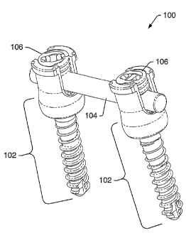
FIG. 1 depicts an embodiment of spinal stabilization system 100 that may be
implanted using a minimally
invasive surgical procedure. Spinal stabilization system 100 may include bone
fastener assemblies 102, elongated
member 104, and/or closure members 106. Other spinal stabilization system
embodiments may include, but are not
limited to, plates, dumbbell-shaped members, and/or transverse connectors.
FIG. 1 depicts a spinal stabilization
system for one vertebral level. In some embodiments, the spinal stabilization
system of FIG. 1 may be used as a
multi-level spinal stabilization system if one or more vertebrae are located
between the vertebrae in which bone
fastener assemblies 102 are placed. In other embodiments, multi-level spinal
stabilization systems may include
additional bone fastener assemblies to couple to one or more other vertebrae.
FIG. 2 depicts a perspective view of bone fastener assembly 102. FIG. 3, FIGS.
4A and 4B, and FIG. 5
depict embodiments of bone fastener assembly components. Components of bone
fastener assembly 102 may
include, but are not limited to, bone fastener 108 (shown in FIG. 3), ring 110
(shown in FIGS. 4A and 4B), and
collar 112 (shown in FIG. 5). Bone fastener 108 may couple bone fastener
assembly 102 to a vertebra. Ring 110
may be positioned between a head of bone fastener 108 and collar 112.
FIG. 6 depicts a cross-sectional representation of bone fastener 108, ring
110, and collar 112 of bone
fastener assembly 102. Bone fastener 108 of bone fastener assembly 102 may
include passage 114. Bone fastener
108 may be cannulated (i.e., passage 114 may run through the full length of
the bone fastener). A guide wire may
be placed through passage 114 so that bone fastener 108 may be inserted into a
vertebra at a desired location and in
a desired angular orientation relative to the vertebra with limited or no
visibility of the vertebra
In some embodiments, a bone fastener assembly may be a fixed angle fastener.
FIG. 7 depicts an
embodiment of a fixed angle bone fastener. Collar and bone fastener may be
formed as a unitary piece of metal. A
fixed angle fastener may be positioned as the first bone fastener assembly
inserted into a vertebra.
CA 02502571 2005-04-15
WO 2004/041100 PCT/US2003/034469
A bone fastener may be, but is not limited to, a bone screw, a ring shank
fastener, a barb, a nail, a brad, or
a trocar. Bone fasteners and/or bone fastener assemblies may be provided in
various lengths in an instrumentation
set to accommodate variability in vertebral bodies. For example, an
instrumentation set for stabilizing vertebrae in
a lumbar region of the spine may include bone fastener assemblies with lengths
ranging from about 30 mm to about
75 mm in 5 mm increments. A bone fastener assembly may be stamped with indicia
(i.e., printing on a side of the
collar). In some embodiments, a bone fastener assembly or a bone fastener may
be color-coded to indicate a length
of the bone fastener. In certain embodiments, a bone fastener with a 30 mm
thread length may have a magenta
color, a bone fastener with a 35 mm thread length may have an orange color,
and a bone fastener with a 55 mm
thread length may have a blue color. Other colors may be used as desired.
Each bone fastener provided in an instrumentation set may have substantially
the same thread profile and
thread pitch. In an embodiment, the thread may have about a 4 mm major
diameter and about a 2.5 mm minor
diameter with a cancellous thread profile. In certain embodiments, the minor
diameter of the thread may be in a
range from about 1.5 mm to about 4 mm or larger. In certain embodiments, the
major diameter of the thread may
be in a range from about 3.5 mm to about 6.5 mm or larger. Bone fasteners with
other thread dimensions and/or
thread profiles may also be used. A thread profile of the bone fasteners may
allow bone purchase to be maximized
when the bone fastener is positioned in vertebral bone.
FIG. 3 depicts an embodiment of bone fastener 108. Bone fastener 108 may
include shank 116, head 118,
and neck 120. Shank 116 may include threading 122. In some embodiments,
threading 122 may include self
tapping start 124. Self-tapping start 124 may facilitate insertion of bone
fastener 108 into vertebral bone.
Head 118 of bone fastener 108 may include various configurations to engage a
driver that inserts the bone
fastener into a vertebra. In some embodiments, the driver may also be used to
remove an installed bone fastener
from a vertebra. In some embodiments, head 118 may include one or more tool
portions 126. Tool portions 126
may be recesses and/or protrusions designed to engage a portion of the driver.
In some embodiments, bone fastener
108 may be cannulated for use in a minimally invasive procedure.
Head 118 of bone fastener 108 may include one or more splines 128, as depicted
in FIG. 3. In some head
embodiments, head 118 may include three splines. Splines 128 may be equally
spaced circumferentially around
head 118 of bone fastener 108. In some head embodiments, splines 128 may be
spaced at unequal distances
circumferentially around head 118. Splines 128 may include various surface
configurations and/or texturing to
enhance coupling of bone fastener 108 with a ring of a bone fastener assembly.
In some embodiments, sides of the
splines may be tapered so that the splines form a dovetail connection with a
ring. In some embodiments, spline
width may be tapered so that a good interference connection is established
when the bone screw is coupled to a ring.
Splines 128 may include one or more projections 130 to facilitate coupling
bone fastener 108 with an inner surface
of a ring. In some embodiments, projections 130 may be positioned on a lower
portion of splines 128. In some
embodiments, the splines may include recessed surfaces that accept projections
extending from surfaces of the ring.
Neck 120 of bone fastener 108 may have a smaller diameter than adjacent
portions of head 118 and shank
116. The diameter of neck 120 may fix the maximum angle that the collar of the
bone fastener assembly can be
rotated relative to bone fastener 108. In some embodiments, neck 120 may be
sized to allow up to about 40° or
more of angulation of the collar relative to the bone fastener. In some
embodiments, the neck may be sized to allow
up to about 30° of angulation of the collar relative to the bone
fastener. In some embodiments, the neck may be
sized to allow up to about 20° of angulation of the collar relative to
the bone fastener.
CA 02502571 2005-04-15
WO 2004/041100 PCT/US2003/034469
FIGS. 4A and 4B depict perspective views of embodiments of ring 110. Outer
surface 132 of ring 110
may have a contour that substantially complements a contour of an inner
surface of a collar in which the ring
resides. A contour of the outer surface of the ring may be a spherical
portion. When the ring is positioned in the
collar, the complementary shape of the ring outer surface and the inner
surface of the collar that contacts the ring
allows angulation of the collar relative to a bone fastener coupled to the
ring. The contour of the outer surface of
the ring and the inner surface of the collar may inhibit removal of the ring
from the collar after insertion of the ring
into the collar.
Outer surface 132 of ring 110 may have a smooth finish. In some embodiments,
outer surface 132 may be
surface treated or include coatings and/or coverings. Surface treatments,
coatings, and/or coverings may be used to
adjust frictional and/or wear properties of the outer surface of the ring. In
some embodiments, a portion of the outer
surface of the ring may be shaped and/or textured to limit a range of motion
of the collar relative to a bone fastener
of a bone fastener assembly.
An inner surface of ring 110 may include one or more grooves 134 and/or one or
more seats 136. Seats
136 may be circumferentially offset from grooves 134. Grooves 134 may be sized
to allow passage of splines of a
bone fastener (e.g., splines 128 shown in FIG. 3) through the ring. When the
splines are inserted through grooves
134, the bone fastener may be rotated until the splines align with seats 136.
The bone fastener may be pulled or
driven so that the splines are positioned in seats 136. In some embodiments,
projections (e.g., projections 130 in
FIG. 3) may pass over ridges 138 of ring 110. Passage of the projections over
ridges 138 may securely couple the
bone fastener to the ring and inhibit separation of the ring from the bone
fastener.
In a ring embodiment, a number of grooves 134 and a number of seats 136 may
equal a number of splines
128 on a head of a bone fastener. Seats 136 and grooves 134 may be equally
spaced circumferentially around the
inner surface of ring 110. In some embodiments, seats 136 may be
circumferentially offset about 60° from grooves
134.
In some embodiments, as shown in FIG. 4A, a ring may be a complete ring
without a split or slots. In
some embodiments, a ring may include a split or slots to facilitate insertion
of the ring into a collar. FIG. 4B
depicts a ring with a split. In some embodiments, a ring with a split and/or
slots may be compressed to ease
insertion into a collar. Once positioned in the collar, the ring may expand to
its original uncompressed dimensions,
thus inhibiting removal from the collar.
As used herein, the term "collar" includes any element that wholly or
partially encloses or receives one or
more other elements. A collar may enclose or receive elements including, but
not limited to, a bone fastener, a
closure member, a ring, and/or an elongated member. In some embodiments, a
collar may couple two or more other
elements together (e.g., an elongated member and a bone fastener). A collar
may have any of various physical
forms. In some embodiments, a collar may have a "U" shape, however it is to be
understood that a collar may also
have other shapes.
A collar may be open or closed. A collar having a slot and an open top, such
as collar 112 shown in FIG. 2
and in FIG. 5, may be referred to as an "open collar." A bone fastener
assembly that includes an open collar may be
referred to as an "open fastener." In some embodiments, an elongated member
may be top loaded into the open
fastener. A closure member may be coupled to the collar to secure the
elongated member to the open fastener.
A collar that does not include a slot and an open top may be referred to as a
"closed collar." A spinal
implant that includes a closed collar may be referred to as a "closed
implant." A closed collar may include an
11
CA 02502571 2005-04-15
WO 2004/041100 PCT/US2003/034469
aperture, bore, or other feature in side surfaces for accommodating other
components of a stabilization system (e.g.,
an elongated member). A setscrew may be used to securely couple an elongated
member to a closed implant.
Collar 112 may include body 140 and arms 142. Arms 142 may extend from body
140. Body 140 of
collar 112 may be greater in width than a width across arms 142 of collar 112
(i.e., body 140 may have a maximum
effective outer diameter greater than a maximum effective outer diameter of
arms 142). A reduced width across
arms 142 may allow a detachable member to be coupled to the arms without
substantially increasing a maximum
effective outer diameter along a length of collar 112. Thus, a reduced width
across arms 142 may reduce bulk at a
surgical site.
A height of body 140 may range from about 3 millimeters (mm) to about 7 mm. In
an embodiment, a
height of body 140 is about 5 mm. Body 140 may include opening 144 in a lower
surface of the body. To inhibit
passage of a ring from collar 112, opening 144 may be smaller than an outer
diameter of the ring. Inner surface 146
may be machined to complement a portion of an outer surface of a ring that is
to be positioned in collar 112.
Machining of inner surface 146 may enhance retention of a ring in collar 112.
Inner surface 146 of body 140 may
be complementary in shape to a portion of outer surface 132 of ring 110 (see
FIG. 4) so that the ring is able to
swivel in the collar. Inner surfaces and/or outer surfaces of collar 112 may
be surface treated or include coatings
and/or coverings to modify frictional properties or other properties of the
collar.
Inner surfaces of arms 142 may include modified thread 148. Modified threads
148 may engage
complementary modified threads of a closure member to secure an elongated
member to a bone fastener assembly.
Modified threads 148 may have a constant pitch or a variable pitch.
A height and a width of arms 142 may vary. Arms 142 may ranges in height from
about 8 mm to about 15
mm. In an embodiment, a height of arms 142 is about 11 mm. A width (i.e.,
effective diameter) of arms 142 may
range from about 5 mm to 14 mm. Arms 142 and body 140 may form slot 150. Slot
150 may be sized to receive an
elongated member. Slot 150 may include, but is not limited to, an elongated
opening of constant width, an
elongated opening of variable width, a rectangular opening, a trapezoidal
opening, a circular opening, a square
opening, an ovoid opening, an egg-shaped opening, a tapered opening, and
combinations and/or portions thereof. In
some embodiments, a first portion of slot 150 may have different dimensions
than a second portion of slot 150. In
certain embodiments, a portion of slot 150 in first arm 142 may have different
dimensions than a portion of slot 150
in second arm 142. When an elongated member is positioned in slot 150, a
portion of the elongated member may
contact a head of a bone fastener positioned in the collar.
In an embodiment of a collar, arms 142 of collar 112 may include one or more
openings and/or indentions
152. Indentions 152 may vary in size and shape (e.g., circular, triangular,
rectangular). Indentions 152 may be
position markers and/or force application regions for instruments that perform
reduction, compression, or
distraction of adjacent vertebrae. In some embodiments, openings and/or
indentions may be positioned in the body
of the collar.
Arms 142 may include ridges or flanges 154. Flange 154 may allow collar 112 to
be coupled to a
detachable member so that translational motion of the collar relative to the
detachable member is inhibited. Flanges
154 may also include notches 156. A movable member of a detachable member may
extend into notch 156. When
the movable member is positioned in notch 156, a channel in the detachable
member may align with a slot in collar
112. With the movable member positioned in notch 156, rotational movement of
collar 112 relative to the
detachable member may be inhibited.
12
CA 02502571 2005-04-15
WO 2004/041100 PCT/US2003/034469
FIGS. 8A-8C show views of collar 112 and ring 110 during top loading insertion
of the ring into the collar.
Ring 110 may be positioned as shown in FIG. 8A and inserted past arms 142 into
body 140. FIG. 8B depicts a
cross-sectional view of ring 110 and collar 112 after insertion of the ring
into the collar through slot 150. After
insertion of ring 110 into collar 112, the ring may be rotated so that a bone
fastener may be positioned through the
ring. FIG. 8C depicts a cross-sectional view of ring 110 and collar 112 after
rotation of the ring in the collar. -
FIGS. 9A-9C show views of collar 112 and ring 110 during bottom loading
insertion of the ring into the
collar. Ring 110 may be positioned as shown in FIG. 9A and inserted into body
140 through an opening in the
bottom of collar 112. In some embodiments, ring 110 may be inserted into body
140 through a groove or a slot in
the bottom of collar 112. In certain embodiments, collar 112 designed for
bottom insertion of ring 110 may have
narrower slot 150 than a collar designed for top insertion of a ring. Collar
112 with narrower slot 150 may allow an
elongated member with a reduced diameter to be used in a spinal stabilization
system. Collar 112 with narrower
slot 150 may be used to reduce bulk at a surgical site.
FIG. 9B depicts a cross-sectional view of ring 110 and collar 112 after
insertion of the ring into the collar
through the opening in the bottom of the collar. After insertion of ring 110
into collar 112, the ring may be rotated
so that a bone fastener may be positioned through the ring. Tolerance between
an outer surface of ring 110 and an
inner surface of body 140 shown in FIGS. 8A-8C and 9A-9C may require force to
be applied to the ring to drive the
ring into the body. Once ring 110 is positioned in body 140, the ring may
expand slightly. In certain embodiments,
significant force may be required to remove ring 110 from body 140 (i.e., the
ring may be substantially unreleasable
from the body). The required force may inhibit unintentional removal of ring
110 from body 140. FIG. 9C depicts
a cross-sectional view of ring 110 and collar 112 after rotation of the ring
in the collar.
FIG. l0A depicts bone fastener 108 before insertion of the bone fastener into
ring 110 positioned in collar
112. Splines 128 may be aligned with grooves 134 to allow passage of head 118
through ring 110 and into collar
112. FIG. lOB depicts bone fastener 108, ring 110, and collar 112 after the
bone fastener has been rotated and head
118 has been coupled to seats in the ring to form bone fastener assembly 102.
Inserting bone fastener 108 through
opening 144 in collar 112 (depicted in FIG. l0A) may allow use of bone
fasteners that have shanks and/or heads
with larger diameters than can pass through slot 150. Bone fasteners with
large diameter shanks may form a bone
fastener assembly (threaded or otherwise) that securely fastens to vertebral
bone during use.
A bone fastener may be rotatably positioned in a collar such that the bone
fastener is able to move radially
and/or rotationally relative to the collar (or the collar relative to the bone
fastener) within a defined range of motion.
The range of motion may be provided within a plane, such as by a hinged
connection, or within a three-dimensional
region, such as by a ball and socket connection. Motion of the bone fastener
relative to the collar (or the collar
relative to the bone fastener) may be referred to as "angulation" and/or
"polyaxial movement". FIG. 11 depicts
bone fastener assembly 102 with central axis 158 of collar 112 aligned with
central axis 160 of bone fastener 108.
Bone fastener 108 may be angulated in a symmetrical conical range of motion
characterized by angle a about the
aligned axes. Bone fastener 108 may be constrained from motion outside of
limit axis 162 by contact between neck
120 of bone fastener 108 and collar 112. Alignment of axis 160 of bone
fastener 108 with central axis 158 of collar
112 may be considered a neutral position relative to the range of motion. The
alignment is a neutral position
because bone fastener 108 may be angulated an equal amount in any direction
from central axis 158. When a driver
is inserted into bone fastener 108, axis 160 of bone fastener 108 may be
substantially aligned with axis 158 of collar
112 to facilitate insertion of the bone fastener into a vertebral body.
13
CA 02502571 2005-04-15
WO 2004/041100 PCT/US2003/034469
In certain embodiments, a range of motion of a collar may be skewed from a
full conical range of motion
relative to aligned central axes of the collar and a bone fastener coupled to
the collar. In some embodiments, a
distal end of a collar may be shaped to skew, or bias, the range of motion
from the range of motion depicted in FIG.
11. FIGS. 12A and 12B depict bone fastener assemblies 102 with biased collars
112. Body 140 of biased collar
112 may be shaped to restrict relative movement of bone fastener 108 (and/or
the collar) to a skewed conical range
of motion defined by limit axes 162. As depicted by limit axes 162 in FIG.
12A, a first arm 142 of collar 112 may
approach bone fastener 108 more closely than a second arm of the collar. As
suggested by limit axes 162 in FIG.
12B, a first opening of the slot between arms 142 of collar 112 may approach
bone fastener 108 more closely than a
second opening of the slot.
Other biased collars may be designed to selectively restrict relative movement
of collars and/or bone
fasteners. In some embodiments, a biased collar may be attached to a
detachable member such that a surgeon
performing a minimally invasive procedure may selectively align the portion of
the collar with the greater range of
motion as needed. For example, the collar depicted in FIG. 12B may be coupled
to a single-level (e.g., C-shaped)
sleeve so that the side of the collar (i.e., the side of the slot) with a
larger range of motion is positioned next to a
channel opening of the sleeve.
When a biased collar of a bone fastener assembly is coupled to a detachable
member and a drive
mechanism is coupled to a bone fastener of the bone fastener assembly, central
axis 158 of collar 112 may align
with central axis 160 of bone fastener 108 to facilitate insertion of the bone
fastener into bone. In some
embodiments, the bias of the collar may be so large that a flexible drive
member is needed to drive the bone
fastener into bone.
In some embodiments, one or more biased collars may be used in a spinal
stabilization system. The spinal
stabilization systems may be single-level systems or multi-level systems.
Biased collars may be used to
accommodate the increasing angle of the pedicle corridor for each lumbar
vertebra. The angle may increase by
about 5 degrees for each successive lumbar vertebra. FIGS. 13A andl3B depict a
single-level spinal stabilization
system including bone fastener assembly 102A coupled to pedicle 164A and
vertebra 166A and bone fastener
assembly 102B coupled to pedicle 164B and vertebra 166B:
A bone fastener of bone fastener assembly 102A may engage pedicle 164A at
pedicle angle ~A relative to
sagittal plane 168. Pedicle angle ~A may range between about 13° and
about 17°. Collar 112A of bone fastener
assembly 102A may be unbiased. Pedicle angle ~B may range between about
18° and about 22°. Collar 112B may
have a bias angle (3 of about 5°. Bone fastener assembly 102B may
engage pedicle 164B at pedicle angle ~B.
Because the bias of collar 112B is approximately equal to the difference
between the pedicle angles of the two
vertebrae, slots 150A and 150B in bone fastener assemblies 102A and 102B,
respectively, may be generally aligned
when both bone fasteners are in neutral positions.
Angulation of either or both collars of the bone fastener assemblies may allow
fme adjustment of
engagement angles of the bone fasteners. In addition, collar angulation may
allow adjustment in the orientation of
bone fasteners in a sagittal plane (i.e., to conform to lordosis of a spine)
while still allowing the collars to be easily
coupled with elongated member 104. Elongated member 104 may be disposed in
slots 150A and 150B and secured
by closure members. In some embodiments, a flexible driver or a polyaxial
driver (e.g., a driver with a universal
joint) may be used to drive the heads of the bone fasteners from a position
that is off axis from the bone fasteners to
reduce the size of an opening of the body needed to implant the spinal
stabilization system.
14
CA 02502571 2005-04-15
WO 2004/041100 PCT/US2003/034469
A closure member may be coupled to a collar of a bone fastener assembly to fix
an elongated member
positioned in the collar to the bone fastener assembly. In some embodiments, a
closure member may be cannulated.
In certain embodiments, a closure member may have a solid central core. A
closure member with a solid central
core may allow more contact area between the closure member and a driver used
to couple the closure member to
the collar. A closure member with a solid central core may provide a more
secure connection to an elongated
member than a cannulated closure member by providing contact against the
elongated member at a central portion
of the closure member as well as near an edge of the closure member.
FIG. 1 depicts closure members 106 coupled to bone fastener assemblies 102.
FIG. 14 depicts closure
member 106 prior to insertion of the closure member into a collar of a bone
fastener assembly. Closure member
106 may include tool portion 170 and male modified thread 172. Tool portion
170 may couple to a tool that allows
closure member 106 to be positioned in a collar. Tool portion 170 may include
various configurations (e.g.,
threads, hexalobular connections, hexes) for engaging a tool (e.g., a driver).
Male modified thread 172 may have a
shape that complements the shape of a female modified thread in arms of a
collar (e.g., modified thread 148
depicted in FIG. 5).
FIG. 15 depicts a cross-sectional representation of closure member 106 taken
substantially along plane 15-
15 of FIG. 14. Closure member 106 may include removal openings 174. A drive
tool may be inserted into removal
openings 174 to allow removal of closure member 106 after tool portion 170 has
been sheared off. Removal
openings 174 may include any of a variety of features including, but not
limited to, sockets, holes, slots, and/or
combinations thereof. In an embodiment, removal openings 174 are holes that
pass through bottom surface 176 of
closure member 106.
A bottom surface of a closure member may include structure and/or texturing
that promotes contact
between the closure member and an elongated member. A portion of the structure
and/or texturing may enter
and/or deform an elongated member when the closure member is coupled to the
elongated member. Having a
portion of the closure member enter and/or deform the elongated member may
couple the elongated member to the
closure member and a bone fastener assembly so that movement of the elongated
member relative to the bone
fastener assembly is inhibited. In a closure member embodiment, such as the
embodiment depicted in FIG. 15,
bottom surface 176 of closure member 106 may include point 178 and rim 180. In
some embodiments, rim 180
may come to a sharp point. In some embodiments, a height of rim 180 may be
less than a height of point 178. In
other embodiments, a height of rim 180 may be the same or larger than a height
of point 178. In some
embodiments, rim 180 may not extend completely around the closure member. For
example, eight or more portions
of rim 180 may be equally spaced circumferentially around closure member 106.
In certain embodiments, a solid
central core including point 178 and rim 180 may enhance the ability of
closure member 106 to secure an elongated
member in a collar.
FIG. 16 depicts a portion of a spinal stabilization system with closure member
106 coupled to collar 112
before tool portion 170 is sheared off. Closure member 106 may couple to
collar 112 by a variety of systems
including, but not limited to, standard threads, modified threads, reverse
angle threads, buttress threads, or helical
flanges. A buttress thread on a closure member may include a rearward-facing
surface that is substantially
perpendicular to the axis of the closure member. Closure member 106 may be
advanced into an opening in a collar
to engage a portion of elongated member 104. In some embodiments, closure
member 106 may inhibit movement
of elongated member 104 relative to collar 112.
CA 02502571 2005-04-15
WO 2004/041100 PCT/US2003/034469
FIG. 17A depicts a cross-sectional view of closure member 106 coupled to bone
fastener assembly 102.
Closure member 106 may include male modified thread 172. Male modified thread
172 may include male distal
surface 182 and male proximal surface 184, as shown in ~'IG. 17B. Collar 112
may include female modified thread
148 on an inside surface of arms 142. Female modified thread 148 may include
female proximal surface 186 and
female distal surface 188. Male proximal surface 184 may couple to female
distal surface 188 during use. Male
proximal surface 184 and female distal surface 188 may be load-bearing
surfaces. A load may result from an
upward load on closure member 106, such as a load resulting when elongated
member 104 positioned in a slot of
collar 112 is secured to bone fastener assembly 102 by closure member 106.
Raised portions 190 and recessed portions 192 may be included on male distal
surface 182 and female
proximal surface 186. Cooperating surfaces 194 of modified threads 172 and 148
may contact or be proximate to
one another during use. As used herein, "proximate" means near to or closer to
one portion of a component than
another portion of a component. Engagement of cooperating surfaces 194 of
modified threads 172 and 148 during
use may inhibit radial expansion of collar 112. Engagement of cooperating
surfaces 194 may inhibit spreading of
arms 142 away from each other (i.e., inhibit separation of the arms). In some
embodiments, cooperating surfaces
194 may be substantially parallel to a central axis of closure member 106. In
other embodiments, cooperating
surfaces 194 may be angled relative to a central axis of closure member 106.
In some embodiments, a proximal surface of a male modified thread may include
raised and recessed
portions. FIG. 18A depicts a cross-sectional view of bone fastener assembly
102 coupled to closure member 106
with raised and recessed portions on a proximal surface of male modified
thread 172. FIG. 18B depicts a cross-
sectional view of raised portions 190 at male proximal surface 184 of male
modified thread 172 and female distal
surface 188 of female modified thread 148. Male proximal surface 184 may
include an overall positive slope S
such that point A near the top of male modified thread 172 is distal from
point B at the base of the male modified
thread. Alternatively, male proximal surface 184 may include an overall
negative slope or a slope of about zero.
In an embodiment, a bone fastener assembly and a closure member may be coupled
with a running fit. A
running fit (i.e., a fit in which parts are,free to rotate) may result in
predictable loading characteristics.of a coupling
of a bone fastener assembly and a closure member. Predictable loading
characteristics may facilitate use of a
closure member with a break-off portion designed to shear off at a
predetermined torque. A running fit may also
facilitate removal and replacement of closure members. In some embodiments, a
closure member may include an
interference fit (e.g., crest-to-root radial interference).
In an embodiment, a position (i.e., axial position and angular orientation) of
a modified thread of a collar
may be controlled, or "timed," relative to selected surfaces of the collar.
For example, a modified thread form may
be controlled relative to a top surface of a collar and an angular orientation
of the slots of the collar. In some
embodiments, positions of engaging structural elements of other coupling
systems (e.g., thread forms) may be
controlled.
Controlling a position of a modified thread form may affect a thickness of a
top modified thread portion of
a collar. In FIG. 5, top modified thread portion 196 is the first modified
thread portion to engage a closure member.
In an embodiment, a position of a modified thread form may be selected such
that the thickness of the leading edge
of a top modified thread portion is substantially equal to the full thickness
of the rest of the modified thread.
Controlling a position of a modified thread form of a collar may increase a
combined strength of engaged
modified thread portions for a collar of a given size (e.g., wall height,
modified thread dimensions, and thread
pitch). Controlling a position of the modified thread form may reduce a
probability of failure of modified thread
16
CA 02502571 2005-04-15
WO 2004/041100 PCT/US2003/034469
portions, and thus reduce a probability of coupling failure between a collar
and a closure member. Controlling the
position of a modified thread form in a collar of a bone fastener assembly may
increase a combined strength of
engaged collar and closure member modified thread portions such that failure
of the modified thread portions does
not occur prior to the intended shearing off of a tool portion of the closure
member. For example, a tool portion of a
closure member may be designed to shear off at about 90 in-lbs of torque,
while the combined modified thread
portions may be designed to withstand a torque on the closure member of at
least 120 in-lbs.
If a thickness of a modified thread portion of a given size and profile is
reduced below a minimum
thickness, the modified thread portion may not significantly contribute to the
holding strength of the modified
thread of a collar. In an embodiment, a position of a modified thread form of
a collar may be controlled such that a
thickness of a top modified thread portion is sufficient for the portion to
increase a holding strength of the collar. In
one embodiment, a top modified,thread portion may have a leading edge
thickness of about 0.2 mm.
In an embodiment, a position of a modified thread form of a collar may be
selected to ensure that a closure
member engages a selected minimum number of modified thread portions on each
arm of the collar. In an
embodiment, at least two modified thread portions having a full thickness over
width w of a collar arm (shown in
FIG. 5) may be engaged by a closure member at each arm. Alternatively, a
closure member may engage parts of
three or more modified thread portions on each arm, with the total width of
the portions equal to at least two full-
width portions. Allowances may be made for tolerances in the components (e.g.,
diameter of the elongated
member) and/or anticipated misalignment between the components, such as
misalignment between an elongated
member and a slot. In an embodiment, a substantially equal number of modified
thread portions in each arm may
. engage the closure member when an elongated member is coupled to a bone
fastener assembly.
Various instruments may be used in a minimally invasive procedure to form a
spinal stabilization system
in a patient. The instruments may include, but are not limited to, positioning
needles, guide wires, dilators, bone
awls, bone taps, sleeves, drivers, tissue wedges, elongated member length
estimating tools, mallets, tissue retractors,
and tissue dilators. The instruments may be provided in an instrumentation
set. The instrumentation set may also
include components of the spinal stabilization system. The components of the
spinal stabilization system may
include, but are not limited to, bone fastener assemblies of various sizes
and/or lengths, elongated members, and
closure members.
Instruments used to install a spinal stabilization system may be made of
materials including, but not
limited to, stainless steel, titanium, titanium alloys, ceramics, and/or
polymers. Some instruments may be
autoclaved and/or chemically sterilized. Some instruments may include
components that cannot be autoclaved or
chemically sterilized. Components of instruments that cannot be autoclaved or
chemically sterilized may be made
of sterile materials. The sterile materials may be placed in working relation
to other parts of the instrument that
have been sterilized.
A targeting needle may be used to locate an entry point in a vertebral body
for a bone fastener of a bone
fastener assembly. In some embodiments, the targeting needle may be a
Jamshidi~ bone marrow biopsy needle.
FIG. 19 depicts an embodiment of targeting needle 198. Targeting needle 198
may include outer housing 200 and
member 202. FIG. 20 depicts an embodiment of outer housing 200. Outer housing
200 may include hollow shaft
204 and handle 206. Scale markings 208 may be printed, etched, or otherwise
placed on hollow shaft 204. Scale
markings 208 may be used to approximate a length of a bone fastener needed for
a vertebra. Handle 206 may
provide a grip that allows a user to manipulate the targeting needle. Handle
206 may include threaded portion 210.
17
CA 02502571 2005-04-15
WO 2004/041100 PCT/US2003/034469
Threaded portion 210 may couple to threading on a portion of a targeting
needle member to secure the member to
outer housing 200.
FIG. 21 depicts an embodiment of member 202 of a targeting needle. Member 202
may include point 212
and cap 214. Point 212 may be placed through a hollow shaft of an outer
housing of the targeting needle. Cap 214
may include threading 216. Member 202 may be rotated relative to the outer
housing to couple threading 216 with
threading in a handle of the outer housing. In some embodiments, the member
may be coupled to the outer housing
by another type of connection system (e.g., by placement of a key in a
keyway). With member 202 positioned in an
outer housing, point 212 may extend from a distal end of a hollow shaft of the
outer housing. Cap 214 may be used
as an impact surface for driving the targeting needle in bone.
FIG. 22 and FIG. 23 depict embodiments of guide wire 218. Guide wire 218 may
be an 18-gauge K-wire.
Guide wire 218 may pass down a shaft of a targeting needle outer housing. A
guide wire may be from about 15 cm
to about 65 cm in length. In some embodiments, guide wires provided in an
instrumentation set are about 46 cm in
length. The length of guide wire 218 may allow a surgeon and/or assistants to
hold at least one portion of the guide
wire at all times when the guide wire is inserted into vertebral bone, even
during insertion, use, and removal of
instruments along a length of the guide wire. A guide wire that can be held
continuously during a surgical
procedure may inhibit removal or advancement of the guide wire from a desired
position during a minimally
invasive surgical procedure.
As depicted in FIG. 22, a distal end of guide wire 218 may include point 220.
Point 220 may facilitate
insertion of the distal end of guide wire 218 into vertebral bone. As depicted
in FIG. 23, a distal end of guide wire
218 may not be pointed. A position of an unpointed guide wire in bone may be
easier to maintain during a spinal
stabilization procedure.
Dilators may be used during a minimally invasive surgical procedure to push
aside tissue and create space
to access vertebral bone. In some embodiments, four tissue dilators of
increasing diameter may be used to establish
sufficient working space to accommodate instruments and spinal stabilization
system components. In some
embodiments, especially for a mid-vertebra or for mid-vertebrae of a mufti-
level stabilization system, only three
dilators may be needed to form sufficient working space. Dilators in an
instrumentation set may increase in
diameter incrementally by a selected amount. For example, outside diameters of
dilators in an instrumentation set
may increase sequentially by increments of about 0.5 mm:
A bone awl may be used to breach cortical bone of a pedicle. FIG. 24 depicts
an embodiment of bone awl
222. Bone awl 222 may include handle 224, passage 226, and tip 228. Handle 224
may provide a secure grip that
allows a surgeon to breach cortical bone of a pedicle with tip 228. A guide
wire that is inserted in vertebral bone in
a desired orientation may be inserted through passage 226 that extends through
bone awl 222. Bone awl 222 may
be moved down the guide wire so that tip 228 contacts the pedicle.
Bone awl 222 may have a length that allows a guide wire positioned in
vertebral bone to always be held in
at least one location when the guide wire is placed through passage 226 in the
needle. In some embodiments,
handle 224 may be removable from a shaft of bone awl 222 so that the guide
wire may always be held during use of
the bone awl.
During some surgical procedures downward force and some rotation of the bone
awl may be sufficient to
breach cortical of a vertebra. During some surgical procedures, an impact
force may be needed for the bone awl to
breach cortical bone. In some embodiments, a guide wire may be removed, the
bone awl may be used to breach
cortical bone, and the guide wire may be reinserted. In some embodiments, a
small dilator may be placed over the
18
CA 02502571 2005-04-15
WO 2004/041100 PCT/US2003/034469
portion of the guide wire extending from the bone awl so that a first end of
the dilator contacts the bone awl. A
mallet or other impact device may be used against a second end of the dilator
so that the bone awl breaches cortical
bone of the vertebra. The dilator may be removed from the bone awl and contact
with the guide wire may be
reestablished.
A bone tap may be used to form a threaded passage of a desired depth through a
pedicle and into a
vertebral body. FIG. 25 depicts an embodiment of tap 230. Tap 230 may include
passage 232, shaft 234,
removable handle 236, flutes 238, and indicia 240. Passage 232 may extend
through a length of shaft 234 and
removable handle 236. A guide wire positioned in vertebral bone may be
inserted into a distal end of passage 232
so that tap 230 can be moved down the guide wire toward the bone.
In an embodiment of tap 230, a proximal portion of shaft 234 may include at
least one flat portion that fits
in a mating portion of removable handle 236. Proximal end of shaft 234 may
also include a detent depression. The
flat portion may allow for rotation of shaft 234 when removable handle 236 is
rotated. An embodiment of
removable handle 236 may include spring-loaded release 242. When spring-loaded
release 242 is compressed (i.e.,
drawn upwards), a detent in removable handle 236 may be movable. When spring-
loaded release 242 is not
compressed, movement of the detent may be inhibited. When shaft 234 is
positioned in removable handle 236, the
detent of the removable handle may be positioned in the detent depression of
shaft 234 to couple the shaft to the
removable handle.
A tap portion of tap 230 may have a known length. As shown in FIG. 25, a tap
portion of tap 230 may
have a length t. In some embodiments, t may be about 20 mm, about 40 mm, about
60 mm, or greater. For
example, t may be about 45 mm. X-ray monitoring of a depth of a tap portion of
known length may allow a
medical practitioner to assess a depth of a hole tapped in a bone. In some
embodiments, the hole may be tapped to
accommodate a bone fastener of a desired length. In certain embodiments, a
bone fastener may be chosen to
accommodate a hole tapped to a desired depth.
A guide wire positioned in vertebral bone may be held near a top of a dilator
inserted over the guide wire
at a surgical site. A proximal end of the guide wire may be positioned through
a distal end of a passage in shaft 234
of tap 230 without a removable handle coupled to the shaft. A proximal portion
of the guide wire may be held
when the proximal portion of the guide wire extends beyond the top of shaft
234. A portion of the guide wire may
always be held during use of tap 230. Shaft 234 may be moved down the guide
wire until the shaft contacts the
vertebral bone. The guide wire may be held near the top of shaft 234 and the
guide wire may be positioned through
passage 232 of removable handle 236. When the guide wire extends out of
passage 232 through removable handle
236, the guide wire may be held above the removable handle. The handle may be
coupled to the shaft using spring-
loaded release 242.
A first reading of indicia 240 relative to a proximal end of a dilator may be
taken when a first flute of
flutes 238 is located at a pedicle. Tap 230 may be rotated so that flutes 238
form a threaded opening through the
pedicle and into a vertebral body. Flutes 238 may have a diameter that is
about 0.1 mm to about 0.7 mm less than a
maximum thread flight of a bone fastener to be positioned in the threaded
opening formed by the flutes. In an
embodiment, tap may form a thread that is about 0.5 mm less than a maximum
thread flight of a bone fastener to be
positioned in the threaded opening formed by the flutes. A position of tap 230
may be monitored using a
fluoroscope. When the threaded opening is formed to a desired depth, a second
reading of indicia 240 relative to
the dilator may be taken. A length of a bone fastener to be inserted into the
vertebral body may be estimated by
taking the difference between the indicia readings.
19
CA 02502571 2005-04-15
WO 2004/041100 PCT/US2003/034469
After a threaded opening is formed to a desired depth, tap 230 may be removed
by rotating the tap until
flutes 238 are disengaged from vertebral bone. Removable handle 236 may be
separated from shaft 234, and the
removable handle may be removed with the guide wire always held in at least
one location. After removable handle
236 is removed from the guide wire, shaft 234 may be removed with the guide
wire always held in at least one
location.
A detachable member may be used as a guide to install bone fasteners of a bone
fastener assembly in
vertebral bone. A detachable member may be coupled to a collar of a bone
fastener assembly. A distal end of a
detachable member may be tapered or angled to reduce bulk at a surgical site.
Instruments may be inserted into the
detachable member to manipulate the bone fastener assembly. Movement of the
detachable member may alter an
orientation of a collar relative to a bone fastener of the bone fastener
assembly. In some embodiments, a detachable
member may be used as a retractor during a spinal stabilization procedure.
A detachable member for a single-level vertebral stabilization system may
include one or more channels in
a wall of the detachable member to allow access to an adjacent vertebra. For
some single-level vertebral
stabilization procedures, only single-channel detachable members (i.e.,
detachable members with a single channel in
a wall of the detachable member) may be used. For other single-level vertebral
stabilization procedures, one or
more mufti-channel detachable members (i.e., detachable members with two or
more channels in a wall of the
detachable member) may be used. Channels may provide flexibility to or enhance
flexibility of a mufti-channel
detachable member. In some embodiments, a proximal portion of a mufti-channel
detachable member may have a
solid circumference. A region of solid circumference in a mufti-channel
detachable member may enhance stability
of the mufti-channel detachable member. In some embodiments, a mufti-channel
detachable member may be longer
than a single-channel detachable member.
A detachable member used at a middle vertebra in a mufti-level stabilization
procedure may be a multi-
channel detachable member. Channels in a mufti-channel detachable member may
allow access to adjacent
vertebrae from a middle vertebra. A detachable member used at an end vertebra
of a mufti-level stabilization
system may be a single-channel detachable member or a mufti-channel detachable
member. A system for coupling
a bone fastener assembly to a mufti-channel detachable member may include a
limiter that inhibits spreading of
arms of the detachable member to inhibit release of the bone fastener assembly
from the detachable member.
A channel in a wall of a detachable member may allow access to a vertebra that
is to be stabilized with a
spinal stabilization system being formed. In some embodiments, a single-
channel detachable member may be
coupled to a bone fastener assembly to be inserted into vertebral bone of a
first vertebra. The single-channel
detachable member may allow access to a second vertebra from the first
vertebra. In other embodiments, a multi-
channel detachable member may be coupled to a bone fastener assembly to be
inserted into vertebral bone of a first
vertebra. The mufti-channel detachable member may allow access from the first
vertebra to adjacent vertebrae.
Instruments may access a bone fastener assembly through a passage in a
detachable member. In some
embodiments, a channel in a wall of a detachable member may extend a full
length of the detachable member. In
some embodiments, especially in embodiments of mufti-channel detachable
members, a channel in a wall of a
detachable member may extend only a portion of the length of the detachable
member. In some embodiments, a
channel in a wall of a detachable member may extend 25%, 50%, 75%, 80%, 90%,
95% or more of the length of the
detachable member. A channel may extend to a distal end of a detachable member
such that an elongated member
inserted in the channel may pass from the detachable member into a slot of a
collar of a bone fastener assembly
coupled to the detachable member.
CA 02502571 2005-04-15
WO 2004/041100 PCT/US2003/034469
A channel in a detachable member may be any of a variety of shapes. A channel
may have a width that
exceeds a width (e.g., a diameter) of an elongated member that is to be
inserted in the channel. In some
embodiments, a channel may be a linear opening parallel to a longitudinal axis
of the detachable member. In some
embodiments, a channel may have a non-linear shape including, but not limited
to, a helical pattern, an arc, an "L"
shape, or an "S" shape. A non-linear channel may allow an elongated member to
travel along a predetermined path.
In certain embodiments, adjacent detachable members may include channels with
matching profiles, allowing ends
of an elongated member to follow similar paths down the detachable member
channels.
Movable members may extend through portions of a detachable member proximate a
channel in the
detachable member. Movable members may engage notches in a collar to establish
a radial orientation of the
detachable member on the collar and/or to inhibit rotation of the collar
relative to the detachable member. A distal
end of a movable member may be flat, curved, or angled. In some embodiments, a
distal end of a movable member
may be threaded. In other embodiments, a distal end of a movable member may be
a projection that engages an
opening in a collar. In some embodiments, an upper surface of a collar and/or
a surface of a distal end of a movable
member may be textured to inhibit rotation of the collar relative to the
detachable member. In certain embodiments,
a proximal end of a movable member may include a tool engaging portion. A tool
engaging portion may include,
but is not limited to, a hex section, a hexalobular section, a tapered
section, a bead, a knot, a keyed opening, a
coating, a threading, and/or a roughened surface for engaging a drive that
rotates or otherwise displaces the movable
member.
A cross section transverse to a longitudinal axis of a detachable member may
have shapes including, but
not limited to, circular, ovoid, square, pentagonal, hexagonal, and
combinations thereof. In some embodiments, a
detachable member may be hollow. In certain embodiments, a thickness of a
hollow detachable member may be
uniform. In certain embodiments, a thickness of a hollow detachable member may
vary along the length of the
detachable member. A detachable member with a passage extending longitudinally
from a first end of the
detachable member to a second end of the detachable member may be referred to
as a "sleeve".
FIG. 26 depicts an embodiment of sleeve 244. Sleeve 244 may be a multi-channel
sleeve. Sleeve 244 may
include wall 246, channels 248, passage 250, movable members 252, and flange
254. Channels 248 may extend
from a distal end of sleeve 244 through a portion of wall 246. Channels 248
may allow instruments to be positioned
and used to form a plane through soft tissue to one or more adjacent
vertebrae. An elongated member may be
inserted in the tissue plane and positioned in collars of bone fastener
assemblies anchored in vertebrae and coupled
to sleeves. Passage 250 may allow instruments to be positioned and used to
manipulate a bone fastener assembly
that is coupled to a distal end of sleeve 244. Movable members 252 may be part
of a system that couples a bone
fastener assembly to sleeve 244. In some embodiments, movable members 252 may
include tool engaging portion
256. A driver may be positioned in tool portion 256. The driver (e.g., a hex
wrench) may be used to extend or
retract a distal end of movable member 252. A distal end of sleeve 244 may
include flange 254 that mates with a
complementary flange on a collar of a bone fastener assembly. A distal end of
sleeve 244 may be tapered to reduce
bulk (e.g., reduce spin diameter) at a surgical site.
FIG. 27 depicts a top view of an embodiment of sleeve 244 coupled to a bone
fastener assembly. Tool
portion 126 of bone fastener 108 is a hexalobular connection.
FIG. 28 depicts a cross-sectional representation of a portion of sleeve 244
with bone fastener assembly 102
taken substantially along line 28-28 of FIG. 27. Flange 254 of sleeve 244 may
mate with flange 154 of collar 112
21
CA 02502571 2005-04-15
WO 2004/041100 PCT/US2003/034469
to inhibit translation of the sleeve relative to the collar. Sleeve 244 may
also include stop 258. Stop 258 may
engage a portion of collar 112 to inhibit separation of walls 246. During use,
stop 258 may inhibit undesired
separation of bone fastener assembly 102 from sleeve 244.
FIG. 29 depicts a cross-sectional representation of a portion of sleeve 244
with bone fastener assembly 102
and elongated member 104 taken substantially along line 29-29 of FIG. 27.
Distal ends of movable members 252
may extend into notches (e.g., notches 156 depicted in FIG. 5) in collar 112.
Portions of walls 246 of sleeve 244
may include threading. Portions of movable members 252 may include threading
complementary to threaded
portions of walls 246. Threading of movable members 252 may engage threading
in walls 246 such that rotation of
the movable members advances or retracts the movable members relative to the
walls.
As shown in FIG. 29, collar 112 may be designed such that elongated member 104
lies below a distal end
of sleeve 244. Coupling sleeve 244 to collar 112 above elongated member 104
may reduce bulk at a surgical site.
With elongated member 104 coupled to collar 112 below a distal end of sleeve
244, the sleeve may be removed
without interference from the elongated member of a spinal stabilization
system.
FIG. 30 depicts an embodiment of sleeve 244. Sleeve 244 may be a single-
channel sleeve for use in
single-level or multi-level spinal stabilization procedures. Sleeve 244 may be
used at the outermost vertebrae to be
stabilized during installation of a mufti-level vertebral stabilization
system. Sleeve 244 may be coupled to a collar
of a bone fastener assembly with movable members 252 and/or flange 254. ,
Instruments may be inserted through
passage 250 of sleeve 244 to access an anchored bone fastener assembly coupled
to the sleeve. An instrument may
be moved through channel 248 toward an adjacent vertebra to form a tissue
plane in soft tissue between sleeve 244
and the adjacent vertebra.
A sleeve may be coupled to a bone fastener assembly in various ways to inhibit
movement of the sleeve
relative to a collar of the bone fastener assembly. A system used to couple
the sleeve to the bone fastener assembly
may inhibit rotation and translation of the sleeve relative to the collar.
FIG. 31 depicts a perspective view of a sleeve embodiment during connection of
the sleeve to collar 112 of
a bone fastener assembly. Sleeve 244 may include movable members 252. Movable
members 252 may include
threaded distal end portions. FIG. 31A depicts a detailed view of a portion of
sleeve 244 and collar 112. Collar 112
may include openings 260. Openings 260 may be threaded. Openings 260 of collar
112 may be aligned with
movable members 252. A drive end of driver 262 may be positioned in tool
engaging portion 256 of movable
member 252. Driver 262 may be rotated to couple a threaded end of movable
member 252 with threads in opening
260. The driver may be positioned in a tool opening of second movable member
252. The driver may be used to
couple a threaded end of second movable member 252 with threads in second
opening 260. Threaded connections
between movable members 252 and collar 112 may inhibit movement of the collar
relative to sleeve 244.
A detachable member may be coupled to a collar of a bone fastener assembly in
various ways. When a
detachable member is coupled to a collar, rotation and translation of the
detachable member relative to the collar
may be inhibited. A system used to couple a detachable member and collar
should be simple, inexpensive to
implement, and should not significantly weaken the mechanical strength of the
collar and/or the detachable
member. Detachable members may be coupled to collars using various coupling
systems including, but not limited
to, flanges, threaded connections, interlocking connections (e.g., ratcheting
connection systems), and/or interference
fits.
In an embodiment of an interlocking connection system, a detachable member may
include an opposing
pair of deflectable arms. Each deflectable arm may include a tooth. The
deflectable arms may be forced outwards
22
CA 02502571 2005-04-15
WO 2004/041100 PCT/US2003/034469
during coupling of a collar to the detachable member. When the collar is
coupled to the detachable member, the
deflectable arms may be positioned in channels in the collar, with the teeth
positioned in indentions in the collar.
The presence of the deflectable arms in the channels of the collar may inhibit
rotation and translation of the
detachable member relative to the collar. Separation of the detachable member
from the collar may be achieved by
insertion of an expander in the detachable member. The expander may be used to
force the deflectable arms
outwards and expel the teeth from the indentions.
FIGS. 32-45 depict embodiments of sleeves coupled to bone fastener assemblies.
In each bone fastener
assembly/sleeve embodiment depicted in FIGS. 32-43 and FIG. 45, an elongated
member seated in the collar of the
bone fastener assembly would lie below a distal end of sleeve 244. Having the
elongated member below the distal
end of sleeve 244 reduces bulk at the surgical site. With sleeve 244
positioned above the elongated member,
interference of the secured elongated member with the sleeve is avoided during
removal of the sleeve.
FIG. 32 depicts a cross-sectional representation of sleeve 244 including
sleeve flange 254. Sleeve 244
may be rotated onto collar 112 until slot 150 aligns with channel 248. Sleeve
flange 254 may engage flange 154 of
collar 112 to inhibit translation of sleeve 244 relative to collar 112 of bone
fastener assembly 102.
In some detachable member and collar coupling embodiments, the detachable
member and the collar may
include members that work together to inhibit radial expansion of walls of the
detachable member. FIG. 33 depicts
an embodiment of sleeve 244 coupled to an embodiment of bone fastener assembly
102. Sleeve 244 may include
sleeve flange 254 and stop 258. Sleeve flange 254 may engage flange 154 of
collar 112 to inhibit translation of
sleeve 244 relative to the collar. Stop 258 may contact ledge 264 of collar
112. Contact of stop 258 against ledge
264 may inhibit release of collar 112 from sleeve 244 caused by radial
expansion of walls of the sleeve. A stop in a
sleeve and a ledge in a collar may be needed in a mufti-channel sleeve
embodiment. A stop in a sleeve andlor a
ledge in a collar may not be needed in a single-channel sleeve embodiment or
in a collar for a single-level
stabilization.
In some detachable member and collar coupling embodiments, a detachable member
may include a
protrusion that mates with a complementary groove in a collar. Alternatively,
a detachable member may include a
groove that mates with a complementary protrusion of a collar. FIG. 34 depicts
a cross-sectional view of sleeve 244
with ridge 266. Ridge 266 may couple with groove 268 in collar 112. Ridge 266
and groove 268 may form a
dovetail joint. The dovetail joint may inhibit radial expansion of sleeve
walls 246. In some embodiments, such as
the embodiment depicted in FIG. 35, ridge 266 and groove 268 may not form a
dovetail joint.
In some embodiments, a detachable member and/or a collar may include a locking
system to inhibit
rotation of the detachable member relative to the collar. The locking system
may be, but is not limited to,
threading, interference fits, frictional engagement, or a press-fit
connection. In some embodiments, a locking
system may inhibit translation and/or rotation of a detachable member relative
to a collar.
FIG. 36 depicts a top view representation of an embodiment of collar 112 of a
bone fastener assembly.
Collar 112 includes openings 260. In some embodiments, openings 260 may be
threaded. In some embodiments,
openings 260 may not include threading. The body of collar 112 adjacent to
openings 260 may include extra
material to provide strength to the collar.
FIG. 37 depicts a partial cross-sectional representation of an embodiment of
sleeve 244 coupled to an
embodiment of collar 112, such as the collar depicted in FIG. 36. Distal end
portions of movable members 252 may
extend into openings 260. When distal end portions of movable members 252 are
positioned in openings 260,
rotational movement of sleeve 244 relative to collar 112 may be inhibited.
Sleeve 244 may include flange 254.
23
CA 02502571 2005-04-15
WO 2004/041100 PCT/US2003/034469
Flange 254 may engage flange 154 of collar 112 to inhibit translation of
sleeve 244 relative to the collar. In an
embodiment in which distal end portions of movable members in a sleeve are
threaded and openings in the collar
are threaded, rotation and translation of the collar relative to the sleeve
may be inhibited when distal end portions of
the movable members are positioned in the openings.
As depicted in FIG. 37, portion 270 of movable member 252 may include
threading. Threading of portion
270 may engage threading in wall 246 of sleeve 244. Engagement of threading of
portion 270 with threading in
wall 246 may allow distal end portion of movable member 252 to advance
towards, or retract from, a distal end of
sleeve 244 when the movable member is rotated.
FIG. 38 depicts a. top view representation of an embodiment of collar 112 of a
bone fastener assembly.
Collar 112 may include notches 156. FIG. 39 depicts a partial cross-sectional
representation of an embodiment of
sleeve 244 coupled to an embodiment of collar 112, such as the collar depicted
in FIG. 38. Distal end portions of
movable members 252 of sleeve 244 may be extended and positioned in notches
156 of collar 112. An interference
fit between the distal end portions of movable members 252 and the body of
collar 112 that defines the notches may
inhibit rotation of sleeve 244 relative to the collar.
Portion 270 of movable member 252 may include threading. Threading of portion
270 may engage
threading in wall 246 of sleeve 244. Engagement of threading of portion 270
with threading in wall 246 may allow
a distal end portion of movable member 252 to advance towards, or retract
from, a distal end of sleeve 244 when the
movable member is rotated.
In an embodiment, an inner sleeve may be positioned in a sleeve to inhibit
translation and/or rotation of the
sleeve relative to a collar of a bone fastener assembly. FIG. 40 depicts a
cross-sectional view of sleeve 244 with
inner sleeve 272. A distal end of inner sleeve 272 may contact an upper end of
collar 112. A proximal portion of
inner sleeve 272 may engage a proximal portion of sleeve 244. The engagement
may allow inner sleeve 272 to
apply a force against collar 112 that presses flange 154 against flange 254 of
sleeve 244 to inhibit translation of the
sleeve relative to the collar. The engagement may be, but is not limited to, a
threaded connection, an interference
fit, a frictional fit, or a keyway type of connection.
In some embodiments, a distal end of an inner sleeve may be roughened or
textured to frictionally engage a
proximal surface of the collar. The frictional engagement may inhibit rotation
of the sleeve relative to the collar. In
some embodiments, inner sleeve 272 may include passage 274. A pin may pass
through passage 274 into an
opening in collar 112. When a pin is positioned through passage 274 into the
opening, rotation of sleeve 244
relative to collar 112 may be inhibited.
In some embodiments, threading may be used to couple a detachable member to a
collar. FIG. 41 and FIG.
42 depict partial cross-sectional representations of sleeves 244 that couple
to collars 112 by threaded connections.
Sleeves 244 may include female threading that is complementary to male
threading of collar 112. In some
embodiments, threading of the sleeve and threading of the collar may be
modified threads.
FIG. 43 depicts a partial cross-sectional representation of sleeve 244 that
couples to collar 112 by a
threaded connection. Sleeve 244 may include male threading, and collar 112 may
include complementary female
threading. In some embodiments, portion 276 of collar 112 that includes
threading which mates with threading of
sleeve 244 may be a break-off section. Collar 112 may be held in a fixed
position. Torque may be applied to sleeve
244 to shear off portion 276.
In some embodiments, a detachable member may include a pair of hinged arms
configured to couple to a
collar. FIG. 44 and FIG. 45 depict embodiments of sleeves that include hinged
portions. Sleeve 244 may include
24
CA 02502571 2005-04-15
WO 2004/041100 PCT/US2003/034469
arms 278. Arms 278 may be pivotally coupled together by hinge 280. Hinge 280
may be located near a proximal
end of sleeve 244. In some sleeve embodiments, sleeve 244 may include a
locking element or a biasing element
(e.g., a spring) near or at hinge 280. A locking element or biasing element
may cause a clamping force to be
exerted on collar 112 to maintain the collar in the sleeve and/or to inhibit
rotation of collar 112 in sleeve 244. In
some embodiments, such as in the embodiment depicted in FIG. 44, flange 254 of
sleeve 244 may contact a bottom
portion of collar 112. In some embodiments, such as in the embodiment depicted
in FIG. 45, flange 254 of sleeve
244 may contact flange 154 of collar 112.
In some detachable member embodiments, proximal portions of detachable members
may be chamfered to
allow ends of the detachable members to more closely approach each other than
detachable members with a
uniform cross section. FIG. 46 depicts sleeves 244 coupled to collars 112
engaged in adjacent pedicles 164.
Sleeves 244 may include chamfered surfaces 282. Chamfered surfaces 282 may
reduce space between proximal
ends of sleeves 244. During some surgical procedures, only one of the sleeves
may be chamfered. During some
surgical procedures, the use of a sleeve with a chamfered surface may allow
for a smaller incision than required
when using non-chamfered sleeves. In some embodiments, other types of
detachable members may be used to
reduce space between proximal ends of detachable members. Other types of
detachable members may include, but
are not limited to, detachable members of different lengths, detachable
members of different diameters, and
detachable members with flexible end portions.
Detachable members may be of various lengths. Detachable members of different
lengths may be used in
the same surgical procedure. A detachable member length used in a spinal
stabilization procedure may be
determined by a patient's anatomy. Detachable members may be just short enough
to allow manipulation by a
medical practitioner above an incision in a patient. In some embodiments,
detachable members may be about 3.5 to
about 11.5 cm long. For example, a single-channel detachable member may be
about 10 cm long. In some
embodiments, detachable members may be about 11.5 cm to about 14 cm long. For
example, a single-channel or a
mufti-channel detachable member may be about 12.5 cm long. A mufti-channel
detachable member may be longer
than a single-channel detachable member. In some embodiments, a mufti-channel
detachable member may be at
least about 15 cm long. For example, a mufti-channel detachable member may be
about 16 cm long. Detachable
members that are too long may require a longer incision andlor a larger tissue
plane for insertion of a spinal
stabilization system. Insertion of an elongated member may be more difficult
with detachable members that are
longer than necessary. Detachable members with excess length may be bulky and
hard to manipulate during a
surgical procedure.
A detachable member may be flexible over its entire length or include a
flexible portion near a proximal
end of the detachable member. A flexible portion may allow positioning of a
proximal portion of a detachable
member in a desired location. A flexible portion may be produced from any of
various materials including, but not
limited to, a surgical grade plastic, rubber, or metal. A flexible portion may
be formed of various elements,
including, but not limited to, a tube, a channel, or a plurality of linked
segments.
FIG. 47 depicts an embodiment of sleeve 244 with a connection that allows
movement of first portion 284
relative to second portion 286. First portion 284 may be coupled to collar 112
of a bone fastener assembly. Second
portion 286 may connect to first portion 284 at linkage 288. Linkage 288 may
include, but is not limited to, a
locking element, a pivot point, a hinge, or a pin. In some embodiments, the
linkage may be a ball and socket type
of connection that allows rotational motion of second portion 286 relative to
first portion 284. During some spinal
stabilization procedures, a detachable member without a second portion that is
able to move relative to a first
CA 02502571 2005-04-15
WO 2004/041100 PCT/US2003/034469
portion may be used at one vertebra, and a detachable member with a second
portion that is able to move relative to
a first portion may be used at one or more vertebrae that are to be
stabilized.
When bone fasteners of polyaxial bone fastener assemblies are positioned in
vertebral bone, detachable
members coupled to collars of the bone fastener assemblies may be moved in
desired positions. During surgery, a
detachable member in a patient may be oriented towards an adjacent vertebra
that is to be stabilized to reduce the
required incision size. In some embodiments, channels of the detachable
members may be aligned so that an
elongated member may be positioned in collars of the bone fastener assemblies.
FIG. 48 depicts an orientation of
three sleeves. Sleeves 244, 244' may couple to collars 112, 112'. Bone
fasteners 108, 108' may be inserted into
vertebrae. Single-channel sleeves 244 may be coupled to collars 112 before
insertion of bone fasteners 108 into two
outer pedicles to be stabilized. Mufti-channel sleeve 244' may be coupled to
collar 112' before insertion of bone
fastener 108' into a central pedicle of the three adjacent pedicles. Single-
channel sleeves 244 may be angled
towards mufti-channel sleeve 244'. In certain embodiments, mufti-channel
detachable members may be coupled to
all three pedicles. In other embodiments, differently shaped detachable
members (e.g., circular, oval) may be used
in one or more of the pedicles. Channels of the detachable members may be
aligned so that an elongated member
may be moved down the detachable members and into collars of the bone fastener
assemblies.
In some embodiments, channels of detachable members may face a direction other
than toward each other.
FIG. 49 depicts sleeves 244 coupled to collars 112 oriented at an angle so
that channels 248 of sleeves 244 face in
different directions. An elongated member may be curved in an appropriate
shape to engage slots 150 in collars
112 when channels 248 of sleeves 244 are angled. In some embodiments, channels
in the detachable member may
not be longitudinal channels down the length of the detachable member. In
embodiments of detachable members
with non-longitudinal channels, the channels of two adjacent detachable
members may not face towards each other
when the openings of collars coupled to the detachable members are aligned.
In an embodiment, a frame may couple to two or more detachable members. FIG.
50 depicts a perspective
view of sleeves 244 coupled to frame 290. As used herein, a "frame" includes
any of a variety of structural
elements including, but not limited, rods, bars, cages, or machined blocks. In
some embodiments, frame 290 may
provide a rigid coupling between sleeves 244. In other embodiments, frame 290
may allow for angular or
translational movement between sleeves. For example, frame 290 may include
slidable elements that allow sleeves
to be translated toward each other or away from each other to facilitate
compression or distraction of vertebrae.
Alternatively, frame 290 may enable sleeves 244 to pivot toward each other or
away from each other. In some
embodiments, frame 290 may allow for movement of sleeves 244 to facilitate
spinal reduction.
After a bone fastener assembly is coupled to a detachable member, a driver may
be coupled to a bone
fastener of the bone fastener assembly. The driver may be used to insert the
bone fastener into vertebral bone.
FIG. 51 depicts an embodiment of driver 292 positioned in sleeve 244. Sleeve
244 is coupled to bone
fastener assembly 102. Driver 292 may be coupled to collar 112 and to bone
fastener 108 of bone fastener
assembly 102. Coupling driver 292 to collar 112 and to bone fastener 108 may
ensure proper alignment of the
driver relative to the bone fastener. Coupling driver 292 to collar 112 and to
bone fastener 108 may also inhibit
movement of the collar relative to the bone fastener during insertion of the
bone fastener.
Driver 292 may include outer shaft 294, inner shaft 296, and removable handle
236. Outer shaft 294 may
include threading 298 and textured portion 300. A portion of outer shaft 294
may be positioned in a passage
through sleeve 244 (passage 250 shown in FIG. 30). Threading 298 may couple to
a modified thread of collar 112.
Textured portion 300 may facilitate rotation of outer shaft 294 so that
threading 298 engages the modified thread of
26
CA 02502571 2005-04-15
WO 2004/041100 PCT/US2003/034469
collar 112. When threading 298 engages the modified thread of collar 112,
driver 292 may be securely coupled to
bone fastener assembly 102, which is securely fastened to sleeve 244.
A distal end of inner shaft 296 may be coupled to bone fastener 108 during
use. Inner shaft 296 may be
coupled at a proximal end to removable handle 236 during use. Inner shaft 296
may be rotatable relative to outer
shaft 294 so that bone fastener 108 can be inserted into vertebral bone. A
proximal portion of inner shaft 296 may
include at least one flat portion that fits in a mating portion of removable
handle 236. Removable handle 236 may
be the same removable handle that is used with a bone tap that forms a
threaded opening in vertebral bone for a
bone fastener. Removable handle 236 may be removed from driver 292 during
insertion of a guide wire through the
driver so that the guide wire may be held in at least one place at all times.
In some embodiments, a removable
handle for the driver may be unnecessary given the length of the guide wire
and/or the length of the driver (e.g., a
long guide wire andlor a short driver).
FIG. 52 depicts a cross-sectional representation of a portion of an embodiment
of a driver that is coupled to
bone fastener 108 and collar 112 of a bone fastener assembly. Collar 112 is
coupled to sleeve 244. Sleeve 244 is
positioned in dilator 302. In some embodiments, clearance between outer shaft
294 and sleeve 244 may be
relatively small. In some embodiments, the clearance between outer shaft 294
and sleeve 244 may range from
about 0.1 mm to about 0.75 mm. For example, the clearance between outer shaft
294 and sleeve 244 may be about
0.25 mm (i.e., an inner diameter of the sleeve may be about 0.5 mm greater
than an outer diameter of the outer
shaft). Also, clearance between sleeve 244 and dilator 302 may be relatively
small. The small clearances may
inhibit undesired movement of the instruments relative to each other and/or
reduce bulkiness at the surgical site.
Thread 298 of outer shaft 294 of the driver may couple to modified thread 148
of collar 112. Head 304 of
inner shaft 296 of the driver may couple to tool portion 126 of bone fastener
108. Head 304 may have a
complementary shape to tool portion 126 of bone fastener 108. A guide wire may
be inserted into a distal end of
passage 114 of bone fastener 108 and through passage 306 of the driver. When
the guide wire is inserted into
passage 114 and passage 306, a removable handle may not be coupled to inner
shaft 296.
During a minimally invasive surgical procedure, a plane may be created in
tissue from a first vertebra to a
second vertebra. An elongated member may be positioned in the plane during the
surgical procedure. In some
embodiments, a tissue plane may be formed using a targeting needle. The
targeting needle may be positioned at the
first vertebra. The distal end of the needle may be moved toward the second
vertebra to form the plane while
maintaining a position of the needle at a surface of the skin. The needle may
be moved back and forth a number of
times to clearly establish the plane. Care may need to be taken to avoid
bending the targeting needle during
establishment of the plane.
In'some embodiments, a tissue wedge may be used to form a plane in tissue
between a first vertebra and a
second vertebra. FIG. 53 depicts an embodiment of tissue wedge 308. Tissue
wedge 308 may include handle 310
and blade 312. Handle 310 may allow blade 312 to be easily positioned at a
desired location.
Blade 312 may be a double-wedged blade. Blade 312 may have a diamond-like
shape. Edges of blade 312
may be blunt to avoid severing tissue during use of tissue wedge 308. Distal
end 314 of blade 312 may be rounded.
A shape of distal end 314 may inhibit damage to tissue and may facilitate
movement of blade 312 towards a target
location during formation of a plane in tissue between vertebrae. In some
tissue wedge embodiments, tissue wedge
308 may include hook 316. Cutting edge 318 in hook 316 may be used to sever
portions of tissue (e.g., fascia)
through which blade 312 cannot form a plane. Cutting edge 318 may be oriented
in blade 312 so that severing of
tissue results when tissue wedge 308 is pulled away from the spine.
27
CA 02502571 2005-04-15
WO 2004/041100 PCT/US2003/034469
An estimating tool may be used to estimate a distance between bone fastener
assemblies anchored in
vertebrae. The bone fastener assemblies may be part of a single-level or multi-
level spinal stabilization system.
The distance estimated by an estimating tool may be used to determine a
desired length of an elongated member to
be positioned in collars of the anchored bone fastener assemblies. FIG. 54
depicts an embodiment of estimating
tool 320 with handle 322 and shaft 324. Arms 326 may be pivotably coupled to
coupling portion 325 of shaft 324.
Distal ends of arms 326 may be rounded. In some embodiments, distal ends of
arms 326 may include members
330. Members 330 may be rounded (e.g., spherical) or elongated (e.g.,
tubular). Members 330 may also have other
shapes to meet specific needs or requirements. In embodiments, a shape and/or
a size of members 330 may be
designed to fit snugly into detachable members coupled to a spinal
stabilization system.
Activator 328 may be located at a proximal end of handle 322. With activator
328 unengaged, a biasing
element (e.g., a spring, springs, and/or elastic member) in coupling portion
325 may allow arms 326 to assume a
fully extended position. With arms 326 in a fully extended position, members
330 may achieve a maximum
separation distance. Estimating tool 320 may be designed such that a maximum
separation distance of members
330 exceeds an expected distance between anchored bone fastener assemblies.
Fully extended arms 326 may be
manually compressed and inserted into passages of sleeves coupled to anchored
bone fastener assemblies. For a
multi-level system, arms 326 may be inserted in detachable members coupled to
the outermost bone fastener
assemblies while one or more detachable members coupled to one or more inner
vertebrae are held out of the way.
With activator 328 unengaged, the biasing element in coupling portion 325 may
force members 330 against inner
walls of the detachable members.
Estimating tool 320 may be advanced toward the anchored bone fastener
assemblies. In some
embodiments, estimating tool 320 may be advanced toward the anchored bone
fastener assemblies until members
330 contact collars and/or bone fasteners of the bone fastener assemblies.
With members 330 contacting collars
and/or bone fasteners, activator 328 of estimating tool 320 may be engaged.
Engaging activator 328 of estimating
tool 320 may limit the biasing element such that the distance between outer
surfaces of members 330 does not
exceed the distance between the anchored bone fastener assemblies. With
activator 328 engaged and the distance
between outer surfaces of members 330 fixed to indicate the distance between
the anchored bone fastener
assemblies, estimating tool 320 may be moved upwards to remove the estimating
tool from the patient. When
estimating tool 320 is moved upwards, arms 326 may compress to facilitate
removal of the estimating tool from the
detachable members.
Once removed from the detachable members, the biasing element may restore the
distance between outer
surfaces of members 330 to indicate the separation between anchored bone
fastener assemblies. The distance
between members 330 (e.g., the distance between outer surfaces of the members)
may be used to estimate a length
of an elongated member needed to couple the anchored bone fastener assemblies.
The distance between members
330 may be read using a scale provided in the instrumentation kit. In some
embodiments, the scale may be indicia
or etching on a surface of the instrumentation kit. In an embodiment, a length
of an elongated member may be
chosen to be greater than a distance between members 330 to allow for bending
of the elongated member and/or to
allow the elongated member to extend beyond the collars of the anchored bone
fastener assemblies. For example,
15 mm may be added to the distance between members 330. In some embodiments, a
length of an elongated
member may be chosen such that the elongated member extends 2 mm or more
beyond the collars. In certain
embodiments, a length of an elongated member may be chosen such that ends of
the elongated member do not
extend from the collars.
28
CA 02502571 2005-04-15
WO 2004/041100 PCT/US2003/034469
In the embodiment shown in FIG. 55, arms 326 of engaging tool 320 may be
substantially parallel to each
other and/or touching each other with activator 328 unengaged. Engaging
activator 328 may cause separation of
arms 326 at an angle, such that a distance between distal ends of the arms is
greater than a distance between
proximal portions of the arms. Estimating tool 320 may be inserted (e.g., with
arms 326 together) in detachable
members coupled to bone fastener assemblies anchored in vertebral bone.
Activator 328 may be engaged and
activated until arms 326 extend through channels of the detachable members and
contact inner surfaces of the
detachable members. Arms 326 may contact bone fasteners in the bone fastener
assemblies. With arms 326
extended to meet resistance in the detachable members, estimating tool 320 may
be withdrawn from the detachable
members. During withdrawal of estimating tool 320 from the detachable members,
arms 326 may be compressed
toward each other as the estimating tool is moved up the detachable members
and out of the body. After
withdrawal of estimating tool 320 from the detachable members, arms 326 may
extend back to the separation
achieved when the arms were touching the bone fasteners. The distance between
extended arms 326 may be used to
estimate a length of an elongated member needed to couple the anchored bone
fastener assemblies.
In some embodiments, an estimating tool may include a gage. FIG. 56 depicts an
embodiment of
estimating tool 320 with gage 332. With arms 326 of estimating tool 320
positioned together, gage 332 may have
or may be set to a zero reading. With arms 326 extended to meet resistance in
sleeves 244, gage 332 may provide
an estimate of the distance between the sleeves. The distance between the
sleeves may be used to estimate a length
of an elongated member needed to couple the anchored bone fastener assemblies.
In an embodiment, a length of an
elongated member may be chosen to be greater than the distance measured by a
gage to allow the elongated
member to extend beyond slots of collars of anchored bone fastener assemblies.
In some embodiments, an elongated member positioner may be used to guide an
elongated member
through detachable members and to position the elongated member in collars
proximate pedicles of vertebrae. FIG.
57 depicts an embodiment of elongated member positioner 334. Elongated member
positioner 334 may include
outer shaft 336, handle 338, inner shaft 340, and grasping member 342. In some
embodiments, grasping member
342 may be a hook. A first end (i.e., proximal end) of outer shaft 336 may be
connected to handle 338. A second
end (i.e., distal end) of outer shaft 336 may be coupled to grasping member
342. Inner shaft 340 may pass through
handle 338 and outer shaft 336. A second end (i.e., distal end 344) of inner
shaft 340 may contact an elongated
member positioned in grasping member 342. A first end (proximal end 346) of
inner shaft 340 may extend from
handle 338. Proximal end 346 of inner shaft 340 may be a knob or a thumb
plate. An amount of force applied to an
elongated member positioned between grasping member 342 and distal end 344 of
inner shaft 340 may be
controlled by the amount of pressure applied to proximal end 346 of inner
shaft 340. Pressure may be applied to
proximal end 346 of inner shaft 340 manually or mechanically. Mechanical means
of applying pressure to proximal
end 346 of inner shaft 340 include, but are not limited to, forceps handles
and an adjustable rotor.
Distal end 344 of inner shaft 340 may be positioned proximate grasping member
342. An elongated
member may be positioned between grasping member 342 and distal end 344 of
inner shaft 340 of positioning tool
334 before or after initial insertion of the elongated member into a sleeve.
The elongated member may be held
between grasping member 342 and distal end 344 of inner shaft 340 with
pressure applied to proximal end 346 of
the inner shaft. Distal end 344 of inner shaft 340 may be contoured (e.g.,
curved) to allow some motion (e.g.,
rocking motion) of the elongated member while the elongated member is coaxed
into position with positioning tool
334. During some installation procedures, positioning tool 334 may remain
coupled to an elongated member until
the elongated member is secured in collars of anchored bone fastener
assemblies with closure members.
29
CA 02502571 2005-04-15
WO 2004/041100 PCT/US2003/034469
In some cases, pressure supplied to an elongated member with an elongated
member positioner may not be
sufficient to seat the elongated member in a collar. A seater may be used in
conjunction with an elongated member
positioner to maneuver an elongated member into one or more collars. During
some procedures, an elongated
member positioner may be removed from the elongated member before using the
seater. During some procedures,
the elongated member positioner may remain attached to the elongated member
until closure members are secured
to bone fastener assemblies to form a spinal stabilization system.
Seater 348, shown in FIG. 58, may include handle 350 and groove or grooves
352. A portion of an
elongated member to be positioned in collars may fit in grooves 352. In an
embodiment, an elongated member
positioner may be used to align an elongated member proximate slots in one or
more collars coupled to pedicles of
vertebrae. Groove 352 of seater 348 may be positioned at a desired position
along a length of the elongated
member. A user may apply downward force with handle 350 to seat the elongated
member in a collar as the
elongated member positioner is used to guide the elongated member into
position.
After an elongated member has been positioned and seated in collars as
desired, closure members may be
used to secure the elongated member to the collars. FIGS. 59A and 59B depict
perspective views of driver 354.
Driver 354 may be used to position a closure member in a collar of a bone
fastener assembly. As shown in FIG.
59A, driver 354 may include handle 356, elongated portion 358, and coupling
portion 360. Coupling portion 360
may be used to engage closure member 106. Coupling portion 360 may engage tool
portion 170 of closure member
106, shown in FIG. 59B. In some embodiments, driver 354 may include an inner
shaft. The inner shaft may couple
the closure member to driver 354. The inner shaft may couple to the tool
portion of the closure member so that the
tool portion is securely held after the tool portion is sheared from the
closure member. In some embodiments, an
end of inner shaft may be press fit into the tool portion. In some
embodiments, the inner shaft may include a
threaded end portion that engages a mating thread in the tool portion.
Rotation of the inner shaft may allow closure
member 106 to be locked in coupling portion 360 of driver 354. Knob 362 may be
used to rotate the inner shaft.
FIG. 60A depicts driver 354 with coupled closure member 106 positioned for
insertion in sleeve 244.
After insertion of driver 354 in sleeve 244, closure member 106 may be
positioned proximate collar 112. With
driver 354 positioned in sleeve 244, as shown in FIG. 60B, the driver may be
rotated to advance closure member
106 in collar 112 and secure elongated member 104 to the collar. When closure
member 106 is snug and elongated
member 104 is secured, driver 354 may be disengaged from the closure member
and removed from sleeve 244. In
an embodiment, driver 354 may be used to shear off the tool portion of secured
closure member 106. In some
embodiments, the coupling portion of the driver may capture the sheared tool
portion of the closure member. In
certain embodiments, driver 354 may include a mechanism to dislodge a closure
member and/or a tool portion of a
closure member from the distal end of the driver.
In some embodiments, a detachable member may be held with a counter torque
wrench as the tool portion
of a closure member is sheared off. In an embodiment, about 90 in-lbs of
torque may be required to shear off the
tool portion of a closure member. A counter torque wrench may inhibit transfer
of force to the patient when a
closure member is being secured to a collar. Figure 61 depicts an embodiment
of counter torque wrench 364 used
to inhibit application of torque to a patient's spine during shearing of a
tool portion of a secured closure member.
Sleeve 244 may fit in opening 366 of counter torque wrench 364. Counter torque
wrench 364 may be positioned
near a proximal end of sleeve 244 during use. Force may be applied to counter
torque wrench 364 in a direction
opposite to rotational force applied to driver 354 to shear off the tool
portion of a secured closure member. Opening
CA 02502571 2005-04-15
WO 2004/041100 PCT/US2003/034469
366 in torque wrench 364 may be of any shape to accommodate a cross-sectional
shape of sleeve 244 and inhibit
rotation of the sleeve during use.
FIG. 62 depicts an embodiment of counter torque wrench 368 designed to
accommodate sleeves. Counter
torque wrench 368 may include hollow shaft 370 and handle 372. Groove 374 may
be located at a distal end of
hollow shaft 370. FIG. 63 depicts counter torque wrench 368 fitted over mufti-
channel sleeve 244. In an
embodiment, hollow shaft 370 may be inserted through an opening in the body
over sleeve 244 and advanced
toward the spine until elongated member 104 is seated in groove 374. Counter
torque wrench 368 may engage the
spinal stabilization system. Force may be applied to counter torque wrench 368
in a direction opposite to rotational
force applied to a driver used to shear off a tool portion of a secured
closure member. During a minimally invasive
spinal stabilization procedure, counter torque wrench 368 may be used with
various types of detachable members,
including single-channel sleeves and mufti-channel sleeves.
Minimally invasive procedures may involve locating a surgical site and a
position for a single skin incision
to access the surgical site. The incision may be located above and between
(e.g., centrally between) vertebrae to be
stabilized. An opening under the skin may be enlarged to exceed the size of
the skin incision. Movement and/or
stretching of the incision, bending of an elongated member, and angulation of
collars of bone fastener assemblies
may allow the length of the incision and/or the area of a tissue plane to be
minimized. In some embodiments,
minimally invasive insertion of a spinal stabilization system may not be
visualized. In certain embodiments,
insertion of a spinal stabilization system may be a top-loading, mini-opening,
muscle-splitting, screw fixation
technique.
Insertion of a spinal stabilization system may include gradually increasing
the diameter of an opening
formed in a pedicle and/or vertebral body to accept a bone fastener assembly.
For example, a targeting needle may
have outer diameter of about D. A bone awl inserted after the targeting needle
may have an outer diameter
incrementally larger than the outer diameter of the targeting needle. As used
herein, an incrementally larger
diameter may be large enough to allow a snug but adjustable fit. For example,
the bone awl may have outer
diameter of about (D + x). A tap portion of a bone tap inserted after the bone
awl may have a minor diameter of
about (D + 2x). A bone fastener may have a minor diameter of about (D + 3x).
In some embodiments, x maybe
between about 0.1 mm and about 1.0 mm. For example, x may be about 0.5 mm.
Incremental sizing of the
targeting needle, bone awl, tap, and bone fastener may promote a proper fit of
the bone fastener in the vertebra to be
stabilized.
In an embodiment of a spinal stabilization system insertion method, the
patient may be placed in a prone
position on a radiolucent table with clearance available for a C-arm of a
fluoroscope. For example, a Jackson table
with a radiolucent Wilson frame attachment may be used. The ability to obtain
high quality images is very
important. Bolsters, frames, and pads may be inspected for radiolucency prior
to the operation. Placing the patient
in a knee-chest position (e.g., using an Andrews table) should be avoided.
Care should be taken to avoid placing
the patient's spine in kyphosis during positioning of the patient.
The C-arm of the fluoroscope should be able to freely rotate between the
anteroposterior, lateral, and
oblique positions for optimal visualization of pedicle anatomy during the
procedure. The arm should be rotated
through a full range of motion prior to beginning the procedure to ensure that
there is no obstruction or radio-
opaque object in the way. The fluoroscope may be positioned so that Ferguson
views and "bullseye" views are
obtainable. Once the patient is positioned and the ability to obtain
fluoroscopic images of the target levels for
instrumentation has been confirmed, the patient may be prepared and draped
sterilely.
31
CA 02502571 2005-04-15
WO 2004/041100 PCT/US2003/034469
For most of the lumbar region, the vertebral pedicle is an obliquely oriented
cylindrical corridor. The
angulation varies by approximately 5 degrees per level (e.g., Ll: 5 degrees;
L5: 25 degrees). A pre-operative fme-
cut computed tomography image may be examined to determine any unique anatomy
of the patient. Acquiring the
pedicle in the most lateral and superior quadrant of the pedicle may be
desirable to avoid the overriding facet during
a minimally invasive procedure. A lateral entry point may allow for better
screw convergence as well as less
interference with the superior adjacent level facet joint. A targeting needle
may be passed in a medial and inferior
trajectory, thus following the natural pathway of the pedicle. Frequent
fluoroscopic inspection in both an
anteroposterior and lateral plane may ensure proper passage of the needle as
the needle is inserted into vertebral
bone.
Various techniques may be used to plan the skin incisions and entry points. In
one embodiment, the
planning sequence for a single-level stabilization may include the following
four steps. First, an anteroposterior
image may be obtained with the spinous processes centered at the target
vertebral bodies. Vertical lines passing
through midpoints of pedicles that are to receive bone fasteners may be marked
on the patient. The lines do not
represent skin entry points. The lines are markers of pedicle entry points
used to estimate angles at which targeting
needles to be inserted to contact the pedicles. In some embodiments, sets of
vertical lines may be drawn
corresponding to the lateral edges of the pedicles instead of lines
corresponding to the midpoints of the pedicles.
Second, horizontal lines may be marked approximately through the centers of
the pedicles (mid-pedicle
lines) on the patient. In some embodiments, the lines may be drawn on the
superior side of the center axes (superior
to the mid-pedicle).
Third, an oblique or "bullseye" view (i.e., down a longitudinal axis of a
pedicle) may be obtained on each
side of the patient for each pedicle that is to be stabilized. Vertical
oblique view lines may be marked on the skin at
the midpoints of each of the pedicles that are to receive a bone fastener. The
oblique view lines may be drawn in a
different color than the vertical lines drawn during the first step. In some
embodiments, vertical lines may be drawn
corresponding to the lateral edges of the pedicles instead of lines
corresponding to the midpoints of the pedicles.
The oblique view lines may be about 2 cm to about 3 cm away from the lateral
pedicle border lines marked
in the first step. For larger patients, the oblique view line may be greater
than about 3 cm away from the midline
marked in the first step. For smaller patients, the oblique view line may be
closer than about 2 cm away from the
midline marked in the first step. The intersection of the oblique view lines
with the horizontal lines drawn in the
second step may represent skin entry points for a targeting needle as the
targeting needle passes through soft tissue
at an angle towards the bony pedicle entry point. A side fluoroscopic image,
the horizontal lines, and the vertical
lines may help the surgeon triangulate between the skin entry points and bony
entry points.
Fourth, an incision may be made in the skin between mid-pedicle lines along
the vertical oblique view
lines. The skin incision may be from about 2 cm to about 4 cm long. In some
embodiments, the incision may be
from about 2.5 cm to about 3 cm long. Limiting the length of the incision may
enhance patient satisfaction with the
procedure. The incisions may be pre-anesthetized with, for example, 1%
lidocaine with 1:200,000 epinephrine. To
blunt the pain response, a long spinal needle may be used to dock on the bone
entry point and inject the planned
muscle path in a retrograde fashion as well. Once the incision has been made,
tissue surrounding the incision may
be pulled and/or stretched to allow access to a target location in a vertebra.
After sterile preparation and draping, the pedicle entry points may be
fluoroscopically rechecked to ensure
that the previously marked lines correspond to the intersection of the midline
of the transverse process and the
lateral joint and pars interarticularis. The intersection of the facet and the
transverse process provides a starting
32
CA 02502571 2005-04-15
WO 2004/041100 PCT/US2003/034469
point that may help avoid the canal and follow the natural inclination of
lumbar pedicles. For the spinal
stabilization system described, in which sleeves coupled to bone fastener
assemblies are substantially unconstrained
by insertion angles of the bone fasteners, patient anatomy may determine the
most advantageous insertion angles of
the bone fasteners.
A scalpel may be used to make a stab wound at the junction of an oblique view
line and a mid-pedicle line.
In an embodiment, the scalpel may be a #11 scalpel. A targeting needle may be
passed through the incision in an
oblique lateral to medial trajectory towards the bony entry point defined by a
lateral pedicle border line. The C-arm
of the fluoroscope may be placed in an anteroposterior position for this
maneuver.
As the targeting needle encounters the bony anatomy, anteroposterior
fluoroscopic images may be used to
place the tip of the needle at the upper outer quadrant of the pedicle. In
some embodiments, the needle may be
walked medially along the transverse process to the pedicle entry point. In
some embodiments, the needle tip may
be docked by lightly tapping the tip into the bone with a mallet or other
impact device to drive the tip into the bone.
In some embodiments, the needle tip may be docked by applying downward
pressure to the targeting needle to force
the tip into the bone.
The fluoroscope may then be moved to a lateral position. The surgeon may
correct the sagittal trajectory
of the needle by moving the needle in an anterior or posterior direction to
match the vector of the pedicle corridor.
In some embodiments, a mallet or other impact device may be used to gently
advance the targeting needle into the
pedicle halfway to the pedicle-vertebral body junction. In other embodiments,
force may be applied to the targeting
needle to drive the targeting needle into the pedicle halfway to the pedicle-
vertebral body junction. An
anteroposterior image may then be obtained to confirm that the needle is
approximately halfway across the pedicle
in the anteroposterior view. If the tip is more than halfway across the
pedicle in a lateral to medial projection, the)
trajectory may be too medial. Further advancement of the needle may risk
passing the needle through the spinal
canal. The needle may be repositioned. A new starting point or new trajectory
may be obtained. If the
anteroposterior image demonstrates that the needle is significantly lateral in
the pedicle, then the needle may have
passed along the lateral portion of the pedicle. A needle that has passed
along the lateral portion of the pedicle may
be withdrawn and repositioned.
Once a good trajectory has been obtained, the targeting needle may be advanced
using a mallet. In some
embodiments, the needle may be pushed in without a mallet. The targeting
needle may be advanced to the junction
of the pedicle and vertebral body under lateral fluoroscopic guidance. FIG.
64A depicts targeting needle 198
advanced to the junction of pedicle 164. At this point, confirmation of
position and trajectory should be repeated
under anteroposterior fluoroscopy. Targeting needle 198 may be further
advanced to a desired depth within
vertebral body 166 using a mallet or applied force. FIG. 64B depicts targeting
needle 198 advanced to the desired
depth.
A scale on targeting needle 198 may be used to approximate a length of a bone
fastener to be used. A first
depth of targeting needle 198 may be measured relative to body surface 376
when pedicle 164 is first encountered.
A second depth of targeting needle 198 may be measured relative to body
surface 376 after the targeting needle has
been advanced to the desired depth in vertebral body 166. An approximate
length of the pedicle screw to be used
may be determined by taking a difference between the depth measurements.
After targeting needle 198 has been advanced into the bone, member 202 of the
targeting needle (shown in
FIG. 64B) may be removed from the targeting needle. FIG. 64C depicts outer
housing 200 with the member
removed. After removal of the member, a guide wire may be placed through a
passage in targeting needle 198 into
33
CA 02502571 2005-04-15
WO 2004/041100 PCT/US2003/034469
vertebral body 166. FIG. 64D depicts targeting needle 198 with guide wire 218
positioned through the passage in
the targeting needle. Lateral fluoroscopic images may be obtained to indicate
the position of guide wire 218. In
some embodiments, guide wire 218 may be pushed into vertebral body 166. In
certain embodiments, guide wire
218 may be advanced about 1 cm beyond an end of outer housing 200 to secure
the guide wire in vertebral body
166. In some embodiments, a small diameter tissue dilator may be placed over
the guide wire and positioned on an
upper surface of the targeting needle. The tissue dilator may provide
stability to the guide wire. Added stability
from the dilator may allow the guide wire to be successfully tapped into the
vertebral body with a small mallet.
Care should be taken to avoid kinking guide wire 218. After guide wire 218 is
secured in vertebral body 166, outer
housing 200 may be removed from the patient. FIG. 64E depicts guide wire 218
after removal of the targeting
needle.
Once the guide wire has been passed through the targeting needle and the
targeting needle has been
removed, the guide wire may be used as a guide to position one or more
successively sized dilators around a target
location in a pedicle. A dilator may be a conduit with a regular shape (e.g.,
cylindrical) or an irregular shape (e.g.,
C-shaped). A dilator may form an opening through soft tissue to the pedicle.
For patients with a thick fascia, it
may be advantageous to make a nick in the fascia with a scalpel blade to
facilitate passage of the dilators. The
dilators may be passed sequentially over the guide wire. The dilators may be
rotated during insertion to facilitate
dilation of surrounding tissue. The dilators may be inserted until the leading
edges contact the pedicle. A distal end
of a dilator may be tapered to facilitate positioning of the dilator proximate
the pedicle. An instrumentation set for a
spinal stabilization system may include two, three, four, or more successively
sized dilators.
FIG. 65A depicts first dilator 302A positioned around guide wire 218. First
dilator 302A may have an
inner diameter just slightly larger than an outer diameter of guide wire 218.
As used herein, "an inner diameter just
slightly larger than an outer diameter" may mean that the inner diameter is
between about 0.03 mm and about 1.0
mm greater than the outer diameter. For example, an inner diameter of first
dilator 302A may be about 0.5 mm
greater than the outer diameter of guide wire 218. FIG. 65B depicts second
dilator 302B positioned around first
dilator 302A. Second dilator 302B may have an inner diameter just slightly
larger than an outer diameter of first
dilator 302A. FIG. 65C depicts third dilator 302C and fourth dilator 302D and
positioned around second dilator
302B. Third dilator 302C may have an inner diameter just slightly larger than
an outer diameter of second dilator
302B. Fourth dilator 302D may have an inner diameter slightly larger than an
outer diameter of third dilator 302C.
Once fourth dilator 302D is in position, dilators 302A, 302B, 302C may be
removed, starting with dilator 302A.
Lengths of dilators in a successively sized set may decrease with increasing
diameter to facilitate removal of the
smaller dilators. Care should be taken to avoid dislodging guide wire 218
during insertion and removal of the
dilators. FIG. 65D depicts fourth dilator 302D positioned around guide wire
218 following removal of dilators
302A,302B,302C.
After tissue dilation has been achieved, a large diameter dilator (e.g., third
dilator 302C or fourth dilator
302D shown in FIG. 65C) may be used to guide a bone fastener assembly and/or
insertion instruments toward a
target location in a pedicle. FIGS. 66A-66F depict portions of a procedure for
preparation of pedicle 164 and
vertebral body 166 for receiving a bone fastener assembly. FIG. 66A depicts
bone awl 222 positioned over guide
wire 218 in dilator 302 such that a tip of the bone awl is on or near a
surface of pedicle 164. Bone awl 222 may be
driven downwards into pedicle 164 to breach cortical bone of the pedicle. FIG.
66B depicts a position of bone awl
222 after pedicle 164 has been breached. After pedicle 164 is breached, bone
awl 222 may be removed from dilator
34
CA 02502571 2005-04-15
WO 2004/041100 PCT/US2003/034469
302. FIG. 66C depicts guide wire 218 and dilator 302 after removal of bone awl
222. In some embodiments, an
initial passage may be formed in the pedicle and the vertebral body using a
drill or a drill and tap combination.
FIG. 66D depicts tap 230 positioned in dilator 302. After pedicle 164 is
breached, tap 230 may be inserted
over guide wire 218 into dilator 302. In an embodiment, dilator 302 may be
third dilator 302C. Tap 230 may be
sized to fit snugly inside third dilator 302C. In some embodiments, dilator
302 may be fourth dilator 302D. In
certain embodiments, fourth dilator 302D may be inserted over third dilator
302C after bone has been tapped
through the third dilator. Tapping through third dilator 302C rather than
fourth dilator 302D may introduce less
bulk at the target site of a pedicle during the tapping procedure. In some
embodiments, an outer diameter of a
sleeve coupled to a bone fastener assembly to be inserted in the pedicle may
be substantially the same as an outer
diameter of third dilator 302C.
Tap 230 may include removable handle 236 and indicia 240. Indicia 240 may be a
scale. When tap 230 is
positioned such that a first thread flight contacts pedicle 164, a first
measurement of the position of the tap relative
to a top of dilator 302 using indicia 240 may be noted. Tap 230 may be rotated
to form a threaded passage through
pedicle 164 and into vertebral body 166 to a desired depth. In some
embodiments, a length of the threaded portion
of tap 230 may be used to determine a depth of a threaded passage formed in a
bone. For a threaded portion of a
known length (e.g., 30 mm, 45 mm, 60 mm), a scaled image (e.g., X-ray image)
of a depth of the threaded portion
in a bone monitored during tapping may allow a medical practitioner to
determine the depth of the threaded
passage. In some embodiments, tap 230 may form threads of major diameter about
0.5 mm smaller than a major
diameter of threads of a bone fastener to be inserted into the threaded
passage. '~
FIG. 66E depicts a position of tap 230 after a threaded passage of a desired
length has been formed in
pedicle 164 and vertebral body 166. Care should be exercised to ensure that
guide wire 218 is not bent or kinked
during the tapping process. The position of tap 230 relative to the end of
guide wire 218 may be monitored to
ensure that guide wire 218 is not dislodged or removed from the vertebra. In
some embodiments, a position of tap
230 may be monitored using fluoroscopic imaging.
After a threaded passage of a desired length has been formed in pedicle 164
and vertebral body 166, a
second measurement of the position of tap 230 relative to a top of dilator 302
using indicia 240 may be noted. A
length of a threaded member may be determined by taking a difference between
the first and second measurements.
In some embodiments, an estimate of length may be derived based upon
fluoroscopic images and a known length of
the tap that is visibly recognizable in the fluoroscopic images. Tap 230 may
be removed from vertebral body 166
and pedicle 164 by rotating the tap out of the vertebral body and the pedicle.
Handle 236 may be removed from a
blade portion of tap 230. The blade portion of tap 230 may be removed from
guide wire 218 with control of the
guide wire initially maintained from above the tap and then from below the
tap. Care may be taken when tap 230 is
removed to maintain guide wire 218 in position and to avoid damage of the
guide wire. FIG. 66F depicts dilator
302 and guide wire 218 after removal of the tap.
A bone fastener assembly with a bone fastener of an appropriate length may be
selected for insertion in a
patient. The size of the bone fastener may be verified with measurement
indicia in an instrumentation set. In some
embodiments, measurement indicia may be etched or printed on a portion of an
instrumentation set. For example,
the chosen bone fastener embodiment may be placed over the outline of a bone
fastener embodiment printed on a
tray of the instrumentation set.
The chosen bone fastener assembly may be attached to a detachable member. In
an embodiment, a bone
fastener assembly may be rotated on a flange of a detachable member. Movable
members of the detachable
CA 02502571 2005-04-15
WO 2004/041100 PCT/US2003/034469
member may be extended into indentations in a collar of the bone fastener
assembly. A driver may be used to
extend the movable members to couple with the collar. When the bone fastener
assembly is coupled to the
detachable member, a drive portion of a fastener driver may be coupled to a
tool portion of the bone fastener. A
shaft of the fastener driver may be positioned in the passage of the
detachable member. A removable handle may
be attached to the shaft of the fastener driver. The detachable member,
collar, and bone fastener may be
substantially co-axial when the fastener driver is positioned in the
detachable member. In some embodiments, the
removable handle may be attached to the shaft of the fastener driver after the
bone fastener, collar, detachable
member, and fastener driver combination is positioned down a guide wire
through a dilator and against a pedicle.
FIGS. 67A-67D depict portions of a procedure for inserting a bone fastener
assembly into a patient. Driver
292 (coupled to the bone fastener), and sleeve 244 (coupled to the collar of
the bone fastener assembly) may be
inserted along guide wire 218 into dilator 302. For spinal stabilization
procedures using four successively sized
dilators, dilator 302 may be fourth dilator 302D. Guide wire 218 represents
the trajectory that a bone fastener or
bone fastener assembly may follow toward pedicle 164 during insertion of a
spinal stabilization system. In some
embodiments, tissue surrounding the incision may be pulled and/or stretched to
allow a desired angular orientation
of the bone fastener assembly relative to pedicle 164. FIG. 67A depicts driver
292 and sleeve 244 positioned in
dilator 302. After insertion of the bone fastener assembly, sleeve 244, and
driver 292 in dilator 302, the driver may
be rotated to thread the bone fastener into pedicle 164 and vertebral body
166. The bone fastener may be advanced
into the pedicle under fluoroscopic guidance to inhibit breaching of the
pedicle walls. When the tip of the bone
fastener advances beyond the posterior margin of vertebral body 166, guide
wire 218 may be removed to inhibit
inadvertent bending of the guide wire or unwanted advancement of the guide
wire.
The bone fastener may be advanced to bring the collar down snug to the facet
joint. The bone fastener
may then be backed off about a quarter of a turn. Backing the fastener off
about a quarter of a turn may allow for
full motion of the collar relative to the bone fastener. FIG. 67B depicts
driver 292 after the bone fastener has been
advanced to the desired depth. After the bone fastener has been advanced to
the desired depth, driver 292 may be
removed from the head of the bone fastener and from dilator 302. FIG. 67C
depicts dilator 302 and sleeve 244 after
removal of the driver. After removal of the driver, dilator 302 may be removed
from the patient. FIG. 67D depicts
collar 112 of bone fastener assembly and sleeve 244 after removal of the
dilator.
After the bone fastener has been secured to the vertebra and the driver has
been removed from the sleeve,
the polyaxial nature of the collar may allow angulation of the sleeve relative
to the bone fastener. Tissue
surrounding the incision may be released such that the sleeve is angled toward
a central location between vertebrae
to be stabilized. The sleeve may be moved to facilitate positioning of
instruments and/or to facilitate access to the
adjacent vertebra that is to be stabilized. For example, the sleeve may be
tilted towards the adjacent pedicle so that
additional length of an opening in the patient is not needed. The channel in
the sleeve may be turned toward the
adjacent pedicle that is to be stabilized with the spinal stabilization system
being formed.
A plane of dilated tissue may be created between a first pedicle and a second
pedicle to be stabilized with a
spinal stabilization system. A bone fastener assembly and a sleeve may be
coupled to the first pedicle. The second
pedicle may be adjacent to the first pedicle. In an embodiment, a tissue wedge
may be placed in the sleeve coupled
to the first pedicle such that the distal end of the tissue wedge contacts the
head of the bone fastener. The proximal
end of the sleeve coupled to the first pedicle may be held such that tissue
around the incision is not pulled or
stretched. The tissue wedge may be wanded through the channel in the sleeve
and the slot in the collar toward the
target location at the second pedicle, thereby creating a plane in muscle and
other tissue between the head of the
36
CA 02502571 2005-04-15
WO 2004/041100 PCT/US2003/034469
installed bone fastener and the target location of a second bone fastener. In
some embodiments, a tissue wedge may
be pivoted about an inside proximal edge of the sleeve such that the distal
end of the tissue wedge bluntly splits the
muscle and fascia along fibers and create a tissue plane between the two
pedicles. The wanding action may be
repeated more than once (e.g., two or three times) to create a good working
plane and displace unwanted tissue
from the plane. The wanding may create a tissue plane. In some embodiments,
the tissue plane may be
substantially trapezoidal. In certain embodiments, a tissue plane may be
created before a bone fastener assembly is
inserted into a vertebra.
FTGS. 68A-D depict some stages during use of a tissue wedge to form a tissue
plane between a sleeve in a
first pedicle and a target location at a second pedicle. FIG. 68A depicts
tissue wedge 308 aligned above pedicle
164A in sleeve 244. With a portion of tissue wedge 308 held proximate to the
proximal end of sleeve 244 or resting
on the proximal end of the sleeve, blade 312 of tissue wedge 308 may be moved
through soft tissue from pedicle
164A toward pedicle 164B. FIG. 68B depicts distal end of tissue wedge 308
positioned at pedicle 164B. After
tissue wedge 308 contacts pedicle 164B, handle 310 may be moved toward pedicle
164B (i.e., away from sleeve
244) to further separate soft tissue in a plane between the pedicles. FIG. 68C
depicts tissue wedge 308 after handle
310 has been angled away from sleeve 244. An initial plane may be created by
wanding tissue wedge from pedicle
164A to pedicle 164B. Tissue wedge 308 may be similarly wanded back to pedicle
164A to further establish the
plane. FIG. 68D depicts tissue wedge 308 realigned in sleeve 244 after the
plane has been established with a back-
and-forth motion. In some embodiments, handle 310 may be maintained proximate
sleeve 244 to minimize the area
of the tissue plane.
A tissue plane may be made in a variety of shapes including, but not limited
to, substantially trapezoidal,
substantially rhomboidal, and substantially triangular. A tissue plane with a
substantially geometric shape may
have the basic geometric shape with, for example, slightly curved edges and/or
slightly rounded corners or apices.
In some embodiments, a sleeve length may be chosen to reduce a size of a
tissue plane that needs to be formed
between pedicles. In certain embodiments, creating a trapezoidal tissue plane
may reduce the invasiveness of a
procedure. Limiting the area of the plane may promote a faster recovery time
and/or may reduce an amount of post-
operative pain experienced by the patient.
In an embodiment, a tissue wedge may be coupled to a portion of a sleeve to
facilitate creation of a tissue
plane. FIG. 69 depicts tissue wedge 308 with blade 312 pivotally coupled to a
proximal extension of sleeve 244.
Tissue wedge 308 may be initially positioned in sleeve 244 with a distal end
of blade 312 proximate pedicle 164A.
Handle 310 may be pivoted toward pedicle 164A to allow wanding of blade 312
towards adjacent pedicle 164B. If
needed, cutting edge 318 may be used to sever fascia that inhibits passage of
blade 312. Sleeve 244 may be pivoted
in conjunction with rotation of collar 112. In another embodiment, sleeve 244
may be extendable (e.g., telescopic)
such that a pivot point may be advanced in the direction of pedicle 164B
during wanding. The extendable portion
of the sleeve may be selectively lockable using a variety of locking
mechanisms including, but not limited to, a
setscrew, a clip, a detent, or a pin.
In an embodiment, two pedicles may be targeted and bone fastener assemblies
anchored in both pedicles
before creation of a tissue plane. A tissue wedge may be inserted at either of
the pedicles. In some embodiments,
the sleeves may be coupled to each other at proximal ends of the sleeves. The
tissue wedge may be coupled to a
sleeve and the sleeve may be used as an anchor during wanding. Insertion of an
elongated member into collars of
bone fastener assemblies, however, may require cutting of some tissue between
the two sleeves.
37
CA 02502571 2005-04-15
WO 2004/041100 PCT/US2003/034469
Other procedures may be used to create a tissue plane. For example, before
targeting pedicle locations
(i.e., before bone fastener insertion), a tissue wedge may be worked downward
from an incision to create a tissue
plane. Alternatively, a scalpel may be used to cut from the surface of the
body to vertebral bone. Extensive use of
a scalpel, however, may remove benefits of a minimally invasive procedure.
In an embodiment, a targeting needle may be passed through the tissue to
create a tissue plane for insertion
of an elongated member. As depicted in FIG. 70A, targeting needle 198 may be
placed in sleeve 244A coupled to
pedicle 164A. Sleeve 244A may be rotated such that channel 248 is directed
toward pedicle 164B. In some
embodiments, a handle portion of targeting needle 198 may be positioned over
pedicle 164B, as depicted in FIG.
70B. The shaft of targeting needle 198 may be wanded from sleeve 244A (e.g.,
from a center of sleeve 244A) in
pedicle 164A to a target location in pedicle 164B to separate the soft tissue
in a plane between the pedicles. FIG.
70C depicts a distal end of targeting needle 198 positioned proximate pedicle
164B. Targeting needle 198 may be
moved back and forth to establish the plane. After targeting needle 198
contacts pedicle 164B and the plane is
established, a bone fastener assembly may be inserted in pedicle 164B using a
procedure similar to the procedure
used to place a bone fastener assembly in an adjacent pedicle. FIG. 70D
depicts sleeves 244A and 244B located
proximate pedicles 164A and 164B, respectively.
Once a well-defined tissue plane has been formed, a targeting needle may be
passed down a first sleeve
coupled to a first vertebra and then wanded along the formed plane over to a
target location at a second pedicle.
The target location at the second pedicle may be fluoroscopically confirmed. A
bone fastener assembly coupled to
a sleeve may be secured in the second pedicle using a procedure similar to the
procedure used to insert a bone
fastener assembly in a first pedicle. FIG. 71 depicts substantially
trapezoidal tissue plane 378 between sleeves 244
coupled to adjacent vertebral bodies 166. Sleeves 244 touch at incision 375
and cross above body surface 376, such
that a length of the incision and/or an area of tissue plane 378 may be
advantageously small. Substantially
trapezoidal tissue plane 378 may have a dimension at body surface 376 equal to
a length of the incision. Sides of
substantially trapezoidal tissue plane 378 may be define by surfaces of
sleeves 244. Opposite the body surface 376,
substantially trapezoidal tissue plane 378 may extend between collars 112. In
some embodiments, the edge of
substantially trapezoidal tissue plane 378 closest vertebral bodies 166 may be
substantially straight. In some
embodiments, the edge of substantially trapezoidal tissue plane 378 closest
vertebral bodies 166 may be curved to
match a contour of bone between the vertebral bodies.
With bone fastener assemblies secured in the vertebral bodies, sleeves coupled
to the bone fastener
assemblies may be oriented to facilitate insertion of an elongated member in
the sleeves. In some embodiments,
sleeves may serve as tissue retractors during a spinal stabilization
procedure. Angular motion of a collar may be
limited by a range of motion allowed between the collar and the bone fastener
that the collar is anchored to.
Angular motion of a collar may be limited by patient anatomy. Angulax motion
or orientation of one collar (i.e.,
sleeve), however, may not depend upon a position of another collar (i.e.,
sleeve). In some embodiments, channel
openings in the sleeves may face each other. In other embodiments, channel
openings in the sleeves may be angled
relative to each other in various arrangements. A distance between the sleeves
may be estimated using an
estimating tool. The distance between the sleeves may be used to select a
length of an elongated member needed to
couple the collars.
In an embodiment, flexible arms of estimating tool 320 depicted in FIG. 54 may
be positioned in sleeves.
With the activator disengaged, the estimating tool may be advanced toward the
pedicles until the arms or members
rest on the collars or bone fasteners of the bone fastener assemblies. The
activator may be engaged. When the arms
38
CA 02502571 2005-04-15
WO 2004/041100 PCT/US2003/034469
are withdrawn from the sleeves, a biasing element may allow the arms to extend
to the length indicative of the
distance between bone fastener assemblies. An elongated member length may be
selected by measuring a distance
between the members of the estimating tool. The measured distance may be
increased by an amount to allow the
elongated member to extend beyond the collars after curvature and/or
insertion. In an embodiment, about 5 mm to
about 30 mm (e.g., about 15 mm) may be added to the measured distance. In some
embodiments, a desired length
of an elongated member may be a length that allows the elongated member to
extend from each collar by about 2
mm or about 3 mm. In certain embodiments, ends of an elongated member may be
flush with the outer surface of
one or more collars.
In an embodiment, an elongated member of desired length may be chosen by
estimating a distance
between the sleeves without the use of an estimating tool. The sleeves may be
positioned as desired (e.g.,
substantially parallel to each other). A distance between the most distant
outer edges of the sleeves may be
estimated. The estimated distance may be increased by an amount to allow the
elongated member to extend beyond
the collars after insertion. In some embodiments, from about 1 mm to about 20
mm may be added to the estimated
distance. In some embodiments, a desired length of elongated member may be a
length that allows the elongated
member to extend from each collar by about 2 mm.
An elongated member may be cut to length and contoured as desired. For
example, a medical practitioner
may use experience and judgment to determine curvature of an elongated member
for a patient. A desired curvature
for the elongated member may be determined using fluoroscopic imaging. In some
embodiments, a curvature of the
elongated member may be chosen such that, when the elongated member is secured
to the collars of the bone
fastener assemblies, sleeves coupled to the bone fastener assemblies cross at
a surface of the skin. Crossing of the
sleeves at a surface of the skin allows the medical practitioner to minimize
trauma to a patient by minimizing
incision length and tissue plane area. The elongated member may be bent or
shaped with a tool (e.g., a rod bender)
to allow insertion of the elongated member through channels of sleeves with
various spatial locations and/or various
angular orientations.
Elongated members may have shapes including, but not limited to, straight,
bent, curved, s-shaped, and z-
shaped. FIG. 72 depicts an embodiment of S-shaped elongated member 104. FIG.
73 depicts an embodiment of
angled elongated member 104. FIG. 74 depicts an embodiment of bent elongated
member 104. FIG. 75 depicts an
embodiment of straight elongated member 104. In some embodiments, elongated
members 104 may have a
substantially circular longitudinal cross section. In certain embodiments,
elongated members 104 may have other
cross-sectional shapes including, but not limited to, regular shapes (oval,
rectangular, rhomboidal, square) and
irregular shapes. An instrumentation kit for a spinal stabilization system may
include straight rods and/or pre-
shaped rods. Straight rods and/or pre-shaped rods may be contoured to
accommodate patient anatomy if needed
during the surgical procedure.
Channels of the sleeves and slots of the collars may be oriented by rotating
the sleeves to accommodate
insertion and seating of the elongated member. In certain embodiments, a
channel opening in a sleeve may be non-
linear (e.g., bent, curved, or angled) to allow portions of the spine to be
selectively stabilized. Sleeve orientation
and/or design may be chosen to allow compression, distraction, and/or
reduction of vertebrae. In some
embodiments, there may be no constraints governing relative location and/or
orientation of the sleeves. Sleeves
may be forced apart or angled toward each other or away from each other to
accommodate insertion of the
elongated member.
39
CA 02502571 2005-04-15
WO 2004/041100 PCT/US2003/034469
Prior to insertion of the elongated member, the tissue wedge or targeting
needle may be used to wand
between the bone fasteners to ensure a clean plane between the bone fasteners.
An end of the elongated member
may be inserted at an angle or substantially longitudinally in a passage
and/or channel of a sleeve coupled to a bone
fastener assembly. Inserting the elongated member at an angle or substantially
longitudinally allows the length of
the incision andlor the area of the tissue plane to remain advantageously
small. In some embodiments, sleeves
coupled to anchored bone fastener assemblies may remain essentially
unconstrained relative to each other during
insertion of the elongated member. In certain embodiments, angular orientation
of the collars may determine a
trajectory of the elongated member down the sleeves and into collars of the
bone fastener assemblies. Inserting the
elongated member down two or more sleeves and through an open path (i.e., the
tissue plane) may allow a medical
practitioner to avoid surgical difficulties associated with anatomical
abnormalities and/or misalignment of system
components (e.g., in multi-level stabilization procedures).
Insertion of the elongated member may not be visualized subcutaneously.
Therefore, a positioning tool
may be used to guide the elongated member down the sleeves into slots in the
collars. A distal portion of the
positioning tool may be contoured. The contour may allow for some rotation of
the elongated member. With slight
pressure, the elongated member may be rotated subcutaneously into a
substantially horizontal position and seated in
the collars. The positioning tool may be held firmly while still allowing a
rocking movement between the elongated
member and the distal end of the positioning tool. Movement of the elongated
member may allow the elongated
member to be maneuvered down the sleeves and into the collars.
FIG. 76A depicts insertion of a first end of elongated member 104 in an
opening of channel 248A of sleeve
244A. In an embodiment, elongated member 104 may be positioned between
grasping member 342 and distal end
344 of the inner shaft of positioning tool 334, as shown in FIG. 76B. The
elongated member may be held between
grasping member 342 and distal end 344 of the inner shaft of positioning tool
334 with pressure applied to a
proximal end of the inner shaft. As the first end of elongated member 104 is
moved along the length of sleeve
244A toward collar 112A, a second end of the elongated member may be inserted
in channel 248B of sleeve 244B.
Channels in sleeves 244A and 2448 may include grooves opposite channel
openings to engage ends of elongated
member 104 and/or to guide the elongated member along the lengths of the
sleeves. Positioning tool 334 may be
used to guide the elongated member along the length of the sleeves through the
plane in the soft tissue.
Slots in collars 112A, 112B may be aligned with channels 248A, 248B of sleeves
244A, 244B,
respectively, to allow elongated member 104 to be positioned in the collars.
Positioning tool 334 may be used to
angle the elongated member through slot 150A such that an end of the elongated
member protrudes through collar
112A away from collar 112B. With one end of elongated member 104 extending
through slot 150A in collar 112A,
positioning tool 334 may be used to guide the other end of the elongated
member the remaining distance down
second sleeve 244B. Positioning tool 334 may then be used to seat the second
end of elongated member 104 in
collar 112B and translate the elongated member to a desired location relative
to the collars. The distal end of the
positioning tool inner shaft may be contoured (e.g., curved and/or grooved) to
allow some motion (e.g., rocking) of
elongated member 104 while the elongated member is coaxed into position and/or
rotated subcutaneously with the
positioning tool. Pressure may be applied to inner shaft 340 to seat elongated
member 104 in the slots of the
collars. FIG. 76C depicts elongated member 104 seated in collars 112A, 112B.
In some embodiments, a Beater may be used to seat the elongated member in the
collars. FIG. 76D depicts
Beater 348 positioned in sleeve 244B. In certain embodiments, seater 348 may
be used to push elongated member
104 into slots in collar 112A and/or 112B while the positioning tool is used
to maneuver the elongated member into
CA 02502571 2005-04-15
WO 2004/041100 PCT/US2003/034469
place. Once the elongated member is positioned in the collars, fluoroscopic
confirmation may ensure that the
elongated member is inserted fully into each collar. Prior to securing the
elongated member to bone fastener
assemblies with closure members, the elongated member may be gripped firmly
with the positioning tool and
persuaded cephalad or caudad as needed. With the elongated member seated in
the collars, orientation of the
sleeves may be constrained relative to each other.
After the elongated member is seated in the collars, additional fluoroscopic
conf'~rmation of elongated
member positioning may be obtained. With the elongated member satisfactorily
positioned, the elongated member
may be secured in place with closure members. FIG. 60A depicts closure member
106 coupled to driver 354.
Driver 354 is positioned for insertion into sleeve 244. A counter torque
wrench may be coupled to the sleeve or to
the elongated member. After insertion of driver 354 in sleeve 244, closure
member 106 may be positioned
proximate collar 112. With driver 354 positioned in sleeve 244, as shown in
FIG. 60B, the driver may be rotated to
advance the closure member in collar 112. To ensure alignment of thread of
closure member with thread of collar,
the driver may initially be rotated in a direction that would result in
removal of the closure member from the collar.
When the user of the driver feels engagement of threading of the closure
member with threading of the collar, the
user may reverse the direction of rotation of the driver to secure the closure
member to the driver. The closure
member may secure the elongated member to the collar. Sleeve 244A may serve as
a coaxial guide to inhibit cross-
threading during insertion of closure members 106. When the closure members
are snug and elongated member
104 is secured, collars 112 are angled such that slots in the collars are
substantially perpendicular to the elongated
member. Driver 354 may be disengaged from the closure member and removed from
sleeve 244. In some
embodiments, driver 354 may be used to shear off a tool portion of a secured
closure member. In certain
embodiments, a coupling portion of driver 354 may capture a sheared tool
portion from a closure member.
Torque required to shear off the tool portion of a closure member may be a
source of pain and/or injury to
a patient. In some embodiments, sleeve 244 may be held with a counter torque
wrench as the tool portion of a
secured closure member is sheared off. In an embodiment, about 90 in-lbs of
torque may be required to shear off
the tool portion of a closure member. A counter torque wrench may inhibit or
reduce transfer of torque to the
patient's spine. Figure 61 depicts an embodiment of counter torque wrench 364
used above the skin to inhibit
application of torque to a patient's spine during shearing of a tool portion
of a secured closure member. Sleeve 244
may fit in opening 366 of counter torque wrench 364. Counter torque wrench 364
may be positioned near a
proximal end of sleeve 244 during use.
Force may be applied to counter torque wrench 364 in a direction opposite to
rotational force applied to
driver 354 to shear off a tool portion of closure member 106. Thus, the tool
portion of closure member 106 may be
sheared off with force exerted above the incision of a patient. In some
embodiments, a collar of a bone fastener
assembly may be designed such that a proximal portion of the collar may be
sheared off with force exerted above
the incision of a patient. In some embodiments, closure member 106 may be
designed (e.g., with a solid central
core) such that the torque required to shear off the tool portion does not
adversely affect the body of the closure
member or the coupling between the closure member and the collar. Opening 366
in torque wrench 364 may be of
any shape to accommodate a cross-sectional shape of sleeve 244.
In some embodiments, counter torque wrench 368 shown in FIG. 63 may be used to
inhibit application of
torque to a patient's spine. Counter torque wrench sleeve 370 may be inserted
through the opening in the body over
sleeve 244. Counter torque wrench sleeve 370 may be advanced toward the spine
until elongated member 104 is
seated in groove 374 of the counter torque wrench sleeve. Force may be applied
to counter torque wrench 368 in a
41
CA 02502571 2005-04-15
WO 2004/041100 PCT/US2003/034469
direction opposite to rotational force applied to a driver used to shear off a
tool portion of a secured closure
member.
Coupling failure between a collar and a closure member of a bone fastener
assembly may be a concern
during surgery. If failure occurs while locking down an elongated member to a
bone fastener assembly in a single-
s or mufti-level system, the failure may require removal of one or more locked
closure members and the elongated
member to extract a failed bone fastener assembly. Coupling failure may occur
during application of other loads,
such as loads used to achieve reduction with a spinal stabilization system.
FIG. 77 depicts a distal portion of driver 380 that may be used to remove
closure member 106 depicted in
FIGS. 14 and 15. A distal end of driver 380 may include two prongs designed to
fit in removal openings 174 of
closure member 106. Driver 380 may be inserted in a sleeve to engage a closure
member. A handle of driver 380
may allow a medical practitioner to apply force in a rotational direction
necessary to remove the closure member.
In some embodiments, a counter torque wrench may be used to inhibit
application of torque to the patient's spine
during removal of a closure member. The closure member may be removed and
replaced as necessary.
After a closure member is successfully secured to a collar and a tool portion
of the closure member has
been sheared off, the driver may be removed from the sleeve coupled to the
anchored bone fastener assembly. FIG.
78A depicts an assembled spinal stabilization system following removal of
driver 354. Key 262, shown in FIG.
78B, may be used to rotate movable members in sleeves 244A, 244B. Rotation of
movable members in sleeves
244A, 244B may release the movable members from the collars. Thus, sleeves
244A, 244B may be uncoupled
from the collars above the incision. FIG. 78C depicts assembled spinal
stabilization system 100 following removal
of sleeve 244A. FIG. 78D depicts assembled spinal stabilization system 100
coupled to adjacent pedicles following
removal of sleeve 244B.
A spinal stabilization system may be used to stabilize two or more vertebral
levels (i.e., at least three
adjacent vertebrae). In an embodiment, an incision may be made in the skin
between the outermost vertebrae to be
stabilized. A first bone fastener assembly may be coupled to a first sleeve.
The first bone fastener may be threaded
into a first pedicle at a target location such that the first sleeve extends
above the body surface. The first sleeve may
rotate about the head of the first bone fastener. A tissue plane may be
created between a channel opening in the first
sleeve and a target location at a second pedicle. In an embodiment, the second
pedicle may be adjacent to the first
pedicle. A second bone fastener assembly may be coupled to a second sleeve and
threaded into the second pedicle
through the incision. Another tissue plane may be created between the first
sleeve or the second sleeve and a target
location in a third pedicle. The third pedicle may be adjacent to the first
pedicle and/or the second pedicle. A third
bone fastener assembly may be coupled to a third sleeve and threaded into the
third pedicle through the incision.
In an embodiment of a method for a two-level spinal stabilization procedure,
an incision may be made
above a target location in a middle pedicle. A first bone fastener may be
anchored to the middle pedicle. After the
first bone fastener is secured, second and third bone fasteners may be coupled
to outer pedicles as desired by pulling
and/or stretching tissue surrounding the incision to allow access to the outer
pedicles.
Channel openings in sleeves coupled to three bone fastener assemblies may be
oriented to allow insertion
of an elongated member to achieve two-level spinal stabilization. FIGS. 79A-
79E depict insertion and seating of an
elongated member in a two-level spinal stabilization system. Use of a rod
positioner and/or seater is implied but not
shown in FIGS. 79A-79E. FIG. 79A depicts insertion of a first portion of
elongated member 104 through channel
248' of mufti-channel sleeve 244 and into channel 248 of sleeve 244B. As the
first portion of elongated member
104 is moved down the length of channels 248, 248' toward collars 112, 112', a
second portion of the elongated
42
CA 02502571 2005-04-15
WO 2004/041100 PCT/US2003/034469
member may be inserted in channel 248 of sleeve 244A. In some embodiments,
elongated member 104 may be
moved down channels 248, 248' using a positioning tool. As elongated member
104 is advanced toward collars
112, 112', the elongated member may pass through an opening in the skin and
into the tissue plane. FIG. 79B
depicts elongated member 104 in channels 248, 248'. Channels 248 in sleeves
244A, 244B may include grooves to
engage ends of elongated member 104 and/or to guide the elongated member down
the lengths of the sleeves. In
certain embodiments, channel openings may be curved or angled to accommodate
various elongated member
configurations.
FIG. 79C depicts elongated member 104 engaged in channels 248, 248'. As
elongated member 104 is
advanced toward collars 112, 112', a first end of the elongated member may
emerge through slot 150 in collar 112
coupled to sleeve 244B. FIG. 79D depicts elongated member 104 after the
elongated member has emerged through
slot 150 in collar 112 coupled to sleeve 244B. In some embodiments, a water
may be used to position elongated
member 104 in collars 112, 112'. FIG. 79E depicts elongated member 104 seated
in collars 112, 112'.
FIGS. 80A-80C depict perspective views of various orientations sleeves 244 may
assume relative to bone
fasteners 108, 108'. In two-level and mufti-level spinal stabilization
systems, an orientation of a sleeve coupled to
an anchored bone fastener assembly is not constrained by an orientation of one
or more other collars coupled to
adjacent bone fastener assemblies. FIGS. 80A-80C also depict various
orientations that bone fasteners 108, 108'
may assume relative to each other. Bone fasteners 108, 108' may be offset from
each other (i.e., non-planar) and/or
be inserted in pedicles at opposing angles. The range of possible orientations
of bone fasteners in pedicles may
allow a spinal stabilization system to securely conform to a patient's spine.
After an elongated member has been positioned and seated in collars as
desired, closure members may be
used to secure the elongated member to the collars. One or more counter torque
wrenches may be used during
shearing of the tool portions of the closure members. In an embodiment,
counter torque wrench 364, depicted in
FIG. 61, may be used with sleeves 244A, 244B. Counter torque wrench 368,
depicted in FIG. 62, may be used with
mufti-channel sleeves and/or single-channel sleeves.
In certain embodiments, an external frame may be used to impose a desired
constraint on one or more
sleeves. For example, an external frame may hold one or more sleeves in a
particular location and/or orientation
such that a desired relative positioning of vertebrae may be achieved. An
external frame may be used to impose a
distance and/or angle between sleeves to achieve distraction or compression of
vertebrae. Reduction of vertebrae
may be achieved when an external frame is used to adjust a relative height of
the sleeves.
In some embodiments, a spinal stabilization system may be inserted using an
invasive procedure. Since
insertion of a spinal stabilization system in an invasive procedure may be
visualized, cannulated components (e.g.,
bone fasteners) and/or instruments (e.g., detachable members) may not be
needed for the invasive (i.e., open)
procedure. Thus, a bone fastener used in an invasive procedure may differ from
a bone fastener used in a minimally
invasive procedure. FIG. 81 depicts a perspective view of an embodiment of
bone fastener 108 that may be used in
an invasive procedure.
Bone fastener 108 may include shank 116, head 118, and neck 120. Shank 116 may
include threading 122.
In some embodiments, threading 122 may include self tapping start 124. Self
tapping start 124 may facilitate
insertion of bone fastener 108 into vertebral bone. Head 118 of bone fastener
108 may include various
configurations to engage a driver that inserts the bone fastener into a
vertebra. In certain embodiments, the driver
may also be used to remove an installed bone fastener from a vertebra.
43
CA 02502571 2005-04-15
WO 2004/041100 PCT/US2003/034469
In some embodiments, head 118 may include one or more tool portions 126. Tool
portions 126 may be
recesses andlor protrusions designed to engage a portion of the driver. Driver
380 depicted in FIG. 77 may be used
to engage bone fastener 108 with tool portions 126 as depicted in FIG. 81.
Head 118 of bone fastener 108 may
include one or more splines. In some embodiments, bone fastener 108 may be
used with a collar, a ring, and/or a
closure member described for use with a cannulated bone fastener. In certain
embodiments, bone fasteners with
closed collars may be used in an invasive spinal stabilization procedure. In
certain embodiments, fixed bone
fasteners (e.g., open fixed bone fasteners) may be used in an invasive spinal
stabilization procedure.
In some embodiments, tools used in an invasive procedure may be similar to
tools used in a minimally
invasive procedure. In certain embodiments, methods of installing a spinal
stabilization system in an invasive
procedure may be similar to methods of installing a spinal stabilization
system in a minimally invasive procedure.
Further modifications and alternative embodiments of various aspects of the
invention will be apparent to
those skilled in the art in view of this description. Accordingly, this
description is to be construed as illustrative
only and is for the purpose of teaching those skilled in the art the general
manner of carrying out the invention. It is
to be understood that the forms of the invention shown and described herein
are to be taken as the presently
preferred embodiments. Elements and materials may be substituted for those
illustrated and described herein, parts
and processes may be reversed, and certain features of the invention may be
utilized independently, all as would be
apparent to one skilled in the art after having the benefit of this
description of the invention. Changes may be made
in the elements described herein without departing from the spirit and scope
of the invention as described in the
following claims.
44