Note: Descriptions are shown in the official language in which they were submitted.
CA 02502919 2005-04-20
WO 2004/047142 PCT/US2003/031839
MICROSTRUCTURED POLYMERIC SUBSTRATE
Field of the Invention
The present invention is directed to a substrate fox use in the retention and
subsequent
desorption of molecules. More specifically, the invention is directed to a
substrate for using
in receiving and releasing samples to be used in analytic processes, such as
mass
spectrometry.
Background
Matrix-assisted laser desorption and ionization (MALDI) has developed into an
important tool for the analysis of numerous compositions, especially complex
biological
materials. MALDI uses a chemical matrix to suspend and retain one or more
analytes prior
to subjecting the matrix and analytes to laser desorption and ionization,
typically during mass
spectrometry. Prior to the development of current organic matrices used in
MALDI, it was
difficult to ionize intact analyte molecules without molecular fragmentation.
Numerous matrices have been developed over the years to fulfill the poorly
understood requirements for successful laser absorbtion and analyte ionization
without
fragmentation of the analyte. The use of these matrices has become important
because they
have permitted the analysis of organic compositions that would otherwise not
be readily
observable using laser desorption and ionization methods.
MALDI has been successfully used to identify peptides, proteins, synthetic
polymers,
oligonucleotides, carbohydrates, and other large molecules. Unfortunately,
traditional
CA 02502919 2005-04-20
WO 2004/047142 PCT/US2003/031839
MALDI has drawbaclcs for the analysis of many small molecules because signals
from the
chemical matrix interfere with signals from analyte molecules. Figures 1 and 2
show spectra
of two common matrices, 2,5-Dihydroxy-benzoic acid (DHBA) and Alpha Cyano-4-
hydroxy-
cinnamic acid (a-CHCA). These spectra show numerous peaks that potentially
interfere with
analysis of the mass spectra of other materials.
Chemical matrices have many other undesirable consequences besides signal
interference. For example, matrices can complicate sample preparation, and the
additional
processing steps and materials risk the introduction of contaminants into the
sample. Both the
matrix and analyte must typically be dissolvable in the same solvent, further
complicating
sample preparation. The matrix can also make it more difficult to interface
separation
techniques, and inhomogeneous sample spots can lead to a sweet-spot phenomenon
wherein
higher amounts of analyte and matrix crystals aggregate along the perimeter of
the sample
drop, leading to reduced reproducibility of spectra.
The co-crystallization process of sample and matrix is also often harsh,
risking the
denaturation or aggregation of proteins. Additionally, it is not always clear
which matrix is
appropriate for a given sample. For example, matrices that are effective for
peptides and
proteins often do not work for oligonucleotides or polymers. Furthermore,
different matrices
may be required in the positive-ion detection mode and the negative-ion
detection mode.
Thus, an exhaustive trial and error search can be required to find the optimal
matrix.
Another difficulty with MALDI is that the currently used desorption substrates
are
typically metal plates. These metal plates are expensive and they typically
must be cleaned
after use so that they can be reused. Cleaning the metal plates is time
consuming and presents
2
CA 02502919 2005-04-20
WO 2004/047142 PCT/US2003/031839
the possibility of carryover contamination, and also does not allow for using
the substrate as a
storage device for archiving the analyte samples for additional analysis.
Therefore, a need
exists for a method and apparatus for reducing or eliminating the need for
matrices.
In 1999, a matrix-free method was described by Wei et al. in U.S. Patent No.
6,288,390. Wei discloses the use of silicon wafers that have been
electrochemically etched
with an HF/ethanol solution under illumination and constant current. The
sample, in solvent,
is applied directly to the silicon without the addition of any matrix. This
new method, labeled
desorption / ionization on silicon (DIOS), allowed for the ionization of
molecules within the
mass range of 100 to 6000 Da without the interference caused by a matrix. Some
spectra
obtained using DIGS, however, have been difficult to reproduce, and the shelf
life of the
DIOS chips is often short. Also, DIOS chips are relatively expensive due to
the high cost of
the materials and processes used in their manufacture.
Therefore, a need remains for an apparatus and method that provides enhanced
laser
desorption in comparison to conventionally used techniques. , There is also a
need for an
analyte desorption substrate that is sufficiently inexpensive so that it can
be used and then
discarded or archived.
3
CA 02502919 2005-04-20
WO 2004/047142 PCT/US2003/031839
Summary of the Invention
The present invention is directed to apparatuses and methods for the high-
energy
desorption/ionization of various compositions. Methods of the invention
utilize
microstructured substrates, optionally in combination with one or more surface
coatings, to
provide enhanced desorption of analytes. Such enhanced desorption is
particularly useful in
fields of analysis such as mass spectroscopy. This enhanced desorption has
various utilities.
For example, use of the microstructured substrate may allow desorption to be
performed
without the use of chemical matrices. In some matrixless implementations,
particularly when
a small molecule (such as those with a molecular weight of less 1000) is being
analyzed, the
methods of the invention may achieve superior performance over that of
conventional matrix
based methods (for example, higher signal to noise ratios and/or better
resolution).
Alternatively, the microstructured substrate may allow desorption to be
performed in
the presence of matrix, but with superior performance compared to standard
matrix based
methods using conventional desorption substrates. For example, using the
microstructured
substrate, an applied analyte/matrix droplet may dry in a more uniform manner
than without a
microstructured substrate. Also, in some implementations lower levels of
matrix may be
used, thereby reducing signal noise from the matrix. Such behavior is
advantageous in
allowing the use of automated sample deposition, location, and analysis. Also,
use of the
microstructured substrate may result in fewer ionic adducts (such as potassium
and sodium)
being formed, resulting in a simpler and easier to interpret spectrum.
The invention also includes structured substrates, such as micro- and nano-
structured
substrates, comprised of polymer materials such as polypropylene and
polycarbonate films.
4
CA 02502919 2005-04-20
WO 2004/047142 PCT/US2003/031839
These structured substrates receive and retain samples and are later used as
desorption
substrates. These structured substrates can have layers of nonvolatile
materials coated onto
their sample receiving surface, such as inorganic coatings including metals,
metal oxides, and
alloys, and organic (carbon containing) coatings including graphite,
silicones, silane
derivativess, diamond like glass (DLG), and parylene.
Specific implementations of the invention are directed to an article having a
structured
surface. The article contains a polymeric substrate with a plurality of
microstructures, and in
certain implementations a nonvolatile coating over at least a portion of the
plurality of
microstructures.
In some implementations the microstructured substrate comprises a
thermoplastic
material, which can be made from one or more of various polymers, such as
polycarbonate
and/or polypropylene. Also, the substrate can contain at least-two layers, the
layers
comprising a first layer of a polymeric substrate, and a second layer of a
nonvolatile material,
the second layer positioned on top of the first layer to form an upper surface
of the substrate;
wherein the upper surface of the substrate comprises a plurality of
microstructures. This
second layer is also referred to herein as a coating, and can be formed using
various methods,
including lamination, electrodeposition, knife coating, etc. The
microstructures may be
formed in the substrate and then subsequently coated with the second layer.
Alternatively, the
substrate may be coated with the second layer, after which the microstructures
are formed in
the substrate. Or, in certain implementations the microstructures may be
formed in the second
layer itself.
5
CA 02502919 2005-04-20
WO 2004/047142 PCT/US2003/031839
The present invention also provides for a desorption substrate that is made
from
relatively inexpensive raw materials and can be economically produced such
that it may be
used and disposed of or alternatively used as a storage device for archiving
analyte samples.
The methods and apparatuses of the invention have many applications including
use in
proteomics, which is the study of protein location, interaction, structure and
function and
seeks to identify and characterize the proteins present in both healthy and
diseased biological
samples. Other applications include DNA analysis, small molecule analysis,
automated high
throughput mass spectrometry, and combinations with separation techniques such
as
electrophoresis, immobilized affinity chromatography, or liquid
chromatography.
Additional features and advantages of the invention will be apparent from the
following detailed description of the invention and the claims. The above
summary of
principles of the disclosure is not intended to describe each illustrated
embodiment or every
implementation of the present disclosure. The detailed description that
follows more
particularly exemplifies certain embodiments utilizing the principles
disclosed herein.
Figures
The invention will be more fully explained with reference to the following
drawings.
Figure 1 is a mass spectrum of the matrix 2,5-dihydroxy-benzoic acid (DHBA).
Figure 2 is a mass spectrum of the matrix alpha cyano-4-hydroxy-cinnamic acid
(a-
CHCA).
Figure 3 is a schematic diagram of an apparatus for performing mass
spectroscopy in
accordance with an implementation of the invention.
G
CA 02502919 2005-04-20
WO 2004/047142 PCT/US2003/031839
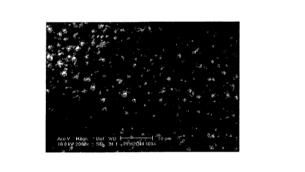
Figure 4 is a scanning electron micrograph of a first microstructured
substrate
manufactured in accordance with the invention.
Figure 5 is a scanning electron micrograph of a second microstructured
substrate
manufactured in accordance with the invention.
Figure 6 is a scanning electron micrograph of a third microstructured
substrate
manufactured in accordance with the invention.
Figure 7 is a scanning electron micrograph of a fourth microstructured
substrate
manufactured in accordance with the invention.
Figure 8 is a scanning electron micrograph of a fifth microstructured
substrate
manufactured in accordance with the invention.
Figure 9 is a mass spectrum of acetaminophen with a-CHCA matrix.
Figure 10 is a mass spectrum of acetaminophen off polypropylene with
microstructured surface TYPE A and an aluminum film.
Figure 11 is a mass spectrum of ascorbic acid with a -CHCA matrix.
Figure 12 is a mass spectrum of ascorbic acid off polypropylene with
microstructured
surface TYPE A and an aluminum film.
Figure 13 is a mass spectrum of penicillin with a -CHCA matrix.
Figure 14 is a mass spectrum of penicillin off polypropylene with
microstructured
surface TYPE A and an aluminum film.
Figure 15 is a mass spectrum of clonidine off polypropylene with
microstructured
surface TYPE A and an aluminum film.
Figure 16 is a mass spectrum of clonidine off Al-coated matte polypropylene.
7
CA 02502919 2005-04-20
WO 2004/047142 PCT/US2003/031839
Figure 17 is a mass spectrum of Substance P off polypropylene with
microstructured
surface TYPE A and an aluminum film.
Figure 18 is a mass spectrum of Substance P off Al-coated matte polypropylene.
Figure 19 is a mass spectrum of Angiotensin II off polypropylene with
microstructured surface TYPE A.
Figure 20 is a mass spectrum of Angiotensin II off Al-coated matte
polypropylene.
Figure 21 is a mass spectrum of clonidine off Al/H-DLG coated smooth
polypropylene.
Figure 22 is a mass spectrum of clonidine off Al/H-DLG coated matte
polypropylene
(via silicone belt tooling)
Figure 23 is a mass spectrum of clonidine off Al/H-DLG coated matte
polypropylene .
(via metal roll tooling).
Figure 24 is a mass spectrum of clonidine off Al/H-DLG coated polypropylene
with
microstructured surface TYPE A.
Figure 25 is a mass spectrum of Substance P off Al/H-DLG coated smooth
polypropylene.
Figure 26 is a mass spectrum of Substance P off Al/H-DLG coated matte
polypropylene (via silicone belt tooling).
Figure 27 is a mass spectrum of Substance P off Al/H-DLG coated matte
polypropylene (via metal roll tooling).
Figure 28 is a mass spectrum of Substance P off Al/H-DLG coated PPTYPE A.
8
CA 02502919 2005-04-20
WO 2004/047142 PCT/US2003/031839
Figure 29 is a mass spectrum of clonidine off uncoated polypropylene with
microstructured surface TYPE A.
Figure 30 is a mass spectrum of bradykinin (1000 ng/~,L) off uncoated
polypropylene
with microstructured surface TYPE A.
Figure 31 is a mass spectrum of clonidine off H-DLG coated polypropylene with
microstructured surface TYPE A.
Figure 32 is a mass spectrum of clonidine off Al-coated polypropylene with
microstructured surface TYPE A.
Figure 33 is a mass spectrum of bradykinin [1000 ng / p.L] off Al-coated
polypropylene with microstructured surface TYPE A.
Figure 34 is a mass spectrum of bradykinin [100 ng/ ~,L] off Al-coated
polypropylene
with microstructured surface TYPE A.
Figure 35 is a mass spectrum of clonidine off Al/H-DLG coated polypropylene
with
microstructured surface TYPE A.
Figure 36 is a mass spectrum of haloperidol off AIIH-DLG coated polypropylene
with
microstructured surface TYPE A.
Figure 37 is a mass spectrum of prazosin off Al/H-DLG coated polypropylene
with
microstructured surface TYPE A.
Figure 38 is a mass spectrum of bradykinin off Al/H-DLG coated polypropylene
with
microstructured surface TYPE A.
Figure 39 is a mass spectrum of clonidine off polypropylene with
microstructured
surface TYPE A freshly coated with aluminum.
9
CA 02502919 2005-04-20
WO 2004/047142 PCT/US2003/031839
Figure 40 is a mass spectrum of clonidine off polypropylene with
microstructured
surface TYPE A coated with aluminum and aged for five months.
Figure 41 is a mass spectrum of prazosin off polypropylene with
microstructured
surface TYPE A freshly coated with aluminum.
Figure 42 is a mass spectrum of prazosin off polypropylene with
microstructured
surface TYPE A coated with aluminum and aged for five months.
Figure 43 is a mass spectrum of clonidine off smooth polycarbonate coated with
colloidal graphite.
Figure 44 is a mass spectrum of clonidine off polycarbonate with
microstructured
surface TYPE B coated with colloidal graphite.
Figure 45 is a mass spectrum of Angiotensin II off smooth polycarbonate film
coated with colloidal graphite.
Figure 46 is a mass spectrum of Angiotensin II off polycarbonate with
microstructured
surface TYPE B
coated with colloidal graphite.
Figure 47 is a mass spectrum of clonidine off polycarbonate with
microstructured
surface TYPE B coated with colloidal graphite.
Figure 48 is a mass spectrum of Angiotensin II off polycarbonate with
microstructured
surface TYPE B coated with colloidal graphite.
Figure 49 is a mass spectrum of clonidine off polycarbonate with
microstructured
surface TYPE B with no coating.
CA 02502919 2005-04-20
WO 2004/047142 PCT/US2003/031839
Figure 50 is a Table showing Signal to Noise versus ionization mode for
various
analytes off Al/H-DLG coated polypropylene with microstructured surface TYPE
A.
Figure 51 is a mass spectrum of clonidine off AIIH-DLG coated structure-within-
structure film.
Figure 52 is a mass spectrum of bradykinin off Al/H-DLG coated structure-
within-
structure film.
Figure 53 is a mass spectrum of clonidine off uncoated polypropylene with
microstructured surface TYPE A with a 10-fold dilution of CHCA matrix.
Figure 54 is a mass spectrum of clonidine off uncoated polypropylene with
microstructured surface TYPE A with a 40-fold dilution of CHCA matrix.
Figure 55 is a mass spectrum of Calmix I off polypropylene with
microstructured
surface TYPE A and an aluminum film, with oc-CHCA matrix.
Figure 56 is a mass spectrum of Calmix I off stainless steel plate, with oc-
CHCA
matrix.
I5 Figure 57 is an expanded mass spectrum of Calmix I off polypropylene with
microstructured surface TYPE A and an aluminum film, with a-CHCA matrix.
Figure 58 is an expanded mass spectrum of Calmix I off Stainless Steel Plate,
with cc-
CHCA matrix.
While principles of the invention are amenable to various modifications and
alternative forms, specifics thereof have been shown by way of example in the
drawings and
will be described in detail. It should be understood, however, that the
intention is not to limit
the invention to the particular embodiments described. On the contrary, the
intention is to
11
CA 02502919 2005-04-20
WO 2004/047142 PCT/US2003/031839
cover all modifications, equivalents, and alternatives falling within the
spirit and scope of the
disclosure and claims.
Detailed Description
A. General Configuration
The present invention is directed to methods and apparatuses for the analysis
of
various compositions, in particular those utilizing high-energy desorption /
ionization of a
sample. For example, laser desorption and ionization of samples for mass
spectroscopy are
suitable applications of the invention. The invention utilizes microstructured
substrates, such
20 as micro- and nano-structured polypropylene and polycarbonate films, as
desorption
substrates. These structured substrates can include films with nonvolatile
layers coated onto
their sample receiving surface, such as inorganic coatings including metals,
metal oxides, and
alloys, and organic (carbon containing) coatings including graphite,
silicones, silane
derivatives, diamond like glass (DLG), and parylene. Substrates made in
accordance with the
25 present invention are typically structured in a manner such that they
promote desorption of a
sample more effectively than non-structured substrates. The structured
substrate serves to
achieve, promote or enhance useful desorption and ionization without
fragmentation. In
addition to providing analyses without the complications of signal due to the
matrix, in some
implementations, such as when a small molecule is being analyzed, the methods
of the
20 invention may achieve superior performance (as manifested by, for example,
higher signal to
noise values) compared to traditional methods and devices.
12
CA 02502919 2005-04-20
WO 2004/047142 PCT/US2003/031839
Various aspects of the invention, including surface structure and topology,
coating
compositions, substrate materials and other aspects of the invention will now
be described in
greater detail.
B. Microstructured Surface
Substrates made in accordance with the invention typically have a
microstructured
surface, and in some cases a microstructured or nanostructured surface. For
the purposes of
this invention, microstructured films are those that have a desirable surface
topography (i.e.,
are non-planar) on at least one surface. Microstructures include
configurations of features
wherein at least two dimensions of the features. are microscopic, as described
in U.S. Patent
Application Publication US 2001/0051264 A1, incorporated herein by reference
in its
entirety. In this context, "microscopic" refers to features that are
sufficiently small so as to
require an optic aid to the naked eye to determine their shape.
In some example implementations, microstructured films can be defined for the
purpose of this invention as those with physical feature sizes in the range of
two hundred
microns or less in at least two of the three possible dimensions (in/out of
the plane of the film,
and in each direction along the plane of the film). Within these general
guidelines, films of
this invention can be more specifically characterized as those that exhibit
surface features
with a desirable characteristic size (such as length measured along any
dimension) and feature
density (features per unit area of film surface). A feature, in this context,
can be anything that
represents a departure or deviation from a flat planar surface. Features can
include those that
protrude (nodules, posts, lumps, ridges, for example), or those which are
recessed (holes, pits,
13
CA 02502919 2005-04-20
WO 2004/047142 PCT/US2003/031839
fissures, crevices, for example). The microstructured surface may also possess
a combination
of protruding and recessed features (for example, furrows and ridges,
protruding and recessed
pyramids). In the case of ridges, furrows, or intersecting planes, a "feature"
may be a corner
or linear intersection of such ridges, furrows or planes.
A feature may be such that its characteristic length in all three dimensions
(i.e. into
and out of the plane of the film, and in each orthogonal direction along the
plane of the film)
is similar. Conversely, a feature may be such that the characteristic length
in one or more
directions is somewhat longer, or even much longer, than in the other
directions (for example,
in the case bf features such as ridges or furrows.)
In some implementations of the invention, microstructured features include
those
possessing a maximum characteristic length in one or more directions of two
hundred
microns. In some implementations, the maximum characteristic length is fifty
microns, while
in yet other implementations; the characteristic length is less than ten
microns. In some
implementations the microstructured fims include those possessing a minimum
characteristic
length in one or more directions of one one nanometer. In other
implementations the
minimum characteristic length is ten nanometers, while in yet other
implementations the
minimum characteristic length is one hundred nanometers. Also, in some
implementations,
microstructured feature densities which are preferable are those in the range
of 100 features or
greater per square mm of film. More preferable are those that possess features
at a density of
greater than 1000 per square mm. Most preferable still are those that possess
features at a
density of greater than 10000 per square mm.
14
CA 02502919 2005-04-20
WO 2004/047142 PCT/US2003/031839
Examples of microstructured substrates according to the present invention are
shown
in the seanning electron micrographs of Figures 4, 5, 6, 7 and 8. The first
structure,
designated as TYPE A, is depicted in Figure 4, and exhibits features in the
size range of
hundreds of nanometers to a few microns. The second structure, referred to as
TYPE B,
exhibits features in the size range of several microns, and is depicted in
Figure 5. The third
structure, depicted in Figure 6, is a so-called matte finish polypropylene
film which exhibits
features in the size range of several hundred nanometers to a few microns. The
fourth
structure, depicted in Figure 7, is another matte finish polypropylene film
which exhibits
features in the size range of several microns.
Smaller scale features can be superimposed upon larger scale features, as
shown for
example in Figure 8. The fine and large-scale features may both serve to
provide enhanced
desorption, or in some cases the fine and large scale features may perform
different functions.
For example, the larger scale features can serve to demarcate a particular
area for sample
placement, may serve as physical barriers to confine a deposited sample within
a desired area,
or may serve as reinforcing ribs to impart greater strength and stiffness to
the film.
The features may be present on a regular repeating basis, such as in the
structure of
Figure 8, or they may be "random" such as in the structures of Figures 4, 5, 6
and 7. The
features may be present over the entire area of the film, or may be present
only in areas in
which sample is to be deposited.
Microstructured films of the invention are typically produced by placing a
formable
precursor (such as a liquid) in contact with a mold bearing the negative
topology (opposite) of
the desired structure, then allowing the precursor to solidify into a solid
film bearing the
CA 02502919 2005-04-20
WO 2004/047142 PCT/US2003/031839
desired structure. One such method is to provide the film precursor in the
form of molten
plastic which is allowed to cool to solidification while in contact with the
mold. This
extrusion !embossing method allows the use of materials that are less subject
to contamination
and disadvantageous byproducts than some prior substrates. An alternative
method is to
utilize an existing film, heat it to the point of softening, bring it into
contact with a mold, and
allow it to cool (embossing). An alternative method is to bring an existing
film into contact
with a mold and conform the film surface to the mold by means of pressure
(calendaring).
Yet another alternative method is to provide the film precursor in the form of
a liquid syrup
consisting of curable, polymerizable or crosslinkable molecules, which are
then cured while
in contact with the mold.
Films can be prepared bearing features of characteristic length and density as
desired,
the features being determined by the mold utilized. In extrusion embossing,
the mold is
typically in the form of a cylinder (roll) or belt. Utilization of cylinders
or belts with various
topographies can provide films with varying microstructures. Fox example,
extrusion of
molten polymer onto an extremely smooth surface (such as polished metal rolls
which are
commonly used in extrusion) will usually result in a film that is smooth,
glossy and
essentially featureless and unstructured for the purposes of this invention.
Extrusion onto a
mold which has had no particular surface modification to make it extremely
smooth (for
example matte finish metal rolls or belts) will provide a film that has a
microstructured
topography in comparison to the smooth film. Such films can provide
enhancement in some
analyte desorption cases.
16
CA 02502919 2005-04-20
WO 2004/047142 PCT/US2003/031839
Extrusion onto a molds which are rough (for example, cloth or fabric-covered
rolls), or
molds that have been subjected to deliberate roughening treatment (for
example, a roll or belt
which has been sandblasted, abraded, etched, etc.) will also provide a film
with more
microstructured topography in comparison to the smooth film. Extrusion onto
molds that
have been designed to provide film specifically engineered for the present
application will
provide a microstructured topography possessing the most advantageous
combination of
feature characteristic length and feature density. Such molds may be generated
by a wide
variety of methods, including physical abrasion, drilling, chemical milling,
lithography, laser
ablation, plasma treatment, engraving, chemical etching, reactive ion etching,
chemical vapor
deposition, physical vapor deposition, and electrochemical deposition. Such
films are
exemplified by the structures of Figures 4 and 5, and are generally the most
useful for a wide
variety of analytes as described in more detail in the examples.
In an alternative implementation, smooth, featureless films are processed to
generate
the desired features. For example, a smooth film may be abraded or modified
by, for
example, embossing, sandblasting, laser ablation, corona treatment, plasma
treatment, or
flame treatment, to impart features. In certain cases the smooth films may be
coated, then
treated to form the desired structure (for example via embossing or
calendaring), as long as
the structure forming process does not damage or adversely affect the coated
layer.
In yet another implementation, it is also possible to coat the substrate with
a coating
that itself forms the features useful in the present invention. For example,
an aluminum layer
might be deposited in the form of nodules or granules, rather than as a smooth
layer. It is also
possible to apply a coating to the film that serves to provide the features
(for example a silica
17
CA 02502919 2005-04-20
WO 2004/047142 PCT/US2003/031839
or other particulate coating), followed by application of a substantially
nonvolatile coating
atop the features.
C. Coatings
The microstructured films of the present invention may be advantageously used
in
combination with one or more coatings applied on top of the microstructured
film to provide
enhanced desorption. Coatings may also serve other purposes; for example,
coatings may
provide a protective or abrasion-resistant burner.
Useful coatings according to the present invention include inorganic materials
such as
metals; for example aluminum, gold, silver, nickel, titanium, palladium, and
platinum; metal
oxides, for example titanium dioxide, silicon oxide and zirconium oxide, and
alloys of metals
or metal oxides, such as inconel or indium tin oxide. Other useful coatings
include organic
materials such as graphite, carbon black, the families of materials referred
to as Diamond-
Like Carbon (DLC), as described in US Patent 6,265,068, and Diamond-Like Glass
(DLG), as
described in PCT publication WO 0166820 entitled Diamond-Like-Glass Thin
Films, and
incorporated herein by reference, silanes and silane derivatives, and
parylene. The coatings
can be conformal (as in the case of parylene and DLG) or particulate in nature
(such as
graphite).
Such surface coatings are generally nonvolatile under conditions used for
laser
desorption. That is, the coating either exhibits negligible volatility, or the
entities that are
volatilized are so low in molecular weight (for example, carbon clusters which
may be
emitted from graphite, or aluminum ions which may be emitted from aluminum)
that they do
18
CA 02502919 2005-04-20
WO 2004/047142 PCT/US2003/031839
not interfere with the analyte being measured. In this regard, the coatings
are distinguished
from conventional matrices. While matrix materials are typically thought of as
"nonvolatile"
in that they have a slow evaporation or sublimation rate under ambient
conditions, they are
volatilized to a significant extent in the actual laser desorption process,
and the volatilized
species have molecular weight such that they may interfere with or obscure the
analyte signal.
This fundamental difference in volatility results in part from the fact that
the coatings
of this invention are typically present in the form of large-scale networks
which possess
bonded iriterconnectivity over many molecular lengths. This bonded
connectivity may be
present in either or both directions along the surface of the film, and/or
perpendicular to the
film. For example, graphite coatings may be employed in which the graphite
particles consist
of many millions of carbon atoms connected by covalent bonds over distances of
up to
microns. Alternatively, metal coatings may be employed which consist of many
millions of
metal atoms connected by metallic bonds, over distances of up to microns and
or even
millimeters. In contrast, matrices are typically applied as crystals comprised
of individual
molecules that are not connected by chemical bonds; or as molecules that are
individually
tethered to attachment sites on the surface of the substrate and are not
connected to each other
by chemical bonds.
Coatings may be applied to the microstructured film via various methods,
including
vapor coating, sputter coating, plasma coating, vacuum sublimation, chemical
vapor
deposition, catholic arc deposition, and so on. These methods are particularly
suited for
coating of metals and metal oxides. Coatings such as graphite are most easily
applied by
obtaining the graphite as a dispersion and applying it to the substrate by any
of the well-
19
CA 02502919 2005-04-20
WO 2004/047142 PCT/US2003/031839
known methods for liquid coating (knife coating, spray coating, dip coating,
spin coating,
etc.).
It can be advantageous to provide the coating in a discontinuous manner as
opposed to
a continuous coating over the entire microstructured surface. For example, the
coating can be
provided at discrete locations, such as spots. In the case of multilayer
coatings, one coating
may be discrete while the other may be continuous, according to the needs of
the particular
instance. Discontinuous coatings may serve several functions. For example,
they may serve
to demarcate the particular area in which the analyte sample is to be
deposited, and then to
allow the area to be located once the film with sample is placed in the mass
spectrometer. A
coating may also be used which provides a discontinuity in the surface energy
of the
microstructured film to advantageously contain a deposited analyte sample
within a desired
area, and to prevent wicking or spreading of the sample over an undesirably
wide area.
Such coatings may be applied in a discrete manner via any number of methods.
If the
coating is applied via vapor coating, a mask, such as a perforated screen or
film, may be used
to limit the coating to the areas defined by the mask. In the case in which it
is desired to have
multilayer, registered discrete coatings (for example spots containing
superimposed
multilayer coatings), the maslc can be attached to the film (for example via
an adhesive)
during coating of the different layers such that the layers are superimposed
in registration.
The masle is then removed after the final coating process. In an alternative
embodiment, the
perforated mask itself can remain on the film, in which case it will serve to
provide wells that
serve to contain the analyte droplet that is placed in the wells. It is also
possible to provide a
perforated layer for this purpose independently of any role in defining the
coating. In the
CA 02502919 2005-04-20
WO 2004/047142 PCT/US2003/031839
case of coatings such as graphite, well-known liquid coating methods such as
gravure coating
can be used to deposit the graphite in a discontinuous manner.
D. Substrate Materials
The present invention relies on substrate materials that are amenable to
formation or
generation of the microstructured surface. Various materials are suitable for
use as substrates
in accordance with the invention. In general the substrate is a polymeric
material, although
non-polymeric materials having the properties descl~bed herein can also be
used. The
substrate is typically non-porous or substantially non-porous.
The microstructured films of the present invention possess advantages over
currently
available porous materials (for example, DIOS chips), in that such porous
materials are
known to be susceptible to contamination via the uptake of impurities from the
atmosphere
during storage or use. In contrast, the microstructured materials are less
susceptible to such
contamination in some implementations because they are typically nonporous.
A wide variety of polymeric materials are useful in this invention. These
include
thermoplastic materials (such as polyolefins, inlcuding polypropylene and
polyethylene) and
thermoset (curable) materials. Suitable materials include crystalline, semi-
crystalline,
amorphous, or glassy polymers. Copolymers may be used as well.
Such polymers may be filled or modified, as long as the filling agent does not
significantly interfere with the enhanced desorption of the analyte. A wide
variety of fillers
and additives are available which impart various of functions and properties.
These include,
for example, fillers to increase strength andlor modulus, additives to provide
increased
21
CA 02502919 2005-04-20
WO 2004/047142 PCT/US2003/031839
resistance to oxidation, increased heat stability, or increased W stability,
processing
additives (for example to provide for improved extrusion properties), pigments
and colorants,
and so on.
The polymeric materials used in this invention can thus be tailored to possess
a wide
variety of physical, chemical, optical, electrical, and thermal properties.
E. Device Assembly and Features
The present invention comprises a substrate bearing a structure, and optional
coatings,
useful for enhanced desorption, particularly in mass spectroscopy. In typical
use the film is
attached to a standard metal plate for insertion into a mass spectrometry
instrument. As such,
a number of useful embodiments of the invention exist. It is advantageous to
provide' the film
with a layer of adhesive applied to the back (non-microstructured) side, to
facilitate
attachment to the metal plate. The adhesive can be a laminating adhesive or
double-faced
tape. The laminating adhesive can be attached to the underside of the
microstructured film,
with a release liner remaining in place on the bottom of the adhesive. The
user can then
simply remove the release liner and attach the film directly to the plate by
means of the
adhesive. Alternatively, a separate piece of laminating adhesive can be
supplied to the user,
who can then apply the adhesive to the metal plate, remove the liner, and
attach the
microstructured film to the top of the adhesive.
The adhesive should be carefully selected such that it does not harbor or
generate any
impurities which might contaminate the microstructured substrate. In addition,
it may be
desirable in some cases for the adhesive to be electrically conductive. Such
conductive
22
CA 02502919 2005-04-20
WO 2004/047142 PCT/US2003/031839
adhesives are readily available, for example conductive adhesive 9713
available from 3M of
Maplewood, Minnesota. The adhesive may be selected such that it is permanently
attached to
the underside of the microstructured film; alternatively, it may be removable.
Typically, the microstructured film, optionally with attached adhesive
underneath, will
be packaged for delivery to the customer. This packaging may consist of any
means that
protects the film and does not act to impart contaminating impurities to the
film. Fox
example, the film could be packaged in a plastic bag or plastic case. As an
additional
protective measure, a protective liner may be placed atop the upper
(microstructured) surface
of the film.
In another embodiment, a bar code label is applied to the microstructured film
so that
the film sample can be readily identified and inventoried for archiving. In
such cases, an area
can be provided outside the working area (i.e. the area upon which samples are
deposited) for
placement of the bar code.
F. Sample Preparation and Methods of Using the Substrates
The present invention is particularly well suited to mass spectrometry
analysis.
Analyte spots deposited on a substrate are hit with short laser pulses to
desorb and ionize the
sample. Ions are formed and then accelerated by one or more electric fields
before arriving at
a detector. The time it takes to reach the detector, or the location on the
detector at which the
particles strike, can be used to determine the mass of the particles.
Time-of-flight analysis (TOF) is one mass spectrometry method that can be
used.
Figure 3 shows a schematic diagram of a time-of-flight setup. For molecules
under 10,000
23
CA 02502919 2005-04-20
WO 2004/047142 PCT/US2003/031839
Da, the reflectron mode is used to condense the kinetic energy distribution of
the ions
reaching the detector. This method was developed to increase the resolution of
mass
spectroscopy and is used primarily for molecules under 10,000 Da. This higher
resolution
often results in a drop in sensitivity and a limited mass range.
G. Examples
The invention can be further understood by means of the following examples.
For these examples, substrates were prepared using polymer melt processing
methods.
Plastic film bearing the "TYPE A" topology of Figure 4 was prepared by
extruding Exxon
Polypropylene 3445 onto a silicone belt tool bearing a structure. The silicone
belt tool had
been prepared by placing liquid silicone in contact with a metal tool by means
of spin casting
and allowing the silicone to solidify. The metal tool had been prepared by
vapor deposition as
described in International Patent Number WO 0116940, hereby incorporated by
reference.
The polymer was extruded at a melt temperature of 400°F, and the tool
temperature setting
was set at 125°F. The nip pressure was set at 20 psi, and the line
speed was set at 5 fpm. The
polypropylene was removed from the tool as it cooled. The polypropylene
extrudate
replicated the tool, resulting in a surface bearing random features ranging
from hundreds of
nanometers to several microns in characteristic dimensions.
Plastic film bearing the "TYPE B" topology of Figure 5 was prepared by
compression
molding. A piece of 0.014" thick film of Makrolon 2407 polycarbonate (produced
by Bayer
AG) was placed between a flat polished metal press plate and a metal tool
bearing a structure.
The metal tool had been prepared by electrochemical deposition of metal onto a
flat metal
24
CA 02502919 2005-04-20
WO 2004/047142 PCT/US2003/031839
surface. The tool, film, and press plate stack was placed into a Wabash
compression molder.
The platens of the compression molder were set to 190°C, and the
platens were closed to
attain 50 psi pressure on the sample. The sample was pressed at this condition
for 2 minutes,
and then the pressure was increased to 200 psi on the sample. This condition
was held for 3
minutes, and then the system was cooled. The samples remained in the
compression molder
at 200 psi until the platens reached 80°C, when the press was opened
and the sample
removed. The feature characteristic dimensions of the polycarbonate film were
in the range
of a few microns.
Film bearing a matte finish (Figure 6) was produced by extruding Exxon
Polypropylene 3445 onto a matte finish silicone belt, under the same
conditions used to
produce the TYPE A pattern described above. The matte finish polypropylene
exhibited
features with characteristic dimensions in the range of several hundred
nanometers to several
microns. The features were in general less pronounced and less well defined
than that of the
TYPE A structure.
Another matte finish film (Figure 7) was produced by extruding polypropylene
onto
an unpolished, matte finish metal roll under typical polypropylene extrusion
conditions. This
film exhibited features with characteristic dimensions generally in the range
of several
microns, with the feature density being generally lower than that of the TYPE
A structure.
Film bearing regular, nonrandom structure-within-structure features (Figure 8)
was
produced by extruding Dow Chemical 7C50 high impact polypropylene copolymer
onto a
metal tool roll bearing the negative of the desired structure. The copolymer
resin was
extruded by means of a Killion single screw 1.25" extruder with die
temperature set at 480°F.
CA 02502919 2005-04-20
WO 2004/047142 PCT/US2003/031839
The molten resin exited the die and was drawn between two nip rollers closed
under pressure.
One roll was rubber coated backing roll and the other was the metal tool roll
bearing the
microstructured pattern. The backing roll was maintained at 100 °F and
the tool roll at 230 °F.
The web speed was between approximately 9.8 and 12.1 feet per minute.
The metal tool roll was engraved with four sets of grooves. There were two
sets of
parallel grooves, which were perpendicular to each other and are referred to
hereinafter as the
major grooves. These two perpendicular sets of helical grooves ran at an angle
of
approximately 45° to the roll axis, and had a depth of approximately 60
micrometers (microns,
or Vim), a width of approximately 18 pm at the bottom and approximately 34 pm
at the top,
and were spaced approximately 250 ~m apart. A third set of grooves ran at an
angle of
approximately 90° to the roll axis, and had a depth of between
approximately 2 and
approximately 4 micrometers (microns, or Vim), a width of approximately 5 pm
at the bottom
and approximately 7 ~m at the top, and were spaced approximately 25 pm apart.
A fourth set
of grooves ran at a direction parallel to the roll axis, and had a depth of
between
approximately 5 micrometers (microns, or Vim), a width of approximately 5 pm
at the bottom
and approximately 7 ~m at the top, and were spaced approximately 25 pm apart.
The third
and fourth sets of grooves are collectively referred to as the minor grooves.
During embossing, the molten polypropylene resin filled the above groove
structures
and solidified, such that a microstructured film was formed bearing features
that were the
negative of the above described grooves. That is, film exhibited a smaller
scale grid of
perpendicular ridges superimposed within a larger scale grid of perpendicular
ridges, as
26
CA 02502919 2005-04-20
WO 2004/047142 PCT/US2003/031839
shown in Figure 11, such as those disclosed in U.S. Patents Docket Numbers
57837US02 and
57838US02, and incorporated herein by reference.
Nonstructured polypropylene film bearing a smooth surface finish was produced
by
extruding polypropylene onto a polished metal roll, under the same extrusion
conditions used
to produce the TYPE A pattern described above. The surface was generally flat
and
featureless.
Nonstructured polycarbonate film bearing smooth surface finish was produced by
extruding polycarbonate onto a polished metal roll, under standard
polycarbonate extrusion
conditions. The surface was generally flat and featureless.
Metal and metal oxide coatings were applied to the films utilizing an NRC 3115
Bell
Jar. For aluminum, the deposition thickness was approximately 950 A. DLG
(Diamond Like
Glass) was applied using a Plasma-Therm vapor coater, according to methods
described in
PCT publication WO 0166820. The DLG coating thickness was approximately 1100
A. In
some cases the DLG coating was post treated to render it hydrophilic
(designated H-DLG), as
described in the same reference. In some cases both coatings were continuous;
in other
cases, one or both coatings were deposited in discrete areas (fox example, in
spots) by use of
masks during the coating process. Masks were either metal foils with areas
removed, or
polymer films lilcewise with areas removed. In some cases the masks were
adhered to the
microstructured film by means of adhesive, particularly when it was desired to
deposit
superimposed, registered, coatings in discrete areas. Specific coating
patterns are described
in the specific examples. Masks were removed after coating.
27
CA 02502919 2005-04-20
WO 2004/047142 PCT/US2003/031839
All mass spectrometry experiments were conducted on an Applied Biosystems
(Framingham, MA) Voyager-DE STR time-of-flight mass spectrometer. The films
were
attached to commercially available metal MALDI plates using double-faced
adhesive tape. A
pulsed 337 nm nitrogen laser with a 3 Hz pulse frequency was used, and laser
intensity was
set at the threshold value. The table below summarizes the main instrument
parameters:
Polarity Positive (Except where specified)
Mode of Operation Reflector
Extraction mode Delayed
Accelerating voltage18,000 -20,000 V for small molecules;
20,000-24,000 V
for peptides
Grid Voltage 76% - 87.5 %
Extraction delay 150 nsec
time
Number of laser 150 shots / spectrum
shots
The mass spectrometry data was processed by using Data Explorers Version 4Ø
Before
measuring the resolution (R) and signal-to-noise (S/N), the "Noise
Filter/Smooth" function
with a 0.7 correlation factor was applied to all spectra.
Example 1
This example illustrates the use of a microstructured substrate with and
without a
chemical matrix.
Polypropylene film bearing the TYPE A structure (henceforth referred to as
PPTYPE
A) was produced as described previously. A metal maslc with a ten by ten grid
array of 1.19
28
CA 02502919 2005-04-20
WO 2004/047142 PCT/US2003/031839
mm diameter holes was adhered to the microstructured side of the film using
ReMount~
removable spray adhesive. The film was then vapor coated with aluminum, as
described
previously, after which the metal mask was removed. The resulting films thus
contained 1.19
mm diameter spots of aluminum. (PPTYPE A coated with aluminum is henceforth
referred to
as polypropylene with microstructured surface TYPE A and an aluminum film).
Samples for analysis were prepared with 0.1 mg of three common drug compounds:
acetaminophen (151.17 Da), ascorbic acid (176.12 Da), and penicillin (389 Da).
These drug
compounds were dissolved in 1.0 ml of a 1:1:0.001 methanol / water / trifluoro
acetic acid
solution. A volume of 0.5 p,L of each analyte solution 'was pipetted directly
onto one of the
aluminum-coated spots on the film. Analyte samples were applied with and
without the
addition of 0.5 ~,L of the matrix alpha cyano-4-hydroxy-cinammic acid (a-
CHCA). The
r
samples were allowed to air dry for approximately fifteen minutes.
Figure 9 shows the mass spectrum of acetaminophen with the addition of a-CHCA
matrix. The matrix signal saturated the detector and no analyte peak can be
seen. Figure 10
shows the mass spectrum of acetaminophen off polypropylene with
microstructured surface
TYPE A and an aluminum film without a matrix. The molecular ion can be clearly
seen at snlz
152.51, along with the sodium and potassium adducts at mlz 174.53 and mlz
190.54
respectively. The spectrum is substantially free from noise, allowing the
analyte to easily be
identified.
Figure 11 shows the mass spectrum of ascorbic acid with the addition of a-CHCA
matrix. Again, the matrix signal saturated the detector and the analyte peak
cannot be seen.
Figure 12 shows the mass spectrum of ascorbic acid off polypropylene with
microstructured
29
CA 02502919 2005-04-20
WO 2004/047142 PCT/US2003/031839
surface TYPE A and an aluminum film without matrix. The molecular ion can be
clearly
seen at mlz 177.53, along with the sodium and potassium adducts at rnlz 199.53
and mlz
215.57 respectively. This method also allows for high resolution allowing the
isotopes of the
molecules to be seen.
Figure 13 shows the mass spectrum of penicillin with a-CHCA matrix. The
molecular
ion does show up at rnlz 390.03, but is hard to identify in the midst of the
matrix noise.
Figure 14 shows the mass spectrum of penicillin off PPTYPE A-A1 without
matrix. The
molecular ion can easily be picked out at rnJz 389.93 with a signal-to-noise
ratio of over forty
times that of the spectrum obtained with matrix.
Example 2
This example illustrates the use of polypropylene with the TYPE A structure
and with
the matte finish structure, coated with aluminum.
Matte finish polypropylene was obtained by extrusion of polypropylene resin
against a
matte finish metal roll as described previously. Polypropylene bearing the
TYPE A structure
was obtained as described previously. Both films were coated with a continuous
layer of
aluminum as described previously.
One small molecule, clonidine (266.6 Da), and two peptides, substance P
(1347.6 Da)
and angiotensin II (1046.2 Da), were obtained from Sigma Chemical Co. (St.
Louis, MO) and
were used without further purification. A solution containing 100 ng / p,L of
each analyte in
50:50 HPLC grade acetonitrile / water with 0.1% trifluoro acetic acid was made
for the small
molecule. A solution containing 1000 ng / p,L of each analyte in 50:50
methanol / water with
CA 02502919 2005-04-20
WO 2004/047142 PCT/US2003/031839
0.1 % trifluoro acetic acid was made for each of the peptides. A volume of 0.5
p,L - 3.0 wL of
analyte was pipetted directly onto the film, followed by drying at room
temperature for
approximately fifteen minutes.
Figure 15 shows the spectrum for clonidine off the polypropylene with the TYPE
A
structure; Figure 16 shows the spectrum for the matte finish polypropylene.
The TYPE A
microstructured film shows over three times the signal-to-noise ratio of the
matte finish
polypropylene. Also, the spectrum off the TYPE A microstructured film shows a
cleaner
baseline due to the lower threshold laser intensity that the microstructured
film allowed to be
used.
20 Figure 17 shows the spectrum for substance P off of the polypropylene with
the TYPE
A structure; Figure 18 shows the spectrum for substance P off of the matte
finish
polypropylene. The signal-to-noise is over twenty times greater on the TYPE A
microstructured film. Additionally, the threshold laser intensity was lower
for the TYPE A
microstructured film leading to a cleaner spectrum and easier identification
of the analyte of
interest.
Figure 19 shows the spectrum for angiotensin IT off of the polypropylene with
the
TYPE A structure; Figure 20 shows the spectrum for angiotensin lI off of the
matte finish
polypropylene. As in the above spectra, the TYPE A microstructured film gives
a much
higher signal-to-noise ratio and a cleaner baseline.
Example 3
31
CA 02502919 2005-04-20
WO 2004/047142 PCT/US2003/031839
This example illustrates the results of mass spectrometry analysis using films
with
various structures. In all cases the film is polypropylene and the coating is
aluminum
followed by hydrophilic DLG (H-DLG). The structures are: nonstructured (made
by
extrusion onto a polished metal roll), matte finish (made by extrusion onto a
matte finish
silicone belt), matte finish (made by extrusion onto an unpolished, matte
finish metal roll) and
the TYPE A structure, all obtained as described previously.
A metal mask with 2.00 mm diameter holes was adhered to each film via ReMount~
removable spray adhesive. The samples were then coated with aluminum followed
by H-
DLG, using methods and apparatus and described previously, after which the
mask was
removed. The resulting films contained superimposed 2.00 mm diameter spots of
aluminum
and H-DLG.
One small molecule, clonidine (266.6 Da), and one peptide, substance P (1347.6
Da),
were obtained from Sigma Chemical Co. (St. Louis, MO). Solutions containing 20
ng / ~,L of
clonidine in 50:50 HPLC grade methanol / water with 0.1% trifluoro acetic acid
and 100 ng /
~.L of substance P in 50:50 HPLC grade methanol / water with 0.1% trifluoro
acetic acid were
made.
For each analyte, a volume of 0.3 ~,L of analyte solution was pipetted
directly onto one
of the Al/H-DLG-coated spots on the film. Due to the difference in surface
energy between
the H-DLG and the surrounding polypropylene, the applied sample remained
confined within
the coated area. The samples were allowed to air dry at room temperature for
approximately
fifteen minutes.
32
CA 02502919 2005-04-20
WO 2004/047142 PCT/US2003/031839
Figures 21-24 show mass spectra of the small molecule clonidine off of
unstructured
polypropylene, matte finish (silicone belt) polypropylene, matte finish (metal
roll)
polypropylene, and polypropylene with the TYPE A structure. With the
unstructured film,
no analyte signal can be obtained, even at high laser power. With the two
matte finish films,
the analyte can be seen, with signal-to-noise of around 600. The spectrum off
the TYPE A
film shows signal-to-noise of 56,000.
Figures 25-28 showy mass spectra of the peptide substance P off of
unstructured,
matte finish (metal and silicone), and the TYPE A microstructured
polypropylene films.
Again, the unstructured film shows zero analyte signal. There is an analyte
signal off each of
the two matte finish films, but signal-to-noise is low. The spectrum quality
off the TYPE A
microstructured film is much better, with higher relative intensity and signal-
to-noise.
Example 4
This example illustrates the results of mass spectrometry analysis using
aluminum and
hydrophilic DLG single layer coatings.
Polypropylene films with the TYPE A structure was obtained without a coating,
with a
continuous coating of hydrophilic diamond-like glass (H-DLG), and with a
continuous
coating of aluminum.
One small molecule, clonidine (266.6 Da), and one peptide, bradylcinin (1060.2
Da),
were obtained from Sigma Chemical Co. (St. Louis, MO) and were used without
any further
purification. A solution containing 100 ng l ~,L of clonidine in 50:50 HPLC
grade methanol /
water with 0.1 % trifluoro acetic acid was made. Two different concentrations
of bradykinin
33
CA 02502919 2005-04-20
WO 2004/047142 PCT/US2003/031839
solution were made in 50:50 methanol / water with 0.1 % trifluoro acetic acid,
one at a
concentration of 1000 ng / p,L and one at a concentration of 100 ng / p.L.
A volume of 3.0 p.L of analyte solution was pipetted directly onto the film,
followed
by drying at room temperature for approximately fifteen minutes.
Figure 29 shows a mass spectrum of clonidine taken off of the polypropylene
film
with the TYPE A structure and no coating. The molecular ion peak can be seen,
but the
relative intensity is low. Figure 30 shows a mass spectrum of the higher
concentration of
bradykinin taken off the same film. No signal can be seen for the peptide.
Figure 31 shows a mass spectrum of clonidine taken off of the polypropylene
film
with the TYPE A structure and H-DLG coating. The spectrum is substantially
free from
chemical noise, but relative intensity is low. No signal was obtained for
either concentration
of bradykinin with this film.
Figure 32 shows a mass spectrum of clonidine taken off of the TYPE A
microstructured polypropylene film with aluminum coating. The spectrum is
relatively clean,
with good signal-to-noise. Figure 33 and Figure 34 show the mass spectra of
the [1000 ng /
p.L] bradylcinin and the [100 ng / p,L] bradykinin off the TYPE A
microstructured
polypropylene film with aluminum coating. The signal to noise is higher than
with the
uncoated or HDLG-coated TYPE A.
Example 5
34
CA 02502919 2005-04-20
WO 2004/047142 PCT/US2003/031839
This example utilizes a multilayer coating of H-DLG on top of aluminum on
polypropylene film with the TYPE A structure. The aluminum coating is
continuous, with the
H-DLG being applied as discontinuous spots atop the aluminum.
Polypropylene film with the TYPE A structure was obtained and coated with
aluminum as described previously. A perforated polymer mask containing 550 pm
diameter
holes was taped to the film, and the film was then coated with H-DLG, after
which the mask
was removed. The resulting films contained 550 p.m diameter spots of H-DLG
over a
continuous layer of aluminum.
Three small molecules, clonidine (266.6 Da), haloperidol (375.9 Da), prazosin
(419.9
Da), and one peptide, bradykinin (1060.2 Da), were obtained from Sigma
Chemical Co. (St.
Louis, MO) and were used without further purification. A solution containing
100 ng / pL of
each analyte in 50:50 HPLC grade methanol / water with 0.1 % trifluoro acetic
acid was made
for each of the analytes.
For each analyte, a volume of 0.5 p,L analyte solution was pipetted directly
onto one of
the H-DLG coated spots on the film. Due to the difference in surface energy
between the H-
DLG and the surrounding aluminum, the applied sample remained confined within
the H-
DLG coated area. The samples were allowed to air dry at room temperature for
approximately fifteen minutes.
Figure 35, Figure 36, and Figure 37 show mass spectra of the small molecules
clonidine, haloperidol, and prazosin taken off of the TYPE A microstructured
polypropylene
films with aluminum plus hydrophilic DLG coating. As can be seen in all the
spectra,
extremely high signal-to-noise ratios are achieved with low laser intensity.
This leads to a
CA 02502919 2005-04-20
WO 2004/047142 PCT/US2003/031839
clean spectrum with no extraneous peaks and easy identification of the
molecule of interest.
Figure 3~ shows a mass spectrum of the peptide bradykinin taken off of the
same film. The
spectrum has high relative intensity, and once again the molecule of interest
is easily picked
out. For all spectra, signal uniformity across the dried droplet was very good
with no "sweet-
spot" phenomenon observed.
Example 6
This example demonstrates the excellent shelf life of aluminum coated TYPE A
films
over several months of storage.
Polypropylene film with the TYPE A structure was obtained as described and
coated
with a continuous layer of aluminum. Some film samples were used for mass
spectrometry
analysis within a few days after coating. Other films were used for analysis
after five months
storage in covered plastic petri dishes at room temperature.
Two small molecules, clonidine (266.6 Da) and prazosin (419.9 Da) were
obtained
from Sigma Chemical Co. (St. Louis, MO) and were used without any further
purification. A
solution containing 100 ng l ~,L of each analyte in 50:50 HPLC grade methanol
/ water with
0.1 % trifluoro acetic acid was made fresh for each of the small molecules. A
volume of 3.0
pI, of analyte solution was pipetted directly onto the film, and allowed to
air dry at room
temperature for approximately fifteen minutes.
Figure 39 shows a mass spectrum of clonidine taken off of the films freshly
coated
with aluminum. Figure 40 shows a mass spectrum of clonidine taken off of film
from the
same batch five months later with fresh analytes applied. No deterioration in
performance is
36
CA 02502919 2005-04-20
WO 2004/047142 PCT/US2003/031839
evident with the aged film, in terms of signal-to-noise and spectrum quality.
Nor is there any
sign of contamination or loss of sensitivity.
Figure 41 shows a mass spectrum of prazosin taken off of freshly coated films.
Figure
42 shows a mass spectrum of prazosin taken off of the same batch of film five
months later
with fresh analytes applied. Again, the aged film shows excellent signal-to-
noise with
excellent spectrum quality.
Example 7
This example illustrates the effect of structure versus nonstructure for the
polycarbonate TYPE B ("PCTYPE B") structure with graphite coating.
Smooth polycarbonate film and polycarbonate film bearing the TYPE B structure
were
obtained as described previously. A 1: 40 dilution of Colloidal Graphite Paint
from Energy
Beam Sciences Inc. (Agawam, MA) in isopropanol was made. A coating of the
diluted
graphite dispersion was applied to the nonstructured polycarbonate and the
TYPE B
microstructured polycarbonate. This was accomplished by dipping a cotton swab
into the
dispersion and swabbing the dispersion onto the film. Two separate swabbings
were
performed, perpendicular to each other, to ensure complete coverage. The
coating was
allowed to dry for several hours prior to sample deposition.
One small molecule, clonidine (266.6 Da), and one peptide, angiotensin II
(1046.2
Da), were obtained from Sigma Chemical Co. (St. Louis, MO) and were used
without further
purification. A solution containing 100 ng / ~,L of the analyte in 50:50 HPLC
grade methanol
37
CA 02502919 2005-04-20
WO 2004/047142 PCT/US2003/031839
/ water with 0.1 % trifluoro acetic acid was made for the small molecule. A
solution
containing 1000 ng / ~,I. of the analyte in water was made for the peptide.
A volume of 1.5 ~,L of analyte was pipetted directly onto the film, and
allowed to air dry for
approximately fifteen minutes.
Figure 43 shows the mass spectrum of clonidine off the nonstructured
polycarbonate
film. The high laser intensity needed to ionize the analyte led to very low
resolution, and the
isotopes of the molecule cannot be distinguished. Figure 44 shows the mass
spectrum of
clonidine off the polycarbonate film with the TYPE B structure. The spectrum
quality is
much improved, with the isotope peaks being clearly resolved and the signal-to-
noise ratio
being much higher than the spectrum taken off the nonstructured film.
Figure 45 shows the mass spectrum of angiotensin II off the nonstructured
polycarbonate film. There is a great deal of baseline noise, and the analyte
peak is hard to
detect. Figure 46 shows the mass spectrum of angiotensin II off the TYPE B
microstructured
polycarbonate film. There is much less noise, the molecular ion is easily
detectable, and the
signal to noise is much improved.
Example S
This example illustrates the effect of graphite coating versus no coating for
the
polycarbonate TYPE B structure.
Polycarbonate bearing the TYPE B structure was obtained and coated with
graphite as
described previously. Separate samples of the polycarbonate with TYPE B
structure were not
coated.
3~
CA 02502919 2005-04-20
WO 2004/047142 PCT/US2003/031839
One small molecule, clonidine (266.6 Da), and one peptide, angiotensin II
(1046.2
Da), were obtained from Sigma Chemical Co. (St. Louis, MO) and were used
without further
purification. A solution containing 100 ng / p,L of the analyte in 50:50 HPLC
grade methanol
/ water with 0.1 % trifluoro acetic acid was made for the small molecule. A
solution
containing 1000 ng / pL of the analyte in water was made for the peptide.
A volume of 1.5 p.L of analyte solution was pipetted directly onto the film,
and allowed to air
dry for approximately fifteen minutes.
Figure 47 shows the mass spectrum of clonidine off the graphite coated
polycarbonate
film with the TYPE B structure. The spectrum quality is good, and the isotope
peaks are
clearly resolved. The signal to noise ratio is excellent. Figure 48 shows the
mass spectrum of
angiotensin II off the same TYPE B microstructured polycarbonate film.
Spectrum quality is
good with the molecular ion being easily detectable.
Figure 49 shows the mass spectrum of clonidine off the polycarbonate film with
the
TYPE B structure and no coating. There is a small analyte peak, but the
relative intensity and
signal-to-noise ratio are low. For angiotensin II off the polycarbonate film
with the TYPE B
structure and no coating, no peptide peaks were found (figure not shown).
Example 9
This example illustrates the use of the microstructured substrate in allowing
both
positive (cation) and negative (anion) analysis off the same substrate. The
example also
demonstrates use of the microstructured substrate for analyte mixtures.
39
CA 02502919 2005-04-20
WO 2004/047142 PCT/US2003/031839
Polypropylene with the TYPE A structure was obtained as described previously.
A
perforated polymer mask containing 550 p,m diameter holes was taped to the
film. The film
was coated with aluminum followed by H-DLG, after which the mask was removed.
The
resulting films contained 550 ~,m diameter spots of H-DLG superimposed over
aluminum.
A proprietary mix of eight compounds in mass range 150-600 Da, representative
of
those often encountered in combinatorial chemistry analysis, was obtained and
was dissolved
in methanol in concentration ranges from 0.1 to 0.3 p,g/ p.L. 0.3 p,L samples
of analyte
solution were pipetted onto the spots on the film and allowed to air dry for
about fifteen
minutes.
In Figure 50 is presented the signal to noise data obtained for the main peak
(or
molecular ion peak) of each of the eight representative compounds and the
average over all
eight compounds. Acceptable signal to noise is seen to be obtainable in both
positive and
negative ionization mode.
Example ZO
This example illustrates the use of a superimposed fine scale/large scale
structure-
within-structure substrate, coated with AI/H-DLG.
Polypropylene copolymer film with the structure-within-structure topology
shown in
Figure 8 was obtained as described previously. An adhesive-backed polymer mask
with an
array of 1.4 mm diameter holes was adhered to the film. The film was coated
with aluminum
followed by H-DLG, after which the mask was removed. The resulting films
contained 1.4
mm diameter spots of H-DLG superimposed over aluminum.
CA 02502919 2005-04-20
WO 2004/047142 PCT/US2003/031839
One small molecule, clonidine (266.6 Da), and one peptide, bradykinin (1060.2
Da),
were obtained from Sigma Chemical Co. (St. Louis, MO) and used without further
purification. Solutions containing 20 ng / p,L of clonidine in 50:50 HPLC
grade methanol /
water with 0.1 % trifluoro acetic acid, and 100 ng / p,L of bradylcinin in
50:50 HPLC grade
methanol / water with 0.1 % trifluoro acetic acid, were made. For each
analyte, a volume of
0.2 p,L of analyte solution was pipetted directly onto one of the coated spots
on the film and
allowed to air dry at room temperature for approximately fifteen minutes.
Figure 51 shows the mass spectrum for clonidine off of the structure-within-
structure
film. The spectrum has high relative intensity, good signal-to-noise and
relatively little
chemical noise. Figure 52 shows bradykinin off of the structure-within-
structure film.
Relative intensity is low, but the analyte peak can be clearly seen.
In both cases the structure-within-structure film was found to result in very
uniform
sample dry-down, as evidenced by easily obtainable spectra with no "sweet-
spot"
phenomenon.
Example 11
This example illustrates the use of uncoated, microstructured film in the
presence of
chemical matrix.
Polypropylene bearing the TYPE A structure was obtained and mounted on a
commercially available metal MALDI plate using double-faced adhesive tape.
The small molecule clonidine (266.6 Da) was obtained from Sigma Chemical Co.
(St.
Louis, MO). A solution containing 20 ng / ~,L of the analyte in 50:50 HPLC
grade methanol /
41
CA 02502919 2005-04-20
WO 2004/047142 PCT/US2003/031839
water with 0.1% trifluoro acetic acid was made. A saturated solution of a-CHCA
matrix in
50:50 HPLC grade methanol / water with 0.1% trifluoro acetic acid was diluted
five-fold and
twenty-fold. A volume of 1 ~,L of each of the diluted matrix solutions were
then mixed with 2
p.L of sample solution, yielding a ten and forty-fold total dilution of the
matrix. A volume of
0.2 ~,I, of the analyte/matrix solution was pipetted directly onto the film,
followed by drying
at room temperature for approximately fifteen minutes.
Figure 53 shows the spectra for clonidine using the 10-fold dilution of a -
CHCA
matrix. The analyte peak has good signal to noise and relative intensity, but
there is
interference from the matrix peaks. Figure 54 shows the spectra for clonidine
using the 40-
fold dilution of a -CHCA matrix. At this dilution level there is less
interference from the
matrix.
Example 12
This example demonstrates the use of microstructured, coated films in the
presence of
matrix.
Polypropylene film with the TYPE A structure was obtained as described
previously.
A metal mask with 500 ~m diameter holes was adhered to the film. The film was
coated with
aluminum, after which the maslc was removed. The resulting film contained 500
~,m diameter
spots of aluminum
The SequazymeT"~ Peptide Mass Standards Kit (PerSeptive Biosystems,
Framingham,
MA) was used for these experiments. Peptide Calibration Mixture 1, contained
the following
peptides: des-Argl-Bradykinin (904.05 Da); Angiotensin T (1296.51 Da); Glul-
Fibrinopeptide
42
CA 02502919 2005-04-20
WO 2004/047142 PCT/US2003/031839
B (1570.61 Da); and Neurotensin (1672.96 Da). A stock solution of the peptide
mixture was
prepared by mixing the peptide standards with 100 ~.L of 30% acetonitrile in
0.01% TFA. A
saturated solution of the matrix, alpha-cyano-4-hydroxycinnamic acid (a-CHCA)
was
prepared by mixing the pre-measured, 5-~ mg of a-CHCA with 1 ml of 50%
acetonitrile in
0.3% trifluoroacetic acid (TFA) diluent. A volume of 24 ~L of the standard oc-
CHCA matrix
solution was mixed with 1 ~L of the peptide stock solution.
Sample volumes of 0.1 ~L or 0.2 ~L were pipetted onto the aluminum-coated
spots on
the film, and allowed to air dry for approximately two minutes. The same
sample deposition
procedure was used to apply analyte spots to a commercially available
stainless steel MALDI
plate. No additional sample preparation or sample clean up was done to the
samples prior to
analysis.
The positive-ion MALDI mass spectrum obtained from 0.1 ~L of Calibration
Mixture
1 with a-CHCA on the TYPE A microstructured, Al coated film produced
protonated
molecular ions (tnlz 1570) with a SlN value of 3,620 for Glu-Fibrinopeptide
(MW 1569 Da),
as shown in Figure 55).
The positive-ion MALDI mass spectrum for the same analyte and matrix
combination
using the commercially available standard stainless steel metal plate is shown
in Figure 56.
The operating conditions used with the metal plate were similar to the
conditions used with
the polypropylene TYPE A microstructured films. The signal to noise was
comparable with
the performance achieved using the aluminum coated PPTYPE A microstructured
film.
Although protonated molecular ions are the primary ionic species produced in
the
laser desorption/ionization process, sodium and potassium cationized species
can also be
43
CA 02502919 2005-04-20
WO 2004/047142 PCT/US2003/031839
formed. Close-up examination of the molecular ion region of Glu-Fibrinopeptide
B from the
stainless steel plate versus the microstructured film shows that cationization
is significantly
reduced when the microstructured film is used, as revealed in comparing
Figures 57 and 58.
In contrast to the conventional metal plates, for which the best signal was
found at
the edge of the dried droplet, for the microstructured film the signal was
much more uniform
across the dried droplet.
The present invention may be embodied in other specific forms without
departing
from its spirit or essential characteristics. The described embodiments are to
be considered in
all respects only as illustrative and not restrictive. The scope of the
invention is, therefore,
indicated by the appended claims rather than by the foregoing description.
44