Note: Descriptions are shown in the official language in which they were submitted.
CA 02503191 2005-04-21
WO 2004/037315 PCT/US2003/031229
SYSTEM AND METHOD FOR FACILITATING HEMOSTASIS OF BLOOD
VESSEL PUNCTURES WITH ABSORBABLE SPONGE
RELATED APPLICATIONS
The present patent application is related to, claims priority under 35 U.S.C.
120 to, and
incorporates by reference herein in their entirety: U.S. patent application
Serial Number
09/613,439 filed July 11, 2000, which is a divisional of U. S. patent
application Serial
Number 091071,284, filed May 1, 1998, now U.S. patent 6,162,192.
BACKGROUND OF THE INVENTION
1. Field of the Invention
The invention relates to a closure system for blood vessel punctures, and more
particularly, the invention relates to a system and method for facilitating
hemostasis of
blood vessel punctures with an absorbable sponge material.
2. Brief Description of the Related Art
A large number of diagnostic and interventional procedures involve the
percutaneous
introduction of instrumentation into a vein or artery. For example, coronary
angioplasty,
angiography, atherectomy, stenting of arteries, and many other procedures
often involve
accessing the vasculature through a catheter placed in the femoral artery or
other blood
vessel. Once the procedure is completed and the catheter or other
instrumentation is
removed, bleeding from the punctured artery must be controlled.
Traditionally, external pressure is applied to the skin entry site to stem
bleeding from a
puncture wound in a blood vessel. Pressure is continued until hemostasis has
occurred at
the puncture site. In some instances, pressure must be applied for a up to an
hour or
more during which time the patient is uncomfortably immobilized. In addition,
a risk of
1
CA 02503191 2005-04-21
WO 2004/037315 PCT/US2003/031229
hematoma exists since bleeding from the vessel may continue beneath the skin
until
sufficient clotting effects hemostasis. Further, external pressure to close
the vascular
puncture site works best when the vessel is close to the skin surface and may
be
unsuitable for patients with substantial amounts of subcutaneous adipose
tissue since the
skin surface may be a considerable distance from the vascular puncture site.
More recently, devices have been proposed to promote hemostasis directly at a
site of a
vascular puncture. One class of such puncture sealing devices features an
intraluminal
anchor which is placed within the blood vessel and seals against an inside
surface of the
vessel puncture. The intraluminal plug may be used in combination with a
sealing
material positioned on the outside of the blood vessel, such as collagen:
Sealing devices
of this type are disclosed in U.S. Pat. Nos. 4,852,568; 4,890,612; 5,021,059;
and
5,061,274.
Another approach to subcutaneous blood vessel puncture closure involves the
delivery
of non-absorbable tissue adhesives, such cyanoacrylate, to the perforation
site. Such a
system is disclosed in U.S. Pat. No. 5,383,899.
The application of an absorbable material such as collagen or a nonabsorbable
tissue
adhesive at the puncture site has several drawbacks including: 1) possible
injection of
the material into the blood vessel causing thrombosis; 2) a lack of pressure
directly on
the blood vessel puncture which may allow blood to escape beneath the material
plug
into the surrounding tissue; and 3) the inability to accurately place the
absorbable
material plug directly over the puncture site.
The use of an anchor and plug system addresses these problems to some extent
but
provides other problems including: 1) complex and difficult application; 2)
partial
occlusion of the blood vessel by the anchor when placed properly; and 3)
complete
blockage of the blood vessel or a branch of the blood vessel by the anchor if
placed
improperly. Another problem with the anchor and plug system involves re-
access. Re-
access of a particular blood vessel site sealed with an anchor and plug system
is not
2
CA 02503191 2005-04-21
WO 2004/037315 PCT/US2003/031229
possible until the anchor has been completely absorbed because the anchor
could be
dislodged into the blood stream by an attempt to re-access.
Yet another approach to subcutaneous puncture closure involves the internal
suturing of
the blood vessel puncture with a specially designed suturing device. However,
these
suturing devices involve a significant number of steps to perform suturing and
require
substantial expertise.
Accordingly, it would be desirable to provide a system for facilitating
hemostasis of
blood vessel punctures which addresses the drawbacks of the known systems.
SCARY ~OF THE INVENTION
One aspect of the present invention relates to a device for facilitating
hemostasis of a
puncture in the wall of a blood vessel including an introduces for hydrating
and
compressing an absorbable sponge pledget for delivery to a patient to
facilitate
hemostasis of the puncture and a plunger insertable into the introduces for
ejection of the
pledget from the introduces into a patient to seal the puncture in the blood
vessel wall.
The introduces includes a staging chamber with a first.diameter configured to
receive the
absorbable sponge pledget, a delivery chamber with a second diameter smaller
than the
first diameter, and a tapered section between the staging chamber and the
delivery
chamber for compressing the pledget.
In accordance with another aspect of the present invention, a system for
facilitating
hemostasis of a puncture in the wall of a blood vessel includes a tract
dilator, an
introduces, and a plunger each having a lumen for allowing the tract dilator,
introduces,
and plunger to be passed over a guidewire. The introduces lumen includes a
staging
chamber configured to receive an absorbable sponge pledget and a delivery
chamber.
The plunger is insertable into the introduces for ejection of the pledget from
the delivery
chamber into a patient to facilitate hemostasis of a puncture in a blood
vessel wall.
3
CA 02503191 2005-04-21
WO 2004/037315 PCT/US2003/031229
In accordance with an additional aspect of the present invention, a method for
facilitating hemostasis of a puncture in the wall of a blood vessel includes
the steps of:
establishing a depth of a blood vessel puncture from the skin of a patient;
loading an
introducer with an absorbable sponge pledget by hydrating and compressing the
pledget;
loading the introducer over a guidewire positioned in the blood vessel by
inserting the
guidewire through the hydrated and compressed pledget; and ejecting the
pledget
adjacent the blood vessel puncture to facilitate hemostasis of the puncture
while
maintaining the guidewire in place.
In accordance with an additional aspect of the present invention a method is
provided for
controlling blood flow from a puncture wound in a blood vessel using a device
comprising an elongated member having a distal tip, a lumen and a wire, the
steps
comprising: inserting the wire through a puncture in the subcutaneous tissue
of the
patient and through the puncture wound into the blood vessel; inserting the
wire through
the lumen of the device; inserting the elongated member through the puncture
in the
subcutaneous tissue and advancing the elongated member along the wire until
the distal
tip of the device is adjacent the puncture wound; and, manipulating the distal
tip so that
at least a portion of the distal tip occupies at least a portion of the
puncture wound to
thereby restrict the flow of blood through the puncture wound.
In accordance with an additional aspect of the present invention a method is
provided for
establishing the depth of a puncture wound in the wall of a blood vessel of a
patient
using a device having a distal tip, the process comprising: introducing the
distal tip of the
device through a patient's subcutaneous tissue and experiencing resistance to
advancement of the device; advancing the device toward the puncture wound in
the wall
of the blood vessel until the distal tip of the device encounters the outer
wall of the
vessel; tentatively determining that distal tip of the device has encountered
the outer wall
of the vessel by experiencing additional resistance to further advancement of
the device;
and, confirming that that distal tip of the device has encountered the outer
wall of the
vessel adjacent to the puncture wound by observing bleed back and further
observing
that by manipulating the distal tip of the device bleed back can be
controlled.
4
CA 02503191 2005-04-21
WO 2004/037315 PCT/US2003/031229
In accordance with an additional aspect of the present invention a method is
provided for
positioning a pledget adjacent to the external wall of a blood vessel puncture
in a patient,
comprising the steps of: advancing a tract dilator through the subcutaneous
tissue of the
patient to determine the depth of the puncture site; advancing an introduces
to a location
so that its distal tip is adjacent to the puncture site wherein the location
is determined at
least in part based on the depth of the puncture site as determined using the
tract dilator;
and, ejecting the pledget from the introduces.
BRIEF DESCRIPTION OF THE DRAWINGS
The invention will now be described in greater detail with reference to the
preferred
embodiments illustrated in the accompanying drawings, in which like elements
bear like
reference numerals, and wherein:
FIG. 1 is a top view of a blood vessel puncture sealing kit;
FIG. 2 is a side cross sectional view of a punctured blood vessel and a tract
dilator for
locating the puncture;
FIG. 3 is a side view of an introduces and pledget prior to placement within
the
introduces;
FIG. 4 is a side view of an introduces having a pledget positioned within the
introduces
staging chamber and a syringe attached to the introduces;
FIG. 5 is a side view of the introduces and syringe with the pledget hydrated
and
advanced to a delivery chamber within the introduces;
FIG. 6 is a diagram of process steps according to one embodiment of the
present
invention;
CA 02503191 2005-04-21
WO 2004/037315 PCT/US2003/031229
FIG. 7 is a diagram of process steps according to one embodiment of the
present
invention;
FIG. 8 a schematic is diagram of the operation of one embodiment of the
present
invention;
FIG. 9 a schematic is diagram of the operation of one embodiment of the
present
invention;
FIG. 10 a schematic is diagram of the operation of one embodiment of the
present
invention;
FIG. 11 is a side cross sectional view of a punctured blood vessel with the
introduces
and plunger positioned for delivery of the pledget;
FIG. 12 is a side cross sectional view of a punctured blood vessel with the
pledget being
deposited at the puncture site;
FIG. 13 is a side cross sectional view of a punctured blood vessel with a
hydrated and
kneaded pledget deposited at the puncture site, the guidewire removed, and the
delivery
system being withdrawn;
FIG. 14 is a side cross sectional view of a punctured blood vessel with a
hydrated and
kneaded pledget facilitating hemostasis of the puncture site;
FIG. 15 is a side cross sectional view of an alternative embodiment of an
introduces;
FIG. 16 is a cross sectional view of a distal end of an introduces according
to another
alternative embodiment having a central channel for receiving the guidewire;
and
FIG. 17 is a cross sectional side view of a distal end of an introduces with a
connector
for connecting a syringe.
6
CA 02503191 2005-04-21
WO 2004/037315 PCT/US2003/031229
DETAILED DESCRIPTION OF THE PREFERRED EMBODIIVVIENTS
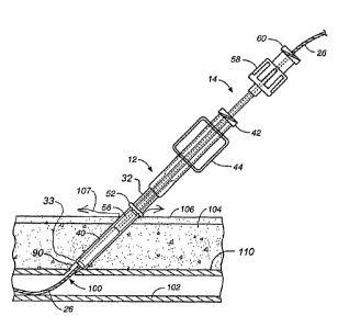
An over the wire delivery system delivers an absorbable sponge pledget in a
hydrated
condition to a blood vessel puncture site to achieve hemostasis. The over the
wire
delivery system includes a tract dilator 10, an introduces 12, and a pusher
14, illustrated
in kit form in FIG. 1. The system allows over the wire delivery of the
absorbable sponge
material directly to the puncture site to achieve hemostasis. Over the wire
delivery
ensures that the sponge material is properly positioned to fully occlude the
puncture. In
addition, the absorbable sponge material is delivered in a hydrated state
which
immediately expands to stop blood flow through the puncture. The introduces
allows the
delivery of more absorbable sponge material through a smaller tract by
hydrating and
compressing the absorbable sponge material.
Prior to discussing the present invention in further detail, the following
terms are
defined:
"Pledget" means a piece of absorbable sponge formed into a generally elongated
shape
having a size which allows delivery in a hydrated state through a delivery
cannula, or
introduces to a site of a puncture in a blood vessel.
"Absorbable sponge" means a biocompatible material which is capable of being
hydrated, is resiliently compressible in a hydrated state, and when implanted
within a
human or other mammalian body is absorbed by the body. Preferably the
absorbable
sponge is non-immunogenic.
"Hydrate" means to partially or fully saturate with a fluid, such as, saline,
water, contrast
agent, thrombin, therapeutic agents, or the like.
"Kneading" of the absorbable sponge material means both dry and wet
manipulation of
sponge material which compresses, enlarges, or changes the shape of the sponge
material causing the sponge material to have improved expansion response.
7
CA 02503191 2005-04-21
WO 2004/037315 PCT/US2003/031229
As shown in FIG. 1, the tract dilator 10, the introduces 12, and the pusher 14
may be
provided to a medical facility in the form of a kit or individually. The tract
dilator 10 as
illustrated in FIGS. 1 and 2 includes a distal tip 20, a proximal end 22, and
a lumen 24
extending from the distal tip to the proximal end of the tract dilator.'The
lumen 24 is
provided to allow the tract dilator 10 to be received over a guidewire 26
which extends
through the puncture wound 100 into the blood vessel 102. The tract dilator 10
may
have a constant cross section or may taper slightly to a smaller diameter at
the distal tip
20. According to an alternative embodiment, the tract dilator 10 may have a
narrow
shaft with an enlarged distal tip. The distal tip 20 has rounded edges to
prevent catching
on subcutaneous tissue 104 as the tract dilator 10 is inserted through the
skin 106 and
tissue to the blood vessel puncture site. The tract dilator distal tip 20 has
a diameter such
that the tip of the tract dilator will not pass into the blood vessel but will
stop and
provide tactile feedback when it reaches the external blood vessel wall 102.
A depth indicator 30 is positioned around the tract dilator 10 and is movable
in an axial
direction. Once the tract dilator has been inserted until the distal tip 20
abuts the external
wall of the blood vessel 102, as shown in FIG. 2, the depth indicator 30 is
manually
positioned adjacent the pat'ient's skin 106. Alternatively, the depth
indicator 30 can be
pushed to a depth indicating position by the skin 106 as the dilator is
inserted.
Preferably, the depth indicator 30 is an elastic ring which is movable axially
on the tract
dilator 10 and maintains a measured position for comparison with the
introduces 12.
A side view of an introduces 12 is illustrated in FIGS. 1 and 3. The
introduces 12
includes a staging chamber 34 for receiving an absorbable sponge pledget 40
and a
delivery chamber 36 for receipt of a hydrated and compressed pledget from the
staging
chamber. A tapered section 38 is provided between the staging chamber 34
having a
larger diameter lumen and the delivery chamber 36 having a smaller diameter
lumen.
The tapered section 38 of the introduces 12 acts as a compression member to
compress
the hydrated pledget 40 into the delivery chamber. The introduces 12 also
includes a luer
fitting 42 at a proximal end for connection to a conventional syringe and wing
members
44 for use in grasping the introduces.
s
CA 02503191 2005-04-21
WO 2004/037315 PCT/US2003/031229
The absorbable sponge pledget 40 according to one preferred embodiment of the
invention is formed from a sheet of absorbable sponge material which has been
cut into
a rectangular shape and rolled to form a compact, substantially cylindrical,
elongated
pledget. The pledget 40 is sized to be received within the staging chamber 34
of the
introduces 12 in a dry rolled state.
Once the pledget 40 has been inserted into the staging chamber 34 of the
introduces 12,
a conventional syringe 50 containing a hydrating fluid is connected to the
luer fitting 42,
as shown in FIG. 4. The pledget 40 is then hydrated within the staging chamber
34 by
injecting a fluid into the staging chamber from the syringe 50 causing the
pledget to
swell, partially or fully blocking the lumen of the introduces. The partial
hydration or
wetting of the exterior surface of the pledget 40 creates a lubricous surface
on the
pledget. The hydrated pledget 40 is then forced into the delivery chamber 36
by
injecting additional fluid with the syringe 50 to force the pledget through
the tapered
section 3~ to the delivery chamber. For a somewhat smaller pledget 40 which
does not
entirely block the lumen of the introduces 12 after hydrating, the venturi
effect will help
to draw the pledget into he delivery chamber 36. As shown in FIG. 5, a finger
may be
placed over the distal end of the introduces 12 during delivery of the pledget
40 to the
delivery chamber 36 to prevent the pledget from being ejected from the
introduces by
the pressure of the fluid. Preferably, one or more vent holes 46 are provided
in the side
walls of the introduces adjacent the distal tip to allow air and liquid to
escape from the
introduces while the pledget 40 is positioned for delivery. These vent holes
46 are small
enough to prevent the pledget 40 from passing substantially into the vent
holes.
As an alternative to placement of a finger at the distal end of the introduces
12 during
advancement of the pledget 40 into the delivery chamber, a removable cap may
be used.
Further, the vent holes 46 may be omitted and a screen or a cap having a
screen may be
used to allow fluid to pass through the screen while the screen prevents the
pledget 40
from being ejected.
CA 02503191 2005-04-21
WO 2004/037315 PCT/US2003/031229
The introduces 12 also includes a depth indicator 52 which is an axially
movable
member used to indicate the depth to which the introduces should be inserted
into the
patient to achieve the proper positioning of the pledget 40 at the puncture
site. The depth
indicator 52 of the introduces 12 is aligned with the depth indicator 30 on
the tract
dilator 10 to achieve proper pledget delivery positioning.
The introduces 12 may be formed in any known manner such as by injection
molding
from a plastic material. Preferably, the introduces 12 is transparent so that
the pledget 40
can be viewed through the introduces and the user can visually confirm the
pledget
position. The introduces lumen may be provided with a reducing coating for
improved
pledget delivery. The delivery fluid also reduces friction for improved
delivery by
wetting the exterior surface of the pledget.
The pusher 14, as illustrated in FIG. 1, includes a distal end 56 which is
configured to
slide within the lumen of the delivery chamber 36 of the introduces 12.
Preferably, there
is a very small clearance or a resilient interference between the outer
diameter at the
distal end 56 of the pusher 14 and the inner diameter of the delivery chamber
36 to
prevent portions of the pledget from getting caught between the pusher and the
.
introduces 12. A resilient pusher distal end 56 or a sealing member on the
pusher 14
may be used to accomplish or approach a resilient fit between the introduces
12 and the
pusher.
The pusher 14 also may include a fitting 58 for connecting the proximal end of
the
pusher to the proximal end of the introduces 12. The fitting 58 acts as a stop
to limit the
motion of the pusher 14 with respect to the introduces 12. A female luer
fitting 60 may
also be included at the proximal end of the pusher 14 for connection of a
syringe to the
pusher for injection of beneficial agent through the pusher.
A method of delivering an absorbable sponge pledget 40 to facilitate
hemostasis of a
blood vessel puncture wound will now be described with respect to the steps
illustrated
in FIGS. 2 and 6-10. After an intravascular procedure has been completed, a
guide wire
26 is already in place, passing through a puncture 90 in the subcutaneous
tissue 104 and
to
CA 02503191 2005-04-21
WO 2004/037315 PCT/US2003/031229
into the blood vessel 102. Alternatively, if a guide wire is not already in
place the guide
wire is inserted through an access sheath used in the intravascular procedure
and the
access sheath is then removed.
Then the following steps are performed in the order indicated.
1. The operator loads pledget 40 into introducer 12, hydrates it and prepares
it for
delivery as shown in Figs. 3, 4, 5, and then sets the introducer 12 aside.
(Step
110)
2. The operator applies occlusive pressure 200 by pushing with the hand
against the
patient's skin to deform the subcutaneous tissue 104 and restrict or
completely
stop the blood flow 202 through the vessel 102. (Step 112) The operator
continues to apply occlusive pressure 200 through step 5.
3. The operator threads the guide wire 26 through the lumen 24 of the tract
dilator
10. (Step 114)
4. The operator advances the tract dilator 10 through tissue 104. (Step 116)
5. The operator continues to advance the tract dilator 10 toward the vessel
106 until
the distal tip 20 encounters and goes through fascia layer 206. The operator
knows that the distal tip 20 has encountered the fascia 206 due to tactile
indication. He often observes increased resistance or a "bump" since the
fascia
206 is tougher than the tissue 104. (Step 118)
6. The operator releases occlusive pressure 200, and blood flows from the
puncture
wound 100, through the lumen 24 and out the proximal end of the tract dilator
10.
(Step 120) We call this flow of blood "bleed back" 210, which is observed by
the
operator.
7. The operator continues to advance the tract dilator 10 toward the vessel
106 while
observing bleed back 210 until the distal tip 20 of the tract dilator 10
encounters
the outer wall 110 of the vessel. (Step 122) The operator determines that the
distal tip 20 has encountered the outer vessel wall 110 because by applying
forward pressure on the vessel wall 102 and puncture wound 100 with the distal
tip 20 he can control bleed back 210. In other words, the distal end 20 of the
tract
dilator 10 can be used to block blood flow from the puncture wound 100 by
11
CA 02503191 2005-04-21
WO 2004/037315 PCT/US2003/031229
carefully manipulating the tract dilator 10. Also, the operator can sometimes
feel
the pulse of the artery through the tract dilator 10, which provides another
indicatio that the distal tip 20 is in contact with the blood vesse, The
operator can
retract or relieve the pressure on the vessel to re-activate the bleed back
210.
Thus, the operator determines that the distal tip 20 is in contact with the
puncture
wound 100 through bleed back observation and tactile feel. The method by
which the operator can control bleed back can be understood with reference to
Figs. 8-10 in which some elements which appear in Fig. 2 are not shown for the
purpose of clarity. It should be noted that Figs 8-10 and the discussion of
those
Figures below represent our best understanding at the present time. In Fig. 8
the
guide wire 26 is in the puncture wound 100 before the distal end has reached
the
surface of the vessel 102. Then in Fig. 9 the distal tip 20 is nearing the
outer
vessel wall 110. It should be noted that the guide wire 26 causes a slight
distortion of the vessel wall 102 at the puncture wound 100, which we call
"tenting" of the vessel wall (due to its tent-like shape), and the guide wire
26 is at
the left-most portion of the puncture wound 100. Then as the tract dilator 10
is
advanced further, as shown in Fig. 10, the distal tip 20 has contacted the
outer
vessel wall 110. The operator can then manipulate the lower part of the distal
tip
20 so that it can block the puncture site thereby preventing bleed back.
Because
the distal tip 20 is located so that the wire 26 abuts the lower part of the
lumen
24, the operator can manipulate the distal tip to restrict blood flow from the
puncture wound and into and through the lumen 24 as well as around the distal
tip 20. However, it can be seen that by moving the tract dilator 10 away from
the
blood vessel, the operator can bring the distal part of the lumen 24 into
fluid-flow
communication with the puncture wound 100 to allow bleed back.
8. The operator maintains the tract dilator 10 with the distal tip 20 against
the blood
vessel and moves the depth indicator 30 on the tract dilator 10 forward
against
the skin surface 106 to measure the depth of the vessel 102. (Step 124)
9. The operator applies occlusive pressure 200 and continues to apply pressure
through step 13. (Step 126)
10. The operator removes the tract dilator 10. (Step 128)
12
CA 02503191 2005-04-21
WO 2004/037315 PCT/US2003/031229
11. Based on the position of the depth indicator 30 of the tract dilator 10
the operator
sets the depth indicator 52 on the introduces 12 which was previously prepared
-
per step 1. (Step 130)
12. The operator threads the guide wire 26 through the introduces 12. (Step
132)
13. The operator advances the introduces 12 until the depth indicator 52 is at
the skin
surface 106 as shown in Fig. 11. This provides the operator an indication that
the
introduces distal tip 16 is in contact with the exterior wall of the vessel
102. (Step
134) To confirm the location of the introduces distal tip 16 relative to the
exterior
wall of the vessel 102, the operator performs a procedure substantially the
same
as he performed to locate the distal tip of the depth 20 of the tract dilator
10 at the
puncture wound 100, as discussed above in step 6. However, the procedure
differs for the introduces 12 since the introduces 12 has no lumen 24 and thus
there is no bleed back. Rather, in the case of the introduces 12 the operator
controls blood flow from the puncture wound 100, which travels through the
hole
formed in the subcutaneous tissue 104 around the exterior of the delivery
chamber 36 of the introduces 12. In other words, blood oozes around the
introduces 12 as indicated by arrows 107 if the operator does not control
blood
flow with the introduces distal tip 16. In this case we consider the oozing
blood
107 to be "bleed back."
14. The operator releases occlusive pressure while maintaining blood flow
control
with the introduces distal tip 16. (Step 136)
15. The operator. delivers the hemostatic sponge as shown in Fig. 12 (Step
13~)
In practice the puncture 90 in the subcutaneous tissue and the puncture wound
100 are
often formed by a device having a diameter in the range of 4 to 10 French. We
have
found that we can use a tract dilator 10 with a diameter of about 12 French
with a lumen
of about 5.5 French with puncture wounds in this size range. In practice, with
a tract
dilator of this size the operator is able to perform step 7 above in a
satisfactory manner,
and more specifically, the operator is able to block the puncture site with
the distal tip 20
and control bleed back. Another parameter we have found in practice to be
effective is
that the angle between the surface of the skin 106 and the elongated member 16
can be
13
CA 02503191 2005-04-21
WO 2004/037315 PCT/US2003/031229
between about 30 degrees and about 40 degrees. This angle is also
substantially the
same as the angle between the blood vessel and the elongated member 16.
Turning again to Figs. 3-5 the loading and staging of the introduces 12 will
be further
explained. A sheet of absorbable sponge material is cut into a rectangle, is
rolled tightly
to form a pledget 40, and is placed into the staging chamber 34 of the
introduces 12. The
steps of cutting and rolling the pledget 40 and placing the dry pledget in the
introduces
staging chamber 34 may be performed before or after the intrevascular
procedure.
Alternatively, the introduces 12 may be provided preloaded with a prepared
pledget 40.
With the pledget 40 placed in the introduces, the syringe 50 is filled with a
hydrating
fluid such as saline, thrombin, contrast agent, other therapeutic agent, or
the like and
attached to the introduces 12 as illustrated in FIG. 4. Fluid is injected
slowly into the
introduces 12 to hydrate the pledget 40. The user then pauses to allow
hydration and
initial swelling of the pledget 40. Sufficient hydration may occur in about 20
to 30
seconds or less depending on the size of the pledget 40.
As shown in FIG. 5, the user then places a finger over the distal end of the
introduces 12
and injects fluid with the syringe 50 to force the pledget 40 through the
tapered section
38 and into the smaller end or delivery chamber 36 of the introduces 12.
Injection of
fluid is stopped when the pledget 40 is positioned at the distal end of the
delivery
chamber 36. At this point the syringe 50 is removed and the introduces is
loaded over
the proximal end of the guidewire 26 for the delivery of the pledget 40 to the
puncture
site.
As shown in FIG. 11, a proximal end of the guidewire 26 is fed into the distal
end of the
introduces 12 though the hydrated and compressed pledget 40 and out the
proximal end
of the introduces. Preferably, the guidewire 26 is fed through substantially
the center of
the pledget 40 to insure that the implanted pledget is centered over the blood
vessel
puncture 100. Alternatively, the guidewire may be inserted along a side of the
pledget
40, through a separate second lumen of the introduces, through an axial lumen
in the
pledget, or through a low density center of the pledget.
14
CA 02503191 2005-04-21
WO 2004/037315 PCT/US2003/031229
After feeding the guidewire 26 through the introduces, the guidewire 26 is fed
through
the pusher 14 and the pusher is advanced into the introduces until the distal
end 56 of the
pusher is in contact with the pledget 40. The introduces 12 and pusher 14 are
then
advanced together down though the skin 106 and the subcutaneous tissue 104
until the
depth indicator 52 on the exterior of the introduces is at the skin level.
In the step illustrated in FIG. 12, the pusher 14 is held stationary while the
introduces 12
is withdrawn proximally preferably to a distance of about 75% of the length of
the
compressed, hydrated pledget 40. This 75% withdrawal distance may be indicated
with
an appropriate marker on the introduces 12 or the plunger 14 or by contact
between the
fittings 42, 58 of the introduces and plunger. The portion of the pledget 40
ejected into
the tissue quickly expands upon delivery to fill the available space and
provide localized
compression. With the pusher 14 and introduces 12 in the position illustrated
in FIG. 12
and the pledget 40 partially ejected, a slight forward pressure is maintained
by the
operator on the introduces and pusher to increase local compression for a
period of time
of approximately 1 minute to allow hemostasis to begin. The forward pressure
causes
the pledget 40 to be compressed around the puncture site, as shown in FIG. 12.
The
guidewire 26 is then completely removed from the introduces 12 and the pusher
14. The
introduces 12 is withdrawn the remaining approximately 25% by engaging the
fitting 58
of the pusher with the female luer fitting 42 of the introduces to completely
discharge
the pledget 40 into the subcutaneous tissue 104 above the puncture site 100. A
slight
forward pressure can then be maintained by the operator on the introduces 12
and pusher
14 for approximately 1 minute before the introduces and pusher are removed
from the
tissue tract leaving the absorbable sponge pledget 40 positioned against the
outer vessel
wall, as shown in FIG. 14, providing local compression and facilitating
hemostasis. The
delivered pledget 40 maintains hemostasis until healing of the blood vessel
102 occurs.
The pledget 40 is absorbed by the body over time.
One type of absorbable sponge material which is acceptable for use in the
present
invention is Gelfoam, manufactured by the Upjohn Company. Gelfoam is a porous,
pliable, cross-linked gelatin material and is available commercially in sheet
form as pre-
compressed or non-compressed sponge. The material may be provided preformed as
a
CA 02503191 2005-04-21
WO 2004/037315 PCT/US2003/031229
pledget 40 or may be cut with a punch or a stencil and knife and rolled to
form a pledget
as described above. Once hydrated, the pledget 40 can be easily compressed to
fit into a
lumen having a smaller cross sectional area than the original cross sectional
area of the
pledget. Additionally, the kneading of the hydrated pledget 40 during delivery
encourages air trapped within the Gelfoam to be expelled and replaced with
fluid,
allowing rapid expansion upon delivery. When a pledget 40 of a pre-compressed
Gelfoam is hydrated and kneaded (expelling air) during delivery, the pledget
will have
the absorption capacity to rapidly expand to many times (e.g., 3 or more
times) its
original dry volume upon delivery. When a pledget 40 of the non-compressed
Gelfoam
is hydrated and kneaded (expelling air) during delivery, the pledget will have
the
absorption capacity to rapidly expand to its original dry volume upon
delivery. These
properties make the Gelfoam sponge material particularly useful for
facilitating
hemostasis of puncture wounds by injection.
Abrupt lumen diameter changes within the introduces 12, such as at the tapered
section
38, will improve "kneading" of the absorbable sponge material passing through
the
introduces. Manipulation of the dry absorbable sponge material, such as the
rolling of
the pledget 40, also provides kneading. Kneading improves hydration of the
sponge
material thereby improving the expansion properties of the hydrated delivered
absorbable sponge.
According to alternative embodiments of the introduces, enlarged, recessed, or
irregular
areas in the lumen of the introduces are provided to impart additional
kneading action to
the absorbable sponge material further improving expansion properties of the
sponge.
FIG. 15 illustrates one such alternative embodiment of the introduces 12a in
which the
delivery chamber of the introduces is provided with two enlarged areas 64. As
the
absorbable sponge pledget 40 passes through the enlarged areas 64 of the
introduces
12a, the material expands and is compressed by the introduces to increase
kneading of
the pledget. According to another alternative embodiment, the introduces may
be
provided with a plurality of staggered irregularities for improved kneading of
the
absorbable sponge pledget 40. The irregularities, enlargements, or recesses
will
preferably have a relatively smooth surface to prevent the absorbable sponge
material
16
CA 02503191 2005-04-21
WO 2004/037315 PCT/US2003/031229
from becoming caught as it passes through the introduces. Preferably, a length
"1"
between a distal end of the introduces 12 and the distal most of the
irregularities,
enlargements, or recesses is sufficient to accommodate the entire hydrated,
compressed
pledget such that the pledget 40 will not become trapped between the plunger
and the
enlargements.
Another alternative embodiment for improved kneading of the pledget 40
includes
features on the guidewixe, such as, irregularities, curves, bends, or the
like. The
guidewire kneading features will improve kneading of the pledget 40 as the
guidewire
26 is inserted through the pledget.
The embodiment of FIG. 15 also includes a delivery chamber 36a provided with
internal
barbs 66 which help to retain the compressed pledget 40 positioned adjacent
the distal
end of the introduces 12a while the guidewire 26 is inserted through the
pledget
material. The internal barbs 66 are small enough to not cause interference
with the
passage of the pusher. In addition to or in place of internal barbs 66 other
features may
be used, such as ribs, a textured surface, holes, or the like.
The barbs 66 help to hold the pledget 40 in place as the guidewire 26 is
inserted through
the pledget. This is particularly useful when using a conventional coiled
guidewire
which creates a significant amount of friction when threaded through the
absorbable
sponge material. Alternatively, a plastic sheathed guidewire or
hydrophilically coated
guidewire can be used which is more easily threaded through the absorbable
sponge
material. A guidewire with a reduced diameter proximal portion will also
facilitate
threading of the guidewire 26 through the pledget 40.
As an alternative to the barbs 66 or a specially designed guidewire, the
plunger 14 can
be used to hold the pledget 40 in place during threading of the guidewire 26
through the
pledget. A hydraulic back pressure can also be created to hold the pledget 40
in place by
blocking the proximal end of the introduces 12, such as by the user's finger.
Such a
hydraulic back pressure will help to hold the pledget in place in the delivery
chamber.
17
CA 02503191 2005-04-21
WO 2004/037315 PCT/US2003/031229
FIG. 16 illustrates a cross section of a distal end of an introduces 12b
according to an
alternative embodiment of the invention in which a central lumen 70 is
provided within
the introduces for receiving the guidewire 26. The central lumen 70 allows the
guidewire
26 to be inserted easily through the pledget 40. According to this embodiment
the
central lumen 70 is formed by a tube 72 which preferably extends at least the
length of
the hydrated pledget 40 when the pledget is positioned within the delivery
chamber 36b.
The tube 72 is supported by one or more ribs 74 connected to the exterior of
the tube
and to the interior wall of the introduces 12b. The pledget 40 for use with
this
embodiment is either formed with a generally U-shaped cross section to be
accommodated in the U-shaped cross section of the delivery chamber 36b or
deforms
during loading to surround the one or more ribs 74 and tube 72.
FIG. 17 shows a proximal end of an introduces 12 connected to a specially
designed
connector 80 for connecting the introduces to the syringe 50. The connector 80
is used
when the proximal end of the introduces 12 is larger in diameter than the
standard
syringe fitting. The connector 80 includes a first end 82 for connection to
the syringe 50
and a second end 84 for connection to the introduces 12. In use, the connector
80 is
removed from the adaptor 12. The pledget 40 is then inserted into the
introduces 12 and
the connector 80 is reattached. The syringe 50 is then connected to the
connector 80 for
injection of fluid into the introduces 12 to hydrate and compress the pledget
40.
Among other advantages this invention permits the delivery of more absorbable
sponge
material down a smaller tract by hydrating and compressing the absorbable
sponge
material. The over the wire delivery method ensures that the absorbable sponge
pledget
40 is delivered directly over the puncture site and remains in the proper
position while
hemostasis is achieved. The vessel depth indicator system ensures that the
absorbable
sponge material is positioned adjacent the exterior of the blood vessel and
does not
extend into the blood vessel to possibly induce thrombosis. The kneading of
the
absorbable sponge material during rolling of the dry sponge and while hydrated
and
passing through the introduces improves the expansion properties of the sponge
material.
18
CA 02503191 2005-04-21
WO 2004/037315 PCT/US2003/031229
The absorbable sponge material can be absorbed by the body in a period of time
between several days and several months depending on the absorbable sponge
material
used. A pledget 40 formed of commercially available Gelfoam material will be
absorbed
by the body within 1 to 6 weeks. However, the pledget material may be
engineered to
provide different rates of absorption.~For example, Gelfoam can be designed to
be
absorbed at different rates by varying the degree of crosslinking. Preferably,
the pledget
40.is designed to be absorbed in less than one month.
Although the pledget 40 has been described as formed from a rectangular shaped
piece
of an absorbable sponge material which is rolled into a cylindrical shape, the
pledget
may also be formed in different shapes. For example, the pledget 40 may be
preformed
in a variety of cross sections including circular, rectangular, star, or other
mufti-sided
shape. The pledget 40 may have a folded cross section and may have through or
blind
holes formed in the dry pledget. In addition, the pledget size and shape can
be matched
to the size and shape of a particular delivery site.
The continuous structure of the delivered absorbable sponge pledget 40
provides more
secure and reliable placement of a plug of material against the blood vessel
puncture
than a paste or liquid. The continuous sponge structure can even facilitate
partial
withdrawal, removal, or movement of the ejected pledget. In accordance with
one aspect
of the invention, the absorbable sponge material can be hydrated with a
clotting agent
such as thrombin, a contrast agent, another beneficial agent, a combination of
fluids, or
the like.
The absorbable sponge pledget 40 may be presoaked with a beneficial agent such
as
thrombin for delivery of the beneficial agent to the punctured blood vessel.
Alternatively, the pledget 40 may be hydrated with a beneficial liquid agent
used as the
hydrating fluid within the syringe 50. Further, the beneficial agent may be
delivered to
the pledget 40 after the pledget is ejected at the blood vessel puncture site
through the
lumen of the pusher 14 or through the introduces 12.
19
CA 02503191 2005-04-21
WO 2004/037315 PCT/US2003/031229
The treatment of a blood vessel puncture with a hydrated and injected pledget
40 of
absorbable sponge to facilitate hemostasis provides substantial advantages in
comfort
over external pressure methods. In addition, the present invention also
provides
advantages over the insertion of a absorbable sponge material in a dry state
or injection
of a liquid or paste. In particular, the hydration and manipulation or
"kneading" of the
hydrated Gelfoam pledget 40 as it is passed through the introduces 12 improves
the
expansion and absorption characteristics of the Gelfoam. The injected Gelfoam
conforms in shape quickly to the shape of the puncture site and immediately
begins
blocking blood flow through the, puncture site and providing local
compression. In
contrast, a dry piece of sponge material does not swell until the blood has
sufficiently
saturated the sponge material, which can take up to hours. The hydrated and
kneaded
sponge material will expand to a larger size much more quickly when wetted
than a
piece of dry sponge material when wetted.
Because the amount of subcutaneous fat and tissue between the skin 106 and the
blood
vessel 102 varies between patients from approximately 0.5 cm to 15 cm or more
the
system may be provided in different lengths for use in different patients. The
pledget 40
size and shape may also be varied for different patients. The absorbable
sponge material
should form a complete plug over the puncture site without expanding into the
blood
vessel or exiting the skin of the patient. In some instances where the amount
of
subcutaneous tissue is great it may be desirable to deliver multiple pledgets
40 in spaced
apart positions along the tract leading to the puncture site.
The particular size and shape of the introduces 12 may vary depending on the
size of the
access site, amount of subcutaneous tissue, and the size of pledget 40 to be
delivered.
According to one example of the present invention, a pledget 40 is formed from
a
rectangular piece of pre-compressed Gelfoam approximately 2 by 3 cm with a
thickness
of 0.15 cm. The Gelfoam is rolled or folded into a pledget having a length of
approximately 3 cm. An introduces 12 for delivery of this pledget to a patient
with an
average amount of subcutaneous tissue has a staging chamber length of about
2.5 to 6
cm, preferably approximately 3 cm, a staging chamber inner diameter of about
0.12 to
1.5 cm, preferably approximately 0.4 cm, and a delivery chamber 36 which is
typically
CA 02503191 2005-04-21
WO 2004/037315 PCT/US2003/031229
longer than the staging chamber and has an inner diameter smaller than that of
the
staging chamber of about 1 cm or less, preferably approximately 0.33 cm or
less. The
particular length of the delivery chamber 36 depends on both the subcutaneous
tissue
depth of the patient and the linear expansion of the pledget 40 as it moves
from the
staging chamber 34 to the delivery chamber. An angle made by a wall of the
tapered
section 38 with a longitudinal axis of the adaptor 12 may vary from about 5
degrees. to
90 degrees., but is preferably between about 30 degrees. and 60 degrees.; more
preferably approximately 45 degrees. The tapered section 38 is illustrated
with a
substantially planar interior surface, when shown in cross section. However,
the tapered
section 38 may also have a convex or concave surface in cross-section. This
example of
pledget 40 and introducer 12 configurations is merely exemplary of the present
invention.
In accordance with an alternative embodiment of the invention, the pledget 40
may be
provided with a rapidly dissolvable tip extending from a distal end of the
pledget.
Examples of rapidly absorbable or dissolvable tip materials include water-
soluble,
biocompatible, non-toxic, and preferably non-immunogenic polymers such as poly
vinyl
alcohol (PVA) and ploy vinyl pyrrolidone (PVP). Other examples could include
gelatin
derived from porcine or bovine sources. Still other possible tip materials
could include,
but are not limited to, poly lactic-glycolic acid, poly (proline), ploy
(ethylene oxide) and
carbowaxes, methyl cellulose, carboxymethyl cellulose, poly (acrylic acid),
poly
(hydroxyethyl methacrylate), poly (acrylamide), natural plant gums, and poly
(methyl
vinyl ether-malefic anhydride). The rapidly dissolvable tip is arranged to
extend slightly
into the blood vessel and will provide an additional locating mechanism which
will hold
the pledget at the proper position over the puncture after the guidewire is
removed in the
step illustrated in FIG. 13. Preferably, the tip extends from the end of the
pledget a
length not shorter than one wall thickness of the target vessel and not
exceeding one
wall thickness plus the lumen diameter of the target vessel. Dissolution rates
are
sufficient to facilitate complete absorption of the rapidly dissolvable tip in
the lumen
within time periods. as short as one minute and not exceeding 72 hours.
Preferably, the
pledget with the dissolvable tip can also be inserted without the use of the
guidewire 26
and the dissolvable tip can serve the locating function of the guidewire for
accurately
21
CA 02503191 2005-04-21
WO 2004/037315 PCT/US2003/031229
positioning the pledget over the blood vessel puncture. The rapidly
dissolvable tip may
be formed from a thin walled tube which extends from an end of the pledget.
For
example, the thin walled tube may be rolled within the pledget. The guidewire
may be
threaded through the thin walled tube of the dissolvable locating tip or along
one side
the locating tip.
It can now be appreciated that the present invention includes at least three
methods:
A method of controlling blood flow from a puncture wound in a blood vessel;
2. A method for establishing the depth of a puncture wound in the wall of a
blood
vessel of a patient; and,
3. A method of positioning a pledget adjacent to a blood vessel puncture.
According to method 1, a method of controlling blood flow ("bleed back") from
a
puncture wound in a blood vessel, an operator inserts a wire 26 through a
puncture 90 in
the subcutaneous tissue 104 of the patient and through the puncture wound 100
into the
blood vessel 102. The operator then inserts the wire through the lumen 24 of a
device
such as a tract dilator 10 or introduces 12. The operator then inserts the
elongated
member 16 or 32 of the device through the puncture 90 in the subcutaneous
tissue and
advances the elongated member 16 or 32 along the wire 26 until the distal tip
20 of the
elongated member 16 or 32 is adjacent the puncture wound 100. The operator
then
applies pressure to the distal tip 20 so that at least a portion of the distal
tip 20 occupies
at least a portion of the puncture wound 90 to thereby restrict the flow of
blood through
the puncture wound.
The operator can use the distal tip 20 and the wire 26 to distort a portion of
the blood
vessel as shown in Figs. 8 and 9. Specifically, the operator can use the wire
26 to create
a "tenting'' of the blood vessel adjacent the puncture wound and use the
distal tip so that
a) the wire is at the edge of the lumen 24, and b) the distal tip 20 occupies
at least a
22
CA 02503191 2005-04-21
WO 2004/037315 PCT/US2003/031229
portion of the puncture wound adjacent the tented portion of the blood vessel
as shown in
Fig. 10 to restrict blood flow "bleed back" from the puncture wound. It should
be
recognized that Figures 8-10 show this process as accomplished with the tract
dilator 10,
but the process can also be accomplished with the introducer 12. In the case
of the tract
dilator 10 which has a lumen 24, the operator can manipulate the distal tip so
that distal
tip of the lumen 24 is not in fluid flow communication with a substantial
portion of the
puncture wound 100.
According to method 2, a method for establishing the depth of a puncture wound
100 in
the wall of a blood vessel of a patient, an operator introduces the distal tip
20 of the
elongated portion 16 of the tract dilator 10 through a puncture 90 in the
patient's
subcutaneous tissue and observes bleed back 210. The operator continues
advancing the
distal tip 20 toward the puncture wound 100 in the wall of the blood vessel.
The
operator determines that that distal tip 20 of the device has encountered the
outer wall of
the vessel 110 adjacent to the puncture wound 100 by observing that by
manipulating the
distal tip 20 bleed back 210 can be controlled. In other words, the operator
advances the
distal tip 20 until he observes that bleed back stops, at which time he knows
that the
distal tip has encountered the surface 110 of the blood vessel and the
puncture site 100.
After the operator has observed that bleed back has stopped, he can confirm
the position
of the distal tip 20 by slightly retracting the distal tip and observe that
bleed back
resumes and then advancing the distal tip slightly to again stop bleed back.
Observing and controlling bleed back is the operator's primary method of
determining
the location of the distal tip. However, the operator can also use tactile
methods as a
secondary means of confirming the location of the distal tip 20. When the
operator
introduces the distal tip 20 of the elongated portion 16 of the tract dilator
10 through a
puncture 90 in the patient's subcutaneous tissue he experiences resistance to
advancement of the distal tip 20. The operator continues advancing the distal
tip 20
toward the puncture wound 100 in the wall of the blood vessel until he
experiences
additional resistance which indicates that the distal tip 20 has encountered
the outer wall
of the vessel 106 or the fascia layer 206. The operator can also in some cases
feel the
23
CA 02503191 2005-04-21
WO 2004/037315 PCT/US2003/031229
patient's pulse through the dilator 10 when the distal tip 20 is in contact
with the blood
vessel 102.
It should also be recognized that in certain cases the lumen 24 of the tract
dilator can be
only slightly larger than the diameter of the guide wire 26. In such a case
there will be
no substantial bleed back through the lumen. However, bleed back can occur as
an
oozing of blood around the elongated member 16, and the operator can observe
such
bleed back and locate the distal tip as described above in the case of a tract
dilator having
a lumen which is substantially larger than the guide wire 26.
According to 3, a method of positioning a pledget adjacent to the external
wall of a blood
vessel puncture 100, the operator advances an introduces 12 so that its distal
tip 33 is
adjacent to the puncture wound 100 and delivers the pledget to the correct
location. The
operator determines that the location is correct at least in part by his
ability to control
bleed back or oozing blood by manipulating the distal tip of the' introduces
12.
While the invention has been described in detail with reference to the
preferred
embodiments thereof, it will be apparent to one skilled in the art that
various changes
and modifications can be made and equivalents employed, without departing from
the
present invention.
24