Note: Descriptions are shown in the official language in which they were submitted.
CA 02503393 2010-05-14
PHOTO CURABLE ENDOPROSTHESIS AND METHOD OF MANUFACTURE
RELATED APPLICATIONS
This application is related to Provisional U.S. Patent Application Serial No.
60/426,734 filed
November 15, 2002, and U.S. Patent Application Serial No. 10/342,771 filed
January 15, 2003.
The above applications are commonly owned.
FIELD OF THE INVENTION
The invention herein relates generally to medical devices and the manufacture
thereof,
and more particularly to improved endoprostheses for use in the treatment of
strictures in lumens
of the body.
BACKGROUND OF THE INVENTION
Ischemic heart disease is the major cause of death in industrialized
countries. Ischemic
heart disease, which often results in myocardial infarction, is a consequence
of coronary
atherosclerosis. Atherosclerosis is a complex chronic inflammatory disease and
involves focal
accumulation of lipids and inflammatory cells, smooth muscle cell
proliferation and migration,
and the synthesis of extracellular matrix. Nature 1993; 362:801-809. These
complex cellular
processes result in the formation of atheromatous plaque, which consists of a
lipid-rich core
covered with a collagen-rich fibrous cap, varying widely in thickness.
Further, plaque disruption
is associated with varying degrees of internal hemorrhage and luminal
thrombosis because the
lipid core and exposed collagen are thrombogenic. J Am Coll Cardiol. 1994;
23:1562-1569
Acute coronary syndrome usually occurs as a consequence of such disruption or
ulceration of a
so called "vulnerable plaque". Arterioseler Thromb Vase Biol. Volume 22, No.
6, June 2002, p.
1002.
1
CA 02503393 2010-05-14
In addition to coronary bypass surgery, a current treatment strategy to
alleviate vascular
occlusion includes percutaneous transluminal coronary angioplasty, expanding
the internal
lumen of the coronary artery with a balloon. Roughly 800,000 angioplasty
procedures are
performed in the U.S. each year (Arteriosclerosis, Thrombosis, and Vascular
Biology Vol. 22,
No. 6, June 2002, p.884). However, 30% to 50% of angioplasty patients soon
develop
significant restenosis, a narrowing of the artery through migration and growth
of smooth muscle
cells.
In response to the significant restenosis rate following angioplasty,
percutaneously placed
endoprostheses have been extensively developed to maintain fluid flow through
a diseased
coronary artery. Such endoprostheses, or stents, which have been traditionally
fabricated using
metal alloys, include self-expanding or balloon-expanded devices that are
"tracked" through the
vasculature and deployed proximate one or more lesions. Stents considerably
enhance the long-
term benefits of angioplasty, but 10% to 50% of patients receiving stents
still develop restenosis.
(J Am Coll Cardiol. 2002; 39:183-193. Consequently, a significant portion of
the relevant
patient population undergoes continued monitoring and, in many cases,
additional treatment.
Continued improvements in stent technology aim at producing easily tracked,
easily
visualized and readily deployed stents, which exhibit the requisite radial
strength without
sacrificing a small delivery profile and sufficient flexibility to traverse
the diseased human
vasculature. Further, numerous therapies directed to the cellular mechanisms
of accumulation of
inflammatory cells, smooth muscle cell proliferation and migration show
tremendous promise for
the successful long-term treatment of ischemic heart disease. Consequently,
advances in
coupling delivery of such therapies to the mechanical support of vascular
endoprostheses,
delivered proximate the site of disease, offer great hope to the numerous
individuals suffering
heart disease.
While advances in the understanding of ischemic heart disease as a complex
chronic
inflammatory process take place, traditional diagnostic techniques such as
coronary angiography
yield to next generation imaging modalities. In fact, coronary angiography may
not be at all
useful in identifying inflamed atherosclerotic plaques that are prone to
producing clinical events.
Imaging based upon temperature differences, for example, are undergoing
examination for use in
detecting coronary disease. Magnetic resonance imaging (1VIRI) is currently
emerging as the
2
CA 02503393 2010-05-14
state of the art diagnostic arterial imaging, enhancing the detection,
diagnosis and monitoring of
the formation of vulnerable plaques. Transluminal intervention guided by MRI
is expected to
follow. However, metals produce distortion and artifacts in MR images,
rendering use of the
traditionally metallic stents in coronary, biliary, esophageal, ureteral, and
other body lumens
incompatible with the use of MRI.
Consequently, an emerging clinical need for interventional devices that are
compatible
with and complementary to new imaging modalities is evident. Further, devices
that exhibit
improved trackability to previously undetectable disease within remote regions
of the body,
especially the coronary vasculature are needed. And finally, devices that both
exhibit improved
mechanical support and are readily compatible with adjunct therapies in order
to lower or
eliminate the incidence of restenosis are needed.
SUMMARY OF THE INVENTION
Novel endoprostheses are provided comprising one or more photocurable
materials. Said
endoprostheses may comprise a first region and a second region, said
photocurable materials
curing according to different parameters in said first region than in said
second region. Said
endoprostheses may exhibit greater cross-linking density in a first region
than in a second region.
BRIEF DESCRIPTION OF THE DRAWINGS
FIGS. IA-B are plan views of an embodiment according to the invention during
and after
photocuring.
FIG. 2 is a plan view of alternative embodiment according to the invention.
FIG. 3 is a plan view of yet another embodiment according to the invention.
FIG. 4 is a plan view of still another embodiment according to the invention.
FIGS. 5A-C illustrate a series of steps in the manufacture of an alternative
embodiment
according to the invention.
3
CA 02503393 2010-05-14
DETAILED DESCRIPTION OF THE INVENTION
Although the invention herein is not limited as such, some embodiments of the
invention
comprise materials that are erodible. "Erodible" refers to the ability of a
material to maintain its
structural integrity for a desired period of time, and thereafter gradually
undergo any of
numerous processes whereby the material substantially loses tensile strength
and mass.
Examples of such processes comprise hydrolysis, enzymatic and non-enzymatic
degradation,
oxidation, enzymatically-assisted oxidation, and others, thus including
bioresorption, dissolution,
and mechanical degradation upon interaction with a physiological environment
into components
that the patient's tissue can absorb, metabolize, respire, and/or excrete.
Polymer chains are
cleaved by hydrolysis and are eliminated from the body through the Krebs
cycle, primarily as
carbon dioxide and in urine. "Erodible" and "degradable" are intended to be
used
interchangeably herein.
A "self-expanding" endoprosthesis has the ability to revert readily from a
reduced profile
configuration to a larger profile configuration in the absence of a restraint
upon the device that
maintains the device in the reduced profile configuration.
"Balloon expandable" refers to a device that comprises a reduced profile
configuration
and an expanded profile configuration, and undergoes a transition from the
reduced
configuration to the expanded configuration via the outward radial force of a
balloon expanded
by any suitable inflation medium.
The term "balloon assisted" refers to a self-expanding device the final
deployment of
which is facilitated by an expanded balloon.
The term "fiber" refers to any generally elongate member fabricated from any
suitable
material, whether polymeric, metal or metal alloy, natural or synthetic.
As used herein, a device is "implanted" if it is placed within the body to
remain for any
length of time following the conclusion of the procedure to place the device
within the body.
The term "diffusion coefficient" refers to the rate by which a substance
elutes, or is
released either passively or actively from a substrate.
4
CA 02503393 2010-05-14
As used herein, the term "braid" refers to any braid or mesh or similar woven
structure
produced from between 1 and several hundred longitudinal and/or transverse
elongate elements
woven, braided, knitted, helically wound, or intertwined any manner, at angles
between 0 and
180 degrees and usually between 45 and 105 degrees, depending upon the overall
geometry and
dimensions desired.
Unless specified, suitable means of attachment may include by melt bond,
chemical
bond, adhesive, sintering, welding, or any means known in the art.
"Shape memory" refers to the ability of a material to undergo structural phase
transformation such that the material may define a first configuration under
particular physical
and/or chemical conditions, and to revert to an alternate configuration upon a
change in those
conditions. Shape memory materials may be metal alloys including but not
limited to nickel
titanium, or may be polymeric. A polymer is a shape memory polymer if the
original shape of
the polymer is recovered by heating it above a shape recovering temperature
(defined as the
transition temperature of a soft segment) even if the original molded shape of
the polymer is
destroyed mechanically at a lower temperature than the shape recovering
temperature, or if the
memorized shape is recoverable by application of another stimulus. Such other
stimulus may
include but is not limited to pH, salinity, hydration, and others. Some
embodiments according to
the invention may comprise one or more polymers having a structure that
assumes a first
configuration, a second configuration, and a hydrophilic polymer of sufficient
rigidity coated
upon at least a portion of the structure when the device is in the second
configuration. Upon
placement of the device in an aqueous environment and consequent hydration of
the hydrophilic
polymer, the polymer structure reverts to the first configuration.
As used herein, the term "segment" refers to a block or sequence of polymer
forming part
of the shape memory polymer. The terms hard segment and soft segment are
relative terms,
relating to the transition temperature of the segments. Generally speaking,
hard segments have a
higher glass transition temperature than soft segments, but there are
exceptions. Natural polymer
segments or polymers include but are not limited to proteins such as casein,
gelatin, gluten, zein,
modified zein, serum albumin, and collagen, and polysaccharides such as
alginate, chitin,
celluloses, dextrans, pullulane, and polyhyaluronic acid; poly(3-
hydroxyalkanoate)s, especially
poly(.beta.-hydroxybutyrate), poly(3-hydroxyoctanoate) and poly(3-hydroxyfatty
acids).
5
CA 02503393 2010-05-14
Representative natural erodible polymer segments or polymers include
polysaccharides
such as alginate, dextran, cellulose, collagen, and chemical derivatives
thereof (substitutions,
additions of chemical groups, for example, alkyl, alkylene, hydroxylations,
oxidations, and other
modifications routinely made by those skilled in the art), and proteins such
as albumin, zein and
copolymers and blends thereof, alone or in combination with synthetic
polymers.
Suitable synthetic polymer blocks include polyphosphazenes, poly(vinyl
alcohols),
polyamides, polyester amides, poly(amino acid)s, synthetic poly(amino acids),
polyanhydrides,
polycarbonates, polyacrylates, polyalkylenes, polyacrylamides, polyalkylene
glycols,
polyalkylene oxides, polyalkylene terephthalates, polyortho esters, polyvinyl
ethers, polyvinyl
esters, polyvinyl halides, polyvinylpyrrolidone, polyesters, polylactides,
polyglycolides,
polysiloxanes, polyurethanes and copolymers thereof.
Examples of suitable polyacrylates include poly(methyl methacrylate),
poly(ethyl
methacrylate), poly(butyl methacrylate), poly(isobutyl methacrylate),
poly(hexyl methacrylate),
poly(isodecyl methacrylate), poly(lauryl methacrylate), poly(phenyl
methacrylate), poly(methyl
acrylate), poly(isopropyl acrylate), poly(isobutyl acrylate) and
poly(octadecyl acrylate).
Synthetically modified natural polymers include cellulose derivatives such as
alkyl
celluloses, hydroxyalkyl celluloses, cellulose ethers, cellulose esters,
nitrocelluloses, and
chitosan. Examples of suitable cellulose derivatives include methyl cellulose,
ethyl cellulose,
hydroxypropyl cellulose, hydroxypropyl methyl cellulose, hydroxybutyl methyl
cellulose,
cellulose acetate, cellulose propionate, cellulose acetate butyrate, cellulose
acetate phthalate,
arboxymethyl cellulose, cellulose triacetate and cellulose sulfate sodium
salt. These are
collectively referred to herein as "celluloses".
Examples of synthetic degradable polymer segments or polymers include
polyhydroxy
acids, such as polylactides, polyglycolides and copolymers thereof;
poly(ethylene terephthalate);
poly(hydroxybutyric acid); poly(hydroxyvaleric acid); poly[lactide-co-
(.epsilon.-caprolactone)];
poly[glycolide-co-(.epsilon.-caprolactone)]; polycarbonates, poly(pseudo amino
acids);
poly(amino acids); poly(hydroxyalkanoate)s; polyanhydrides; polyortho esters;
and blends and
copolymers thereof.
For those embodiments comprising a shape memory polymer, the degree of
crystallinity
of the polymer or polymeric block(s) is between 3 and 80%, more often between
3 and 65%.
6
CA 02503393 2010-05-14
The tensile modulus of the polymers below the transition temperature is
typically between 50
MPa and 2 GPa (gigapascals), whereas the tensile modulus of the polymers above
the transition
temperature is typically between 1 and 500 MPa. Most often, the ratio of
elastic modulus above
and below the transition temperature is 20 or more.
The melting point and glass transition temperature of the hard segment are
generally at
least 10 degrees C., and preferably 20 degrees C., higher than the transition
temperature of the
soft segment. The transition temperature of the hard segment is preferably
between -60 and 270
degrees C., and more often between 30 and 150 degrees C. The ratio by weight
of the hard
segment to soft segments is between about 5:95 and 95:5, and most often
between 20:80 and
80:20. The shape memory polymers contain at least one physical crosslink
(physical interaction
of the hard segment) or contain covalent crosslinks instead of a hard segment.
The shape
memory polymers can also be interpenetrating networks or semi-interpenetrating
networks.
Rapidly erodible polymers such as poly(lactide-co-glycolide)s, polyanhydrides,
and
polyorthoesters, which have carboxylic groups exposed on the external surface
as the smooth
surface of the polymer erodes, also can be used. In addition, polymers
containing labile bonds,
such as polyanhydrides and polyesters, are well known for their hydrolytic
reactivity. Their
hydrolytic degradation rates can generally be altered by simple changes in the
polymer backbone
and their sequence structure.
Examples of suitable hydrophilic polymers include but are not limited to
polyethylene
oxide), polyvinyl pyrrolidone, polyvinyl alcohol, poly(ethylene glycol),
polyacrylamide
poly(hydroxy alkyl methacrylates), poly(hydroxy ethyl methacrylate),
hydrophilic polyurethanes,
HYPAN, oriented HYPAN, poly(hydroxy ethyl acrylate), hydroxy ethyl cellulose,
hydroxy
propyl cellulose, methoxylated pectin gels, agar, starches, modified starches,
alginates, hydroxy
ethyl carbohydrates and mixtures and copolymers thereof.
Hydrogels can be formed from polyethylene glycol, polyethylene oxide,
polyvinyl
alcohol, polyvinyl pyrrolidone, polyacrylates, poly (ethylene terephthalate),
polyvinyl acetate),
and copolymers and blends thereof. Several polymeric segments, for example,
acrylic acid, are
elastomeric only when the polymer is hydrated and hydrogels are formed. Other
polymeric
segments, for example, methacrylic acid, are crystalline and capable of
melting even when the
7
CA 02503393 2010-05-14
polymers are not hydrated. Either type of polymeric block can be used,
depending on the desired
application and conditions of use.
Curable materials include any material capable of being able to transform from
a fluent or
soft material to a harder material, by cross-linking, polymerization, or other
suitable process.
Materials may be cured over time, thermally, chemically, or by exposure to
radiation. For those
materials that are cured by exposure to radiation, many types of radiation may
be used,
depending upon the material. Wavelengths in the spectral range of about 100-
1300 nm may be
used. The material should absorb light within a wavelength range that is not
readily absorbed by
tissue, blood elements, physiological fluids, or water. Ultraviolet radiation
having a wavelength
ranging from about 100-400 nm may be used, as well as visible, infrared and
thermal radiation.
The following materials are some examples of curable materials: urethanes,
polyurethane
oligomer mixtures, acrylate monomers, aliphatic urethane acrylate oligomers,
acrylamides, UV
curable epoxies, photopolymerizable polyanhydrides and other UV curable
monomers.
Alternatively, the curable material can be a material capable of being
chemically cured, such as
silicone based compounds which' undergo room temperature vulcanization.
Alternatively,
photocurable polyanhydrides solids are desirable. When cured at approximately
365 nm, less
than I% free monomer remains.
Some embodiments according to the invention comprise materials that are cured
in a
desired pattern. Such materials may be cured by any of the foregoing means.
Further, for those
materials that are photocurable, such a pattern may be created by coating the
material in a
negative image of the desired pattern with a masking material using standard
photoresist
technology. Absorption of both direct and incident radiation is thereby
prevented in the masked
regions, curing the device in the desired pattern. A variety of biocompatibly
eroding coating
materials may be used, including but not limited to gold, magnesium, aluminum,
silver, copper,
platinum, inconel, chrome, titanium indium, indium tin oxide. Projection
optical
photolithography systems that utilize the vacuum ultraviolet wavelengths of
light below 240 nm
provide benefits in terms of achieving smaller feature dimensions. Such
systems that utilize
ultraviolet wavelengths in the 193 nm region or 157 nm wavelength region have
the potential of
improving precision masking devices having smaller feature sizes.
8
CA 02503393 2010-05-14
Photopolymerization of multifunctional monomers readily allows for the
production of
high density crosslinked polymer networks having increased thermal stability,
mechanical
strength, and resistance to solvent absorption. Additionally,
photopolymerization can be
performed in a matter of between seconds and minutes, conferring great
clinical advantages, and
control over the extent of polymerization desired. Endoprostheses fabricated
utilizing
photopolymerization thereby can transition rapidly and to a desired extent
from a nearly liquid,
highly flexible (and therefore easily tracked) form to a semi-rigid, stable,
device exhibiting the
requisite radial strength. Additionally, regions of endoprostheses can be
selectively
photopolymerized to achieve desired physical characteristics that vary from
the physical
characteristics of other regions of the device. For example, middle portions
of an endoprosthesis
where a high degree of structural rigidity is desired may be selectively
photopolymerized to
achieve a higher cross-linking density than the outer end portions, which may
be desired to be
more highly compliant. As another example, structural support members may be
selectively
photopolymerized to achieve greater structural rigidity than, for example,
longitudinal
connecting members. Materials may be selected for the wavelength at which they
polymerize,
and an endoprosthesis fabricated from materials at varied wavelengths disposed
in regions
according to the desired physical properties of the region. Examples of
multifunctional
monomers include diethylene glycol dimethacrylate, methacrylated 1,6-
bis(carboxyphenoxy)hexane, and methacrylated pyromellitylimidoalanine.
Though not limited thereto, some embodiments according to the invention have
been
surface treated to comprise one or more therapeutic substances that will elute
from the structure
or prosthesis independently or as the material comprising the stent erodes.
The diffusion
coefficient of various regions of an endoprosthesis, for example, a luminal
surface, may be
varied according to the desired diffusion coefficient of a particular surface.
Alternatively,
therapeutic substances may be incorporated into the materials that comprise
the endoprosthesis.
According to the invention, such surface treatment and/or incorporation of
therapeutic substances
may be performed utilizing one or more of numerous processes that utilize
carbon dioxide fluid,
e.g., carbon dioxide in a liquid or supercritical state.
A supercritical fluid is a substance above its critical temperature and
critical pressure (or
"critical point"). Compressing a gas normally causes a phase separation and
the appearance of a
separate liquid phase. However, all gases have a critical temperature above
which the gas cannot
9
CA 02503393 2010-05-14
be liquefied by increasing pressure, and a critical pressure or pressure which
is necessary to
liquefy the gas at the critical temperature. For example, carbon dioxide in
its supercritical state
exists as a form of matter in which its liquid and gaseous states are
indistinguishable from one
another. For carbon dioxide, the critical temperature is about 31 degrees C
(88 degrees D) and
the critical pressure is about 73 atmospheres or about 1070 psi.
The term "supercritical carbon dioxide" as used herein refers to carbon
dioxide at a
temperature greater than about 31 degrees C and a pressure greater than about
1070 psi. Liquid
carbon dioxide may be obtained at temperatures of from about -15 degrees C to
about -55
degrees C and pressures of from about 77 psi to about 335 psi. One or more
solvents and blends
thereof may optionally be included in the carbon dioxide. Illustrative
solvents include, but are
not limited to, tetrafluoroisopropanol, chloroform, tetrahydrofuran,
cyclohexane, and methylene
chloride. Such solvents are typically included in an amount, by weight, of up
to about 20%.
In general, carbon dioxide may be used to effectively lower the glass
transition
temperature of a polymeric material to facilitate the infusion of
pharmacological agent(s) into the
polymeric material. Such agents include but are not limited to hydrophobic
agents, hydrophilic
agents and agents in particulate form. For example, following fabrication, an
endoprosthesis and
a hydrophobic pharmacological agent may be immersed in supercritical carbon
dioxide. The
supercritical carbon dioxide "plasticizes" the polymeric material, that is, it
allows the polymeric
material to soften at a lower temperature, and facilitates the infusion of the
pharmacological
agent into the polymeric endoprosthesis or polymeric coating of a stent at a
temperature that is
less likely to alter and/or damage the pharmacological agent.
As an additional example, an endoprosthesis and a hydrophilic pharmacological
agent
can be immersed in water with an overlying carbon dioxide "blanket". The
hydrophilic
pharmacological agent enters solution in the water, and the carbon dioxide
"plasticizes" the
polymeric material, as described above, and thereby facilitates the infusion
of the
pharmacological agent into a polymeric endoprosthesis or a polymeric coating
of an
endoprosthesis.
As yet another example, carbon dioxide may be used to "tackify", or render
more fluent
and adherent a polymeric endoprosthesis or a polymeric coating on an
endoprosthesis to
facilitate the application of a pharmacological agent thereto in a dry,
micronized form. A
CA 02503393 2010-05-14
membrane-forming polymer, selected for its ability to allow the diffusion of
the pharmacological
agent therethrough, may then be applied in a layer over the endoprosthesis.
Following curing by
suitable means, a membrane that permits diffusion of the pharmacological agent
over a
predetermined time period forms.
In alternative embodiments of the present invention, at least one monomer or
comonomer
can be solubilized in carbon dioxide and copolymerized with a fluoromonomer.
Any suitable
monomers or comonomers can be employed, including, but not limited to,
acrylate,
methacrylate, acrylamide, methacrylamide, styrenics, ethylene, and vinyl ether
monomers. The
copolymerizations of the present invention may be carried out under
temperature and pressure
conditions similar to those given above.
Objectives of therapeutics substances incorporated into materials forming or
coating an
endoprosthesis according to the invention include reducing the adhesion and
aggregation of
platelets at the site of arterial injury, block the expression of growth
factors and their receptors;
develop competitive antagonists of growth factors, interfere with the receptor
signalling in the
responsive cell, promote an inhibitor of smooth muscle proliferation.
Antiplatelets,
anticoagulants, antineoplastics, antifibrins, enzymes and enzyme inhibitors,
antimitotics,
antimetabolites, anti-inflammatories, antithrombins, antiproliferatives,
antibiotics, and others
may be suitable. More specific examples of the foregoing examples are set
forth in related
Patent Application Serial No. 60/426,125.
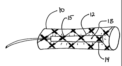
FIGS IA-B represent plan views of another embodiment according to the
invention.
Endoprosthesis 10 of FIGS 1A-B, shown in its expanded configuration, comprises
a generally
tubular structure formed from one or more fibers 12. One or more fibers 12
comprise fiber
points of intersection 14. Prior to deployment, fibers 12 comprise a photo-
curable coating 15, at
or near points of intersection 14, in semi-cured form. Following delivery and
expansion of the
endoprosthesisl 0 by suitable means, the delivery catheter (not shown) is
replaced by ultraviolet
light delivery catheter 18. Ultraviolet radiation within the ranges discussed
above is delivered
via ultraviolet light delivery catheter 18, and photocurable coating 15 is
cured. Ultraviolet light
delivery catheter 18 is then removed from the vessel, and endoprosthesis 10 is
left in place.
Alternatively, substantially the entire endoprosthesis -may comprise a
photocurable
coating. As shown in FIG. 2, endoprosthesis 30 may be disposed on distal end
33 of expanded
11
CA 02503393 2010-05-14
balloon 35, over which photolithographic masking material 32 has been applied
in a pattern.
Masking material 32 prevents the delivery of radiation, to leave desired
portions, for example
fiber points of intersection, exposed. Endoprosthesis 30 may then be exposed
to ultraviolet or
other suitable form of radiation, allowing the exposed portions of the coated
device to cure.
Following delivery of radiation, balloon 35 is removed. In time, the
photolithographic masking
material, and eventually endoprosthesis 30 may erode biocompatibly.
An alternative embodiment according to the invention is shown in FIG. 3. As
discussed
above with respect to FIG. 2, endoprosthesis 40 is mounted upon distal end 43
of balloon 45,
which has been coated with photolithographic masking material 42 in a pattern
such radiation is
selectively delivered to endoprosthesis 40, allowing curing in selected
regions of endoprosthesis
40.
In an alternative embodiment, an endoprosthesis can comprise multiple
materials that are
curable at different wavelengths, in order to confer varied physical
properties on the prosthesis
according to the desired properties of a particular region of the
endoprosthesis. For example, the
proximal and distal ends of a prosthesis can comprise one or more materials
that are curable at a
wavelength distinct from that at which the remainder of the prosthesis is
curable, and can be
selected for greater compliance. It has been shown clinically that restenosis
occurs in response
to vessel trauma at the proximal and distal ends of prostheses. By controlling
the physical
properties to enhance flexibility and to minimize compliance mismatch at the
proximal and distal
ends of the prosthesis, tremendous clinical benefit can be conferred upon the
device. As a
second example, the material selected to comprise the longitudinal connecting
members can cure
at a different wavelength than that at which the remainder of the prosthesis
cures, to impart
greater compliance and flexibility of longitudinal members while allowing the
structural rigidity
needed in support members.
Turning now to FIG 4, a further embodiment according to the invention is
provided.
Endoprosthesis 50 comprises a generally tubular element 52. Although
alternative
configurations are possible, generally tubular element 52 is formed by
weaving, as defined
above, one or more fibers 54. Hollow element 56 is then woven or affixed to
generally tubular
element 52. Hollow element 56 comprises curable material 58 in its interior.
Following
expansion of endoprosthesis 50, curable material 58 is allowed to cure or, if
it is photocurable, is
12
CA 02503393 2010-05-14
exposed to radiation in order to initiate curing. Hollow element 56, following
curing of curable
material 58, confers structural support upon endoprosthesis 50.
FIGS. 5A-C represent steps in the preparation of an alternative embodiment
according to
the invention. FIG. 5A is an enlarged end view of composite flat sheet 60.
Composite flat sheet
60 comprises a first polymeric laminate layer 62, a photocurable and/or
chemically reactive
membrane 64, and second polymeric laminate layer 66. After formation of
composite flat sheet
60, it is rolled to form unexpanded endoprosthesis 68, as shown in FIG. 5C.
Upon expansion of
endoprosthesis 68, light delivery source 70 is introduced within
endoprosthesis 68, as seen in an
end view in FIG. 5C. Photocurable and/or chemically reactive membrane 64 is
thereby cured,
conferring the requisite structural rigidity to endoprosthesis 68.
While particular forms of the invention have been. illustrated and described
above, the
foregoing descriptions are intended as examples, and to one skilled in the art
will it will be
apparent that various modifications can be made without departing from the
spirit and scope of
the invention.
13