Note: Descriptions are shown in the official language in which they were submitted.
CA 02504279 2005-04-15
-1 -
TITLE: Materials and Method of Modulating the Immune Response Using T
Helper-Antigen Presenting Cells
FIELD OF THE INVENTION
The invention relates to a method of modulating the immune response
to treat or prevent a disease. In particular, the method relates to a method
of
using T helper-antigen presenting cells to modulate the immune response to
treat or prevent a disease.
BACKGROUND OF THE INVENTION
Generation of effective cytotoxic T lymphocyte (CTL) responses to
minor histocompatibility or tumor antigens not associated with danger signals
often requires help from CD4+ T helper (Th) cells via cross-priming (1 ). Such
help was originally thought to be mediated by CD4+T cell IL-2 acting at short
range to promote CD8+T cell proliferation (2).
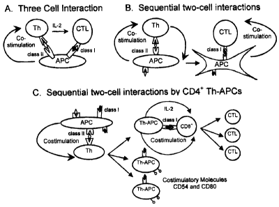
Two models of CD4+ T help for CD8+ CTL responses have been
proposed previously, including the passive model of three-cell interaction
(3,4)
and the dynamic model of sequential two-cell interactions by antigen
presenting cells (APC) (5). The three-cell model suggested that activated
CD4+ T cells and naive CD8+ T cells must interact simultaneously with a
common APC, and that the CD4+ Th cells provide CD8+ T cell help via
expression of Interleukin 2 (IL-2) (Figure 1A). The conundrum, however, is
how a rare antigen-specific CD4+ Th cell and an equally rare antigen-specific
CD8+ T cell (typically 1 in 105-106 T cells) would simultaneously find the
same
antigen peptide-carrying APC in an unprimed animal (6). Instead, Ridge et al
(5) have proposed a dynamic model of two sequential interactions, in which
an APC first offers co-stimulatory signals to a CD4+ Th cell and then to a
CD8+ CTL cell (Figure 1 B). According to this model, the APC-stimulated CD4+
Th cells must first reciprocally counter-stimulate the APCs (through CD40
ligand signaling) such that this newly "conditioned" APC can then directly co-
stimulate CD8+ CTLs. Support for this model comprises evidence that
antigen-specific CTL responses can be induced by vaccination with either
large numbers of APC activated in vitro through CD40 signaling or, in either
major histocompatibility complex (MHC) class II knockout (KO) or CD4+ T cell-
CA 02504279 2005-04-15
-2 -
depleted mice, by high level activation of APCs in vivo with anti-CD40 Ab (5,7-
9). Although this model provides a possible explanation for the conditional
nature of T-cell help for CTL responses, the experimental conditions used in
the above studies may well not accurately model the physiology of Th cell-
dependent immune responses in vivo. In addition, a scarcity caveat remains
(10), in that very small numbers of antigen-bearing APCs (11) must first
activate and be conditioned by the rare naive antigen-specific CD4+ Th cells,
and then find and activate in turn equally rare naive Ag-specific CD8+ CTL. In
addition, this model does not explain how IL-2 from Th cells' would be
precisely targeted to Ag-specific CD8+ Ag-specific CTLs. Furthermore, the life
span of an activated dendritic cell (DC) in the T cell zone of a lymph node is
around 48 hours (11-13), possibly due to CD4+ T cell killing of the cognate
APCs (14-15), whereas the antigen-specific CTL response is first detected at
around day 5 in the lymph nodes (11,16). Thus, this dynamic model also does
not explain compellingly the temporal gap between antigen presentation and
the acquisition of CTL effector function in vivo.
It is recognized that stimulation of T cells by APCs involves at least two
signaling events: one elicited by TCR recognition of peptide-MHC complexes
and the other by costimulatory molecule signaling (e.g., T cell CD28/APC
CD80) (17). A consequence of such Ag-specific T cell-APC interactions is the
formation an immunological synapse, comprising a central cluster of TCR-
MHC-peptide complexes and CD28-CD80 interactions surrounded by rings of
engaged accessory molecules (e.g., complexed LFA-1-CD54) (18,19). One
important feature of synapse physiology is that APC-derived surface
molecules are transferred to the Th cells during the course of their TCR
internalization followed by recycling (20,21 ).
SUMMARY OF THE INVENTION
The present inventor has demonstrated that CD4+ T cells can acquire
the synapse-composed MHC class II and costimulatory molecules (CD54 and
CD80), and bystander MHC class (/peptide complexes from antigen
presenting cells. In addition, the inventor has demonstrated that the
molecules
acquired by the CD4+ T cells are functional, and that these CD4+ T cells can
CA 02504279 2005-04-15
-3 -
act as CD4+ T helper-antigen presenting cells (Th-APC) to stimulate the
immune system in vitro and in vivo, particularly the CTL response.
Accordingly, the invention provides a method of modulating the
immune response to treat or prevent a disease comprising administering an
effective amount of T helper-antigen presenting cells to an animal in need
thereof. The present invention also provides a use of an effective amount of T
helper-antigen presenting cells to treat or prevent a disease.
In addition, the invention provides a pharmaceutical composition for
preventing or treating a disease comprising an effective amount of T helper-
antigen presenting cells and a pharmaceutically acceptable carrier, diluent or
excipient.
Other features and advantages of the present invention will become
apparent from the following detailed description. It should be understood,
however, that the detailed description and the specific examples while
indicating preferred embodiments of the invention are given by way of
illustration only, since various changes and modifications within the spirit
and
scope of the invention will become apparent to those skilled in the art from
this detailed description.
BRIEF DESCRIPTION OF THE DRAWINGS
The invention will now be described in relation to the drawings in
which:
Figure 1 shows three models for the delivery of CD4+ T help to CD8+
CTL. (A) The "passive", three-cell interaction model, in which APC
simultaneously present Ag to the T helper and the CTL, but deliver co-
stimulatory signals only to the helper. The CD4+ Th cell in turn produces IL-
2,
which is required for CTL activation. (B) The dynamic model of sequential
two-cell interactions by APCs, in which the APC offers co-stimulatory signals
to the CD4+ T helper, which reciprocally "licenses" the APC (left side of
panel)
such that it can only then directly co-stimulate the CTL (right side). (C) The
new dynamic model of sequential two-cell interactions, in which APCs
"license" CD4+ T helper cells to act as APCs (Th-APCs). APCs directly
transfer MHC class I/Ag complexes and co-stimulatory molecules to
CA 02504279 2005-04-15
' -4 -
expanding populations of IL-2- producing Th cells, which thereby act directly
as Th1-APCs to simulate CTL activation.
Figure 2 shows analysis of OVA expression by flow cytometry. (a) EG7
(thick solid lines) and EL4 (thick dotted lines), and (b) BL6-10ovA (thick
solid
lines) and BL6-10 (thick dotted lines) tumor cells were stained with the
rabbit
anti-OVA antibody (Sigma), followed with the FITC-goat anti-rabbit IgG
antibody, and then analyzed by flow cytometry. Tumor cells stained with
normal rabbit serum were employed as control populations (thin dotted lines).
One representative experiment of two is displayed.
Figure 3 shows transfer of DC membrane molecules to active CD4+ T
cells. (A) CFSE-labeled DCova were incubated with Con A-stimulated CD4+ T
cells from OT II mice. T cells with (thick solid lines) and without (thick
dotted
lines) incubation of DCovA were stained with Abs and analyzed for expression
of H-2Kb, lab, CD54 and CD80 by flow cytometry, respectively. (B) CFSE-
labeled DCova were incubated with Con A-stimulated CD4+ T cells from
H-2Kb, lab, CD54 and CD80 gene KO OT II mice, respectively. T cells with
(thick solid lines) and without (thick dotted lines) incubation of DCovA were
stained with Abs and analyzed for expression of the above molecules,
respectively. T cells with incubation of DCovA were also stained with isotype-
matched Abs and employed as control populations (thin dotted lines). (C)
DCovA-activated CD4+ T cells (Th-APCs) from OT II mice were stained with a
panel of Abs (thick solid lines) and analyzed by flow cytometry. The control
CD4+ T cells (thin dotted lines) were only stained with isotype-matched Abs.
(D) DCovA-activated CD4+ T cells (Th-APCs) from H-2Kb, lab, CD54 and CD80
gene KO OT II mice, respectively, were stained with a panel of Abs (thick
solid lines). The control CD4+ T cells (thin dotted lines) were only stained
with
isotype-matched Abs. One representative experiment of two in the above
different experiments is shown.
Figure 4 shows membrane acquisition analysis by confocal
fluorescence microscopy. CFSE-labeled DCovA were incubated with Con A-
stimulated CD4+ T cells from (A) H-2Kb, (B) CD54 and (C) CD80 gene KO OT
II mice, stained with fluorochrome-labeled Abs, and analyzed by confocal
CA 02504279 2005-04-15
-5 -
fluorescence microscopy. Images include DCs (larger cells) alone, T (smaller)
cells alone or a mixture of DC and T cells (a) under differential interference
contrast, (b) with a cell-surface stain consisting of ECD (red)-Ab for either
H-2Kb, CD54, or CD80, (c) with cytoplasmic CFSE stain (green), and (d) with
both stains. The data confirm that (i) DCovA (larger cells), but not gene-
deleted T cells (smaller cells), express H-2Kb, CD54, and CD80 molecules
(arrows), and (ii) during co-culture of DCovA with T cells, the T cells
acquire
H-2Kb, CD54, and CD80 molecules (arrow heads). One representative
experiment of two is shown.
Figure 5 shows in vivo membrane transfer assay. The CD4+ T cells
purified from OT II/ lab-~~ and OT II/CD80-~- mice were transferred into wild-
type
C57BU6 mice, respectively. The first group of mice remained untreated and
the second group of mice were immunized with DCovA. The CD4+ OT II/ lab-
~- and OT II/CD80-~~ T cells were then purified from the first (thick dotted
lines)
and the second group (solid lines) of mice and then stained with the FITC-
anti-lab and FITC-anti-CD80 antibodies and the FITC-conjugated isotype-
matched antibodies (thin dotted lines) for flow cytometric analysis,
respectively. One representative experiment of three is shown.
Figure 6 shows that CD4+ T-APCs stimulate RF3370 and OT I CD8+ T
cells. (A) MHC class I presentation of OVA to RF3370 hybridoma by Th-
APCs. The amount of IL-2 secretions of stimulated RF3370 cells in examining
wells were subtracted by the amounts of IL-2 in wells containing DCovA, Th-
APC and Con A-OT II alone, respectively. *, p<0.01 (Student t test) versus
cohorts of Con A-OT II. (B) In vitro CD8+ T cell proliferation assay. Varying
numbers of irradiated Th-APCs, Kb~~- Th-APCs, Con A-OT II and DCovA cells
were co-cultured with naive OT I or B6 CD8+ T cells. After three days, the
proliferative responses of the CD8+ T cells were determined by 3H-thymidine
uptake assays. (C) Th-APCs were cultured with OT I CD8+ T cells either
separated in transwells (transwell) or not (all other bars). In the latter
cultures,
the impact on OT I CD8+ T cell proliferation of adding each of the
neutralizing
reagents, all neutralizing reagents together (mixed reagents), or all control
Abs and fusion proteins (control reagents) was assessed. In one set of wells,
CA 02504279 2005-04-15
-6 -
supernatants from cultured Th-APCs (supernate) were added to the CD8+ T
cells in place of the Th-APCs themselves. *, p<0.01 (Student t test) versus
cohorts of Th-APC. (D) In vivo CD8+ T cell proliferation assay. CFSE-labeled
OT I CD8+ T cells were i.v. injected into C57BL/6 mice. Twelve hours later,
each mouse was i.v. given Th-APCs or Con A-OT II cells or DCovA or PBS,
then 3 days later the numbers of division cycles of the CFSE-labeled CD8+ T
cells in the recipient spleens were determined by flow cytometry. One
representative experiment of three in the above different experiments is
shown.
Figure 7 shows that CD4+ T-APC induce the development of antigen-
specific CTL activity in vitro and in vivo. In vitro cytotoxicity assay. (A)
Three
types of activated CD8+ T cells (DCovA/OT I, Th-APC/ OT I, and Con A-OT
II/OT I) were used as effector (E) cells, whereas 5'Cr-labeled EG7 or control
EL-4 tumor cells used as target (T) cells. (B) Th-APCs were used as effector
(E) cells, whereas 51 Cr-labeled EG7, DCs, DCovA, LB27 and EG70VAll cells
used as target (T) cells. The data are presented as the percent specific
target
cell lysis in 5'Cr-release assay. Each point represents the mean of triplicate
cultures. (C) In vivo cytotoxicity assay. C57BL/6 splenocytes differentially
labeled to be CFSE"'g" and CFSE~°"', were pulsed with OVAI and Mut1
peptide, respectively. These splenocytes were then i.v. injected at ratio of
1:1
into mice immunized with DCovA, Th-APCs or Con A-OT II cells, or PBS.
Sixteen hours later, the CFSE"'9" or CFSE~°"' cells remaining in the
spleens
were determined by flow cytometry. The value in each panel represents the
percentage of CFSE"~9" cells versus CFSE~°'" cells remaining in the
spleens.
Figure 8 shows immune protection of lung metastasis in mice
immunized with Th-APCs. Pulmonary metastases were formed in different
groups of immunized mice by intravenous injection of 0.5x10⁶ BL6-10ovA or
BL6-10 tumor cells. Four weeks later, mouse lungs were removed. The extent
of lung metastasis in 6 different groups of mice described in Exp I of Table 1
was displayed.
CA 02504279 2005-04-15
-7 -
DETAILED DESCRIPTION OF THE INVENTION
The inventor has demonstrated that T helper cells can acquire antigen-
presenting machinery from antigen presenting cells. In particular, the T
helper
cells can acquire MHC class II/peptide complexes, MHC class (/peptide
complexes and co-stimulatory molecules from antigen presenting cells. The
inventor has demonstrated that these molecules are functional on the T helper
cells. Thus the T helper cells can act as T helper-antigen presenting cells
and
directly stimulate the immune response, particularly CTL activity.
Accordingly, the invention provides a method of modulating the
immune response to treat or prevent a disease comprising administering an
effective amount of T helper-antigen presenting cells to an animal in need
thereof. The invention also provides a method of modulating the immune
response for use in transplantation comprising administering an effective
amount of a T helper-antigen presenting cell to an animal in need thereof.
The present invention also provides a use of an effective amount of T helper-
antigen presenting cells to treat or prevent a disease.
The term "disease" term disease as used herein includes, and is not
limited to, cancer, immune diseases, such as an autoimmune disease, or
infections.
As used herein, the phrase "to treat or prevent a disease" refers to
inhibition or reducing the occurrence of a disease. For example, if the
disease
is cancer "preventing cancer" refers to prevention of cancer cell replication,
inhibition of cancer spread (metastasis), inhibition of tumor growth,
reduction
of cancer cell number or tumor growth, decrease in the malignant grade of a
cancer (e.g., increased differentiation), or improved cancer-related symptoms;
and "treating cancer" refers to preventative treatment which decreases the
risk of a patient from developing a cancer, or inhibits progression of a pre-
cancerous state (e.g. a colon polyp) to actual malignancy.
As used herein, the phrase "effective amount" means an amount
effective, at dosages and for periods of time necessary to achieve the desired
result, e.g. to treat or prevent a disease. Effective amounts of T helper-
antigen
presenting cells may vary according to factors such as the disease state, age,
CA 02504279 2005-04-15
-$ -
sex, weight of the animal. Dosage regime may be adjusted to provide the
optimum therapeutic response. For example, several divided doses may be
administered daily or the dose may be proportionally reduced as indicated by
the exigencies of the therapeutic situation.
As used herein, the term "animal" includes all members of the animal
kingdom, including humans.
The term "a cell" as used herein includes a single cell as well as a
plurality or population of cells.
The term "T helper-antigen presenting cells" refers to T helper cells
that can stimulate cytotoxic T lymphocytes by acting as antigen presenting
cells. Specifically, the T helper-antigen presenting cells express MHC
(/antigen complexes and co-stimulatory molecules, such as CD54 and CD80,
and can act as antigen presenting cells to stimulate cytotoxic T lymphocytes.
The T helper cells can acquire the antigen/MHC class I complexes and co
stimulatory molecules directly from other antigen presenting cells, such as
dendritic cells, B cells and macrophages.
In one embodiment, the T helper-antigen presenting cells are
generated by immunizing an animal with an antigen of interest, and then
purifying T helper cells using CD4 as a marker.
A person skilled in the art will appreciate that T helper-antigen
presenting cells can be generated by recombinant technology. In one
embodiment, T helper cells are genetically engineered to express MHC class I
complexes with an antigen of interest and co-stimulatory molecules, such as
CD54 and CD80. Necessary techniques are explained fully in the literature,
such as, "Molecular Cloning: A Laboratory Manual", second edition
(Sambrook et al., 1989); "Oligonucleotide Synthesis" (M. J. Gait, ed., 1984);
"Animal Cell Culture" (R. I. Freshney, ed., 1987); "Methods in Enzymology"
(Academic Press, Inc.); "Handbook of Experimental Immunology" (D. M. Weir
& C. C. Blackwell, eds.); "Gene Transfer Vectors for Mammalian Cells" (J. M.
Miller & M. P. Calos, eds., 1987); "Current Protocols in Molecular Biology"
(F.
M. Ausubel et al., eds., 1987); "PCR: The Polymerase Chain Reaction",
CA 02504279 2005-04-15
_g _
(Mullis et al., eds., 1994); "Current Protocols in Immunology" (J. E. Coligan
et
al., eds., 1991 ).
The term "modulating the immune response" as used herein refers to
enhancing or suppressing the immune system of an animal. In a preferred
embodiment, the T helper-antigen presenting cells enhance the immune
response, particularly the CTL response. The immune response of an animal
can be readily tested using techniques known in the art. In one embodiment,
in vivo or in vitro CD8+ T cell proliferation assays can be used. In another
embodiment, in vivo or in vitro CD8+ cytotoxic assays can be used.
In one embodiment, T helper-antigen presenting cells are used alone
to modulate the immune response to treat or prevent a disease. In another
embodiment, T helper-antigen presenting cells are used in combination with
other immune cells to modulate the immune response to treat or prevent a
disease. Other immune cells include, and are not limited to, dendritic cells,
macrophages, B cells and cytotoxic T lymphocytes.
In a further embodiment, the method of the invention includes the use
of an immune adjuvant. Immune adjuvants are known to persons skilled in
the art and include, without being limited to, the lipid-A portion of a gram
negative bacteria endotoxin, trehalose dimycolate or mycobacteria,
phospholipid bromide (DDA), certain linear polyoxypropylene-polyoxyethylene
(POP-POE) block polymers, mineral salts such as aluminum hydroxide,
liposomes, cytokines and inert vehicles such as gold particles.
The T helper-antigen presenting cells may be formulated into
pharmaceutical compositions for administration to subjects in a biologically
compatible form suitable for administration in vivo. By "biologically
compatible
form suitable for administration in vivo" is meant a form of the substance to
be
administered in which any toxic effects are outweighed by the therapeutic
effects. The substances may be administered to living organisms including
humans, and animals. Administration of a therapeutically active amount of
the pharmaceutical compositions of the present invention is defined as an
amount effective, at dosages and for periods of time necessary to achieve the
desired result. For example, a therapeutically active amount of a substance
CA 02504279 2005-04-15
-10 -
may vary according to factors such as the disease state, age, sex, and weight
of the individual, and the ability of antibody to elicit a desired response in
the
individual. Dosage regime may be adjusted to provide the optimum
therapeutic response. For example, several divided doses may be
administered daily or the dose may be proportionally reduced as indicated by
the exigencies of the therapeutic situation.
Accordingly, the present invention provides a pharmaceutical
composition for preventing or treating a disease comprising an effective
amount of T helper-antigen presenting cells and a pharmaceutically
acceptable carrier, diluent or excipient.
The active substance may be administered in a convenient manner
such as by injection (subcutaneous, intravenous, intramuscular, etc.), oral
administration, inhalation, transdermal administration (such as topical cream
or ointment, etc.), or suppository applications. Depending on the route of
administration, the active substance may be coated in a material to protect
the
T helper-antigen presenting cells from the action of enzymes, acids and other
natural conditions which may inactivate the T helper-antigen presenting cells.
The compositions described herein can be prepared by per se known
methods for the preparation of pharmaceutically acceptable compositions
which can be administered to subjects, such that an effective quantity of the
active substance is combined in a mixture with a pharmaceutically acceptable
vehicle. Suitable vehicles are described, for example, Remington's
Pharmaceutical Sciences (2003 - 20th edition) and in The United States
Pharmacopeia: The National Formulary (USP 24 NF19) published in 1999.
On this basis, the compositions include, albeit not exclusively, solutions of
the
substances in association with one or more pharmaceutically acceptable
vehicles or diluents, and contained in buffered solutions with a suitable pH
and iso-osmotic with the physiological fluids.
The following non-limiting examples are illustrative of the present
invention:
EXAMPLES
Materials and Methods
CA 02504279 2005-04-15
-11 -
Tumor cells, reagents and animals
The highly lung metastatic B16 mouse melanoma BL6-10 and OVA-
transfected BL6-10 (BL6-10ovA) cell lines were generated by the inventor (22).
Both cell lines form numerous lung metastasis after i.v. tumor cell (0.5X10⁶
cells/mouse) injection. The mouse B cell hybridoma cell line LB27 expressing
both H-2Kb and lab, the mouse thymoma cell line EL4 of C57BL/6 mice and
the OVA-transfected EL4 (EG7) cell line which is sensitive to CTL killing were
obtained from American Type Culture Collection (ATCC, Rockville, MD). Both
BL6-10 and BL6-10ovA express similar level of H-2Kb, but not lab. Both BL6-
10ovA and EG7 cells expressed OVA by flow cytometric analysis, whereas
BL6-10 and EL4 cells did not (Figure 2). T cell hybridoma cell line RF3370
expresses TCR specific for H-2Kb/OVA peptide complexes (23). The biotin-
labeled monoclonal Abs specific for H-2Kb (AF6-88.5), lab (AF6-120.1 ), CD3
(145-2C11), CD4 (GK1.5), CD8 (53-6.7), CD11b (MAC-1), CD11c (HL3),
CD25 (7D4), CD54 (3E2), CD69 (H1.2F3), CD80 (16-10A1) and Va2V~35+
TCR (MR9-4) were obtained from BD Pharmingen, Mississauga, ON,
Canada. The OVAI (SIINFEKL) and OVAII (ISQAVHAAHAEINEAGR)
peptides (24,25) are OVA tumor peptides for H-2Kb and lab, respectively,
whereas Mut1 (FEQNTAQP) peptide is an irrelevant 3LL lung carcinoma for
H-2Kb (26). These peptides were synthesized by Multiple Peptide Systems
(San Diego, CA). The OVA-specific TCR transgenic OT I and OT II mice, and
H-2Kb, lab, CD4, CDB, CD54 and CD80 KO mice on a C57BL/6 background
were obtained from the Jackson Laboratory (Bar Harbor, MA). Homozygous
OT II/H-2Kb-'-, OT II/lab-'-, OT II/CD54~'~ and OT II/CD80~'- mice were
generated
by backcrossing the designated gene KO mice (H-2Kb) onto the OT II
background for three generations; homozygosity was confirmed by PCR
according to Jackson laboratory's protocols. All mice were maintained in the
animal facility at the Saskatoon Cancer Center and treated according to
animal care committee guidelines of University of Saskatchewan.
Preparation of dendritic cells
Activated, mature bone marrow-derived DCs, expressing high levels of
MHC class II, CD40, CD54 and CD80, were generated from C57BU6 mice,
CA 02504279 2005-04-15
' -12 -
as described previously (27). To generate OVA-pulsed DC (DCovA), DCs were
pulsed overnight at 37°C with 0.1 mg/ml OVA (Sigma, St. Louis, MO),
then
washed extensively (26).
Preparation of OT II CD4+ and OT I CD8+ T cells
Naive OVA-specific CD4+ T and CD8+ T cells were isolated from OT II
or OT I mouse spleens, respectively, and enriched by passage through nylon
wool columns. CD4+ and CD8+ cells were then purified by negative selection
using anti-mouse CD8 (Ly2) or CD4 (L3T4) paramagnetic beads (DYNAL Inc,
Lake Success, NY) to yield populations that were >98% CD4+/Va2V~5+ or
CD8+/Va2V~5+, respectively. To generate DCovA-activated CD4+ T cells,
CD4+ T cells (2X10⁵ cells/ml) from OT II mice or designated gene-deleted OT
II mice were stimulated for three days with irradiated (4,000 rads) BM-derived
DCovA (1X10⁵ cells/ml) in the presence of IL-2 (10 U/ml), IL-12 (5 ng/ml) and
anti-IL-4 antibody (10 Ng/ml) (R&D Systems, Minneapolis, MN) (28). These in
vitro DCovA-activated CD4+ T cells, also referred to herein as CD4+ Th-Ag
presenting cells (Th-APCs), were then isolated by Ficoll-Paque (Sigma)
density gradient centrifugation, or further purified using CD4 microbeads
(Milttenyi Biotec, Auburn, CA) in some experiments. Con A-stimulated OT II
C D4+ T (Con A-OT II) cells were similarly generated by incubating
splenocytes from OT II or OT II/KO mice with Con A (1 Ng/ml) and IL-2 (10
U/ml) for 3 days, after which the CD4+ T cells were purified on density
gradients. To ascertain that no DCs were in purified Th-APCs or Con A-OT II
cells, these active T cells were further purified by using CD4 microbeads
(Milttenyi Biotec).
Phenotypic characterization of DCovA-activated CD4'' T cells
For the phenotypic analyses, Th-APCs were stained with Abs specific
for H-2Kb, lab, CD3, CD4, CDB, CD11b, CD11c, CD25, CD54, CD69, CD80
and Va2V(35+ TCR (BD Pharmingen), respectively, and analyzed by flow
cytometry. For the intracellular cytokines, cells were restimulated with 4000
rad-irradiated BL27 tumor cells pulsed with OVAI! peptide for 4 hours (28),
and then processed using a 'Cytoflx/CytoPerm Plus with GoIgiPlug' kit (BD
Pharmingen), with R-phycoerythrin (PE)-conjugated anti-IL-4, -perforin and
CA 02504279 2005-04-15
-13 -
-IFN-'y Abs (R&D Systems), respectively. Culture supernatants of the re-
stimulated Th-APCs were analyzed for IFN-~y, IL-2 and IL-4 expression using
ELISA kits (Endogen, Cambridge, MA), as reported previously (26).
In vitro and in vivo membrane molecule transfer assays
In in vitro membrane transfer assay, DCovA or DC were incubated with
5-carboxy-fluorescein diacetate succinimidyl ester (CFSE; 0.5 NM) at
37°C for
minutes and washed 3 times with PBS. CFSE-labeled DCovA or DC were
incubated with Con A-OT II cells at 37°C for 4 hours, then the cell
mixtures,
the original DCovA and Con A-OT II cells were stained with a panel of
10 phycoerythrin-Texas red-X (ECD)-Abs specific for H-2Kb, CD54 and CD80,
respectively, and analyzed by confocal fluorescence microscopy. CD4+ T cells
in the cell mixture were also purified by cell sorting and analyzed by flow
cytometry. Con A-OT II cells stained with biotin-labeled isotype-matched Abs
and ECD-avidin (BD Pharmingen) were used as controls.
15 In in vivo membrane transfer assay, naive T cells were isolated from
OT II/lab'~' and OT II/CD80'~' mouse spleens, respectively, and enriched by
passage through nylon wool columns. The CD4+ T cells (5x10⁶ cells/mouse)
were further purified by negative selection using the anti-mouse CD8 (Ly2)
paramagnetic beads (DYNAL Inc), and then i.v. injected into wild-type
C57BU6 mice. One group of mice remained untreated. One day subsequent
to the injection, another group of mice were i.v. immunized with irradiated
(4,000 rads) DCova, (0.2X10⁶ cells/mouse). Three days after the immunization,
mice were sacrificed. T cells were isolated from the spleens of these two
groups of mice, and enriched by passage through nylon wool columns. The
OVA-specific CD4+ OT II T cells were further purified from these T cells by
positive selection using the biotin-anti-TCR antibody and anti-biotin
microbeads (Milttenyi Biotec), and then stained with FITC-anti-lab and FITC-
anti-CD80 antibodies for flow cytometric analysis, respectively.
Antigen presentation
RF3370 hybridoma cells (0.5X10⁵ cells/well) were cultured with
irradiated (4,000 rad) DCova or Th-APCs or Con A-OT II (1X10⁵ cells/well) for
24 hr. To investigate the fate of acquired MHC class (/peptide expression, Th-
CA 02504279 2005-04-15
-14 -
APCs alone were cultured for 1, 2 and 3 days in culture medium containing IL-
2 (10 U/ml), termed Th-APC (1, 2 and 3 Day), and then harvested for
stimulation of RF3370 cells, respectively. The supernatants were harvested
for measurement of IL-2 secretion using ELISA kit (Endogen).
CD8+ T cell proliferation assays
For in vitro CD8+ T cell proliferation assay, irradiated (4,000 rads)
stimulators, the Th-APCs, Con A-OT II cells (0.4X10⁵ cells/well), DCovA
(0.1X10⁵ cells/well) and their 2-fold dilutions were cultured with a constant
number of responders, the naive OT I or C57BU6 (B6) CD8+ T cells (0.5X10⁵
cells/well). To rule out the potent effect of endogenous H-2Kb, Th-APCs
generated from H-2Kb~~- OT II T cells were termed Kb-~- Th-APCs and used as
stimulators. In some experiments, each of a panel of neutralizing reagents
(anti-IL-2, -H-2Kb or -LFA-1 Abs, and CTLA-4/Ig fusion protein) (each 15
Ng/ml; R&D Systems) or a mixture of the above reagents were added to the
cells, while control cells received a mixture of isotype-matched irrelevant
Abs
and fusion protein. In other experiments, the irradiated CD4+ Th-APCs and
naive OT I CD8+ T cells were cultured in transwell plates (Costar, Corning,
NY), separated by 0.4 pM pore-sized membranes. After 48 hrs, thymidine
incorporation was determined by liquid scintillation counting (26).
For in vivo CD8+ T cell proliferation assay, purified naive OT I CD8+ T
cells were labeled with CFSE (1.5 NM) and i.v. injected into C57BL/6 mice
(2X10⁶ cells each). Twelve hours later, each mouse was i.v. injected with
2X10⁶ Th-APCs and Con A-OT II cells, respectively, or 0.2X10⁶ DCovA. In
another group, mice were injected with PBS. Three days later, the splenic T
cells from the recipients were stained with ECD-anti-CD8 Ab (Beckman
Coulter, Miami, FL), and then analyzed by flow cytometry.
Cytotoxicity assays
For in vitro cytotoxicity assay, the activated CD8+ T cells derived from
the above three day co-culture with irradiated (4,000 rads) DCovA, Th-APCs
and Con A-OT II cells were purified on density gradients and termed
DCovA/OT I, Th-APC/OT I and Con A-OT II/OT I, respectively. These cells as
well as Th-APCs were used as effector (E) cells, while 5'Cr-labeled EG7, the
CA 02504279 2005-04-15
-15 -
control EL-4 tumor cells, DCovA~ LB27 and OVAII-pulsed LB27 (LB27ovAn)
tumor cells were used as target (T) cells, respectively. Specific killing was
calculated as: 100 X [(experimental cpm - spontaneous cpm)/(maximal cpm -
spontaneous cpm)], as previously described (26).
The inventor adapted a recently reported in vivo cytotoxicity assay (29).
Briefly, C57BU6 mice were i.v, immunized with DCovA (0.5X10⁶ cells), Th-
APCs or Con A-OT II cells (2X10⁶ cells). Seven days later, mice were boosted
once. In another group, mice were injected with PBS. Naive mouse
splenocytes were incubated with either high (3.0 NM, CFSEn~9n) or low (0.6
pM, CFSE~°W) concentrations of CFSE, to generate differentially labeled
target
cells. The CFSE"~9n cells were pulsed with OVAI, whereas the CFSE~°'"
cells
were pulsed with the irrelevant 3LL lung carcinoma H-2Kb peptide Mut1 and
served as internal controls. These peptide-pulsed target cells were washed
extensively to remove free peptide, and then i.v. co-injected at 1:1 ratio
into
the above immunized mice three days after the boost. Sixteen hours after
target cell delivery, the spleens were removed and residual CFSEn'9n and
CFSE~°w target cells remaining in the recipients' spleens were
sorted and
analyzed by flow cytometry.
Animal studies
Wild-type C57BU6 mice (n=8) were injected i.v. with 0.2X10⁶ DCovA,
2X10⁶ Th-APCs and Con A-OT II cells, respectively, and then 7 days later
they were boosted once. To study the immune mechanism, CD4 and CD8 KO
mice (n=8) were injected i.v. with 2X10⁶ Th-APCs, and then 7 days later the
mice were boosted once. Three days subsequent to the boost, the mice were
i.v. given 0.5X10⁶ BL6-10ova, or BL6-10 tumor cells. The mice were sacrificed
4 weeks after tumor cell injection and the lung metastatic tumor colonies were
counted in a blind fashion (22). Metastases on freshly isolated lungs appeared
as discrete black pigmented foci that were easily distinguishable from normal
lung tissues and confirmed by histological examination. Metastatic foci too
numerous to count were assigned an arbitrary value of >100.
Results
CD4+ Th-APCs acquire the synapse-composed MHC class II and CD54
CA 02504279 2005-04-15
-16 -
molecules and the bystander MHC Class I from APCs by APC stimulation
In order to explore DC membrane-derived APC machinery acquisition
by CD4+ T cells, Con A-stimulated CD4+ T cells from OVA-specific TCR
transgenic OT II mice were cultured for 4 h either alone or with OVA-pulsed
DCs (DCovA) or DC. The CD4+ T cells were then sorted and examined for
expression of MHC class I and II, CD54 and CD80 by flow cytometry. The
control Con A-stimulated OT II CD4+ T cells expressed some MHC class I and
II, CD54 and CD80. However, following incubation with DCovA, these T cells
displayed moderately augmented levels of these molecules (Figure 3A),
suggesting that DC molecules could have been transferred to the T cells. The
membrane transfer can be mostly blocked by addition of anti-H-2Kb and LFA-
1 antibodies and CTLA-4/Ig fusion protein, indicating that the membrane
acquisition of Th-APCs from DCovA is mediated by TCR and co-stimulatory
molecules. In addition, these T cells following interaction with DCs without
OVA pulsing also displayed augmented levels of these molecules, but to a
lesser extent, indicating that these DC molecule transfer is mediated by both
the antigen-specific and non-specific manners.
Since all T cells express MHC class I and CD54, and some activated T
cells also express MHC class II and CD80 molecules (30,31), it was
necessary to confirm that the increased levels of T cell-associated MHC class
I and II, CD54 and CD80 were not due to endogenous T cell up-regulation of
these molecules. Thus, we incubated CFSE-labeled DCovA with Con A-
stimulated CD4+ T cells derived from OT II mice with homozygous H-2Kb, lab,
CD54 and CD80 gene KO, respectively, then sorted the T cells and assessed
their expression of these markers. The T cells did not express their
respectively deleted gene products when cultured alone, but did discernibly
express H-2Kb, lab, CD54 and CD80 after 4 hr incubation with DCovA, as
determined by flow cytometry (Figure 3B) or confocal fluorescence
microscopy (Figure 4). These results indicate that, besides previously
reported MHC class I transferred onto CD8+ T cells during DC/CD8+ T cell
interaction and MHC class II and CD80 molecules transferred onto CD4+ T
cells during DC/CD4+ T cell interaction (21,32,33), CD4+ T cells can also
CA 02504279 2005-04-15
-17 -
acquire CD54 forming the immune synapse (18,19) as well as the bystander
MHC class I molecules from DCs after DC stimulation of CD4+ T cells. In
addition to the mechanism of antigen-specific MHC-TCR mediated
internalization and recycling (20,21 ), the uprooting of APC molecules or APC-
released vesicles may also contribute to the above membrane transfer,
especially the bystander MHC class I (34).
The inventor then examined whether naive T cells can also acquire DC
Ag-presenting machinery in culture. Naive OT II CD4+ T cells were first
purified by using nylon column to remove DCs and B cells and anti-CD8
paramagnetic beads (DYNAL Inc) to remove CD8+ T cells, and then incubated
for three days with irradiated DCovA. The activated OT II CD4+ T cells were
then purified by using ficoll-Paque density gradient centrifugation and CD4
microbeads (Milttenyi Biotec), and then analyzed by flow cytometry. These T
cells, which proliferated in response to DCovA stimulation, expressed cell
surface CD4, CD25 and CD69, and intracellular perforin and IFN-y, but not
IL-4 (Figure 3C); they also secreted IFN-y (~2 ng/ml/106 cells/24 hr) and IL-2
(~2.5 ng/ml/106 cells/24 hr), but not IL-4, in culture. This data indicates
that
these OVA-TCR transgenic CD4+ T cells were type 1 T helpers (Th1). In
addition, there was no CD11b+/11c+DC population existing in these purified
CD4+ T cells (Figure 3C). This is because that any survival irradiated DCovA
cells and the potential small amount of contamination of spleen DCs or B cells
within the original naive OT II CD4+ T cell preparation, which might picked up
OVA peptides from irradiated DCovA in the culture, would be eliminated by the
killing activity of these activated Th1 cells expressing perforin (Figure 7B)
(35,36). In addition to the common H-Kb expression, these Th cells also
expressed lab, CD54 and CD80 molecules, and here too they did so whether
they were derived from wild-type or homozygous H-2Kb-~-, lab~~-, CD54-~- or
CD80-~~ KO mice (Figure 3D). Thus, the inventor demonstrates that naive
CD4+ T cells can also acquire MHC class II and costimulatory molecules
(CD54 and CD80) composing the immune synapse as well as the bystander
MHC class I from DCs by in vitro DC stimulation.
To further confirm the membrane acquisition in vivo, wild-type C57BL/6
CA 02504279 2005-04-15
-18 -
mice were first injected with purified CD4+ OT II/ lab-~~ and OT II/CD80-~- T
cells, and then immunized with DCovA. Three days after the immunization,
mice were sacrificed. CD4+ OT II T cells were purified from these immunized
mouse spleens, and then stained with FITC-anti-lab and FITC-anti-CD80
antibodies for flow cytometric analysis, respectively. As shown in Figure 5,
CD4+ OT II/ lab-- and OT II/CD80-~- T cells derived from mice immunized with
DCovA became slightly lab and CD80 positive, respectively, whereas these T
cells derived from mice without immunization remained lab and CD80
negative, indicating that CD4+ OT II T cells acquire lab and CD80 molecules
by in vivo DCovA stimulation.
Th-APCs stimulate CD8+ T cell proliferation in vitro and in vivo
The ability of the CD4+ T cells, which acquired H-2Kb/OVAI peptide
complexes and the DC Costimulatory molecules, to act as direct APCs
(termed CD4+ TL-APLs) for CD8+ T cell stimulation was then examined. To
examine the functionality of these putative Th-APC cells, the inventor
initially
assessed their ability to stimulate IL-2 secretion of T cell hybridoma RF3370.
As shown in Figure 6A, RF3370 cells alone did not secret IL-2. However, Th-
APCs significantly stimulated RF3370 to secret IL-2 (95 pg/ml) as did DCova,
(220 pg/ml), indicating that Th-APCs expressed functional H-2Kb/OVAI
peptide complexes. The stability of the acquired MHC I/OVAI peptide
complexes was then assessed. The rate of their decay was assessed by
culturing these Th-APCs after MHC class I acquisition for varying time
periods. As shown in Figure 6A, the ability to stimulate IL-2 secretion of
RF3370 cells did decay over time. However, readily detectible MHC class
(/peptide expression was still observed as much as 3 days after in vitro
culture.
To further confirm the results, the inventor then assessed the ability of
the Th-APCs to induce proliferation of naive OT I CD8+ T cells in vitro. The
positive control DCovA cells which previously demonstrated to possess a
highly activated phenotype (27) strongly induced OT I cell proliferation
(Figure
6B). DCovA-activated CD4+ Th-APCs which were purified by Ficoll-Paque
density gradient centrifugation and using CD4 microbeads did indeed
CA 02504279 2005-04-15
-19 -
stimulate proliferation of OT I CD8+ T cells, but to a lesser extent due to
(i)
less costimulatory molecules and (ii) lacking the third signal, DC-secreted IL-
12 (37), compared with DCovA. However, they did not stimulate responses of
the control naive C57BU6 (B6) mouse CD8+ T cells, nor did Con A-stimulated
OT II CD4+ T (Con A-OT II) cells [secreting IFN-y (~4.0 ng/m1/106 cells/24 hr)
and IL-2 (~3.3 ng/m1/106 cells/24 hr), but lacking self IL-4 and acquired H-
2Kb/OVA peptide complexes] stimulate OT I CD8+ T cell proliferation. In
addition, Kb-~~ Th-APCs derived from the H-2Kb-~- OT II KO mice (Figure 3D)
showed similar CD8+ T cell stimulatory activity as Th-APCs derived from the
wild-type OT II mice (Figure 6B), indicating that the activation of CD8+ OT I
T
cells is mediated via the acquired H-2Kb/OVA peptide complexes, but not the
endogenous H-2Kb of Th-APCs. In separate experiments, we demonstrated
that CD8+ T cell stimulatory activity of the Th-APCs was contact-dependent
since transwells blocked CD8+ T cell proliferation (Figure 6C). Furthermore,
adding anti-MHC class I or -LFA-1 Abs, or cytotoxic T lymphocyte-associated
Ag (CTLA)-4/Ig fusion protein could significantly inhibit the OT I CD8+ T cell
proliferative response in the co-cultures by 38, 50, and 58%, respectively,
while anti-IL-2 antibody had less effect (19% inhibition) (p<0.01).
Simultaneous addition of all blocking reagents reduced the proliferative
response by 92% (p<0.01 ). Taken together, this data indicates that this
response is critically dependent on H-2Kb/OVAI/TCR specificity and
greatly affected by nonspecific co-stimulatory CD54/LFA-1 and CD80/CD28
interactions between the CD4+ Th-APCs and CD8+ T cells. That this
proliferative effect was not simply an in vitro artifact was confirmed by
demonstrating that these Th1-APCs can also stimulate proliferative responses
in vivo. The inventor adoptively transferred CFSE-labeled naive OT I CD8+ T
cells into mice that were also given Th-APCs, ConA-OT II cells, DCovA or
PBS. The labeled CD8+ T cells did not show any division in mice treated with
PBS. However, the labeled CD8+ T cells underwent some cycles of cell
division in the mice given either Th-APCs or DCovA, but did not respond in the
animals given Con A-OT II cells (Figure 6D).
Th-APCs stimulate CD8+ T cell differentiation into CTL effectors in vitro and
in
CA 02504279 2005-04-15
-20 -
vivo
As a critical test of the functionality of these purified CD4+ Th-APCs, we
tested their ability to induce the differentiation of naive OT I CD8+ T cells
into
CTL effectors, as determined using in vitro 5'Cr release assays with EG7
tumor cells expressing an OVA transgene. The Th-APC- activated OT I CD8+
T (Th-APC/OT I) cells displayed substantial cytotoxic activity (33% specific
killing; E:T ratio, 12) against an OVA-expressing EG7 cell line as did the
DCovA-activated OT I CD8+ T (DCovA/OT I) cells (46% killing; E:T ratio, 12),
but not against its parental EL4 tumor cells (Figure 7A), indicating that the
killing activity of these CTLs is OVA-tumor specific. In addition, these CD4+
Th-APCs expressing perforin (Figure 3C) displayed killing activities for DCovA
and LB27ovAn cells with lab/OVAII expression (Figure 7B). However, they
themselves did not show any killing activity to LB27 and EG7 (Figure 7B) or
BL6-10ovA cells without lab/OVAII expression. As with the proliferation
assays,
the in vitro CD8+ CTL induction capacity of CD4+ Th-APCs can also be
translated into an induction of effector CTL function in vivo. The inventor
adoptively transferred OVAI peptide-pulsed splenocytes that had been
strongly labeled with CFSE (CFSE"'9"), as well as the control peptide Mut1-
pulsed splenocytes that had been weakly labeled with CFSE (CFSE~°'"),
into
recipient mice that had been vaccinated with these purified Th-APCs, DCovA,
Con A-OT II cells or PBS. We assessed the disappearance of the labeled
cells from the mice by flow cytometric analysis and found that the CFSE~o'"
(irrelevant Mut1 peptide-pulsed) cells were unaffected by the vaccination
protocol. In addition, we found that no substantial loss (1 %) of the CFSE"'9"
(OVAI peptide-pulsed) cells from the PBS-immunized mice. However, there
was substantial loss of the CFSE"'g" (OVAI peptide-pulsed) cells from the Th-
APC-immunized (86%) or DCovA-vaccinated (97%) mice, but not from the Con
A-OT II cell-vaccinated (2%) mice (Figure 7C). These data indicate that CD4+
Th-APCs carrying H-2Kb/OVAI complexes and DC co-stimulatory molecules
can stimulate the development of OVA-specific CTL effector cells in vivo.
Th-APCs induce OVA-specific antitumor immunity in vivo
In addition, Th-APCs can also stimulate OVA-specific CTL-mediated
CA 02504279 2005-04-15
-21 -
antitumor immunity in vivo. We injected these purified Th-APCs i.v. into mice,
followed by i.v. challenge with OVA-expressing BL6-10ovA or OVA-negative
BL6-10 tumor cells. All mice immunized with Con A-OT II cells (i.e., cells
lacking acquired H-2Kb/OVAI complexes and co-stimulatory molecules) as
well as the control mice (8/8) without any immunization had large numbers
(>100) of lung metastatic tumor colonies four weeks after tumor cell challenge
(Exp I of Table 1 and Figure 8). In addition, all mice (8/8) immunized with
naive OT II T cells also died of lung metastasis. However, all mice (8/8)
immunized with Th-APCs had no lung tumor metastasis. DCovA immunization
was equally effective in inducing anti-tumor immunity. The specificity of the
protection was confirmed with the observation that Th-APCs did not protect
against BL6-10 tumors that did not express OVA, with all mice having large
numbers (>100) of lung metastatic tumor colonies after tumor cell challenge.
To study the immune mechanism, CD4 and CD8 KO mice were used for
immunization of Th-APCs. As shown in Exp II of Table 1, all of the CD4 KO
mice (8/8) were still protected from BL6-10ovA tumor challenge, indicating
that
activation of CD8+ CTL response by Th-APCs is independent on the host
CD4+ T cells. However, all CD8 KO mice (8/8) had numerous lung tumor
metastases, indicating that the Th-APCs-driven antitumor immunity is
mediated by CD8+ CTLs. The Th-APC-induced CD8+ CTL response is more
likely through direct interaction between Th-APCs and CD8+ CTLs rather than
cross-presentation of the host DCs picking up OVA peptides released from
Th-APCs, because the former is CD4+ T cell independent whereas the latter is
CD4' T cell dependent.
Discussion
A long-standing paradox in cellular immunology has been the
conditional requirement for CD4+ Th cells in priming of CD8+ CTL responses.
CTL responses to non-inflammatory stimuli (e.g., MHC class I alloantigen Qa-
1, the male HY Ag) are CD4+ T cell-dependent (2,38,39). The inventor
demonstrates the critical helper requirement for CTL induction, as have two
other recent reports. Wang et al showed that the primary CD8+ T cell
responses to Ags presented in vivo by peptide-pulsed DCs are also
CA 02504279 2005-04-15
-22 -
dependent on help from CD4+ T cells (40). More importantly, Behrens et al
have demonstrated that coinjection of Ag-presenting DC-activated, but not
naive, CD4+ OT II T cells induces CTL responses against islet ~i cell OVA Ag
and leads to diabetes in rat insulin promoter (RIP)-OVA"' transgenic mice.
They also found that activated CD4+ OT II T cells provide CD40-mediated
help to CD8+ T cell responses without these T cells necessarily seeing Ag on
the same APC (41 ). On the other hand, some have suggested that CD4+ T
cell help is only essential for memory CTL responses (29). Thus, the
generation of effectors from naive CD8+ T cells is reported to be helper
independent in mice immunized with irradiated embryonic cells expressing an
adenovirus type 5 E1A transgene (42). Having said that it is highly relevant
that such adenoviral challenge would also introduce potent inflammatory
signals into the sensitizing microenvironment (leading to high level DC
maturation) (43), to say nothing of the potential for help from natural killer
cells
(44). In addition, the E1A adenoviral Ag features multiple CD8+ T cells
epitopes (45), and therefore also a greater base of Ag-specific CD8+ T cell
precursors from which to draw (46). A strong and direct activation of DCs (47)
would explain the previous demonstrations that induction of some anti-viral
CTL responses is CD4+ T helper cell-independent.
T cell-to-T cell (T-T) Ag presentation, dependent upon activated CD4+
T cells first acquiring MHC class II and CD80 molecules from APCs and then
stimulating other CD4+ T cells, is increasingly attracting attention (32,33).
However, the roles such T-APCs may play in vivo have been as yet ill defined
and the results of the relevant in vitro studies disparate, in part because
multiple experimental systems have been employed. For example, CD4+ T-
APCs can induce IL-2 production and proliferative responses among naive
responder T cells (48,49), which is consistent with the results in this study.
However, these T-APCs have also been shown to induce apoptosis in
activated CD4+ T cells or anergization of CD4+ T cell lines (33,50-52). In
contrast, the inventor found that in vivo transfer of CD4+ Th1-APCs
expressing high levels of INF-y and IL-2, which were generated by incubation
of OT II CD4+ T cells with DCovA in the presence of IL-12 and anti-IL-4
CA 02504279 2005-04-15
-23 -
antibody, were able to stimulate OVA-specific CTL responses. Interesting, the
inventor also found that in vivo transfer of CD4+ Th2-APCs expressing high
levels of IL-4 and IL-10, which were generated by incubation of OT II CD4+T
cells with DCova in the presence of IL-4 and anti-IFN-~y antibody, were able
to
induce OVA-specific immune suppression. In other reports, however, in vivo
transfer of CD4+ Th1-APCs derived from IL-2-dependent transformed T cell
lines, has been reported to induce immunosuppressive, but not
immunostimulatory effects in the context of autoimmune responses (52,53). In
these studies, the T-APCs employed were derived from rather
uncharacterized Con A-stimulated allogeneic or Ag-pulsed CD4+ T cell lines.
Therefore, it is difficult to assess the extent to which they are
representative of
T-APCs as they would be generated in vivo. In addition, these studies have
addressed only the activation of CD4+ T cell responses.
In this study, we have shown that CD4+ T cells can acquire synapse-
composed MHC class II, CD54 and CD80 molecules from APCs by APC
stimulation. In addition, for the first time, the inventor has shown that CD4+
T
cells can also acquire the bystander MHC class I/OVAI peptide complexes
which are critical molecules in stimulation of OVA-specific CTL responses.
Furthermore, the inventor has provided a complete line of evidence that
compellingly substantiates the practical aspects of CD4+ T cells acting as
APCs for effective CD8+T cell responses in vitro and in vivo. A model of CD4+
T cell help for CTL induction that takes these observations into account would
address multiple important aspects of this paradigm in cellular immunology. A
central caveat in models of CD4+ T cell help for CTL responses is that of
scarcity, or how rare Ag peptide-carrying DCs, Ag-specific CD4+, and Ag-
specific CD8+ T cells manage to encounter each other with enough efficiency
to ensure that we expeditiously and appropriately respond to all
Ags/pathogens (i.e., to maintain the integrity of the organism). It is counter-
intuitive that a function as critical as this not be optimized in some way.
The
model wherein APCs that are themselves licensed by Th cells to directly
activate CD8+ T cells (Figure 1 B) (5) offers the advantage that a single
licensed APC can contact multiple CD8+ T cells, and thereby expand the
CA 02504279 2005-04-15
-24 -
activation signal. However, a very limited number of DCs arriving in lymph
nodes would interact with many CD4+ T cells, and the evidence demonstrates
that they both induce marked proliferative responses among the naive Ag-
specific CD4+ T cell population, and also bestow on them of these progeny
Th-APC functionality. In turn, each new Th-APC can interact with and activate
naive CD8+ CTL precursor cells, such that they also undergo expansion. The
gain in this system is thereby dramatically increased even before the newly
activated CTL precursors begin to proliferate. The discovery of the inventor
also fits in well with the practical and theoretical constraints of Th-cell-
dependent CTL responses in the host. Experimental evidence clearly shows
that provision of IL-2 dramatically augments the efficiency of precursor CTL
expansion (2-4). The inventor has shown that Th-APCs produce IL-2, and the
data explains how CD4+ Th cells' IL-2 would be efficiently and precisely
targeted to Ag-specific CD8+ T cells. It also addresses the requirement for
cognate CD4+ T cell help for CD8+ CTL precursors (3,4,54), with the APCs in
this case being by definition a cognate T helper cell.
Taken together, this study clearly delineates the role CD4+ Th-APCs
can play in stimulation of CD8+ CTL responses. It also provides a solid
experimental foundation for each of the tenants of a new dynamic model of
sequential two-cell interactions by CD4+ Th-APCs in Th-cell- dependent CTL
immune responses. Not only are Th-APC effective inducers of Ag-specific
CTL activity in vitro, but also they efficiently induce protective anti-tumor
immunity in vivo, thereby confirming their physiological relevance. While the
inventor has addressed multiple parameters of this new model in the context
of Th-cell-dependent CTL responses, in principle its conditions could be
equally well met in regulatory T cell-dependent tolerance induction. Thus, T
helper-antigen presenting cells can be used in antitumor immunity, cancer
vaccine development and other immune disorders (e.g., autoimmunity).
While the present invention has been described with reference to what
are presently considered to be the preferred examples, it is to be understood
that the invention is not limited to the disclosed examples. To the contrary,
the
CA 02504279 2005-04-15
-25 -
invention is intended to cover various modifiications and equivalent
arrangements included within the spirit and scope of the appended claims.
All publications, patents and patent applications are herein
incorporated by reference in their entirety to the same extent as if each
individual publication, patent or patent application was specifically and
individually indicated to be incorporated by reference in its entirety.
CA 02504279 2005-04-15
-26 -
Table 1. Vaccination with CD4+ Th-APC protects against lung tumor
metastases in mice
Immunization Tumor cell challengeTumor-bearing Median number
mice of
(%) lung tumor
colonies
Experiment 18
DCovA BL6-10ovA 0/8 (0) 0
Th-APCs BL6-10ovA 0/8 (0) 0
Con A-OT II BL6-10ovA 8/8 (100) >100
cells
PBS BL6-10ovA 8/8 (100) >100
Th-APCs BL6-10 8/8 (100) >100
PBS BL6-10 8/8 (100) >100
Experiment II°
Th-APCs (B6 mice) BL6-10ovA 0/8 (0) 0
Th-APCs (CD4 KO) BL6-10ovA 0/8 (0) 0
Th-APCs (CD8 KO) BL6-10ovA 8/8 (100) >100
a. In experiment I, C57BL/6 mice (n=8) were immunized with DCovA, Th-
APCs, Con A-OT II cells or PBS. Following the immunization, each mouse
was challenged i.v. with OVA transgene-expressing (BL6-10ovA) or wild-type
BL6-10 tumor cells. The mice were sacrificed 4 weeks after tumor cell
challenge and the numbers of lung metastatic tumor colonies were counted.
One representative experiment of two is shown.
b. In experiment II, wild-type C57BL/6 (B6) and CD4 or CD8 KO mice (n=8)
were immunized with Th-APCs. Following the immunization, each mouse was
challenged i.v. with OVA transgene-expressing (BL6-10ovA) tumor cells. The
mice were sacrificed 4 weeks after tumor cell challenge and the numbers of
lung metastatic tumor colonies were counted. One representative experiment
of two is shown.
CA 02504279 2005-04-15
-27 -
FULL CITATIONS FOR REFERENCES REFERRED TO IN THE
SPECIFICATION
1. Pardoll, D. 1998. Cancer vaccines. Nat. Med. 4: 525.
2. Keene, J. A., and J. Forman. 1982. Helper activity is required for the in
vivo
generation of cytotoxic T lymphocytes. J. Exp. Med. 155:768.
3. Mitchison, N. A., and C. O'Malley. 1987. Three-cell-type clusters of T
cells
with antigen-presenting cells best explain the epitope linkage and noncognate
requirements of the in vivo cytolytic response. Eur. J. Immunol. 17:1579.
4. Bennett, S. R., F. R. Carbone, F. Karamalis, J. F. Miller, W. R. Heath.
1997.
Induction of a CD8+ cytotoxic T lymphocyte response by cross-priming
requires cognate CD4+ T cell help. J. Exp. Med. 186:65.
5. Ridge, J. P., F. Di Rosa, P. Matzinger. 1998. A conditioned dendritic cell
can be a temporal bridge between a CD4+ T-helper and a T-killer cell. Nature.
393:474.
6. Bousso, P., and E. Robey. 2003. Dynamics of CD8+ T cell priming by
dendritic cells in intact lymph nodes. Nat. Immunol. 4:579.
7. Bennett, S.R., F.R. Carbone, F. Karamalis, R.A. Flavell, J.F. Miller, W.R.
Heath. 1998. Help for cytotoxic-T-cell responses is mediated by CD40
signalling. Nature. 393:478.
8. Schoenberger, S. P., R. E. Toes, E. I. van der Voort, R. Offringa, C. J.
Melief. 1998. T-cell help for cytotoxic T lymphocytes is mediated by CD40
CD40L interactions. Nature. 393:480.
CA 02504279 2005-04-15
-28 -
9. Yang, Y., and J. M. Wilson. 1996. CD40 ligand-dependent T cell activation:
requirement of B7-CD28 signaling through CD40. Science. 273:1862.
10. Bretscher, P. A. 1999. A two-step, two-signal model for the primary
activation of precursor helper T cells. Proc. Natl. Acad. Sci. USA 96:185.
11. Ingulli, E., A. Mondino, A. Khoruts, and M. K. Jenkins. 1997. In vivo
detection of dendritic cell antigen presentation to CD4+ T cells. J. Exp.
Med.185:2133.
12. Ruedl, C., P. Koebel, M. Bachmann, M. Hess, and K. Karjalainen. 2000.
Anatomical origin of dendritic cells determines their life span in peripheral
lymph nodes. J. Immunol. 165:4910.
13. Norbury, C. C., D. Malide, J. S. Gibbs, J. R. Bennink, J. W. Yewdell.
2002.
Visualizing priming of virus-specific CD8 + T cells by infected dendritic
cells in
vivo. Nat. Immunol. 3:265.
14. Tite, J. P. 1990. Evidence of a role for TNF-alpha in cytolysis by CD4+,
class II MHC-restricted cytotoxic T cells. Immunology. 71:208.
15. Rathmell, J.C., M.P. Cooke, W.Y. Ho, J. Grein, S.E. Townsend, M.M.
Davis, C.C. Goodnow. 1995. CD95 (Fas)-dependent elimination of self-
reactive B cells upon interaction with CD4+ T cells. Nature. 376:181.
16. Lanzavecchia, A. and F. Sallusto. 2000. Dynamics of T lymphocyte
responses: intermediates, effectors, and memory cells. Science. 290:92.
17. Lenschow, D. J., T. L. Walunas, J. A. Bluestone. 1996. CD28/B7 system
of T cell costimulation. Annu. Rev. Immunol. 14:233.
18. Grakoui, A., S.K. Bromley, C. Sumen, M.M. Davis, A.S. Shaw, P.M. Allen,
CA 02504279 2005-04-15
-29 -
M.L. Dustin. 1999. The immunological synapse: a molecular machine
controlling T cell activation. Science. 285:221.
19. Viola, A., S. Schroeder, Y. Sakakibara, A. Lanzavecchia, 1999. T
lymphocyte costimulation mediated by reorganization of membrane
microdomains. Science. 283:649.
20. Hwang, I., J.F. Huang, H. Kishimoto, A. Brunmark, P.A. Peterson, M.R.
Jackson, C.D. Surh, Z. Cai, J. Sprent. 2000. T cells can use either T cell
receptor or CD28 receptors to absorb and internalize cell surface molecules
derived from antigen-presenting cells. J. Exp. Med. 191:1137.
21. Huang, J.F., Y. Yang, H. Sepulveda, W. Shi, I. Hwang, P.A. Peterson,
M.R. Jackson, J. Sprent, Z. Cai. 1999. TCR-Mediated internalization of
peptide-MHC complexes acquired by T cells. Science. 286:952.
22. Kimura, A. K., and J. H. Xiang. 1986. High levels of Met-72 antigen
expression: correlation with metastatic activity of B16 melanoma tumor cell
variants. J. Nat. Cancer Inst. 76:1247.
23. Mitchell, D., S. Nair, E. Gilboa. 1998. Dendritic cell/macrophage
precursors capture exogenous antigen for MHC class I presentation by
dendritic cells. Eur.J. Immunol. 28:1923.
24. Li M., G. M. Davey, R. M. Sutherland, K. Christian, A. M. Lew, C. Hirst,
F.
R. Carbone, R. H. William. 2001. Cell-associated ovalbumin is cross-
presented much more efficiently than soluble ovalbumin in vivo. J. Immunol.
166:6099.
25. Slingluff, C.1996. Tumor antigens and tumor vaccines: peptides as
immunogens. Seminars in Surgical Oncol.12:446.
CA 02504279 2005-04-15
-30 -
26. Zhang, W., Z. Chen, F. Li, H. Kamencic, B. Juurlink, J.R. Gordon. Xiang,
J. 2003. Tumour necrosis factor-alpha (TNF-alpha) transgene-expressing
dendritic cells (DCs) undergo augmented cellular maturation and induce more
robust T-cell activation and anti-tumour immunity than DCs generated in
recombinant TNF-alpha. Immunology. 108:177.
27. Liu, Y., H. Huang, Z. Chen, L. Zong, J. Xiang. 2003. Dendritic cells
engineered to express the FIt3L stimulate type 1 immune response, and
induce enhanced cytotoxic T and natural killer cell cytotoxicities and
antitumor
immunity. J. Gene Med. 5:668.
28. Nishimura, T., K. Iwakabe, M. Sekimot, Y. Ohmi, T. Yahata, M. Nakui, T.
Sato, S. Habu, H. Tashiro, M. Sato, A. Ohta. 1999. Distinct role of antigen-
specific T helper type 1 (Th1) and Th2 cells in tumor eradication in vivo. J.
Exp. Med. 190:617.
29. Bourgeois, C., B. Rocha. C. Tanchot. 2002. A role for CD40 expression on
CD8+ T cells in the generation of CD8+ T cell memory. Science. 297:2060.
30. Azuma, M., H. Yssel, J.H. Phillips, H. Spits, L. L. Lanier. 1993.
Functional
expression of B7/BB1 on activated T lymphocytes. J. Exp. Med. 177:845.
31. Sansom, D. M. and N. D. Hall. 1993. B7/BB1, the ligand for CD28, is
expressed on repeatedly activated human T cells in vitro. Eur. J. Immunol.
23:295.
32. Tatari-Calderone, Z., R. T. Semnani, T. B. Nutman, J. Schlom, H.
Sabzevari. 2002. Acquisition of CD80 by human T cells at early stages of
activation: functional involvement of CD80 acquisition in T cell to T cell
interaction. J. Immunol. 169:6162.
33. Tsang, J. Y., J.G. Chai, R. Lechier. 2003. Antigen presentation by mouse
CA 02504279 2005-04-15
-31 -
CD4+ T cells involving acquired MHC class Il:peptide complexes: another
mechanism to limit clonal expansion. Blood. 101:2704.
34. Hudrisier, D. P. Bongrand. 2002. Intercellular transfer of antigen-
presenting cell determinants onto T cells: molecular mechanisms and
biological significance. The FASEB J. 16:477.
35. Huang, H., F. Li, J. Gordon, J. Xiang. 2002. Synergistic enhancement of
antitumor immunity wirh adoptively transferred tumor-specific CD4 and CD8 T
cells and intratumoral lymphotactin transgene expression. Cancer Res.
62:2043.
36. Li, Y., N. Wang, H. Li, K. King, R. Bassi, H. Sun, A. Santiago, A. Hooper,
P. Bohlen, D. Hicklin. 2002. Active immunization against the vascular
endothelial growth factor receptor flk1 inhibits tumor angiogenesis and
metastasis. J.Exp.Med.195:1575.
37. Curtsinger, J., C. Schmidt, A. Mondino, D. Lins, R. Kedl, M. Jenkins, M.
Mescher. 1999. Inflammatory cytokines provide a third signal for activation of
nave CD4 and CD8 T cells. J. Immunol. 162:3256.
38. Rosenberg, A. S., T. Mizuochi, S. O. Sharrow, A. Singer. 1987.
Phenotype, specificity, and function of T cell subsets and T cell interactions
involved in skin allograft rejection. J. Exp. Med. 165:1296.
39. Rees, M.A., A.S. Rosenberg, T. I. Munitz, A. Singer. 1990. In vivo
induction of antigen-specific transplantation tolerance to Qa1a by exposure to
ailoantigen in the absence of T-cell help. Proc. Natl. Acad. Sci. USA 87:
2765.
40. Wang, J. C. and A. M. Livingstone. 2003. CD4+ T cell help can be
essential for primary CD8+ T cell responses in vivo. J. Immunol. 171:6339.
CA 02504279 2005-04-15
-32 -
41. Behrens, G. M. et al. 2004. Helper requirements for generation of effector
CTL to islet ~i cell antigens. J Immunol. 172:5420.
42. Janssen, E.M., E.E. Lemmens, T. Wolfe, U. Christen, M.G. von Herrath,
S.P. Schoenberger. 2003. CD4+ T cells are required for secondary expansion
and memory in CD8+ T lymphocytes. Nature. 421:852.
43. Lyakh, L.A., G.K. Koski, H.A. Young. S.E. Spence, P.A. Cohen, N.R. Rice.
2002. Adenovirus type 5 vectors induce dendritic cell differentiation in human
CD14(+) monocytes cultured under serum-free conditions. Blood. 99:600.
44. Mailliard, R.B., Y.I. Son, R. Redlinger, P.T. Coates, A. Giermasz, P.A.
Morel. W.J. Storkus, P. Kalinski. 2003. Dendritic cells mediate NK cell help
for
Th1 and CTL responses: two-signal requirement for the induction of NK cell
helper function. J. Immunol. 171:2366.
45. Wang, B., C. N. Christopher, R. Greenwood, J. R. Bennink, J. W. Yewdell,
J.A. Frelinger. 2001. Multiple paths for activation of naive CD8+ T cells:
CD4+-
independent help.J. Immunol. 167:1283.
46. Mintern, J. D., G. M. Davey, G. T. Belz, F. R. Carbone, W. R. Heath.
2002. Precursor frequency affects the helper dependence of cytotoxic T cells.
J. Immunol. 168:977.
47. Hou, S., X. Y.Mo, L. Hyland, P. C. Doherty. 1995. Host response to
Sendai virus in mice lacking class II major histocompatibility complex
glycoproteins. J Virol. 69:1429.
48. Mauri,D., T. Wyss-Coray, H. Gallati, W. J. Pichler. 1995. Antigen-
presenting T cells induce the development of cytotoxic CD4+ T cells. I.
Involvement of the CD80-CD28 adhesion molecules. J. Immunol. 155:118.
CA 02504279 2005-04-15
-33 -
49. Taams, L. S., W. van Eden, M. H. Wauben. 1999. Antigen presentation by
T cells versus professional antigen-presenting cells (APC): differential
consequences for T cell activation and subsequent T cell-APC interactions.
Eur. J. Immunol. 29:1543.
50. Nisini, R., A. Fattorossi, C. Ferlini, R. D'Amelio. 1996. One cause for
the
apparent inability of human T cell clones to function as professional
superantigen-presenting cells is autoactivation. Eur. J. Immunol. 26:797.
51. Satyaraj, E., S. Rath, V. Bal. 1994. Induction of tolerance in freshly
isolated alloreactive CD4+ T cells by activated T cell stimulators. Eur. J.
Immunol. 24:2457.
52. Mannie, M. D., S. K. Rendall, P. Y. Arnold, J. P. Nardella, G. A. White.
1996. Anergy-associated T cell antigen presentation. A mechanism of
infectious tolerance in experimental autoimmune encephalomyelitis. J.
Immunol. 157:1062.
53. Patel, D. M., P. Y. Arnold, G. A. White, J. P. Nardella, M. D. Mannie.
1999.
Class II MHC/peptide complexes are released from APC and are acquired by
T cell responders during specific antigen recognition. J. Immunol. 163:5201.
54. Ossendorp, F., E. Mengede, M. Camps, R. Filius, C. J. Melief. 1998.
Specific T helper cell requirement for optimal induction of cytotoxic T
lymphocytes against major histocompatibility complex class II negative
tumors. J. Exp. Med. 187:693.