Note: Descriptions are shown in the official language in which they were submitted.
CA 02504586 2005-05-02
WO 2004/041350 PCT/US2003/034059
AUXILARY CENTRAL NERVOUS SYSTEM PRE-PULSE FOR SHOCK PAIN
INHIBITION
The present invention relates generally to an implantable device for
delivering a
pain inhibiting stimulation pulse to the central nervous system prior to
delivering
cardioversion shock therapy.
Implantable cardioverter defibrillators (ICDs) are capable of detecting
cardiac
arrhythmias and delivering electrical stimulation therapies to terminate
arrhythmias.
Tachycardia may be terminated by anti-tachycardia pacing therapies or high-
voltage
cardioversion shocks. Fibrillation may be terminated by high-voltage
defibrillation
shocks. These high-voltage shocks, which are referred to inclusively herein as
"cardioversion shocks," can be life-saving to a patient but can be very
painful. Some
patients have recurring arrhythmias and are subject to repeated shock
therapies. Patient
anxiety over receiving a painful shock therapy can affect a patient's overall
quality of life
and their acceptance of ICD use.
Some types of arrhythmias, such as atrial fibrillation may not be directly
life-
threatening but may put a patient at risk for developing more serious
ventricular
tachycardia or fibrillation, stroke, or injuries due to dizziness or loss of
consciousness.
Therefore, while not immediately life-threatening, it may be desirable to
treat atrial
arrhythmias with cardioversion shocks in order to prevent precipitating
complications.
Such treatment, however, may not be readily accepted by a patient due to the
cardioversion pain to which he or she will be subjected.
One approach for reducing the pain associated with cardioversion shocks is to
minimize the energy of the shocking pulse. While this approach may reduce the
amount
of pain perceived by the patient, it does not eliminate the pain and
potentially
compromises the effectiveness of the shock therapy.
Another approach to alleviating cardioversion pain is to deliver an analgesic
therapy prior to delivering a cardioversion shock. An implantable cardioverter
for
providing cardioversion electrical energy and applying a pain alleviating
therapy at an
appropriate site in the patient's body prior to or in conjunction with the
delivery of the
cardioversion energy is generally disclosed in U.S. Pat. No. 5,662,689, issued
to Elsberry
CA 02504586 2005-05-02
WO 2004/041350 PCT/US2003/034059
2
et al., incorporated herein by reference in its entirety. The pain alleviating
therapy for the
associated cardioversion energy induced and propagated pain is preferably
either an
analgesic drug or electrical neurostimulation to one or more specific sites of
the peripheral
and central pain pathways. An analgesic drug may require a few minutes to one
hour to
suppress pain, depending on the specific analgesic administered. Delivery of
an analgesic
drug may be useful in alleviating pain associated with atrial cardioversion
since rapid
cardioversion is not necessary for atrial fibrillation as opposed to
ventricular fibrillation.
The alleviation of pain through spinal cord stimulation (SCS) is practiced
clinically
and commercial devices, such as the Medtronic Itrel~II implantable
neurostimulation
system, are widely available for treating intractable pain. Spinal cord
stimulation has also
been proposed for relieving pain associated with angina as generally disclosed
in U.S. Pat.
No. 5,824,021, issued to Rise. See also, for example, Mannheimer C, et al.,
"Effects of
spinal cord stimulation in angina pectoris induced by pacing and possible
mechanism of
action," BMJ, 1993;307:477-80. It is postulated that spinal cord stimulation
relieves pain
by inhibiting impulse transmission in small fiber afferents by the activation
of the large
fiber afferents on the spinal segmental level. See Eliasson T, et al., "Spinal
cord
stimulation in angina pectoris with normal coronary arteriograms," Coronary
Artery
Disease, 1993;4:819-27.
Another approach to reducing the pain that a patient experiences during
cardioversion is to deliver pain-inhibiting stimuli prior to delivering the
therapeutic painful
stimulus as generally disclosed in U.S. Pat. No. 6,438,418, issued to Swerdlow
et al.,
incorporated herein by reference in its entirety. Prepulse inhibition (PPI) is
the suppression
of a patient's perception of the intensity of and the motor response to a
startling or painful
stimulus by preceding the painful stimulus with a significantly less intense
pre-stimulus
(see, for example, Cohen et al, "Sensory magnitude estimation in the context
of reflex
modification," J Exper Psychology 1981;7:1363-70, and Swerdlow et al,
"Neurophysiology and neuropharmacology of short lead interval startle
modification," in
Startle Modification: Implication for Neuroscience, Cognitive Science, and
Clinical
Science, ed. Dawson et al., Cambridge Univ. Press, 1997, Chapter 6). Prepulse
inhibition
is effective when a prepulse stimulus is delivered on the order of 30 to 500
ms prior to a
more intense, painful stimulus.
CA 02504586 2005-05-02
WO 2004/041350 PCT/US2003/034059
3
The effectiveness of prepulse inhibition decreases when a prepulse stimulus is
delivered more than one second prior to a painful stimulus. Therefore, the
timing of
prepulse stimuli is important in achieving a desired pain-inhibiting effect.
The short time
delay required between a prepulse stimulus and a painful stimulus may be used
advantageously in inhibiting cardioversion shock pain since the prepulse
stimulus may be
delivered just prior to an urgently needed cardioversion shock. Pain
inhibition may be
achieved without a clinically significant delay in delivering the
cardioversion shock.
The PPI effect may be realized by delivering prepulse stimuli along the same
or a
different sensory pathway than the painful stimulus. PPI is thought to
activate
sensorimotor gating processing regulated by the forebrain, thus any sensory
pathway that
activates this forebrain circuitry may be effective in inducing the PPI pain
suppression
effects. Perhaps the most direct pathway to this forebrain circuitry rnay be
through the
central nervous system itself. What is needed therefore, is a method and
apparatus for
reducing or eliminating cardioversion shock pain that activates the prepulse
inhibitory
pathways directly via the central nervous system.
The present invention provides an implantable cardioverter deftbrillator
system for
detecting cardiac arrhythmias, delivering cardioversion shock therapy when
indicated and
preceding the cardioversion shock therapy with a prepulse inhibition (PPI)
stimulus
delivered directly to the spinal cord. The system for detecting arrhythmias,
delivering
cardioversion shock therapy, and delivering a PPI stimulus prior to shock
therapy may be
integrated into one implanted medical device with an associated system of one
or more
cardiac leads and at least one spinal cord stimulation (SCS) lead.
Alternatively, the
system may include two separate implantable devices, one for detecting
arrhythmias and
delivering shock therapy and a second for delivering a PPI stimulus upon
receiving a
command from the first device that a pain-inhibiting prepulse is needed.
In accordance with a method provided by the present invention, after detecting
an
arrhythmia, which may be an atrial or ventricular arrhythmia, the cardioverter
defibrillator
device selects an anti-arrhythmia therapy to be delivered according to
selectable or
programmable therapy options. If the therapy to be delivered is a
cardioversion shock, a
PPI stimulus trigger is generated. Output circuitry within the cardioverter
defibrillator
device may respond to the PPI stimulus trigger by generating a pulse of a
predetermined or
CA 02504586 2005-05-02
WO 2004/041350 PCT/US2003/034059
4
programmable energy. The PPI pulse is delivered directly to the spinal cord
via the SCS
lead. A timing control circuit controls the delivery of the PPI pulse at a
given time
interval prior to the delivery of the cardioversion shock. In an alternative
embodiment, the
PPI stimulus trigger signal is transmitted via a "body bus" to a separate PPI
stimulation
device implanted elsewhere in the patient's body. The PPI stimulation device
receives the
transmitted trigger signal and generates an output PPI pulse that is delivered
directly to the
spinal cord via a SCS lead.
By effectively inhibiting cardioversion shock pain through prepulse
stimulation of
the central nervous system, a patient is relieved of bearing the pain normally
associated
with cardioversion shocks. Cardioversion therapy may be more readily accepted
by
patients and physicians allowing broader application of the therapy for the
treatment of
arrhythmias.
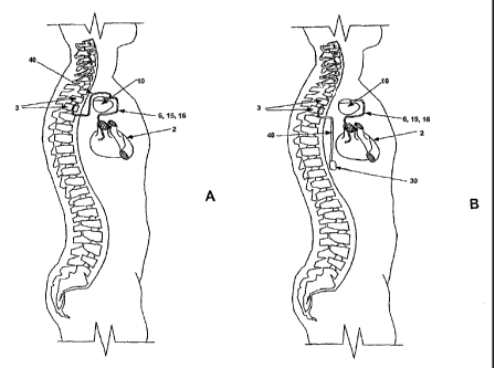
FIG. lA is a schematic illustration of an implantable cardioverter
defibrillator and
cardioversion pain inhibiting system implanted in a patient in accordance with
one
embodiment of the present invention.
FIG. 1B is a schematic illustration of an implantable cardioverter
defibrillator and
cardioversion pain inhibiting system implanted in a patient in accordance with
an
alternative embodiment of the present invention.
FIG. 2 is an illustration of an implantable cardioverter defibrillator (ICD)
that may
be included in the systems of FIGS. lA and 1B and a partially cut-away view of
a
patient's heart depicting placement of an associated cardiac lead system.
FIG. 3A is a functional block diagram of the ICD of FIG. 1A.
FIG. 3B is a functional block diagram of the ICD and PPI stimulation device
shown in FIG. 1B.
FIG. 4 is a flow diagram providing an overview of the operations included in a
preferred embodiment of the present invention for delivering a PPI stimulus
directly to the
central nervous system prior to a cardioversion shock.
The present invention is aimed at providing a system and method for
automatically
delivering a prepulse inhibition (PPI) stimulus directly to the central
nervous system to
reduce or eliminate cardioversion shock pain. FIG. lA is a schematic
illustration of an
CA 02504586 2005-05-02
WO 2004/041350 PCT/US2003/034059
implantable cardioverter defibrillator and cardioversion pain inhibiting
system implanted
in a patient in accordance with one embodiment of the present invention. The
system
includes a set of cardiac leads 6, 15, and 16 in communication with a
patient's heart 2, and
a spinal cord stimulation lead 40 in communication with the patient's spinal
cord 3. The
spinal cord stimulation (SCS) lead 40 may be provided as an epidural lead as
generally
described in commonly assigned U.S. Pat. No. 5,733,322 issued to Starkebaum
and U.S.
Pat. No. 6,308,103 issue to Gielen, both patents incorporated herein by
reference in their
entirety. Numerous types of spinal cord or epidural leads known for
stimulating the spinal
cord may be used successively with the present invention. Methods for
implanting an
epidural lead are generally disclosed in commonly assigned U.S. Pat. Nos.
5,255,691 and
5,360,441 issued to Otten, both patents incorporated herein by reference in
their entirety.
A SCS lead may include a plurality, e.g. four, spaced apart electrodes adapted
to be placed
in the epidural space adjacent to spinal segments. A PPI stimulus may be
optimally
effective in inhibiting cardioversion pain when delivered to the spinal cord
generally in the
region of the upper thoracic segments, such as spinal segments Tl and T2 as
approximately depicted in FIG. lA. The proximal end of SCS lead 40 is
connected to an
implantable cardioverter defibrillator device 10 that includes circuitry for
delivering a PPI
stimulus as will be described below.
FIG. 1B is a schematic illustration of an implantable cardioverter
defibrillator and
cardioversion pain inhibiting system implanted in a patient in accordance with
an
alternative embodiment of the present invention. Identically numbered
components in
FIG. 1B correspond to those in FIG. 1A, however, in the embodiment of FIG. 1B,
circuitry for delivering a PPI stimulus is contained in a separate implantable
device 30.
PPI stimulation device 30 is controlled by commands transmitted to device 30
from ICD
10 through a "body bus,".as will be described in greater detail below. SCS
lead 40 is
connected to PPI stimulation device 30. An advantage of including PPI
stimulation
circuitry in a separate device is that device 30 may be implanted at a site
different than
ICD 10 which may allow SCS lead 40 to be more easily implanted and tunneled to
PPI
stimulation device 30. The length of SCS lead 40 may be reduced depending on
the
location of device 30.
CA 02504586 2005-05-02
WO 2004/041350 PCT/US2003/034059
6
FIG. 2 is an illustration of an implantable cardioverter defibrillator (ICD)
that may
be included in the systems of FIGs. lA and 1B and a partially cut-away view of
a
patient's heart depicting placement of an associated cardiac lead system. A
connector
block 12 receives the proximal end of a right ventricular lead 16, a right
atrial lead 15 and
a coronary sinus lead 6, used for positioning electrodes for sensing and
stimulation in three
or four heart chambers. Connector block 12 includes a port 18 for receiving
SCS lead 40
for delivering a PPI stimulus directly to the spinal cord when PPI stimulus
circuitry is
included within ICD 10.
In FIG. 2, the right ventricular lead 16 is positioned such that its distal
end is in the
right ventricle for sensing right ventricular cardiac signals and delivering
pacing or
shocking pulses in the right ventricle. For these purposes, right ventricular
lead 16 is
equipped with a ring electrode 24, an extendable helix electrode 26 mounted
retractably
within an electrode head 28, and a coil electrode 20, each of which are
connected to an
insulated conductor within the body of lead 16. The proximal end of the
insulated
conductors are coupled to corresponding connectors carried by bifurcated
connector 14 at
the proximal end of lead 16 for providing electrical connection to the ICD 10.
The right atrial lead 15 is positioned such that its distal end is in the
vicinity of the
right atrium and the superior vena cava. Lead 15 is equipped with a ring
electrode 21 and
an extendable helix electrode 17, mounted retractably within electrode head
19, for
sensing and pacing in the right atrium. Lead 15 is further equipped with a
coil electrode
23 for delivering high-energy shock therapy. The ring electrode 21, the helix
electrode 17
and the coil electrode 23 are each connected to an insulated conductor with
the body of the
right atrial lead 15. Each insulated conductor is coupled at its proximal end
to a connector
carried by bifurcated connector 13.
The coronary sinus lead 6 is advanced within the vasculature of the left side
of the
heart via the coronary sinus and great cardiac vein. The coronary sinus lead 6
is shown in
the embodiment of FIG. 2 as having a defibrillation coil electrode 8 that may
be used in
combination with either the coil electrode 20 or the coil electrode 23 for
delivering
electrical shocks for cardioversion and defibrillation therapies. In other
embodiments,
coronary sinus lead 6 may also be equipped with a distal tip electrode and
ring electrode
for pacing and sensing functions in the left chambers of the heart. The coil
electrode 8 is
CA 02504586 2005-05-02
WO 2004/041350 PCT/US2003/034059
7
coupled to an insulated conductor within the body of lead 6, which provides
connection to
the proximal connector 4.
The electrodes 17 and 21 or 24 and 26 may be used for cardiac pacing as
bipolar
pairs, commonly referred to as a "tip-to-ring" configuration, or individually
in a unipolar
configuration with the device housing 11 serving as the indifferent electrode,
commonly
referred to as the "can" or "case" electrode. Housing 11 may also serve as a
can electrode
in combination with electrodes carried by SCS lead 40 for unipolar stimulation
of the
spinal cord. Housing 11 may also serve as a subcutaneous defibrillation
electrode in
combination with one or more of the defibrillation coil electrodes 8, 20 or 23
for
defibrillation of the atria or ventricles. It is recognized that alternate
lead systems may be
substituted for the three cardiac lead system illustrated in FIG. 2.
Although three or four-chamber pacing, cardioversion and defibrillation
capacity is
not necessary for practicing the invention, a multi-chamber system is
illustrated so as to
indicate the scope of the invention. It is understood that the invention may
normally be
practiced with a single chamber atrial or ventricular cardioversion device, a
dual chamber
cardioversion device, or a multichamber cardioversion device. The device may
include
pacemaking capabilities in addition to arrhythmia detection and cardioversion
therapy
capabilities.
A functional block diagram of the ICD 10 of FIG. lA is shown in FIG. 3A. This
diagram should be taken as exemplary of the type of device with which the
invention may
be embodied and not as limiting. 'The disclosed embodiment shown in FIG. 3A is
a
microprocessor-controlled device, but the methods of the present invention may
also be
practiced with other types of devices such as those employing dedicated
digital circuitry.
With regard to the electrode system illustrated in FIG. 2, the ICD 10 is
provided
with a number of connection terminals for achieving electrical connection to
the cardiac
leads 6, 15, and 16 and their respective electrodes. The connection terminal
311 provides
electrical connection to the housing 11 for use as the indifferent electrode
during unipolar
stimulation or sensing. The connection terminals 320, 310, and 318 provide
electrical
connection to coil electrodes 20, 8 and 23 respectively. Each of these
connection
terminals 311, 320, 310, and 318 are coupled to the high voltage output
circuit 234 to
CA 02504586 2005-05-02
WO 2004/041350 PCT/US2003/034059
8
facilitate the delivery of high energy shocking pulses to the heart using one
or more of the
coil electrodes 8, 20, and 23 and optionally the housing 11.
The connection terminals 317 and 321 provide electrical connection to the
helix
electrode 17 and the ring electrode 21 positioned in the right atrium. The
connection
terminals 317 and 321 are further coupled to an atrial sense amplifier 204 for
sensing atrial
signals such as P-waves. The connection terminals 326 and 324 provide
electrical
connection to the helix electrode 26 and the ring electrode 24 positioned in
the right
ventricle. The connection terminals 326 and 324 are further coupled to a
ventricular sense
amplifier 200 for sensing ventricular signals.
The atrial sense amplifier 204 and the ventricular sense amplifier 200
preferably
take the form of automatic gain controlled amplifiers with adjustable sensing
thresholds.
The general operation of the ventricular sense amplifier 200 and the atrial
sense amplifier
204 may correspond to that disclosed in U.S. Pat. No. 5,117,824, by Keimel, et
al.,
incorporated herein by reference in its entirety. Whenever a signal received
by atrial sense
amplifier 204 exceeds an atrial sensing threshold, a signal is generated on
the P-out signal
line 206. Whenever a signal received by the ventricular sense amplifier 200
exceeds a
ventricular sensing threshold, a signal is generated on the R-out signal line
202.
Switch matrix 208 is used to select which of the available electrodes are
coupled to
a wide band amplifier 210 for use in digital signal analysis. Selection of the
electrodes is
controlled by the microprocessor 224 via data/address bus 218. The selected
electrode
configuration may be varied as desired for the various sensing, pacing,
cardioversion and
defibrillation functions of the ICD 10. Signals from the electrodes selected
for coupling to
bandpass amplifier 210 are provided to multiplexer 220, and thereafter
converted to multi-
bit digital signals by A/D converter 222, for storage in random access memory
226 under
control of direct memory access circuit 228. Microprocessor 224 may employ
digital
signal analysis techniques to characterize the digitized signals stored in
random access
memory 226 to recognize and classify the patient's heart rhythm employing any
of the
numerous signal processing methodologies known in the art. A tachyarrhythmia
recognition system is described in U.S. Pat. No. 5,545,186 issued to Olson et
al.,
incorporated herein by reference in its entirety.
CA 02504586 2005-05-02
WO 2004/041350 PCT/US2003/034059
9
The telemetry circuit 330 receives downlink telemetry from and sends uplink
telemetry to an external programmer, as is conventional in implantable anti-
arrhythmia
devices, by means of an antenna 332. Data to be uplinked to the programmer and
control
signals for the telemetry circuit 330 are provided by microprocessor 224 via
address/data
bus 218. In accordance with the present invention, control parameters for
delivering a PPI
stimulus may be downloaded to device 10 from an external programmer via
telemetry
circuit 330. PPI stimulus control parameters may include the pulse amplitude
and width of
the PPI stimulus and the time interval between a PPI stimulus and a succeeding
cardioversion shock. Received telemetry is provided to microprocessor 224 via
multiplexer 220. Numerous types of telemetry systems known for use in
implantable
devices may be used.
Circuitry illustrated in FIG. 3A includes an exemplary embodiment of circuitry
dedicated to providing cardiac pacing, cardioversion and defibrillation
therapies. The
pacer timing and control circuitry 212 includes programmable digital counters
which
control the basic time intervals associated with various single, dual or mufti-
chamber
pacing modes or anti-tachycardia pacing therapies delivered in the atria or
ventricles.
Pacer circuitry 212 also determines the amplitude of the cardiac pacing pulses
under the
control of microprocessor 224.
During pacing, escape interval counters within pacer timing and control
circuitry
212 are reset upon sensing of R-waves or P-waves as indicated by signals on
lines 202 and
206, respectively. In accordance with the selected mode of pacing, pacing
pulses are
generated by atrial pacer output circuit 214 and ventricular pacer output
circuit 216. The
pacer output circuits 214 and 216 are coupled to the desired electrodes for
pacing via
switch matrix 208. The escape interval counters are reset upon generation of
pacing
pulses, and thereby control the basic timing of cardiac pacing functions,
including anti-
tachycardia pacing.
The durations of the escape intervals are determined by microprocessor 224 via
data/address bus 218. The value of the count present in the escape interval
counters when
reset by sensed R-waves or P-waves can be used to measure R-R intervals and P-
P
intervals for detecting the occurrence of a variety of arrhythmias.
CA 02504586 2005-05-02
WO 2004/041350 PCT/US2003/034059
The microprocessor 224 includes associated ROM in which stored programs
controlling the operation of the microprocessor 224 reside. A portion of the
random
access memory 226 may be configured as a number of recirculating buffers
capable of
holding a series of measured intervals for analysis by the microprocessor 224
for
5 predicting or diagnosing an arrhythmia. In response to the detection of
tachycardia, anti-
tachycardia pacing therapy can be delivered by loading a regimen from
microcontroller
224 into the pacer timing and control circuitry 212 according to the type of
tachycardia
detected.
In the event that higher voltage cardioversion or defibrillation pulses are
required,
10 microprocessor 224 activates the cardioversion and defibrillation control
circuitry 230 to
initiate charging of the high voltage capacitors 246 and 248 via charging
circuit 236 under
the control of high voltage charging control line 240. The voltage on the high
voltage
capacitors 246 and 248 is monitored via a voltage capacitor (VCAP) line 244,
which is
passed through the multiplexer 220. When the voltage reaches a predetermined
value set
by microprocessor 224, a logic signal is generated on the capacitor full (CF)
line 254,
terminating charging. The defibrillation or cardioversion pulse is delivered
to the heart
under the control of the pacer timing and control circuitry 212 by high
voltage output
circuit 234 via a control bus 238. The output circuit 234 determines the
electrodes used
for delivering the cardioversion or defibrillation pulse and the pulse wave
shape.
In accordance with the present invention, prior to delivering the
cardioversion
pulse, a PPI stimulus is delivered under the control of PPI timing and control
circuit 360.
PPI timing and control circuit 360 is in communication with microprocessor 224
via data
bus 218. When the voltage on VCAP line 244 reaches a predetermined value,
which may
be a value indicating the high voltage capacitors 246 and 248 are fully
charged or,
alternatively, are charged to a predetermined percentage of full charge, a PPI
stimulus may
be generated by PPI output circuit 362 under the control of timing and control
circuit 360.
PPI control circuit 360 determines the pulse width and pulse amplitude of the
PPI
stimulus, which may be programmable values received from telemetry circuit
330. The
PPI stimulus generated by PPI output circuit 362 is delivered directly to the
spinal cord via
SCS lead 40 connected to a terminal 350 provided for electrically coupling SCS
lead 40 to
device 10.
CA 02504586 2005-05-02
WO 2004/041350 PCT/US2003/034059
11
In alternative embodiments, a dedicated PPI output circuit 362 may be
eliminated,
and a PPI stimulus may be generated by either of pacing output circuits 214
and 216 or
high-voltage output circuit 234. Terminal 350 connected to SCS lead 40 may be
selectively coupled to of output circuits 214, 216 or 234 by switch matrix 208
at the
appropriate time for delivering a PPI stimulus. When either of pacing output
circuits 214
or 216 is used for delivering the PPI stimulus, both the PPI stimulus pulse
width and the
pulse amplitude may be selected, under the control of PPI timing and control
360, from the
settings available for atrial or ventricular pacing. When high-voltage output
circuit 234 is
used for delivering a PPI stimulus, the pulse amplitude will equal the
amplitude of a high-
voltage shock therapy, but the pulse width may be selected to be very narrow
such that the
PPI stimulus is weaker than the succeeding high-voltage shock. The use of high-
voltage
defibrillation circuitry for delivery of an atrial or ventricular prepulse is
generally
described in the previously incorporated '418 patent.
FIG. 3B is a functional block diagram of the ICD 10 and PPI stimulation device
30
shown in FIG. 1B. In FIG. 3B, identically numbered components correspond to
those in
FIG. 3A, however in FIG. 3B, PPI timing and control circuit 360 and PPI output
circuit
362 for delivering a PPI stimulus are removed from ICD 10 and included in
separate PPI
stimulation device 30. Device 30 preferably receives commands from ICD 10 via
a "body
bus," as generally disclosed in U.S. Pat. No. 4,987,897, issued to Funke,
incorporated
herein by reference in its entirety. ICD 10 is provided with a transmitter 150
and
transducer 152 for transmitting frequency modulated signals from ICD 10 to PPI
stimulation device 30. Modulated signals for transmission from ICD 10 to
device 30
include information relating to PPI stimulus pulse amplitude and width, which
information
is provided to transmitter 150 from microprocessor 224. Transmitted signals
are received
by transducer 364 of device 30 and demodulated by timing and control circuit
360.
Device 30 may optionally include a transmitter for transmitting signals back
to ICD 10.
Device 30 receives a PPI stimulus trigger command from ICD 10 at the
appropriate time
for delivering a PPI stimulus, prior to a cardioversion shock. The PPI
stimulus is
delivered by PPI output circuit 362 with a pulse width and amplitude set by
timing and
control circuit 360 based on commands received from ICD 10. The PPI stimulus
is
delivered directly to the central nervous system via terniinal 350 connected
to SCS lead 40
CA 02504586 2005-05-02
WO 2004/041350 PCT/US2003/034059
12
and terminal 351, which may be connected to the housing of device 30 for
serving as a can
electrode during unipolar PPI stimulation. Alternatively, terminal 351 may be
provided
for connection to one or more anode electrodes included on SCS lead 40 for
bipolar
stimulation of the spinal cord.
In FIG. 4 a flow diagram is shown providing an overview of the operations
included in a preferred embodiment of the present invention for delivering a
PPI stimulus
directly to the central nervous system prior to a cardioversion shock. At step
405, cardiac
signals are sensed to determine various intervals associated with P-waves and
R-waves by
pacer timing and control 212. Step 405 is executed continuously to monitor the
heart's
rhythm at all times, except for during blanking intervals applied to
ventricular and atrial
sense amplifiers 200 and 204 during pacing or shocking pulse delivery. If an
arrhythmia
is detected at decision step 410, an appropriate anti-arrhythmia therapy is
selected.
Depending on the type of arrhythmia detected, a cardioversion shock therapy
may not be
indicated. For example, when a tachycardia detection is made, programmed
therapies may
include tiered therapies beginning with anti-tachycardia pacing therapies
which are
attempted prior to delivering cardioversion shocks. If a cardioversion or
defibrillation
shock therapy is not indicated at decision step 415, the appropriate anti-
arrhythmia pacing
therapy is delivered at step 417. If the arrhythmia is terminated (as
determined at step
410), the method 400 returns to step 405 and continues monitoring the heart
rhythm.
If a cardioversion or defibrillation shock therapy is indicated in response to
a
detected arrhythmia, as determined at decision step 415, charging of the high
voltage
capacitors is initiated at step 420. After the capacitor charge has reached a
predetermined
PPI stimulus trigger value, as determined at step 425, microprocessor 224
verifies that an
arrhythmia is still being detected at decision step 430, and then triggers the
delivery of the
PPI stimulus at step 435. A PPI stimulus trigger is preferably generated upon
full
charging of the high-voltage capacitors such that the capacitors are ready to
deliver a
cardioversion shock after a short time delay, e.g. after less than 500 ms,
after a PPI
stimulus is delivered. Alternatively, the PPI stimulus trigger may be
generated once high-
voltage capacitors are charged to a certain percentage of full charge, for
example 90%
fully charged, so that by the time the PPI stimulus has been delivered and the
PPI-shock
delay period has elapsed, the capacitors are fully charged and the
cardioversion shock may
CA 02504586 2005-05-02
WO 2004/041350 PCT/US2003/034059
13
be immediately delivered. In this alternative embodiment, the PPI stimulus
would be
generated by either dedicated or pacing output circuitry, not the high-voltage
output
circuitry since high-voltage capacitors would still be charging during PPI
stimulus
delivery. As described above, a PPI stimulus may be delivered from output
circuitry
S included in device 10 according to the embodiment of FIG. 3A. Alternatively,
the PPI
stimulus trigger may generate a telemetry signal transmitted by ICD 10 to PPI
stimulation
device 30 which in turn triggers a PPI stimulus to be delivered from PPI
stimulation
device 30 according to the embodiment of FIG. 3B. The amplitude, duration, and
wave
shape of the PPI stimulus may be set according to fixed or programmable values
and may
be selected based on an individual patient's response. Generally monophasic or
biphasic
pulses or pulse trains could be utilized for a PPI stimulus. If the arrhythmia
has self
terminated during capacitor charging, as determined at decision step 430, the
method 400
returns to step 405 and continues monitoring the heart rhythm.
If arrhythmia detection is still occurring at step 430, the PPI stimulus is
delivered
at step 435. Timer and control circuitry 212 then sets a PPI-shock delay
interval that must
expire prior to delivering the cardioversion shock at step 445. The time
interval between
the PPI stimulus and the cardioversion shock may be fixed or programmable
according to
an individual patient's response. The time interval required for an optimal
PPI effect may
vary between approximately 20 and 500 ms, and is typically on the order of
approximately
100 ms.
After delivering the cardioversion shock at step 445, method 400 returns to
step
430 to determine if an arrhythmia is still detected. If so, steps 435 through
445 are
repeated. If the arrhythmia is successfully terminated, method 400 returns to
step 405 to
continue monitoring the heart rhythm.
Thus a system and method for delivering a prepulse inhibition stimulus
directly to
the central nervous system prior to a cardioversion shock therapy has been
disclosed. The
embodiments described herein are considered the preferred embodiments
contemplated to
date and are intended to be exemplary, not limiting, with regard to the
following claims.