Note: Descriptions are shown in the official language in which they were submitted.
CA 02504605 2012-11-30
WO 2004/043387
PCT/US2003/035777
-1-
Treatment Of Chronic Lymphocytic Leukemia And Prostate Cancer With
MicroRNA miR15
Reference to Government Grant
The invention described herein was supported in part by grant nos.
P01CA76259, P01CA81534, and P30CA56036 from the National Cancer
Institute. The U.S. government has certain rights in this invention.
Field of the Invention
The invention relates to the diagnosis of cancers, in particular to the
diagnosis of chronic lymphocytic leukemias or prostate cancer by detecting
miR15 and miR16 copy number, mutational status, or gene expression. The
invention also relates to the treatment of cancers, involving the reduction or
absence of miR15 or miR16 gene expression, in particular to the treatment of
chronic lymphocytic leukemias or prostate cancer by administering miR15 or
miR16 gene products.
Background of the Invention
Cancers are a significant source of mortality and morbidity in the U.S.
and throughout the world. In particular, chronic lymphocytic leukemia ("CLL")
and prostate cancer are clinically important neoplastic diseases of adult
humans.
CLL is the most common form of adult leukemia in the Western world. Also,
the age-adjusted incidence of prostate cancer now surpasses that of all other
cancers among men in the United States, and, after lung cancer, is the second
leading cause of all male cancer deaths in the country.
Hemizygous and/or homozygous loss at 13q14 occurs in more than half
of the reported CLL cases, and constitutes the most frequent chromosomal
abnormality in CLL. The karyotyping of tissue samples from CLL patients
identified relatively few chromosomal abnormalities, suggesting that the
CA 02504605 2005-05-02
WO 2004/043387
PCT/US2003/035777
-2-
specificity and frequency of observed deletions at 13q14 have pathologic
significance. In addition, 13q14 deletions also occur in 60% of prostate
cancers,
suggesting that one or more tumor suppressor genes located at 13q14 are
involved in the pathogenesis of both CLL and prostate cancers.
The presence of both clonal homozygous and heterozygous deletions,
and the very high frequency of 13q14 loss in CLL and prostate cancers,
indicates that deletions in this region are related to the etiology of certain
cancer
types. Several groups have used positional cloning in order to identify the
gene
or genes in the deleted areas. To date, a total of eight genes from the
deleted
regions of 13q14 in sporadic and familial cases of CLL have been identified
and
screened for alterations at the DNA and/or RNA level: Leul (BCMS or
EST70/Leul), Leu 2 (ALT1 or 1B4/Leu2), Leu 5 (CAR), CLLD6, KPNA3,
CLLD7, L0051131 (putative zinc finger protein NY-REN-34 antigen) and
CLLD8. However, detailed genetic analyses, including extensive loss of
heterozygosity (LOH), mutation and expression studies, have failed to
demonstrate the consistent involvement of any of these genes in
carcinogenesis.
Micro RNAs (miRNAs) are found in over one hundred distinct
organisms, including fruit flies, nematodes and humans. miRNAs are believed
to be involved in a variety of processes that modulate development in these
organisms. The miRNAs are typically processed from 60- to 70-nucleotide
foldback RNA precursor structures, which are transcribed from the miRNA
gene. The RNA precursor or processed miRNA products are easily detected,
and a lack of these molecules can indicate a deletion or loss of function of
the
corresponding miRNA gene.
Current therapies for CLL typically involve chemotherapy, administered
alone or in combination with autologous bone marrow transplantation. The
chemotherapy agents employed are generally toxic to the patient and cause only
partial remissions in a relatively large proportion of patients. Therapies for
prostate and other cancer therapies can also involve chemotherapy, often
following surgical resection of a tumor. However, as with CLL, the curative
properties of the chemotherapeutic agents (with or without surgery) are
limited.
CA 02504605 2005-05-02
WO 2004/043387
PCT/US2003/035777
-3-
Prostate cancer can also be treated with external beam radiation or
brachytherapy (e.g., with radioactive "seeds"), again either alone or in
combination with surgery. Such treatments risk exposing normal tissue of the
patient to the radiation, and may not be entirely effective.
There is a need for a rapid, economical and accurate diagnostic test for
CLL or prostate cancer. There is also a need for an economical and effective
treatment for cancers, especially CLL or prostate cancer, which does not have
a
significant negative impact on the patient.
Summary of the Invention
It has now been discovered that the miR15 or miR16 genes are localized
to 13q14 in humans, and that the 13q14 region is deleted in a significant
portion
of subjects suffering from CLL or prostate cancer. It has also been found that
the RNA products of the miR15 or miR16 genes inhibit the neoplastic or
tumorigenic growth of CLL and prostate cancer cells. The RNA products can
be used as a therapy for cancers which involve downregulation of the miR15 or
miR16 genes.
The miR15 and miR16 micro RNA genes are located at 13q14 within a
30 kb region of loss in CLL and prostate cancer, and both genes are deleted or
down-regulated in the majority of CLL and prostate cancer cases. Thus, the
invention provides a diagnostic test for CLL or prostate cancer comprising
detection of the gene product from these genes, detection of miR15 or miR16
gene copy number, or determination of the mutational status.
In one embodiment, the diagnostic test comprises isolating RNA from a
subject suspected of having CLL or prostate cancer, and detecting the levels
of
the miR15 or miR16 gene product by Northern blot hybridization using probes
for miR15 or miR16 RNA precursor or processed miRNA, wherein a reduction
in miR15 or miR16 precursor or processed microRNA as compared to a control
normal sample is diagnostic of CLL or prostate cancer.
In another embodiment, the diagnostic test comprises isolating DNA
from a subject suspected of having an miR15 or miR16 mediated cancer such as
CA 02504605 2005-05-02
WO 2004/043387 PCT/US2003/035777
-4-
CLL or prostate cancer, and detecting the miR15 or miR16 gene copy number
by Southern blot hybridization using probes for miR15 or miR16 gene
sequences, wherein a reduction in gene copy number to one or zero is
diagnostic
of CLL or prostate cancer.
In another embodiment, the diagnostic test comprises detecting a
reduction in miR15 or miR16 gene copy number by evaluating the loss of
heterozygosity of the D135273 and D135272 markers, wherein a loss of
heterozygosity at these markers is diagnostic of CLL or prostate cancer.
In a further embodiment, the diagnostic test comprises isolating DNA
from a subject suspected of having CLL or prostate cancer, and detecting
deletions or mutations in the miR15 or miR16 genes by PCR amplification of
miR15 or miR16 gene fragments and comparing the amplified fragments with
amplified fragments from a control normal sample, wherein the detection of a
mutation in one or more copies of the miR15 or miR16 genes is diagnostic of
CLL or prostate cancer. The amplified fragments can be compared by the single
stranded conformational polymorphism technique. In one aspect the mutation is
a partial deletion in the miR15 or miR16 gene sequences.
In another embodiment, the diagnostic test comprises isolating RNA
from a subject suspected of having CLL or prostate cancer and detection of a
mutation in miR15 or miR16 gene products is diagnostic of CLL or prostate
cancer.
In a further embodiment, the diagnostic test comprises isolating RNA
from a subject suspected of having CLL or prostate cancer, and detecting the
levels of the miR15 or miR16 gene product by amplification of the miR15 or
miR16 precursor or processed microRNA by reverse-transcriptase polymerase
chain reaction, wherein a reduction in miR15 or miR16 precursor or processed
microRNA as compared to an internal control amplified RNA is diagnostic of
CLL or prostate cancer.
The invention also provides a method of treating an miR15 or miR16
mediated cancer in a subject in need of such treatment, comprising
CA 02504605 2005-05-02
WO 2004/043387 PCT/US2003/035777
-5-
administering an effective amount of an miR15 or miR16 gene product to the
subject, such that proliferation of cancer cells in inhibited.
The invention also provides a method of treating miR15 or miR16
mediated cancer in a subject in need of such treatment, in which cells from
the
subject are isolated and transfected ex vivo with an effective amount a
nucleic
acid comprising sequences encoding the miR15 or miR16 gene product. The
expression of the miR15 or miR16 gene product in the transfected cells can be
confirmed. The cells are then reimplanted into the subject, and proliferation
of
cancer cells in the subject is inhibited.
The invention further provides a method of inhibiting proliferation of
miR15 or miR16 mediated cancer cells in a subject, comprising delivering to
the
cells an effective amount of an miR15 or miR16 gene product.
The invention still further provides a pharmaceutical composition for
treating a subject having miR15 or miR16 mediated cancer, comprising an
isolated miR15 or miR16 gene product, or a nucleic acid encoding an miR15 or
miR16 gene product, and a pharmaceutically acceptable carrier.
Brief Description of the Figures
FIGS. 1A and 1B are schematic representations of the predicted
secondary structure of the miR15 and miR16 precursor RNA, respectively. The
RNA secondary structure prediction was performed using the "mfold" program,
version 3.1 of Matthews et al. (1999), J. Mol. Biol. 288:911-940, and manually
refined to accommodate G/U wobble base pairs in the helical segments. The
sequence of the processed miR15 and miR16 miRNA is underlined. Adapted
from Lagos-Quintana et al. (2001), Science 294:853-858.
FIG. 2A is map of genes within the 13q14 tumor suppressor locus in
CLL showing the localization of the miR15/16 gene cluster. The position of
genetic markers and the position of genes on the map are shown.
FIG. 2B is a map of previously reported 13q14 deletions, marked by
horizontally striped boxes.
CA 02504605 2005-05-02
WO 2004/043387
PCT/US2003/035777
-6-
FIG. 2C is a map of the locus between the D1351150 and D135272
markers. The orientation of each gene in this locus is marked by an arrow
under
the gene name, and colored vertical bars mark the position of corresponding
exons for each gene.
FIG. 2D is a map of the locus between the Alu 18 and D135272
markers. Bars and boxes mark the position of exons for LEU2/ALT1 and LEUI.
The short vertical arrows mark the position of miR15 and miR16 genes. Circles
mark the position of PCR primers used to screen somatic cell hybrid clones
derived from a fusion of two independent leukemia cases (CLL-A and CLL-B).
Filled boxes represent portions of chromosome 13 present in the hybrids.
"<-31.4 kb¨>" indicates an approximately 31.4 kb deleted region in clone
CLL-A, which was derived from a patient with CLL carrying a t(2;13)(q12;q13)
translocation, bilateral retinoblastoma, and ulcerative colitis. The long
vertical
arrow represents the position of the breakpoint in clone CLL-B carrying a
t(2;13)(q32;q14) translocation, and "E-29 kb->" indicates an approximately 29
kb deleted region in this clone.
FIG. 3A is a Northern blot analysis of miR15 and miR16 gene
expression in normal human kidney, prostate, liver, skeletal muscle ("Sk
muscle"), testicle, CD5+ B cells (CD5+), leukemia cells ("Per B1 Leuk"), and
bone marrow ("BM").
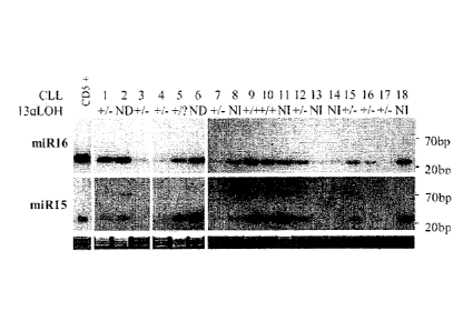
FIG. 3B is a loss of heterozygosity ("LOH") analysis of microsatellite
makers D135272 and D135273 in 18 CLL patients. DNA from normal human
CD5+ cells was used as a control. The LOH status for the samples is shown as
"-V+ heterozygosity," "+/- LOH," "-I- homozygous deletion," "NI" (not
informative), "?" (not enough material) and "ND" (not done). Ethidium
bromide-stained Northern gels were used as normalization controls.
Detailed Description of the Invention
All nucleic acid sequences herein are given in the 5' to 3' direction. In
addition, all deoxyribonucleotides in a nucleic acid sequence are represented
by
CA 02504605 2011-08-22
=
-7-
capital letters (e.g., deoxythymidine is "T"), and ribonucleotides in a
nucleic
acid sequence are represented by lower case letters (e.g., uridine is "u").
CLL or prostate cancer can be diagnosed by detecting a reduction in
miR15 or miR16 gene copy number, or by detecting mutations in one or more
copies of the miR15 or miR16 genes. A reduction in miR15 or miR16 gene
copy number from diploid to haploid, or to no copies, is diagnostic of CLL or
prostate cancer. Likewise, a mutation in one or both copies of the miR15 or
miR16 genes implies a loss of gene function, and is diagnostic of CLL or
prostate cancer.
As used herein, a "CLL cell" is a lymphocyte from a subject who has or
is suspected of having CLL, which lymphocyte has a "CLL Score" of at least 4
as determined according to the scoring system of Matutes et al. (1994),
Leukemia 8(10) :1640-1645'µ.
As used herein, a "prostate cancer cell" is a neoplastic or
tamorigenic cell of prostate origin, whether or not located in the prostate.
One
skilled in the art can readily identify a CLL or prostate cancer cell.
The miR15/miR16 gene cluster has been mapped to 13q14. The nucleic
acid sequences of these genes are contained within clone 317g11, the
nucleotide
sequence of which is given in GenBank record accession no. AC069475.
20A deletion
=
or mutation in the miR15 or miR16 genes can be detected by determining the
structure or sequence of these genes in tissue from a subject suspected of
having
CLL or prostate cancer, and comparing this with the structure or sequence of
these genes in a sample of unaffected tissue from the subject, or in a sample
of
tissue from a normal control. Such a comparison can be made by any suitable
technique.
According to the practice of the invention, to diagnose an miR15 or
miR16 mediated cancer, a tissue sample is derived from a subject. The sample
is then prepared for determination of miR15 or raiR16 gene product expression
or deletion or mutation of miR15 or miR16 genes. A tissue sample includes a
biopsy of interest, as well as blood and fluid samples.
CA 02504605 2005-05-02
WO 2004/043387
PCT/US2003/035777
-8-
As used herein, an "miR15 or miR16 mediated cancer" is any cancer in
which the expression of the miR15 or miR16 genes is reduced or absent in at
least a portion of tumorigenic or neoplastic cells associated with that
cancer.
Examples of miR15 or miR16 mediated cancers include CLL and prostate
cancer.
The presence of miR15 or miR16 deletions or mutations can be detected
by Southern blot hybridization of the genomic DNA from a subject, using
probes for miR15 or miR16 genes, e.g., as described below. For example, a
sample of tissue or blood can be removed from a subject suspected of having
CLL or prostate cancer by conventional biopsy techniques. Alternatively, a
blood sample can be removed from a subject suspected of having CLL or
prostate cancer, and white blood cells isolated for DNA extraction. The blood
or tissue sample is preferably obtained from the patient prior to initiation
of
radiotherapy or chemotherapy. A corresponding tissue or blood sample can be
obtained from unaffected tissues of the subject, or from a normal human
individual, for use as a control.
Southern blot hybridization techniques are within the skill in the art. For
example, the genomic DNA isolated from tissue or blood from a subject
suspected of having CLL or prostate cancer can be digested with restriction
endonucleases. This digestion generates restriction fragments of the genomic
DNA, which can be separated by electrophoresis, for example on an agarose gel.
The restriction fragments are then blotted onto a hybridization membrane
(e.g.,
nitrocellulose or nylon), and hybridized with labeled probes specific for the
miR15 or miR16 genes. A deletion or mutation of these genes is indicated by an
alteration of the restriction fragment patterns on the hybridization membrane,
as
compared to a control DNA sample which has been treated identically to the
DNA sample from the subject. Probe labeling and hybridization conditions
suitable for detecting miR15 or miR16 gene copy number or mutations can be
readily determined by one of ordinary skill in the art. The term "deletion,"
as
used herein, refers to partial deletion of a gene or to deletion of the entire
gene.
CA 02504605 2011-08-22
-9-
The miR15 and miR16 nucleic acid probes for Southern blot
hybridization can be designed based upon the published sequence of the miR15
and miR16 microRNAs as described in Lagos-Quintana et al. (2001), Science
294:853-858.
The nucleotide sequence of the miR15 ruicroRNA is uagcagcacanaauggtmugug
(SEQ ID NO:3). The nucleotide sequence of the miR16 microRNA is
uagcagcacg-uaaauauuggcg (SEQ ID NO:4). Suitable probes for detecting miR15
and miR16 DNA are, respectively:
CACAAACCATTATGTGCTTGCTA (SEQ ID NO:5)
GCCAATAITIACGTGCTGCTA (SEQ ID NO:6)
The complements of SEQ ID NO:5 and SEQ NO:6 can also be used
to probe for miR15 or miR16 DNA.
Methods for preparation of labeled DNA and RNA probes, and the
conditions for hybridization thereof to target nucleotide sequences, are
described
in Molecular Cloning: A Laboratory Mannal, J. Sambrook et al., eds., 2nd
edition, Cold Spring Harbor Laboratory Press, 1989, Chapters 10 and 11.
For example, the nucleic acid probe can be labeled with, e.g., a
, 32p, 33p,
radionuclide such as 3H or 35S; a
heavy metal; or a ligand capable
of functioning as a specific binding pair member for a labeled ligand (e.g.,
biotin, avidin or an antibody), a fluorescent molecule, a chemiluminescent
molecule, an enzyme or the like.
Probes can be labeled to high specific activity by either the nick
tranclation method of Rigby et al. (1977), J. Mol. Biol. 113:237-251 or by the
random priming method of Fienberg et al. (1983), Anal. Biochem. 132:6-13.
\The latter is
the method of choice for synthesizing 32P-labeled probes of high specific
activity from single-stranded DNA or from RNA templates. For example, by
replacing preexisting nucleotides with highly radioactive nucleotides
according
CA 02504605 2005-05-02
WO 2004/043387
PCT/US2003/035777
-10-
to the nick translation method, it is possible to prepare 32P-labeled nucleic
acid
probes with a specific activity well in excess of 108 cpm/microgram.
Autoradiographic detection of hybridization can then be performed by exposing
hybridized filters to photographic film. Densitometric scanning of the
photographic films exposed by the hybridized filters provides an accurate
measurement of miR15 or miR16 gene copy number. Alternatively, miR15 or
miR16 gene copy number can be quantified by computerized imaging systems,
such the Molecular Dynamics 400-B 2D Phosphorimager available from
Amersham Biosciences, Piscataway, NJ.
Where radionuclide labeling of DNA or RNA probes is not practical, the
random-primer method can be used to incorporate the dTTP analogue 5-(N-(N-
biotinyl-epsilon-aminocaproy1)-3-aminoallypdeoxyuridine triphosphate into the
probe molecule. The biotinylated probe oligonucleotide can be detected by
reaction with biotin-binding proteins such as avidin, streptavidin, or anti-
biotin
antibodies coupled with fluorescent dyes or enzymes which produce color
reactions.
Deletions or mutations of the miR15 or miR16 genes can also be
detected by amplifying a fragment of these genes by polymerase chain reaction
" (PCR), and analyzing the amplified fragment by sequencing or by
electrophoresis to determine if the sequence and/or length of the amplified
fragment from the subject's DNA sample is different from that of the control
DNA sample. Suitable reaction and cycling conditions for PCR amplification of
DNA fragments can be readily determined by one of ordinary skill in the art.
Exemplary PCR reaction and cycling conditions are given in the methods used
for the Examples, below.
Diagnosis of an miR15 or miR16 mediated cancer can be performed by
detecting deletions of 13q14 between various chromosome markers, such as the
markers indicated in FIGS. 1A, 1B, and 2A-2D. For example, a deletion in the
region of 13q14 between microsatellite markers D135272 and D13S273
comprising miR15 or miR16, indicates the presence of an miR15 or miR16
mediated cancer. In addition, when the deletion in 13q14 is between
CA 02504605 2011-08-22
-11-
microqFftllite markers D13S1150 and D138273 or between locus Alul 8 and
rnicrollite marker D13S273, where miR15 or miR16 are deleted, the
presence of an mal5 or miR16 mediated cancer is indicated.
An alternative method of determining the number of miR15 or miR16
-- genes per diploid genome in a sample of tissue relies on the fact that the
mal5/miR16 gene cluster is located in 13q14, and is linked to the markers
D135272 and D13S273. The loss of a copy of the miR15 or miR16 genes in an
individual who is heterozygous at a locus linked to the DI3S272 and. DI3S273
markers can be inferred from the loss of heterozygosity in these markers.
-- Methods for determining loss of heterozygosity of chromosomal markers are
within the skit] in the art. An exemplary loss of heterozygosity study is
given in
Example 3 below.
Another technique for determining whether the miR15 or miR16 genes
in a subject suspected of having CLL or prostate cancer are mutated is single
-- strand conformational polymorphism (SSCP), for example as described in
Orita
et al. (1989), Genomics 5: 874-879 and Hayashi (1991), PCR Methods and
Applic. 1: 34-38,.
,The SSCP technique consists of amplifying a fragment of the gene of
interest by PCR; denaturing the fragment and electrophoresing the two
denatured single strands under non-denaturing conditions. The single strands
assume, a complex sequence-dependent intrastrand secondary structure that
aflècz hestiauds electrophoretic mobility.
A deletion or mutation in. one or both miR15 or miR16 genes can also
cause a redaction in miR15 or miR16 gene expression. Thus, CLL or prostate
cancer can also be diagnosed by detecting expression levels of the RNA
produced from the raiR15 or miR16 genes, where a reduction in miR15 or
miR16 gene expression is diagnostic of CLL or prostate cancer.
The miR15 and miR16 genes are each transcribed to produce a -70 kb
precursor RNA which forms a stem-loop structure. The precursor RNA is not
translated into a protein, but is rather processed into a "micro RNA" or
"miRNA," which is believed to be the functional gene product.
CA 02504605 2005-05-02
WO 2004/043387
PCT/US2003/035777
-12-
As used herein, an "miR15 or miR16 gene product" means the processed
or unprocessed RNA transcripts from the miR15 or miR16 genes, as described
more fully below. The terms "RNA," RNA transcripts," and "gene product," are
used interchangeably herein in the context of miR15 or miR16 gene expression.
The miR15 and miR16 precursor RNAs are described in Lagos-Quintana
et al. (2001), Science 294, 853-858, the entire disclosure of which is
incorporated herein by reference. The sequences of the miR15 and miR16
precursor RNAs are given in SEQ ID NO:1 and SEQ ID NO:2. The predicted
stem-loop structures of SEQ ID NO:1 and SEQ ID NO:2, are shown in Figs. 1A
and 1B, respectively.
[SEQ ID NO:1]:
ccuuggaguaaaguagcagcacauaaugguuuguggauuuugaaaaggugcaggccauauu
gugcugccucaaaaauacaagg
[SEQ ID NO:2]:
gucagcagugccuuagcagcacguaaauauuggcguuaagauucuaaaauuaucuccaguau
uaacugug cugcugaagu aagguugac
Without wishing to be bound by any theory, it is believed that the miR15
and miR16 precursor RNAs are co-expressed from the miR15/miR16 gene
cluster, and are processed by the Dicer/Argonaute complex into the functional
miRNA products. See, e.g., Lee et al. (2001), Science 294:862. Both functional
miRNA products from these genes are single-stranded RNA molecules of 22
nucleotides in length which have a 5' terminal monophosphate and a 3' terminal
hydroxyl group. The nucleotide sequence of the processed miR15 microRNA is
uagcagcacauaaugguuugug (SEQ ID NO:3). The nucleotide sequence of the
processed miR16 microRNA is uagcageacguaaauauuggcg (SEQ ID NO:4),In the
practice of the invention, the 60-70 nt RNA precursor molecules produced from
the miR15 or miR16 genes can be detected. Alternatively, the shorter miR15
and miR16 microRNA gene products, which are produced through processing of
the precursor RNAs by the Dicer and Argonaute proteins, can be detected.
CA 02504605 2005-05-02
WO 2004/043387
PCT/US2003/035777
-13-
Methods for determining RNA expression levels are within the level of
skill in the art. For example, a sample of tissue or blood from a subject
suspected of having CLL or prostate cancer is obtained as described above. As
a control, a corresponding tissue or blood sample can be obtained from
unaffected tissues of the subject, or from a normal human individual as
described above. The control tissue or blood sample is then processed along
with the sample from the subject. The levels of miR15 or miR16 gene
expression in the subject can then be compared to those from unaffected tissue
from the subject, or to the miR15 or miR16 expression levels in tissue or
blood
from the normal control. For example, the relative miR15 or miR16 expression
level in CLL cells or cells of the sampled prostate tumor are conveniently
determined with respect to one or more standards. The standards may comprise,
for example, a zero expression level on the one hand and the expression level
of
the gene in normal tissue of the same patient, or the expression level in the
tissue of a normal control group on the other hand. The standard may also
comprise the miR15 or miR16 expression level in a standard cell line. The size
of the decrement in miR15 or miR16 expression in comparison to normal
expression levels is indicative of the future clinical outcome following
treatment.
Alternatively, the levels of miR15 or miR16 gene expression in a subject
suspected of having CLL or prostate cancer can be compared to average levels
of miR15 or miR16 gene expression previously obtained for a population of
normal human controls.
Suitable techniques for determining the level of RNA transcripts of a
particular gene in cells are well known to those skilled in the art. According
to
one such method, total cellular RNA can be purified from cells by
homogenization in the presence of nucleic acid extraction buffer, followed by
centrifugation. Nucleic acids are precipitated, and DNA is removed by
treatment with DNase and precipitation. The RNA molecules are then separated
by gel electrophoresis on agarose gels according to standard techniques, and
transferred to nitrocellulose filters by, e.g., the so-called "Northern"
blotting
CA 02504605 2011-08-22
-14-
technique. The RNA is then immobilized on the filters by heating. Detection
and quantification of specific RNA is accomplished using appropriately labeled
DNA or RNA probes complementary to the RNA in question. See, for example,
Molecular Cloning: A Laboratory Manual, J. Sambrook et al., eds., 2nd edition,
Cold Spring Harbor Laboratory Press, 1989, Chapter 7.
Suitable probes for Northern blot
hybridization of miR15 or miR16 RNA include SEQ ID NO:5 and SEQ ID
NO:6.
Autoradiographic detection of probe hybridization to raiR15 or miR16
RNA can be performed by exposing hybridized filters to photographic film.
Densitometric scanning of the photographic films exposed by the hybridized
filters provides an accurate measurement of RNA transcript levels.
Alternatively, RNA transcript levels can be qnantified by computerized imaging
of the hybridization blot, for example with the Molecular Dynamics 400-B 2D
Phosphorimager available from Amersham Biosciences, Piscataway, NJ.
In addition to Northern and other RNA blotting hybridization techniques,
the levels of RNA transcripts can be carried out according to the technique of
in
situ hybridization. This technique requires fewer cells than the Northern
blotting technique, and involves depositing whole cells onto a microscope
cover
slip and probing the nucleic acid content of the cell with a solution
containing
radioactive or otherwise labeled cDNA or cRNA probes. This technique is
particularly well-suited for analyzing tissue biopsy samples from subjects
suspected of having prostate cancer. The practice of the in situ hybridi7ation
technique is described in more detail in U.S. Pat. No. 5,427,916,.
Suitable probes for in
situ hybridization of taiR15 or miR16 RNA include SEQ ID NO:5 and SEQ ID
NO:6.
The relative number of miR15 or miR16 transcripts can also be
determined by reverse transcription of miR15 or miR16 transcripts, followed by
amplification in a polymerase chain reaction (RT-PCR). The levels of miR15 or
miR16 transcripts can be quantified in comparison with an internal standard,
for
CA 02504605 2005-05-02
WO 2004/043387
PCT/US2003/035777
-15-
example, levels of mRNA from a "housekeeping" gene present in the same
sample. A suitable "housekeeping" gene for use as an internal standard
includes
myosin or glyceraldehyde-3-phosphate dehydrogenase (G3PDH). The methods
for quantitative RT-PCR and variations thereof are well known to those of
ordinary skill in the art.
Other techniques for measuring miR15 and miR16 expression are also
known to those of skill in the art and include various techniques for
measuring
the rates of RNA transcription and degradation.
An miR15 or miR16 mediated cancer can be treated by administering the
isolated gene product of the miR15 or miR16 genes, either alone or in
combination, to an miR15 or miR16 mediated cancer cell. Without wishing to
be bound by any theory, it is believed that the miR15 or miR16 gene products
suppress the neoplastic or tumorigenic growth of such cancer cells.
In particular, CLL or prostate cancer can be treated by administering the
isolated gene product of the miR15 or miR16 genes, either alone or in
combination, to a CLL or prostate cancer cell.
As used herein, an "miR15 or miR16 mediated cancer cell" is a
tumorigenic or neoplastic cell isolated from a subject suffering from an miR15
or miR16 mediated cancer. An miR15 or miR16 mediated cancer cell can be
identified by detecting a reduction or absence of miR15 or miR16 gene products
in the cell, or by detecting a cancerous or neoplastic phenotype in the cell.
One
skilled in the art can readily identify cells with a cancerous or neoplastic
phenotype. For example, such cells are insensitive to contact-induced growth
inhibition in culture, and will form foci when cultured for extended periods.
Cancerous or neoplastic cells also exhibit characteristic morphological
changes,
disorganized patterns of colony growth and acquisition of anchorage-
independent growth. Cancerous or neoplastic cells also have the ability to
form
invasive tumors in susceptible animals, which can be evaluated by injecting
the
cells, for example, into athymic mice using techniques within the skill in the
art.
As used herein, an "isolated" gene product is one which is altered or
removed from the natural state through human intervention. For example, an
CA 02504605 2005-05-02
WO 2004/043387
PCT/US2003/035777
-16-
RNA naturally present in a living animal is not "isolated." A synthetic RNA,
or
an RNA partially or completely separated from the coexisting materials of its
natural state, is "isolated." An isolated RNA can exist in substantially
purified
form, or can exist in a cell into which the RNA has been delivered. Thus, an
miR15 or miR16 gene product which is deliberately delivered to or expressed in
a cell, such as a CLL or prostate cancer cell, is considered an "isolated"
gene
product.
The miR15 and miR16 gene products can be obtained using a number of
standard techniques. For example, the gene products can be chemically
synthesized or recombinantly produced using methods known in the art.
Preferably, the RNA products are chemically synthesized using appropriately
protected ribonucleoside phosphoramidites and a conventional DNA/RNA
synthesizer. Commercial suppliers of synthetic RNA molecules or synthesis
reagents include Proligo (Hamburg, Germany), Dharmacon Research (Lafayette,
CO, USA), Pierce Chemical (part of Perbio Science, Rockford, IL, USA), Glen
Research (Sterling, VA, USA), ChemGenes (Ashland, MA, USA) and
Cruachem (Glasgow, UK).
Alternatively, the miR15 and miR16 gene products can be expressed
from recombinant circular or linear DNA plasmids using any suitable promoter.
Suitable promoters for expressing RNA from a plasmid include the U6 or H1
RNA pol III promoter sequences, or the cytomegalovirus promoters. Selection
of other suitable promoters is within the skill in the art. The recombinant
plasmids of the invention can also comprise inducible or regulatable promoters
for expression of the miR15 and miR16 gene products in CLL, prostate cancer,
or other cells.
The miR15 and miR16 gene products which are expressed from
recombinant plasmids can be isolated from cultured cell expression systems by
standard techniques. The miR15 and miR16 gene products which are expressed
from recombinant plasmids can also be delivered to and expressed directly in
the CLL or prostate cancer cells. The use of recombinant plasmids to deliver
CA 02504605 2011-08-22
-17-
the miR15 and miR16 gene products to CLL or prostate cancer cells is discussed
in more detail below.
The miR15 and miR16 gene products can be expressed from a separate
recombinant plasmid, or can be expressed from the same recombinant plasmid.
Preferably, the miR15 and miR16 gene products are expressed as the RNA
precursor molecules from a single plasinid, and the precursor molecules are
processed into the functional miRNA molecules by a suitable processing
system. Suitable processing systems include the in vitro Drosophila cell
lysate
system as described in U.S. published application 2002/0086356 of Tuschl et
al.,
Selection of plasmids suitable for expressing the miR15 and miR16 gene
products, methods for inserting nucleic acid sequences for expressing the gene
products into the plasmid, and methods of delivering the recombinant plasmid
to
the cells of interest are within the skill in the art. See, for example Zeng
et al.
(2002), Molecular Cell 9:1327-1333; Tuschl (2002), Nat. Biotechnol, 20:446-
448; Brummellcamp et al. (2002), Science 296:550-553; Miyagishi et al. (2002),
Nat. Biotechnol. 20:497-500; Paddison et al. (2002), Genes Dev. 16:948-958;
Lee et al. (2002), Nat. Biotechnol. 20:500-505; and Paul et al. (2002), Nat.
Biotechnol. 20: 505-508.
In a preferred embodiment, a plasmid expressing the miR15 or rniR16
gene products comprises a sequence encoding the miR15 or miR16 precursor
RNA under the control of the CMV intermediate-early promoter. As used
herein, "under the control" of a promoter means that the nucleic acid
sequences
encoding the miRNA product are located 3' of the promoter, so that the
promoter can. initiate transcription of the miRNA product coding sequences.
The miR15 or miR16 gene products can also be expressed from
recombinant viral vectors. It is contemplated that the miR15 and miR16 gene
products can be expressed from two separate recombinant viral vectors, or from
the same viral vector. The RNA expressed from the recombinant viral vectors
can either be isolated from cultured cell expression systems by standard
CA 02504605 2011-08-22
-18-
techniques, or can be expressed directly in CLL or prostate cancer cells. The
use of recombinant viral vectors to deliver the miR15 or miR16 gene products
to
CLL or prostate cancer cells is discussed in more detail below.
The recombinant viral vectors of the invention comprise sequences
encoding the miR_15 and miR16 gene products and any suitable promoter for
expressing the RNA sequences. Suitable promoters include, for exam'ple, the
U6 or H1 RNA pol HI promoter sequences, or the cytomegalovirus promoters.
Selection of other suitable promoters is within the skill in the art. The
recombinant viral vectors of the invention can also comprise inducible or
regulatable promoters for expression of the miR15 and raiR16 gene products in
a CLL or prostate cancer cell.
Any viral vector capable of accepting the coding sequences for the
miR15 and miR16 gene products can be used; for example, vectors derived from
adenovirus (AV); adeno-associated virus (AAV); retroviruses (e.g.,
lentiviruses
.15 (LV), Rhabdoviruses, murine leukemia virus); herpes virus, and the
like. The
tropism of the viral vectors can also be modified by pseudotyping the vectors
with envelope proteins or other surface antigens from other viruses. For
example, an AAV vector of the invention can be pseudotyped with surface
proteins from vesicular, stomatitis virus (VSV), rabies, Ebola, Mokola, and
the
like. Selection of recombinant viral vectors suitable for use in the
invention,
methods for inserting nucleic acid sequences for expressing RNA into the
vector, methods of delivering the viral vector to the cells of interest, and
recovery of the expressed RNA products are within the skill in the art. See,
for
example, Dornburg (1995), Gene Therap. 2:301-310; Eglifis (1988),
Biotechniques 6:608-614; Miller (1990), Hum. Gene Therap. 1:5-14; and
Anderson (1998), Nature 392:25-30.
Preferred viral vectors are those derived from AV and AAV. A suitable
AV vector for expressing the miRNA of the invention, a method for
constructing the recombinant AV vector, and a method for delivering the vector
into target cells, are described in Xia et al. (2002), Nat. Biotech. 20:1006-
1010.
=
CA 02504605 2011-08-22
-19-
, Suitable
AAV vectors for expressing the miRNA of the invention, methods for
constructing the recombinant AAV vector, and methods for delivering the
vectors into target cells are described in Samulsld et al. (1987), J. Virol.
61:3096-3101; Fisher et al. (1996), J. Virol., 70:520-532; Samulski et al.
(1989),
J. Viral. 63:3822-3826; U.S. Pat. No. 5,252,479; U.S. Pat No. 5,139,941;
International Patent Application No. WO 94/13788; and International Patent
Application No. WO 93/24641.
Preferably, the miR15 and miR16 gene products are
expressed from a single recombinant AAV vector comprising the CMV
intermediate early promoter.
In one embodiment, a recombinant AAV viral vector of the invention
comprises a nucleic acid sequence encoding the miR15 or miR16 precursor
RNA in operable connection with a polyT termination sequence under the
control of a hnman U6 RNA promoter. As used herein, "in operable connection
with a polyT termination sequence" mesns that the nucleic acid sequences
encoding the sense or antisense strands are immediately adjacent to the polyT
termination signal in the 5' direction. During transcription of the miR15 or
miR16 sequences from the vector, the polyT termination signals act to
terminate
transcription.
In the practice of the invention, the miR15 or miR16 gene products are
used to inhibit the neoplastic or tumorigenic growth of miR15 or miR16
mediated cancer cells, in particular of CLL or prostate cancer cells. Without
wishing to be bound by any theory, it is believed that the processed miRl5 and
miR16 miRNAs bind to complementary sequences in one or more target
mRNAs which are necessary to initiate and/or maintain neoplastic or
tumorigenic growth in these cells. Thus, the invention provides a method of
treating an miR15 or miR16 mediated cancer, for example CLL or prostate
cancer, in a subject in need of such treatment. The method comprises
administering an effective amount of an miR15 or miR16 gene product to the
CA 02504605 2005-05-02
WO 2004/043387
PCT/US2003/035777
-20-
subject, such that proliferation of miR15 or miR16 mediated cancer cells is
inhibited.
As discussed above, an miR15 or miR16 mediated cancer is a cancer in
which expression of either or both the miR15 or miR16 genes is reduced or
absent in at least a portion of tumor or neoplastic cells associated with the
disease. Expression of the miR15 or miR16 genes is reduced or absent in tumor
or neoplastic cells from CLL or prostate cancer; thus, CLL and prostate cancer
are considered to be miR15 or miR16 mediated cancers. A reduction or absence
of miR15 or miR16 gene expression may also be found in other cancers; such
cancers would therefore also be considered miR15 or miR16 mediated cancers.
For example, expression of the miR15 or miR16 genes may be reduced
or absent in primary or metastatic tumor or neoplastic cells from cancers of
at
least the following histologic subtypes: sarcoma (cancers of the connective
and
other tissue of mesodermal origin); melanoma (cancers deriving from pigmented
melanocytes); carcinoma (cancers of epithelial origin); adenocarcinoma
(cancers
of glandular epithelial origin); cancers of neural origin (glioma/glioblastoma
and
astrocytoma); and hematological neoplasias, such as leukemias and lymphomas
(e.g., acute lymphoblastic leukemia and chronic myelocytic leukemia).
Expression of the miR15 or miR16 genes may also be reduced or absent
in cancers having their origin in at least the following organs or tissues,
regardless of histologic subtype: breast; tissues of the male and female
urogenital system (e.g. ureter, bladder, prostate, testis, ovary, cervix,
uterus,
vagina); lung; tissues of the gastrointestinal system (e.g., stomach, large
and
small intestine, colon, rectum); exocrine glands such as the pancreas and
adrenals; tissues of the mouth and esophagus; brain and spinal cord; kidney
(renal); pancreas; hepatobiliary system (e.g., liver, gall bladder); lymphatic
system; smooth and striated muscle; bone and bone marrow; skin; and tissues of
the eye.
Expression of the miR15 or miR16 genes may also be reduced or absent
in cancers or tumors in any prognostic stage of development, for example as
measured by the "Overall Stage Groupings" (also called "Roman Numeral") or
CA 02504605 2011-08-22
-21-
the "Tumor, Nodes, and Metastases" (TNM) staging systems. Appropriate
prognostic staging systems and stage descriptions for a given cancer are known
in the art, for example as described in the National Cancer Tnstitute's
"CancerNet" Internet website.
A subject in need of treatment for an miR15 or miR16 mediated cancer
can be identified by obtaining a sample of tumor or neoplastic cells (or cells
suspected of being tumor or neoplastic) from the subject, and determining
whether the expression of miR15 or miR16 is reduced or absent in at least a
portion of the cells as compared to cells from normal tissue obtained from the
subject. Methods for detecting miR15 or raiR16 gene expression levels in cells
are within the skill in the art (see above). Alternatively, the miR15 or miR16
expression in cells obtained from a subject can be compared to average
expression levels of these genes in cells obtained from a population of normal
subjects. A subject in need of treatment for CLL can be readily identified by
a
physician using standard diagnostic techniques. See, e.g., "Chronic
lymphocytic
leukemia: recommendations for diagnosis, staging, and response criteria.
International Workshop on Chronic L3rmphocytic Leukemia," (1989) Annals of
Internal Medicine 110(3):236-238,.
For example, subjects with CLL exhibit circulating
CLL cells, lymphocytosis (i.e., lymphocyte counts in the blood equal to or
higher than 10,000 cells per cubic millimeter), and a progressive accumulation
of CLL cells in the bone marrow and lymphatic tissues.
The identity of CLL cells in a subject's blood or other tissue can be
confirmed by direct visual observation of a blood sample, and/or by
determining
the "CLL Score" of lymphocytes. The CLL Score indicates the presence or
absence of five lymphocyte surface markers characteristic of CLL cells: CD5+,
CD23+, FMC7-, and weak expression (+/-) of surface immunoglobulin (SmIg)
and CD22. This scoring system gives a value of 1 or 0 for each of these five
markers according to whether it is typical or atypical for CLL. CLL cells have
a
CLL Score 4 or 5, while lymphocytes from other leukemias have a CLL Score
of <1 to 3. See Matutes et al. (1994), Leukemia 8(10):1640-1645 and Moreau et
CA 02504605 2011-08-22
6
_ .
-22-
al. (1997), American Journal of Clinical Pathology, 108:378-8Z.
CLL cells also have
relatively low levels of surface-membrane immunoglobnlin as compared with
normal peripheral blood B cells. Surface-membrane immunoglobulin levels on
lymphocytes can be readily detected according to standard techniques; see,
e.g.,
Roilitan et al. (1995), New England Journal of Medicine 333:1052-1057.
A subject in need of treatment for prostate cancer can also be readily
identified by a physician according to standard diagnostic techniques, using
criteria such as patient age, detection of an enlarged prostate by digital
rectal
exam, prostate-specific antigen ("PSA") level, Gleason score of biopsy
material,
and the presence of immunohistochemically detectable genetic markers such as
p53, p21, and cyclin.s on cells from prostate tissue. A serum PSA level of 20
ng/ml or greater and a poorly differentiated prostate tissue histology (e.g.,
Gleason score 8 or higher) is indicative of prostate cancer. The presence of
prostate tumors in a subject can also be confirmed by non-invasive imaging
techniques, such as CT scan, tansrectal ultrasound of the prostate ("TRUSP"),
and magnetic resonance imaging ("MRI"), as are known in the art.
As used herein, an "effective amount" of miR15 or miR16 gene products
is an amount sufficient to inhibit proliferation of an miR15 or miR16 mediated
cancer cell in a subject suffering from an miR15 or miR16 mediated cancer. For
example, an effective amount of miR15 or miR16 gene products can be an
amount sufficient to inhibit proliferation of CLL cells in a subject suffering
from
CLL, or inhibit proliferation of prostate cancer cells in a subject suffering
from
prostate cancer. It is understood that a "prostate tumor cell" is not
necessarily
located in the prostate, but includes cells from metastatic tumors of prostate
origin.
As used herein, to "inhibit the proliferation of an raiR15 or miR16
mediated cancer cell" means to kill the cell, or permanently or temporarily
arrest
the growth of the cell. Inhibition of miR15 or miR16 mediated cancer cell
proliferation can be inferred if the number of such cells in the subject
remains
CA 02504605 2005-05-02
WO 2004/043387
PCT/US2003/035777
-23-
constant or decreases after administration of the miR15 or miR16 gene
products.
An inhibition of miR15 or miR16 mediated cancer cell proliferation can also be
inferred if the absolute number of such cells increases, but the rate of tumor
growth decreases.
The number of miR15 or miR16 mediated cancer cells in a subject's
body can be determined by direct measurement, or by estimation from the size
of primary or metastatic tumor masses.
For example, the number of CLL cells in a subject can be readily
determined, for example by a whole blood or white blood cell count. The
number of CLL cells can also be readily determined by immunohistological
methods, flow cytometry, or other techniques designed to detect the
characteristic surface markers of CLL cells.
The size of a prostate tumor mass or a tumor mass can be ascertained by
direct visual observation, or by diagnostic imaging methods such as X-ray,
magnetic resonance imaging, ultrasound, and scintigraphy. Diagnostic imaging
methods used to ascertain size of the tumor mass can be employed with or
without contrast agents, as is known in the art. The size of a tumor mass can
also be ascertained by physical means, such as palpation of the tissue mass or
measurement of the tissue mass with a measuring instrument such as a caliper.
For prostate tumors, a preferred physical means for determining the size of a
tumor mass is the digital rectal exam.
One skilled in the art can readily determine an effective amount of the
miR15 or miR16 gene products to be administered to a given subject, by taking
into account factors such as the size and weight of the subject; the extent of
disease
penetration; the age, health and sex of the subject; the route of
administration; and
whether the administration is regional or systemic.
For example, an effective amount of the compounds of the invention can
be based on the approximate weight of a tumor mass to be treated. The
approximate weight of a tumor mass can be determined by calculating the
approximate volume of the mass, wherein one cubic centimeter of volume is
roughly equivalent to one gram. An effective amount of the miR15 or miR16
CA 02504605 2005-05-02
WO 2004/043387
PCT/US2003/035777
-24-
gene products based on the weight of a tumor mass can be at least about 10
micrograms/gram of tumor mass, and is preferably between about 10-500
micrograms/gram of tumor mass. More preferably, the effective amount is at
least about 60 micrograms/gram of tumor mass. Particularly preferably, the
effective amount is at least about 100 micrograms/gram of tumor mass. It is
preferred that an effective amount based on the weight of the tumor mass be
injected directly into the tumor.
An effective amount of the miR15 or miR16 gene products can also be
based on the approximate or estimated body weight of a subject to be treated.
Preferably, such effective amounts are administered parenterally or enterally,
as
described below. For example, an effective amount of the miR15 or miR16
gene products administered to a subject can range from about 5 ¨ 3000
micrograms/kg of body weight, and is preferably between about 700 - 1000
micrograms/kg of body weight, and is more preferably greater than about 1000
micrograms/kg of body weight.
One skilled in the art can also readily determine an appropriate dosage
regimen for the administration of the miR15 and miR16 gene products to a given
subject. For example, the miR15 and miR16 gene products can be administered
to the subject once (e.g., as a single injection or deposition).
Alternatively, the
gene products can be administered once or twice daily to a subject for a
period of
from about three to about twenty-eight days, more preferably from about seven
to
about ten days. In a preferred dosage regimen, the miR15 and miR16 gene
products are administered once a day for seven days. Where a dosage regimen
comprises multiple administrations, it is understood that the effective amount
of
the miR15 and miR16 gene products administered to the subject can comprise the
total amount of gene product administered over the entire dosage regimen.
The miR15 and miR16 gene products can be administered to a subject by
any means suitable for delivering the gene products to cells of the subject,
such
as hematopoietic stem cells (HSCs), CLL cells or prostate cancer cells. For
example, the miR15 and miR16 gene products can be administered by methods
suitable to transfect cells of the subject with miR15 or miR16 gene products,
or
CA 02504605 2011-08-22
=
-25-
with nucleic acids coirising sequences encoding the miR15 or miR16 gene
products. The cells c.7-1 be transfected directly with the miR15 or miR16 gene
products (as these are nucleic acids), or can be transfected with nucleic
acids
comprising sequences encoding the miR15 or miR16 gene products. Preferably,
the cells are transfected with a plasraid or viral vector comprising sequences
encoding the miR15 or miR16 gene products, as described above.
Transfection methods for eukaryotic cells are well known in the art, and
include direct injection of the nucleic acid into the nucleus or pronucleus of
a
cell; electroporation; liposome transfer or transfer mediated by lipophilic
materials; receptor mediated nucleic acid delivery, bioballistic or particle
acceleration; calcium phosphate precipitation, and transfection mediated by
viral
vectors.
For example, cells can be transfected with a liposomal transfer
compound, e.g., DOTAP (N41-(2,3-dioleoyloxy)propyll-N,N,N-trimethyl-
ammonium methylsulfate, Boehringer - Mannheim) or an equivalent, such as
LIPOFECTIN. The amount of nucleic acid used is not critical to the practice of
the invention; acceptable results may be achieved with 0.1-100 micrograms of
nucleic acid/105 cells. For example, a ratio of about 0.5 micrograms of
plasmid
vector in 3 micrograms of DOTAP per 105 cells can be used.
In one embodiment, miR15 or miR16 mediated cancer cells, for example
CLL or prostd_e --ncter cells, are isolated from the subject, transfected with
nucleic acids MnalT2 the naiR15 and miR16 gene products, and reintroduced
into the subject In a preferred embodiment, the transfected and reimplanted
cells are CLL rTts In a particularly preferred embodiment, the transfected and
reimplanted cells are HSCs from a subject who has been diagnosed with CLL.
Techniques for isolating CLL cells from a subject are within the skill in
the art, for example as described in Example 7 below. Techniques for
isolating,
identifying, separating, and culturing HSCs from a subject are also within the
skill in the art, for example as disclosed in U.S. Pat. Nos. 5,635,387 and
5,643,741, and Campana et al. (1995) Blood 85:1416-1434.
Preferably, harvested
CA 02504605 2011-08-22
_
-26-
bone marrow is purged of tumorigerlic or neoplastic cells prior to
transfection of
the HSCs. Suitable purging techniques include, for exRmple, leukopheresis of
mobili7ed peripheral blood cells, immunoaffinity-based selection or killing of
tumor cells, or the use of cytotoxic or photosensitizing agents to selectively
kill
tumor cells, as are km own in the art. See, for example, Bone Marrow
Processing
and Purging, Part 5 (A. Gee, ed.), CRC Press, Boca Raton, Fla., 1991; Lydald
et
al. (1996) J. Photochem. and Photobiol. 32:27-32; and Gazitt et al. (1995),
Blood, 86:381-389.
The isolated CLL cells or HSCs can be trRnsfected by any suitable
technique, as discussed above. After transfection, a portion of the CLL cells
or
HSCs can be examined to confirm the presence of appropriate expression levels
of the gene products. Once appropriate expression of the miR15 or iniR16 gene
products has been confirmed, the remaining transfected cells can then be
reintroduced into the subject Transfected CLL cells or HSCs can be
reintroduced into the subject by parenteral methods, including intravenous
infusion or direct injection into the bone marrow. The transfected cells are
preferably reintroduced into the subject in a saline solution or other
pharmaceutically acceptable carrier. A suitable number of transfected cells
for
reintroduction is from about 105 to about 108 cells per kilogram of subject
body
weight. The number of transfected cells available for re-introduction can be
increased by expanding the cells in culture prior to transfection.
Preferably, the CLL cells or HSCs are transfected with a nucleic acid
comprising sequences which encode an miR15 or miR16 gene product, e.g. a
plasmid expression vector, that stably integrates into the CLL cell or HSC
genome to provide long-term expression of the compound. Stable integration
and expression can be confirmed by techniques known in the art, such as a
Southern blot of genomic DNA using miR15 or miR16 cDNA (or fragments
thereof) as a probe. Expression of miR15 or miR16 gene products can also be
detected by standard Northern blot techniques. The CLL cells or HSCs stably
transfected with sequences encoding the miR15 or miR16 gene products
CA 02504605 2005-05-02
WO 2004/043387
PCT/US2003/035777
-27-
continue to express the compound once they are re-implanted into the subject.
An exemplary method of isolating HSC from a subject, transfecting them with
plasmids expressing miR15 and miR16 gene products, and reimplanting the
transfected HSC into the subject is given in Example 8 below.
The miR15 and miR16 gene products can also be administered to a
subject by any suitable enteral or parenteral administration route. Suitable
enteral administration routes for the present methods include oral, rectal, or
intranasal delivery.
Suitable parenteral administration routes include
intravascular administration (e.g. intravenous bolus injection, intravenous
infusion, intra-arterial bolus injection, intra-arterial infusion and catheter
instillation into the vasculature); pen- and intra-tissue injection (e.g.,
peri-
tumoral and intra-tumoral injection, intra-retinal injection, or subretinal
injection); subcutaneous injection or deposition including subcutaneous
infusion
(such as by osmotic pumps); direct application to the tissue of interest, for
example by a catheter or other placement device (e.g., a retinal pellet or a
suppository or an implant comprising a porous, non-porous, or gelatinous
material); and inhalation. Preferably, the miR15 and miR16 gene products are
administered by injection or infusion. For the treatment of miR15 or miR16
mediated cancers which involve solid tumors, the miR15 and miR16 gene
products are preferably administered by direct injection into the tumor.
In the present methods, the miR15 and miR16 gene products can be
administered to the subject either as naked RNA, in conjunction with a
delivery
reagent, or as a nucleic acid (e.g,, a recombinant plasmid or viral vector)
comprising sequences which expresses the gene product. Suitable delivery
reagents for administration of the miR15 and miR16 gene products include the
Minus Transit TKO lipophilic reagent; lipofectin; lipofectamine; cellfectin;
or
polycations (e.g., polylysine), or liposomes.
Recombinant plasmids comprising sequences which express the miR15
or miR16 gene products are discussed above. Recombinant viral vectors
comprising sequences which expresses the miR15 or miR16 gene products are
CA 02504605 2011-08-22
-28-
also discussed above, and methods for delivering such vectors to CLL or
prostate cancer cells of a subject are within the skill in the art.
In a preferred embodiment, liposomes are used to deliver the miR15 or
miR16 gene products, or nucleic acids comprising sequences encoding the gene
products, to a subject Liposomes can also increase the blood half-life of the
gene products or nucleic acids. In the practice of this embodiment of the
invention, the miR15 or miR16 gene products, or nucleic acids comprising
sequences encoding the gene products, are encapsulated in liposomes prior to
administration to the subject.
Liposomes suitable for use in the invention can be formed from standard
vesicle-forming lipids, which generally include neutral or negatively charged
phospholipids and a sterol such as cholesterol. The selection of lipids is
generally guided by consideration of factors such as the desired liposome size
and half-life of the liposomes in the blood stream. A variety of methods are
known for preparing liposomes, for example as described in Szoka et al.
(1980),
Ann. Rev. Biophys. Bioeng. 9:467; and U.S. Pat. Nos. 4,235,871, 4,501,728,
4,837,028, and 5,019,369..
The liposomes encapsulating the miR15 or miR16 gene products, or
nucleic acids comprising sequences encoding the gene products, can comprise a
ligand molecule that targets the liposome to an miR15 or miR16 mediated
cancer cell, such as a CLL or prostate cancer cell. Ligands which bind to
receptors prevalent in such cancer cells, such as monoclonal antibodies that
bind
to tumor cell antigens or CLL cell surface markers, are preferred.
The liposomes encapsulating the miR15 or miR16 gene products, or
nucleic acids comprising sequences encoding the gene products, can also be
modified so as to avoid clearance by the mononuclear macrophage system
("MMS") and reticuloendothelial system ("RES"). Such modified liposomes
have opsonization-inhibition moieties on the surface or incorporated into the
liposome structure. In a particularly preferred embodiment, a liposome of the
invention can comprise both opsonization-inhibition moieties and a ligand.
CA 02504605 2011-08-22
-29-
Opsonization-inhibiting moieties for use in preparing the liposomes of
the invention are typically large hydrophilic polymers that are bound to the
liposome membrane. As used ,herein, an opsoni7ation inhibiting moiety is
"bound" to a liposome membrane when it is chemically or physically attached to
the membrane, e.g., by the intercalation of a lipid-soluble anchor into the
membrane itself, or by binding directly to active groups of membrane lipids.
These opsonintion-inhibiting hydrophilic polymers form a protective surface
layer which significantly decreases the uptake of the liposomes by the MMS and
RES; e.g., as described in U.S. Pat. No. 4,920,016,1
Opsonization inhibiting moieties suitable for modifying liposomes are
preferably water-soluble polymers with a number-average molecular weight
from about 500 to about 40,000 daltons, and more preferably from about 2,000
to about 20,000 daltons. Such polymers include polyethylene glycol (PEG) or
polypropylene glycol (PPG) derivatives; e.g., methoxy PEG or PPG, and PEG
or PPG stearate; synthetic polymers such as polyacrylamide or poly N-vinyl
pyrrolidone; linear, branched, or dendrimeric polysmidoamines; polyacrylic
acids; polyalcohols, e.g., polyvinylalcohol and polyxylitol to which
carboxylic
or amino groups are chemically linked, as well as gangliosides, such as
ganglioside GMi. Copolymers of PEG, methoxy PEG, or methoxy PPG, or
derivatives thereof, are also suitable. In addition, the opsonization
inhibiting
polymer can be a block copolymer of PEG and either a polyaraino acid,
polysaccharide, polyamidoamine, polyethyleneamine, or polynucleotide. The
opsonization inhibiting polymers can also be natural polysaccharides
containing
amino acids or carboxylic acids, e.g., galacturonic acid, glucuronic acid,
mannuronic acid, hyaluronic acid, pectic acid, neuraminic acid, alginic acid,
carrageenan; aminated polysaccharides or oligosaccharides (linear or
branched);
or carboxylated polysaccharides or oligosaccharides, e.g., reacted with
derivatives of carbonic acids with resultant linking of carboxylic groups.
Preferably, the opsonization-inhibiting moiety is a PEG, PPG, or derivatives
CA 02504605 2011-08-22
-30-
thereof. Liposomes modified with PEG or PEG-derivatives are sometimes
called "PEGylated liposomes."
The opsonintion inhibiting moiety can be bound to the liposome
membrane by any one of numerous well-known techniques. For example, an N-
hydroxysuccinimide ester of PEG can be bound to a phosphatidyl-ethanolamine
lipid-soluble anchor, and then bound to a membrane. Similarly, a dextral'
polymer can be derivatized with a stearylamine lipid-soluble anchor via
reductive amination using Na(CN)BH3 and a solvent mixture such as
tetrahydrofuran and water in a 30:12 ratio at 60 C.
Liposomes modified with opsonization-inhibition moieties remain in the
circulation much longer than unmodified liposomes. For this reason, such
liposomes are sometimes called "stealth" liposomes. Stealth liposomes are
known to accumulate in tissues fed by porous or "leaky" microvasculature.
Thus, tissue characterized by such microvasculature defects, for example solid
tumors, will efficiently accumulate these liposomes; see Gabizon, et al.
(1988),
Proc. Natl. Acad. Sci., USA, 18: 6949-53. In addition, the reduced uptake by
the
RES lowers the toxicity of stealth liposomes by preventing significant
accumulation of the liposomes in the liver and spleen. Thus, liposomes that
are
modified with opsoni7ation-inhibition moieties are particularly suited to
deliver
the miR15 or miR16 gene products, or nucleic acids comprising sequences
encoding the gene products, to tumor cells.
The raiR15 or miR16 gene products are preferably formulated as
pharmaceutical compositions prior to administering to a subject, according to
techniques known in the art. Pharmaceutical compositions of the present
invention are characterized as being at least sterile and pyrogen-free. As
used
herein, "pharmaceutical formulations" include formulations for human and
veterinary use. Methods for preparing pharmaceutical compositions of the
invention are within the skill in the art, for example as described in
Remington's
Pharmaceutical Science, 17th ed., Mack Publishing Company, Easton, Pa.
(1985),
CA 02504605 2012-11-30
WO 2004/043387
PCT/US2003/035777
-31-
The present pharmaceutical formulations comprise the miR15 or miR16
gene products, or a nucleic acid comprising sequences encoding the gene
products (e.g., 0.1 to 90% by weight), or a physiologically acceptable salt
thereof, mixed with a pharmaceutically-acceptable carrier. The pharmaceutical
formulations of the invention can also comprise the miR15 or miR16 gene
products, or nucleic acids comprising gene products encoding the gene
products,
which are encapsulated by liposomes and a pharmaceutically-acceptable carrier.
Preferred pharmaceutically-acceptable carriers are water, buffered water,
normal saline, 0.4% saline, 0.3% glycine, hyaluronic acid and the like.
In a preferred embodiment, the pharmaceutical compositions of the
invention comprise miR15 or miR16 gene products which are resistant to
degradation by nucleases. One skilled in the art can readily synthesize miR15
and miR16 gene products which are nuclease resistant, for example by
incorporating one or more ribonucleotides which are modified at the 2'
position
into the miR15 and miR16 gene products. Suitable 2'-modified ribonucleotides
include those modified at the 2'-position with fluoro, amino, alkyl, alkoxy,
and
0-ally!.
For example, the pharmaceutical compositions of the invention comprise
miR15 or miR16 gene products incorporating one or more 2'-modified
ribonucleotides of the formulae 2'AR-nucleotide, wherein:
A is oxygen or a halogen (preferably fluorine, chlorine or
bromine); and
R is hydrogen or straight or branched chain C1..6 alkyl;
provided that when A is a halogen, then R is omitted. A
preferred modified 2-ribonucleotide is 2'-0 methyl ribonucleotide. Preferably,
pharmaceutical compositions of the invention comprise raiR15 or miR16 gene
products in which each ribonucleotide is a 2'-modified ribonucleotide.
Pharmaceutical compositions of the invention can also comprise
conventional pharmaceutical excipients and/or additives. Suitable
pharmaceutical excipients include stabili7ers, antioxidants, osmolality
adjusting
CA 02504605 2011-08-22
-32-
agents, buffers, and pH adjusting agents. Suitable
additives include
physiologically biocompatible buffers (e.g., tromethamine hydrochloride),
additions of chelants (such as, for example, DTPA or DTPA-bisamide) or
calcium chelate complexes (as for example calcium DTPA, CaNaDTPA-
bisamide), or, optionally, additions of calcium or sodium salts (for example,
calcium chloride, calcium ascorbate, calcium gluconate or calcium lactate).
Pharmaceutical compositions of the invention can be packaged for use in liquid
form, or can be lyophilized.
For solid pharmaceutical compositions of the invention, conventional
nontoxic solid pharmaceutically-acceptable carriers can be used; for example,
pharmaceutical grades of mannitol, lactose, starch, magnesium stearate, sodium
saccharin, talcum, cellulose, glucose, sucrose, magnesium carbonate, and the
like.
For example, a solid pharmaceutical composition for oral administration
can comprise any of the carriers and excipients listed above and 10-95%,
preferably 25%-75%, of the miR15 or miR16 gene products. A pharmaceutical
composition for aerosol (inhalational) administration can comprise 0.01-20% by
weight, preferably 1%-10% by weight, of the miR15 or miR16 gene products
encapsulated in a liposome as described above, and a propellant. A carrier can
also be included as desired; e.g., lecithin for intranasal delivery.
The invention will now be illustrated by the following non-limiting
examples.
Examples
The following techniques were used in the Examples:
Patient samples and cell lines - Patient samples were obtained after
informed consent from patients diagnosed with CLL at the CLL Research
Consortinm institutions. Briefly, peripheral blood was obtained from CLL
TM
patients, and mononuclear cells were isolated through Ficoll-Hypaque gradient
centrifugation (Amersham Pharmacia Biotech, Piscataway, NJ) and then
CA 02504605 2011-08-22
-33-
piocessed for RNA and DNA extraction according to standard protocols as
described in Sambrook J et al. (1989), Molecular cloning: A Laboratory Manual
(Cold Spring Harbor Laboratory Press, Cold Spring Harbor, NY).
As normal controls for
LOH studies, DNA from buccal mucosa from the corresponding patients was
included on small (1-2 mm2) pieces of paper.
Thirty human cell lines were obtained from the American Type Culture
Collection (ATCC; Manassas, VA) and maintained according to ATCC
instructions. These cell lines were AS283, BL2, Bla, BJAB, CA46, Namalva,
P3HRI, PAPB 682, PABm, Raji (Burkitt's lymphoma), Dell, SICDHL, ST486
(T-cell lymphoma), JM (immunoblastic B cell lymphoma), MC116
(undifferentiated lymphoma), Molt3, Supt 11 (T-ALL), U266 (multiple
myeloma), A549, H1299 (lung carcinoma), TE2, 1E10 (esophageal carcinoma),
HeLa (cervical carcinoma), RC48 (kidney carcinoma) and 2220, 2221, 11609,
11611, LNCAP, TSUR (prostate carcinoma).
CD5+ B-cell separation - Tonsils were obtained from patients in the
pediatric age group (3-9 years) undergoing routine tonsillectomies. Purified B
cells were obtained by rosetting the mononuclear cells with neurarninidase
treated sheep erythrocytes. The B
cells were further fractionated by
discontinuous Percoll gradients (Pharmacia Biotech, Uppsala, Sweden) as
described in Dono M et al. (2000), .1. 1777771111101. 164:5596-5604,.
The B cells collected
from the 50% Perco11 fraction were incubated with and CD5 mAb followed by
zoat and mouse Ig conjugated with magnetic microbeads. CD5+ B cells were
obtained by positive selection by collecting the cells retained on the
magnetic
colvinin MS using the MiniMACS system (Miltenyi Biotec).
Somatic cell hybrids - Somatic cell hybrids were generated following
conventional methods and selected in hypoxanthine-aminopterin-thymidine
(HAT) medium as described in Negrini M et al. (1994), Cancer Res. 54:1818-
1824. DNA
derived from single cell clones and subclones was isolated with the DNeasy
CA 02504605 2005-05-02
WO 2004/043387
PCT/US2003/035777
-34-
tissue kit (Qiagen) and screened by PCR for the presence or absence of
chromosome 13 and chromosome 2 markers (see Table 1 below for primer
sequences). Fifteen clones were isolated from fusion of a CLL case (CLL-B)
carrying a t(2;13)(q32; q14) translocation, and one clone was isolated from
fusion of another CLL case (CLL-A) carrying a t(2;13)(q12; q13) translocation.
Twelve CLL-B derived clones carried a full complement of both chromosomes
13 and 2, whereas three carried the del(13q) and a full complement of
chromosome 2. The single clone from CLL-A carried a chromosome 13 with a
small deletion at 13q14 and no part of chromosome 2.
Northern blotting - Total RNA isolation was performed using the Tr-
Reagent protocol (Molecular Research Center, Inc). RNA samples (30 vig each)
were run on 15% acrylamide denaturing (urea) Criterion precast gels (Bio-Rad
Laboratories, Hercules, CA) and then transferred onto Hybond-N+ membrane
(Amersham Pharmacia Biotech). The hybridization with oc-32P ATP was
performed at 42 C in 7% SDS, 0.2M Na2PO4 pH 7.0 overnight. Membranes
were washed at 42 C, twice in 2x SSPE, 0.1% SDS and twice with 0.5x SSPE,
0.1% SDS. The probes used to detect miR15 and miR16 RNA were,
respectively:
CACAAACCATTATGTGCTTGCTA (SEQ ID NO:5)
GCCAATATTTACGTGCTGCTA (SEQ ID NO:6)
Blots were stripped by boiling in 0.1% aqueous SDS/0.1x SSC for 10
minutes, and were reprobed several times. As loading control, 5S rRNA stained
with ethidium bromide was used.
Reverse Transcriptase Polymerase Chain Reaction (RT-PCR) - RT-PCR
was performed to analyze the levels of gene expression in normal CD5+ cells
and 23 B-CLL samples. One microliter of cDNA was used for each
amplification reaction using the Advantage2 PCR kit (Clontech), with 10 pmol
of each gene-specific primer for 35 cycles of 94 C for 20 seconds, 65 C for 30
seconds, 68 C for 1 minute (for a list of primers used, see Table 1 below). To
CA 02504605 2011-08-22
-35-
ensure that the RNA was of sufficient purity for RT-PCR, a PCR assay with
primers specific for G3PDH cDNA (Clontech, Palo Alto, CA) was used.. RT-
PCR products were separated by agarose gel electrophoresis following standard
procedures as described in Sambrook J et al. (1989), supra.
Western blotting - SDS/PAGE gels of cell lysates from 9 B-CLL patients
were probed with GST-SLUG Middle antibody (a gift from Dr. Thomas Look ¨
Harvard, MA) and SNX2 (N17) antibody (Santa Cruz Biotechnology, CA).
Detection was performed using ECL Western Blotting detection kit (Amersham
Pharmacia, UK) according to the manufacturer's instructions.
Database analysis - Searches against the "nr" and "dbEST" databases,
and a search for short, nearly exact matches were performed with the BLAST
alignment tool accessed through the National Center for Biotechnology
Information website, maintained by the National Institutes of Health and the
National Library of Medicine. See also Altschul et al. (1990), J. Ada. Biol.
215:
403-10 and Altschul et al. (1997), Nucleic Acids Res. 25:3389-3402_
Searches for
homology of short sequences were also performed with the FASTA alignment
tool provided by the Biology worlcBench website.
CA 02504605 2005-05-02
WO 2004/043387 PCT/US2003/035777
-36-
Table 1 - Primers Used for Screening Somatic Cell Hybrids
Name Primer Sequence SEQ
ID NO.
D2S396L ATA CAC CTC TAA ATA TCT GTT CCA G 7
D2S396R AAG TAG GAC CAT TCT AAT AGC C 8
D2S112L GAG TGG CGG TGA GAA GGT AT 9
D2S112R AGC CAT TGC TAT CTT TGA GG 10
D2S2243L TGG GAT ATG CTT CAG GGA C 11
D2S2243R AGC TGA CCT TOG AAT CTG GTT 12
D13 S2601, AGA TAT TGT CTC CGT TCC ATG A 13
D13 S26OR CCC AGA TAT AAG1 GAC CTG OCT A 14
D13S2631, CCT GGC CTG TTA GTT TTT ATT GTT A 15
D13S263R CCC AGT CTT GGG TAT GTT TTT A 16
D13S165L GTT TCG CCA AGC CTG TT 17
D13S165R GTT GAC AAT AAA ATA CGC CAC A 18
D13S273L CTG NGG CAA AAA CAA CTC TT 19
D13S273R ATC TGT ATG TCC TCC TTT CAA TG 20
D13S1168L AAC CTC ATT TAA ATG TAA AGC ATC A 21
D13S1168R GTA ATG TCA TTG CTT TTG ATT TGC 22
D13S1150L CTC TTG AGG GAA AAA AAA AAT CA 23
D13S115OR CCA GGC AAC CAA CCA GTC 24
D13 S272L ATA CAG ACT TCC CAG TGG CT 25
D13S272R ,AGC TAT TAA AGT TCC CTG GAT AAA T 26
GCT16C05L AAG GAA TCA GAG AAA TGG GG 27
GCT16C05R OCT GAG TCA GAG GGA TTT GA 28
D13S25FOR AGA GGT AAA CAA ACC AAA CCC 29
D13S25REV OCT GAC AAT CAA GAG AAG ATG 30
D13 S2 84L AAA ATC AGO TGG AAA CAG AAT 31
CA 02504605 2005-05-02
WO 2004/043387
PCT/US2003/035777
-37-
D13S284R AAA GGC TAA CAT CGA AGG GA 32
01ALU18 CAG AAC CAG AGA AAC AGC 33
02ALU18 ATG GCA CAA CAG CTT AAC 34
AFMA301WB5 GAA TGC AGG TGT ACC TAT CAA C 35
AFMA301WB5 ACT GAG TGA CTG CTA CCC AG 36
D13S272L1 AGC TAG CCC TAT CAG GGT 37
D13S272R1 GTA AGT GGA GGT TAC CTG 38
5279F GAA TCA TTC GTG CTA AGT GGA T 39
5451R TGC CAA CTG CTT GAA GAA TCT C 40
7130F ACA CCT AAC TCC TGG GTT GTT C 41
7371R ACT AAA TGC CAG CGT TTG CAT G 42
9530F GGT CTT ACT CTG GTT AAA TCT 43
9757R CAT TGG TAG CTA AGG AAA CAC 44
11521F CCA TTC AAG CCT GGA CAA TCT T 45
11802R GAA ACT TGA GAC AAT AAG GAG C 46
12440F CAT GTA ACC AAG ATA AAT CCG T 47
12558R CTG GAA AAT GTA TGT GAT GAG G 48
17261F CTG TTG CTA TCT GTA ATA ACA C 49
17494R CTT GGA ATT TTC CAC TGA ATC 50
18701R TCA TCA GAA GAA ATC AAG GCA G 51
18560F CAG TGT TAG GAA TAC GCA TTC A 52
GSP2F4 CCT TGC CAG TAC GCC CAC AAG CTG 53
GSP1R1 CCC CAC CTA TGG TTG TAG TGA GCA TCC 54
CA 02504605 2005-05-02
WO 2004/043387
PCT/US2003/035777
-38-
Example 1 - A 30 kb Deletion Region in Somatic Cell Hybrids of CLL
Patients
Heretofore, there has been no clear definition of the minimal region of
loss at 13q14 in CLL patients. Previously, various and relatively large
(between
130 to 550 kb) regions deleted in 13q14 have been described in CLL (see Figure
2B). LOH and Southern blot analyses were used to identify the centromeric
boundary of homozygous loss at the Alul 8 locus (Figure 2D), which is located
between D13S11.50 and D13S272 less than 65 kb centromeric to exon 5 of the
LEU2 gene. However, no small or overlapping homozygous deletions were
found that allowed a better localization of the target tumor suppressor.
To better define the region of loss in CLL, somatic cell hybrids of mouse
LM-TIC and CLL cells carrying 13q14 translocations and/or deletions were
generated. PCR screening of resulting hybrid clones allowed the segregation of
the two copies of chromosome 13 present in the tumors. In this manner, a 31.4
kb deletion was identified in one case, and the chromosomal breakpoint was
precisely localized in the other (Figure 2D). These results indicated that the
13q14 tumor suppressor genes lay within a 29 kb region between exons 2 and 5
of the LEU2 gene. The primers used to screen the somatic cell hybrids are
given
in Table 1.
As shown in Figure 2, the region deleted in the somatic cell hybrids was
consistent with all reported regions of loss, including a 10 kb region
reported
several years ago by Liu et al. (1997) Oncogene 15:2463-2473. Exons 1 and 2
of LEU2 also lay within that region, and within the one defined here. However,
LEU2 has been excluded as a likely candidate tumor suppressor gene for B-CLL
(see Bullrich et al. (2001), Cancer Res. 61:6640-6648; Migliazza et al.
(2001),
Blood 97:2098-2104; Wolf et al. (2001), Hum. Mol. Genet. 10:1275-1285; and
Mertens et al. (2002) Blood 99:4116-4121).
CA 02504605 2005-05-02
WO 2004/043387
PCT/US2003/035777
-39-
Example 2 - The miR15 and miR16 Genes Are Localized in the Minimally
Deleted Region of Chromosome 13 and Are Highly Expressed in CD5+
Cells
Publicly available sequence information and databases were screened for
new regulatory genes in the minimal region of loss at 13q14. A cluster of two
recently cloned miRNA genes, miR15 and miR16, were located exactly in the
deleted region (Figure 2A). To evaluate the level of expression of miR15 and
miR16 in normal tissues, Northern blot analysis of miR15 and miR16 RNA was
performed on a panel of normal tissues, including CD5+ B cells isolated from
tonsils of normal individuals (Figure 3A). CD5+ B cells were used as controls,
because B-CLL is characterized by a progressive accumulation of CD5+ B-
lymphocytes. Ubiquitous expression of both miR15 and miR16 genes was
found, with the highest level in normal CD5+ lymphocytes. In addition, miR16
was consistently expressed at higher levels than miR15 in normal tissues.
These
data indicated that the miR15 and miR16 genes play an important role in normal
CD5+ B-cell homeostasis.
Example 3 - The miR15 and miR16 Genes are Frequently Deleted or
Downregulated in CLL Samples with Deletions at 13q14
To investigate whether the miR15 and miR16 genes were involved in
CLL pathogenesis, 60 CLL samples and 30 human cancer cell lines were
analyzed for miR15 and miR16 expression by Northern blotting (Figure 3A).
68% of CLL patients (41/60), as well as 5 out of 6 analyzed prostate cancer
cell
lines, showed a significant reduction in expression when compared with their
_
normal tissue counterparts. These findings demonstrated that the miR15 and
miR16 genes are down-regulated in the majority of B-CLL and prostate cancer
cases tested.
In addition, 23 out of 60 CLL samples (38%) presented a clearly
identifiable band of about 70 nt representing the miR15 precursor RNA. The 70
nt miR15 band was not found in any normal tissue analyzed except for bone
CA 02504605 2005-05-02
WO 2004/043387
PCT/US2003/035777
-40-
marrow (Figure 3A), which indicated that miR15 precursor RNA could be
inefficiently processed in CLL.
To determine whether the observed down-regulation of expression
correlated with allelic loss in CLL, LOH studies were performed with
microsatellite makers D13S272 and D13S273 on 46 CLL patients from whom
normal DNA was available (Figure 3B). We found that 68% of informative
samples displayed LOH in at least one marker (24 out of 35 cases). In all but
four samples (75%), expression of the miR15/16 gene products was reduced.
For 12 samples, reproducible results were not obtained due to the poor quality
of
the starting material. Additionally, expression levels were reduced in 6 out
of
11 cases (55%) without apparent LOH. In these cases, deletions may have been
too small to be detected with the markers analyzed.
Northern blot analysis indicated that both miR15 and miR16 gene
products were expressed in cases with known large homozygous deletions at
13q14 and with less than 5% normal cells, pointing to the presence of other
highly similar micro RNA genes in the genome. Indeed, a cluster very similar
to miR15/miR16 gene cluster (but with different precursors) has been reported
on chromosome 3q25-26.1 (see Lagos-Quintana et al. (2002), Curr. Biol.
12:735-739). To show that the variation in miR15/16 gene expression was
strictly related to deletions on chromosome 13q, probes specific for miR16
precursor RNA on chromosome 13 and for the miRNA precursor RNA
produced from the gene on chromosome 3 were designed and used to probe
Northern blots.
While the miR16 precursor RNA from chromosome 13 was detected at
low levels, no specific hybridization with the chromosome 3 probe was found in
the same samples. In addition, an LOH study was performed with two
micro satellite markers spanning a region of 2 Mb located immediately
centromeric to this cluster. Four of 17 informative samples showed LOH in at
least one marker, and no correlation with the levels of expression of miR15/16
was found. These data clearly demonstrated that down-regulation of miR15 and
CA 02504605 2005-05-02
WO 2004/043387
PCT/US2003/035777
-41-
miR16 gene expression in CLL correlates with allelic loss at 13q14, and
indicated a role for miR15 and miR16 gene products in CLL pathogenesis.
Example 4 - miR15 and miR16 are Also Involved in CLL Pathogenesis in
Mice
To further investigate whether the miR15 and miR16 genes were
involved in CLL pathogenesis, studies were extended to Eg-TCL1 transgenic
mice which develop CLL (Bichi et al., (2002), Proc. NatL Acad. Sci. USA
99:10:6955-6960). Cytogenetic and genetic alterations were examined in p..t-
TCL1 transgenic mice. Northern blot analyses were performed as described
above (see Examples- "Northern blotting," and Example 3).
In approximately 80% of the transgenic mice, there was a knock down
of the mouse homologues of miR15 and miR16 in CLL cells, compared to
normal mouse spleen lymphocytes. These results are similar to those described
in Example 3 for human CLLs compared to normal human cells.
Comparisons were made between mouse chromosome 15 and human
chromosome 12. Comparative gene hybridization (CGH) of the transgenic
mouse leukemias showed that approximately 35% had an amplification of a
region of mouse chromosome 15, which corresponds to a region of human
chromosome 12. Cytogenetic analyses of these mouse leukemias also showed
trisomies or tetrasomies of mouse chromosome 15. Trisomies of chromosome
12 are known to occur in approximately 25% of human CLLs.
Comparative gene hybridization also showed a loss of a region of mouse
chromosome 14 (51.6-78.5 Mb) which corresponds to region 13q14 in humans.
The results of the studies indicate that the CLL mouse model
recapitulates events occurring in the pathogenesis of human CLL. Taken
together, the data presented in Examples 1-4 indicate a role for miR15 and
miR16 in CLL pathogenesis in mammals.
CA 02504605 2005-05-02
WO 2004/043387
PCT/US2003/035777
-42-
Example 5 - Analysis of Mutations Did Not Reveal Point Mutations in
miR15 and miR16 genes in CLL and Gastrointestinal Cancers
In order to further evaluate involvement of the miR15 and miR16 genes
in CLL, a set of 120 B-CLLs and 80 colorectal and gastric cancers were
screened for mutations by direct sequencing of PCR amplification products. A
720 bp genomic region containing the entire cluster was amplified. In three
cases, the same alteration was found in the miR16 precursor RNA; a T to C
substitution at position 2. This change was not predicted to alter the hairpin
structure of the miRNA. Several extragenic polymorphisms were also found.
The paucity of mutations in the miR16 gene was not surprising given the small
size (70 bp) of the miR16 gene.
In order to identify alternative mechanisms for inactivation of the
remaining allele in CLL cases showing LOH, "in silico" cloning was used to
identify a putative promoter region located about 215 bp downstream of the
miR16 gene. Down regulation by promoter hypermethylation was reported for
several cancer related genes including p16NK4a, p73, hMLH1, or VHL (see
Esteller (2002), Oncogene 21:5427-5440). Methylation-specific PCR was
therefore used to analyze the methylation status of one CpG rich region
located
5' from the putative miR16 promoter. There was no detectable difference in the
methylation patterns in any of the analyzed CpG sites independent of the
levels
of miR15 or miR16 gene expression in ten CLL samples (eight with decreased
expression and two with high expression). However, methylation of different
regions or of small regions of CpG sites which escaped detection by the
methylation-specific PCR cannot be excluded.
CA 02504605 2011-08-22
-43-
Example 6 - Expression of miR15 and miR16 Gene Products in Human
Cells
The cDNA sequences encoding the entire 70 nucleotide miR15 and
miR16 RNA precursors are separately cloned into the context of an irrelevant
mRNA expressed under the control of the cytomegalovirus immediate early
(CMV-IE) promoter, according to the procedure of Zeng et al. (2002), Mol. Cell
9:1327-1333õ
Briefly, Xho I linkers are placed on the end of double-stranded cDNA
sequences encoding the miR15 and raiR16' RNA precursors, and these
constructs are separately cloned into the Xlio I site present in the pBC12/CMV
plasmid. The pBC12/CMV plasmid is described in Cullen, (1986), Cell 46:
973-9824 .The
plasmid containing the miR15 precursor RNA sequences is called pCMV-
miR15, and the plasmid containing the miR16 precursor RNA sequences is
called pCMV-miR16.
pCMV-miR15 and pCMV-miR16 are separately transfected into
cultured human 293T cells by standard techniques using the FuGene 6 reagent
(Roche). Total RNA is extracted as described above, and the presence of
processed miR15 or miR16 RNA is detected by Northern blot analysis with
miR15 and miR16 specific probes.
pCMV-miR15 and pCMV-miR16 are also separately transfected into
cultured hiiman prostate carcinoma cell lines 2220, 2221, 11609, 11611,
LNCAP, TSUR. Total RNA is extracted as described above, and the presence
of processed miR15 or miR16 RNA in the prostate carcinoma cells is detected
by Northern blot analysis with miR15 and miR16 specific probes. The
transfected prostate carcinoma cells are also evaluated for changes in
morphology, the ability to overcome contact inhibition, and other markers
indicative of a transformed phenotype.
Example 7 - Transfection of CLL Cells with miR15 and raiR16 Gene
Products
CA 02504605 2011-08-22
-44-
CLL cells from a subject diagnosed with CLL are isolated and
transfected with plasmids encoding miR15 and raiR16 micro RNAs as follows.
CD5+ B cells are isolated as described above and CLL cells are
identified by visual inspection or by determining the CLL Score according to
the
scoring system of Matutes et al. (1994), Leukemia 8(10):1640-1645
CD5+ B cells with a
CLL Score of at least 4 are considered CLL cells. Deletions in the 13q14
region
which remove the miR15/miR16 gene cluster are confirmed in the isolated CLL
cells.
The isolated CLL cells are transfected with pCMV-miR15 and pCMV-
miR16 by standard techniques. Total RNA is extracted as described above, and
the presence of processed miR15 or miR16 RNA detected by Northern blot
analysis with miR15 and miR16 specific probes. Stable integration of raiR15
and miR16 gene sequences is also confirmed by Southern blot hybridization
using probes specific for miR15 and miR16 gene sequences.
Example 8 - Transfection of Hematopoietic Stem Cells with miR15 and
miR16 Gene Products
Hematopoietic stem cells (HSC) from subjects diagnosed with CLL die
obtained from bone marrow as follows.
Bone marrow is harvested from the iliac bones of a subject under general
anesthesia in an operating room using standard techniques. Multiple aspira-6.r-
---;-,
are taken into heparinized syringes for a total of about 750 to 1000 ml bone
marrow. The aspirated marrow is transferred immediately into a transport
medium (TC-199, Gibco, Grand Island, New York) containing 10,000 units of
preservative-free heparin per 100 ml of medium. The aspirated marrow is
filtered through three progressively finer meshes to obtain a cell suspension
devoid of cellular aggregates, debris and bone particles. The filtered marrow
is
then processed further into an automated cell separator (e.g., Cobe 2991 Cell
CA 02504605 2005-05-02
WO 2004/043387
PCT/US2003/035777
-45-
Processor) to obtain the "buffy coat" (i.e., leukocytes devoid of red cells
and
platelets).
The buffy coat preparation is partially enriched for hematopoietic stem
cells (HSC) by positively selecting for CD34+ cells with immunomagnetic beads
(Dynal A.S., Oslo, Norway) as follows. The buffy coat preparation is
suspended in supplemented alpha medium and incubated with mouse anti-
HPCA-I antibody in 1:20 dilution, 45 minutes, at 4 C with gentle inverting of
tubes. The cells are washed 3X in supplemented alpha medium, and then
incubated with beads coated with the Fc fragment of goat anti-mouse IgGI (75
1 of immunobeads/107 CD34+ cells). After 45 minutes of incubation at 4 C,
cells adherent to the beads are positively selected using a magnetic particle
concentrator, as directed by the manufacturer.
2 x 10⁴ cells from the preparation enriched for HSC are incubated in 5
ml polypropylene tubes (Fisher Scientific, Pittsburgh, PA) in a total volume
of
0.4 ml of Iscove's modified Dulbecco's medium (IMDM) containing 2% human
AB serum and 10 mM Hepes buffer, and are transfected with pCMV-miR15 and
pCMV-miR16 by standard techniques. Expression of miR15 or miR16 RNA is
confirmed in a portion of the transfected HSC by Northern blot analysis, and
stable integration of miR15 or miR16 gene sequences in a portion of the HSC is
confirmed by Southern blot analysis. Approximately 4 x 10⁸/kg body weight to
about 8 x 10⁸/kg body weight of the remaining transfected cells are
reimplanted
into the subject according to standard bone marrow transplant techniques.
The experiment is repeated, but the bone marrow is purged of neoplastic
cells with ionizing radiation prior to transfection and reimplantation, as
follows.
Cells in the buffy coat preparation are adjusted to a cell concentration of
about 2
x 107/m1 in TC-199 containing about 20% autologous plasma. Recombinant
human hematopoietic growth factors rH IL-3 or rEl GM-CSF are added to the
cell suspension to stimulate growth of hematopoietic neoplasms and thereby
increase their sensitivity to ionizing radiation. The cells are then exposed
to 5 -
10 Gy ionizing radiation, washed once at 4 C in TC-199 containing about 20%
CA 02504605 2011-08-22
-46-
autologous plasma, and transfected with pCMV-miR15 and pCMV-miR16 as
above.
Example 9 - Preparation of Liposomes Encapsulating miR15 or miR16
Liposome Preparation] - Liposomes composed of lactosyl cerebroside,
phosphatidylglycerol, phosphatidylcholine and cholesterol in molar ratios of
1:1:4:5 are prepared by the reverse phase evaporation method described in U.S.
Pat. No. 4,235,871.
The liposomes are prepared in an aqueous solution of 100 lig/n3.1
processed miR15 or miR16 MA or 500 p.g/m1pCMV-miR15 or pCMV-miR16.
The liposomes thus prepared encapsulate either the processed miR15 or miR16
RNA, or the pCMV-miR15 or pCMV-miR16 plasmids.
The liposomes are then passed through a 0.4 polycarbonate membrane
and suspended in saline, and are separated from non-encapsulated material by
column chromatography in 135 mM sodium chloride, 10 mM sodium phosphate
pH 7.4. The liposomes are used without further modification, or are modified
as
described below.
A quantity of the liposomes prepared above are charged to an
appropriate reaction vessel to which is added, with stirring, a solution of 20
mM
sodium metaperiodate, 135 mM sodium chloride and 10 mM sodium phosphate
(pH 7.4). The resulting mixture is allowed to stand in darkness for 90 minutes
at a temperature of about 20 C. Excess periodate is removed by dialysis of the
reaction mixture against 250 ml of buffered saline (135 mM sodium chloride, 10
mM sodium phosphate, pH 7.4) for 2 hours. The product is a liposome having a
surface modified by oxidation of carbohydrate hydroxyl groups to aldehyde
groups. Targeting groups or opsonintion inhibiting moieties are conjugated to
the liposome surface via these aldehyde groups.
Liposome Preparation 2 - A second liposome preparation composed of
maleimidobenzoyl-phosphatidylethanolarnine (MBPE), phosphatidylcholine and
cholesterol is obtained as follows. MBPE is an activated phospholipid for
CA 02504605 2011-08-22
-47-
coupling suLfhydryl-containing compounds, incluriing proteins, to the
liposomes.
Dimyristoylphosphatidylethanolamine (DMPE) (100 mmoleS) is
dissolved in 5 ml of anhydrous methanol containing 2 equivalents of
triethylamine and 50 mg of m-maleimidobenzoyl N-hydrox-ysuccinimide ester,
as described in Kitagawa et al. (1976), J. Blochein. 79:233-236õ
The resulting reaction
fs allowed to proceed under a nitrogen gas atmosphere overnight at room
temperature, and is subjected to thin layer chromatography on Silica gel H in
chloroform/methanol/water (65/25/4), which reveals quantitative conversion of
the DMPE to a faster migrating product. Methanol is removed under reduced
pressure and the products re-dissolved in chloroform. The chloroform phase is
extracted twice with 1% sodium chloride and the maleimidobenzoyl-
phosphatidylethanolamine (MBPE) purified by silicic acid chromatography with
chlorofoliu/methanol (4/1) as the solvent. Following purification, thin-layer
chromatography indicates a single phosphate containing spot that is ninhydrin
negative.
Liposomes are prepared with MBPE, phosphatidylcholine and
cholesterol in molar ratios of 1:9:8 by the reverse phase evaporation method
of
U.S. Pat. No. 4,235,871, supra, in an aqueous solution of 100 ,g/m1 processed
miR15 or miR16 RNA or a solution of 500 jig,/m1 PCMV-miR15 or pCMV-
raiR16. Liposomes are separated from non-encapsulated material by column
chromatography in 100 mM sodium chloride-2 m1\4 sodium phosphate (pH 6.0).
Example 10 - Attachment of Anti-CD5+ or Anti-Prostate Tumor Antibodies
to Liposomes Encapsulating miR15 or miR16
An appropriate vessel is charged with 1.1 ml (containing about 10
mraoles) of Liposome Preparation 1 carrying reactive aldehyde groups, or
Liposome Preparation 2 above. 0.2 ml of a 200 mM sodium cyanoborohydride
solution and 1.0 ml of a 3 mg/ml solution of a monoclonal antibody directed
against the CD5+ cell surface marker or a prostate tumor cell antigen is added
to
CA 02504605 2012-11-30
_
-48-
the preparation, with stirring. The resulting reaction mixture is allowed to
sMnd
overnight while maintained at a temperature of 4 C. The reaction mixture is
separated on a Biogel A5M agarose column (Biorad, Richmond, Ca.; 1.5 X 37
cm).
Example 11 - Inhibition of Human Prostate Tumor Growth In Vivo with
miR15 or miR16 Gene Products
A hormone refractory human prostate adenocarcinoma cell line (PC-3) is
inoculated into nude mice, and the mice are divided into treatment and control
groups. When tumors in the mice reached 100 to 250 cubic millimeters,
processed iniR15 and miR16 encapsulated in liposomes are injected directly
into
the tumors of the control group. The tumors of the control group are injected
with liposomes encapsulating carrier solution only. Tumor volume is measured
throughout the study. The efficacy of miR15 and miR16 gene products to
inhibit prostate tumor growth in the Dunning R-3327 rat prostate
adenocarcinoma model is also evaluated, as follows. A highly metastatic and
malignant clone (RT-3.1) of Dunning R-3327 prostate adenocarcinoma cells is
inoculated into Copenhagen rats, which are then divided into treatment and
control groups. Both groups form solid tumor masses in approximately one
week. The tumors of rats in the treatment group are then injected with
processed miR15 and miR16 encapsulated in liposomes twice a week for 5
weeks. The tumors of the control group are injected with liposomes
encapsulating carrier solution only. Tumor volume is measured throughout the
study.
One
skilled in the art will readily appreciate that the present invention is well
adapted
to carry out the objects and obtain the ends and advantages mentioned, as well
as those inherent therein. The present invention may be embodied in other
specific forms.
CA 02504605 2005-05-02
SEQUENCE LISTING
<110> Thomas Jefferson University
<120> Compositions and Methods for Cancer
Diagnosis and Therapy
<130> 08903040CA
<140> not yet known
<141> 2003-11-12
<150> 60/425,864
<151> 2002-11-13
<150> 60/469,464
<151> 2003-05-09
<160> 54
<170> FastSEQ for Windows Version 4.0
<210> 1
<211> 83
<212> RNA
<213> Homo sapiens
<400> 1
ccuuggagua aaguagcagc acauaauggu uuguggauuu ugaaaaggug caggccauau 60
ugugcugccu caaaaauaca agg 83
<210> 2
<211> 89
<212> RNA
<213> Homo sapiens
<400> 2
gucagcagug ccuuagcagc acguaaauau uggcguuaag auucuaaaau uaucuccagu 60
auuaacugug cugcugaagu aagguugac 89
<210> 3
<211> 22
<212> RNA
<213> Homo sapiens
<400> 3
uagcagcaca uaaugguuug ug 22
<210> 4
<211> 22
<212> RNA
<213> Homo sapiens
<400> 4
uagcagcacg uaaauauugg cg 22
<210> 5
<211> 23
<212> DNA
<213> Artificial Sequence
<220>
<223> Probe
Page 1
_
CA 02504605 2005-05-02
WO 2004/043387
PCT/US2003/035777
08321-0126PC1.TXT
<400> 5
cacaaaccat tatgigcttg cta 23
<210> 6
<211> 21
<212> DNA
<213> Artificial Sequence
<220>
<223> Probe
<400> 6
gccaatattt acgtgctgct a 21
<210> 7
<211> 25
<212> DNA
<213> Artificial Sequence
<220>
<223> Primer
<400> 7
atacacctct aaatatctgt tccag 25
<210> 8
<211> 22 '
<212> DNA
<213> Artificial sequence
<220>
<223> Primer
<400> 8
aagtaggacc attctaatag cc 22
<210> 9
<211> 20
<212> DNA
<213> Artificial Sequence
<220>
<223> Primer
<400> 9
gagtggcggt gagaaggtat 20
<210> 10
<211> 20
<212> DNA
<213> Artificial Sequence
<220>
<223> Primer
<400> 10
agccattgct atctttgagg 20
<210> 11
<211> 19
<212> DNA
<213> Artificial sequence
Page 2
CA 02504605 2005-05-02
WO 2004/043387
PCT/US2003/035777
08321-0126PC1.TXT
<220>
<223> Primer
<400> 11
tgggatatgc ttcagggac 19
<210> 12
<211> 21
<212> DNA
<213> Artificial Sequence
<220>
<223> Primer
<400> 12
agctgacctt ggaatctggt t 21
<210> 13
<211> 22
<212> DNA =
<213> Artificial Sequence
<220>
<223> Primer
<400> 13
agatattgtc tccgttccat ga 22
<210> 14
<211> 22
<212> DNA
<213> Artificial Sequence
<220>
<223> Primer
<400> 14
cccagatata aggacctggc ta 22
<210> 15
<211> 25
<212> DNA
<213> Artificial Sequence
<220>
<223> Primer
<400> 15
cctggcctgt tagtttttat tgtta 25
<210> 16
<211> 22
<212> DNA
<213> Artificial Sequence
<220>
<223> Primer
<400> 16
cccagtcttg ggtatgtttt ta 22
<210> 17
Page 3
CA 02504605 2005-05-02
WO 2004/043387
PCT/US2003/035777
08321-0126PC1.TXT
<211> 17
<212> DNA
<213> Artificial Sequence
<220>
<223> Primer
<400> 17
gtttcgccaa gcctgtt 17
<210> 18
<211> 22
<212> DNA
<213> Artificial Sequence
<220>
<223> Primer
<400> 18
gttgacaata aaatacgcca ca 22
<210> 19
<211> 20
<212> DNA
<213> Artificial Sequence
<220>
<223> Primer
<221> misc_feature
<222> (1)...(20)
<223> n = A,T,C or G
<400> 19
ctgnggcaaa aacaactctt 20
<210> 20
<211> 23
<212> DNA
<213> Artificial Sequence
<220>
<223> Primer
<400> 20
atctgtatgt cctcctttca atg 23
<210> 21
<211> 25
<212> DNA
<213> Artificial Sequence
=
<220>
<223> Primer
<400> 21
aacctcattt aaatgtaaag catca 25
<210> 22
<211> 24
<212> DNA
<213> Artificial Sequence
Page 4
CA 02504605 2005-05-02
WO 2004/043387
PCT/US2003/035777
08321-0126PC1.TXT
<220>
<223> Primer
<400> 22
gtaatgtcat tgcttttgat ttgc 24
<210> 23
<211> 23
<212> DNA
<213> Artificial Sequence
<220>
<223> Primer
<400> 23
ctcttgaggg aaaaaaaaaa tca 23
<210> 24
<211> 18
<212> DNA
<213> Artificial Sequence
<220>
<223> Primer
<400> 24
ccaggcaacc aaccagtc 18
<210> 25
<211> 20
<212> DNA
<213> Artificial Sequence
<220>
<223> Primer
<400> 25
atacagactt cccagtggct 20
<210> 26
<211> 25
<212> DNA
<213> Artificial Sequence
<220>
<223> Primer
<400> 26
agctattaaa gttccctgga taaat 25
<210> 27
<211> 20
<212> DNA
<213> Artificial Sequence
<220>
<223> Primer
<400> 27
aaggaatcag agaaatgggg 20
<210> 28
<211> 20
Page 5
CA 02504605 2005-05-02
WO 2004/043387
PCT/US2003/035777
08321-0126PC1.TXT
<212> DNA
<213> Artificial Sequence
<220>
<223> Primer
<400> 28
gctgagtcag agggatttga 20
<210> 29
<211> 21
<212> DNA
<213> Artificial Sequence
<220>
<223> Primer
<400> 29
agaggtaaac aaaccaaacc c 21
<210> 30
<211> 21
<212> DNA
<213> Artificial Sequence
<220>
<223> Primer
<400> 30
gctgacaatc aagagaagat g 21
<210> 31
<211> 21
<212> DNA
<213> Artificial Sequence
<220>
<223> Primer
<400> 31
aaaatcaggt ggaaacagaa t 21
<210> 32
<211> 20
<212> DNA
<213> Artificial Sequence
<220>
<223> Primer
<400> 32
aaaggctaac atcgaaggga 20
<210> 33
<211> 18
<212> DNA
<213> Artificial Sequence
<220>
<223> Primer
<400> 33
cagaaccaga gaaacagc 18
Page 6
CA 02504605 2005-05-02
WO 2004/043387
PCT/US2003/035777
08321-0126PC1.TXT
<210> 34
<211> 18
<212> DNA
<213> Artificial Sequence
<220>
<223> Primer
<400> 34
atggcacaac agcttaac 18
<210> 35
<211> 22
<212> DNA
<213> Artificial Sequence
<220>
<223> Primer
<400> 35
gaatgcaggt gtacctatca ac 22
<210> 36
<211> 20
<212> DNA
<213> Artificial Sequence
<220>
<223> Primer
<400> 36
actgagtgac tgctacccag 20
<210> 37
<211> 18
<212> DNA
<213> Artificial Sequence
<220>
<223> Primer
<400> 37
agctagccct atcagggt 18
<210> 38
<211> 18
<212> DNA
<213> Artificial Sequence
<220>
<223> Primer
<400> 38
gtaagtggag gttacctg 18
<210> 39
<211> 22
<212> DNA
<213> Artificial Sequence
<220>
<223> Primer
Page 7
CA 02504605 2005-05-02
WO 2004/043387
PCT/US2003/035777
08321-0126PC1.TXT
<400> 39
gaatcattcg tgctaagtgg at 22
<210> 40
<211> 22
<212> DNA
<213> Artificial Sequence
<220>
<223> Primer
<400> 40
tgccaactgc ttgaagaatc tc 22
<210> 41
<211> 22
<212> DNA
<213> Artificial Sequence
<220>
<223> Primer
<400> 41
acacctaact cctgggttgt tc 22
<210> 42
<211> 22
<212> DNA
<213> Artificial Sequence
<220>
<223> Primer
<400> 42
actaaatgcc agcgtttgca tg 22
<210> 43
<211> 21
<212> DNA
<213> Artificial Sequence
<220>
<223> Primer
<400> 43
ggtcttactc tggttaaatc t 21
<210> 44
<211> 21
<212> DNA
<213> Artificial Sequence
<220>
<223> Primer
<400> 44
cattggtagc taaggaaaca c 21
<210> 45
<211> 22
<212> DNA
<213> Artificial Sequence
Page 8
CA 02504605 2005-05-02
WO 2004/043387
PCT/US2003/035777
08321-0126PC1.TXT
<220>
<223> Primer
<400> 45
ccattcaagc ctggacaatc tt 22
<210> 46
<211> 22
<212> DNA
<213> Artificial Sequence
<220>
<223> Primer
<400> 46
gaaacttgag acaataagga gc 22
<210> 47
<211> 22
<212> DNA
<213> Artificial Sequence
<220>
<223> Primer
<400> 47
catgtaacca agataaatcc gt 22
<210> 48
<211> 22
<212> DNA
<213> Artificial Sequence
<220>
,<223> Primer
_<400> 48
ctggaaaatg tatgtgatga gg 22
<210> 49
<211> 22
<212> DNA
<213> Artificial Sequence
<220>
<223> Primer
<400> 49
ctgttgctat ctgtaataac ac 22
<210> 50
<211> 21
<212> DNA
<213> Artificial Sequence
<220>
<223> Primer
<400> 50
cttggaattt tccactgaat c 21
<210> 51
Page 9
CA 02504605 2005-05-02
WO 2004/043387
PCT/US2003/035777
08321-0126PC1.TXT
<211> 22
<212> DNA
<213> Artificial Sequence
<220>
<223> Primer
<400> 51
tcatcagaag aaatcaaggc ag 22
<210> 52
<211> 22
<212> DNA
<213> Artificial Sequence
<220>
<223> Primer
<400> 52
cagtgttagg aatacgcatt ca 22
<210> 53
<211> 24
<212> DNA
<213> Artificial Sequence
<220>
<223> Primer
<400> 53
ccttgccagt acgcccacaa gctg 24
<210> 54
<211> 27
<212> DNA
<213> Artificial Sequence
<220>
<223> Primer
<400> 54
ccccacctat ggttgtagtg agcatcc 27
Page 10