Note: Descriptions are shown in the official language in which they were submitted.
CA 02505437 2000-03-03
78406-13D
INTERBODY SPINAL FUSION IIvIPLANT HAVING AN
ANATOMICALLY CONFORMED TRAILING END
Background of the Invention
Field-ef the Invention
The present invention relates generally to interbody spinal fusion
implants that are securely placed into the intervertebral space created across
the
spinal disc between two adjacent vertebral bodies after the removal of damaged
spinal disc material and preferably at least some vertebral bone from each of
the
adjacent vertebral bodies for the purpose of achieving interbody spinal
fusion,
which fusion occurs preferably at least in part through the spinal fusion
implant
itself. In particular, the present invention is directed to an improved,
interbody
spinal fusion implant having opposed arcuate surfaces for penetrably engaging
each of the vertebral bodies adjacent a disc space in the human spine and
having
a trailing end configured to conform to the anatomic contour of the anterior
and/or lateral aspects of the vertebral bodies, so as to not protrude beyond
the
curved contours thereof, and in one preferred embodiment of the present
invention the above described implants -are structurally adapted to be rotated
for proper insertion.
Description of the Related Art
Surgical interbody spinal fusion generally refers to the methods for
achieving a bridge of bone tissue in continuity 'between adjacent vertebral
bodies and across the disc space to thereby substantially eliminate relative
motion between the adjacent vertebral bodies. The term "disc space" refers to
the space between adjacent vertebrae normally occupied by a spinal disc.
Human vertebral bodies have a hard outer shell of conlpact bone
(sometimes referred to as the cortex) and a relatively softer, inner mass of
cancellous bone. Just below the cortex adjacent the disc is a region of bone
referred to herein as the "subchondral zone". The outer shell of compact bone
(the boney endplate) adjacent to the spinal disc and the underlying
subchondral
1
CA 02505437 2000-03-03
78406-13D
zone are together herein referred to as the boney "end plate region" and, for
the purposes of this application, is hereby so defined to avoid ambiguity. A
circumferential ring of dense bone extends around the perimeter of the
endplate
region and is the mature boney successor of the "apophyseal growth ring".
This circumferential ring comprises of very dense bone and for the purposes of
this application will be referred to as the "apophyseal rim". The spinal disc
that
normally resides between the adjacent vertebral bodies maintains the spacing
between those vertebral bodies and, in a healthy spine, allows for the normal
relative motion between the vertebral bodies.
Reference is made throughout this Background section to the attached
drawings in order to facilitate an understanding of the related art and
problems
associated therewith. In FIG. 1, a cross-sectional top plan view of a
vertebral
body V in the lumbar spine is shown to illustrate the dense bone of the
apophyseal rim AR present at the perimeter of the vertebral body V about the
endplate region and an inner mass of cancellous bone CB. The structure of the
vertebral body has been compared to a core of wet balsa wood encased in a
laminate of white oak. From the top plan view in FIG. 1, it can be seen that
the best structural bone is peripherally disposed.
FIG. 2 is a top plan view of a fourth level lumbar vertebral body V
shown in relationship anteriorly with the aorta and vena cava (collectively
referred to as the "great vessels" GV).
FIG. 3 is a top plan view of a fifth lumbar level vertebral body V shown
in relationship anteriorly with the iliac arteries and veins referred to by
the
designation "IA-V". The location of these fragile blood vessels along the
anterior aspects of the lumbar vertebrae makes it imperative that no hardware
protrude dangerously therefrom where the vessels could be endangered.
Implants for use in human spinal surgery can be made of a variety of
materials such as surgical quality metals, ceramics, plastics and plastic
composites, cortical bone and other materials suitable for the intended
purpose,
CA 02505437 2000-03-03
78406-13D
and further may be absorbable and or bioactive as in being osteogenic. Fusion
implants preferably have a structure designed to promote fusion of the
adjacent
vertebrae by allowing bone to grow through the implant from vertebral body
to adjacent vertebral body to thereby fuse the adjacent vertebrae. This type
of
implant is intended to remain indefinitely within the patient's spine or if
made
of bone or other resorbable material to eventually be replaced with the
patient's
bone.
Michelson, Ray, Bagby, Kuslich, and others have taught the use of
hollow, threaded perforated cylinders to be placed across a disc space between
two adjacent vertebrae in the human spine to encourage interbody spinal fusion
by the growth of bone from one vertebra adjacent a disc to the other vertebra
adjacent that disc through such implants. Michelson, Zdeblick and othen have
also taught the use of similar devices that either have truncations of their
sides
such that they are not complete cylinders, and/or are tapered along their
longitudinal axis much like a cylinder which has been split longitudinally and
then wedged apart. AII of these implants have in common opposed arcuate
surfaces for penetrably engaging into each of the vertebral bodies adjacent a
disc
space to be fused. Such implants now in common use throughout the spine,
may be used individually or inserted across the disc space in side-by-side
pairs,
and may be insertable from a variety of directions.
It is commonly held by surgeons skilled in the art of spinal fusion that
the ability to achieve spinal fusion is inter alia directly related to the
vascular
surface area of contact over which the fusion can occur, the quality and the
quantity of the fusion mass (e.g. bone graft), and the stability of the
construct.
However, the overall size of interbody spinal fusion implants is limited by
the
shape of the iinplants relative to the natural anatomy of the human spine.
.For
example, such implants cannot dangerously protrude from the spine where they
might cause injury to one or more of the proximate vital structures including
the large blood vessels.
3
CA 02505437 2000-03-03
78406-13D
With reference to FIG. 4, a top plan view of the endplate region of a
vertebral body V is shown to illustrate the area H available to safely receive
an
implant(s) inserted from the anterior aspect (front) of the spine, with the
blood
vessels retracted.
As can be seen in FIG. 5, a top plan view of the endplate region of a
vertebral body V with the outlines of two differentially sized prior art
implants
A and B installed, one on each side of the midline of the vertebral body V,
are
shown. The implantation of such prior art implants A and B is limited by their
configuration and the vascular structures present adjacent anteriorly to the
implantation space. For example, the great vessels GV present at the L+ level
and above are shown in solid line in FIG. 5, and for the Ls and Si levels, the
iliac artery and vein IA-V are shown in dotted line. As shown in FIG. 5, prior
art implant A represents an attempt by the surgeon to optimize the length of
the iunplant which is inhibited by a limiting corner LC. Implant A, the
longest
prior art implant that can be inserted without interfering with the great
vessels
GV adjacent the vertebral body V, leaves cross-hatched area X of a cross
section
the vertebral body at the endplate region wasted which would be a very useful
surface for contact for fusion and for support of the implant by the vertebral
body. Similarly, implant B is an attempt by the surgeon to optimize the width
of an implant which is also inhibited by a limiting corner I.C. Implant B, the
widest prior art implant that can be inserted without interfering with the
great
vessels GV adjacent the vertebral body V, leaves cross-hatched area Y of the
cross section of the vertebral body adjacent the endplate region wasted which
could otherwise be a very useful surface area for contact for fusion and for
support of the implant by the vertebral body. The presence of limiting corners
LC and LC' on any such implants precludes the surgeon from safely utilizing
an implant having both the optimal width and length, that is the length of
implant A and the width of implant B combined, as such an implant would
markedly protrude from the spine and endanger the large blood vessels.
4
CA 02505437 2000-03-03
78406-13D
FIG. 5 illustrates the maximum dimensions for the above discussed
prior art implants A and B to be safely contained within the spine so that a
corner LC or LC' of the trailing end (side wall to trailing end junction) or
the
most rearward extension of that sidewall does not protrude outward beyond
the rounded contour of the anterior (front) or the anterolateral (front to
side)
aspect of the vertebral bodies. Prior art implant A maximizes length, but
sacrifices width and for the most part fails to sit over the best supportive
boae
peripherally the apophyseal rim as previously shown in FIG. 1. Prior implant
B maximizes width, but sacrifices length and again fails to sit over the best
structural bone located peripherally in the apophyseal rim of the vertebral
body, comprising of the cortex and dense subchondral bone. Both prior art
implants A and B fail to fill the area available with a loss of both vital
surface
area over which fusion could occur and a loss of the area available to bear
the
considerable loads present across the spine.
Similarly, FIG. 6A shows the best prior art cross-sectional area fill for a
pair of inserted threaded implants G as per the current prior art. Note the
area
Y anterior to the implants G, including the excellent structural bone of the
apophyseal rim AR, is left unused, and thus implants G fail to find the best
vertebral support. Since the wasted area Y anterior to the implants G is three
dimensional, it also wastes a volume that optimally could be utilized to hold
a
greater quantity of osteogenic material. Finally, the implants of the prior
art
fail to achieve the optimal stability that could be obtained by utilizing the
greater available surface area of contact and improved length that an implant
with the maximum width and length would have, and thereby the best lever
arms to resist rocking and tilting, and increased contact area to carry
further
surface protrusions for providing stability by engaging the vertebrae, such as
with the example shown of a helical thread.
FIG. 11 shows the best fill obtained when a prior art implant C is
inserted, from a lateral approach to the spine (from a position anterior to
the
5
CA 02505437 2000-03-03
78406-13D
transverse processes of the vertebrae) referred to herein as the "translateral
approach" or "translaterally" across the transverse width W of vertebral body
V. . Some examples of implants inserted from the translateral approach are the
implants disclosed in U.S. Patent No. 5,860,973 to Michelson and preferably
inserted with the method disclosed in U.S. Patent No. 5,772,661 to Michelson.
Implant C does not entirely occupy the cross-sectional area of the end plate
region and leaves cross-hatched area Z of the vertebral body V unoccupied by
the implaat which area would be useful for contact for fusion and for support
of the implant. The configuration of the trailing corner LC" of the prior an
implant C prevents i.mplant C from being sized larger and prevents the full
utilization of the surface area of contact of the vertebral body cross-
sectional
area resulting in a sub-optimal fill of the disc space with the implant, and
little
of the implant sitting on the apophyseal rim.
The configuration of prior art implants prevents the utilization of the
apophyseal rim bone, located at the perimeter of the vertebral body to support
the implants at their trailing ends. The utilization of this dense bone would
be
ideal.
Therefore, there is a need for an interbody spinal fusion implant having
opposed arcuate portions for penetrably engaging adjacent vertebral bodies,
including implants requiring rotation for proper insertion into an
intervertebral
space formed across the disc space between two adjacent vertebrae, that is
capable of fitting within the external perimeter of the vertebral bodies
between
which the implant is to be inserted to maximize the surface area of contact of
the implant and vertebral bone without the danger of interfering with the
great
vessels adjacent to the vertebrae into which the implant is to be implanted.
There exists a further need for an implant that is adapted to utilize the
dense
cortical bone in the perimeter of the vertebral bodies in supporting such an
implant installed in a disc space.
6
CA 02505437 2000-03-03
78406-13D
SUMMARY OF THE INVENTION
The present invention relates to preformed,
manufactured interbody spinal fusion implants for placement
between adjacent vertebral bodies of a human spine at least
in part across the disc space between those adjacent
vertebral bodies, without dangerously extending beyond the
outer dimensions of the two adjacent vertebral bodies
adjacent that disc space, to maximize the area of contact
of the implant with the vertebral bone. For example, the
present invention specifically excludes bone grafts
harvested from a patient and shaped by a surgeon at the
time of surgery such as those of cancellous or
corticocancellous bone. The present invention can benefit
implants requiring an element of rotation for proper
insertion into the implantation space, and more generally,
any and all interbody spinal fusion implants having opposed
arcuate surfaces spaced apart to penetrably engage within
the substance of the opposed adjacent vertebral bodies, as
opposed to merely contacting those vertebral bodies at
their exposed boney endplates.
The invention provides a manufactured preformed
interbody spinal fusion implant for insertion at least in
part across a surgically corrected height of a disc space
between two adjacent vertebral bodies of a human spine, the
vertebral bodies having an anterior aspect, a posterior
aspect, and to each side a lateral aspect, said implant
comprising: a leading end for insertion into the disc
space, an opposite trailing end, and a mid-longitudinal
axis passing through said leading and trailing ends, said
trailing end having an outer perimeter configured to be
7
CA 02505437 2008-02-21
78406-13D
rotationally asymmetrical about the mid-longitudinal axis;
and opposed arcuate convex portions adapted for placement
toward and into the adjacent vertebral bodies, said implant
having a height between said opposed arcuate convex
portions defining an implant height greater than the
surgically corrected height of the disc space into which
said implant is to be implanted, said trailing end of said
implant having a maximum width as measured across the mid-
longitudinal axis and being transverse to the height of
said implant, each of said opposed arcuate convex portions
having at least one opening, said openings being in
communication with one another to permit for the growth of
bone from adjacent vertebral body to adjacent vertebral
body through said implant, said trailing end having an
exterior surface that is curved in a plane parallel to the
mid-longitudinal axis across a substantial portion of the
maximum width of said implant.
The invention also provides a manufactured
preformed interbody spinal fusion implant for insertion at
least in part across a surgically corrected height of a
disc space between two adjacent vertebral bodies of a human
spine, the vertebral bodies having an anterior aspect, a
posterior aspect, and to each side a lateral aspect, said
implant comprising: a leading end for insertion into the
disc space, an opposite trailing end, and a mid-
longitudinal axis passing through said leading and trailing
ends; opposed arcuate convex portions adapted for placement
toward and into the adjacent vertebral bodies, said implant
having a height between said opposed arcuate convex
portions defining an implant height greater than the
surgically corrected height of the disc space into which
7a
CA 02505437 2000-03-03
78406-13D
said implant is to be implanted, said trailing end of said
implant having a maximum width as measured across the mid-
longitudinal axis and being transverse to the height of
said implant, each of said opposed arcuate convex portions
having at least one opening, said openings being in
communication with one another to permit for the growth of
bone from adjacent vertebral body to adjacent vertebral
body through said implant; and opposed first and second
side walls connecting said opposed arcuate convex portions,
said trailing end being asymmetrical in a cross sectional
plane transverse to the mid-longitudinal axis, said
trailing end having an outer perimeter that curves at least
in part along a plane transverse to the mid-longitudinal
axis and along a plane parallel to the mid-longitudinal
axis, said trailing end having an exterior surface that is
curved across a substantial portion of the maximum width of
said implant.
The invention further provides a manufactured
preformed interbody spinal fusion implant for insertion at
least in part across a surgically corrected height of a
disc space between two adjacent vertebral bodies of a human
spine, the vertebral bodies having an anterior aspect, a
posterior aspect, and to each side a lateral aspect, said
implant comprising: a leading end for insertion into the
disc space, an opposite trailing end having an exterior
surface, and a mid-longitudinal axis passing through said
leading and trailing ends; opposed arcuate convex portions
adapted for placement toward and into the adjacent
vertebral bodies, said implant having a height between said
opposed arcuate convex portions defining an implant height
7b
CA 02505437 2000-03-03
78406-13D
greater than the surgically corrected height of the disc
space into which said implant is to be implanted, each of
said opposed arcuate convex portions having at least one
opening, said openings being in communication with one
another to permit for the growth of bone from adjacent
vertebral body to adjacent vertebral body through said
implant; and opposed first and second side walls connecting
said opposed arcuate convex portions, said trailing end
having a segment with a first distance between the mid-
longitudinal axis of said implant and said first side wall
that is different from another segment of said trailing end
having a second distance between the mid-longitudinal axis
of said implant and said second side wall, said trailing
end having an outer perimeter defining an area being non-
planar across the mid-longitudinal axis of said implant.
7c
CA 02505437 2000-03-03
78406-13D
More specifically, in the present invention, while the implant overall
may be enlarged relative to the sizes possible with prior implants, the
limiting
corner of the trailing end and side wall at the trailing end has been removed.
It
has been the need in the past to keep this limiting corner of the implant from
protruding beyond the perimeter of the disc space that has prevented these
same implants from being of the optimal size overall so as to maximize the
area
of contact and to seat upon and be supported by the peripheral rim of densely
compacted bone.
As another example, for an implant to be inserted from the lateral aspect
of the spine, the implant of the present invention has opposed arcuate
surfaces
for penetrably engaging each of the vertebral bodies adjacent the disc space
to
be fused, a leading end which is inserted first into the disc space, and an
opposite trailing end. The trailing end is configured to conform to the
curvature of the lateral aspect of the perimeter of the vertebral bodies
adjacent
the disc space and without dangerously extending beyond the outer dimensions
of the two vertebral bodies, such that when the implant is inserted in the
disc
space, the surface area of the vertebral bone in contact with the implant is
maximized without interfering with any of the vital structures adjacent to
those
vertebral bodies.
The spinal implants of the present invention may also have at least one
opening allowing for communication between the opposed upper and lower
vertebrae engaging surfaces to permit for bone growth in continuity through
the implant from the adjacent vertebral bodies for fusion across the disc
space
of the adjacent vertebral bodies, and through the implant.
For any of the embodiments of the present invention described herein,
the implants may include protrusions or surface roughenings for engaging the
bone of the vertebral bodies adjacent to the implant. The material of the
implant may be an artificial material such as titanium or one of its implant
quality alloys, cobalt chrome, tantalum, or ariy other metal appropriate for
8
CA 02505437 2000-03-03
78406-13D
surgical implantation and use as an interbody spinal fusion implant, or
ceramic, or composite including various plastics, carbon fiber composites, and
can include materials which are at least in part bioresorbable. The materials
of
the implant also can include transplants of cortical bone or other naturally
occurring materials such as coral, and the implants may further comprise
osteogenic materials such as bone morphogenetic proteins, or other chemical
compounds, the purpose of which is to induce or otherwise encourage the
formation of bone, or fusion, including genetic material coding for production
of bone.
BRIEF DESCRIPTION OF THE DRAWINGS
FIG. 1 is a top plan view of a horizontal cross-section through a boney
endplate region of a vertebral body.
FIGS. 2-3 are top plan views of the fourth and fifth level lumbar
vertebral bodies in relationship to the blood vessels located anteriorly
thereto.
FIG. 4 is a top plan plan view of an endplate region of a vertebral body
illustrating the area available to safely receive an implant(s) inserted from
the
anterior aspect of the spine and the area of the annulus that typically
remains
from an implantation from an anterior approach.
FIG. 5 is a top plan view of a lumbar venebral body depicting the safe
area of insertion for variously proportioned prior art implants for placement
to
either side of the midline.
FIG. 6A is a top plan view of the endplate region of a vertebral body
depicting the best fit for two threaded spinal implants of the prior art
implanted
on either side of the midline.
FIG. 6B is a top plan view of the endplate region of the vertebral body
shown in FIG. 6A illustrating the optimal proportions and shape of an
embodiment of an implant in accordance with the present invention.
FIG. 6C is a top plan view of the endplate region of the vertebral body
shown in FIG. 6A and two threaded spinal fusion implants of the present
"9
CA 02505437 2000-03-03
78406-13D
invention depicting the optimal proportions and shape for such interbody
fusion implants.
FIG. 7A a top plan view of threaded spinal fusion implant of the present
invention with a driver instrument for engaging the trailing end of the
implant.
FIG. 7B is cross-sectional view along lines 7B-7B of FIG. 7A.
FIG. 8 is a front elevatioaal view of two adjacent vertebral bodies with
the outline of another embodiment of the implant of the present invention
inserted centrally from an anterior approach to the spine.
FIG. 9 is a top plan view of the endplate region of a vertebral body and
implant along line 9-9 of FIG. 8.
FIG. 10 is a top plan view of the endplate region of a. vertebral body with
the outlines of two implants in accordance with another embodiment of the
present invention implanted to either side of the midline.
FIG. 11 is a top plan view of the endplate region of a vertebral body with
a prior art implant implanted translaterally across the transverse width of
the
vertebral body from a lateral aspect of the spine.
FIG. 12A is a top plan view of the endplate region of the vertebral body
of FIG. 11 with an implant of the present invention implanted translaterally
across the transverse width of the vertebral body from a lateral aspect of the
spine.
FIG. 12B is a top plan view of the endplate region of the vertebral body
of FIG. 11 with an alternative embodiment of implants of the present invention
implanted translaterally across the transverse width of the vertebral body
from
a lateral aspect of the spine, with the gap between the implants exaggerated
for
visual effect.
FIG. 12C is a trailing end view of a first of the implants shown in FIG.
12B.
FIG. 12D is a leading end view of a second of the implants shown in
FIG. 12B.
CA 02505437 2000-03-03
78406-13D
FIG. 13A is a side elevational view of two adjacent vertebral bodies with
two implants of. another embodiment of the present invention implanted
translaterally side-by-side across the transverse width of the vertebrae from
a
lateral aspect of the spine.
FIG. 13B is a top plan view of the endplate region of a vertebral body
along lines 13B-13B of FIG. 13A.
FIG. 14A is a side elevational view of two adjacent vertebral bodies with
two implants of another embodiment of the present invention implanted
translaterally across the transverse width of the vertebral from a lateral
aspect.of
the spine.
FIG. 14B is a top plan view of the endplate region of a vertebral body
along line 14B-4B of FIG. 14A.
FIGS. 15A and 15B are top plan views of alternative embodimenu of the
implant of the present invention iIlustrated in outline form.
FIG. 16A is a top view of an alternative embodiment of the implant of
the present invention illustrated in outline form.
FIG. 16B is a side elevational view of the implants of FIGS. 15A, 15B,
and 16A from long side "L'.
DETAILED DESCRIPTION OF THE INVENTION
FIG. 6B shows in outline form the optimal area available to be occupied
by one fusion implant 100' to be inserted into the intervertebral space in
side
by side pairs.
With reference to FIGS. 6C, 7A, and 7B, a first embodiment of the
present invention comprising an interbody spinal implant generally referred by
the numeral 100, is shown inserted from the anterior aspect of a vertebral
body
V to each side of the midline M in the lumbar spine. In one embodiment of 'the
present invention, implant 100 has a leading end 102 for insertion into the
disc
space, an opposite trailing end 104 configured to generally conform to at
least a
portion of the natural anatomical curvature of the anterior aspect of the
11
CA 02505437 2000-03-03
78406-13D
vertebral bodies adjacent the disc space, and more narrowly to be
foreshortened at that aspect of the implant trailing end, that would be most
lateral within the disc space when implanted within the spine. Implant 100 has
opposed arcuate portions 106 and 108 that are oriented toward and adapted to
penetrably engage within the adjacent vertebral bodies when inserted across
the
intervertebral space. Opposed arcuate portions 106 and 108 have a distance
therebetween defining an implant height greater than the height of the disc
space at implantation. Preferably, each of the opposed arcuate portions 106
and
108 have at least one opening 110 in communication with one another 'to
permit for the growth of bone in continuity from the adjacent vertebral bodies
and through implant 100, and as herein shown implant 100 may further be
hollow or at least in part hollow. Implant 100 may also include surface
roughening such as thread 100 for penetrably engaging the boned of the
adjacent vertebral bodies.
As a result of its configuration, when implant 100 is inserted between
two adjacent venebral bodies, implant 100 is contained within the vertebral
bod.ies and does not dangerously protrude from the spine. Specifically, the
most lateral aspect of the implanted implant at the trailing end has been
relieved, foreshortened, or contoured so as to allow the remainder of the
implant to be safely enlarged so as to be larger overall than the prior art
implants without the trailing end lateral wall protruding from the disc space
so
as to endanger the adjacent blood, vessels (though overall enlargement is = a
requisite element of the invention).
The present invention is not limited to use in the lumbar spine and is
useful throughout the spine. In regard to use in the cervical spine, by way of
example, in addition to various blood vessels the esophagus and trachea would
also be at risk.
Further, the present invention includes such implants having opposed
arcuate surface portions as just described whether said opposed portions are
12
CA 02505437 2000-03-03
78406-13D
generally parallel along the length of the implant or in angular relationship
to
each other such that the opposed arcuate surfaces are closer to each other
proximate one end of the implant than at the longitudinally opposite other
end,
or allowing for a variable surface, or any other configuration and
relationship
of the opposed arcuate surfaces.
As shown in FIG. 6C, two implants 100 are implanted into the
intervertebral space side-by side. The isnplants 100 of the present invention
optimally fill the available area and optimally sit on the anterior
aphophyseal
rim. It can be seen that in one embodiment of the implant 100 of the present
invention, trailing end 104 is arcuate to be in conformation to the peripheral
profile of the anterior aspect of the vertebral bodies where the implant is in
contact with the vertebral bodies so as to allow the implant to have both a
maximum safe width and length, and to sit on the peripheral vertebral body
rim, including the anterior cortex and/or the apophyseal rim. This allows the
implants of the present invention to have the maximum surface area of contact
with the vertebrae, the greatest volume for holding osteogenic material, to
sit
upon the very good structural bone present at the periphery of the vertebral
bodies, to have a greater surface over which to have bone engaging surface
irregularities, and as a result of this combination to have the greatest
stability of
the iinplant itself and in turn to stabilize the vertebrae relative to each
other.
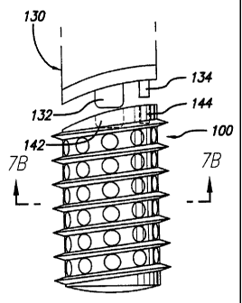
As shown in FIG. 7A, trailing end 104 may be configured to
complementary engage and instrument 130 for driving implant 100 into the
installation space. Instrument 130 may have a centrally disposed projection
132
and an off-center -projection 134 for engaging recesses 142 and 144 of
trailing
end 104, respectively. Projection 132 is preferably threaded as is recess 142.
While the implants of FIGS. 6C, 7A, and 7B are shown as cylindrical, the
implant of the present invention includes the novel teaching as applied to any
implants having opposed, at least in part, arcuate surfaces for penetrably
engaging into the vertebral bodies adjacent the disc space across which the
13
CA 02505437 2000-03-03
78406-13D
implant is implanted for the purpose of achieving fusion. These implants may
have flattened or modified sides to be less wide. Some examples of such
implants are taught by Michelson in U.S. Patent Nos. 5,593,409 and 5,559,909.
With reference to FIGS 8 and 9, when such a teacHing is applied for use
with a solitary, centrally placed implant 200 to be implanted anteriorly and
generally along the midline of the disc space, the trailing end 204 of implant
200
would be arcuate as shown, such that trailing end 204 is not rotationally
symmetrical about the mid-longitudinal axis MLA of implant 200, the trailing
end 204 might, in a preferred embodiment, for such use be symmetrical left and
right of the midlongitudinal axis MLA when properly inserted alternatively,
though not preferred, the implant 200 of FIG. 9 for implantation anteriorly
could have a rotationally symmetrical, or even a. rounded trailing end.
With reference to FIG. 10, while not achievirng the maximum advantage
of the present inventive teaching, implanu 300a and 300b (shown in. outlined
form) may be used in side by side pairs, each being symmetrically arcuate left
and right, but not rotationally, about the mid-longitudinal axis MLA to
provide
the advantage that there need not be mirror image implants or oppositely
threaded (left and right) implants provided, and such implaats if requiring
continuing rotation for their insertion (designed to be screwed in) can be
properly situated by half turns rather than full turns. That is, the correct
alignment of the implant occurs every 180 of rotation.
FIG. 10 further shows the area available to safely be filled and the
silhouette of a pair of implants 300a and.300b having symmetricaIly extended
trailing ends 304a and 304b for allowing for improved filling of the disc
space
and having relieved trailing end to side wall junctions to avoid the implant
from
protruding dangerously from the disc space ancerolaterally. While the fill is
not
quite as good as with the fully asymmetric trailing end embodiment, the
14
CA 02505437 2000-03-03
78406-13D
implants of FIG. 10 when inserted by rotation can be positioned by half-turn
increments and the need for different left and right implants has been
eliminated
As shown in FIG. 11, the best fill relative to a vertebral body achievable
by prior art implant C disposed anterolaterally across the transverse width of
the vertebral body is limited by corner LC" and leaves cross-hatched area Z
unoccupied by implant C.
With reference to FIG. 12A, another embodiment of the implant of the
present invention referred to by the numeral 400 is shown. Implaat 400 is for
insertion from the anterolateral aspect of the vertebral body and FIG. 12A
illustrates the greatly improved best fill made possible with implant 400.
Implant 400 has a general configuration as described in U.S. Patent No.
5,860,973 to Michelson, and has a trailing end 404 that is arcuate to
generally
conform to at least a portion of the natural anatomical curvature of the
lateral
aspect of the vertebral bodies. It is appreciated that implant 400 may include
the features of implant 100 described above and trailing end 404 may be
arcuate,
symmetrically or asymmetrically (left and right), about the mid-longitudinal
axis MLA of implant 400. In this manner, the area Z illustrated in FIG. 11 is
occupied and utilized by implant 400 which can actually be not only longer
overall, but also wider, or of a larger diameter, as the limiting corner LC"
of
the prior art implant and FIG. 11 has been removed. As evident from the
drawings, the present invention moves the limiting corner LC formed by the
junction of the side wall to the trailing wall or the most rearwardly
protruding
aspect of the laterally placed sidewall inward away from escaping the disc
space.
FIG. 12B is a top plan view of the endplate region of the vertebral body
of FIG. 11 with an alternative embodiment of first and second implants 450a
and 450b of the present invention implanted translaterally across the
transverse
width of the vertebral body from a lateral aspect of the spine. Implants 450a
and 450b are configured such that when they are installed, they have a general
CA 02505437 2000-03-03
78406-13D
configuration similar to a single implant 400 described above. Typically,
Implant 450a is inserted into the implantation space first, and then implant
450b is inserted into the same implantation space behind, and preferably
coaxial
to, implant 450a in a "box car" arrangement.
As shown in FIGS. 12C and 12D, trailing end 454a of implant 450a is
configured to be placed in contact with leading end 452b of implant 450b, and
preferably complementary engage leading end 452b. For example, trailing end
454a saay include raised portions 460a and 462a that cooperatively engage
raised
portions 460b and 462b of leading end 452b of implant 450b. When implants
450a and 450b are in contact, it is possible to impart movement of implant
450a
within the implantation space by movemeni of implant 450b. In this manaer,
it is possible to fine tune the depth of insertion of implant 450a without
removing implant 450b. The ability to move implant 450a in this manner also
prevents stripping of implant 450b due to the failure of movement of implant
450a.
Implant 450b can have a trailing end with a conventional configuration
or it can have a trailing end 454b that is arcuate to generally conform to at
least
a portion of the natural anatomical curvature of the lateral aspect of the
vertebral bodies. It is appreciated that implant 450b may include the features
of
implant 100 described above and trailing end 454b may be arcuate,
symmetrically or asymmetricaIly (left and right), about the mid-longitudinal
axis MLA of implant 450b. Leading end 452b may include a removable erid cap
470 with a hex drive 472.
Trailing end 454a of implant 450a is preferably flat or indented
concavely, may include a threaded opening 480 and a slot 482 for engaging
insertion instrumentation for driving the implants. The leading end 452b of
implant 450b may be flat, preferably with a bevel, chamfer, or radius, or
convex
to fit into the trailing end 454a of implant 450a. The radius of the leading
flat
edge of leading end 452b of implant 450b allows implant 450b to thread into an
16
CA 02505437 2000-03-03
78406-13D
already tapped path created by the insertion of implant 450a and pemlits the
external thread of implants 450a and 450b to functionally align easily.
FIGS. 13A and 13B demonstrate a pair of implants 500a and 500b of the
present invention being used in a side-by-side relationship inserted generally
laterally or anterolaterally into the spine. As shown in FIGS. 14A and 14B,
two implants 600a and 600b, one anterior, one posterior, the anterior one may
be of a larger diameter than the posterior one. The posterior one may be
longer than the anterior one. Each may have a trailing end that is curved from
side to side symmetrically or asymmetrically.
The prior art threaded implants, be they for rotation for screwing them
in or for less than a full turn rotation for locking them in after they have
already been linearly advanced into the spine, have all had geaerally straight
trailing ends or trailing ends that have been rotationally symmetrical in
regard
to length. In contradistinction, the implants of the present invention in the
preferred embodiment have trailing ends that are either arcuate or truncated
to
generally conform to the anterior and/or lateral (anterolateral) peripheral
contours of the vertebral bodies to be fused at their trailing ends and are
specifically for insertion from the anterior and anterolateral aspects of the
spine
aad from a position anterior to the transverse processes of the vertebrae to
be
fused, aad preferably are not rotationally symmetrical about their
longitudinal
axis.
While the exact curvature of a particular vertebral body may not be
known, the teaching of having the implant trailing end be arcuate or truncated
along one side or from side to side so as to eliminate the size limiting
corner or
the side wall or lateral'aspect junction to the implant trailing end is of
such
benefit that minor differences do not detract from its benefit. Further, the
range of describable curvatures may be varied proportionately with the size of
the implants as well as their intended location within the spine and direction
of
17
CA 02505437 2000-03-03
78406-13D
insertion to be most appropriate and easily determinable by those of ordinary
skill in the art.
Generally in the lumbar spine, the arc of radius of the curvature should
be from 15 to 30 millimeters to be of greatest benefit, though it could be
greater
or less, and still be beneficial. The same is true for the cervical spine
where the
arc of radius is 10-30 mm, with 15-20 nun being preferred. Similarly, the
trailing end could be curved at least in part, but not be an arc of a circle
and still
practice the present invention.
With reference to FIGS. 15A and 15B, as a substitute for contouring the
entire trailing end, the trailing end may have a configuration that may be
straight across and then chamfered as illustrated by implant 200 or radiused
to
one side only as illustrated by implant 800, sufficient to eliminate what
would
otherwise be a protruding corner when said implant would be properly inserted
and as previously described both lateral wall rear end junctions eould be
chamfered or radiused.
The implants of the present invention can be configured to have a
ma$imum distance from a horizontal plane HP perpendicular to and bisecting a
length along the mid-longitudinal axis MLA of the implant and the trailing end
of the implant that is greater than the distance from the horizontal
? perpendicular plane HP to the trailing end of at least one of the opposite
side
walls of the implant. This maximum distance may be greater than the distance
from the perpendicular plane HP to the trailing end of both of the side walls,
or
the distance from the perpendicular plaae HP to the trailing end of the second
side waU can greater than the distance from the perpendicular plane HP to the
trailing end of the first side wall. Alternatively, the distance from the
perpendicular plane to the trailing end of the second side wall can be greater
than the distance along the mid-longitudinal axis from the perpendicular plane
HP to the trailing end and greater than the distance from the perpendicular
plane HI' to the trailing end of the first side wall. The implants of the
present
18
CA 02505437 2000-03-03
78406-13D
invention may also have a maximum first length measured along a first
implant side wall that s longer than a secoad maaimum length measured along a
second implant side wall.
As should be evident from the above discussioa, all of these
embodiments allow for an interbody spinal fusion implant utilizing an element
of rotation for the proper insertion of the implants having at least one
relieved
or foreshortened aspect of at least one sidewall to end junction for placement
laterally so as to not protrude unsafely from the disc space.
As per FIGS. 16A and 16B, it should be appreciated then that a top view
of the trailing end must have a convex type profile as illustrated by implant
900
while the side view will not or to a much lesser extent. That is, the trailing
end
of the present invention implants are rotationally asynimetrical about the mid-
longitudinal axis MLA even when symmetrical from side to side, which side to
side symmetrically is not a requirement of the broad inventive concept of the
present invention. To have the opposed vertebrae engaging surfaces protrude
dangerously beyond the perimeter of the disc space so as to impinge on the
blood vessels or other vital structures proximate the spine is absolutely
contrary
to the teachings of the present invention which teaches a safe meaas for
allowing the optimal sizing of the implant(s). As shown in FIG. 16B, the long
sides "L' of implants 700-900 are generally the same.
While the present invention has been taught using implants requiring
rotation for their insertion, this has been done to highlight that the present
invention is counterintuitive and non-obvious. The additional implant length
made possible by the present inventive teaching actually provides for aa
implant that would seem to in all but the final selected position protrude
dangerously from. the spine. And indeed it would except that all implants
require at a minimum a clear path for their insenion: Thus, while the extended
trailing portion does extend from the spine until its final rotation into
correct
alignment it does so when the vital structures, organs, vessels, etc., are
retracted
19
CA 02505437 2000-03-03
78406-13D
and protected and ceases to do so thereafter when those strucnues are released
back to their normal positions in relationship to the spine.
Thus, while the present invention has been explained in regard to such
implants requiring rotation for their insertion, the present invention is not
so
limited and is useful for all interbody spinal fusion implants having opposed
arcuate upper and lower surfaces or surface portions for penetrable engagement
into the bodies of vertebrae adjacent a disc space to be implanted. Moreover,
such implants may include at least one opening therethrough to allow for the
growth of bone from vertebral body to vertebral body aad through the
implant. -
While particular embodiments of the present invention have been shown
and described, it will be obvious to those skilled in the art that changes and
modifications may be made without depaning from this invention in its
broader aspects and, therefore, the aim in the appended daims is to cover all
such changes and modifications as fall within the true spirit and scope of
this
invention.
While specific innovative features may have been presented in reference
to specific exaaznples, they are just examples, and it should be understood
that
various combinations of these innovative features beyond those specifically
shown are taught such that they may now be easily alternatively combined and
are hereby anticipated and claimed.