Note: Descriptions are shown in the official language in which they were submitted.
CA 02505464 2012-11-26
CATHETER TRACKING WITH PHASE INFORMATION
FIELD OF THE INVENTION
The present invention relates to a method for determining the position
and/or orientation of a catheter or other interventional access device or
surgical
probe using phase patterns in a magnetic resonance (MR) signal.
BACKGROUND OF THE INVENTION
Catheters and other endovascular access tools have long been used in the
art as medical devices to advance therapeutic agents to an anatomic point of
interest for examination, diagnosis, and intervention. Accurate positioning of
such
interventional devices requires monitoring to ensure that the device is being
advanced through the correct structures without causing injury, failing
mechanically, and for other reasons known to one skilled in the art.
Methods existing in the art for such monitoring include X-ray visualization,
as well as MRI tracking of any component of the device designed to be visible
on
MRI. Many conventional vascular interventional procedures use X-ray imaging
technology in which catheters or other probes are inserted into a vein or
artery and
navigated to specific locations for diagnostic and therapeutic procedures. For
1
CA 02505464 2005-04-27
example, over 3,000 trans-septal procedures are performed each year in the
United States for left sided radio-frequency ablation therapy and mitral
valvoplasty procedures. However, 3-6% of these are complicated by aortic or
atrial perforations due to incorrect needle positioning. This relatively high
complication rate can in part be attributed to the inability to directly
visualize
critical endocardial landmarks using a two-dimensional projection x-ray
fluoroscopy. Conventional X-ray guided interventions suffer from a number of
limitations, including: (1) limited anatomical visualization of the body and
blood
vessels during the examination, (2) limited ability to obtain a cross-
sectional view
of the target tissue or blood vessel, (3) inability to characterize important
pathologic features of atherosclerotic plaques, (4) limited ability to obtain
functional information on the state of the related organ, and (5) exposure of
the
subject to potentially damaging x-ray radiation.
Many invasive cardiovascular procedures, such as traversing total chronic
vascular occlusions, would benefit from using MR guidance to accurately
deliver
interventional medical devices to target locations because MRI methods have
fewer limitations than conventional X-ray techniques. For example, United
States
Patent No. 6,606,513 to Lardo et al describes a method means for an MR trans-
septal needle that can be visible on an MRI, can act as an antenna and receive
MRI signals from surrounding subject matter to generate high-resolution
images,
and can enable real-time active needle tracking during MRI guided trans-septal
puncture procedures. Interventional cardiology would also benefit from using
MR
guidance by exploiting MRI's excellent soft tissue contrast. For example, MRI
is
2
CA 02505464 2012-11-26
able to distinguish between infarcted and healthy myocardium to identify an
appropriate location for stem cell delivery [1]. MRI is also able to
distinguish
between plaque and vessel walls [2-3] which facilitates traversing total
chronic
occlusions. Furthermore, recent research has demonstrated that a complete
electrophysiologic study can now be performed entirely under MR1 guidance,
including the ability to navigate catheters and characterize the temporal and
spatial
formation of ventricular radiofrequency ablation lesions in vivo.
Additionally, MRI has been shown to guide mitral valvoplasty procedures.
As these two therapies account for 95% of all trans-septal procedures
performed, it
is clear that the ability to perform safe trans-septal needle puncture under
MRI
guidance will be of great importance as the field of interventional
cardiovascular
MRI continues to evolve.
In order to perform such procedures under MR guidance, it is necessary to
visualize a cardiac catheter and track it using MRI. Another key requirement
in
minimally-invasive or non-invasive procedures is to integrate the positioning
of
instruments, needles, or probes with image guidance to confirm that the
trajectory
or location is as safe as possible, and to provide images that enhance the
ability of
the physician to distinguish between tissue types. Placement may require
acquisition of static images for planning purposes, either in a prior MRI
examination or during the interventional MRI session, or real-time images in
arbitrary scan planes during the positioning process. (Dumoulin et al., "Real-
Time
Position Monitoring of Invasive Devices Using Magnetic Resonance", Mag Reson
Med 1993; 29: 411-415; Coutts et al., "Integrated and Interactive Position
3
CA 02505464 2012-11-26
Tracking and Imaging of Interventional Tools and Internal Devices Using Small
Fiducial Receiver Coils.", Magnetic Resonance in Medicine 1998, 40:908-913).
Despite the several distinct advantages MRI has over x-ray fluoroscopy,
MRI guided positioning of catheters and other devices has numerous challenges
related to imaging artifacts, electromagnetic interference, and the necessity
for
cardiac and respiratory gating and rapid imaging and display. The invention
described herein describes a procedure and required hardware to perform MR
guided procedures with active tracking of the tip of the interventional
device. Such
a procedure may improve imaging applications in a number of interventional MRI
guided therapies.
Device tracking techniques using MRI can generally be separated into
passive and active tracking methods. Passive tracking is based on device
visualization due to magnetic susceptibility artifacts. Magnetic
susceptibility is a
quantitative measure of a material's tendency to interact with and distort an
applied
magnetic field. Magnetic susceptibility artifacts are generated by
inhomogeneities
in the magnetic field due to a material with a magnetic susceptibility
different than
that of tissue [3-6]. Initial attempts to position and visualize endovascular
devices in
MR imaging were based on passive susceptibility artifacts produced by the
device
when exposed to the MR field.
United States Patent No. 4,827,931, to Longmore and United States Patent
No. 5,154,179 and 4,989,608 to Ratner disclose the incorporation of
paramagnetic
material into endovascular devices to make the devices visible based on
magnetic
susceptibility imaging. United States Patent No. 5,211,166 to
4
CA 02505464 2005-04-27
Sepponen similarly discloses the use of surface impregnation of various
"relaxants", including paramagnetic materials and nitrogen radicals, onto
surgical
instruments to enable their MR identification. However, these patents do not
provide for artifact-free MR visibility in the presence of rapidly alternating
magnetic fields, such as would be produced during echo-planar MR imaging
pulse sequences used in real-time MR guidance of intracranial drug delivery
procedures. Nor do these patents teach a method for monitoring with MR-visible
catheters the outcomes of therapeutic interventions, such as, for example,
drug
delivery into brain tissues or cerebral ventricles.
Ultrafast imaging sequences generally have significantly lower spatial
resolution than conventional spin-echo sequences. Image distortion may include
general signal loss, regional signal loss, general signal enhancement,
regional
signal enhancement, and increased background noise. The magnetic
susceptibility artifact produced by the device should be small enough not to
obscure surrounding anatomy, or mask low-threshold physiological events that
have an MR signature, and thereby compromise the physician's ability to
perform
the intervention. These relationships will be in part dependent upon the
combined
or comparative properties of the device, the particular drug, and the
surrounding
environment (e.g., tissue). No additional processing or hardware is required
with
passive tracking techniques. However, an additional limitation with passive
tracking is that quantitative information about the catheter position or
orientation
is not obtained. This inhibits automatic scan plane prescription and the
catheter
must be manually kept within the scan plane.
5
CA 02505464 2005-04-27
United States Patent No. 5,470,307 to LindaII discloses a low-profile
catheter system with an exposed MR-visible coating containing a therapeutic
drug agent, which can be selectively released at remote tissue sites by
activation
of a photosensitive chemical linker. However, in common with other currently
used endovascular access devices, such as catheters, microcatheters, and
guidewires, the catheter tip is difficult to see on MRI because of inadequate
contrast with respect to surrounding tissues and structures. This makes
accurate
localization difficult and degrades the quality of the diagnostic information
obtained from the image. Also, the mere observation of the location of the
catheter in the drug delivery system does not reliably or consistently
identify the
position, movement and/or efficient delivery of drugs provided through the
system. Thus, one objective of this invention is to provide for an MR-
compatible
and visible device that significantly improves the efficacy and safety of drug
delivery at various tissue locations using MR guidance.
An improved method for passive MR visualization of implantable medical
devices is disclosed in United States Patent No. 5,782,764 to Weme. This
invention minimizes MR susceptibility artifacts and controls eddy currents in
the
electromagnetic scattering environment, so that a bright "halo" artifact is
created
to outline the device in its approximately true size, shape, and position. In
the
method of the invention disclosed by Weme, an ultra thin coating of conductive
material comprising 1-10% of the theoretical skin depth of the material being
imaged is applied, wherein the susceptibility artifact due to the metal is
negligible
6
CA 02505464 2012-11-26
due to the low material mass. At the same time, the eddy currents are limited
due
to the ultra-thin conductor coating on the device.
A similar method employing a nitinol wire with Teflon coat in combination
with extremely thin wires of a stainless steel alloy included between the
nitinol wire
and Teflon coat, has been reported in the medical literature by Frahm at at,
"Passive Visualization ¨ A Suitable Approach for MR-Guided Vascular
Interventions in Low-Field Systems? In Vivo Evaluation of a New Guide Wire.",
Proc. ISMRM (Proceedings of the 5th International Society for Magnetic
Resonance in Medicine), Vancouver, 1997, p. 1931.
Active catheter tracking requires the reception of the MR signal by the
catheter through a receive coil located on a device that is coupled via a
cable to
the input port of an MR scanner. Henceforth, a receive coil located on a
device
and coupled via a cable to the scanner will be referred to as a microcoil.
Most
active tracking techniques project the magnitude sensitivity pattern of small
microcoils located on the catheter onto three orthogonal axes. The location of
the
micro-coils can then be determined by identifying the peaks of the projections
[7-9].
There is, however, a weakness with this approach in that the peaks of the
projections do not necessarily correspond to the center of the micro-coil. The
magnitude sensitivity profile of a coil also changes with different coil
orientations.
This makes peak finding through curve fitting difficult. The method is also
inherently susceptible to noise; high-resolution scans are needed; and it is
not
possible to obtain orientation information from the magnitude projections of a
single
coil [10].
7
CA 02505464 2012-11-26
Exemplary of methods for active MR visualization of interventional medical
devices is United States Patent No. 5,211,165 to Dumoulin et at., which
discloses
an MR tracking and localizing system for a catheter based on transmit/receive
microcoils positioned near the end of the catheter. Applications of such
catheter-
based devices in endovascular and endoscopic imaging have been described in
the medical literature, for example, Hurst et al., "Intravascular (catheter)
NMR
receiver probe: preliminary design analysis and application to canine
iliofemoral
imaging." Magnetic resonance in medicinel 992, vol. 24(2) pp. 343-357; Bonert
P.
et al., "In-Plane Tracking of Medical Instruments during mRr. Proceedings of
the
5th International Society for Magnetic Resonance in Medicine (Proc. ISMRM),
Vancouver, 1997, p. 1925; Coutts et at., "Integrated Position Tracking and
Imaging
o Interventional Tools and Internal Devices Using Small Fiducial Receiver
Coils",
Proceedings of the 5th International Society for Magnetic Resonance in
Medicine,
Vancouver, 1997. p. 1924 ; Wendt et at., "Simultaneous Shifted Rotated Keyhole
Imaging and Active Tip Tracking for Interventional Procedure Guidance",
Proceedings of the 5th International Society for Magnetic Resonance in
Medicine,
Vancouver, 1997. p. 1926; Langsaeter et al., "Tracking of an MR-Compatible
Microendoscope for Interventional MRI of the Paranasal Sinuses", Proceedings
of
the 5th International Society for Magnetic Resonance in Medicine, Vancouver,
1997. p. 1929; Zimmerman et al., "Evaluation of Catheter/Guide Wire
Steerability:
In Vitro Comparison of Fluoroscopic Guidance with Active MR-Tracking in an
Open
0.5T MR-System", Proceedings of the 5th International Society for Magnetic
Resonance in Medicine, Vancouver, 1997. p. 30; and, Ladd et at., "Vascular
8
CA 02505464 2012-11-26
Guidevvire Visualization for MR Fluoroscopy." Proceedings of the 5th
International
Society for Magnetic Resonance in Medicine, Vancouver, 1997. p. 1937.
Various imaging coils for interventional MR1 are known in the art. United
States Patent No. 5,738,632 to Karasawa discloses an endoscopetrigidoscope
with
MRI coils located in the distal section of the device. United States Patent
No.
5,699,801 to Atalar et al describes a loop antenna for interventional MR1 and
spectroscopy applications. United States Patent No. 5,348,010 to Schnall et
al.
discloses an inflatable MR1 receiver coil employing a balloon.
United States Patent No. 5,271,400 describes a tracking system for the
position and orientation of an invasive device within a patient, wherein the
device
includes a receiver coil and an MR active sample. The receiver picks up
magnetic
resonance signals generated by the sample. The frequencies are proportional to
the location of the coil along the applied field gradients, since the signals
are
received in the presence of these magnetic field gradients. The
6a
CA 02505464 2005-04-27
system is designed to enable location of the invasive device and enhanced
imaging of a region around the invasive device.
United States Patent No. 6,587,706 and United States Patent No.
6,560,475 to Viswanathan disclose microcoils which can be used in medical
devices to enhance RF response signals and to create fields to enhance imaging
capability in MRI imaging systems. The microcoil design includes at least one
pair of radially opposed microcoils, each microcoil having an outside
microcoil
diameter of 6 mm or less, individual windings of each microcoil together
defining
a geometric plane for each microcoil, and the plane of each microcoil being
parallel to the plane of another microcoil in the pair of radially opposed
microcoils.
United States Patent No. 6,549,800 to Atalar et al discloses methods for in
vivo magnetic resonance imaging, wherein MRI probes are adapted for insertion
into a plurality of body orifices in order to evaluate the anatomy of
proximate
anatomic structures, to diagnose abnormalities thereof and to treat the
diagnosed
abnormalities. MRI probes are described that are suitable for use in the
mediastinum, in the pancreaticohepaticobiliary system, in the
tracheobronchopulmonary system, in the head and neck, in the genitourinary
system, the gastrointestinal system, the vascular system, and in the
evaluation,
diagnosis and treatment of internal fluid collections.
United States Patent No. 6,061,587 to Kucharczyk et al. discloses an
apparatus and method for targeted drug delivery into a living patient using
catheter-based microcoils and magnetic resonance (MR) imaging. The apparatus
9
CA 02505464 2005-04-27
and method uses MRI to track the location of drug delivery and estimating the
rate of drug delivery.
A different approach for remote sensing of location is disclosed by United
States Patent No. 5,042,486 to Pfeiler et al. and by United States Patent No.
5,391,199 to Ben Haim. These technologies are based on generating weak
radio-frequency signals from three different transmitters, receiving the
signals
through an RF antenna inside the device, and calculating the distances from
the
transmitters, which define the spatial location of the device. However, the
application of this technology to MRI is problematic due to the simultaneous
use
of RF signals by the MR scanning. Potential difficulties are the heating of
the
receiving antenna in the device by the high amplitude excitation RF
transmissions of the MRI scanner and artifacts in the MR image.
United States Patent No. 5,271,400 and United States Patent No.
5,211,165 to Dumoulin et al. disclose a tracking system employing magnetic
resonance signals to monitor the position (since mentioned below that
orientation information is not available) of a device within a human body. The
device disclosed by Dumoulin's inventions have an MR-active sample and a
receiver coil which is sensitive to MR signals generated by the MR-active
sample. These signals are detected in the presence of MR field gradients and
thus have frequencies which are substantially proportional to the location of
the
coil along the direction of the applied gradient. Signals are detected by
sequentially applied, mutually orthogonal magnetic gradients to determine the
device's position in several dimensions. The position of the device as
determined
CA 02505464 2005-04-27
by the tracking system is superimposed upon independently acquired medical
diagnostic images. However, this method cannot directly determine the
orientation of the device, may be subject to heating of the coil, and requires
time
to implement that reduces the temporal resolution available for repeated MRI
acquisitions.
Although the patented inventions referenced above provide useful
technological advances in the field of image-guided interventions, each
invention
also has significant inherent limitations. Unlike the present invention, which
is
based on phase information, the prior art references are based on magnitude
information. Phase information can track an interventional device more
accurately than magnitude sensitivity information because phase information is
more spatially varying than magnitude projections. The present invention also
provides notable advantages over the prior art by enabling the position and
orientation of a catheter tip to be reliably tracked using low resolution MR
scans
for real-time interventional MRI applications. The prior art methods are also
inherently susceptible to noise; high-resolution scans are needed; and it is
not
possible to obtain orientation information from the magnitude projections of a
single coil [10].
SUMMARY OF THE INVENTION
The present invention provides a phase sensitivity method for tracking the
position of endovascular access devices and interventional probes which is
more
accurate than magnitude sensitivity methods disclosed in the prior art.
11
CA 02505464 2005-04-27
The present invention provides a phase sensitivity method for reliably
tracking the position of catheters using low-resolution clinically acceptable
MRI
scans that enable real-time interventional MRI applications.
The present invention also provides a phase sensitivity method which
enables real-time MRI tracking of the position of endovascular access devices
and interventional probes, including surgical tools and tissue manipulators,
devices for in vivo delivery of drugs, angioplasty devices, biopsy and
sampling
devices, devices for delivery of RF, thermal, microwave or laser energy or
ionizing radiation, and internal illumination and imaging devices, such as
catheters, endoscopes, laparoscopes, and related instruments.
The present invention discloses a method for determining the position
and/or orientation of a catheter or other interventional access device or
surgical
probe using phase patterns in a magnetic resonance (MR) signal. In the method
of the invention, the position and orientation of a microcoil at the distal
tip of the
interventional device is established from the receive phase pattern of the
microcoil, wherein an axial projection image of the microcoil's sensitivity
pattern
is initially created. The position of the microcoil in the axial plane is
determined
based on lines of constant phase which extend in the radial direction from the
microcoil's edges, and the roll angle 0 of the microcoil about the static
magnetic
field is established from the angle at which the phase pattern is rotated with
respect to a reference axial phase pattern. Further in the method of the
invention, an oblique slice is prescribed through the center of the microcoil
and
perpendicular to the plane in which the microcoil lies. According to the
invention,
12
CA 02505464 2005-04-27
the microcoil is then located in a third orthogonal direction based on
discontinuities in the oblique phase pattern which extend radially from the
microcoil's edges.
The pitch angle 4) of the coil about a vector perpendicular to the oblique
plane is determined by calculating the angle at which the oblique phase
pattern is
rotated with respect to a reference phase pattern. In the method of the
invention,
the phase in the two regions is then sampled to verify the calculation of the
roll
angle 0 wherein the roll angle 0 and the pitch angle 4) form two Euler angles
that
can be used to determine the normal of the microcoil.
According to the invention, phase information can track the position of a
catheter or other interventional device more accurately than magnitude
sensitivity
information because the receive phase sensitivity of a microcoil is unique for
any
given position and orientation of the microcoil, and phase information is more
spatially varying than magnitude projections. In the method of the invention,
both
the position and orientation of a microcoil can be determined using phase
information. Catheter tip tracking is carried out by obtaining two phase
images;
one integrated phase image projected onto the axial plane and one in an
oblique
plane through the center of the coil and normal to the microcoil plane. In
another
preferred embodiment of the invention, phase information adequate for MRI
localization can also be obtained from only one plane, for example, a plane
through the microcoil perpendicular to the axial plane, wherein such phase
information is used together with position information obtained from
projections,
or based on a locator that produces substantial phase patterns. Furthermore,
the
13
CA 02505464 2010-07-23
phase images obtained for catheter tracking using the method of the present
invention can be low-resolution MR images, wherein clinically useful MRI scans
can be obtained relatively rapidly thereby making catheter tracking using
phase
information clinically useful for real-time interventional MRI applications.
In one aspect of the present invention there is provided a method of
determining one or both of position and orientation of a medical device in a
patient's body, the method comprising the steps of:
a) placing the medical device in a patient's body and placing the
patient in a magnetic resonance imaging scanner, the magnetic resonance
imaging scanner having means for producing magnetic resonance signals
having a phase and magnitude and detection means for detecting magnetic
resonance signals, and the medical device including at least one marker for
perturbing the phase of the magnetic resonance signal;
b) acquiring magnetic resonance signals with perturbed phase from
the patient's body using the detection means; and
c) reconstructing from the acquired magnetic resonance signals with
perturbed phase at least one map of a spatial distribution of the phase of
the received signals, and using selected characteristics of the spatial
distribution of the phase to determine one or both of the device position
and orientation of the medical device in the patient's body.
In another aspect of the present invention there is provided a
method for determining the position and orientation of a medical device
inserted into a body of a patient, comprising the steps of:
14
CA 02505464 2010-07-23
placing a medical device in a patient's body and placing the patient in a
magnetic resonance imaging scanner, the magnetic resonance imaging scanner
having means for producing magnetic resonance signals each having a phase
and magnitude and detection means for detecting magnetic resonance signals;
the medical device including at least one marker for perturbing the phase
of the magnetic resonance signals, the at least one marker including a
microcoil;
detecting phase images with perturbed phase in magnetic resonance
(MR) signals received by the microcoil;
determining the position and orientation of the medical device in the
patient's body by producing an axial projection image of the at least one
microcoil's
integrated phase image in an axial plane, determining a position of the at
least
one microcoil in the axial plane based on lines of constant phase which extend
in a radial direction from the at least one microcoil's edges,
determining a roll angle 0 of the at least one microcoil from an angle at
which the phase image is rotated with respect to a reference axial phase
image,
prescribing an oblique slice, perpendicular to the axial plane, through a
center of the at least one microcoil and perpendicular to the plane in which
the at
least one microcoil lies and then determining a location of the microcoil in a
third
orthogonal direction by noting that discontinuities in an oblique phase image
extend radially from the at least one microcoil's edges,
determining a pitch angle by calculating an angle at which the oblique
phase image is rotated with respect to a reference phase image; and
CA 02505464 2010-07-23
determining a normal of the at least one microcoil wherein the roll angle 0
and the pitch angle (I) form two Euler angles used to determine the normal of
the
at least one microcoil.
The present invention also providesn apparatus for determining one or
both of position and orientation of a medical device in a patient's body,
comprising:
a) medical device for insertion into a patient's body, the medical device
including at least one marker for perturbing a phase of magnetic resonance
signals;
b) a magnetic resonance imaging scanner, the magnetic resonance
imaging scanner having means for producing said magnetic resonance signals
each having said phase and a magnitude and detection means for detecting
magnetic resonance signals responsively emitted from the patient's body; and
c) the magnetic resonance imaging scanner including processing
means for reconstructing from the detected magnetic resonance signals
having perturbed phase at least one map of a spatial distribution of the
phase of the detected signals, and using selected characteristics of the
spatial distribution of the phase to determine one or both of the position
and orientation of the medical device in the patient's body.
16
CA 02505464 2010-07-23
BRIEF DESCRIPTION OF THE FIGURES
The catheter tracking with phase information produced according to
the present invention will now be described, by way of example only,
reference being made to the accompanying drawings, in which:
Figures 1 a-d depicts various properties of phase patterns in the
axial plane. The orientation of the micro coil is depicted with respect to a
16a
CA 02505464 2005-04-27
coordinate system (Left), the scan plane is depicted with respect to the
microcoil (Middle), and the simulated phase image that corresponds to the
plane are shown (Right).
Figure 1a shows a simulated phase image in the axial plane through
the center of the microcoil.
Figure lb shows a simulated phase image in the axial plane with the
microcoil rotated by angle (1) about the x-axis.
Figure 1c shows a simulated phase image in the axial plane with the
microcoil rotated by angle 0 about the x-axis and by angle 0 about the z-
axis.
Figure 1d shows a simulated projected phase image in the axial
plane with the microcoil rotated by angle 0 about the x-axis and by angle
0 about the z-axis.
Figures 2a-c depicts various properties of phase patterns in an
oblique plane perpendicular to the axial plane through the center of the
microcoil. The orientation of the micro coil is depicted with respect to a
coordinate system (Left), the scan plane is depicted with respect to the
microcoil (Middle), and the simulated phase image that corresponds to the
plane are shown (Right).
Figure 2a shows a simulated phase image in an oblique plane
through the center of the microcoil.
17
CA 02505464 2005-04-27
Figure 2b shows a simulated phase image in an oblique plane
through the center of the microcoil with the microcoil rotated by angle (I)
about a vector perpendicular to the oblique plane.
Figure 2c shows a simulated phase image in an oblique plane
through the center of the microcoil with the microcoil rotated by angle 0
about the z-axis and rotated by angle 4 about a vector perpendicular to the
oblique plane.
Figure 3a-c shows a graphical demonstration of a potential
localization scheme in one embodiment of the invention.
Figure 3a shows a graphical demonstration of step 1 of a potential
localization scheme in one embodiment of the invention. A projected phase
image is obtained in the axial plane. The micro coil is positioned on the
plane using the lines of constant phase as shown. The rotation of the coil 0
about the z-axis is determined by measuring the rotation on the phase
pattern with respect to a reference.
Figure 3b shows step 2 of a potential localization scheme in one
embodiment of the invention. An oblique plane perpendicular to the axial
plane, passing through the center of the coil, and perpendicular to the plane
in which the circular microcoil lies is prescribed.
Figure 3c shows step 3 of a potential localization scheme in one
embodiment of the invention. Using the phase image corresponding to the
oblique slice prescribed in Figure 3b, the microcoil is positioned on the z-
axis using discontinuities between regions of constant phase as show. The
18
CA 02505464 2005-04-27
rotation of the coil 4) about a vector perpendicular to the oblique plane is
determined by measuring the rotation on the phase pattern with respect to
a reference.
Figures 4a-d shows a comparison between actual and simulated
phase images obtained from a 4-mm-outside diameter microcoil at the tip of
a 6F angiographic catheter positioned in an agar phantom using the
method of the present invention. The phase images were acquired with a
spin-echo imaging pulse sequence.
Figure 4a shows a projection phase image acquired in the axial
plane.
Figure 4b shows a simulated projection phase image in the axial
plane.
Figure 4c shows a phase image acquired in the oblique plane.
Figure 4d shows a simulated phase image in the oblique plane.
Figures 5a-d shows a comparison between actual and simulated
phase images obtained from a 4-mm-outside diameter microcoil at the tip of
a 6F angiographic catheter positioned in an agar phantom using the
method of the present invention. The phase images were acquired using a
spiral acquisition.
Figure 5a shows a projection phase image acquired in the axial
plane.
Figure 5b shows a simulated projection phase image in the axial
plane.
19
CA 02505464 2005-04-27
Figure 5c shows a phase image acquired in the oblique plane.
Figure 5d shows a simulated phase image in the oblique plane.
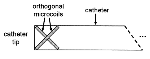
Figure 6 shows an embodiment of the invention which uses two
microcoils oriented perpendicularly to each other.
Figures 7a-b illustrates one possible application of the invention involving
the guidance of a catheter through a chronic total occlusion.
Figure 7a graphically illustrates how position and orientation
information about a catheter tip can be used to place a device in a blood
vessel at the proximal end of an occlusion and parallel to the vessel wall.
Figure 7b shows one embodiment of the invention where phase
information is used to provide position and orientation information about a
catheter tip. The information is subsequently displayed on an MR image of
a chronic total occlusion for guidance purposes.
DETAILED DESCRIPTION OF THE INVENTION
Definitions:
As used herein the phrase "phase pattern" refers to a spatial map of
phase in the MR signal in a particular plane of interest.
As used herein the phrase "catheter tracking" refers to the act of
determining information about the position and/or orientation of a catheter
tip.
As used herein the phrase "marker for perturbing the phase of the
magnetic resonance signal" means using a receive coil of arbitrary shape used
to
CA 02505464 2005-04-27
introduce phase into the MR signal through its receive sensitivity field or
using a
material of arbitrary shape and sufficient magnetic susceptibility to perturb
the
static magnetic field in the volume surrounding the material.
As used herein the phrase "microcoil" refers to small tuned radiofrequency
antenna used to receive MR signal or transmit an MR excitation field.
As used herein the phrase "field map" refers to a spatial map of the static
magnetic field in a plane of interest.
Active MRI tracking of catheters and other interventional probes has been
a subject of research for more than a decade, but most studies have focused on
magnitude sensitivity methods for localizing such devices. The fundamental
weakness with this approach is that it produces inaccurate localization when
the
peak of the magnitude projections does not correspond to the location of the
center of the microcoil. The magnitude sensitivity profile of the coil also
changes
with different coil orientations. The magnitude projection method is also
inheritably susceptible to noise, high-resolution projections are needed, and
orientation information cannot be obtained from projections alone.
The present invention addresses these various limitations of magnitude
sensitivity methods by providing a novel method for determining the position
and
orientation of interventional access devices and surgical probes based on
phase
patterns in the MR signal around a marker positioned on the device or probe.
More particularly, the present invention provides a method of determining the
position and/or orientation of a medical device in a patient's body which
involves
placing the medical device in a patient's body and placing the patient in a
21
CA 02505464 2005-04-27
magnetic resonance imaging (MRI) scanner. The medical device includes at
least one marker for perturbing the phase of the magnetic resonance signal
which is measured by the MRI scanner. Based on the received magnetic
resonance signals acquired from the patient's body using the magnetic
resonance imaging scanner, at least one map of the spatial distribution of the
phase of the received signals is reconstructed, and using characteristics of
the
spatial distribution of the phase, the device position and/or orientation are
determined.
In a preferred embodiment of the invention the marker is a small circular
microcoil positioned on the device or probe. In this embodiment, the microcoil
is
used to acquire the MR signal. However, it will be appreciated that the
microcoil
does not need to be circular, it may for example be elliptical or square, and
in
addition any other type of marker may be used so long as it perturbs the phase
of
the magnetic resonance signal, for instance through any type of susceptibility
mechanism. In this latter case, the marker is selected so that the difference
in
magnetic susceptibility between the marker and adjacent water in the body
yields
unique phase patterns in the signal around the marker which can be mapped
using the MR signal received by an external coil.
Another example includes the introduction of a small piece of
ferromagnetic material onto the probe which disrupts the local magnetic field.
In
this embodiment, MR signals can be acquired using an MR reception coil located
external to the body. A further example is the introduction of a small bubble
of
carbon dioxide in a balloon attached to the device. Again, the difference in
22
CA 02505464 2005-04-27
magnetic susceptibility between the gaseous carbon dioxide and adjacent water
in the body yields unique phase patterns in the signal around the balloon
which
can be mapped using the MR signal received by an external coil.
When using a small circular microcoil positioned on the device or probe
the method of the present invention determines the position and orientation of
interventional access devices and surgical probes based on phase patterns in
the MR signal around the small circular microcoil positioned on the device or
probe.
The microcoil may be connected electrically to the MR system or signals
from the coil may be coupled optically or inductively to a transducer which is
electrically connected to the MR scanner. The latter embodiments provide some
electrical isolation between the patient and the scanner. One can configure
the
system so that it is possible to separately receive signals from different
coils at
the same time. That said, when using microcoils, phase images are derived from
the signal from each microcoil.
In the method of the invention, accurate position and orientation
information can be obtained over a circular area of at least 4 microcoil
diameters.
Moreover, since only a sample of the information is needed to position and
orient
the catheter, highly redundant localization information is available and
global
correlations can be used to identify the phase patterns.
The present invention will now be described further with particular
reference to certain non-limiting embodiments and to the accompanying
drawings in Figures 1 to 4. In the method of the invention, the sensitivity
field of a
23
CA 02505464 2005-04-27
circular microcoil is evaluated. The theory of reciprocity states that the
sensitivity
field of an MR receive coil is equivalent to the magnetic field produced when
current I is passed through the coil. The receive sensitivity of a circular
receive
coil can be described in cylindrical coordinates by the following equations:
B= ______________________________________________________ Pik [K(k)+a 2 - r2
¨ZQ E(k)1 [1]
4747ir - rY + z'2
Arke ____________________________________ [ a2 +r 2 +2 '2
B r = K(k)+ __________ E(k)1
[2]
4,7r1rir - rY +
where
k = 4ar
j(a_r)2+z2
[3]
a is the coil's radius, p is the magnetic permeability of the medium
surrounding
the coil, r is radial distance from the centre of the coil and z' is distance
in the
perpendicular direction. K(k) and E(k) are elliptic integrals of the first and
second
kind respectively. The directional component of the sensitivity introduces
spatially varying phase patterns into the MR signal that are dependent solely
on
the position and orientation of the coil within the magnetic field. Assuming
constant phase in the MR signal in the absence of the receive coil, the phase
distribution introduced by the receive coil can be identified through a phase
reconstruction of an image acquired from the receive coil.
With reference to Figure la, when the radius of the microcoil is smaller
compared to the field of view (FOV) (field of view being the size, in linear
24
CA 02505464 2005-04-27
dimensions, of the acquired phase pattern), so that it satisfies the condition
(a <
FOV/10), the simulated magnetic field receive phase pattern around the
circular
microcoil 1 in the axial plane (perpendicular to the static field pointing
along z)
through the center of the coil has lines of constant phase extending in the
radial
direction from its edges 2. According to the invention, the phase pattern in
the
axial plane is independent of coil pitch about the x axis (magnet coordinate
system) (Figure 1b) and rotates by angle 0/2 3 with coil rotation about the z
axis
by angle 0 (Figure 1c), wherein this phase pattern is also independent of
axial
slice, wherein the same phase pattern results if the signal is integrated over
the
z-direction (magnet coordinate system) (Figure 1d). Further in the method of
the
invention, the phase pattern in an oblique slice through the center of a micro
coil
lying in the z-x plane and normal to the coil plane (Figure 2a) consists of
two
areas of constant phase, wherein the areas of constant phase have a value of
n/2 +0 above and below the coil and a value of -n/2 +0 to the sides of the
coil.
The discontinuities 6 between the two areas extend in the radial direction
from
the coil edges. According to the invention, the phase pattern rotates by an
angle
0/2 7 with coil rotation by angle 4) about the direction normal to the oblique
plane
(Figure 2b). With reference to Figure 2c, when the coil is rolled by angle 0
about
the z axis, the values of phase in the two constant areas increase by 0 to
form a
value of n/2+0 above and below the coil 8 and ¨7c/2+0 to the sides of the coil
9.
With reference to Figure 3, according to a preferred embodiment of the
invention, the phase patterns around a circular microcoil can be used to
determine the position and orientation of the microcoil. In the method of the
CA 02505464 2005-04-27
invention, an axial projection image (no slice selection gradient) of the
microcoil's
sensitivity pattern is first created (Figure 3a). In a preferred embodiment,
the
microcoil can be positioned on the x-y plane with reference to lines of
constant
phase which propagate radially from the edges of the microcoil. The amount 0
by which the microcoil is rotated about the static field Bo is determined by
determining the amount by which the phase pattern is rolled. Further in the
method of the invention, an oblique slice is then prescribed (drawn) through
the
center of the microcoil and perpendicular to the plane in which the microcoil
lies
(Figure 3b). Prescribing" means selecting an imaging plane, often done by
10 drawing a line on an MR image to define a plane perpendicular to the
current
image passing through that line.
With reference to Figure 3c, the microcoil can then be located on a third
axis based on discontinuities in the oblique phase pattern 6 which extend
radially
from the microcoil's edges.
Further in the method of the invention, the pitch angle (1) by which the coil
is rotated about a vector perpendicular to the oblique plane, can be
determined
by calculating the angle at which oblique phase pattern is rotated with
respect to
a reference phase pattern. The phase in the two regions can then be sampled to
verify the calculation of 0. The angles 0 and 4) form two Euler angles from
which
the microcoil's normal can be determined.
In order to evaluate the utility of the present invention, a small microcoil
with diameter 4-mm-outside diameter (30 gauge insulated magnet wire, 4
windings) was placed on the distal tip of a 6F angiographic catheter and
26
CA 02505464 2005-04-27
embedded in an agar phantom. Phase patterns were obtained using a 1.5T GE
Signa CV/i MR scanner with a spin echo pulse sequence (FOV=8cm, TE=17ms,
TR=5000ms, 256x256). The phase patterns obtained from the microcoil are
shown in FIG. 4a and 4c with reference to simulated phase patterns around
microcoils in a similar orientation. In the axial phase pattern obtained
without
slice selection (FIG. 4a) radial lines of constant phase extending from the
coil's
edges can clearly be seen, wherein the phase pattern resembles the simulations
(FIG. 4b). The phase pattern in the oblique slice (FIG. 4c) consists of two
regions of constant phase, wherein phase values in these regions are in
agreement with the predicted values (FIG. 4d).
In another embodiment of the invention, phase inhomogeneities can be
corrected for by obtaining two separate phase images at different echo times
and
generating a field map to perform post-acquisition corrections of static
magnetic
field inhomogeneities.
In another embodiment, the necessary phase images can be acquired
rapidly using a fast acquisition technique. In this embodiment the methods for
tracking the device are identical to previous embodiments with the exception
that
a rapid acquisition technique is used to collect the phase data in the planes
described in FIGs. 3a-3c. In this embodiment, phase image quality is
sacrificed
for acquisition speed to enable tracking with a high temporal resolution. An
example of a rapid imaging technique is spiral acquisition which collects data
in a
rapid and efficient manner. The utility of this embodiment was investigated by
acquiring phase patterns in the planes of interest using the same microcoil
27
CA 02505464 2005-04-27
discussed above using a spiral acquisition (FOV=8cm, TR=36ms, TE=10ms,
FA=45 deg, 3 4096-point spiral interleaves in k-space with an additional
acquisition for field map calculation, total acquisition time = 216 ms). The
phase
patterns obtained from the microcoil using a spiral acquisition are shown in
FIG.
5a and 5c with reference to simulated phase patterns around microcoils in a
similar orientation. In the axial phase pattern obtained without slice
selection
(FIG. 5a) radial lines of constant phase extending from the coil's edges can
clearly be seen, wherein the phase pattern resembles the simulations (FIG.
5b).
The phase pattern in the oblique slice (FIG. 5c) consists of two regions of
constant phase, wherein phase values in these regions are in agreement with
the
predicted values (FIG. 5d).
The results of the simulation studies underscore several advantages of the
phase sensitivity method of the present invention. Unlike magnitude
sensitivity
methods disclosed in the prior art, phase patterns are unique to a microcoil's
position and orientation and yield accurate information about both
localization
parameters. According to the present invention, phase pattern information also
provides a more robust localization algorithm, since phase patterns are more
spatially varying than magnitude projections and yield clear position and
orientation information over a circular area of at least 4 coil diameters.
Furthermore, since global two-dimensional correlations can be used to
identify the phase patterns, low-resolution scans may be sufficient for
locating the
coil. As a result, localization using the phase pattern methods disclosed by
this
invention may prove particularly useful in real-time applications, wherein as
one
28
CA 02505464 2005-04-27
non-limiting example, if the coil were to be located within a 2cm volume, a
single
spiral acquisition at sufficient resolution (2cm FOV, 1.05 mm resolution,
31.25
kHz bandwidth, 1024 readout) could be performed on a 1.5T GE Signa CV/i
system in 16 ms. According to this application of the method of the invention,
catheter position and orientation could be determined in under 100ms even with
extra acquisitions to correct for inhomogeneities.
In another preferred embodiment of the present invention, active MR
localization of the orientation and position of an interventional device may
also be
achieved by means of several microcoils positioned along the longitudinal axis
of
a catheter or other interventional device. Particularly preferred are
microcoils
consisting of a circular loop of conductive material positioned around the
functional parts of an interventional device, such as a drug delivery
catheter.
Depending on the orientation of the coil with the magnetic Bo, single
microcoils
may be used separately or may be constructed in an array. In order to reduce
the thickness of the microcoil, the coil material may be sputter-coated onto
the
surface of the device. To provide information about angular twist of the
device,
the loop may be sputter-coated onto the side of the device rather than in a
plane
perpendicular to the longitudinal axis of the device. Also preferred is a
microcoil
able to move and rotate inside a catheter sleeve attached to another component
of the device. This could be used to provide position and/or orientation
information about this component (eg. shaft) inside the catheter). In another
preferred embodiment, more than one microcoil may be present, wherein the
distribution of microcoils along a length of the catheter defines the MR-
visible
29
CA 02505464 2005-04-27
region of the interventional device. In general, this embodiment of the
invention is
best practiced by employing an array of microcoils, such that an MR image is
obtained for any orientation of the interventional device.
According to the invention, where functional elements are combined into a
single interventional device, the positioning and orientation of several
microcoils
may be tailored for a particular interventional procedure. In the method of
the
invention, one or more microcoils may be positioned near or at the distal end
of
the central catheter to assist in positioning the device at a target
anatomical
location. Other configurations of microcoils, such as parallel alignment of
the
microcoils in a strip-like orientation, stacking of microcoils in rows and
columns,
or mixtures of these and other configurations may also be useful in the
practice
of the present invention.
In another embodiment of the present invention, two microcoils are placed
in orthogonal directions at the distal tip of the catheter, as shown in Figure
6. In
this configuration tracking is performed using both coils. Advantages with
this
configuration include the ability to track the device with adequate signal
regardless of device orientation. In this configuration there is also minimal
inductive coupling between microcoils. Furthermore, the configuration is well
suited for acquiring high-resolution images of the anatomy immediately
surrounding the device.
Other functional elements of interventional probes which may be localized
using the method of the invention include thermal elements for providing heat,
radiation carrying elements (e.g., ultraviolet radiation, visible radiation,
infrared
CA 02505464 2005-04-27
radiation), optical fibers, detection elements (e.g., pH indicators,
electronic
activity indicators, pressure detectors, ion detectors, thermal detectors,
etc.), and
any other sensing or detection element which would be useful during medical
procedures. In accordance with the method of the invention, these individual
elements would benefit from accurate directed placement of the functional tip
of
the device within the target site in a tissue. For example, in the treatment
of
neurological diseases and disorders, targeted drug delivery can significantly
improve therapeutic efficacy while minimizing systemic side-effects of the
drug
therapy. Image-guided placement of the tip of a drug delivery catheter
directly
into specific regions of the brain can initially produce maximal drug
concentration
close to the loci of tissue receptors following injection of the drug. High-
resolution visual images denoting the actual position of the drug delivery
device
within the brain are extremely useful to the clinician in maximizing the
safety and
efficacy of the procedure. In a particularly preferred embodiment, drug
delivery
devices, such as catheters, could be monitored by the MR phase tracking
method of the present invention, thus making intra-operative verification of
catheter location possible.
The present invention also overcomes other limitations of the prior art.
For example, the limited distribution of drug injected from a single catheter
location can reduce the therapeutic efficacy of the intervention in cases of
anatomically extensive neurological damage, such as, for example, with certain
brain tumors and stroke. Since the volume flow rate of drug delivery typically
must be very low in order to avoid indiscriminate damage to brain cells and
nerve
31
CA 02505464 2005-04-27
fibers, delivery of a drug from a single point source limits the distribution
of the
drug by decreasing the effective radius of penetration of the drug agent into
the
surrounding tissue receptor population. Another aspect of this invention is
therefore to overcome these inherent limitations of single point source drug
delivery by devising a catheter tracking method which provides the ability for
accurately monitoring the placement of a catheter tip at several tissue
locations
in order to allow multiple drug release sources, which effectively disperse
therapeutic drug agents over a brain region containing receptors for the drug,
or
over an anatomically extensive area of brain pathology.
The availability of an MR-visible drug delivery device combined with
clinically acceptable low-resolution imaging would make it possible to obtain
near
real-time information on drug delivery during interventional procedures in an
intra-operative MR system, as well as for pre-operative and post-operative
confirmation of the location of the drug delivery device. Medical and surgical
applications of the present invention would include vascular surgery and
interventional radiology, cardiac surgery and cardiology, thoracic surgery and
radiology, gastrointestinal surgery and radiology, obstetrics, gynecology,
urology,
orthopedics, neurosurgery and neurointerventional radiology, head and neck
surgery and radiology, ENT surgery and radiology, and oncology. In addition to
direct tissue injection, the method of the invention applies to drug delivery
via
intraluminal, intracavitary, laparoscopic, endoscopic, intravenous,
intraarterial
applications.
32
CA 02505464 2005-04-27
The present invention also provides clinical benefits for certain
cardiovascular procedures, such as, for example, traversing chronic total
occlusions, where an intravascular device is pushed through a chronic
occlusion
in an artery to re-establish blood flow. Knowledge of the orientation and
position
of the device tip with respect to both the occlusion and vessel wall is
extremely
important because of significant risk of incidental surgical damage to the
vessel
wall (Figure 7a). MRI can now reliably differentiate vessel wall from
surrounding
tissue. The method of the present invention can be used to determine and image
both the position and orientation of a device with respect to critical
anatomic
landmarks resulting in improved safety and procedural efficacy. In a preferred
embodiment, device position and orientation would be imaged in real-time on an
MR image thereby providing clinically beneficial guidance for the procedure
(Figure 7b).
In a particularly preferred embodiment, the present invention can also be
used for targeted delivery of stem cells in the myocardium. In order to
achieve
preferential migration of stem cells into infracted myocardium, injections
must be
made at the border between diseased and healthy tissue. MR can be used to
identify such sites through delayed contrast-enhanced images, wherein the
method of the present invention can be used to guide an injection device to
appropriate target sites in the penumbra of the ischemic myocardium and to
properly orient the injection needle for delivery of cells into region best
suited for
establishing functional improvement.
33
CA 02505464 2012-11-26
It should be understood that the foregoing description is merely illustrative
of
the invention. Various alternatives and modifications can be devised by those
skilled in the art without departing from the scope or spirit of the
invention. As used
herein, the terms "comprises", "comprising", "including" and "includes" are to
be
construed as being inclusive and open ended, and not exclusive. Specifically,
when used in this specification including claims, the terms "comprises",
"comprising", "including" and "includes" and variations thereof mean the
specified
features, steps or components are included. These terms are not to be
interpreted
to exclude the presence of other features, steps or components.
The foregoing description of the preferred embodiments of the invention has
been presented to illustrate the principles of the invention and not to limit
the
invention to the particular embodiment illustrated. It is intended that the
scope of
the invention be defined by all of the embodiments encompassed within the
following claims and their equivalents.
34
CA 02505464 2005-04-27
References Cited:
U.S. Patent Documents
4827931 May, 1989 Longmore 128/334.
4984573 Jan., 1991 Leunbach 128/653.
4989608 Feb., 1991 Ratner 128/653.
5154179 Oct., 199 Ratner 128/653.
5155435 Oct., 1992 Kaufman et al 324/309.
5188111 Feb., 1993 Yates et al. 128/657.
5201314 Apr., 1993 Bosley et al. 128/662.
5211166 May., 1993 Sepponen 128/653.
5218964 Jun., 1993 Sepponen 128/653.
5262727 Nov., 1993 Behbin et al. 324/318.
5271400 Dec., 1993 Dumoulin et al 128/653.
5290266 Mar., 1994 Rohling et al. 604/272.
5318025 Jun., 1994 Dumoulin et al. 128/653.
5353795 Oct., 1994 Souza et al. 128/653.
5357958 Oct., 1994 Kaufman 128/653.
5409003 Apr., 1995 Young 128/653.
5419325 May., 1995 Dumoulin et al. 128/653.
5534778 Jul., 1996 Loos et al. 324/318.
Other Documents
[1] Dick AJ, Guttman MA, Raman VK, Peters DC, Pessanha BS, Hill JM,
Smith S, Scott G, McVeigh ER, Lederman RJ. "Magnetic resonance
fluoroscopy allows targeted delivery of mesenchymal stem cells to infarct
borders in Swine." Circulation 108(23): 2899-904.
[2] Barkhausen J, Ebert W, Heyer C, Debatin JF, Weinmann HJ. "Detection
of atherosclerotic plaque with Gadofluorine-enhanced magnetic resonance
imaging." Circulation 108(5): 605-9.
[3] Wentzel JJ, Aguiar SH, Fayad ZA. "Vascular MRI in the diagnosis and
therapy of the high risk atherosclerotic plaque." J Interv Cardiol 16(2): 129-
42.
[4] Glowinski A, Adam G, Bucker A, Neuerburg J, van Vaals JJ, Gunther RW.
"Catheter visualization using locally induced, actively controlled field
inhomogeneities." Magn Reson Med 38(2): 253-8.
CA 02505464 2005-04-27
[5] Bakker CJG, Weber J, van Walls JJ van, Mali WPTM, Viergever MA.
"Dedicated Catheters for Susceptibility-Based MR Visualization." 4th
ISMRM, New York, 1996. Pg. 900.
[6] Lenz G, Drobnitzky M, Dewey Ch. "MR-visible catheters for intro-
vascular
interventional MRI procedures." 4th ISMRM, New York, 1996. Pg. 901.
[7] Dumoulin CL, Souza SP, Darrow RD. "Real-time position monitoring of
invasive devices using magnetic resonance." Magn Reson Med 29(3):
411-5.
[8] Elgort, DR, Wong E.Y, Hillenbrand C, Wacker FK, Lewin JS, Duerk, JL.
"Real-time catheter tracking and adaptive imaging." J Magn Reson
Imaging 18(5): 621-6.
[9] Santos JM, McConnell M, Scott G, Hyon MS, Pauly JM. "Multi-Coil Real-
Time Interventional System." 11th ISMRM, Toronto, 2003. 1197.
[10] Hillenbrand CM, Elgort DR, Wong EY, Wacker FK, Lewing JS, Duerk JL. "
A Catheter Based, Opposed Solenoid Phased Array Coil for Active Device
Tracking and High Resolution lntravascular MRI." 11th ISMRM, Toronto,
2003. 1186.
36