Note: Descriptions are shown in the official language in which they were submitted.
CA 02505587 2005-05-19
WO 2004/052445 PCT/US2003/038794
METHODS AND SYSTEMS FOR SELECTIVE CONTROL OF
BLADDER FUNCTION
CROSS-REFERENCE TO RELATED APPLICATIONS
[0001] This application claims priority to and benefit of USSN 10/313,960,
filed on
December 6, 2002, which is incorporated herein by reference in its entirety
for all purposes.
STATEMENT AS TO RIGHTS TO INVENTIONS MADE UNDER FEDERALLY
SPONSORED RESEARCH AND DEVELOPMENT
f Not Annlicable 1
FIELD OF THE INVENTION
[0002] This invention relates to methods and systems for regulating neural
transmission. Specifically described is a method for selectively inhibiting
somatic nerve
transmission in a mixed nerve containing both somatic and autononuc nerve
fibers. The
methods find application in treatment of chronic pain, spastic contractions
and fox
controlling visceral functions. The method directed to the utilization of one
or more
electrodes an selected nerve bundles and the application of alternate phase
rectangular
electrical pulses to the electrodes) to regulate neural transmission or to
control muscle
contraction.
BACKGROUND OF THE INVENTION
[0003] Various medical patients lose voluntary control over their bladder
andlor
bowel. Although vesicostomy, augmentation cystoplasty or an artificial
sphincter implanted
around the urethra are commonly used to provide partial control over the
evacuation
function of the bladder and to control continence, these solutions have
drawbacks well
known to those skilled in the medical profession and related arts. Other
patients who
achieve a modicum of control over their bladder functions are equally in need
of a system to
rehabilitate their nerve and muscle dysfunctions. Similar problems arise in
respect to
involuntary bowel control.
-1-
CA 02505587 2005-05-19
WO 2004/052445 PCT/US2003/038794
[0004] The physiology of the bladder and bowel is closely linked to the
urethral
muscle physiology of the pelvic floor (levator and muscle) and its related
urethral and anal
sphincters. For the bladder to store urine and for the bowel to serve as a
reservoir for feces,
two opposite, but complementary, behaviors are found. In particular, for
storage, the
bladder and rectum must relax and the urethral and anal sphincters must remain
contracted.
The reverse is true during evacuation of either urine or feces, i.e., the
urethral or anal
sphincter relaxes, along with the pelvic floor, and subsequently the bladder
and rectum
contracts.
[0005] The sequence will reverse once voiding and defecation is completed,
i.e., the
sphincters and pelvic floor muscles will revert to their tonic closure states
and the bladder
and rectum will revert to their storage states. This behavior has been
demonstrated by
simultaneous manometric (or EMG/pressure) recordings of this bladder/rectum,
urethral/anal behavior during filling and emptying of the bladder. This
sequence of events
is well-established and is accepted universally.
[0006] Reduced bladder capacity and laclc of volitional urinary voiding are
experienced by spinal cord injured patients. With the present state-of-the-
art, implanted
pulse generators that are connected to electrodes attached to sacral roots to
electrically
stimulate the sacral roots provide patient-controlled bladder voiding.
Additionally, dorsal
rhizotomies are used to improve the bladder capacity, which is reduced by
hyperactivity of
the afferent fibers in the dorsal roots. -
[0007] Currently, voiding by electrical stimulation of the sacral roots is
accomplished by stimulating both the somatic and the parasympathetic nerves in
the sacral
roots. This technique causes both the striated sphincter muscles and the
detrusor smooth
muscles to contract simultaneously. As a result, the increased sphincter
pressure is still able
to bloclc the passage of urine in spite of the increased bladder pressure.
After a few seconds
of stimulation, the electrical stimulus pulses are turned off and the striated
somatic muscles
relax to decrease the sphincter pressure before the slower smooth muscle of
the bladder
relaxes, thus providing a pressure differential, higher in the bladder, so
that a momentary
passage of urine results. The electrical stimulation is again turned on, then
off, to obtain
another burst of urine. This procedure is repeated until the bladder is
effectively emptied.
-2-
CA 02505587 2005-05-19
WO 2004/052445 PCT/US2003/038794
[0008] In most prior nerve stimulators the typical shape of the current or
voltage
pulses that are used are rectangular and monophasic, that is, current or
voltage is one
direction. The current (or voltage) is applied for a short duration (typically
0.05 to 2
milliseconds) and then the current (or voltage) supply is turned off, and then
turned on again
in the same direction. This on-off sequence is continued and produces a train
of pulses
continued, e.g., at a nominal rate of ~0 pulses per second, to stimulate the
fibers. Thus, the
pulse generators produce essentially monophasic unidirectional pulses of
current (or
voltage).
SUMMARY OF THE INVENTION
[0009] This invention provides a method of selectively inhibiting neural
transmission of a somatic fiber in a mixed nerve containing both somatic and
autonomic
nerve fibers. In one embodiment the method involves applying alternate phase
high
frequency, low amplitude pulse pairs to the nerve in an amount sufficient to
inhibit neural
transmission of somatic fibers without inhibiting the autonomic nerves. The
method can
further involve additionally applying an alternate phase low frequency, high
amplitude
pulse pairs to the nerve to maintain the inhibition of the somatic fibers
while stimulating the
autonomic nerve fibers. Preferred frequencies and amplitudes are as described
herein.
[0010] A preferred use of the invention is to control bladder and bowel
function
where the method further includes providing an electrical pulse generator. The
electrical
pulse generator is coupled to at least one nerve responsible for controlling
bladder and
bowel function. Alternate phase high frequency, low amplitude pulse pairs are
applied to
the nerve to block sphincter muscle contraction. Low frequency, high amplitude
pulse pairs
are applied to the nerve to void the bladder or bowel while the alternate
phase high
frequency, low amplitude pulse pairs are continued. In a preferred embodiment,
the low
frequency, high amplitude pulse pairs are alternate phase. They may also be
monophasic.
[0011] Thus, in one embodiment, this invention provides a method of
selectively
inhibiting neural transmission of a somatic fiber in a mixed nerve containing
both somatic
and autonomic nerve fibers. The method comprises applying alternate phase high
frequency, low amplitude pulse pairs to the nerve in an amount sufficient to
inhibit neural
transmission of somatic fibers without substantially inhibiting autonomic
fibers in said
-3-
CA 02505587 2005-05-19
WO 2004/052445 PCT/US2003/038794
mixed nerve. The method can further comprise applying an alternate phase low
frequency,
high amplitude pulse pairs to the nerve to inhibit both somatic and autonomic
nerve fibers.
[0012] In another embodiment, this invention provides a method of controlling
a
bladder or a bowel. The method involves applying alternate phase high
frequency, low
amplitude pulse pairs to a nerve that innervates the bowel or bladder; and
applying alternate
phase low frequency, high amplitude pulse pairs to the nerve to void the
bladder or bowel,
while continuing to apply the alternate phase high frequency, low amplitude
pulse pairs. In
certain embodiments, the high frequency pulse pairs are at least 50 pulse
pairs per second.
In certain embodiments, the high frequency pulse pairs range from about 50 to
about 200 or
about 80 to about 120 pulse pairs per second (ppps). In certain embodiments,
the low
frequency pulse pairs are in a range of up to about 40 pulse pairs per second.
In certain
embodiments, the low frequency pairs are in a range of about 15 to about 25
pulse pairs per
second. The amplitude of high frequency pulse pairs typically ranges from
about 0.1
milliamperes to about 1.5 milliamperes. In certain embodiments, the amplitude
of high
frequency pulse pairs ranges from about 0.3 milliamperes to about 0.8
milliamperes. The
amplitude of low frequency pulse pairs typically ranges from about 1
milliampere to about 3
milliamperes. In certain embodiments, the amplitude of low frequency pulse
pairs ranges
from about 1.3 milliamperes to about 1.7 milliamperes. In certain embodiments,
the high
frequency pulses have pulse widths in a range of about 0.01 ms to about 0.5
ms, more
preferably about 0.08 ms to about 0.12 ms. In certain embodiments, the low
frequency
pulses have pulse widths in a range of about 0.1 ms to about 1.0 ms, and more
preferably
have pulse widths of approximately 0.5 ms.
[0013] This invention also provides a system for controlling a bladder or
bowel. The
system typically comprises an electrical pulse generator configured to produce
alternate
phase high frequency, low amplitude pulses and an alternate phase low
frequency, high
amplitude pulses to a sacral nerve, and at least one electrode that can be
coupled to a nerve
and transmit an electrical signal from the pulse generator to the nerve. In
certain
embodiments, the electrode is coupled to the sacral nerve and in electrical
communication
with the electrical pulse generator. In certain embodiments, the system
comprises two
electrodes coupled to the electrical pulse generator with four wires. The
system can further
comprise an external power source electromagnetically coupled to the
electrical pulse
-4-
CA 02505587 2005-05-19
WO 2004/052445 PCT/US2003/038794
generator. The system can further comprise a pressure sensitive switch on the
electrical
pulse generator.
[0014] In certain embodiments, this invention also provides a method of
inhibiting
neural transmission in a nerve by applying electrical impulses to the nerve
where the
electrical impulses have the following characteristics: alternate phase and a
frequency in of
at least 60 pulse pairs per second at an amplitude sufficient to inhibit
neural transmission.
In certain embodiments, the frequency is at least 60 pulse pairs per second.
In certain
embodiments, the frequency is in a range of about 60 to about 500 pulse pairs
per second,
more preferably about 60 to about 300 pulse pairs per second.
[0015] Also provided is a method of reducing chronic pain. The method involves
of
applying electrical impulses having the following characteristics: alternate
phase and a
frequency of at least 60 pulse pairs per second to a nerve at an amplitude
sufficient to inhibit
neural transmission. In certain embodiments, the frequency is at least 60
pulse pairs per
second. In certain embodiments, the frequency is in a range of about 60 to
about 500 pulse
pairs per second, more preferably about 60 to about 300 pulse pairs per
second.
[0016] This invention provides a method of reducing muscle spasticity in
muscles
innervated with nerves. This method involves applying electrical impulses
having the
following characteristics: alternate phase and a frequency range of at least
60 pulse pairs per
second to the nerve at an amplitude sufficient to inhibit neural transmission.
In certain
embodiments, the frequency is in a range of about 60 to about 500 pulse pairs
per second,
more preferably about 60 to about 300 pulse pairs per second.
[0017] A method is provided for inhibiting neural transmission in nerves by
contacting the nerves with an electrode connected to an electrical pulse
generator and
applying electrical impulses having the following characteristics: alternate
phase and a
frequency in a range of 60 pulse pairs per second at an amplitude sufficient
to inhibit neural
transmission. In certain embodiments, the electrode is a ribbon of conducting
metal. The
nerve can be an intact extradural root. The nerve can be a human nerve or a
nerve of a non-
human mammal.
[0018] In still yet another embodiment, this invention provides a method of
controlling a bladder or a bowel. The method involves providing an electrical
pulse
generator operatively linlced to at least one nerve or operatively linl~ing
the electrical pulse
-5-
CA 02505587 2005-05-19
WO 2004/052445 PCT/US2003/038794
generator to at least one nerve; applying alternate phase high frequency, low
amplitude
pulse pairs to the nerve; and applying low frequency, high amplitude pulse
pairs to the
nerve to void the bladder while continuing to apply the alternate phase high
frequency, low
amplitude pulse pairs. In certain embodiments, the high frequency pulse pairs
are at least
50 pulse pairs per second In certain embodiments, the high frequency pulse
pairs range
from about 50 to about 200 pulse pairs per second, preferably about 80 to
about 120 pulse
pairs per second. In certain embodiments, the low frequency pulse pairs are in
a range of up
to about 40 pulse pairs per second, preferably about 15 to about 25 pulse
pairs per second.
In certain embodiments, the amplitude of high frequency pulse pairs ranges
from about 0.1
milliamperes to about 1.5 milliamperes, preferably from about 0.3 milliamperes
to about 0.8
milliamperes. In certain embodiments, the amplitude of low frequency pulse
pairs ranges
from about 1 milliamperes to about 3 milliamperes, preferably from about 1.3
milliamperes
to about 1.7 milliamperes. The high frequency pulses typically have pulse
widths in a range
of about 0.01 ms to about 0.5 ms, preferably in a range of about 0.08 ms to
about 0.12 ms.
The low frequency pulses typically have pulse widths in a range of about 0.1
ms to about
1.0 ms. In certain embodiments, the low frequency pulses have pulse widths of
approximately 0.5 ms.
[0019] This invention also provides a method of stimulating nerve fibers by
applying alternate phase pulse pairs.
[0020] Also provided is a method of selectively inhibiting neural transmission
of
somatic nerve fibers while selectively stimulating neural transmission of
autonomic nerve
fibers in a mixed nerve containing both somatic and autonomic nerve fibers.
The method
involves applying alternate phase high frequency, low amplitude pulse pairs to
inhibit
neural transmission'of somatic nerve fibers without substantially inhibiting
neural
transmission of autonomic nerve fibers; and applying alternate phase low
frequency, high
amplitude pulse pairs to the nerve in an amount sufficient to stimulate neural
transmission
of autonomic nerve fibers.
[0021] In still another embodiment, this invention provides a method of
controlling
a bladder or a bowel. The method involves (i) applying alternate phase high
frequency, low
amplitude pulse pairs to inhibit neural transmission of somatic nerve fibers
without
substantially inhibiting neural transmission of autonomic nerve fibers; and
(ii) applying
-6-
CA 02505587 2005-05-19
WO 2004/052445 PCT/US2003/038794
alternate phase low frequency, high amplitude pulse pairs to the nerve in an
amount
sufficient to stimulate neural transmission of autonomic nerve fibers.
[0022] In certain embodiments, the invention provides a system for controlling
a
bladder or a bowel. The system comprises: at least one electrical pulse
generator configured
to produce alternate phase low frequency, high amplitude pulse pairs and/or
alternate phase
high frequency, low amplitude pulse pairs in communication with at least one
mixed nerve
containing both somatic and autonomic nerve fibers through at least one
electrode.
[0023] In certain embodiments, this invention provides a system for
controlling a
bladder or a bowel, the method comprising: at least an electrical pulse
generator configured
to produce alternate phase low frequency, high amplitude pulse pairs and
alternate phase
high frequency, low amplitude pulse pairs in communication with at least one
mixed nerve
containing both somatic and autonomic nerve fibers through at least one
electrode.
[0024] In still another embodiment, this invention provides a system for
controlling
a bladder or a bowel. The system typically comprises: at least an electrical
pulse generator
configured to produce alternate phase low frequency, high amplitude pulse
pairs and
alternate phase high frequency, low amplitude pulse pairs in communication
with a
multitude of mixed nerves containing both somatic and autonomic nerve fibers
through an
array of electrodes.
DEFINITIONS
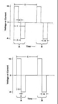
[0025] The term "alternate phase" when used with respect to a pulse pair,
refers to a
pulse pair comprising two pulses of opposite sign, where the potential (for a
voltage pulse)
or the current (for a current pulse) remains at a common potential or point of
no current
flow for some measurable time (x in Figure lA) between the first and second
pulse
comprising the pulse pair. In certain embodiments, where the period between
the pulse
pairs is given by 7~ and the delay between the end of the first pulse of the
pulse pair and the
beginning of the second pulse of the pulse pair is given by x (see Figure lA)
then x is
preferably equal to or greater than 7J20, more preferably equal to or greater
than ?x,/10, and
still more preferably equal to or greater than 7~,/6 or ~,/5, or a,/3.
CA 02505587 2005-05-19
WO 2004/052445 PCT/US2003/038794
[0026] The phrase "a pulse pair comprising two pulses of the opposite sign"
refers to
a pulse pair where if the first pulse is positive going, the second pulse is
negative going and
if the first pulse is negative going, the second pulse is positive going.
[0027] A "simple biphasic pulse pair" refers to a pair of (electronic) pulses
where
the first pulse is positive going (current or voltage relative to common) and
a second pulse
is negative going (current or voltage relative to common) or where the first
pulse is negative
going and followed by a second positive going pulse and the delay "x" between
the first and
second pulse is less than 7,120, more preferably less than 7140, or a,/80 and
most preferably is
about zero (see, e.g., Figures lA and 1B).
[0028] The term "nerve" refers to, but is not limited to, sacral roots, spinal
roots,
bundles of roots or nerves, mixed fiber nerve bundles, small and large nerve
fibers, dorsal
roots, ventral roots, somatic nerve bundles and autonomic nerve bundles.
[0029] The terns "mixed nerve" or "mixed fiber nerve" or "mixed fiber nerve
bundle" are used interchangeably and refer to groups or bundles of nerve
axons.
[0030] The term "small fibers" refers to nerve fibers having a diameter less
than
about 2 ,um.
[0031] The term "large fibers" refers to nerve fibers generally having a
diameter
greater than about 2 ,um.
[0032] The phrase "operatively linking" when referring to operatively linlcing
a
pulse generator to a nerve indicates that the pulse generator is disposed in a
manner that
permits the application of generated pulses to the nerves. Such operative
linking can be
accomplished in any of a number of ways, e.~. by connecting the pulse
generator via
conductive leads to one or more electrodes applied, positioned, juxtaposed
next to, inserted
into the nerve, nerve root, etc., by direct juxtaposition of the pulse
generator to the subject
nerves, by radio, magnetic, inductive, or other electromagnetic coupling of
the pulse
generator to one or more electrodes in contact with the nerves, and so forth.
BRIEF DESCRIPTION OF THE DRAWINGS
[0033] Figures lA and 1B illustrate two alternate phase pulse pairs (A and B)
(Figure lA) and a two simple biphasic pulse pairs (A and B) (Figure 1B).
_g_
CA 02505587 2005-05-19
WO 2004/052445 PCT/US2003/038794
[0034] Figure 2 shows graphs that compare the effects of two different pulse
patterns on sphincter pressure.
[0035] Figure 3 schematically illustrates the pelvic plexus region in a human,
including the nervous system for controlling bladder evacuation and related
functions, and
further illustrates an operative procedure for controlling such functions.
[0036] Figure 4 illustrates an implantable pulse generator 10 operably linlced
to
nerves 12 via leads 13.
[0037] Figure 5 is schematic illustration of a system for controlling a
bladder in
accordance with the presentinvention.
[0038] Figures 6A and 6B are examples of a pattern for pulses for controlling
a
bladder in accordance with the present invention.
[0039] Figure 7 is a block diagram illustrating an example of an implantable
pulse
generator in accordance with the present invention.
[0040] Figure 8 illustrates a wave form for controlling a bladder in
accordance with
the present invention.
[0041] Figure 9 illustrates the connection of electrodes to various nerve
bundles. A
multiplicity of electrode pairs 63, 65, and 66 are attached to separate nerve
bundles, and
multiple electrodes 63 and 64 are attached to the same nerve bundle.
[0042] Figure 10 illustrates a multiplicity of active electrode contacts
employed on a
single nerve.
[0043] Figures 11 and Figure 12 are views similar to Figure 3, but illustrate
additional operative procedures for controlling bladder evacuation and related
functions.
[0044] Figure 13 schematically illustrates the percutaneous implantation of an
electrode adjacent to the S3 sacral roots through the dorsum for the purpose
of selectively
stimulating such nerve.
[0045] Figures 14, 15, 16, 17, and 18 are views similar to Figure 3, but
illustrate
additional operative procedures for controlling bladder evacuation and related
functions.
[0046] Figure 19 illustrates one embodiment of a pulse generator suitable for
practice of the methods of this invention.
-9-
CA 02505587 2005-05-19
WO 2004/052445 PCT/US2003/038794
[0047] Figure 20 illustrates one embodiment of a pulse generator suitable for
practice of the methods of this invention.
[0048] Figure 21 illustrates shows a circuit, wave form, and a block diagram
of a
device that can produce the alternate phase pulses using a single voltage
source without an
output transformer.
[0049] Figure 22 illustrates one embodiment of a pulse generator suitable for
practice of the methods of this invention.
DETAILED DESCRIPTION
[0050] This invention pertains to novel devices and methods for selective
control of
somatic fibers or autonomic fibers in a mixed fiber nerve. Such mixed fiber
nerves
innervate various organs such as the bowel and bladder. By selectively
regulating
autonomic and somatic fiber activity, organ control can be effected where such
control has
been compromised (e.g. by neurological damage). Thus, for example, bladder
and/or bowel
retentions and evacuation can be more effectively controlled particularly in
subjects where
voluntary control is non-existent or inhibited.
I. Selective control of lame and small fibers in a mixed fiber nerve
[0051] This invention is based, in part, on the discovery that mixed
frequency,
alternate phase current or voltage pulses can selectively bloclc impulses in
somatic fibers to
achieve' flaccid paralysis of the sphincter muscles and selectively excite the
autonomic
fibers) to produce detrusor muscle contraction. High frequency, low amplitude
current or
voltage pulses make the sphincter muscles unresponsive to low frequency, high
amplitude
current or voltage pulses which contract the detrusor muscles to effectively
achieve bladder
voiding in spinal cord injured subjects.
[0052] This differential activation/inhibition of small and large fibers can
readily be
exploited in the regulation of bladder (or other organ) function in subjects
in which such
function is compromised. In particular, the ability to utilize high frequency
small amplitude
pulse pairs applied, e.g. to a motor root to control sphincter activity,
obviates the need for
surgical separation, dissection and resection, e.g., of the sacral somatic
nerve Ss and
possible attendant complications (e.g. incontinence) can thereby be avoided.
-10-
CA 02505587 2005-05-19
WO 2004/052445 PCT/US2003/038794
[0053] More generally, it was discovered that effective differential
excitation of
large and small fibers in a bundle of mixed nerve fibers can be achieved by
the use of
alternate phase pulse pairs. In particular, the application of alternate phase
high frequency
current or voltage pulse pairs is effective in blocl~ing post-synaptic
responses, while leaving
small fibers essentially unaffected. Mixed frequency, alternate phase current
or voltage
pulses can selectively bloclc impulses in somatic and selectively excite
autonomic fibers) in
a mixed fiber nerve.
[0054] It was also discovered that high current/voltage pulses are able to
produce
blockage in the smaller fibers, for example, of the autonomic system. Thus, in
another
embodiment, high current/voltage pulses are used to block small fibers of the
autonomic
nervous system.. This bloclc is also applicable to the efferent and/or
afferent fibers, for
example, the fibers used in proprioception, pain, and temperature.
[0055] This invention thus provides the means for differential
activation/inhibition
of small and large fibers in a mixed fiber nerve. This differential
activation/inhibition of
small and large (autonomic and somatic fibers) finds use in a wide variety of
contexts. Such
differential inhibition/activation can be exploited to provide effective
control of bladder
function and/or to provide effective control of other organs, such as the
bowel, colon and
associated sphincters, (e.g., anus). The methods of this invention can also
provide means
for eliminating or suppressing spastic detrusor activity, spastic urethral and
pelvic floor
activity and spastic anal sphincter, and the like.
[0056] It was also a surprising discovery that, as explained herein, alternate
phase
pulse pairs are particularly effective in providing selective control of small
and large nerve
fibers.
[0057] In certain embodiments, the stimulation is provided as "alternate
phase"
voltage or current pulses (see, e.g., FigurelA). While the pulse are
illustrated as "square
wave" pulses in FigurelA, they need not be so limited. Other alternate phase
waveforms
(e.g. sinusoidal, triangular, ramped, stepped, etc.) can also be utilized.
[0058] As illustrated in FigurelA, an alternate phase pulse pair refers to a
pulse pair
where the potential (for a voltage pulse) or the current (for a current pulse)
remains at a
common potential (or no current) for some measurable time between the first
and second
pulse. This is in contrast to the typical biphasic pulse pair or biphasic
pulse (illustrated in
-11-
CA 02505587 2005-05-19
WO 2004/052445 PCT/US2003/038794
FigurelB) where there is essentially no delay at common or ground between the
first and
second phase of the pulse pair. In addition, when the first pulse is positive
going, the
second pulse is negative going and when the first pulse is negative going, the
second pulse
is positive going.
[0059] The pulse pair is characterized by a frequency f or period ~, (time
between
pulse pairs), an amplitude (current or voltage), a pulse width w, a time
between the
beginning of the first phase/pulse in a pulse pair and the beginning of the
second phase of
the pulse pair e, a time between the end of the first phase of a pulse pair
and the beginning
of the second phase of a pulse pair x, and/or a pulse pair duration t as
illustrated in
Figure 1 A.
[0060] In accordance with one aspect of this invention, high frequency low
amplitude pulse pairs are used to disable large fibers and thereby inhibit
skeletal muscle
activity (e.g. to paralyze sphincters). In certain embodiments, a high
frequency pulse pair
has a frequency f of at least about 50 pulse pairs/second up to a frequency of
about 200 to
300 pulse pairs per second. In typical embodiments, the frequency is in a
range of about 60
to about 500 pulse pairs per second, preferably about 60 to about 200 pulse
pairs per
second, more preferably about 80 to about 150 pulse pairs per second, and most
preferably
from about 90 to about 120 pulse pairs per second.
[0061] In accordance with one aspect of the present invention, the low
frequency
pulse pairs range from about 10 pulse pairs per second up to about 40 pulse
pairs per
second. In certain embodiments, the low frequency pulse pairs range in
frequency from
about 15 to about 25 pulse pairs per second.
[0062] The high frequency pulses typically have pulse widths w ranging from
about
0.01 ms to about 0.5 ms, preferably from about 0.01 ms to about 0.20 ms, more
preferably
from about 0.02 ms to about 0.15 ms, and most preferably from about 0.08 ms to
about 0.12
ms.
[0063] The low frequency pulses typically have pulse widths raging from about
0.1
ms to about 1 ms, preferably from about 0.2 ms to about 1.0 ms, and most
preferably have
pulse widths of approximately 0.5 ms.
-12-
CA 02505587 2005-05-19
WO 2004/052445 PCT/US2003/038794
[0064] The pulse pairs used in the methods of this invention can vary in
amplitude,
as well as frequency f, width w, and delay D or x. As indicated herein, high
frequency low
amplitude pulse pairs are used to disable (inhibit) large fiber activity. The
low amplitude
pulses range from about 0.1 milliamps to about 1.5 milliamps or range from a
voltage
sufficient to produce a current of about 0.1 milliamps to about 1.5 milliamps.
In certain
embodiments, the low amplitude pulses range from about 0.3 to about 1.0
milliamps,
preferably from about 0.4 to about 0.9 milliamps, more preferably from about
0.4 to about
0.8 milliamps.
[0065] High amplitude, low frequency pulse pairs are used to activate small
fibers
(e.g. to evacuate bowel or bladder). In accordance with this invention, high
amplitude pulse
pairs range in amplitude from about 1.0 to about 3.0 milliamps or range from a
voltage
sufficient to produce a current of about 1.0 to about 3.0 milliamps. In
certain embodiments,
the high amplitude pulse pairs rang in amplitude from about 1.3 to about 1.7
milliamps.
[0066] It was a surprising discovery that alternate phase pulse pairs provide
improved efficacy in regulating bladder function and/or the function of other
organs. As
illustrated in Figure 2 the effects of alternate phase pulses on sphincter
pressure are different
compared with the effects produced by the "biphasic" pulses. The pulse
patterns are shown
at the bottom of the figure. On the left are two "alternate phase" pulse pairs
and on the right
are two simple "biphasic" pulse pairs. The pulse rate of each pattern is 100
pulse pairs per
second. Each pattern was turned on for about 2.5 seconds at three different
current
amplitudes: 0.5, 0.7, and 1.0 milliamperes, as shown on the bottom trace. The
effect of the
stimulation is shown in the top two traces.
[0067] The bladder pressures are similar for the two different pulse patterns.
The
sphincter pressures, however, are different. The "alternate phase" pulses
produce a very
short, high-pressure burst in the sphincter followed by the bladder pressure
that is reflected
in the sphincter area. The simple "biphasic" pulses produced a prolonged
sphincter pressure
that was always higher than the bladder pressure during the period of
stimulation. The
"alternate phase" pulses relaxed the sphincter immediately so that voiding
could be initiated
far sooner than would be possible with the "biphasic" pulses. Thus, greater
bladder voiding
was obtained with lower sphincter pressure.
-13-
CA 02505587 2005-05-19
WO 2004/052445 PCT/US2003/038794
[0068] The delay x between the two pulses comprising the pulse pair is
typically
optimized to produce maximum efficacy (e.g., of bladder voiding). Typically,
the most
desirable effect is produced by the symmetrical" alternate phase" pulse pair,
that is, equal
time between the plus and minus and minus and plus phases. As the "alternate
phase" pulse
pair becomes closer to a simple "biphasic" pulse pair the effect (e.g. x
approaches zero) the
enhanced effect shifts accordingly.
[0069] In various embodiments, of the present invention, the "alternate phase"
pulse
pair ranges from equal timing (2:1 ratio 7,./x) between phases to about a 4:1,
6:1 or ~:1 ratio
(7Jx). Thus, for example, if ~, is the time between two plus phases, then a
symmetric wave
form will be obtained with x= 7,,/2. While ~, and x are illustrated with
respect to the leading
edge of the pulses) (see, e.g., Figure 1A), they could also be measured with
respect to the
midpoint of the pulse or with respect to any other convenience reference
point.
[0070] The pulse amplitude A, frequency f, delay x, 0, and width w can be
optimized for maximum efficacy in each subject. Typically, this is
accomplished after
surgical placement of the electrodes. In certain embodiments, a programmable
controller is
used to vary these parameters as well as electrode selection/activation in
multiple electrode
configurations to achieve maximum efficacy. If possible, a configuration that
produces
maximum efficacy with minimal power consumption is selected.
[0071] It is noted that, while the alternate phase pulse pairs are illustrated
with both
pulses comprising the pair having the same width w and amplitude A, these
parameters can
be individually varied for each pulse.
[0072] In brief summary, high frequency, low amplitude pulse pairs, preferably
alternate phase pulse pairs act to disable the function of organs innervated
by larger fibers.
Such pulse pairs, can be used to flaccidly paralyze sphincter muscles without
activating
small fibers. Low frequency, high amplitude pulse pairs, preferably alternate
phase pulse
pairs activate small fibers. High frequency, high amplitude pulse pairs,
preferably alternate
phase pulse pairs, act to selectively bloclc small fibers. These effects are
summarized in
Table 1.
-14-
CA 02505587 2005-05-19
WO 2004/052445 PCT/US2003/038794
Table 1. Summary of effects of pulse pairs, preferably alternate phase pulse
pairs.
Pulse Type Effect
High frequency, low amplitude Disables large fibers. Flaccidly paralyze
sphincter. No effect on small fibers
Low frequency, high amplitude Activate small and large fibers. Activate
detrusor muscles.
High frequency, high amplitude Produce blockage in small fibers (e.g. efferent
and afferent fibers). Bloclc proprioception, pain,
temperature, etc.
II. Control of bladder function.
[0073] In certain embodiments, this .invention provides systems and methods
for
controlling a bladder (e.g. maintaining continence and/or effecting evacuation
when
desired). The pulse generation systems of this invention typically include an
electrical pulse
generator configured to produce alternate phase high frequency, low amplitude
pulses and
alternate phase low frequency, high amplitude pulses that can be transmitted
by one or more
electrodes on one or more sacral root(s). The system thus includes at least
one electrode
that can be coupled to a sacral root and in electrical communication with the
electrical pulse
generator.
[0074] As described in U.S. Patent No. 4,607,639, Figure 3 schematically
illustrates
the pelvic plexus region of a human, including the nervous system for
controlling bladder
evacuation and related functions. The nervous system includes a somatic nerve
system of
fibers (or nerve bundles) S and an autonomic nerve system of fibers or nerve
bundles A,
finding their immediate origin at sacral segments S2, S3, and S4 of the spinal
cord and
sacrum. The main nerve supply to the detrusor muscle of a bladder B emanates
primarily
from sacral segment S3, a lesser amount from sacral segment S2, and a still
lesser amount
from sacral segment S4.
[0075] One aspect of this invention is directed to a method for controlling
the
evacuation of bladder B. The method can involve identifying the anatomical
location of at
least one nerve or component thereof that controls at least one function of
the bladder, e.g.,
continence andlor contraction of the bladder. One or more electrodes are then
positioned,
-15-
CA 02505587 2005-05-19
WO 2004/052445 PCT/US2003/038794
either surgically or percutaneously, at least in close proximity to the nerve
or nerve root and
selectively energized as described herein to stimulate particular fibers.
[0076] Further, this invention contemplates either permanent surgical
implantation
or temporary percutaneous of the devices described herein implantation for
nerve
stimulation purposes.
[0077] As further illustrated in Figure 3, the main nerve supply emanating
from each
sacral segment S2, S3, and S4 comprises two components or roots, namely, a
dorsal root D
and a ventral root V. The dorsal root is primarily sensory to transmit
sensation to the spinal
cord whereas the ventral root is primarily motor to transmit motor impulses
from the spinal
cord to bladder B and associated sphincter. Although illustrated as being
separated, the
dorsal and ventral roots for each nerve are, in fact, normally joined together
and their fibers
mixed to progress as a single trunk.
[0078] Fibers of the nerve trunlc are divided into somatic fibers S that
connect to
voluntary muscles and autonomic fibers A that connect to visceral organs, such
as bladder
B. In various embodiments, methods of this invention involve isolation of
various
components of these nerve fibers at various levels in the nervous system.
Although not
required by the devices described herein, dorsal root D can be separated from
ventral root V
to facilitate stimulation of only the motor fibers of a particular ventral
root. In this manner,
the motor fibers can be stimulated without inducing pain and without
generating impulses
along the sensory passage way.
[0079] ~f course, the use of dual mode (e.g. high frequency low amplitude
pulse
pairs followed by low frequency high amplitude pulse pairs) pulse pairs as
described herein
can obviate the need for sectioning ventral root from dorsal root.
[0080] Previous methods required that somatic nerves S and autonomic nerves A
be
separated from each other to effectuate independent stimulation. Indeed to
eliminate
sphincter activity and facilitate bladder voiding, the sacral somatic Ss nerve
is often
sectioned bilaterally.
[0081] A significant advantage of the devices and methods described herein is
the
independent inhibition/activation of small and large fibers. Accordingly,
using only
electronic means, the sphincter muscles can be selectively relaxed while the
detrusor
-16-
CA 02505587 2005-05-19
WO 2004/052445 PCT/US2003/038794
muscles are actuated to effectuate bladder voiding. The methods of this
invention thus
eliminate the need to section somatic nerve and continence is more easily
maintained.
[0082] In addition, particular pulse trains can be delivered to particular
roots to
maximize efficacy. For example, responses obtained with pre-operative
evaluation of
responses to stimulation recorded urodynamically could indicate that the S2
sacral nerve
constitutes the main motor supply to external sphincter E, whereas the S3
sacral nerve
constitutes the main motor supply to bladder B. Thus, the S3 sacral nerve
would be utilized
to control the detrusor muscle and thus the contracting function of bladder B
and
stimulation predominantly with low frequency high amplitude alternate phase
pulse pairs as
described herein can optimize bladder evacuation. Conversely, in this context,
the S2 sacral
nerve could be utilized to control the muscles controlling the continence
function of external
sphincter E and application of high frequency small amplitude pulse pairs
predominantly to
this root can effectuate sphincter relaxation. Studies have shown that in
certain patients,
only the nerves on one side of the sacrum provide the main motor control over
a particular
organ, i.e., unilateral control rather than bilateral control. Pre-operative
testing of a
particular patient can determine which variation will provide the best choice
for a
subsequent operative procedure. The ability of this invention to selectively
activate or
inhibit particular components of the various nerves or roots, with the
combined ability to
test a patient intraoperatively and record responses enables the surgeon to
isolate and
selectively stimulate the particular nerve fibers that will effect the
specific function or
functions required.
[0083] Figure 4 illustrates an implantable pulse generator 10 activated by a
pressure
sensitive switch 15. One example of a generator includes a battery to provide
power to the
circuits. Electrodes 11 that attach to nerves 12 are shown with their leads 13
connected to
the pulse generator, e.g. via connector 17 (e.g. a 4 wire connector).
[0084] Figure 5 illustrates an example of placement of the electrodes and
pulse
generator inside a patient. Preferably, there are several electrodes, one or
more for each
nerve root. After the electrodes are placed around appropriate sacral roots
12, leads 13 are
brought around to the implantation site of the pulse generator. The leads are
preferably
attached to the pulse generator via mating connectors on the leads and the
pulse generator.
In certain embodiments, the implanted pulse generator is programmed via an
external
-17-
CA 02505587 2005-05-19
WO 2004/052445 PCT/US2003/038794
controller 21 that communicates with the implant 10 by coded electromagnetic
pulses. The
external device can be held over the site of the implant to accomplish the
programming.
After the implant is programmed, the implant can be activated (e.g. by
electromagnetic
pulses, a magnet, by manually operating a pressure-sensitive switch, etc.).
[0085] The electrodes can be applied to any nerve including roots such as the
sacral
root and peripheral nerves such as the sciatic nerve. Contact can be anywhere
along the
nerves including intradurally and extradurally. In preferred embodiments
contact is at the
sacral roots, more preferably at a plurality of sacral roots, e.g. using an
electrode array.
[0086] In accordance with the present invention, when the implant is activated
it
generates continuous high frequency, low amplitude, "alternate phase" pulses,
i.e., alternate
phase pulse pairs, that are applied to the nerves via the leads and electrodes
to selectively
inhibit neural transmission in somatic fibers.
[0087] In cases where bladder capacity is adequate and the bladder is not too
spastic, bladder voiding is accomplished using high frequency, low amplitude
alternate
phase pulses to render the sphincter unresponsive to subsequently applied low
frequency,
higher amplitude pulses that induce contraction of the bladder detrusor
muscles. The pulse
pairs are typically applied to one or more dorsal roots.
[0088] A typical pulse pattern to effect bladder evacuation is illustrated in
Figures
6A and 6B. Initial high frequency low amplitude stimulation (e.g., nominally
100 pulse
pairs/sec) for about 1 to about 10 seconds, more typically from about 2 to
about 6 or 8
seconds, and most typically for about 4 seconds 4 seconds disables
transmission in somatic
fibers that innervate the sphincter muscles and produces sphincter relaxation.
The
application of a second electrical signal comprising low frequency high
amplitude pulse
pairs (e.g. nominally 20 pulse pairs/sec) then stimulates bladder contraction
to effect
evacuation. . While the high frequency, low amplitude alternate phase pulses
are present,
the sphincter is not responsive to the low frequency, higher amplitude pulses
and thus does
not impede the flow of urine and the bladder empties in a continuous stream.
Both the high
frequency, low amplitude and the low frequency, higher amplitude pulses are
turned off
either after a preset time in the pulse generator or by the patient.
[0089] This second electrical signal can be delivered via different
electrodes) than
the electrodes) delivering the low amplitude, high frequency pulse pairs
and/or it can be
1~_
CA 02505587 2005-05-19
WO 2004/052445 PCT/US2003/038794
delivered on the same electrode(s). The second electrical signal can be
generated from a
separate generator than that generating the first electrical signal, or both
electrical signals
can be generated by the same pulse generator. That is the signals can be
separate or
overlaid in a complex signal from a single generator. Where both signals are
delivered by
the same electrode or electrode pair, in certain embodiments, one of the low
amplitude
pulses of the high frequency pulse train higher is periodically increased in
amplitude (to
produce the low frequency high amplitude signal). The periodicity of these
higher
amplitude pulses is preferably the inverse of the effective frequency of the
desired low
frequency pulses
[0090] In the case of spasticity in the bladder, e.g., bladder hyperactivity,
the
alternate phase pulse generator systems of this invention can be used to
increase bladder
capacity. The application of the high frequency, high amplitude alternate
phase pulses
blocks impulse transmission in the dorsal roots, thus releasing the bladder
spasticity to
allow the bladder to fill. The high amplitude of the high frequency pulses
blocks the
transmission of impulses in the smaller fibers associated with the sensory
fibers from the
bladder. (These high amplitude high frequency pulses also block impulse
transmission in
the larger somatic fibers.)
[0091] While it is preferable that the low frequency, high amplitude pulse
pairs are
alternate phase, it is not required. The low frequency, high amplitude pulse
pairs can be
simple biphasic pulse pairs or even monophasic.
[0092] When it is desired to void the bladder, reducing the amplitude of the
high
frequency alternate phase pulses allows the smaller fibers to recover and to
transmit their
impulses from the bladder to the spinal cord, thus causing the bladder to
become spastic and
increasing the bladder pressure. The resulting increased bladder pressure
along with the
continued bloclcing of the sphincter muscles by the lower amplitude high
frequency
alternate phase pulses allows the bladder to empty. In addition, low frequency
high
amplitude pulse pairs can activate the detrusor muscles effecting bladder
evacuation. At the
completion of bladder emptying the amplitude of the high frequency can
increased after a
predetermined time of by the patient (or patient's attendant) to again reduce
spasticity.
[0093] In the instance of a patient desiring to empty a bladder, the operator
(e.g.,
patient, attendant, doctor, etc.) activates the pulse generator e.g. using an
external actuator
-19-
CA 02505587 2005-05-19
WO 2004/052445 PCT/US2003/038794
(radio controller, magnetic controller, induction controller, etc.) or by
manipulation of an
implanted pulse generator (e.g. by locating the bulge of the pressure
sensitive switch
located on the implanted pulse generator). In certain embodiments, the
operator is required
to press the switch on the pulse generator or on the controller in a coded
manner in order to
prevent accidental activation of the implant. For, example, pressing the
switch for one
second then releasing the switch for a second and then pressing the switch for
more than
two seconds may activates the voiding cycle.
(0094] In certain embodiments, the system can provide feedback to the operator
(e.g.~via a signal to the controller) to indicate that the sphincter is
relaxed and detrusor
muscles are ready to be activated. The activation of the detrusor muscles can
then be
manual (e.g., under control by the operator), or automatic/timed.
[0095] In certain embodiments, the device can signal the operator when the
void
cycle is completed, can complete the void cycle at a present time, or in
response to a signal
(e.g. a pressure signal, a proprioceptive signal, etc.). Termination of the
voiding period may
be either automatic or manual. The implant may be programmed to turn off the
high
amplitude, low frequency, and "alternate phase" pulses after a period of time
(e.g. fifteen
seconds). Alternatively, the patient may stop the voiding period sooner by
pressing the
switch that is on the implant. For example, a simple stop signal may be
continuous
depression of the switch for at least 3 seconds.
III. Other uses for the methods and devices of this invention.
A) Control of other organs.
[0096] As the bowel and colon are also controlled by sacral roots, the methods
described above to effect bladder evacuation can also be used to effect
evacuation of the
bowel or colon. Basically colon evacuation is effected by administering a high
frequency,
low amplitude current or voltage pulses to appropriate nerve roots) to relax
the external
sphincter (anus). Then a low frequency, high amplitude current or voltage
pulses is
superimposed on the first signal to produce contraction of muscles to
effectively achieve
evacuation of the colon. Bowel evacuation can similarly be accomplished.
[0097] Bladder, bowel, or colon evacuation can be concurrently or
independently as
desired and determined by the particular clinical presentation.
-20-
CA 02505587 2005-05-19
WO 2004/052445 PCT/US2003/038794
B~ Blockage of pain, proprioception, temperature, muscle spasticity and the
like.
[0098] As explained herein, high frequency, high amplitude electrical
stimulation
(e.g. of nerve roots) can produce bloclcage in small fibers. This is effective
in both efferent
and afferent fibers.
[0099] Bloclcage of efferent fibers can be exploited in a wide variety of
contexts.
For example where a bladder, bowel or colon is spastic, the spasticity
prevents complete
filling of the subject organ. Administration of high frequency, high amplitude
electrical
stimulation to the appropriate nerve roots can inhibit small fiber activity
and thereby
suppress the spasticity permitting proper filling of the subject organ.
[0100] Blockage of afferent fibers can inhibit sensory nerves, e.g.
proprioception.
pain, temperature, and the like. This can be exploited to treat chronic pain
(e.g. severe
arthritis, chronic back pain, neuropathy, and the like).
[0101] Typically, as indicated above, blockage of small fibers is accomplished
by
administering a high frequency high amplitude alternate phase electrical
stimulation to one
or more nerve roots, e.g., as described herein. In certain embodiments, the
impulses are
alternate phase impulses with a frequency of at least 60 pulse pairs per
second at an
amplitude sufficient to inhibit neural transmission. The nerve is optionally a
mixed nerve
having both somatic and autonomic nerve fibers.
IV Devices and electrodes for the selective control of lar a and small fibers
[0102] Devices and electrodes for generating and applying alternate phase
pulse
pairs according to this invention can take a wide variety of embodiments.
Typically such
devices comprise a pulse generator for generating the current or voltage pulse
pairs, and one
or more electrodes for conducting the pulse pairs to the site of pulse
application, usually a
nerve root.
A) Pulse generator.
[0103] The alternate phase pulse pairs used in the methods of this invention
are
typically created by a pulse generator. In certain embodiments, the pulse
generator is
-21-
CA 02505587 2005-05-19
WO 2004/052445 PCT/US2003/038794
implantable (within the body of the subject) (e.g. subcutaneous or visceral)
while in certain
embodiments, the pulse generator is external.
[0104] Figure 7 illustrates a block diagram of an implantable pulse generator
10.
The pulse generator comprises a control system adapted to transmit electrical
current pulses
to electrodes implanted on selected nerves or nerve roots as described herein.
The control
system comprises an external control-transmitter system 21, and a receiver
system 20
implanted on a patient for transmitting electrical current pulses to the
electrodes. The
purpose of the illustrated system is to efficiently provide high frequency low
amplitude
alternate phase pulse pairs to disable large fibers with the subsequent
addition of low
frequency high amplitude pulse pairs to effect small fiber activity.
[0105] An electromagnetic detector 20 receives electromagnetic emissions from
an
external transmitting device 21. The emissions are preferably a series of
electromagnetic
pulses that are coded according to information that is sent to the implant. An
FM detector
22 converts the electromagnetic pulses into voltages that can be used by a
micro-controller
23 to penorm operations that are sent from transmitter 21.
[0106] During the programming process the high frequency amplitude and the low
frequency amplitude information can be stored as digital information in their
respective
RAM (read only memory) data storage 24,25. Digital-to-analog converters 30, 31
convert
the digital data into a constant voltage that is sent to their respective
analog gates 32, 33.
These constant voltages are gated into a high frequency and low frequency
mixer 40. The
gate control comes from the respective high frequency and low frequency pulse
generators
41, 42. The pulse generators preferably operate at frequencies that are two
times the
frequency of the desired out put pulse pairs. Micro-controller 23 provides the
signals that
turn the pulse generators on and off. As described previously, the high
frequency pulses are
turned on to increase the bladder capacity and to relax the urethra sphincter.
The low
frequency pulses are turned on when bladder voiding is required. A voltage to
current
converter 43 converts the string of voltage pulses of the combined high and
low frequency
pulses into constant current pulses. The direction of every other pulse in the
pulse train that
comes from the voltage to current converter is reversed by pulse phase
reversing switches
44. This produces the "alternate phase" pulses that are sent to the sacral
nerves via the
electrodes and leads that are attached to the implant. A square wave voltage
from the
-22-
CA 02505587 2005-05-19
WO 2004/052445 PCT/US2003/038794
micro-controller controls the reversing switches. The resulting wave form is
shown in
Figure 8. The high frequency, low amplitude, alternate phases are identified
as 50 while the
low frequency, high amplitude, alternate phases are identified as 51.
[0107] In certain embodiments, the electromagnetic detector 20 can comprise a
standard implantable antenna coil adapted to receive the "rf" signal(s)
transmitted from the
controller 21. Such receivers are well l~nown to those of shill in the art. In
certain
instances, for example, the receiver can be similar to the type manufactured
by Avery
Laboratories, Inc. under Model No. I-110 (bipolar).
[0108] The illustrated pulse generator comprises a pressure sensitive switch
that can
be activated at will by the operator (patient, attendant, physician, etc.).
Typically the switch
is activated by palpitation by the operator. In various embodiments,
activation/control can
be effected by use of the controller 21 without recourse to the pressure
sensitive switch.
[0109] While the generator is illustrated with four leads to electrodes,
generators
that can drive more (e.g. 8, 10, 12, 16 leads, etc.) or fewer leads are also
contemplated. In
certain embodiments, the pulse generators allow independent control of
essentially all signal
parameters. Thus, pulse pair frequency, delay between pulses comprising a
pair, amplitude
of each pulse within a pulse pair, and pulse width of each pulse comprising a
pulse pair can
be independently controlled. In addition, pulse generators can readily be
fabricated that
deliver different signals to different electrode leads
[0110] The implantable pulse generator is typically encased and sealed in a
biocompatible material (e.g. silastic, ceramic, etc.).
[0111] In certain embodiments, various functions embodied by the illustrated
implantable pulse generator can be effected by an external pulse generator.
Indeed, in
certain embodiments, the implanted components can comprise little more than
one or more
antennal coils connected to appropriate electrodes and all pulse generation
functions can be
regulated by an external or pericutaneous pulse generator. In this regard, it
is noted that
U.S. Patent 6,366,815 describes an implantable stimulator electrode for
stimulation of
nerves adapted to be surgically implanted around a nerve bundle. The
stimulator comprises
one or more electrode means which, when implanted around the nerve bundle,
surrounds the
nerve bundle totally or partly. The electrode and associated electronic
circuit are coupled to
-23-
CA 02505587 2005-05-19
WO 2004/052445 PCT/US2003/038794
and powered by one or more receiving coils. The electrode, when implanted,
acts as a
remote addressable maintenance free unit that is powered telemetrically.
[0112] In certain embodiments, an external pulse generator can be used to
supplement an internal implantable pulse generator. The external generator can
then be
used to optimize stimulus signals for the particular electrode configuration
and/or patient
physiology. Once an optimal signal program is determined, this program can
then be
downloaded to and stored by the implanted pulse generator.
[0113] In certain embodiments, the use of an implant that utilizes an external
device
that sends a voiding code to the implant via an electromagnetic coupling is
contemplated.
Also contemplated are implants that do not include batteries in the implant.
Power to
operate such implants) can be provided by an external device that is located
on the outside
of the body opposite the implant. The implant is programmed by the external
device, and
the voiding period is preferably initiated by pressing a button on the
external device.
Illustrative circuits that can be used in the devices of this invention or
that can be modified
for use in the methods described herein are illustrated, e.g., in Figure 22.
[0114] The pulse generator is typically connected to electrode leads for
transmitting
the electrical pulses) to the nerves or nerve roots. Typically a connector is
used for
coupling the implantable pulse generator to the electrode leads. This permits
surgical
implantation of the electrodes and then subsequent coupling of the electrodes
to the pulse
generator. Suitable connectors are well known to those of skill in the art
(see, e.g., U.S.
Patents 6,366,820 and 6,327,502).
B) Electrodes.
[0115] The electrodes can be fabricated of any of a variety of biocompatible,
non-
toxic conducting materials commonly utilized as implantable electrodes. Such
materials are
well know to those of dull in the art and include, but are not limited to
iridium, titanium,
titanium alloy (e.g. TiN, titanium dioxide), platinum, platinum alloy, gold,
gold alloy, and
the like (see, e.g., U.S. Patents 5,181,526; 5,074,313; 4,352,360; 5,931,862 ;
5,755,762;
5,824,016 ; 3,749,101; 5,628,778 ; and 4,502,492).
[0116] The electrodes can be of a variety of shapes and sizes. Such shapes
include,
but are not limited to example, ribbons, buttons, pads, collars, etc. The
electrodes can be
-24-
CA 02505587 2005-05-19
WO 2004/052445 PCT/US2003/038794
malleable and shaped for metal for partially or completely surrounding the
nerve or nerve
root or the electrodes can have tapered shape like a needle.
[0117] Figure 5 illustrates an example of placement of the electrodes and
pulse
generator inside a patient. Preferably, there are multiple electrodes, one or
more for each
nerve root. After the electrodes are placed around appropriate sacral roots
12, leads 13 are
brought around to the implantation site of the pulse generator. The leads are
preferably
attached to the pulse generator via mating connectors on the leads and the
pulse generator.
[0118] The electrodes can be applied to any nerve including roots such as the
sacral
root and peripheral nerves such as the sciatic nerve. Contact can be anywhere
along the
nerves including intradurally and extradurally.
[0119] In various embodiments, the electrodes are monopolar, bipolar,
tripolar, or
quadrapolar, or various combinations thereof.
[0120] Figure 9, illustrates the connection of electrodes to various nerve
bundles.
This figure illustrates a multiplicity of electrode pairs 63, 64, 65, and 66
are attached to
separate nerve bundles. In various embodiments, each pair of electrodes can be
activated
independently or all together to insure effective stimulation of the nerve.
[0121] Figure 10 illustrates another embodiment where a multiplicity of active
electrode contacts are employed on a single electrode.
[0122] It is noted that because the methods of this invention provide
differential
activation/inhibition of small or large fibers based simply on the stimulus
signal, unlike
nerve stimulation electrodes previously known, the electrodes used in this
invention need
not be contacted predominantly to different fiber types. Rather, the
electrodes of this
invention are conveniently connected to a mixed nerve fiber or a nerve root.
[0123] The embodiments described above are meant to be illustrative and not
limiting. Other methods of contacting nerves or nerve roots with electrodes
are known to
those of shill in the art and can be routinely implemented.
C~ Integrated control system.
[0124] In certain embodiments, this invention contemplates integrated control
systems for control of bladder or other internal organs. Such control systems
typically
-25-
CA 02505587 2005-05-19
WO 2004/052445 PCT/US2003/038794
comprise an electrical pulse generator configured to produce an alternate
phase high
frequency, low amplitude pulse and an alternate phase low frequency, high
amplitude pulse
to one or more a sacral nerves) or nerve root(s), and at least one electrode
and more
typically a plurality of electrodes that can be coupled to a nerve and
transmit an electrical
signal from the pulse generator to the nerve root(s).
[0125] The system can further comprise a means for actuating and/or for
programming the pulse. Such means can comprise a controller (e.g. an rf
controller, a
magnetic controller, an induction controller, etc.). In certain embodiments,
the system
comprises a pressure sensitive switch coupled to or on the electrical pulse
generator that
permits mechanical activation of the device.
[0126] In various embodiments, the system comprises a plurality of electrodes
(e.g.
at least two electrodes, preferably at least 4 electrodes, in certain
embodiments, at least 6 or
8 electrodes and in certain other embodiments, at least 10 or 16 electrodes)
that can be
coupled to one or more pulse generators, e.g. through an electrode connector.
[0127] The system can comprise an internal power source (e.g. a battery) as a
component of the pulse generator or separate from the pulse generator, e.g. an
implantable
battery pack or an external battery pack. In certain embodiment, the system
comprises an
external power source that can transmit power to the internal pulse generator
and/or to the
internal electrode(s).
V. Surgical Procedures for inserting device to control bladder evacuation.
[0128] U.S. Patent 4,607,639 describes various surgical procedures for
insertion of a
pulse generator and associated electrodes for controlling bladder function
and/or the
function of other internal organs. The devices of this invention can be
surgically inserted in
a similar manner.
[0129] Figure 3 illustrates an operative procedure whereby continence and
evacuation of bladder B is closely controlled in a particular patient, such as
a quadriplegic
using devices according to this invention. It is noted that many of the
procedures described
in U.S. Patent 4,607,639 contemplate sectioning of the superior somatic nerve
Ss to
eliminate activation of the external sphincter. The methods of this invention
are particularly
advantageous in obviating the necessity for sectioning the superior somatic
nerve because of
-26-
CA 02505587 2005-05-19
WO 2004/052445 PCT/US2003/038794
the independent inhibition of small and large fibers in a mixed fiber nerve.
Thus, in the
methods described herein, such sectioning is optional and preferably omitted.
[0130] Similarly, using the methods of this invention it is also unnecessary
to do
selective sectioning of motor roots. Using the stimulation patterns described
herein, simple
application of the electrode to the nerve root permits independent
inhibition/activation of
small and large fibers in a mixed fiber nerve. Thus, if desired, the
sectioning of nerve roots
described herein can be omitted.
[0131] The particular operative procedure utilized will depend upon a
particular
patient's ability to respond to electrical stimuli at strategic locations on
his or her nervous
system in the pelvic plexus region. For example, it is assumed in operative
procedure
illustrated in Figure 3 that the patient is unable to self-control his or her
bladder functions
and that such locations have been evaluated pre-operatively.
[0132] As illustrated in Figure 3, after the anatomical location of the S3
sacral nerve
is identified, such as by the percutaneous insertion and electrical
energization of an
electrode placed at least in close proximity to such nerve, as illustrated in
Figure 13, the
dorsal (sensory) root D and ventral (motor) root V can be surgically separated
bilaterally on
each side of sacral segment S3 although such separation is not necessary. An
electrode 2
can then be attached, e.g. by sutures and implanted on each ventral root V for
purposes of
external excitation and stimulation, with the devices) described herein.
[0133] In previous practice, after bilateral implantation of electrodes 2 on
ventral
components or roots V, each superior somatic nerve Ss is often sectioned
bilaterally, e.g., at
3 to eliminate any increase in the resistance normally provided by the levator
ani muscles at
least partially surrounding external sphincter E and controlled by superior
somatic nerve Ss.
However, using the devices of the present invention, selective inhibition of
the somatic
fibers can be achieved (using high frequency small amplitude pulse pairs as
described
above) and consequently sectioning of the superior somatic nerve Ss can be
avoided.
[0134] Superior somatic innervation Ss is commonly described in anatomy books
(e.g., CIBA or Gray's) as part of the innervation to the levator ani muscles,
whereas inferior
somatic innervation SI is classically described as the pudendal nerve in
Alcoclc's Canal. It
should be noted that an internal sphincter I will normally open when the
bladder contracts
and thus requires no artificial control.
_27_
CA 02505587 2005-05-19
WO 2004/052445 PCT/US2003/038794
[0135] The operative procedure illustrated in Figure 3 is generally preceded
by
identification of the S3 sacral nerve and confirmation that it controls
bladder and related
functions by use of intraoperative stimulation and urodynamic recordings.
Conditions
sufficient to effect bladder evacuation without sacrificing continence, i.e.,
the ability to
retain contents of the bladder until conditions are proper for urination, are
assumed to be
confirmed.
[0136] Pre-operative electrostimulation can be achieved by the use of a
monopolar
or bipolar probe for stimulating the various nerve bundles. A nerve stimulator
can be used
to deliver a DC square wave (or other stimulus) for stimulation purposes.
Suitable nerve
stimulators are known to those of slcill in the art. For example, in certain
embodiments, the
nerve stimulator can be of the type manufactured by Grass medical instruments
of Quincy,
Mass., under Model No. S-44.
[0137] Suitable electrodes and electrode materials will be known to those of
ordinary skill in the art. For example, the electrodes can be of the type
disclosed in U.S.
patent application Ser. No. 597,502. In certain embodiments, each electrode
can constitute
a bipolar cuff electrode having an inside diameter approximating, e.g., 3-5
mm. and
provided with, e.g., 1 mm by 2 mm platinum contacts having a 3 mm separation
placed
opposite each other about the periphery of ventral nerve root V. This type of
electrode is
manufactured by Avery Laboratories, Inc. under Model No. 390.
Alternative procedure for controlling bladder evacuation (Figure 11).
i
[013] Figure 11 illustrates optional variations to the operative procedure
illustrated
in Figure 3 which can potentially enhance bladder evacuation. After the
various critical
nerves for controlling bladder evacuation have been identified by
intraoperative stimulation
and urodynamic recordings, each of the S2, S3, and S4 sacral nerves are
separated to isolate
the respective ventral and dorsal roots thereof. Pudendal or inferior somatic
nerve S 1 is
then sectioned unilaterally to isolate external sphincter E on one side.
Dorsal root D of the
S3 sacral nerve can be sectioned at 2a to isolate the sensory function
thereof. Although
illustrated as being performed unilaterally, and as stated above, in certain
applications it
may prove desirable to perform such sectioning bilaterally.
[0139] An electrode 3a can be implanted on the entire S3 sacral nerve
unilaterally,
with or without dorsal rhizotomies at other sacral levels. The S3 sacral nerve
can then
-28-
CA 02505587 2005-05-19
WO 2004/052445 PCT/US2003/038794
sectioned at 4a unilaterally (or bilaterally), downstream of pelvic nerve P to
isolate this
nerve's contribution to inferior somatic nerve SI. It should be noted that
electrode 3a is thus
positioned on the S3 sacral nerve to stimulate the detrusor muscles of bladder
B, via pelvic
nerve P.
[0140] After appropriate separation of the dorsal and ventral roots of the S2
sacral
nerve, the dorsal root is sectioned at Sa unilaterally (or bilaterally) and an
electrode 6a is
suitably implanted on the ventral root V of the S2 sacral nerve. While
superior somatic
nerve Ss can be sectioned bilaterally, as described above in reference to the
Figure 3
operative procedure, to eliminate any additional increase in resistance from
contraction of
the levator ani muscle when the bladder is contracting for evacuation
purposes, utilization
of the devices of this invention typically obviates the need for such
sectioning.
[0141] The above options will also tend to eliminate or minimize a response in
the
pelvic floor sphincter which could otherwise prevent low resistance voiding of
the bladder
synchronous with stimulation. These optional variations address the
possibility that
excessive residual sphincter activity remains with stimulation after the
operative procedure
has been attempted. Sphincter response may be reflexively produced which
suggests the
need for dorsal sectioning at 2a and Sa in Figure 11, or directly produced to
suggest
sectioning 1a of inferior somatic SI, unilaterally or bilaterally. The above
steps must, of
course, be carefully evaluated prior to the selected operative procedure so as
not to
compromise continence or the contraction of the bladder or bowel or nerves
controlling the
erection process.
[0142] Additional optional procedures may include percutaneous implantation of
an
electrode 7a' on sacral nerve S3 and/or S4, upstream of the point whereat the
autonomic
nerve roots forming pelvic nerve P separate from the respective sacral nerve
proper, to aid
in bladder contraction through the pelvic nerve. A further option contemplates
implantation
of a cuff electrode 8a' around sacral nerve S4, either unilaterally as shown
or bilaterally, to
assist in the control of bladder evacuation. It should be understood that
above sectioning
steps 2a and Sa, as well as the implantation of electrodes 3a, 6a and 8a', can
require
laminectomy, i.e., incision of the posterior arch of the vertebrae.
_29_
CA 02505587 2005-05-19
WO 2004/052445 PCT/US2003/038794
B) Non-laminectomy procedure for controlling visceral organs (Figures 12
and 13
[0143] Figure 12 illustrates an operative procedure in which an electrode 1b
is
implanted onto the S3 sacral nerve through a sacral foramen without excising
the posterior
arch of the vertebrae. A second electrode 2b can be implanted in a like manner
on the S4
sacral nerve, either in addition to or in lieu of electrode 1b. These
electrode implants can be
effected unilaterally, as illustrated, or bilaterally, depending on the pre-
operative test results.
Although not required with the devices of this invention, superior somatic
nerve Ss can,
optionally, be sectioned at 3', either unilaterally or bilaterally as
illustrated in Figure 12.
[0144] This operative procedure will normally provide means for selectively
eliminating or suppressing spastic detrusor activity, spastic urethral and
pelvic floor activity
and spastic anal sphincter. Such an approach can further suppress or enhance
erection.
[0145] Figure 13 illustrates the percutaneous implantation of electrode 1b
through
the dorsum and the sacral foramen of sacral segment S3 for the purpose of
selectively
stimulating the S3 sacral nerve. After the appropriate depth and location of
the S3 nerve has
been verified by electrostimulation and recorded urodynamically, electrode lb
can be
inserted through the hollow spinal needle used for such stimulation with the
wire lead
connected to the electrode being suitably sutured in place, as shown, for
attachment to a
receiver (not shown), as will be described more fully hereinafter. This
percutaneous method
can also be used to temporarily implant an electrode on any one or more of the
sacral nerves
for testing purposes, i.e., to record activity in the bladder in response to
stimulation of one
or more of the nerves by the electrodes to thus determine which nerve or
nerves are
controlling the bladder functions. This procedure can be conducted
unilaterally or
bilaterally.
[0146] For example, electrode lb can be percutaneously placed on the S3 sacral
nerve with the external extremity of the wire attached to the electrode then
being taped to
the slim along with a receiver connected thereto. The patient could then
resume his day-to-
day lifestyle and be allowed to stimulate the nerve or nerves artificially via
a transmitter
compatible with the receiver. If the response is positive and complete relief
is achieved, the
electrode or electrodes could be permanently implanted or temporarily
implanted for the
purpose of correcting any dysfunction by "retraining" the nerve and associated
muscles.
-30-
CA 02505587 2005-05-19
WO 2004/052445 PCT/US2003/038794
Should little or no improvement result, the same procedure could be followed
to accurately
ascertain which nerve or nerves require stimulation. Thus, this invention
contemplates not
only the implantation of one or more electrodes in the sacral nervous system
for controlling
evacuation of a visceral organ or the like, but also contemplates use of such
electrodes and
procedures to rehabilitate muscle dysfunction by neuromodulation of muscular
behavior.
C) Additional optional operative procedures (Figures 14-18)
[0147] In another procedure, illustrated in Figure 14, electrodes 1d are
implanted
bilaterally on inferior somatic nerve SI and electrodes 2d are implanted
bilaterally on the S3
sacral nerve percutaneously. As illustrated in Figure 15, electrodes le can be
implanted
bilaterally on superior somatic nerve Ss and electrodes 2e are implanted
bilaterally on
inferior somatic nerve SI.
[0148] Figure 16 illustrates another operative procedure for controlling
continence
and bladder contraction. In the illustrated operative procedure, electrodes if
are implanted
bilaterally on inferior somatic nerve SI. Superior somatic Ss is, optionally,
sectioned
bilaterally, as illustrated, and a pair of second electrodes Zf can be
implanted bilaterally on
the separated ventral root V of the S3 sacral nerve.
[0149] Figure 17 illustrates an operative procedure particularly adapted for
achieving continence due to muscle weakness of the bladder or bowel.
Electrodes 1g and
2g are implanted bilaterally on inferior somatic nerve SI and on the S3 sacral
nerve, as
illustrated. As an option to implantation of the electrode unilaterally on the
S3 sacral nerve,
an electrode 2g' could be implanted on the ventral root thereof. In addition,
an electrode 3g
is implanted unilaterally on the S3 sacral nerve percutaneously.
Alternatively, an electrode
3g' could be implanted on the S4 sacral nerve, also percutaneously.
[0150] As another option, illustrated in Figure 17, another electrode 2g could
be
implanted on superior somatic nerve Ss, either unilaterally or bilaterally, as
illustrated. The
Figure 17 operative procedure illustrates the two components of sphincter
contraction with
the number of implants and their locations being dependent on recruitability
of muscle
activity in individual patients and/or the ability of percutaneous technique
to adequately
couple the electrodes with the appropriate nerve fibers.
-31-
CA 02505587 2005-05-19
WO 2004/052445 PCT/US2003/038794
[0151] Figure 18 illustrates an operative procedure particularly adapted for
controlling autonomic dysreflexia and bladder storage.. Electrodes 2h are
implanted on the
inferior somatic nerve bilaterally. Although not required with the devices of
this invention,
superior somatic nerve Ss can be sectioned bilaterally at 1h. Alternatively
electrode 2h
could be implanted unilaterally with the opposite side of the inferior somatic
nerve being
sectioned at 2h'.
[0152] The operative procedures illustrated in Figures 3, and 11-18 are
examples of
specific procedures applicable to particular patients. These examples are
intended to be
illustrative and not limiting. The various steps described above in connection
with one
particular procedure could be included with or substituted in lieu of steps
included in one or
more of the other procedures to meet a particular case study. For example,
many of the
above steps could be performed bilaterally where disclosed unilaterally, and
vice versa.
[0153] In addition, other suitable surgical procedures for implantation of
electrodes
and/or pulse generators will be known to those of skill in the art.
[0154] It follows when reciting the method steps of "implanting" or
"attaching" an
electrode to a particular nerve or "sectioning" a particular nerve, etc:,
intend to cover both
unilateral and bilateral procedures.
[0155] Various combinations of operative procedures are described herein for
effecting the desired neurostimulation for specific case studies (male or
female). For
example, a quadriplegic who has suffered a neck injury that damages the spinal
cord will
normally require an operative procedure wherein control of bladder B and
external sphincter
E are of utmost importance. In addition, the quadriplegic will suffer
uncontrolled bowel
evacuation, for example, which is concurrently controlled when bladder control
is effected
by such operative procedure. In addition, it may prove desirable to modulate
other voiding
dysfunctions that may occur as a result of one or more of a multitude of other
neurological
reasons.
[0156] Selection of the various options described herein would typically be
based
upon evaluation of responses obtained from preoperative stimulation recorded
urodynamically. The ability of a particular procedure to be conducted
percutaneously or
surgically, or a combination thereof, further expands application of this
invention. Those
spilled in the medical arts relating hereto will also appreciate that the
above operative
-32-
CA 02505587 2005-05-19
WO 2004/052445 PCT/US2003/038794
procedures can be utilized to control not only bladder functions but also
functions of other
organs, such as the colon, bowel, anal sphincter, etc.
[0157] Thus, it is emphasized that the specific operative procedures herein
described
can be combined with one or more of the other procedures described herein or
otherwise
known to those of shill in the art, as dictated, e.g., by pre-operative
evaluation of responses
to stimulation recorded urodynamically. For example, when a particular
procedure (e.g.,
electrode implant, nerve separation, sectioning, etc.) is described as being
performed
bilaterally, clinical testing may indicate that in certain other patients, a
unilateral procedure
is necessary (and vice versa). Lilcewise, the specific steps or procedures
utilized in one
operative procedure (e.g. Figures 3, and 11-1~) may be utilized in combination
with one or
more steps utilized in other operative procedures, as will be appreciated by
those skilled in
the arts relating hereto.
VI. Kits.
[0158] In various embodiments, this invention contemplates lots for the
practice of
the methods described herein. Typical kits comprise a container containing one
or more
electrodes and/or one or more pulse generators, and/or one or more pulse
generator
controllers as described herein. In certain embodiments, the electrodes)
and/or the pulse
generator are packaged in sterile packaging or in packaging that can be
sterilized (e.g.
autoclaved).
[0159] In addition, the bits optionally include labeling and/or instructional
materials
providing directions for the surgical implantation of electrodes and/or the
pulse generators)
described herein, and/or for activating or programming the pulse generators,
for optimizing
electrical signals to provide a particular response, and the like.
[0160] While the instructional materials typically comprise written or printed
materials they are not limited to such. Any medium capable of storing such
instructions and
communicating them to an end user is contemplated by this invention. Such
media include,
but are not limited to electronic storage media (e.g., magnetic discs, tapes,
cartridges,
chips), optical media (e.g., CD ROM), and the like. Such media may include
addresses to
Internet sites that provide such instructional materials.
-33-
CA 02505587 2005-05-19
WO 2004/052445 PCT/US2003/038794
EXAMPLES
[0161] The following examples are offered to illustrate, but not to limit the
claimed
invention.
Example 1
Means and Device for Selective Activation of Small Fibers in a Mixed Nerve
[0162] The purpose of this experiment was to investigate the use of electrical
stimuli
to selectively stimulate small diameter fibers to the exclusion of stimulating
the larger
fibers. The general idea was to use high frequency sinusoidal current or
voltage to disable
the larger fibers, thus producing flaccid paralysis of the skeletal sphincter
muscles. Then
the' application of low frequency sinusoidal current or voltage excites the
small diameter
fibers that contract the detrusor muscles.
[0163] The sinusoidal wave form, while effective in blocking the generation of
the
impulse in nerve fibers was not an efficient form of energy. Since energy
(power) must be
conserved when an implantable stimulating device is powered by a battery, the
most
efficient form of stimulus energy is important.
[0164] A device was designed and built that allowed comparison of the effects
of
the sinusoidal wave with an alternated phase rectangular wave. With this
device, the
sinusoidal wave could be gradually changed into alternate phase rectangular
pulses.
Surprisingly, the latter proved to be as effective in blocking the
initialization and
transmission of impulses.
[0165] Bladder voiding was accomplished by applying the high frequency
alternate
current to one bipolar electrode placed on a sacral root and applying a low
frequency
alternate phase current to another bipolar electrode placed on the same root.
The low
frequency pulse generator was turned on after the high frequency pulse
generator produced
sphincter fatigue. The resulting bladder pressure produced voiding.
[0166] In another experiment, an electrode was used that had three contacts.
One
stimulus generator was connected to one pair of adjacent contacts and the
other stimulus
generator was connected to the other pair of adjacent contacts with the center
contact
common to both generators. This change was as effective as using two separate
bipolar
electrodes, but now the total length of the electrode attached to the root was
shorter. The
-34-
CA 02505587 2005-05-19
WO 2004/052445 PCT/US2003/038794
shorter the electrode that is placed around a nerve the lower the risk of
trauma or damage to
the nerve.
[0167] We built a device that combines the high and low frequency biphasic
stimulus wave forms and applies the mixed pattern to a single electrode pair
(see, Figure
19). Each wave form had a separate amplitude control so that the proper
amplitude of each
stimulus could be applied to the nerve. Upon receiving a start signal, the
device generates a
train of high frequency biphasic current or voltage pulses. After a time, for
example, of up
to about 15 seconds, to be certain that the sphincter was flaccidly paralyzed,
a train of low
frequency alternate phase current or voltage pulses were superimposed on the
high
frequency pulses to produce a combined pulse pattern. The combined pulse
pattern could
be stopped either manually or automatically after a preset on period. The
single electrode
pair permitted the shortest electrode possible to enclose the nerve, thus
imparting the least
amount of trauma to the nerve. This device was tested and shown to be
effective in
producing voiding in the acute canine model.
[0168] The device could be used in any situation that requires the flaccid
paralysis
of skeletal muscles and the excitation of smaller fibers in a mixed nerve.
[0169] The described device can generate a low frequency wave form (20
biphasic
pulses per second or 20 alternate phase pulses per second), a high frequency
(100 biphasic
pulses per second) or the combination of the high frequency and low frequency
pulses (see,
e.g. Figures 20 and 21). In a simplified embodiment, the implantable
stimulator could have
only one version of the low frequency wave form to mix with the high frequency
wave
form.
Example 2
Means and Device for Producing Selectiye Nerve Blockade
[0170] In this example we demonstrated that alternate phase pulses of equal
amplitude can block the generation and transmission of impulses in nerve
fibers. These
high frequency, relatively low amplitude (current or voltage) alternate phase
pulses can act
on the large fibers to flaccidly paralyze skeletal muscles. Higher amplitude
pulses are able
to produce blockage in small fibers, for example, of the autonomic system. The
block is
-35-
CA 02505587 2005-05-19
WO 2004/052445 PCT/US2003/038794
applicable to both afferent and efferent fibers and can be used, for example
to block the
fibers used in proprioception, pain, and temperature..
[0171] During an acute experiment with a canine model, it was demonstrated
that
high frequency alternate phase pulses applied to sacral nerve roots could
produce flaccid
paralysis in skeletal muscles. The goal of the experiment was to demonstrate
that low
amplitude, high frequency sinusoidal currents that are applied to specific
sacral roots could
reduce the external sphincter pressure of the urethra without effecting the
detrusor muscle of
the bladder so that voiding might be achieved. The fibers to the detrusor
muscle, which are
in the same nerve as the fibers to the skeletal muscles, must remain
functional so that
subsequent electrical stimulation of those fibers would produce enough bladder
pressure to
assure bladder voiding.
[0172] At the onset of stimulation with sinusoidal current (100 Hz ), there
was an
initial contraction of the skeletal muscles indicated by increased sphincter
pressure and
deflection of the dog's tail. After a few seconds, while the electrical
current remained on,
the sphincter pressure and the tail deflection subsided. After the slceletal
muscles relaxed a
higher amplitude lower frequency signal was applied to the same nerve and
detrusor
contraction was observed without any increase in sphincter pressure. After the
sinusoidal
current was stopped, the sphincter still responded to low frequency stimulus
pulses showing
that the paralysis was reversible. Additional tests using monophasic (but
charge balanced)
rectangular pulses were not able to achieve the same results. Thus, it was
demonstrated that
relatively high frequency sinusoidal currents were capable of producing
reversible flaccid
paralysis.
[0173] While a sinusoidal current was shown to be the most effective wave form
to
achieve flaccid paralysis of skeletal muscle, it is not a practical current
source for clinical
applications. Sinusoidal current requires a large amount of energy when
compared with
typical present implantable pulse generators which generate very short pulses
with relatively
long times between (e.g. a 0.2 msec pulse every 50 msec).
[0174] To test whether alternate phase pulse stimulation could result in
similar
efficacy, a device was built that could trim a full sinusoidal wave into short
alternate phase
pulses (see, e.g. Figures 21 and 22). The change could be accomplished
gradually in order
-36-
CA 02505587 2005-05-19
WO 2004/052445 PCT/US2003/038794
to determine how much trimming could occur before the sinusoidal shape no
longer
produced the flaccid paralysis.
[0175] This pulse generator was tested on an acute canine model. The effect of
stimulation of the sacral roots with the full sinusoidal wave and the
gradually trimmed
sinusoidal were compared. Surprisingly, the later was as effective as the
former in
producing flaccid paralysis of the skeletal muscles. Thus a practical battery-
powered
implantable pulse generator could be used clinically to obtain flaccid
paralysis.
[0176] Alternate phase pulses applied at the rate of 100 pulse pairs per
second
(ppps) to nerve can reversibly block the propagation of impulses. Figure 21
shows a circuit
wave form, and a block diagram of a device that can produce the alternate
phase pulses
using a single voltage source without an output transformer.
[0177] The alternate phase rectangular pulse generator was successfully
tested.
Although higher frequency pulses are just as effective, the higher frequencies
require more
energy and are not as practical for clinical use particularly in an
implantable device.
[0178] Lower frequencies require less energy, but below about 40 or 60 pulse
pairs
per second do not produce the impulse transmission block. Larger diameter
fibers have a
lower threshold to impulse initiation than do the smaller diameter fibers,
therefore,
relatively low amplitudes of cunent can affect the larger diameter fibers and
leave only the
small diameter fibers unaffected and remain responsive to higher amplitude
lower frequency
electrical stimuli.
[0179] Blocking the transmission of impulses in the small fibers can be
accomplished by using a higher amplitude stimulus current. Although the effect
was
demonstrated on efferent nerve fibers, the effect is also applicable to
afferent nerve fibers.
The large sensory fibers that respond to proprioception, for example, could be
disabled,
while the smaller afferent fibers could continue to respond to stimuli,
natural or induced. In
addition, higher amplitude alternate phase stimuli applied to the small fibers
would bloclc
transmission in fibers associated, with pain and temperature.
[0180] It is understood that the examples and embodiments described herein are
for
illustrative purposes only and that various modifications or changes in light
thereof will be
suggested to persons skilled in the art and are to be included within the
purview of this
-37-
CA 02505587 2005-05-19
WO 2004/052445 PCT/US2003/038794
application and scope of the appended claims. All publications, patents, and
patent
applications cited herein are hereby incorporated by reference in their
entirety for all
purposes.
-3 8-