Note: Descriptions are shown in the official language in which they were submitted.
CA 02505732 2005-05-13
WO 2004/045370 PCT/US2003/036318
Aortic Valve Implantation Device
Field of the Invention
The present invention relates to fastening devices and a method for assisting
implantation of an aortic bioprosthetic valve in a body channel, and more
particularly, to
reusable minclip apparatuses to facilitate orienting and releasably securing
bioprosthetic
heart valve leaflets during the valve implantation.
Bacl~~round of the Invention
Various surgical techniques may be used to repair a diseased or damaged valve,
including annuloplasty (contracting the valve annulus), quadrangular resection
(narrowing
the valve leaflets), commissurotomy (cutting the valve commissures to sepaxate
the valve
leaflets), or decalcification of valve and annulus tissue. Alternatively, the
valve may be
replaced, by excising the valve leaflets of the natural valve, and securing a
replacement
valve in the valve position, usually by suturing the replacement valve to the
natural valve
annulus.
Prosthetic heart valves are used to replace damaged or diseased human heart
valves. The heart is a hollow muscular organ having four pumping chambers: the
left and
right atria and the left and right ventricles, each provided with its own one-
way valve.
Human heart valves under the conditions of normal physiological functions are
passive
devices that open under the pressure of blood flow on their leaflets. There
are four valves
in the heart that serves to direct the flow of blood through all chambers in a
forward
direction.
In general, blood leaves the heart lower chambers in the direction to the rest
of the
body or to the lungs for required oxygenation, or blood enters the lower
chambers from
the upper chambers of the heart. Similarly, they close under the pressure
exerted on the
same leaflet elements when blood flow is retrograde, thus impeding return of
blood flow
to the chamber it has just left. This, under normal conditions, (that is, when
the body is not
under physical stresses and the heart is beating at the normal resting state
of about 70 beats
per minute) equates to the leaflets opening by separation from each other,
thereby
producing an opening or closing by apposing to each other approximately 38
million times
per year. It can be surmised that under stress conditions this may be
happening at higher
CA 02505732 2005-05-13
WO 2004/045370 PCT/US2003/036318
2
rates, thus increasing the number of separations and appositions, as well as
the forces of
impact between the leaflets during the closing. Prosthetic heart valves can be
used to
replace any of these naturally occurring valves, although repair or
replacement of the
aortic or mitral valves is most common because they reside in the left side of
the heart
where pressures are the greatest.
When disease conditions affect the structure of the materials of the
components of
the native valve apparatus, the valve itself will decay, degenerate or disrupt
and require
repair or replacement to restore proper function necessary for the
continuation of life.
Where replacement of a heart valve is indicated, the dysfunctional valve is
typically cut out and replaced with either a mechanical valve, or a tissue
valve. Tissue
valves are often preferred over mechanical valves because they typically do
not require
long-term treatment with anticoagulants. The most common tissue valves are
constructed
with whole porcine (pig) valves, or with separate leaflets cut from bovine
(cow) or equine
(horse) pericardium. U.S. Pat. No. 6,461,382, entire contents of which are
incorporated
herein by reference, discloses a typical flexible heart valve construct with
reduced
vibration-related strain.
Cox in U.S. Pat. No. 6,270,526, entire contents of which are incorporated
herein by
reference, discloses a replacement aortic valve with the inlet end of a
tubular segment
sutured to the valve annulus while the outlet end of the tube is sutured
longitudinally along
three lines. It is one aspect of the present invention to simplify the
suturing operation of
the outlet end via reusable miuclip apparatuses to facilitate accurately and
precisely
orienting and releasably securing bioprosthetic heart valve leaflets during
the valve
implantation.
The open-heart valve replacement is a long tedious procedure. For implantation
of
a bioprosthetic valve in the aortic position, a surgeon typically opens the
aorta and excises
the native valve. The surgeon then inserts the prosthetic valve through the
opening in the
aortic wall and secures the prosthesis at the junction of the aorta and the
left ventricle. The
inflow annulus of the valve faces the left ventricle and, relative to the
surgeon's
perspective, may be termed the distal annulus, while the outflow annulus of
the valve
faces the aorta and may be termed the proximal annulus.
Cosgrove et al. in U.S. Pat. No. 6,197,053, entire contents of which are
incorporated herein by reference, discloses a holding apparatus for
facilitating
implantation of a prosthetic heart valve within a heart, the apparatus
comprising a cage
CA 02505732 2005-05-13
WO 2004/045370 PCT/US2003/036318
3
having a prosthesis retention space and is releasably attached to the proximal
end of the
heart valve prosthesis. The releasable attachment of the prosthesis to the
holding apparatus
may be accomplished by a number of suture threads which are passed through the
prosthesis and threaded upon the holding apparatus. Such a holding apparatus
is bulky and
difficult to operate within a confined heart valve space.
After the prosthetic tissue valve ring is placed and implanted in the aortic
annulus
9 position, the leaflets need to be attached to the aorta. A conventional
procedure for
releasably securing the commissure of the leaflets to the artery wall is
usually
accomplished by a clamp followed by suturing. Since the commissures are
oriented toward
the artery wall one at a time, the relative location of the commissures onto
the aortic artery
temporarily held by an atraumatic clamp may be re-positioned several times for
intended
spacing apart and fastening, which exposes the patient to unnecessary longer
surgery
duration. Therefore, it would be desirable to provide a reusable miniclip
apparatus that is
simple, useful, less expensive to manufacture, and easy to use so as to
overcome the
disadvantages of the current clamping practice. The improved miniclip
apparatus is to
facilitate precisely and accurately orienting and releasably securing a
bioprosthetic heart
valve leaflet during the valve implantation that saves time of the open-chest
operation.
Summary of the Invention
It is one object of the present invention to provide a miniclip apparatus for
releasably stabilizing a leaflet onto an aortic wall during an aortic valve
implantation. In
one aspect, the miniclip apparatus comprises a clip base having a first clip
member
consisting of a plurality of first prongs and an opposite second clip member
consisting of a
plurality of second prongs, wherein the first prongs and the second prongs are
sized and
configured for releasably clipping and stabilizing the leaflet in conjunction
with the aortic
wall. In one embodiment, the first clip member is configured essentially
parallel to the
second clip member. The aortic valve herein may be a porcine valve or a valve
fabricated
from pericardium tissue selected from a group consisting of equine, bovine,
porcine, and
ovine.
In another aspect, the miniclip apparatus further comprises an actuator
assembly
operable using one hand, the actuator assembly being located at the clip base,
wherein the
first clip member moves away from the second clip member when the actuator
assembly is
activated. In one embodiment, the first clip member and the second clip member
are
preshaped and configured enabling the two clip members to clip and stabilize
the leaflet in
CA 02505732 2005-05-13
WO 2004/045370 PCT/US2003/036318
4
conjunction with the aortic artery wall when the actuator assembly is not
activated. In a
particular embodiment, the actuator assembly is absent of a coiled spring
construct.
The plurality of first prongs of the miniclip apparatus further comprises a
first set
. of prongs and a second set of prongs, and wherein a proper distance is
configured between
the first set and the second set of prongs for releasably holding a pledget
therebetween,
and wherein the proper distance is increased when the actuator assembly is
activated.
It is another object of the present invention to provide a method for
releasably
stabilizing three leaflets of an aortic valve onto an aortic artery wall
during aortic valve
implantation. The method comprises orienting all three commissures of the
three leaflets
toward the aortic artery wall to form double-layer composites spaced apart at
about 120
degrees, each double-layer composite having an interior side and an exterior
side. In one
aspect, the method further comprises selecting miniclip apparatus and
activating the
actuator assembly of the miniclip apparatus while simultaneously inserting the
miniclip
apparatus over the double-layer composite, wherein the first clip member lies
on the
interior side of the composite and the second clip member lies on the exterior
side of the
composite. Finally, the method comprises a step of passing a suture through
the three-layer
composite and deactivating the actuator assembly to releasably clipping and
stabilizing the
first leaflet in conjunction with the aortic artery wall.
Brief Description of the Drawiilgs
Additional objects and features of the present invention will become more
apparent
and the invention itself will be best understood from the following Detailed
Description of
Exemplary Embodiments, when read with reference to the accompanying drawings.
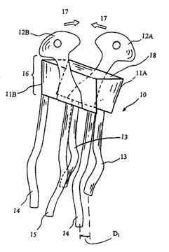
FIG. 1 is a reusable miniclip apparatus to facilitate locating, orienting and
releasably securing a bioprosthetic heart valve leaflet during the valve
implantation in
accordance with one embodiment of the present invention.
FIG. 2 is a simple miniclip apparatus of FIG. 1 at a released state.
FIG. 3 is a prior art clipping using a clamp for holding the valve leaflet and
a
portion of the aortic artery wall together during implantation of an aortic
valve in a body
channel.
FIG. 4 is an illustrative example of the current device holding a pledget as
part of
the aortic valve leaflet fastening procedures.
CA 02505732 2005-05-13
WO 2004/045370 PCT/US2003/036318
FIG. 5 is another illustration of applying the miniclip apparatus for holding
the
valve leaflet and a portion of the aortic artery wall together for fastening.
FIG. 6 is a traverse cross-sectional view of the composite to be sutured
together,
section 1-1 of FIG. 5.
5 Detailed Description of Exemplary Embodiments
Referring to FIGS. 1 to 6, what is shown is an embodiment of a releasably
fastening device used in aortic valve implantation, wherein the device is to
facilitate
accurate and quick locating, orienting, and releasably securing bioprosthetic
heart valve
leaflets during the valve implantation. While the description sets forth
various
embodiment specific details, it will be appreciated that the description is
illustrative only
and should not to be construed in any way as limiting the invention.
Furthermore, various
applications of the invention, and modifications thereto, which may occur to
those who are
skilled in the art, are also encompassed by the general concepts described
below.
Aortic stenosis is a disease of the aortic valve in the left ventricle of the
heart. This
aortic valvular orifice can become tightly stenosed, and therefore the blood
cannot
anymore be freely ej ected from the left ventricle. In fact, only a reduced
amount of blood
can be ejected by the left ventricle which has to markedly increase the
ventricular chamber
pressure to pass the stenosed aortic orifice. In such aortic diseases, the
patients can have
syncope, chest pain, and mainly difficulty in breathing. Aortic stenosis is a
very common
disease in people above sixty years old and occurs more and more frequently as
the subject
gets older. The evolution of such a disease is disastrous when symptoms of
cardiac failure
appear and many patients die in the year following the first symptoms of the
disease. The
commonly available treatment is the replacement of the stenosed aortic valve
by a
prosthetic valve via open-heart surgery.
The natural leaflets include arcuate cusp portions separated by common
commissure portions. If the natural valve has three leaflets, and has a
vertically oriented
flow axis, the leaflets are evenly distributed circumferentially 120 degrees
apart with
lower cusp portions and upstanding commissure portions. The commissure
portions are
connected between the cusp portions and are generally axially aligned along
the aortic
wall. The annular root of an aortic valve is composed of fibrous tissue and
generally
conforms to the undulating perimeter of the valve to support the leaflets.
CA 02505732 2005-05-13
WO 2004/045370 PCT/US2003/036318
6
Carpentier in U.S. Pat. No. 6,338,740, entire contents of which are
incorporated
herein by reference, discloses a heart valve with radially moveable cusps and,
commissures wherein the commissures may be pivotally or flexibly coupled.
Carpentier
'740 also discloses a multi-legged holder having legs alternating between each
cusp and
commissure to be used in the implantation. Brendzel et al. in U.S. Pat. No.
6,391,053,
entire contents of which are incorporated herein by reference, discloses a
prosthetic heart
valve having valve housing and a cuff positioned such that prosthesis is
attached in a
supraannular position relative to a tissue annulus of the heart. Neither
patent discloses a
simple miniclip apparatuses to facilitate orienting and releasably securing
bioprosthetic
heart valve leaflets during the valve implantation.
The tissue valve or tissue valve leaflets are generally chemically treated to
render
the valve suitable for long-term implantation in human. Glutaraldehyde is a
chemical most
often used for tissue fixation. The tissue fixation is well known to an
ordinary artisan who
is skilled in the art and does not constitute a part of the present invention.
In this respect, implanting the aortic heart valve of the present invention
involves
excising the natural leaflets and attaching the prosthetic heart valve
proximate the fibrous
annulus, but also in part up the aortic wall. The attachment means may be
sutures, staples,
adhesives, or otherwise, that is anchored into the aortic wall itself,
adjacent to the fibrous
annulus.
Suture is biocompatible, flexible and long lasting. The suture arrangement
useful in
the present invention comprises a first needle and a second needle connected
by length of
suture. After passing the first and the second needles from within the aorta
through the
wall of aorta and valve leaflet outwardly, the needles may then be pulled away
from the
aorta wall to thread the suture through the tissue.
FIG. 1 shows a simple miniclip apparatus to facilitate accurately and quickly
orienting and releasably securing a bioprosthetic heart valve leaflet during
the valve
implantation in accordance with one embodiment of the present invention. The
miniclip
apparatus is absent of a coiled spring or other complicate structure that may
retain debris
from previous surgeries, even after autoclaving.
The miniclip apparatus 10 of the present invention for releasably stabilizing
or
fixing a leaflet onto an aortic artery wall during aortic valve implantation
may comprise a
clip base 16 having a first clip member 11B consisting of a plurality of first
prongs (14 and
15) and an opposite second clip member 11A consisting of a plurality of second
prongs
CA 02505732 2005-05-13
WO 2004/045370 PCT/US2003/036318
7
13, wherein the first prongs (14, 15) and the second prongs 13 sized and
configured for
releasably clipping and stabilizing the leaflet 25 in conjunction with the
aortic artery wall
22 (shown in FIGS. 5 and 6). The miniclip apparatus 10 further comprises an
actuator
assembly 12A, 12B operable using one hand located at the clip base 16, wherein
the first
clip member 11B moves away from the second clip member 11A when the actuator
assembly 12A/12B is activated. The first clip member 11B and the second clip
member
11A are connected through a middle member 1S with a preset spring effect. One
method
for activating the actuator assembly is to press the assembly elements 12A and
12B toward
each other as shown by an arrow 17 in FIG. 1.
In a further aspect of the present invention, the first clip member is
configured
essentially parallel to the second clip member. In another aspect, the first
clip member and
the second clip member are preshaped and configured enabling the two clip
members to
clip and stabilize the leaflet in conjunction with the aortic artery wall when
the actuator
assembly is not activated. Elements of the miniclip may be made of stainless
steel, Nitinol
or other suitable metal that could be preshaped and configured with the
intended clipping
properties. In some aspect, the plurality of first prongs further comprises a
first set of
prongs 14 and a second set of prongs 15, and wherein a proper distance, Dl, is
sized and
configured between the first set 14 and the second set 15 of prongs for
releasably holding
a pledget 31 therebetween. The proper distance D1 is sized and configured to
snugly hold
the pledget 31. The proper distance is increased from D1 of FIG. 1 to DZ of
FIG. 2 when
the actuator assembly is activated. FIG. 2 shows a simple miniclip apparatus
of FIG. 1 at a
released state when the actuator assembly is activated.
FIG. 3 is a prior art clipping illustration using a clamp 26 for holding the
valve
leaflet 25 and a portion of the aortic artery wall 22 together during
implantation of an
aortic valve in a body channel. The clamp 26 generally includes two jaws 24A,
24B that
may have a wide variety of preset clamping pressures, which are mostly used
for vessel
occlusion. During operations, one hand is needed to hold the clamp 26 for
fastening
purposes. The conventional clamp does not have additional features of holding
at least one
pledget along with the general releasably clipping Rinction as shown in FIG.
4, wherein
the miniclip of the present invention is simply lightweight and can be left
alone without a
hand to hold it.
FIG. 4 is an illustrative example of the current device 10 holding a pledget
31 as
paxt of the aortic valve leaflet fastening procedures. FIG. 5 shows another
illustration of
applying the miniclip apparatus 10 for holding the valve leaflet 25 and a
portion of the
aortic artery wall 22 together for fastening. In operations, the miniclips
each holding the
CA 02505732 2005-05-13
WO 2004/045370 PCT/US2003/036318
8
composite of a commissure of one leaflet toward the aortic artery wall can be
placed at the
edge 21 of the aortic artery wall 22 at an angle a, (3, and 0, wherein each
angle of a, (3, or
0, may be about 120 degrees or with any predetermined angles.
FIG. 6 shows a traverse cross-sectional view of the composite to be sutured
together, section 1-1 of FIG. 5. The composite comprises a first set of prongs
14 and a
second set of prongs 15 sandwiching a first pledget 31B. The composite further
comprises
the combined set of prongs 14/15 and the plurality 13 of second prongs
sandwiching the
aortic artery wall 22, the commissure portion of the leaflet 25 and optionally
a second
pledget 31A. In operations, the composite is temporarily held by a miniclip 10
of the
present invention and is ready for passing a suture to fasten the composite
together. After
fastening, the miniclip 10 is easily released from the composite by slightly
activating the
actuator assembly 12A/12B. In another aspect, the miniclip is to releasably
stabilize and
hold the composite that comprises a synthetic tab that is securely attached to
the distal end
of the leaflet, rather than the leaflet itself, wherein the synthetic tab may
be made of
expanded polytetrafluoroethylene (Teflon), polyester (Dacron~), silicone
(SllastlCTM), ,
polyurethane (Pellethane~) or other suitable synthetic material.
The edge 23 of the commissure 25 is generally oriented at a distance D3 lower
than
the edge 21 of the aortic artery wall 22. The distance D3 is at least one
millimeter,
preferably at 2-3 millimeters.
It is one aspect of the present invention to utilize the miniclip 10 of the
present
invention for assisting the aortic valve implantation. Therefore, it is one
object of the
present invention to provide a method for releasably stabilizing three
leaflets of an aortic
valve onto an aortic artery wall during aortic valve implantation comprising:
(a) orienting
a com~nissure of one of the three leaflets toward the aortic artery wall to
form a double-
layer composite, having an interior side and an exterior side; (b) selecting
one miniclip
apparatus of claim 1; (c) activating the actuator assembly of the miniclip
apparatus while
simultaneously inserting the miniclip apparatus over the double-layer
composite, wherein
the first clip member lies on the interior side of the composite and the
second clip member
lies on the exterior side of the composite; (d) deactivating the actuator
assembly to
releasably clipping and stabilizing the first leaflet in conjunction with the
aortic artery
wall; and (e) repeating the steps of (a) to (d) for additional two miniclip
apparatuses on the
remaining two leaflets, wherein the three miniclip apparatuses are spaced
apart at about
120 degrees or any predetermined angle.
CA 02505732 2005-05-13
WO 2004/045370 PCT/US2003/036318
9
In one aspect, the method may fixrther comprise, after the step (a), a step of
inserting at least a pledget along with at least one of the double-layer
composites to form a
three-layer composite or a multiple-layer composite, the multiple-layer
composite having
an interior side and an exterior side. The pledget may be selected from a
group consisting
of an expanded polytetrafluoroethylene (Teflon), polyester (DacronTM),
silicone
(SilasticTM), polyurethane (Pellethane~) or other suitable synthetic material.
In another aspect, the method may further comprise, after the step (e), a step
of
passing a suture through the three-layer or multiple-layer composite, wherein
the step of
passing a suture may be carried out by passing a needle of the suture from the
anterior side
of the multiple-layer composite. The method may fiu-ther comprise a step of
passing a
second needle of the suture from the anterior side of the multiple-layer
composite,
followed by a step of removing the miniclip apparatus from the multiple-layer
composites.
In one embodiment, the method May further comprise a step of removing at least
a
portion of a patient's heart valve by means of a cutting tool. In some aspect
of the present
invention, the cutting tool may be made of an electrically conductive metal
and
radiofrequency energy is provided to the cutting tool for enhanced valve
removal. The
high frequency energy ablation is well known to an ordinary artisan who is
skilled in the
ar t.
In operations, the step of orienting the commissure of the leaflets against
the aortic
artery wall may be earned out by inserting a dilator into a center of the
aortic valve. The
dilator can be a balloon-based device or a basket-type expandable device. The
dilator and
its use are well known to an ordinary artisan skilled in the art.
From the foregoing description, it should now be appreciated that a miniclip
apparatuses to facilitate locating, orienting, and releasably securing
bioprosthetic heart
valve leaflets during the valve implantation and method of use thereof have
been
disclosed. While the invention has been described with reference to a specific
embodiment, the description is illustrative of the invention and is not to be
construed as
limiting the invention. Various modifications and applications may occur to
those who are
skilled in the art, without departing from the true spirit and scope of the
invention, as
described by the appended claims.