Note: Descriptions are shown in the official language in which they were submitted.
CA 02505760 2005-O1-17
WO 2004/045675 PCT/US2003/037045
A METHOD AND APPARATUS FOR ANCHORING OF PACING LEADS
Related Applications
The present application is related to U.S. Provisional Patent Applications
serial
no. 60/426,773, filed on Nov. 15, 2002; serial no. 60/476,487, filed on June
6, 2003;
60/479,399, filed on June 18, 2003; and serial no. 60/464,437, filed on April
22, 2003,
which are each incorporated herein by reference and to which priority is
claimed
pursuant to 35 USC 119.
Background of the Invention
1. Field of the Invention
The invention relates to the field of cardiology and in particular to
apparatus and
methods for pacemaker implantations.
2. Description of the Prior Art
In many cases pacemaker introducers are precurved for steerability and use in
the coronary sinus. Being precurved, such introducers cannot be rotated for
the
purpose of setting a screw-in anchor into myocardium, since their distal ends
wobble
uncontrollably while being rotated. It is nevertheless advantageous to anchor
an
introduces when similarly implanting or anchoring the much more flexible
pacemaker
lead. If the pacemaker lead is not supported by a fixed-in-place introduces,
the
pacemaker lead itself can either push back and displace the introduces with
the result
that the pacemaker lead is implanted in the wrong place, or the pacemaker lead
simply
1
CA 02505760 2005-O1-17
WO 2004/045675 PCT/US2003/037045
"spaghettis" or unpredictably folds on itself rather than being controllably
driven in or
implanted into the myocardial position selected by the surgeon.
Pacing of the left ventricle 14 diagrammatically shown in Fig. 1 is
conventionally
performed by placement or implantation of a wire or lead 10 in the coronary
venous
system 12 as diagrammatically shown in the cutaway view of the coronary vein
16 in
Fig. 2. However, not every location of the wall of the left ventricle 14,
which needs to be
paced, is accessible to a lead through the coronary venous system 12 as can be
readily
appreciated by the vascular anatomy depicted of the coronary venous system 12
and
coronary arterial system 18 depicted in the simplified perspective view of
Fig. 2.
Brief Summary of the Invention
The invention is an introduces having a distal end comprising an anchor
provided
on the distal end of the introduces for attachment of the distal end of the
introduces into
tissue. The anchor attaches at or near the surface of a body cavity, for
example in. the
vascular space, or more particularly in the cardial space. When the introduces
is
anchored in the cardial space a pacemaker lead is guided through the
introduces and
anchored therein while the introduces is anchored within cardial space.
The invention is directed to anchoring introducers and leads throughout the
vascular space, including anchoring into any one of the walls of the heart
chambers,
into any vascular location, and into body cavities, usually closely related to
the vascular
system such as the pericardial space. The anchoring can be realized through a
plurality
of different means such as screw anchors, barbed anchors, piercing tools with
barbs or
distal inflatable balloons, grabbing tools or suction anchors.
In one embodiment the invention is defined as a method of implanting a
pacemaker lead into the pericardial space or microvasculature of a heart
comprising the
2
CA 02505760 2005-O1-17
WO 2004/045675 PCT/US2003/037045
steps of: disposing an elongate instrument into the venous system of the
heart; exiting
the venous system; disposing the elongate instrument or a different elongate
instrument
into the pericardial space or microvasculature at a predetermined location;
and
implanting a pacemaker lead at the predetermined position.
The step of implanting a pacemaker lead at the predetermined position
comprises the step of implanting the lead in a position adjacent to, on or in
the surface
of the left ventricle.
Preferably, the step of implanting a pacemaker lead at the predetermined
position comprises the step of implanting the lead in a position of optimized
pacing
efficacy.
In one embodiment the step of disposing an elongate instrument or a different
elongate instrument into the pericardial space or microvasculature at a
predetermined
location comprises the step of disposing the elongate instrument or a
different elongate
instrument through the microvasculature into the pericardial space.
In another embodiment the step of disposing an elongate instrument or a
different elongate instrument into the pericardial space or microvasculature
comprises
the step of disposing the elongate instrument or a different elongate
instrument into a
first venous bed.
In another embodiment the step of disposing an elongate instrument or a
different elongate instrument into the pericardial space or microvasculature
comprises
the step of disposing the elongate instrument or a different elongate
instrument into the
vascular mesh.
In still another embodiment the step of disposing an elongate instrument or a
different elongate instrument into the pericardial space or microvasculature
comprises
3
CA 02505760 2005-O1-17
WO 2004/045675 PCT/US2003/037045
the step disposing the elongate ,instrument or a different elongate instrument
from the
first venous bed, through the vascular mesh into a second venous bed.
t
The method further comprises the step of anchoring the implanted pacemaker
lead in the pericardial space or microvasculature.
In one embodiment the step of implanting the pacemaker lead comprises
implanting the pacemaker lead in the microvasculature and further comprises
the step
of dilating the microvasculature prior to implanting the pacemaker lead
therein.
In another embodiment the step of exiting the venous system comprises the step
of puncturing a vein in the venous system.
In still another embodiment the step of exiting the venous system comprises
the
step of entering the vasculature communicated with the venous system.
In yet another embodiment the step of exiting the venous system comprises the
step of exiting the vasculature and entering the pericardial space.
The step of disposing the elongate instrument or a different elongate
instrument
into the pericardial space or microvasculature at a predetermined location
comprises
the step of disposing an introduces, catheter, guidewire, balloon, dilator,
needle and/or
lead.
The invention is also defined as apparatus or a surgical kit of instruments
for
performing each of the foregoing steps separately or in any combination.
The invention is still further defined as an apparatus for implanting a
pacemaker
lead into heart tissue comprising an inner introduces which is steered into
the heart; a
first anchor provided on a distal end of the introduces; a pacemaker lead
telescopically
disposed through the inner introduces; and a second anchor provided on a
distal end of
the pacemaker lead.
4
CA 02505760 2005-O1-17
WO 2004/045675 PCT/US2003/037045
The first anchor has an inner diameter large enough to permit telescopic
disposition therethrough of the second anchor and pacemaker lead.
The apparatus may in some embodiments further comprise an outer biased
introduces through which the inner introduces is telescopically disposed and
steered to
an implantation site.
In one embodiment the pacemaker lead is rotatable within the inner introduces
and where the second anchor screws into the heart tissue at an implantation
site and
wherein the first anchor maintains the inner introduces in position while the
second
anchor screws into the heart tissue at the implantation site. The first anchor
can be
disengaged from the heart tissue at the implantation site, after the second
anchor is
implanted without dislodgement of the second anchor and pacemaker lead.
In a first embodiment the first anchor is a screw anchor with a first sense of
screw advancement and where the second anchor is a screw anchor with a second
sense of screw advancement opposite to the first sense of screw advancement.
In all embodiments it is possible that the outer introduces and inner
introduces are
separable, including sliceable, splittable, peelable, or tearable.
In yet another embodiment the second anchor is rotatable on and captured by
the inner introduces and drivable by an elongate instrument. In this case the
apparatus
further comprises the elongate instrument and a lumen defined through the
inner
introduces through which the elongate instrument is disposed. The second
anchor is
typically, but not necessarily telescopically disposable from the distal end
of the inner
introduces.
The apparatus may further comprise a plurality of first anchors coupled to the
inner introduces.
The invention contemplates that the inner, or outer both introducers may
biased.
5
CA 02505760 2005-O1-17
WO 2004/045675 PCT/US2003/037045
In an illustrated embodiment the first anchor comprising a fish-hook anchor.
The first anchor may be movably coupled to the inner introduces and is
deployed
when the inner introduces is telescopically advanced out of the distal end of
the outer
introduces. The first anchor is resiliently dispose within the inner
introduces and
automatically resiliently deployed when the inner introduces is telescopically
advanced
out of the distal end of the outer introduces. In one implementation the
apparatus
further comprises a wire coupled to the first anchor and which is operative
when
manipulated to rotate the anchor to extend out of or be retracted in the inner
introduces.
The first anchor is resiliently biased to be normally retracted within the
inner introduces
and where the wire is operated by applying a tensile force to rotate the first
anchor to an
extended configuration out of the inner introduces.
In still a further embodiment the apparatus further comprises an elongate
instrument and a lumen defined through the inner introduces through which the
elongate
instrument is disposed, the first anchor being a piercing tool coupled to a
distal end of
the elongate instrument and having at least one barb disposed thereon. The
first anchor
comprises a plurality of barbs on the piercing tool, which may take the form
of a plurality
of stiff angled fibers disposed on the piercing tool.
In another embodiment the first anchor comprises a hollow needle with an
inflatable tip balloon.
In still another embodiment the first anchor comprises a bimetallic wire which
can
be differentially tensioned and curved to form a temporary distal hook.
In all of the embodiments the apparatus may further comprise a hemostatic
valve
coupled to the inner introduces.
In yet more embodiments the first anchor is a suction device. In one example,
the suction device comprises a suction cavity defined in the inner introduces
with a
6
CA 02505760 2005-O1-17
WO 2004/045675 PCT/US2003/037045
peripheral lip to assist in allowing a suction attachment to the heart tissue.
The suction
cavity may be positioned on a lateral surface of the inner introduces.
The inner introduces has a lumen and the suction device comprises a means for
providing suction to the lumen and communicating the suction to the distal
orifice of the
lumen at the distal tip of the inner introduces. The means for communicating
the suction
to the distal orifice of the lumen at the distal tip of the inner introduces
comprises a
central lumen defined through the inner introduces through which central lumen
the
pacemaker lead is disposed. In another embodiment the lumen is an auxiliary
lumen
defined through the inner introduces and where the means for communicating the
suction to the distal orifice of the lumen at the distal tip of the inner
introduces comprises
a communication of the lumen with the distal orifice of the lumen at the
distal tip of the
inner introduces.
While the apparatus and method has or will be described for the sake of
grammatical fluidity with functional explanations, it is to be expressly
,understood that
the claims, unless expressly formulated under 35 USC 112, are not to be
construed as
necessarily limited in any way by the construction of "means" or "steps"
limitations, but
are to be accorded the full scope of the meaning and equivalents of the
definition
provided by the claims under the judicial doctrine of equivalents, and in
the.case where
the claims are expressly formulated under 35 USC 112 are to be accorded full
statutory
equivalents under 35 USC 112. The invention can be better visualized by
turning now
to the following drawings wherein like elements are referenced by like
numerals.
7
CA 02505760 2005-O1-17
WO 2004/045675 PCT/US2003/037045
Brief Description of the Drawings
Fig. 1 is a side cross sectional view of the distal end of an outer introducer
comprised of a conventional precurved or biased outer introducer with an
unbiased or
flexible, but rotationally stiff inner telescopic introducer.
Fig. 2 is a side cross sectional view of the distal end of a second embodiment
of
the telescopic introducer, which is comprised of a biased introducer through
which the
pacemaker lead with the distal anchor is implanted.
Fig. 3 is a side cross sectional view of the distal end of a third embodiment
of
telescopic introducer which is comprised of a prebiased introducer through
which the
pacemaker lead with the distal anchor is implanted.
Fig. 4 is directed to another embodiment where an outer biased introducer is
used to steer to the approximate site in the heart.
Fig. 5 is a simplified diagrammatic side cross section view of a human heart
illustrating the pericardial space.
Fig. 6 is a simplified diagrammatic, partially cut-away side view of a human
heart
illustrating the vascular system of the heart.
Fig. 7 is a simplified diagram of the venous vascular mesh on the ventricular
surface.
Fig. 8 is a simplified diagrammatic side cross section view of in enlarged
scale of
the region in circular dotted outline in Fig. 3 illustrating dilation of the
microvasculature
with a balloon.
Fig. 9 is a simplified diagrammatic side cross section view of Fig. 4 showing
the
disposition of a catheter into the microvasculature.
8
CA 02505760 2005-O1-17
WO 2004/045675 PCT/US2003/037045
Fig. 10 is a simplified diagrammatic side cross section view of Fig. 5 showing
the
disposition of a guidewire and introducer into the microvasculature.
Fig. 11 is a simplified diagrammatic side cross section view of Fig. 5 showing
the
disposition of a pacemaker lead into the microvasculature.
Fig. 12 is a simplified diagrammatic side view of the disposition of an
elongate
instrument from a first venous bed through the venous vascular mesh and
implantation
in a second venous drainage area.
Fig. 13 is a simplified side cross-sectional view of a heart having a chamber
into
which an introducer with a suction anchor has been endovascularly disposed.
Fig. 14 is an enlarged simplified side cross-sectional view of the distal tip
of the
introducer of Fig. 13.
Fig. 15 is a side elevational view of the distal tip of the introducer of Fig.
14.
Fig. 16 is a diagrammatic side cross-sectional view of another embodiment of a
suction anchor where the suction is provided to the distal orifice of catheter
either
through main lumen between the clearance of lumen and pacemaker lead or
through an
auxiliary lumen defined in the catheter with a distal communication with lumen
at or near
orifice.
Fig. 17 is a diagrammatic side cross-sectional view of an embodiment wherein
suction is provided only through the axial lumen.
Fig. 18 is a diagrammatic side cross-sectional view of an embodiment wherein a
grabbing tool is provided as the anchor through the axial lumen.
The invention and its various embodiments can now be better understood by
turning to the following detailed description of the preferred embodiments
which are
presented as illustrated examples of the invention defined in the claims. It
is expressly
9
CA 02505760 2005-O1-17
WO 2004/045675 PCT/US2003/037045
understood that the invention as defined by the claims may be broader than the
illustrated embodiments described below.
Detailed Description of the Preferred Embodiments
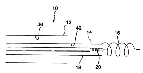
Fig. 1 is a side cross sectional view of the distal end of an outer introduces
10
comprised of a conventional precurved or biased outer introduces 12 with an
unbiased
or flexible, but rotationally stiff inner telescopic introduces 14.
Alternatively, inner
introduces 14 may be biased. Telescopic inner introduces 14 is provided at or
near its
distal end with a conventional screw-in anchor 16. Anchor 16 has an inner
diameter
which is large enough so that when pacemaker lead 18 is telescopically
disposed
through a axial lumen 42 in inner introduces 14 with its corresponding distal
anchor 20,
distal anchor 20 can be telescopically disposed through anchor 16 during
fixation into
the myocardium without engaging anchor 16.
Being in the preferred embodiment unbiased, inner introduces 14 easily stays
on
position at the location defined by the distal end of outer introduces 12 when
inner
telescopic introduces 14 is rotated. When pacemaker lead 18 is then rotated
and
anchored, the support of anchored introduces 14 keeps pacemaker lead 18
supported
and similarly on-position at the location defined by the distal end of outer
introduces 12.
Once pacemaker lead 18 is implanted, inner introduces 14 can then be
unscrewed without dislodging anchor 20 and removed. To enhance the
compatibility of
adjacent implantations of anchor 20 coaxially inside anchor 16, anchor 20 and
anchor
16 can be provided with helicity of opposite senses. For example, if anchor 20
is a
right-hand screw, anchor 16 is provided as a left-hand screw. In this manner,
when
anchor 16 is unscrewed, it is rotated clockwise when viewed from the proximal
end of
CA 02505760 2005-O1-17
WO 2004/045675 PCT/US2003/037045
introduces 10. Any clockwise rotation transmitted by anchor 16 in any way to
anchor 20
thereby serves to screw in or tighten anchor 20.
It must be understood that introducers 12 and 14 may be sliceable, tearable,
peelable, or separable in any way now known or later devised, so that they are
easily
removed over any pacemaker hub or connector (not shown) at the proximal end of
pacemaker lead 18.
Fig. 2 is a side cross sectional view of the distal end of a second embodiment
of
telescopic introduces 10 which is comprised of a biased introduces 22 through
which
pacemaker lead 18 with distal ,anchor 20 is implanted. A parallel lumen 32 is
defined in
the wall of introduces 22 through which a torsional wire 30 is disposed. Wire
30 has a
termination 28 which is or can be coupled with a captive screw 34 which has a
distal
anchor 24. It is contemplated that screw 34 will be retained within lumen 32
until
deployment, at which time it is then distally extended by being pushed by
wire' 30 and
then rotated by wire 30 to fix anchor 24 into the adjacent myocardium. In this
embodiment, anchors 20 and 24 are not telescopic, but are deployed in
parallel.
It is to be understood that instead of a captive screw 34, wire 30 and anchor
24
may be integral and simply delivered through auxiliary lumen 32. A plurality
of such
auxiliary lumens 32 and wire 30/anchor 24 combinations may be provided and
employed in a radial pattern at the distal end of introduces 22.
Fig. 2 is described above as a nontelescopic system. However, it must be
understood as shown in Fig. 2 that introduces 22 may be biased or unbiased and
similarly telescopically disposed through an axial lumen 44 of a biased outer
introduces
12. As in the case of Fig. 1, the outer introduces 12 is used to steer inner
introduces 22
to the approximately vicinity of the implantation site, inner introduces 22 is
telescopically
11
CA 02505760 2005-O1-17
WO 2004/045675 PCT/US2003/037045
advanced out of introduces 12 and anchored in place. Pacemaker lead 18 is then
telescopically advanced in lumen 42 and implanted using anchor 20.
Fig. 3 is a side cross sectional view of the distal end of a third embodiment
of
telescopic introduces 10 which is comprised of a prebiased introduces 36
through which
pacemaker lead 18 with distal anchor 20 is implanted. The distal portion or
end of
introduces 36 is provided with one or more barbless hooks or "fish hooks",
which are
normally resiliently retained within recesses defined in the wall of
introduces 36. Hooks
38 can be deployed by pulling tension wires 40 coupled to hooks 38 to rotate
hooks 38
out of the recesses in introduces 36 to a position where they may penetrate
radially
adjacent myocardium or vascular tissue. Once hooks 38 are deployed by pulling
on
wires 40, pacemaker 18 is anchored from the end of anchored introduces 36.
Once
pacemaker lead 18 is anchored, the tension on wires 40 is released, allowing
springs or
other resilient means attached to hooks 38 to return them to their undeployed
configuration with recesses within introduces 36.
Again it must be understood that while the embodiment of Fig. 3 is described
above as a nontelescopic system, in another embodiment introduces 36 may be a
prebiased on unbiased telescopic introduces disposed through axial lumen 44 in
a
biased outer introduces 12. As in the case of Fig. 1, the outer introduces 12
is used to
steer inner introduces 36 to the approximately vicinity of the implantation
site, inner
introduces 36 is telescopically advanced out of introduces 12 and anchored in
place.
Pacemaker lead 18 is then telescopically advanced in lumen 42 and implanted
using
anchor 20. Thus, the parallel-lumen screw anchors of and hooks as shown in
Figs. 2
and 3 respectively can be substituted for the distal screw anchor 16 and
single lumen
introduces 14 of Fig. 1.
12
CA 02505760 2005-O1-17
WO 2004/045675 PCT/US2003/037045
Fig. 4 is directed to another embodiment where outer biased introduces 12 is
used to steer to the approximate site in the heart. Inner biased or unbiased
introduces
36 is then telescopically disposed through axial lumen 44 in introduces 12 and
position
near or adjacent to a position on the heart wall 52 where pacemaker
implantation is to
occur. In the illustrated embodiment implantation is made into the intra-
atrial septum 52
from the right atrium. A wire 48 is disposed in a parallel or auxiliary lumen
46 in inner
introduces 36 and extends distally from the end of introduces 36. The distal
end of wire
48 is or is fitted with a solid needle 54 which is able to puncture and to be
forced
through the smooth wall of intra-atrial septum 52. Wire 48 is further arranged
or fitted
with one or more flexible barbs, such as a plurality of flexible, but stiff
fibers or filaments
50, which are biased to angle proximally on wire 48 like a brush. Needle 54
and
filaments 50 can thus easily be advanced distally and pushed through intra-
atrial
septum 52 to serve as a temporary anchor of inner introduces 36 to intra-
atrial septum ,
52. Needle 54 may penetrate entirely through intra-atrial septum 52 into the
left atrium.
Wire 48 resists backing out or being pulled out of septum 52 by means of the
proximally
directed bias of filaments 50 which act as barbs. However, the anchoring
strength is not
so great that wire 48 cannot be later manually withdrawn taking needle 54 and
filaments
50 with it.
With inner introduces 36 temporarily anchored to intra-atrial septum 52,
pacemaker lead 18 with its distal screw anchor 20 is distally extended from
axial lumen
42 in introduces 36 and screwed into intra-atrial septum 52. The anchored
inner
introduces 36 provides enough purchase or support to allow anchor 20 to be
screwed
into intra-atrial septum 52 without pacemaker lead 18 backing off or simply
bending and
collapsing against septum 52. The stiffness, degree of bias and number of
filaments 50
are chosen to provide enough anchoring force that inner introduces 36, which
is
13
CA 02505760 2005-O1-17
WO 2004/045675 PCT/US2003/037045
frictionally coupled to wire 48 in lumen 46, cannot be backed out by the
reaction force
applied to pacemaker' lead 18, which is frictionally coupled to inner
introduces 36 in
lumen 42, by the screwing action of anchor 20 of pacemaker lead 18 into septum
52.
Once pacemaker lead 18 is firmly anchored into or through septum 52, wire 48,
filaments 50 and needle 54 can be pull out by applying sufficient tension to
wire 48 from
its proximal end.
It must be understood that while the temporary anchoring of wire 48 is shown
by
means of a plurality of filaments 50, there are many other equivalent ways by
which
temporary anchoring of wire 48 can be achieved. For example, needle 54 may be
hollow and carry a small inflatable and deflatable tip balloon, or wire 48
and/or needle
54 may be a bimetallic wire which can be differentially tensioned and curved
to form a
temporary distal hook by means of an electrical current and ohmic heating of
the
bimetallic wire, or simply by exposure to the body heat.
Fig. 18 is another embodiment similar to Fig. 4 in which instead of the
elongate tool
or wire 48 in lumen 46 a stylet or other tool 56 is used which is terminated
with a grabbing
tool 58. In the illustrated embodiment grabbing tool 58 comprises a set of
pincers with one
or two movable jaws which are manipulated by stylet 56 to open and close, as
well as to
be telescopically disposable in lumen 46. The mechanism used to manipulate
tool 58 may
be any type of actuation device now known or later devised for actuating one
or more
opposing jaws, such as a hollow wire with an inner telescoping solid core wire
with one
jaw fixed to the hollow wire and the second jaw rotated about the first jaw
and coupled to
the core wire to rotate the second jaw about its pivot point on the first jaw.
Alternatively,
grabbing tool 58 may have one jaw fixed to the distal end of introduces 36
with the second
jaw rotatable about a pivot point also fixed to the distal end of introduces
36 or the first jaw.
The two jaws may be resiliently biased to a closed position. Style 56 then
takes the form
14
CA 02505760 2005-O1-17
WO 2004/045675 PCT/US2003/037045
of a tension wire 56 to rotate the second jaw relative to the first jaw to
open and close the
grabbing tool 58. In any embodiment grabbing tool 58 is operative to
temporarily pinch,
seize, or grab hold of some heart or vascular tissue to temporarily fix
introduces 36 in
place. In the case where stylet 56 is telescopically slidable within lumen 46,
style 56 can
be vascularly disposed and grabbing tool 58 actuated to fix it to a location.
Introduces 36
is then guided into position over anchored stylet 56 until the distal end of
introduces 36 is
proximate to or adjacent the tissue site to which the lead 18 is to be
implanted. Stylet 56
may then be longitudinally fixed to introduces 36, such as by a proximal
locking or pinching
device (not shown), and then lead 18 implanted in a conventional manner with
introduces
36 securely but temporarily fixed into position.
While this disclosure is directed to the anchoring of a catheter or
introduces, it
must be expressly understood that the disclosed catheter or introduces
includes within
its scope any sliceable, splittable, peelable, tearable or separable catheter
or introduces
now known or later devised, as well as catheters or introducers, which cannot
be
separated in any of these manners. In addition, whether or not a hemostatic
valve is
associated with the catheter or introduces of the invention, and if so,
whether or not the
hemostatic valve is separable or not, together with or separately from the
catheter or
introduces, is all expressly included within the scope of the disclosed
invention.
For example, splittable valves of the type disclosed in Lee, "Splittable
Hemostatic
Valve and Sheath and the Method for Using the Same", U.S. Patent 5,125,904
(1992)
and 5,312,355 (1994), which are incorporated herein by reference, are
included.
Catheters and introducers are included of the type as disclosed in: Kurth,
"Permanent
Catheter with an Exterior Balloon Valve and Method of Using the Same," U.S.
Patent
5,792,118 (1998), "Method and Apparatus for Insertion of Elongate Instruments
Within a
Body Cavity," U.S. Patent Application serial no. 09/708,150 (2000), "A
Temporarily
CA 02505760 2005-O1-17
WO 2004/045675 PCT/US2003/037045
Secured Guidewire and Catheter for Use in the Coronary Venous System and
Method
of Using the Same," U.S. Patent Application serial no. 10/365,890 (2003), "A
Method
and Apparatus for a Suction-Anchored Introducer for Pacemaker Implantation,"
U.S.
Provisional Patent Application serial no. 60/464,437 (2003), "Method and
Apparatus for
Implantation of Left Ventricular Pacing Leads Between the Epicardium and
Pericardium," U.S. Provisional Patent Application serial no. 60/426,773
(2002), and "A
Tool for Placement of Dual Angioplasty Wires in the Coronary Sinus
Vasculature," U.S.
Provisional Patent Application serial no. 60/408,385 (2002); Worley et.al.,
"Introducer
for Accessing the Coronary Sinus of a Heart," U.S. Patent Application serial
no.
10/139,551 (2002), "A Telescopic, Peel-Away Introducer and Method of Using the
Same," U.S. Patent Application serial no. 10/139,554 (2002), "A Telescopic,
Peel-Away
Introducer and Method of Using the Same," U.S. Patent Application serial no.
10/139,554 (2002), "A Telescopic Introducer with a Compound Curvature for
Inducing '
Alignment and Method of Using the Same", U.S. Patent Application serial no.
10/202,158; and Kurth et.al;, "Introducer and Hemostatic Valve Combination and
Method of Using the Same, " U.S. Patent Application serial no. 10/234,686
(2002), "A
Compression Fitting for an Introducer Coupled to a Hemostatic Valve," U.S.
Patent
Application serial no. 10/277,476 (2002), which are all incorporated herein by
reference.
Consider now pacemaker anchoring in the pericardial space. As shown in the
diagrammatic view of Fig. 5 the human heart 128 is contained within a conical
sac of
serous membrane called the pericardial sac or simply the pericardium 124 that
encloses
the heart and the roots of the great blood vessels of humans and vertebrates
in general.
The epicardium 126 is the visceral part of the pericardium 124 that closely
envelops the
heart. In between the epicardium 126 and the outer surface of the cardiac
muscle and
vasculature is a space lying above the coronary vasculature 112, 118 called
the
16
CA 02505760 2005-O1-17
WO 2004/045675 PCT/US2003/037045
pericardial space 122. This pericardial space 122 may be void or partially
filled with a
lubricious fluid. In diseased states the pericardium may contain a substantial
amount of
fluid.
A pacemaker lead is implanted in a heart into the pericardial space, on or in
the
epicardium or in the microvasculature by: disposing an elongate instrument
into the
venous system of the heart; puncturing system at a predetermined position or
entering
the microvasculature of the venous system; disposing the elongate instrument
into the
pericardial space, epicardium or in the microvasculature at a predetermined
location in
the pericardial space, epicardium or in the microvasculature; and implanting a
pacemaker lead at the predetermined position. It should be clear that the lead
can be
implanted either into the pericardial space or into the vascular mesh in or on
the heart
wall surface just adjacent to the pericardial space.
The step of implanting a pacemaker lead at the predetermined position
comprises implanting the lead in a position on the surface of the left
ventricle in a
position of optimized pacing efficacy through the venous microvasculature on
the
ventricular surface or in the pericardial space.
In one embodiment the elongate instrument may be disposed into a first venous
bed through the vascular mesh and subsequently into a second venous drainage
bed
for optimal positioning at or near the ventricular surface or adjacent
pericardial space.
In either case the implanted pacemaker lead is then anchored in the venous
microvasculature on the ventricular surface or in the pericardial space.
The microvasculature may also be dilated prior to implanting the pacemaker
lead
in order to allow for access of the guiding instrument or lead.
Consider first implantation of a lead into the pericardial space 122. In this
embodiment the invention is directed to a method and apparatus in which a
wire,
17
CA 02505760 2005-O1-17
WO 2004/045675 PCT/US2003/037045
catheter, lead, introducer or other instrument 110 is endovascularly disposed
by
conventional means into the coronary venous system 112 to a point 120 in the
coronary
venous system 112 where a puncture of the venous system 112 may take place as
depicted in Fig. 6.
At this point 120, the coronary vein 116 is punctured or otherwise opened to
allow the disposition of the wire, catheter, lead, introducer or other
instrument 110 to be
disposed through the vein 116 and then inserted, steered or disposed in the
pericardial
space 122 to the desired location on the heart's surface, or in this case in
the vicinity of
the left ventricle 130. Once in position it is anchored by conventional means
in the
pericardial space 122.
There are many means whereby the incision or puncture through the wall of vein
116 may be accomplished. A hollow or solid needle 134 shown in Figs. 7 and 8
can be
disposed through a catheter 110 and positioned at the venous site 120
selected.
Advancement of the needle 134 beyond the distal tip of the catheter 110 allows
the
needle to puncture the vein 116 at the desired location 120. The vein 116 may
also be
punctured employing cutting or puncturing probes using ohmic heating, laser
light,
radiofrequency or microwave heating, ultrasonic or other energy sources, a
balloon or
blunt probe may also be used to open the vein into the pericardial space.
Once the vein 116 is punctured confirmation must be obtained that entry into
the
pericardial space 122 is accomplished. This can be practiced by injecting a
contrast
agent through the puncture site 120 into the pericardial space 122, obtaining
an
ultrasound image of the field of operation, or inserting a guidewire or other
radio opaque
means into the puncture site 120 for fluoroscopic confirmation.
With confirmation of entry into the pericardial space 122 a guidewire or probe
138 is then advanced into the space 122 through catheter 110, which may be
removed
18
CA 02505760 2005-O1-17
WO 2004/045675 PCT/US2003/037045
and then followed, if desired, by an introducer or other introducing
instrument 140 which
is steerable or otherwise navigable to the desired location in the pericardial
space 122
adjacent to or proximal to the desired location in the left ventricular wall
as shown in Fig.
10.
Finally, a pacing lead 142 is then brought or disposed at the desired location
using the introducer or other introducing instrument 140 or the pacing lead
142 itself
may be self-guiding as shown in Fig. 11. It is contemplated that once the
desired
location has been accessed; the pacing lead 142 will be anchored to the site
in a
conventional manner. Conventional anchoring means 144 can be employed or a
mechanism with is optimized to the special environment of the pericardial
space can be
employed. In the case of patients who have cardiac bypass surgery, the
pericardial
space 22 often includes adhesive tissues, which provide a naturally adhesive
or
embedding tissue field and a pericardial-epicardial anchor 144 may not be
required or is
of less concern.
No restrictions or limitations are envisioned as being included which would in
any
way reduce the scope of the means whereby the wire, catheter, lead or other
instrument
110, 138, 140, or 142 may be steered, by which the vein 116 is punctured, by
which the
vein is sealed around the wire, catheter, lead, other instrument, 110, 138,
140, or 142 or
implanted pacing lead, 142 or by which the implanted pacing lead 142 is
anchored at
the desired location.
Consider now the implantation of a lead into the vascular mesh. Ventricular
surface of the heart has disposed therein and/or thereon a microvasculature
132 as
diagrammatically shown in Fig.7 between the arterial system 118 and venous
system
112 forming what comprises a vascular mesh. The vascular mesh is comprised of
a
multiplicity of small vessel radiating from the more distal portions of the
coronary venous
19
CA 02505760 2005-O1-17
WO 2004/045675 PCT/US2003/037045
system. The vascular mesh subdivides into smaller and smaller multi-branched
vessels
and ultimately communicates ,to the capillary system in the heart walls. Blood
drains
from the heart muscle into the vascular mesh and then into the coronary veins.
Generally, a flow path can be found through the vascular mesh communicating
one
vessel of the coronary venous system with another vessel of the coronary
venous
system.
The microvasculature 132 may also be opened or dilated with a balloon 136 or
blunt instrument that opens the distal microvasculature 132 to allow for a
catheter or
other instrument 110 to be advanced. The balloon 136 may be withdrawn, or a
central
channel through a balloon catheter 110 may be used to withdraw needle 134, so
that
another catheter, lead or other instrument 110 can be deployed into the
microvasculature 132.
In one embodiment access to the venous system 112 through the coronary sinus
is accomplished using a fine, flexible 0.014 inch guidewire 138. The guidewire
138 is
steered through a selected venous path to the very end of a venous bed 146
shown in
Fig. 12. At the end of a venous bed 146, the vascular system 112, 118
communicates
with an adjacent vascular bed through a vascular mesh 132 located on the
epicardium
126 and also communicating with one or more other venous beds 146'.
Theoretically, a
path can be traced through the vascular mesh 32 between any two venous beds
146
and 146' in the entire cardiac vascular system 112, 118. In theory the wire
138 can be
advanced through the vascular mesh 132 into an adjacent or another venous bed
146
and ultimately looping back to the coronary sinus.
In this manner the wire 138 can be then steered from a first venous bed 146 to
a
selected position in a second venous bed 146', which position 146 might be
accessible
or easily accessible through the coronary sinus and the second venous bed
146',
CA 02505760 2005-O1-17
WO 2004/045675 PCT/US2003/037045
accessible as a practical matter only by a path through the coronary sinus
112, the first
venous bed 146, the vascular mesh 132 and into the second venous bed 146'.
Therefore, the ideal or, desired position for a pacing lead 142 becomes
accessible even
if located in the second venous bed 146' through the first venous bed 146.
The pacemaker lead 142 is anchored in its position by virtue of its frictional
engagement or intimacy with the terminal end of the first venous bed 146 and
with the
vascular mesh 132. If necessary, the end of the first venous bed 146 and the
vascular
mesh 132 can be opened by positioning an angioplasty balloon 136 on the
guidewire
138 at the position of terminal constriction of the first and second venous
beds 146, 146'
and in the vascular mesh 132. This allows for the easy passage then of a
pacemaker
lead 142 through the terminal constriction of the first and second venous beds
146, 146'
and the vascular mesh 132. In some cases an introduces 140 may be disposed
through
the terminal constriction of the first and second venous beds 146, 146' and
the vascular
mesh 132 and employed to deliver the pacemaker lead 142. Removal of the
introduces
140 leaves the lead 142 anchored in position in the second venous bed 146' by
virtue of
its embedment in the terminal constriction of the first and second venous beds
146, 146'
and/or the vascular mesh 132.
Similarly, if the ideal or desired position for a pacing lead 142 happens not
to lie
in the vicinity of any venous bed, then direct access from the first venous
bed 146
through the vascular mesh 32 can be achieved, using a pericardial-epicardial
anchor
144 on the pacemaker lead tip. Use of the angioplasty balloon 136 as described
above '
opens up access to the vascular mesh 132 and allows a steerable introduces 140
or
lead 142 to then be selectively placed in the vascular mesh.
It is further possible that use of the balloon 136 may be used to
intentionally
rupture the microvasculature 132 allowing the lead 142 to then enter the
pericardial
21
CA 02505760 2005-O1-17
WO 2004/045675 PCT/US2003/037045
space 122 and become anchored therein as described above in a manner similar
to
venous puncture.
Fig. 13 is a simplified side cross-sectional view of a heart 210 having a
chamber
212 into which an introduces 214 has been endovascularly disposed. Introduces
214
may be employed in an body cavity or portion of the vascular system.
Introduces 214
has a suction anchor portion 218 on its distal end identified in the dotted
portion of Fig.
13 and which is shown in enlarged simplified side cross-sectional view in Fig.
14.
Introduces 214 is provided with an axial lumen 226 through which a pacemaker
lead 228 is or can be disposed. As shown in Fig. 13 pacemaker lead 228 has a
tissue
anchor or screw 216 on its distal end for attachment or embedding into the
wall of heart
210. Any type of distal or other anchoring device now known or later devised
may be
used with pacemaker lead 228 without departing from the spirit and scope of
the
invention. In general, regardless whatever type of anchoring device 216 is
used with
pacemaker lead 228, it tends to push against pacemaker lead 228 and hence
against
introduces 210 and tends to dislodge or move introduces 210 away from its
intended
implantation position or configuration.
To provide for a noninvasive or nontraumatic anchoring of the distal end of
introduces 210, the invention provides a suction anchor 218 for the distal end
of
introduces 210 as best shown in Fig. 14. Introduces 210 has its distal portion
formed so
that there is a suction cavity 220 defined therein and opening on one side of
introduces
210. A lumen 222 communicates with cavity 220 and provides a means of
providing
and maintaining a suction on cavity 220. The periphery of cavity 220 is
provided with a
circumscribing lip 224 to facilitate sealing and suction attachment to the
wall of heart
210, even when there are slight irregularities in the wall's surface as best
seen in the
side elevational view of Fig. 15. The amount of suction applied to cavity 220
is
22
CA 02505760 2005-O1-17
WO 2004/045675 PCT/US2003/037045
controlled proximally by the surgeon by means of a suction pump (not shown).
Introducer 214 is therefore able to temporarily attach itself to a body cavity
wall like a
sucker fish without traumatic damage to the tissue where attachment is
realized.
Nevertheless, the degree of attachment is secure, but can be selectively and
quickly
released.
A preferred embodiment is shown in the diagrammatic side cross-sectional view
of Fig. 16 wherein the suction is provided to the distal orifice 234 of
catheter 214 either
through main lumen 226 between the clearance of lumen 226 and pacemaker lead
218
or through an auxiliary lumen 230 defined in catheter 214 with a distal
communication
with lumen 226 at or near orifice 234. To clarify these alternative
embodiments, Fig. 17
is a diagrammatic side cross-sectional view of an embodiment wherein suction
is
provided only through axial lumen 226. In either case the suction is provided
at the
distal orifice 234 of catheter 214 which is position into contact with tissue
or heart wall
232. As seen in Fig. 16 the suction may draw some to the tissue 236 of wall
232 into
orifice 234 and seal directly with wall 232. Anchor 216 may then be advanced
in lumen
226 through orifice 234 and fixed in a conventional manner into tissue 236.
Continuous
suction is applied to lumen 226 during the fixation operation so that tissue
236 of wall
232 is keep in position in or adjacent to orifice 234. After conventional
fixation of lead
228 is achieved. The suction is terminated, tissue 236 released, and catheter
214 is
removed.
The embodiments of Figs. 13 - 15 may require an additional cylindrical
introducer to enable the device to be smoothly~introduced through the tissue
planes and
inserted into the vascular system due to the irregularities in outline of the
suction anchor
on the distal tip of the catheter. While any structures departing from a
smooth
cylindrical envelope can be made of very soft or pliable materials, they may
nonetheless
23
CA 02505760 2005-O1-17
WO 2004/045675 PCT/US2003/037045
present some resistance to easy introduction. On the other hand the
embodiments of
Figs. 16 and 17 are preferred in that they are completely and smoothly
cylindrical in
their outer envelope and therefore are adaptable to over the wire techniques
without the
necessity for an additional introducer. The distal most end of catheter 214
may be
modified to include forward extending lips or any other structure which would
be
beneficial in establishing a suction seal with the tissue.
Many alterations and modifications may be made by those having ordinary skill
in
the art without departing from the spirit and scope of the invention.
Therefore, it must
be understood that the illustrated embodiment has been set forth only for the
purposes
of example and that it should not be taken as limiting the invention as
defined by the
following claims. For example, notwithstanding the fact that the elements of a
claim are
set forth below in a certain combination, it must be expressly understood that
the
invention includes other combinations of fewer, more or different elements,
which are
disclosed in above even when not initially claimed in such combinations.
The words used in this specification to describe the invention and its various
embodiments are to be understood not only in the sense of their commonly
defined
meanings, but to include by special definition in this specification
structure, material or
acts beyond the scope of the commonly defined meanings. Thus if an element can
be
understood in the context of this specification as including more than one
meaning, then
its use in a claim must be understood as being generic to all possible
meanings
supported by the specification and by the word itself.
The definitions of the words or elements of the following claims are,
therefore,
defined in this specification to include not only the combination of elements
which are
literally set forth, but all equivalent structure, material or acts for
performing substantially
the same function in substantially the same way to obtain substantially the
same result.
24
CA 02505760 2005-O1-17
WO 2004/045675 PCT/US2003/037045
In this sense it is therefore contemplated that an equivalent substitution of
two or more
elements may be made for any one of the elements in the claims below or that a
single
element may be substituted for two or more elements in a claim. Although
elements
may be described above as acting in certain combinations and even initially
claimed as
such, it is to be expressly understood that one or more elements from a
claimed
combination can in some cases be excised from the combination and that the
claimed
combination may be directed to a subcombination or variation of a
subcombination.
Insubstantial changes from the claimed subject matter as viewed by a person
with ordinary skill in the art, now known or later devised, are expressly
contemplated as
being equivalently within the scope of the claims. Therefore, obvious
substitutions now
or later known to one with ordinary skill in the art are defined to be within
the scope of
the defined elements.
The claims are thus to be understood to include what is specifically
illustrated
and described above, what is conceptionally equivalent, what can be obviously
substituted and also what essentially incorporates the essential idea of the
invention.