Note: Descriptions are shown in the official language in which they were submitted.
CA 02505967 2005-05-09
WO 2004/051283 PCT/US2003/037385
-1-
BIOLOGICAL GROWTH PLATE SCANNER
FIELD
[0001] The invention relates to scanners for analysis of biological growth
media to
analyze bacteria or other biological agents in food samples, laboratory
samples, and the
like.
BACKGROUND
[0002] Biological safety is a paramount concern in modern society. Testing for
biological contamination in foods or other materials has become an important,
and
sometimes mandatory requirement for developers and distributors of food
products.
Biological testing is also used to identify bacteria or other agents in
laboratory samples
such as blood samples taken from medical patients, laboratory samples
developed for
experimental purposes, and other types of biological samples. Various
techniques and
devices can be utilized to improve biological testing and to streamline and
standardize
the biological testing process.
[0003] In particular, a wide variety of biological growth media have been
developed.
As one example, biological growth media in the form of growth plates have been
developed by 3M Company (hereafter "3M") of St. Paul, Minnesota. Biological
growth plates are sold by 3M under the trade name PETRIFILM plates. Biological
growth plates can be utilized to facilitate the rapid growth and detection and
enumeration of bacteria or other biological agents commonly associated with
food
contamination, including, for example, aerobic bacteria, E. coli, coliform,
enterobacteriaceae, yeast, mold, Staphylococcus aureus, Listeria,
Campylobacter, and
the like. The use of PETRIFILM plates, or other growth media, can simplify
bacterial
testing of food samples.
[0004] Biological growth media can be used to identify the presence of
bacteria so
that corrective measures can be performed (in the case of food testing) or
proper
diagnosis can be made (in the case of medical use). In other applications,
biological
growth media may be used to rapidly grow bacteria or other biological agents
in
laboratory samples, e.g., for experimental purposes.
[0005] Biological growth plate scanners refer to devices used to read or count
bacterial colonies, or the amount of a particular biological agent on a
biological growth
CA 02505967 2005-05-09
WO 2004/051283 PCT/US2003/037385
-2-
plate. For example, a food sample or laboratory sample can be placed on a
biological
growth plate, and then the plate can be inserted into an incubation chamber.
After
incubation, the biological growth plate can be placed into the biological
growth plate
scanner for automated detection and enumeration of bacterial growth. In other
words,
biological growth plate scanners automate the detection and enumeration of
bacteria or
other biological agents on a biological growth plate, and thereby improve the
biological
testing process by reducing human error.
SUMMARY
[0006] In general, the invention is directed to a biological growth plate
scanner. The
biological growth plate scanner may include a mufti-color illumination system
that
illuminates the biological growth plate with different illumination colors. A
monochromatic image capture device captures images of the biological growth
plate
during illumination of the growth plate with each of the illumination colors.
A
processor combines the images to form a composite mufti-color image, and
analyzes
the composite image to produce an analytical result such as a colony count.
[0007] The biological growth plate scanner may include both front and back
illumination components. The front illumination component provides
illumination for
a front side of the biological growth plate, which is scanned by the scanner.
The back
illumination component provides illumination for a back side of the biological
growth
plate. The back illumination component may include an optical diffuser element
disposed behind the biological growth plate, e.g., under the biological growth
plate
when the major plane of the growth plate is oriented horizontally. The
diffuser element
receives light from one or more laterally disposed illumination sources, and
distributes
the light to illuminate a back side of the biological growth plate. The
illumination
sources in the front and back illumination components may take the form of
light
emitting diodes (LEDs) that can be controlled by the processor.
[0008] In one embodiment, the invention provides a device for scanning
biological
growth plates. The device comprises a mufti-color illumination system that
selectively
illuminates a biological growth plate with different illumination colors, a
monochromatic camera oriented to capture an image of the biological growth
plate, and
CA 02505967 2005-05-09
WO 2004/051283 PCT/US2003/037385
-3-
a processor that controls the camera to capture images of the biological
growth plate
during illumination with each of the different illumination colors.
[0009] In another embodiment, the invention provides a method for scanning
biological growth plates. The method comprises selectively illuminating a
biological
growth plate with different illumination colors, and capturing images of the
biological
growth plate, using a monochromatic camera, during illumination with each of
the
different illumination colors.
[0010] In an added embodiment, the invention provides a system for scanning
biological growth plates. The system comprises means for selectively
illuminating a
biological growth plate with different illumination colors, and means for
capturing
images of the biological growth plate, using a monochromatic camera, during
illumination with each of the different illumination colors.
[0011] In a further embodiment, the invention provides a device for scanning
biological growth plates. The device comprises a mufti-color illumination
system that
selectively illuminates a biological growth plate with one or more different
illumination
colors, a camera oriented to capture an image of the biological growth plate,
and a
processor. The processor controls the camera to capture one or more images of
the
biological growth plate during illumination with each of the different
illumination
colors, and controls the illumination system to produce desired illumination
intensities
and illumination durations.
[0012] In one embodiment, the invention provides a device comprising an
optical
diffuser element, and an illumination source oriented to direct light into the
optical
diffuser element, wherein the optical diffuser element directs the light
toward a side of
a biological growth plate.
[0013] In another embodiment, the invention provides a method comprising
directing
light into an optical diffuser element to illuminate a side of a biological
growth plate.
[0014] In an added embodiment, the invention provides a device comprising an
optical diffuser element, a first illumination source oriented to direct light
into the
optical diffuser element, wherein the optical diffuser element directs the
light toward a
first side of a biological growth plate, a second illumination source oriented
to direct
light toward a second side of the biological growth plate, and means for
scanning the
CA 02505967 2005-05-09
WO 2004/051283 PCT/US2003/037385
-4-
second side of the biological growth plate during illumination of the first
and second
sides by the optical diffuser element and the second illumination source.
[0015] The invention can provide a number of advantages. For example, the use
of a
monochromatic camera results in resolution benefits and cost savings. In
particular, a
monochromatic camera offers increased spatial resolution relative to mufti-
color
cameras and a resulting cost reduction per unit resolution. Rather than
obtaining a
single, mufti-color image, the monochromatic camera captures multiple high
resolution
images, e.g., red, green and blue, and then combines them to produce a high
resolution,
mufti-color image.
[0016] The use of different illumination colors can be achieved by independent
sets
of color LEDs, e.g., red, green and blue LEDs. The LEDs offer an extended
lifetime
relative to lamps and have inherently consistent output spectra and stable
light output.
A processor can control the LEDs to perform sequential illumination of the
biological
growth plates with different colors.
[0017] In addition, the color LEDs can be controlled independently to provide
different output intensities and exposure durations. This feature is
advantageous
because the LEDs may exhibit different brightness characteristics, and
reflector
hardware or other optical components associated with the LEDs may present
nonuniformities.
[0018] Also, the camera and associated lens, or different types of culture
films, may
exhibit different responses to the illumination colors. For example, the
camera may be
more or less sensitive to red, green and blue, presenting additional
nonuniformities.
however, the LED's can be independently controlled to compensate for such
nonuniformities.
[0019] A back illumination component as described herein offers a convenient
structure for effectively illuminating the back side of the biological growth
plate with
good uniformity while conserving space within the scanner. For example, the
back
illumination component may provide a diffuser element that serves to support a
biological growth plate and distribute light injected into the diffuser
element from
laterally disposed illumination sources. In addition, the back illumination
component
may incorporate a set of fixed illumination sources that do not require
movement
CA 02505967 2005-05-09
WO 2004/051283 PCT/US2003/037385
-5-
during use, thereby alleviating fatigue to electrical wiring and reducing
exposure to
environmental contaminants.
[0020] Additional details of these and other embodiments are set forth in the
accompanying drawings and the description below. Other features, objects and
advantages will become apparent from the description and drawings, and from
the
claims.
BRIEF DESCRIPTION OF DRAWINGS
[0021] FIG. 1 is a perspective view of an exemplary biological growth plate
scanner.
[0022] FIG. 2 is another perspective view of an exemplary biological growth
plate
scanner.
[0023] FIGS. 3 and 4 are front views of an exemplary growth plate bearing an
indicator pattern for image processing profile selection.
[0024) FIG. 5 is a block diagram illustrating internal operation of a
biological growth
plate scanner.
[0025] FIG. 6 is a block diagram illustrating the biological growth plate
scanner of
FIG. 5 in greater detail.
[0026] FIG. 7 is a side view illustrating a front illumination component for a
biological growth plate scanner.
[0027] FIG. 8 is a front view illustrating a front illumination component for
a
biological growth plate scanner.
[0028] FIG. 9 is a side view illustrating a back illumination component for a
biological growth plate scanner in a loading position.
[0029) FIG. 10 is a side view illustrating the back illumination component of
FIG. 9
in a scanning position.
[0030) FIG. 11 is a bottom view illustrating the back illumination component
of
FIGS. 9 and 10.
[0031] FIG. 12 is a side view illustrating the combination of front and back
illumination components for a biological growth plate scanner.
[0032] FIG. 13 is a circuit diagram illustrating a control circuit for an
illumination
system.
CA 02505967 2005-05-09
WO 2004/051283 PCT/US2003/037385
-6-
[0033] FIG. 14 is a functional block diagram illustrating the capture of mufti-
color
images for preparation of a composite image to produce a plate count.
[0034] FIG. 15 is a flow diagram illustrating a technique for the capture of
multi-
color images for preparation of a composite image to produce a plate count.
[0035] FIG. 16 is a flow diagram illustrating the technique of FIG. 15 in
greater
detail.
DETAILED DESCRIPTION
[0036] The invention is directed to a biological growth plate scanner for
biological
growth plates. A biological growth plate can be presented to the biological
growth
plate scanner, which then generates an image of the plate and performs an
analysis of
the image to detect biological growth. For example, the scanner may count or
otherwise quantify an amount of biological agents that appear in the image,
such as a
number of bacteria colonies. In this manner, the biological growth plate
scanner
automates the analysis of biological growth plates.
[0037] A biological growth plate scanner, in accordance with the invention,
may
include a mufti-color illumination system that illuminates the biological
growth plate
with different illumination colors. A monochromatic image capture device
captures
images of the biological growth plate during illumination of the growth plate
with each
of the illumination colors. A processor combines the images to form a
composite
mufti-color image, and analyzes the composite image and/or individual
components of
the composite image to produce an analytical result such as a colony count or
presence/absence result.
[0038] In addition, the biological growth plate scanner may include both front
and
back illumination components. The back illumination component may include a
diffuser element disposed under the biological growth plate. The optical
diffuser
element receives light from one or more laterally disposed illumination
sources, and
distributes the light to illuminate a back side of the biological growth
plate. The
illumination sources in the front and back illumination components may take
the form
of light emitting diodes (LEDs) that can be controlled by the processor.
Various
embodiments of a biological growth scanner will be described.
CA 02505967 2005-05-09
WO 2004/051283 PCT/US2003/037385
_7_
[0039] The invention may be useful with a variety of biological growth plates.
For
example, the invention may be useful with different plate-like devices for
growing
biological agents to enable detection andlor enumeration of the agents, such
as thin-
film culture plate devices, Petri dish culture plate devices, and the like.
Therefore, the
term "biological growth plate" will be used broadly herein to refer to a
medium suitable
for growth of biological agents to permit detection and enumeration of the
agents by a
scanner. In some embodiments, the biological growth plate can be housed in a
cassette
that supports multiple plates, e.g., as described in LT.S. Patent No.
5,573,950 to Graessle
et al.
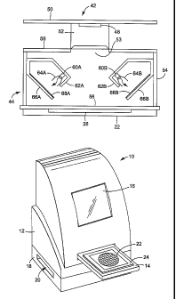
[0040] FIG. 1 is a perspective view of an exemplary biological growth plate
scanner
10. As shown in FIG. 1, biological growth plate scanner 10 includes a scanner
unit 12
having a drawer 14 that receives a biological growth plate (not shown in FIG.
1).
Drawer 14 moves the biological growth plate into biological growth plate
scanner 10
for scanning and analysis.
[0041] Biological growth plate scanner 10 also may include a display screen 16
to
display the progress or results of analysis of the biological growth plate to
a user.
Alternatively or additionally, display screen 16 may present to a user an
image of the
growth plate scanned by biological growth plate scanner 10. The displayed
image may
be optically magnified or digitally scaled upward.
[0042] A mounting platform 18 defines an ejection slot 20 through which the
growth
plate can be ejected following analysis by biological growth plate scanner 10.
Accordingly, biological growth plate scanner 10 may have a two-part design in
which
scanner unit 12 is mounted on mounting platform 18. The two-part design is
depicted
in FIG. 1 for purposes of example, and is not intended to be required by or
limiting of
the inventions described herein.
[0043] Scanner unit 12 houses an imaging device for scanning the biological
growth
plate and generating an image. The imaging device may take the form of a
monochromatic line scanner or an area scanner, in combination with a mufti-
color
illumination system to provide front and back illumination to the biological
growth
plate. In addition, scanner unit 12 may house processing hardware that
performs
analysis of the scanned image, e.g., in order to determine the number or
amount of
biological agents in the growth plate. For example, upon presentation of the
biological
CA 02505967 2005-05-09
WO 2004/051283 PCT/US2003/037385
_g_
growth plate via draw er 14, the plate may be positioned adjacent an optical
platen for
scanning.
[0044] When drawer 14 is subsequently opened, the growth plate may drop
downward into the mounting platform 18 for ejection via ejection slot 20. To
that end,
mounting platform 18 may house a conveyor that ejects the growth plate from
biological growth plate scanner 10 via ejection slot 20. After a biological
growth plate
is inserted into drawer 14, moved into scanner unit 12, and scanned, the
biological
growth plate drops downward into mounting platform 18, where a horizontal
conveyor,
such as a moving belt, ejects the plate via slot 20.
[0045] FIG. 2 is another perspective view of biological growth plate scanner
10. As
shown in FIG. 2, drawer 14 extends outward from biological growth plate
scanner 10 to
receive a biological growth plate 22. As illustrated, a biological growth
plate 22 may
be placed on a platform 24 provided within drawer 14. In some embodiments,
platform
24 may include positioning actuators such as cam levers to elevate the
platform for
precise positioning of growth plate 22 within biological growth plate scanner
10. Upon
placement of biological growth plate 22 on platform 24, drawer 14 retracts
into scanner
unit 12 to place the biological growth plate in a scanning position, i.e., a
position at
which the biological growth plate is optically scanned.
[0046] FIGS. 3 and 4 are front views of an exemplary biological growth plate
22. By
way of example, a suitable growth plate 22 may comprise biological growth
plates sold
by 3M under the trade name PETRIFIL,M plates. Alternatively, biological growth
plate
22 may comprise other biological growth media for growing particular bacteria
or other
biological agents. In some embodiments, biological growth plate 22 may carry a
plate
type indicator 28 to facilitate automated identification of the type of
biological media
associated with the growth plate.
[0047] Plate type indicator 28 presents an encoded pattern that is machine-
readable.
In the example of FIGS. 3 and 4, plate type indicator 28 takes the form of an
optically
readable pattern. In particular, FIGS. 3 and 4 depict a four-square pattern of
light and
dark quadrants formed in a corner margin of biological growth plate 22. In
other
words, plate type indicator 28 defines a two-dimensional grid of cells
modulated
between black and white to form an encoded pattern.
CA 02505967 2005-05-09
WO 2004/051283 PCT/US2003/037385
-9-
[0048] A wide variety of optical patterns such as characters, bar codes, two-
dimensional bar codes, optical gratings, holograms and the like are
conceivable. In
addition, in some embodiments, plate type indicator 28 may take the form of
patterns
that are readable by magnetic or radio frequency techniques. Alternatively,
plate type
indicator 28 may take the form of apertures, slots, surface contours, or the
like that are
readable by optical or mechanical techniques. In each case, plate type
indicator 28
carries information sufficient to enable automated identification of the type
of
biological growth plate 22 by biological growth plate scanner 10.
[0049] Biological growth plates may facilitate the rapid growth and detection
and
enumeration of bacteria or other biological agents including, for example,
aerobic
bacteria, E. coli, coliform, enterobacteriaceae, yeast, mold, Staplrylococcus
aureus,
Listeria, Campylobacter and the like. The use of PETRIFILM plates, or other
growth
media, can simplify bacterial testing of food samples. Moreover, biological
growth
plate scanner 10 can further simplify such testing by providing automated
plate type
detection, and automated selection of image processing profiles based on the
detected
plate type to analyze biological growth plate 22, e.g., by counting bacterial
colonies on
an image of the plate.
[0050] As shown in FIG. 3, biological growth plate 22 defines a growth area
26. A
determination of whether a given sample being tested in plate 22 is
acceptable, in terms
of bacterial colony counts, may depend on the number of bacterial colonies per
unit
area. Accordingly, scanner 10 quantifies the amount of bacterial colonies per
unit area
on plate 22, and may compare the amount, or "count," to a threshold. The
surface of
biological growth plate 22 may contain one or more growth enhancing agents
designed
to facilitate the rapid growth of one or more types of bacteria or other
biological agents.
[0051] After placing a sample of the material being tested, typically in
liquid form,
on the surface of biological growth plate 22 within growth area 26, plate 22
can be
inserted into an incubation chamber (not shown). In the incubation chamber,
bacterial
colonies or other biological agents being grown by growth plate 22 manifest
themselves, as shown in biological growth plate 22 of FIG. 4. The colonies,
represented by various dots 30 on biological growth plate 22 in FIG. 4, may
appear in
different colors on plate 22, facilitating automated detection and enumeration
of
bacterial colonies by scanner 10.
CA 02505967 2005-05-09
WO 2004/051283 PCT/US2003/037385
-10-
[0052] FIG. 5 is a block diagram illustrating internal operation of a
biological growth
plate scanner 10. As illustrated in FIG. 5, a biological growth plate 22 is
positioned
within biological growth plate scanner 10 on a platform (not shown in FIG. 5).
The
platform places biological growth plate 22 at a desired focal plane of an
imaging device
32. In accordance with the invention, imaging device 32 may include mufti-
color
illumination systems for front and back illumination of growth plate 22, as
well as a
monochromatic line or area scanner that captures an image of the surface of
growth
plate 22. In some embodiments, for example, imaging device 32 may take the
form of
a two-dimensional, monochromatic camera.
[0053] In general, imaging device 32 captures images of biological growth
plate 22,
or at least a growth region within the biological growth plate, during
illumination of the
biological growth plate with one or more different illumination colors. In
some
embodiments, illumination durations and illumination intensities may be
controlled
according to requirements of different biological growth plates. In addition,
selective
illumination of a first side and a second side of the biological growth plate
can be
controlled according to requirements of different biological growth plates.
[0054] A processor 34 controls the operation of imaging device 32. In
operation,
processor 34 controls imaging device 32 to illuminate biological growth plate
22 with
different illumination colors, and capture images of biological growth plate
22.
Processor 34 receives image data representing the scanned images from imaging
device
32 during illumination with each of the different illumination colors, and
combines the
images to form a mufti-color composite image. Processor 34 analyzes the
composite
image of biological growth plate 22 and analyzes the image to produce an
analytical
result, such as a colony count or a presence/absence result.
[0055] In some embodiments, processor 34 may extract or segregate a portion of
the
image to isolate plate type indicator 28. Using machine vision techniques, for
example,
processor 34 may analyze plate type indicator 28 to identify a plate type
associated with
biological growth plate 22. Processor 34 then retrieves an image processing
profile
from image processing profile memory 36. The image processing profile
corresponds
to the detected plate type, and may specify image capture conditions and image
analysis
conditions. Processor 34 may take the form of a microprocessor, digital signal
processor, application specific integrated circuit (ASIC), field programmable
gate array
CA 02505967 2005-05-09
WO 2004/051283 PCT/US2003/037385
-11-
(FPGA) or other integrated or discrete logic circuitry programmed or otherwise
configured to provide functionality as described herein.
[0056] Using the image processing profile, processor 34 loads appropriate
image
processing parameters and proceeds to process the scanned image of biological
growth
plate 22. In this manner, processor 34 forms an image processing device in the
sense
that it processes the image data obtained from biological growth plate 22. The
image
processing parameters may vary with the image processing profile and detected
plate
type, and may specify particular imager analysis conditions, including
parameters such
as color, size, shape and proximity criteria for analysis of the scanned
image. The
criteria may differ according to the type of plate 22 to be analyzed, and may
significantly affect colony count or other analytical results produced by
biological
growth plate scanner 10. The image processing profile also may specify image
capture
conditions such as illumination colors, intensities, and durations suitable
for a particular
type of biological growth plate.
[0057] Upon selection of the appropriate image processing parameters,
processor 34
processes the scanned image and produces an analytical result, such as a
colony count
or a presencelabsence result, which is presented to a user via display 16.
Processor 34
also may store the analytical result in memory, such as count data memory 38,
for later
retrieval from scanner 10. The data stored in count data memory 38 may be
retrieved,
for example, by a host computer that communicates with biological growth plate
scanner 10 via a communication port 40, e.g., a universal serial bus (USB)
port. The
host computer may compile analytical results for a series of biological growth
plates 22
presented to biological growth plate scanner 10 for analysis.
[0058] Automated selection of image processing profiles within biological
growth
plate scanner 10 can provide a convenient and accurate technique for selecting
the
appropriate image processing profile. Automated selection of image processing
profiles can promote the accuracy of bacterial colony counts and other
analytical
procedures. In particular, automatic image processing profile selection can
avoid the
need for a technician to visually identify and manually enter the plate type.
In this
manner, plate identification errors sometimes associated with human
intervention can
be avoided. Consequently, the combination of a scanner 10 and a biological
growth
plate 22 that carries plate type indicator 28 can promote efficiency and
workflow of
CA 02505967 2005-05-09
WO 2004/051283 PCT/US2003/037385
-12-
laboratory technicians while enhancing analytical accuracy and, in the end,
food safety
and human health.
[0059] FIG. 6 is a block diagram illustrating biological growth plate scanner
10 of
FIG. 5 in greater detail. Imaging device 32 (FIG. 5) of biological growth
plate scanner
may include, as shown in FIG. 6, a camer a 42, front illumination component 44
and
back illumination component 46. In accordance with the invention, front and
back
illumination systems 44, 46 may produce different illumination intensities,
colors and
durations on a selective basis. In particular, processor 34 controls front and
back
illumination systems 44, 46 to expose biological growth plate 22 to different
illumination colors, intensities and durations. In addition, processor 34
controls camera
42 to capture images of biological growth plate 22 during illumination with
the
different colors.
[0060] For example, processor 34 may provide coordinated control of
illumination
systems 44, 46 and camera 42 to capture multiple images of biological growth
plate 22.
Processor 34 then combines the multiple images to form a multi-color,
composite
image. Using the mufti-color, composite image, and/or individual components of
the
composite image, processor 34 analyzes biological growth plate 22 to produce
an
analytical result such as a detection or colony count. In one embodiment,
front and
back illumination systems 44, 46 may expose biological growth plate 22 to red,
green
and/or blue illumination colors on a selective basis under control of
processor 34. In
this example, camera 42 captures red, green and blue images of biological
growth plate
22. Processor 34 then combines the red, green and blue images to form the
mufti-color,
composite image for analysis.
[0061] As an illustration, processor 34 may first activate red illumination
sources
within front and back illumination components 44, 46 to expose biological
growth
plate 22 to red illumination. In particular, processor 34 may control the
intensity and
exposure duration of the red illumination sources. In synchronization with the
red
illumination exposure, camera 42 captures a red image of biological growth
plate 22
and stores the captured image in an image memory 47 within scanner 10.
[0062] Processor 34 then activates green illumination sources within front and
back
illumination components 44, 46 to expose biological growth plate 22 to green
illumination, followed by capture of a green image by camera 42. Similarly,
processor
CA 02505967 2005-05-09
WO 2004/051283 PCT/US2003/037385
-13-
activates blue illumination sources within front and back illumination
components 44,
46 to expose biological growth plate 22 to blue illumination, followed by
capture of a
blue image by camera 42.
[0063] Camera 42 captures monochromatic images for each of the red, green and
blue illumination exposures, and may store the images in separate files. Using
the files,
processor 34 combines the captured images to form the composite image for
analysis.
The order in which biological growth plate 22 is exposed to the multiple
illumination
colors may vary. Therefore, exposure to red, green and blue illumination
sources in
sequence should not be considered limiting of the invention.
[0064] The individual images captured by camera 42 may be represented in terms
of
optical intensity or optical density. In other words, camera 42 captures gray
scale data
that can be used to quantify the reflected output of biological growth plate
22 for each
exposure channel, e.g., red, green and blue. The use of a monochromatic camera
42 to
capture the individual images can result in image resolution benefits and cost
savings.
In particular, a less expensive monochromatic camera 42 may offer increased
spatial
resolution relative to multi-color cameras that capture red, green and blue
spectra
simultaneously. Accordingly, camera 42 can obtain high resolution imagery
needed for
effective analysis of biological growth plate 22 with reduced cost. Rather
than obtain a
single, mufti-color image monochromatic camera 42 captures multiple high
resolution
images, e.g., red, green and blue, and then processor 34 combines them to
produce a
high resolution, mufti-color image.
[0065] The different illumination sources within front and back illumination
systems
44, 46 may take the form of LEDs. In particular, the different illumination
colors can
be achieved by independent sets of color LEDs, e.g., red, green and blue LEDs.
As an
advantage, LEDs offer an extended lifetime relative to other illumination
sources such
as lamps. LEDs also may provide inherently consistent output spectra and
stable light
output.
[0066] Also, processor 34 can readily control the output intensities and
exposure
durations of the LEDs to perform sequential illumination of the biological
growth
plates 22 with appropriate levels of illumination. Processor 34 can be
programmed to
control the different sets of color LEDs independently to provide different
output
CA 02505967 2005-05-09
WO 2004/051283 PCT/US2003/037385
-14- '
intensities and exposure durations for each illumination color applied to
biological
growth plate 22.
[0067] This ability to independently control the LEDs via processor 34 can be
advantageous because the LEDs may exhibit different brightness
characteristics, and
reflector hardware or other optical components associated with the LEDs may
present
nonuniformities. In addition, camera 42 and one or more associated camera
lenses may
exhibit different responses to the illumination colors. For example, camera 42
may be
more or less sensitive to red, green and blue, presenting additional
nonuniformities in
the color response for a given illumination channel.
[0068] Processor 34 can independently control the LEDs, however, in order to
compensate for such nonuniformities. For example, scanner 10 may be calibrated
at
the factory or in the field to characterize the response of camera 42 to the
different
illumination sources, and then compensate the response by storing appropriate
drive
values to be applied by processor 34. Hence, processor 34 may apply different
drive
values to the LEDs for different illumination colors and intensity levels to
produce a
desired degree of uniformity in the images captured by camera 42.
[0069] In some embodiments, scanner 10 may process images of different
biological
growth plates 22 according to different image processing profiles. The image
processing profiles may be selected by processor 34 based on user input or
identification of the type of biological growth plate 22 presented to scanner
10. The
image processing profile may specify particular image capture conditions, such
as
illumination intensities, exposure durations, and colors, for capturing images
of
particular plate types. Thus, the scanner may apply different image capture
conditions,
including different illumination conditions, in processing images of different
biological
growth plates 22.
[0070] As an illustration, some types of biological growth plates 22 may
require
illumination with a particular color, intensity and duration. In addition,
some biological
growth plates 22 may require only front or back illumination, but not both.
For
example, an aerobic count plate may require only front illumination as well as
illumination by only a single color such as red. Alternatively, an E.
coli/Coliform plate
may require only back illumination and a combination of red and blue
illumination.
Similarly, particular intensity levels and durations may be appropriate. For
these
CA 02505967 2005-05-09
WO 2004/051283 PCT/US2003/037385
-15-
reasons, processor 34 may control illumination in response to image capture
conditions
specified by an image processing profile.
[0071] FIG. 7 is a side view illustrating a front illumination component 44
for
biological growth plate scanner 10. As shown in FIG. 7, front illumination
component
44 may be integrated with camera 42. For example, camera 42 may include a
camera
body with a CMOS or CCD camera chip 48 mounted to a camera backplane 50, such
as
a printed circuit board, which may carry circuitry to drive camera chip 48 and
receive
image data for processor 34. A camera lens 52 may be oriented to capture
images of a
biological growth plate 22 via an aperture 53 in a housing defined by front
illumination
component 44. In the example of FIG. 7, front illumination component 44
includes a
side wall 54, a front wall 56, and an optical platen 58. Optical platen 58 may
simply be
a transparent sheet of glass or plastic that permits transmission of
illuminating light and
capture of imagery from biological growth plate 22 by camera 42. In some
embodiments, optical platen 58 may be eliminated such that the growth area 26
of plate
22 is illuminated with no intervening structure between growth area 26 and the
emitted
light. Biological growth plate 22 may be elevated into contact or close
proximity with
optical platen 58 to permit camera 42 to capture images.
[0072] A number of components may be housed within front illumination
component
44. For example, front illumination component 44 may include one or more
illumination sources 60A, 60B, preferably arranged in linear arrays about a
periphery of
growth area 26 of biological growth plate 22. In particular, a linear array of
red, green
and blue illumination sources 60A, 60B may extend along each of four edges of
biological growth plate 22, e.g., in a square pattern. In other embodiments,
the
illumination sources may be arranged in alternative patterns, e.g., circular
patterns.
Again, illumination sources 60A, 60B may take the form of LEDs and may be
arranged
in groups of one red, one green and one blue LED.
[0073] Illumination sources 60A, 60B may be mounted within illumination
chambers
62A, 62B. Reflective towels 64A, 64B are mounted about illumination sources
60A,
60B and serve to reflect and concentrate the light emitted by the illumination
sources
toward inwardly extending walls 66A, 66B of chambers 62A, 62B. The reflective
material may be coated, deposited, or adhesively affixed to an interior
surface of
reflective towels 64A, 64B. An example of a suitable reflective material for
reflective
CA 02505967 2005-05-09
WO 2004/051283 PCT/US2003/037385
-16-
cowels 64A, 64B is the 3M Radiant Mirror Reflector VM2000 commercially
available
from 3M Company of St. Paul, Minnesota.
[0074] Walls 66A, 66B may carry a diffusing material such as an optical
diffusing
film 68A, 68B that serves to diffuse light received from illumination sources
60A, 60B.
The diffuse light is transmitted into an interior chamber of front
illumination
component 44 to illuminate growth region 26 of biological growth plate 22. An
example of a suitable diffusing material for diffusing film 68A, 68B is the
Mitsui WS-
180A diffuse white film, commercially available from Mitsui & Co., Inc., of
New
York, New York. The diffusing film 68A, 66B may be coated or adhesively
affixed to
an interior surface of walls 66A, 66B.
[0075] FIG. 8 is a front view illustrating front illumination component 44 in
greater
detail. As shown in FIG. 8, front illumination component 44 may include four
illumination chambers 62A, 62B, 62C, 62D arranged around a periphery of
biological
growth plate 22. Each illumination chamber 62 may include two sets of
illumination
sources 60. For example, chamber 62A may contain illumination sources 60A,
60C,
chamber 62B may contain illumination sources 60B, 60D, chamber 62C may contain
illumination sources 60E, 60F, and chamber 62D may contain illumination
sources
60G, 60H. In addition, chambers 62A, 62B, 62C, 62D may include respective
walls
66A, 66B, 66C, 66D carrying diffusing film. In other embodiments, each
respective
chamber 62 may include any number of illumination sources 60, which may or may
not
be the same number of illumination sources in other chambers.
[0076] Illumination sources 60 may include an array of illumination elements
grouped together, e.g., in groups of three. In particular, each illumination
source 60
may include a red LED, a green LED, and a blue LED that can be separately
activated
to illuminate biological growth plate 22. Upon activation of the individual
LEDs, an
inner chamber defined by front illumination component 44 is filled with
diffused light
to provide front illumination to biological growth plate 22. Camera 42
captures an
image of biological growth plate 22 during successive exposure cycles with
each of the
different illumination colors.
[0077] FIG. 9 is a side view illustrating back illumination component 46 for a
biological growth plate scanner 10 in a loading position, i.e., a position in
which
biological growth plate 22 is initially loaded into the scanner. In some
embodiments,
CA 02505967 2005-05-09
WO 2004/051283 PCT/US2003/037385
-17- ,
biological growth plate 22 may be loaded into scanner via drawer 14, as shown
in FIG.
2. In particular, drawer 14 carries a diffuser element 74 that serves as a
platform for
biological growth plate 22. Drawer 14 may be configured to permit retraction
of
biological growth plate 22 into the interior of scanner 10, and elevation of
the
biological growth plate into a scanning position.
[0078] Once loaded, biological growth plate 22 can be supported by optical
diffuser
element 74 or, alternatively, supported by a transparent platform in close
proximity to
the optical diffuser element. Optical diffuser element 74 serves to diffuse
light that is
laterally injected into the diffuser element and radiate the light upward to
provide back
side illumination of biological growth plate 22. Back illumination component
46
effectively illuminates the back side of biological growth plate 22 with good
uniformity
while conserving space within scanner 10.
[0079] In addition, back illumination component 46 incorporates a set of fixed
illumination sources 76A, 76B that do not require movement during use, thereby
alleviating fatigue to electrical wiring and reducing exposure to
environmental
contaminants. Rather, biological growth plate 22 and diffuser element 74 are
elevated
into position in alignment with the fixed illumination sources 76A, 76B: In
summary,
back illumination component 46 offers good illumination uniformity across the
surface
of biological growth plate 22, a flat illumination surface, a fixed
arrangement of
illumination sources 76A, 76B, and an efficient size and volume for space
conservation.
[0080] Illumination sources 76A, 76B are positioned adjacent a lateral edge of
diffuser element 74, when the diffuser element occupies the elevated, scanning
position. Each illumination source 76A, 76B may include a reflector cowl 78A,
78B to
reflect and concentrate light emitted by the illumination sources toward
respective
edges of diffuser element 74. In this manner, illumination sources 76A, 76B
inject
light into optical diffuser element 74. The reflective material may be coated,
deposited,
or adhesively affixed to an interior surface of reflective towels 78A, 78B. An
example
of a suitable reflective material for reflective towels 78A, 78B is the 3M
Radiant
Mirror Reflector VM2000 commercially available from 3M Company of St. Paul,
Minnesota.
CA 02505967 2005-05-09
WO 2004/051283 PCT/US2003/037385
_18_
[0081] A platen support 80A, 80B may be provided to support an optical platen
58
(FIG. 7), and provide an interface for engagement of back illumination
component 46
with front illumination component 44. As further shown in FIG. 9, a support
bracket
82A, 82B provides a mount for optical diffuser element 74. In addition,
illumination
sources 76A, 76B are mounted to backplanes 84A, 84B, which may carry a portion
of
the circuitry necessary to drive the illumination sources. However, backplanes
84A,
84B and illumination sources 76A, 76B may be generally fixed so that travel of
the
illumination sources and associated fatigue to wiring and other electrical
components is
not necessary, and exposure to environmental contaminants is reduced.
[0082] A back side of diffuser element 74 may be defined by a reflective film
88 that
promotes inner reflection of light received from illumination sources 76A,
76B, i.e.,
reflection of light into an interior chamber defined by diffuser element. In
this manner,
the light does not exit the back region of diffuser element 74, but rather is
reflected
inward and upward toward biological growth plate 22. Reflective film 88 may be
coated, deposited, or adhesively bonded to a wall defined by diffuser element
74.
Alternatively, reflective film 88 may be free-standing and define the back
wall of
diffuser element 74. An example of a suitable material for reflective film 88
is 3M
Radiant Mirror Film, 2000F1A6, commercially available from 3M Company of St.
Paul, Minnesota.
[0083] A front side of diffuser element 74, adjacent biological growth plate
22, may
carry an optical diffusing material such as an optical light guide and
diffusing film 86.
Diffuser element 74 may define an internal chamber between reflective film 88,
optical
light guide and diffusing film 86, and respective light transmissive layers
89A, 89B
forming side walls adjacent illumination sources 76A, 76B. As will be
described,
opposing side walls of optical diffuser element 74 on sides not adjacent
illumination
sources 76A, 76B may be formed by reflective layers to promote internal
reflection of
light injected into the diffuser element.
[0084] The internal chamber defined by optical diffuser element 74 may simply
be
empty and filled with air. Optical light guide and diffusing film 86 serves to
diffuse
light emitted from diffuser element 74 toward biological growth plate 22. An
example
of a suitable optical diffusing film is 3M Optical Lighting Film, printed with
a pattern
of diffuse white dots having 30% area coverage, with prism orientation facing
down
CA 02505967 2005-05-09
WO 2004/051283 PCT/US2003/037385
-19-
toward the diffuser element. In particular, the prisms of optical light guide
and
diffusing film 86 face into diffuser element 74 and the orientation of the
prisms is
generally perpendicular to illumination sources 76A, 76B. The 3M Optical
Lighting
Film is commercially available from 3M Company of St. Paul, Minnesota.
[0085] In addition, diffuser element 74 may include a scratch-resistant, light
transmissive layer 87 over optical light guide and diffusing film 86.
Biological growth
plate 22 may be placed in contact with scratch-resistant layer 87. Additional
scratch-
resistant, light transmissive layers 89A, 89B may be disposed adjacent the
lateral edges
of diffuser element 74. In particular, layers 89A, 89B may be disposed between
illumination sources 76A, 76B and diffuser element 74.
[0086] Scratch-resistant, light transmissive layers 89A, 89B are placed over
light
entry slots at opposite sides of diffuser element 74 to permit transmission of
light from
illumination sources 76A, 76B into the diffuser element, and also provide a
durable
surface for upward and downward sliding movement of the diffuser element. An
example of a suitable scratch-resistant, light transmissive material for use
as any of
layers 87, 89A, 89B resides in the class of acrylic glass-like materials,
sometimes
referred to as acrylglass or acrylplate. Alternatively, layers 87, 89A, 89B
may be
formed by glass.
[0087] An acrylic or glass plate as layer 87 can be used to provide a stable,
cleanable
platform for the biological growth plate, and protect diffuser element 74 from
damage.
An approximately 1 mm gap may be provided between layer 87 and optical light
guide
and diffusing film 86 to preserve the optical performance of the diffusing
film, which
could be altered by contact with materials other than air.
[0088] FIG. 10 is a side view illustrating the back illumination component 46
of FIG.
9 in a scanning position. In particular, in FIG. 10, diffuser element 74 is
elevated
relative to the position illustrated in FIG. 9. Diffuser element 74 may be
elevated by a
variety of elevation mechanisms, such as ramming, lead screw or pulley
arrangements.
As diffuser element 74 is elevated into scanning position, biological growth
plate 22 is
placed in proximity or in contact with optical platen 58 (FIG. 7).
[0089] Upon elevation into scanning position, illumination sources 76A, 76B
inject
light into diffuser element 74, which diffuses the light and directs it upward
to provide
back illumination for biological growth plate 22. As will be described,
illumination
CA 02505967 2005-05-09
WO 2004/051283 PCT/US2003/037385
-20-
sources 76A, 76B may incorporate differently colored illumination elements
that are
selectively activated to permit camera 42 to separate monochromatic images for
each
color, e.g., red, green and blue.
[0090] FIG. 11 is a bottom view illustrating back illumination component 46 of
FIGS. 9 and 10. As shown in FIG. 11, multiple illumination sources 76A-76H may
be
disposed in linear arrays on opposite sides of diffuser element 74. FIG. 11
provides a
perspective of back illumination components from a side opposite biological
growth
plate 22, and therefore shows reflective layer 88. Each illumination source 76
may
include three illumination elements, e.g., a red (R) element, a green (G)
element, and a
blue (B) element. The red, green and blue elements may be red, green and blue
LEDs.
Back illumination component 46 may be configured such that all red elements
can be
activated simultaneously to illuminate the back side of biological growth
plate 22 with
red light in order to capture a red image with camera 42. The green elements
and blue
elements, respectively, may be similarly activated simultaneously.
[0091] As further shown in FIG. 11, reflective layers 93A, 93B form opposing
side
walls of diffuser element 74 on sides not adjacent illumination sources 76.
Reflective
layers 93A, 93B may be formed from materials similar to reflective layer 88,
and may
be affixed to interiors or respective side walls or form free-standing walls
themselves.
In general, reflective layers 88, 93A, 93B serve to reflect light injected by
illumination
sources 76 into the interior chamber defined by diffuser element 74,
preventing the
light from escaping from the back side or side walls of the diffuser element.
Instead,
the light is reflected inward and toward diffusing material 86. In this
manner, the light
is concentrated and then diffused by diffusing material 86 for transmission to
illuminate
a back side of biological growth plate 22.
[0092] FIG. 12 is a side view illustrating the combination of front and back
illumination components 44, 46, as well as camera 42, for biological growth
plate
scanner 10. As shown in FIG. 12, optical platen 58 serves as an interface
between front
illumination component 44 and back illumination component 46. In operation,
biological growth plate 22 is elevated into proximity or contact with optical
platen 58.
Front and back illumination components 44, 46 then selectively expose
biological
growth plate 22 with different illumination colors to permit camera 42 to
capture
images of the biological growth plate. For example, front and back
illumination
CA 02505967 2005-05-09
WO 2004/051283 PCT/US2003/037385
-21- ,
component 44, 46 may selectively activate red, green and blue LEDs in sequence
to
form red, green and blue images of biological growth plate 22.
[0093] FIG. 13 is a circuit diagram illustrating a control circuit 90 for an
illumination system. Control circuit 90 may be used to control illumination
sources in
front and back illumination components 44, 46. In the examples of FIGS. 7-12,
front
and back illumination components 44, 46 each include eight separate
illumination
sources 60, 76. Each illumination source 60, 76 includes a red, green and blue
illumination element, e.g., red, green and blue LEDs. Accordingly, FIG. 13
illustrates
an exemplary control circuit 90 equipped to simultaneously drive eight
different LEDs
on a selective basis. In this manner, control circuit 90 may selectively
activate all red
LEDs to illuminate biological growth plate 22 with red light. Similarly,
control circuit
90 may selectively activate all green or blue LEDs for green and blue
illumination,
respectively. FIG. 13 depicts control circuit 90 as controlling eight LEDs
simultaneously, and hence controlling either front illumination component 44
or back
illumination component 46: However, the output circuitry controlled by
processor 34
may essentially be duplicated to permit control of sixteen LEDs
simultaneously, and
therefore both front illumination component 44 and back illumination component
46.
[0094] As shown in FIG. 13, processor 34 generates digital output values to
drive a
set of LEDs. Digital-to-analog converters (DAC) 91A-91H convert the digital
output
values to an analog drive signals. Buffer amplifiers 92A-92H amplify the
analog
signals produced by DACs 91A-91H and apply the amplified analog drive signals
to
respective arrays of LEDs 94A-94H, 96A-96H, 98A-98H. DACs 91A-91H and
amplifiers 92A-92H serve as programmable controllers to selectively control
illumination durations and illumination intensities of LEDS94A-94H, 96A-96H,
98A-
98H. Processor 34 drives the controllers, i.e., DACs 91A-91H and amplifiers
92A-
92H, according to requirements of different biological growth plates 22 to be
processed
by scanner 10.
[0095] Advantageously, processor 34 may access particular sets of digital
output
values to produce a desired output intensity for LEDs 94A-94H, 96A-96H, 98A-
98H.
For example, the digital output values can be determined upon factory or field
calibration of scanner 10 in order to enhance the uniformity of the
illumination
provided by the various LEDs 94A-94H, 96A-96H, 98A-98H. Again, the red, green
CA 02505967 2005-05-09
WO 2004/051283 PCT/US2003/037385
-22-
and blue LEDs may be characterized by different output intensities and
responses, and
associated reflector and optics hardware may present nonuniformities, making
independent control by processor 34 desirable in some applications.
[0096] Also, the digital output values may be determined based on the
requirements
of different biological growth plates 22, i.e., to control the intensity and
duration of
illumination applied to the growth plates. Accordingly, processor 34 may
selectively
generate different output values for different durations, enable different
sets of LEDs
94-94H, 96A-96H, 98A-98H, and selectively enable either front illumination,
back
illumination or both, based on the particular types of biological growth
plates 22
presented to scanner 10.
[0097] The anodes of all LEDs 94A-94H, 96A-96H, 98A-98H are coupled to the
respective outputs of drive amplifiers 92A-92H for simultaneous activation of
selected
LEDs. To permit selective activation of LEDs for particular illumination
colors, the
cathodes of LEDs 94A-94H (Red) are coupled in common to a switch, e.g., to the
collector of a bipolar junction transistor 100A with an emitter coupled to a
ground
potential. Similarly, the cathodes of LEDs 96A-96H (Green) are coupled in
common to
the collector of a bipolar junction transistor 100B, and the cathodes of LEDs
98A-98H
(Blue) are coupled in common to the collector of a bipolar junction transistor
1000.
[0098] Processor 34 drives the base of each bipolar transistor 100A-100C with
a
RED ENABLE, GREEN ENABLE or BLUE ENABLE signal. In operation, to expose
biological growth plate to red illumination, processor 34 selects digital
values for the
red LEDs 94A-94H, and applies the digital values to DACs 91A-91H, which
produce
analog drive signals for amplification by buffer amplifiers 92A-92H. In
synchronization with application of the digital values for the red LEDs 94A-
94H,
processor 34 also activates the RED ENABLE line to bias transistor 100A "on,"
and
thereby pull the anodes of red LEDs 94A-94H to ground.
[0099] Using the ENABLE lines, processor 34 can selectively activate red LEDs
94A-94H to expose biological growth plate 22 to red illumination.
Simultaneously,
processor 34 controls camera 42 to capture a red image of biological growth
plate 22.
To capture green and blue images, processor 34 generates appropriate digital
drive
values and activates the GREEN ENABLE and BLUE ENABLE lines, respectively.
As an advantage, the ENABLE lines can be used to independently control the
exposure
CA 02505967 2005-05-09
WO 2004/051283 PCT/US2003/037385
-23-
durations of the illumination colors. For example, it may be desirable to
expose
biological growth plate 22 to different durations of red, green and blue
illumination.
[00100] FIG. 14 is a functional block diagram illustrating the capture of
multi-color
images for preparation of a composite image to produce a plate count. As shown
in
FIG. 14, monochromatic camera 42 captures a red image lO2A, green image 102B
and
blue image 102C from biological growth plate 22. Processor 34 then processes
the red,
green and blue images 102 to produce a composite image 104. In addition,
processor
34 processes the composite image to produce an analytical result such as a
colony count
106. Once the composite image has been prepared, combining the red, green and
blue
images, processor 34 may apply conventional image analysis techniques to
produce the
colony count.
[00101] FIG. 15 is a flow diagram illustrating a technique for the capture of
multi-
color images for preparation of a composite image to produce a plate count. As
shown
in FIG. 15, the technique may involve selective illuminating of a biological
growth
plate 22 with different illuminant colors (108), and capturing plate images
during
exposure to each of the illumination colors ( 110). The technique further
involves
forming a composite image (112) based on the separately captured images .for
each
illumination color, and processing the composite image (114) to produce an
analytical
result such as a colony count (116). The colony count may be displayed to the
user and
logged to a date file. As mentioned above, techniques for capture of some
images may
involve illumination with one, two or more illumination colors, as well as
front side
illumination, back side illumination or both, depending on the requirements of
the
particular biological growth plate 22 to be processed by scanner 10.
[00102] FIG. 16 is a flow diagram illustrating the technique of FIG. 15 in
greater
detail. As shown in FIG. 16, in operation, processor 34 first outputs digital
values to
drive the red illumination LEDs 94A-94H (FIG. 13) (11~), and activates the
front and
back red illumination LEDs with the RED ENABLE line (120) to illuminate
biological
growth plate 22. Camera 42 then captures an image of biological growth plate
22
during illumination by the red LEDs 94A-94H ( 122).
[00103] Next, processor 34 outputs digital values to drive the green
illumination LEDs
96A-96H ( 124), and activates the front and back green illumination LEDs with
the
GREEN ENABLE line ( 126) to illuminate biological growth plate 22. Camera 42
then
CA 02505967 2005-05-09
WO 2004/051283 PCT/US2003/037385
-24-
captures an image of biological growth plate 22 during illumination by the
green LEDs
96A-96H (128). Processor 34 then outputs digital value to drive the blue
illumination
LEDS 98A-98H (130), and activates the blue illumination LEDs with the BLUE
ENABLE line ( 132).
[00104] After the blue image is captured by camera 42 (134), processor 34
combines
the red, green and blue images to form a composite red-green-blue image (136).
Processor 34 then processes the composite red-green-blue image ( 138) and/or
individual components of the composite image to generate a colony count (140).
Again, in some embodiments, processor 34 may process the individual red-green-
blue
images prior to combining the red, green and blue images to form a composite
image.
Again, the red-green-blue order of illumination and capture is described
herein for
purposes of example. Accordingly, biological growth plate 22 may be
illuminated and
scanned in a different order.
[00105] In operation, processor 34 executes instructions that may be stored on
a
computer-readable medium to carry out the processes described herein. The
computer-
readable medium may comprise random access memory (RAM) such as synchronous
dynamic random access memory (SDRAM), read-only memory (ROM), non-volatile
random access memory (NVRAM), electrically erasable programmable read-only
memory (EEPROM), FLASH memory, magnetic or optical data storage media, and the
like.
[00106] Various modification may be made without departing from the spirit and
scope of the invention. For example, it is conceivable that some of the
features and
principles described herein may be applied to line scanners as well as area
scanners.
These and other embodiments axe within the scope of the following claims.