Note: Descriptions are shown in the official language in which they were submitted.
CA 02506935 2005-05-20
WO 2004/046712 PCT/US2003/037205
ISOLATION OF SPERM CELLS FROM OTHER BIOLOGICAL
MATERIALS USING MICROFABRICATED DEVICES AND RELATED
METHODS THEREOF
This application claims priority to Provisional Patent Application No.
60/427,734, filed November 20, 2002.
FIELD OF THE INVENTION
The present invention relates to cell separation using microfabricated
devices.
In particular, the present invention provides methods and devices for
separation of
sperm from biological materials, such as other cells and molecular species, in
a cell
mixture in a microfabricated device through the use of electroosmotic flow,
electrophoretic mobility, pressure gradient, differential adhesion, and/or
combinations
thereof.
BACKGROUND OF THE INVENTION
The use of DNA typing to verify and often convict suspects in sexual crime
cases relies on the separation of the perpetrator DNA from that of the victim.
The
perpetrator DNA is most easily obtained from sperm cells collected on vaginal
swabs,
taken in the routine collection of sexual assault evidence. The majority of
cells
collected on such swabs are epithelial cells from the victim, however, and
these cells
must be separated from the sperm cells before DNA from the sperm cells can be
recovered and STRs amplified for analysis by capillary electrophoresis (or
other
analytical methods). Effective separation of the victim's and perpetrator's
DNA,
combined with the ensuing preparatory/analysis steps, are time- and labor-
intensive
processes. At the present time, this is carried out by chemical means
involving
CA 02506935 2005-05-20
WO 2004/046712 PCT/US2003/037205
2
differential lysis of the cells collected on the vaginal swab. The multistep
procedure
begins by lysing the epithelial cells while still adsorbed to the cotton swab.
During
this time, the intact sperm cells (mainly heads since the tails have been
degraded) are
desorbed from the cotton swab, collected and then lysed for DNA extraction
using a
MicroconT~~ concentration step or other methods known in the art.
The multistep nature of this current method affords it the same disadvantages
from which many conventional isolation methodologies suffer. First, the time-
consuming and labor-intensive procedure translates into cost-ineffectiveness.
Second,
extensive sample handling presents opportunities for loss of biological
material,
which may be problematic if only small amounts of starting material are
available. In
addition, extensive sample handling increases the chance of contamination with
exogenous DNA, which can complicate interpretation of the results.
One possible solution to the conventional, time-consuming differential
extraction could be provided by microminiaturization of the analytical
methodology
in an embodiment that allowed for cell sorting to be executed rapidly and
efficiently.
Much progress has been made developing microchip-based analytical systems to
carry
out simple processes. In the early stages, a number of groups demonstrated the
analytical power of microchips for carrying out fast separations (Harnson et
a1.
Towards miniaturized electrophoresis and chemical analysis systems on silicon:
an
alternative to chemical sensors. Sensors and Act. B. 10:107-116, 1993; Mann,
A.,
Graber, N., Widmer, H.M. Miniaturized Total Chemical Analysis Systems: A Novel
Concept for Chemical Sensing., Sensors and Actuators, BI. 8:244-248, 1990; and
Jacobson et al. Integrated Microdevice for DNA Restriction Fragment Analysis.
Anal. Chem. 1996 68:720-723). Patents have also been issued for these
microfluidic
devices.
CA 02506935 2005-05-20
WO 2004/046712 PCT/US2003/037205
U.S. Patent No. 5,46,335 to Wilding et al. discloses devices and methods for
detecting the presence of a preselected analyte in a fluid sample. The
invention
provides a device comprising a solid substrate, typically on the order of a
few
millimeters thick and approximately a 0.2 to 2.0 centimeters square,
microfabricated
to define a sample inlet port and a microscale flow system. A sample is passed
through the microfabricated device, and the restriction or blockage of flow
through
the flow system is detected as a positive indication of the presence of the
analyte. The
device may be adapted for operation in conjunction with a pump, for example,
to
induce flow of a sample through the flow system.
Despite the laxity in the field that fast separations are adequate, our
experience
with clinical diagnostics indicates that sample preparation will have to be
integrated
with electrophoresis in order for this technology to be fully exploited.
Therefore, the
present invention addresses a key sample preparation step, cell sorting, in
cell
analyses, especially for forensic analyses.
Other groups addressing the cell-separation problem use an antibody-based
separation scheme. Eisenberg et al. (unpublished report) uses magnetic beads,
to
which sperm-specific antibodies are attached. There may be numerous problems
associated with this approach including: clogging of the column by the large
numbers
of epithelial cells in a "real-world" sample, inability to integrate the cell
separation
with other microminiaturization analyses, expense of materials, and numerous
steps
still required to result in PCR-ready DNA. A reliable separation may not
result using
the antibody/magnetic bead approach due to extensive clogging of the column.
The
present invention described herein overcomes the shortfalls of the
conventional
procedures as well as this antibody/magnetic bead capture system. The non- or
low-
CA 02506935 2005-05-20
WO 2004/046712 PCT/US2003/037205
affinity based separation described utilizes the differing physical and
biological
properties of the sperm and epithelial cells to effect a separation.
Microfabricated devices have recently been developed for cell separations and
transport. Kricka et al. (Applications of a microfabricated device for
evaluating
sperm function. Clin Chem. 39(9):1944-7, 1993) used a microfabricated device
for
the electrophoretic separation of live and dead sperm based upon their
differences in
surface charge.
U.S. Patent No. 5,296,375 and 5,427,946, both to Kricka et al., discloses
devices and methods similar to Wilding et al. above for clinical analysis of a
sperm
sample. In one embodiment, a sperm sample is applied to the inlet port, and
the
competitive migration of the sperm sample through the mesoscale flow channel
is
detected to serve as an indicator of sperm motility. In another embodiment,
the
substrate of the device is microfabricated with a sperm inlet port, an egg
nesting
chamber, and an elongate mesoscale flow channel communicating between the egg
nesting chamber and the inlet port. In this embodiment, a sperm sample is
applied to
the inlet port, and the sperm in the sample are permitted to competitively
migrate
from the inlet port through the channel to the egg nesting chamber, where in
vitro
fertilization occurs. The devices may be used in a wide range of applications
in the
analysis of a sperm sample, including the analysis of sperm morphology or
motility,
to assess sperm binding properties, and for in vitro fertilization.
Li and Harrison (Transport, manipulation and reaction of biological cells on-
chip using electrokinetic effects. Anal. Claern. 69: 1564-1568, 1997) showed
the
transport (not separation) and lysis of E. Coli, yeast, and canine
erythrocytes in a
microchip exploiting electrokinetic effects. Using electric fields of 100-600
V/cm,
CA 02506935 2005-05-20
WO 2004/046712 PCT/US2003/037205
cells were directed from the loading reservoir to the waste or outlet
reservoirs of the
microdevice.
Fu et al. (A microfabricated fluorescence-activated cell sorter. Nature
Biotech.
17:1109-1111, 1999; and An integrated microfabricated cell sorter. Anal Chern.
74
5 (11):2451 -2457, 2002) developed a microfabricated fluorescence-activated
cell
sorter. This system requires that the sorted cells be fluorescently labeled,
by means of
expression of green fluorescent protein or in some other manner. This method
of cell
sorting requires interrogation/identification of the particle, and subsequent
valuing of
the flow to direct the cell into the correct reservoir on the rnicrodevice.
U.S. Patent No. 6,193,647 to Beebe et al. discloses a microfluidic embryo
handling device and method in which biological rotating of embryos is
simulated.
Fluid flow is used to move and position embryos without assistance of
electrical
stimulus or other means which may produce undesired heating of biological
medium
used as the fluid for transporting and position. The device provides an
excellent
simulation of biological conditions and may be used for culturing, sorting,
testing,
evaluating, fertilizing and other similar typical handling operations. No cell
separation is disclosed in this patent.
Separation and identification of various bacteria have been shown by
Armstrong et al. (Rapid identification of the bacterial pathogens responsible
for
urinary tract infections using direct inj ection CE. Anal. Chem. 72:4474-6,
2000; and
Separating microbes in the manner of molecules: 1. Capillary electrokinetic
approaches. Anal. Chem. 71, 5465-5469, 1999) using conventional capillary
electrophoresis. Armstrong et al. separated and identified E. coli and Staph.
Saprophyticus, which are bacterial pathogens commonly responsible for urinary
tract
infections, by using polyethylene) oxide as a sieving matrix. In 2001,
Armstrong et
CA 02506935 2005-05-20
WO 2004/046712 PCT/US2003/037205
al. used conventional capillary electrophoresis for the separation of various
bacteria
from yeast. The microbes were detected using laser-induced fluorescence and,
therefore, required staining with a fluorescent dye. They used this
separation/detection method to evaluate cell viability using a commercially-
available
viability stain and detecting the difference in fluorescence emission.
SUMMARY OF~THE INVENTION
The speed and efficiency of the conventional differential extraction procedure
warrants improvement by the micro-miniaturization of cell sorting. A stand-
alone
microdevice for rapidly sorting sperm cells from epithelial cells and
extracting DNA
would improve DNA analysis in the crime laboratories by reducing the cost of
analysis through improved speed, reduced reagent consumption, decreased
technician
time, reduced sample handling-induced contamination, and ease of automation.
The
present invention provides a novel method and device for separation of sperm
and
epithelial cells on a microdevice. A separation method that does not require a
high
affinity interaction with the cells but, instead, one that exploits
electrophoretic
mobility, electroosmotic flow, pressure-based flow and/or combination thereof
is
exploited. This present invention utilizes the differential physical and
biological
properties of the cells, such as their propensity for adhesion, specific
gravity, cell
surface charge, and size. Two important aspects of such a cell separation
mechanism
separation are, but not limited thereto, the magnitude of the flow, which can
be
controlled by a number of mechanisms, such as electrophoretic mobility,
electroosmotic flow, pressure gradient (pump as well as gravity), and/or
combinations
thereof, as well as the surface properties of the channel walls. The present
invention
provides techniques for the isolation of sperm cells from biological
materials,
CA 02506935 2005-05-20
WO 2004/046712 PCT/US2003/037205
preferably other cells and molecular species, most preferably epithelial
cells, for
forensic applications using microfabricated devices.
In a further embodiment, the present invention is used to isolate sperm from
either other cells or other biologically-derived molecular species enables
sperm to be
concentrated in smaller volumes. This effect could find utility with in vitro
fertilization applications. Beebe et al. has shown that human eggs (oocytes)
can be
manipulated in microfabricated devices in processes pertinent to in
vitf°o fertilization.
The device described herein for sperm cell isolation could be utilized to
enhance the
concentration (number) of sperm in the collection chamber. One could envision
how
the presence of an oocyte in the collection chamber where an enhanced
concentration
of sperm are collected, might improve the efficiency of in vitro
fertilization.
In a further embodiment, the present invention is used to isolate sperm from
either other cells or other biologically-derived molecular species enables
sperm
quantitation. This could be accomplished with a number of on-line counting
sperm
approaches as the migrate through the microchannel to the collection
reservoir.
Included in these means would be optical detection using either light
scattering from a
laser or other focused light source, impedance spectroscopy, fluorescence
detection
(assuming the cells were fluorescently tagged), or some form of imaging
software that
was capable of counting cells based on size. This would find utility in
forensic
applications defining when the requisite number of sperm from the biological
sample
required for the analysis had been collected in the collection reservoir.
In a further embodiment, the present invention is used to isolate sperm from
other cells or other biologically-derived molecular species via some flow-
driven
separation process presents the possibility of quantitating subpopulations of
sperm
from the sample. This could facilitate the evaluation of sperm that are
dysfunctional
CA 02506935 2005-05-20
WO 2004/046712 PCT/US2003/037205
with respect to fertilization ability, the separation of sperm subpopulations
that are
functional relative to those that are dysfunctional due to exposure to
toxicants
(apoptotic) or cryostorage.
BRIEF DESCRIPTION OF THE DRAWINGS
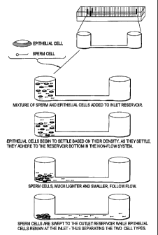
Figure 1 shows the size difference between sperm and epithelial cells.
Figure 2 outlines microchannel cell separation based on cell density/adhesion
differences.
Figure 3 outlines microchannel cell separation in an electric field-driven
system based upon density, proclivity for adhesion, and electrophoretic
mobility. The
sperm are swept with the flow to the cathodic reservoir (right).
Figure 4 shows an alternate manifestation of the microchannel cell separation
in an electric field-driven system based upon density, proclivity for
adhesion, and
electrophoretic mobility. In this three-reservoir system, the cell mixture is
deposited
in the central reservoir, and the epithelial cells and sperm cells are
collected in the
outside reservoirs.
Figure 5 shows the present invention being used as part of a mufti-function
(multiple 'domain') totally-integrated system.
DETAILED DESCRIPTTON OF THE PREFERRED EMBODIMENTS
The present invention exploits physical and/or biological properties of sperm
and other biological materials, such as epithelial cells, to effect a robust
and reliable
separation of the two cell types. Biological materials used herein includes,
but is not
limited to, other cells, such as epithelial cells, red blood cells, white
blood cells, etc.;
molecular species, such as nucleic acids (RNA and DNA), proteins, etc.; cell
CA 02506935 2005-05-20
WO 2004/046712 PCT/US2003/037205
membranes; and organelles. Two separation approaches can be utilized to invoke
separation of cells, with a main focus on the separation of sperm from other
cells for
forensic analysis where both the sperm and the other cells can be important in
the
forensic process. The first mode amenable to a microfabricated device is a
separation
driven by an electric field - this inherently involves both an electrophoretic
component (mobility of cells based on size and their surface charge) and a
flow
component in the form of electroosmotic flow (EOF - the flow that results from
the
presence of ions in glass channel). The second type, one that does not invoke
the use
of electric fields but is based solely on flow, can be driven by a number
means
including gravity-driven (siphoning), hydrostatic pressure (or vacuum)-driven
flow, or
centrifugal driven flow.
I. Cell Separation Exploiting Electrokinetic Phenomena
In microchip electrophoresis, analytes are acted upon by two forces, intrinsic
electrophoretic mobility (yep), governed by the charge-to-size ratio of the
analyte, and
EOF, generated by charge on the microchannel surface. For cell separations,
these
forces can be employed together, or EOF can be reduced (or close to zero),
with the
electrophoretic mobilities providing the main governing force for the
separation.
Consequently, three scenarios emerge where separation is driven by 1)
electrokinetic
phenomena specific to the cells themselves; 2) a combination of electrokinetic
phenomena specific to the cells and the EOF; and 3) the low volume, plug-type
flow
resulting from EOF. These are addressed individually below.
A. Separation based solely on cell electrophoresis
CA 02506935 2005-05-20
WO 2004/046712 PCT/US2003/037205
The simplest scenario in microchips, one where the chip surface was treated to
negate the EOF, cell electrophoretic mobility becomes the dominant separation
force.
This has already been demonstrated in the literature (Kricka et al., 1993) by
the
separation of live and dead sperm in an electric field, which is likely due to
S differences in the cell surface charge, however, the role of EOF in this
separation
cannot be ruled out. For sperm and epithelial cells, the significant size
difference (4-S
p,m vs SO pm) presents a scenario where there is likely to be significant
differences in
charge-to-size ratio, and this may be exploited for the sake of separation
(Fig. 1 ). In
addition, the surface charge of the cells can be varied with pH, solution
composition,
10 and ionic strength of the separation buffer. This allows altering the
surface charges in
the electrophoretic-based separation scheme to optimize the separation speed
and
efficiency.
An electrophoretically-driven system is attractive because, in addition to
separation of the cells, there is a cellular concentrating effect. Therefore,
the buffer
1 S volume used to desorb the biological material from the swab would have
minimal
impact on downstream sample preparation or analytical processes where volume
limitations may exist. In addition, any free DNA in the biological material is
not
captured in the sperm fraction.
B. Separation based on cell electrophoresis and EOF
In addition to exploiting cell electrophoretic mobility, a significant EOF
provides a flow bulk component to the separation and, under the appropriate
circumstances, can enhance the separation. Under conditions with a reasonable
EOF,
the differential movement of sperm and epithelial calls exists under low
electric field
2S strengths (about S-1000 V/cm, preferably about 2S-300 V/cm, most preferably
about
CA 02506935 2005-05-20
WO 2004/046712 PCT/US2003/037205
11
75-100 V/cm). Sperm migrate toward the cathode, while epithelial cells have an
opposite mobility (to the anode). However, in the same way that the surface
charge of
the cells can be altered by the pH, solution composition, and ionic strength
of the
separation buffer, so can the EOF. A high solution ionic strength reduces the
charge
on the microchannel surface (the zeta potential) and, hence, reduces the EOF.
Reducing or even eliminating the charge on the microchannel surface by
covalent,
dynamic or absorptive coating can similarly reduce or minimize EOF. A similar
effect can be achieved by reducing the solution pH, but this is less
attractive with cells
that will need to be maintained in the biological pH range. Consequently a
number of
approaches can be used to optimize the EOF that allows for optimal separation
of the
analytes involved, in this particular case, different biological materials,
specifically,
sperm and epithelial cells.
C. Separation based solely on electric field-driven flow (EOF)
See EOF section below.
II. Flow-based separations
A critical aspect of this mechanism is the magnitude of the flow used for the
separation. A flow that is low in magnitude (about 0.1-1000 ~,L/hr, preferably
about
0.3-10 ~,L/hr, and most preferably about 0.6 ~.L/hr) and reproducibly-
controlled flow
' is utilized for these separations and can be achieved with a number of
approaches.
A. EOF
The low magnitude, plug-type flow associated with EOF (no turbulence) is
ideal for separating cells based on physical characteristics. Modification of
the silica
CA 02506935 2005-05-20
WO 2004/046712 PCT/US2003/037205
12
surface charge allows control of EOF and provides a support for electrostatic
interactions that can further increase the cell separation efficiency. Under
low electric
field strength (e.g., ~33V per cm of microchannel), we have observed the
differential
movement of sperm and epithelial cells in phosphate-buffered saline (pH 7.4) -
the
sperm cells migrate to the cathode and epithelial cells migrated to the anode.
Hence,
placement of a mixture of sperm and epithelial cells in a reservoir on a
microdevice,
and proper placement of electrodes results in the separation of sperm cells
from the
mixture into another reservoir containing the cathode. An applied field is
used to
direct the sperm cells into the desired reservoir on the microdevice (Figure
3). The
migration of epithelial cells to the anode is due to their negative surface
charge.
Sperm cells also have an overall negative surface charge, but the sperm
migrate
toward the cathode because the magnitude of electroosmotic flow is greater
than the
magnitude of the electrophoretic mobility of the sperm cells. In a separation
based
upon EOF flow, we can also take advantage of the other mechanisms of
separation
described herein such as density differences, proclivity for adhesion to the
microchannel surface as well as to other cells, as well as the electrophoretic
mobility
differences. In this manner, the selectivity and efficiency of separation can
be
enhanced.
In an alternate embodiment of this concept, the mixture reservoir can be
placed between two reservoirs connected in a linear fashion by a microchannel
etched
into the glass (Figure 4). By placing electrodes in these two outside
reservoirs, the
mixture in the center can be separated and the two cell types and/or
biological
materials collected in the separate outside reservoirs. It should be noted
that, in either
manifestation, the use of a separation using electrokinetic effects has the
added
benefit in that any DNA in the cell mixture from cells lysed prior to the
separation is
CA 02506935 2005-05-20
WO 2004/046712 PCT/US2003/037205
13
attracted to the anode and, thus, is separated from the sperm cell fraction.
This is
particularly important in forensic applications.
B. Gravity-driven flow
Gravity-driven flow (siphoning) can also provide a low magnitude flow that
can be controlled with some accuracy and, hence, could be employed to
differentially
move the cells in microchannels. Under these conditions, the effect of gravity
not
only drives the flow of fluid from one reservoir to the other, but density
differences in
the cells in a mixture can be,exploited, in which the epithelial cells settle
more readily
than sperm cells. For example, in the case of sperm and epithelial cells,
approximately 5 minutes is sufficient to allow the epithelial cells to
'settle' to the
bottom of the reservoir/channel before flow is induced. Flow is then induced
by
mismatched liquid heights in connecting reservoirs. The data shows that the
fluid
flow rate remains constant at an acceptable magnitude for at least 10 minutes,
allowing adequate time for a cell separation where sperm were observed leaving
the
mixture reservoir at a rate of approximately 5 sperm/sec.
C. Pressure (or vacuum)-driven flow
More reproducible and controllable flow rates can be generated in a pressure-
driven
system employing the appropriate volume syringes and pumps. This uses the same
mechanism of separation as the gravity-driven flow, but would provide greater
opportunity for automation due to the external control of the flow rate.
Clearly what
was accomplished with gravity-driven flow could be achieved with this system
but in
a much more automatable manner.
CA 02506935 2005-05-20
WO 2004/046712 PCT/US2003/037205
14
III. Combined separation
Techniques discussed above are can be used alone or in combination. Various
combinations are appropriate for the present invention. A successful
separation
typically utilizes both flow and electrokinetic separations. The following are
non-
limiting examples of combined separations that are appropriate for the present
invention: 1) separation utilizing electrokinetic phenomena and pressure-
driven flow;
2) separation utilizing pressure-driven flow and EOF; and 3) separation
utilizing
electrolcinetic phenomena, pressure-driven flow, and EOF. Further, gravity,
vacuum-
driven and centrifugally-driven flow can easily substitute for the pressure-
driven flow
discussed in the possible combined separation regimes.
IV. Other considerations for isolation of sperm cells from a biological
mixture
A. Surface Area-to-volume considerations
There are a number of channel design modifications that result in an increased
surface-to-volume ratio, which we believe will also increase the separation
efficiency.
These include placing microfabricated posts in the separation channel. In this
way,
the posts (separated by approximately 8 pm) act as a physical filter allowing
sperm
cells to freely flow through the barriers, while the epithelial cells are too
large (Chen
et al., 1998). They utilized filters of varying size (5-35 Vim) to separate
the cells
(based solely upon cell size) prior to DNA extraction of each fraction.
Wilding et al.
(1998) used 7 ~.m-spaced barriers in microchannels to effect a size-based
separation
of white and red blood cells. An s-curve channel shape will create a similar
increase
in surface-to-volume ratio without the incorporation of posts. An alternate
manifestation of this cell separation invention involves the use of increased
surface-
to-volume ratios in conjunction with the electroosmotic, pressure-driven and
gravity-
CA 02506935 2005-05-20
WO 2004/046712 PCT/US2003/037205
flow in the microchannel to optimize the separation efficiency resulting from
various
physical andlor biological characteristics of the cells such as proclivity for
adhesion,
size, and density.
S. Exploiting differential adhesion
An inherent biological characteristic of white blood cells (WBCs) is their
ability to adhere to surfaces in biological systems. Wilding et al. (1998)
exploited
this, trapping WBCs using a series of weir-type filters, with efficient
trapping relying
on increasing the surface-to-volume ratio and enhancing the opportunity for
WBCs to
10 bind to the channel surface. A similar phenomenon is exploited in the
current
invention where sperm and epithelial cell mixtures may be separated as the
epithelial
cells adhere to each other and to glass microchannel surfaces to a much
greater extent
than do sperm cells. This results from the larger surface/contact area of the
typically
flat epithelial cells. In addition to exploiting the high proclivity for
adhesion of
15 epithelial cells (to the glass surface and to other epithelial cells) in
comparison to
sperm, the cell separation shown in Figure 2 is also based upon their size and
density.
The sperm cells, smaller and less dense, are swept by the fluid movement into
the
channel and to the outlet reservoir.
C. Capture of Free DNA and Other Non-sperm Components
The sperm separation method of the present invention may be optimized to
effectively remove other non-sperm components of the mixture that may be
problematic to the user. These components can include, but are not limited to,
DNA
and other cells such as white blood cells, red blood cells bacteria and yeast.
DNA can
be effectively prevented from contaminating the sperm cell fraction with the
use of a
CA 02506935 2005-05-20
WO 2004/046712 PCT/US2003/037205
16
positively-charged microchannel coating combined with the appropriate buffer
(possessing the appropriate ionic strength, pH, etc.), or with the use of a
buffer
(possessing the appropriate ionic strength, pH, etc.) needed for use of a bare
(untreated) microchannel wall. In a similar manner, a positive, neutral, or
negative
rnicrochannel coating (covalent or dynamic) may be needed in conjunction with
the
appropriate buffer (ionic strength, pH, etc.) to optimize the separation of
sperm from
other non-sperm components. In addition, the ionic strength, pH,
concentration, and
viscosity of the electrolyte solution may be optimized by the addition of
other
modifiers (e.g., detergent) to optimize the removal of unwanted cellular,
protein,
nucleic acid or low molecular weight components that may interfere with
analysis.
V. Microfabricated devices
Microfabricated or microfluidic devices are used to perform the separation of
the present invention. "Microfabricated" or "microfluidic," as used herein,
refers to a
system or device having fluidic conduits or microchannels that are generally
fabricated at the micron to submicron scale, e.g., typically having at least
one cross-
sectional dimension in the range of from about 0.1 ~Cm to about 500 ~,m. The
microfluidic system of the invention is fabricated from materials that are
compatible
with the conditions present in the particular experiment of interest. Such
conditions
include, but are not limited to, pH, temperature, ionic concentration,
pressure, and
application of electrical fields. The materials of the device are also chosen
for their
inertness to components of the experiment to be carried out in the device.
Such
materials include, but are not limited to, glass, quartz, silicon, and
polymeric
substrates, e.g., plastics, depending on the intended application.
CA 02506935 2005-05-20
WO 2004/046712 PCT/US2003/037205
17
The device generally comprises a solid substrate, typically on the order of a
few millimeters thick and approximately 0.2 to 12.0 centimeters square,
microfabricated to define at least one inlet reservoir, at least one outlet
reservoir, and
a microchannel flow system, preferably a network of flow channels, extending
from
the at least one inlet reservoir to the at least one outlet reservoir. In the
embodiment
depicted in Figures 2 and 3, a sperm containing biological sample is applied
to the
inlet reservoir; and the sperm moves, under various forces) discussed above,
from the
inlet reservoir through the microchannel to the outlet reservoir.
In the embodiment depicted in Figure 4, the device comprises at least three
reservoirs and at least two channels. The inlet reservoir is connected to a
first outlet
reservoir by a first channel, and is connected to a second outlet reservoir by
a second
channel. A sperm containing biological sample is applied to the inlet
reservoir; and
the sperm moves, by EOF and electrophoretic mobility, from the inlet reservoir
through the microchannel to the first outlet reservoir, while the other cells,
preferably
epithelial cells, moves from the inlet reservoir to the second outlet
reservoir.
Although the drawings show only one separation apparatus, multiple
separations may be accomplished on a single chip. These multiplexed
separations can
be done in parallel or at different times, depending on the load requirements
of the
user. Further, the main separation channel can intersect and connect with
other
channels. This is important, for example, for diluting the sample, adjusting
the pH of
the sample, adding reactants to the sample, coating the channel, etc. For the
case of
adjusting the pH, the intersection can be used to inject acid and/or base to
the solution
flowing in the main separation channel. In doing so, the pH of the solution
flowing in
the main separation channel can be controlled and varied along the length of
the
channel.
CA 02506935 2005-05-20
WO 2004/046712 PCT/US2003/037205
18
Analytical devices having microfabricated flow systems can be designed and
fabricated in large quantities from a solid substrate material. They are
preferably easy
to sterilize. Silica and silicon are the preferred substrate materials because
of the
well-developed technology permitting its precise and efficient fabrication,
but other
materials may be used including cast or molded polymers including
polytetrafluoroethylenes and polydimethylsiloxane (PDMS). The sample inlet and
other reservoirs, the microfabricated flow system, including the flow
channels) and
other functional elements, may be fabricated inexpensively in large quantities
from a
silicon substrate by any of a variety of micromachining methods known to those
skilled in the art. The micromachining methods available include filin
deposition
processes such as spin coating and chemical vapor deposition, laser
fabrication or
photolithographic techniques such as UV or X-ray processes, or etching methods
which may be performed by either wet chemical processes or plasma processes.
Flow channels of varying widths, depths, and shape can be fabricated with
microfluidic dimensions for use in sperm separation. The silica substrate
containing a
fabricated microchannel may be covered and sealed, e.g., thermally bonded,
with a
thin glass cover. Other clear or opaque cover materials may be used.
Alternatively,
two silica substrates can be sandwiched, or a silicon substrate can be
sandwiched
between two glass covers. The use of a transparent cover results in a window
which
facilitates dynamic viewing of the channel contents, and allows optical
probing of the
micro-flow system either visually, by machine, and/or by laser interrogation.
Other
fabrication approaches can also be used.
The capacity of the devices is very small and therefore the amount of sample
fluid required for an analysis is low. For example, in a 3 cm x 3 cm silicon
substrate,
having on its surface an array of 50 channels which are 120 ~.m wide x 40 ~.m
deep x
CA 02506935 2005-05-20
WO 2004/046712 PCT/US2003/037205
19
2 cm (2x10⁴ ~,m) long, the volume of each groove is 0.096 ~,L and the total
volume of
the 50 grooves is 4.~ ~,L. The low volume of the microfabricated flow systems
allows
assays to be performed on very small amounts of a liquid sample (<5 ~.L). The
devices may be microfabricated with microliter volumes, or alternatively
nanoliter
volumes or less, which advantageously limits the amount of sample, buffer or
other
fluids required for an analysis. Thus, an important consequence and advantage
of
employing flow channels having microscale dimensions is that very small scale
analyses can be performed.
To provide appropriate electric fields, the system generally includes a
voltage
controller that is capable of applying selectable voltage levels, sequentially
or, more
typically, simultaneously, to each of the reservoirs, including ground. Such a
voltage
controller is implemented using multiple voltage dividers and multiple relays
to
obtain the selectable voltage levels. Alternatively, multiple independent
voltage
sources are used. The voltage controller is electrically connected to each of
the
reservoirs via an electrode positioned or fabricated within each of the
plurality of
reservoirs. In one embodiment, multiple electrodes are positioned to provide
for
switching of the electric field direction in a microchannel, thereby causing
the
analytes to travel a longer distance than the physical length of the
microchannel. Use
of electrokinetic transport to control material movement in interconnected
channel
structures was described, e.g., in WO 96/04547 to Ramsey, which is
incorporated by
reference.
Modulating voltages are concomitantly applied to the various reservoirs to
affect a desired fluid flow characteristic, e.g., continuous or discontinuous
(e.g., a
regularly pulsed field causing the sample to oscillate direction of travel)
flow of
labeled components toward a waste reservoir. Particularly, modulation of the
CA 02506935 2005-05-20
WO 2004/046712 PCT/US2003/037205
voltages applied at the various reservoirs can move and direct fluid flow
through the
interconnected channel structure of the device.
Another way to control flow rates is through creation of a pressure
differential.
For example, in a simple passive aspect, a cell suspension is deposited in a
reservoir
5 or well at one end of the channel, and at sufficient volume or depth, that
the cell
suspension creates a hydrostatic pressure differential along the length of the
channel,
e.g., by virtue of its having greater depth than a well at an opposite
terminus of the
channel. Typically, the reservoir volume is quite large in comparison to the
volume
or flow through rate of the channel, i.e., 1 p,L reservoirs or larger as
compared to a
10 100 p,m channel cross section. Another pressure based system is one that
displaces
fluid in the microfluidic channel using, e.g., a probe, piston, pressure
diaphragm, or
any other source capable of generating a positive or negative pressure.
Alternatively, a pressure differential is applied across the length of the
channel. For example, a pressure source is optionally applied to one end of
the
15 channel, and the applied pressure forces the material through the channel.
For
example, pressure applied at the inlet reservoir would force the cell mixture
contained
therein through the rnicrochannel, and into the outlet reservoir. The pressure
is
optionally pneumatic, e.g., a pressurized gas or liquid, or alternatively a
positive
displacement mechanism, i.e., a plunger fitted into a material reservoir, for
forcing the
20 material along through the channel. Pressure can, of course, also be due to
electrokinetic force, thermal expansion, or a variety of other methods and
devices.
Alternatively, a vacuum source (i.e., a negative pressure source) is applied
to a
reservoir at the opposite end of the channel to draw the suspension through
the
channel. A vacuum source can be placed iri the outlet reservoir to draw a cell
suspension from the inlet reservoir. Pressure or vacuum sources are optionally
CA 02506935 2005-05-20
WO 2004/046712 PCT/US2003/037205
21
supplied external to the device or system, e.g., external vacuum or pressure
pumps
sealably fitted to the inlet or outlet of the channel, or they are internal to
the device,
e.g., microfabricated pumps integrated into the device and operably linked to
the
channel, such as those disclosed in WO 97/02357 to Anderson et al., which is
incorporated herein by reference.
Alternatively, flow in this system could be established by centrifugal forces
generated by spinning microdevices around a central axis. The channels in the
microdevices would be situated at least partly radially outward from the
central axis
with the inlet reservoir closer to the central axis than the outlet reservoir.
Spinning
instrumentation (e.g. centrifuge) external to the microdevice would be used to
generate the required rotational motion. Flow rates through the microchannels
would
be controlled by changing the speed of the rotation, the distance from the
central axis,
or both.
The microchip-based cell separator can be designed as a mono-tasking stand-
alone unit that serves a single function - cell separation. This would be
consistent
with the above discussion. With this system, cells extracted or desorbed from
the
sampling instrument, such as cotton applicator, would be added to the inlet
reservoir
in the appropriate volume where application of the appropriate forces would
used to
facilitate the cell separation. The separated material, sperm and other cells,
would be
removed from their respective reservoirs for subsequent analysis.
The microchip-based cell separator can also be envisioned as part of a multi-
function (multiple 'domain') totally-integrated system that caries out
numerous
processes, either simultaneously or serially (Figure 5). This involves the
cell
separator as only one of many domains in an integrated system that could
provide
'sample in/answer out' capability. This arrangement has the cell separation
domain
CA 02506935 2005-05-20
WO 2004/046712 PCT/US2003/037205
22
receiving a cell mixture from 'upstream' processing, via fluidic transfer,
from a cell
extraction (e.g., elution and/or desorption) domain where the cell mixture is
obtained
and removed from the original sampling instrument. Following separation of the
sperms from other cells, the sperms and other cells are transferred for
downstream
processing which involves fluidic transfer to one of two subsequent domains
for
processing. In one embodiment, the sperms and/or others cells are transferred
to a
'DNA extraction' domain and then to the 'PCR' domain for select target DNA
amplification prior to STR typing. Alternatively, the sperms and/or other
cells would
be transferred directly to the PCR domain for select target DNA amplification.
Such integrated system can be carried out with a 'valueless' microchip where
control of fluidic movement is carried out with pumps or electrokinetically.
Alternatively, the use of a valued system can be invoked. This integrated
approach
allows for insulation of each of the domains more effectively and minimizes
leakage
or contamination of reagents from one domain to another.
Although certain presently preferred embodiments of the invention have been
specifically described herein, it will be apparent to those skilled in the art
to which the
invention pertains that variations and modifications of the various
embodiments
shown and described herein may be made without departing from the spirit and
scope
of the invention. Accordingly, it is intended that the invention be limited
only to the
extent required by the appended claims and the applicable rules of law.