Note: Descriptions are shown in the official language in which they were submitted.
CA 02507967 2005-05-30
WO 2004/058079 PCT/US2003/040318
1
SURGICAL STAPLE-CLIP AND APPLIER
This is a non-provisional application claiming the priority of provisional
application Serial No. 60/434,344, filed on December 17, 2002, entitled
"Surgical
Staple-Clip and Applier," which is fully incorporated herein by reference.
BACKGROUND OF THE INVENTION
Field of the Invention
This invention generally relates to medical devices and, more specifically,
to a staple-clip and applier adapted for use in surgical procedures.
Discussion of the Prior Art
Clips and staples are widely used in many surgical procedures such as
occlusion, ligation and fixation of various body tissues and vessels. Clips
are
generally U-shaped, open-ended wires that are positioned around a target
tissue
and clamped together to constrict or occlude the tissue. A clip applier is
typically
used for applying the clips. Clip appliers can be configured for applying a
single
clip or multiple clips in both open and minimally invasive or laparoscopic
surgeries. The individual clips are provided in a loading cartridge or rack of
a clip
applier. With minimally invasive surgery, a clip applier must be able to
deliver
clips through a small access port or trocar having a diameter of about 10 mm
to
12 mm. Accordingly, the size of a deliverable laparoscopic clip must be
smaller
than the inside diameter of the trocar through which it is introduced.
Typically, a
CA 02507967 2005-05-30
WO 2004/058079 PCT/US2003/040318
12 mm clip applier is capable of delivering a clip that is no longer than 7-8
mm. It
is not uncommon for laparoscopic surgeons to discover that even the largest
clip
available is undersized for a particular surgical procedure. Moreover, clips
will
occasionally move about or slip off the tissue to which they have been
applied.
In some cases, a surgeon may apply an excessive compressive force to the clip
and tissue to minimize movement or slippage of the clip. This excessive
compression may cause necrosis of tissue since nutrition to the tissue is
interrupted or eliminated.
Staples are also widely used in many surgical procedures to constrict or
occlude,a body tissue or vessel. A surgical staple typically includes a pair
of
penetrating legs connected by a base portion. Surgical staples are applied
using
a stapler, which compresses the penetrating legs as the legs advance through
the body tissue and are bent against an opposing jaw of the stapler to secure
the
staple to the body tissue. A feature of the staple is it defines open portions
that
provide nourishment to the tissue even when the staple is bent. Surgical
staples
have proved to be effective, however, the staplers used for applying the
staples
are often bulky and require a very strong closing or compressing force, which
is
not ideal for minimally invasive or laparoscopic surgeries. As such, it is
desirable
to find a staple/clip providing good traction to prevent movement and slippage
while requiring only a force to close or compress the staple/clip.
Specifically, it is
desirable to have a staple/clip where the force required to constrict or
occlude a
body tissue is separate from the force required to secure and maintain the
device
in position. The staple-clip would provide good traction while maintain proper
CA 02507967 2005-05-30
WO 2004/058079 PCT/US2003/040318
3
nourishment to the body tissue. It is advantageous to use the staple/clip, for
example, to secure the renal vessel in donor nephrectomy.
SUMMARY OF THE INVENTION
The present invention is directed to a surgical staple-clip for use in a wide
range of surgical procedures. The staple-clip comprises a plurality of
individual
elements including a clip component and a securing or fixation member
connected with the clip component to form a composite staple-clip. The
composite staple-clip may be configured for use in both open and minimally
invasive or laparoscopic surgeries. The staple-clip may be introduced to a
surgical site in an un-assembled condition through a small port or trocar. An
applier for the staple-clip comprising a pair of opposed jaw-like channels is
provided to position and apply the clip component and the securing member.
The clip component is positioned around a target tissue and is compressed or
clamped upon the tissue using only the force required for a specific surgical
procedure such as occlusion, ligation or fixation. When the clip component is
properly applied and the desired effects are observed, the securing member is
urged forward and over the clip component to secure the staple-clip.
In one aspect of the invention, the clip component includes traction
enhancement features including surface interruptions, bumps, valleys, ridges
and
the like. In another aspect of the invention, the clip component includes
tissue-
penetrating features similar to those of a staple. It is appreciated that with
the
staple-clip of the invention, the force required to constrict or occlude the
tissue is
CA 02507967 2005-05-30
WO 2004/058079 PCT/US2003/040318
separate from the force required to secure and maintain the staple-clip in
position. That is, only the compressive force needed to perform a specific
surgical procedure such as occlusion, ligation or fixation is applied to the
body
tissue, and the force normally required to secure and maintain a clip of the
prior
art is not applied since traction and security are supplied by the clip
component
and securing member of the staple-clip. As a result, the body tissue is not
over-
compressed and nourishment to the body tissue is maintained.
Other aspects of the invention include thumb actuated clip appliers for use
in hand assisted laparoscopy (HAL). In one embodiment, a clip applier includes
a handle and a thumb actuated mechanism that is used to slidably release clips
onto a body tissue or vessel by sliding the thumb actuated mechanism forward
and backward using only one hand. This design closes the jaws around the body
tissue or vessel and allows a closed clip to slide into position. In another
embodiment, a clip is provided having a first arm and a second arm folded over
the first arm. The first arm includes a latch mechanism such as an inwardly
turned portion or hook at its distal end that is configured to interlock or
mate with
a distal end of the second arm when the arms are clamped together. The latch
mechanism operates in a similar way to a hair clip and responds to thumb
pressure. An operator may single-handedly access the clip and slide it onto a
body tissue or vessel as needed. The arms of the clip may include tissue-
penetrating elements on the inner, opposed faces of the arms.
In another aspect of the invention, a two-stage clip is disclosed having a
clip component and a staple component for securing the clip after it has been
CA 02507967 2005-05-30
WO 2004/058079 PCT/US2003/040318
properly positioned. The clip component is formed from a first piece of
material
and includes opposed arms, each of which includes openings allowing
penetration of legs of the staple component. The staple component is formed
from a second piece of material and is used to puncture a body tissue or
vessel
5 and to interlock the clip component. The arms of the clip component may
include
a latch mechanism at the distal ends to mate with each other when the arms are
closed or clamped together. During use, the first stage closes the arms of the
clip. After the first stage, the clip can still be safely removed. The second
stage
secures the clip permanently onto the body tissue or vessel by applying the
staple component to the clip component. In another embodiment, a two-stage
clip is formed entirely from a single piece of material and includes a first
arm and
an opposed second arm. The second arm further includes securing elements, all
of which are formed as an integral, one-piece construction. With this
construction, the securing elements may still remain open after the first
stage
when the arms are clamped upon a body tissue or vessel. A second action then
presses the securing elements into the body tissue or vessel.
These and other features and advantages of the invention will become
more apparent with a discussion of preferred, embodiments in reference to the
associated drawings.
DESCRIPTION OF THE DRAWINGS
FIGS. 1 (a) and 1 (b) illustrate a typical surgical clip in an open condition
and a closed condition, respectively;
CA 02507967 2005-05-30
WO 2004/058079 PCT/US2003/040318
6
FIG. 2 illustrates the movement and slippage of a typical surgical clip;
FIGS. 3(a) and 3(b) illustrate a typical surgical staple in an open condition
and a closed condition, respectively;
FIGS. 4(a) and 4(b) illustrate a typical arrangement of open and closed
surgical staples, respectively;
FIGS. 5(a) and 5(b) illustrate a typical surgical clip applier and staple
applier, respectively;
FIG. 6(a) is a perspective view of the clip components and securing
member of the staple-clip in accordance with an embodiment of the invention;
FIG. 6(b) is a perspective view of the assembled staple-clip of FIG. 6(a);
FIG. 7 is a perspective view of the assembled staple-clip of FIG. 6(b)
placed upon a body conduit or passage;
FIG. 8 is a perspective view of the staple-clip and applier configured for
use in a minimally invasive surgical procedure;
FIGS. 9(a), 9(c), 9(e) and 9(g) are side views of the staple-clip of the
invention in an open, closing, closed and locked condition, respectively;
FIGS. 9(b), 9(d), 9(f) and 9(h) are end views of the staple-clip of FIGS.
9(a), 9(c), 9(e) and 9(g), respectively;
FIGS. 10(a) and 10(b) are perspective views of a clip in an open and
closed condition, respectively, having a monolithic construction in accordance
with another embodiment of the invention;
FIGS. 10(c) and 10(d) are side views of the clip of FIGS. 10(a) and 10(b),
respectively, having pointed tissue-penetrating elements;
CA 02507967 2005-05-30
WO 2004/058079 PCT/US2003/040318
7
FIGS. 10(e) and 10(f) are end views of the clip of FIGS. 10(c) and 10(d),
respectively;
FIGS. 11 (a) and 11 (b) are side views of the staple-clip before and after
placement upon a body conduit or passage, respectively;
FIGS. 11 (c) and 11 (d) are end views of the staple-clip of FIGS. 11 (a) and
11 (b), respectively;
FIGS. 12(a) and 12(b) are schematic side views of a staple-clip applier in
a closed and open condition, respectively;
FIGS. 12(c) and 12(d) are front, end views of the staple-clip applier of
FIGS. 12(a) and 12(b), respectively;
FIGS. 13(a) and 13(b) are perspective views of the tissue contacting face
portions of a staple-clip in another aspect of the invention;
FIG. 14 illustrates the sequence of placing the securing member upon the
tissue-engaging members of the staple-clip of the invention;
FIGS. 15(a), 15(b) and 15(c) illustrate the invention sized and configured
to pass through a small trocar port in a first condition, second condition and
final
condition, respectively;
FIG. 16 illustrates placement of a single staple-clip of the invention;
FIG. 17 illustrates parallel placement of the staple-clips of the invention;
FIG. 18 illustrates parallel placement of the staple-clips of the invention
with a cutting element placed between the staple-clips;
FIG. 19 illustrates a laparoscopic stapler configured for use in donor
nephrectomy in accordance with another aspect of the invention;
CA 02507967 2005-05-30
WO 2004/058079 PCT/US2003/040318
8
FIGS. 20(a) - 20(e) illustrate various thumb actuated clip appliers for use
in hand assisted laparoscopy (HAL) in accordance with additional aspects of
the
invention;
FIG. 21 illustrates a spring like coil for suturing a body tissue or vessel in
accordance with another aspect of the invention;
FIG. 22 illustrates a plurality of staples formed from a single piece of
material in accordance with another aspect of the invention;
FIG. 23 illustrates a temporary HAL clamp for use in donor nephrectomy in
accordance with another aspect of the invention;
FIGS. 24 - 26 illustrate various two-stage staple-clips in accordance with
additional aspects of the invention; and
FIG. 27 illustrates a holder to press a body tissue flat and to provide
clearance during placement of a staple-clip having sharp features.
DESCRIPTION OF PREFERRED EMBODIMENTS
AND BEST MODE OF THE INVENTION
FIGS. 1 and 2 illustrate a surgical clip 10 of the prior art having a
generally
open shape including a first leg 15, a second leg 20 and a base 30 connecting
the first leg 15 and the second leg 20. The clip 10 can be positioned around a
body conduit or passage, e.g., a blood vessel, and compressed using a clip
applier 150 as illustrated in FIG. 5(a). The clip applier 150 generally
comprises
an elongate shaft 155, sized and configured to fit.through a surgical trocar
port, a
distal end 160 having a pair of opposed jaws 165, 167 and a proximal end 170
CA 02507967 2005-05-30
WO 2004/058079 PCT/US2003/040318
9
having a handle 180 to open and close the jaws 165, 167. The clip 10, held in
an
open condition, is supplied to the jaws 165, 167 either automatically or
manually.
The clip 10 is advanced to a desired site and subsequently closed or
compressed. With the first and second legs 15 and 20 properly formed, the jaws
165, 167 can be opened and moved away from the tissue leaving the clip 10
clamped around the tissue. The clip 10 derives its strength from the material
from which it is made. The material chosen must be sufficiently malleable to
allow the clip to be compressed as illustrated in FIG. 1 (b) while preventing
spring-back of the material after application of a compressive force.
There are many factors that must be considered in applying a surgical clip
to a body tissue or vessel. First, the clip must be sufficiently wide to
completely
encompass the tissue. As illustrated in FIG. 1 (a), the clip 10 must have an
open
area 50 that is wide enough to encompass the target tissue. Second, the clip
must be compressible with a reasonable application force, i.e., a force that
an
operator feels comfortable applying to the body tissue. Third, the clip, once
applied, must not move from the location where it was applied. As illustrated
in
FIG. 2, the clip 10 must not slip along axes 80(a) or 80(b) or slip off of
body
vessel 60. A fourth factor is that the clip 10 should not compress the body
tissue
so much that it constricts, interrupts or destroys the nourishment of the
tissue.
As can be seen, the above factors of clip-based ligation or fixation are
difficult to
achieve using a single wire-formed clip. Moreover, the issues of compression
and traction must be separated if proper nourishment of the tissue is to be
preserved while maintaining a secure placement of the clip.
CA 02507967 2005-05-30
WO 2004/058079 PCT/US2003/040318
Surgical staples are another device that is commonly used for occlusion,
ligation and fixation of body tissues and vessels. Referring to FIG. 3(a), a
typical
surgical staple 100 comprises generally straight, penetrating leg portions 110
and
120 connected together by a base portion 130. As the penetrating leg portions
5 110, 120 are applied to a body tissue or vessel, the leg portions advance
through
the tissue and are bent against the opposing jaw of the stapler and toward
each
other as illustrated in FIG. 3(b). The bending of the staple 100 forms a
confinement of the body tissue that is, for the most part, independent of the
compression of the tissue itself. FIG. 5(b) illustrates a laparoscopic stapler
190
10 that operates in much the same way as a clip applier. During use, the
stapler
190 is advanced over a portion of body tissue and is compressed to deliver at
least one, and more often several, penetrating surgical staples. See, for
example, FIGS. 4(a) and 4(b). The compressed surgical staple 100 has a
general shape of the capital letter "B" where fluid nourishment of the tissue
is
provided through open portions 140 of the folded staple 100. Surgical staples
have proved to be effective and are a standard in surgery. However, the
staplers
used for applying the staples are often bulky and require a very strong
closing or
compressing force, which is not ideal for minimally invasive or laparoscopic
surgeries. Accordingly, there is a need in the art for a device having
features and
advantages of both the staple and clip. The staplelclip would provide good
traction to prevent the device from moving or dislodging from the body tissue
while maintaining proper nourishment to the tissue. In addition, the force
required to constrict or occlude the body tissue would be separated from the
CA 02507967 2005-05-30
WO 2004/058079 PCT/US2003/040318
11
force required. to secure and maintain the device in position. Specifically,
the
portion of tissue to be treated would not be compressed more than is necessary
to achieve the desired result.
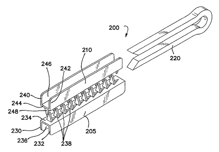
FIG. 6(a) illustrates a surgical staple-clip 200 in accordance with a first
embodiment of the invention. The staple-clip 200 comprises a first tissue-
engaging member 205, a second tissue-engaging member 210 opposed to the
first tissue-engaging member 205, and a securing or fixation member 220 for
securing the first and second tissue-engaging members 205, 210. The first
tissue-engaging member 205 comprises generally opposed walls 230, 232 and a
connecting wall 234 that together define an elongate channel 236. The second
tissue-engaging member 210 is similar to the first tissue-engaging member 205
and comprises generally opposed walls 240, 242 and a connecting wall 244 that
together define an elongate channel 246. The tissue-engaging members 205,
210 are normally held such that the front faces of the connecting walls 234,
244,
respectively, are opposed to each other. The opposing front faces of the
connecting walls 234, 244 may include a plurality of tissue-penetrating
elements
238, 248, respectively.
An advantage of the staple-clip of the invention is it provides good traction
without requiring an excessive compressive force to be applied to the staple-
clip.
In particular, the securing member 220 is sized and configured to slide into
the
elongate channels 236, 246 to securely clamp the tissue-engaging members
205, 210 around a body tissue or vessel with minimal compressive force. More
specifically, the force required to secure and maintain the staple-clip (to
provide
CA 02507967 2005-05-30
WO 2004/058079 PCT/US2003/040318
12
adequate traction) is independent from the force required to constrict or
occlude
a body tissue or vessel. With the staple-clip of the invention, only the
compressive force needed to perform a specific surgical procedure such as
occlusion, ligation or fixation is applied to the body tissue, and the force
normally
required to secure and maintain a clip of the prior art is not applied since
traction
and security are supplied by the tissue-engaging members 205, 210. In other
words, the staple-clip of the present invention provides the necessary
traction
without requiring an excessive compressive force to keep the staple-clip from
moving or becoming loose. As a result, nourishment of the lightly compressed
tissue is maintained and tissue necrosis due to over-compression is
eliminated.
In another aspect of the invention, the tissue-engaging members include
traction enhancing features including bumps, ridges, slots, holes, etc. as
generally illustrated in FIGS. 13(a) and 13(b). The traction enhancing
features
are sized and configured to grip tissue and provide traction and security
beyond
that which might be achieved by over-compressing a typical clip. The securing
member 220 may be a spring-clip or a deformable clip acting as a retention
member and providing a uniform pressure across the occluded tissue or vessel
to prevent loosening of the staple-clip 200 over time as illustrated in FIGS.
7, 9(a)
- 9(h) and 14. The tissue-penetrating elements 238, 248 are configured to
penetrate the tissue and to prevent the tissue from moving or sliding when
clamped as illustrated in FIGS. 11 (a) - 11 (d), 16 and 17.
It is appreciated that the connecting walls 234, 244 and the respective
tissue-penetrating elements 238, 248 may be formed as an integral, one-piece
CA 02507967 2005-05-30
WO 2004/058079 PCT/US2003/040318
13
construction. It is further appreciated that the number of rows of tissue-
penetrating elements and the number of tissue-penetrating elements per row
may vary according to each application and the shape and size of the clip and
body tissue. It is further noted that the tissue-penetrating elements in each
row
may be aligned or staggered as desired. The tissue-engaging members 205,210
and the securing member 220 may have cross-sections of any configuration
including polygonal, circular and elliptical configurations.
Referring to FIG. 10, there is shown a monolithic staple-clip 500 in
accordance with another aspect of the invention. The monolithic staple-clip
500
has a general shape of the capital letter "U". The staple-clip 500 comprises a
first tissue-engaging portion or leg 505, an opposed second tissue-engaging
portion or leg 510 and a deformable connecting portion 520 connecting the
first
and second tissue-engaging portions 505 and 510. Each of the opposing faces
of the tissue-engaging portions 505, 510 comprises a plurality of tissue
penetrating elements or protrusions 525, 530, respectively. The protrusions
are
sized and configured to penetrate tissue that is captured between the tissue-
engaging portions and provide traction and security beyond that which might be
achieved by over-compressing a typical clip. Similar to other aspects of the
invention, only the force required to perform a specific surgical procedure
such
as occlusion, ligation or fixation is applied to a body tissue and the force
previously needed to secure and maintain the clip is no longer applied.
Nourishment of the lightly compressed tissue is thus maintained and tissue
necrosis due to over-compression is eliminated.
CA 02507967 2005-05-30
WO 2004/058079 PCT/US2003/040318
14
The monolithic staple-clip 500 may be formed from a flat metal sheet that
is die-cut, stamped or etched forming a first notched or toothed portion, a
smooth
connecting portion and a second notched or toothed portion. The notches or
teeth of the first and second portions are then bent so as to extend in the
same
direction or plane and to form channels within the notches or teeth. The
staple-
clip is then formed in a U-shape by bending the connecting portion so that the
notches or teeth of the two tissue-engaging portions are opposed. A preferred
embodiment of the monolithic staple-clip comprises a malleable material such
as
Titanium or stainless steel. Other materials include any medically acceptable
metal or plastic material that is ductile, malleable or deformable.
It is appreciated that the staple-clips of the invention can be applied to a
body tissue or vessel using an applier 600 as illustrated in FIGS. 12(a) -
12(d).
The staple-clip applier 600 generally comprises an elongate shaft 605, sized
and
configured to fit through a surgical trocar port, a distal end 610 having a
pair of
opposed jaws 615, 620, and a proximal end (not shown) having a handle to open
and close the jaws 615, 620. The staple-clip applier 600 further comprises a
sliding member 625 to advance the securing member 220 over the tissue-
engaging members 205, 210 after closure of the jaws 615, 620 as further
described below. The jaws 615, 620 operate to apply the tissue-engaging
members 205, 210, respectively, around a target body tissue or vessel. The
tissue-engaging members are supplied to the jaws either manually or
automatically. With the tissue-engaging members 205, 210 properly placed, the
jaws 615, 620 can be compressed using only the force required for a specific
CA 02507967 2005-05-30
WO 2004/058079 PCT/US2003/040318
surgical procedure such as occlusion, ligation or fixation. When the tissue-
engaging members 205, 210 are properly applied, the sliding member 625 can
then urge the securing member 220 forward and over the tissue-engaging
members 205, 210 to secure the staple-clip 200 as illustrated in FIGS. 9(a) -
5 9(h), 11 (a) - 11 (d) and 14. The tissue-engaging members 205, 210 and
securing
member 220 may be introduced to a surgical site in an un-assembled condition
through a small port or trocar. FIGS. 8 and 15(a) - 15(c) further illustrate
the
staple-clip and applier sized and configured for use in a minimally invasive
or
laparoscopic surgical procedure.
10 Multiple staple-clips may also be loaded in a staple-clip applier and
advanced individually or simultaneously between the jaws. In the case of
simultaneously applying the staple-clips, the applier must include a plurality
of
slots in the opposed jaws to receive the multiple staple-clips. A cutting
member
such as a blade may be included in the applier to be advanced between the
15 staple-clips after they have been applied to transect the body tissue
between the
staple-clips as illustrated in FIGS. 17 and 18.
In another aspect of the invention, FIG. 19 depicts a traditional
laparoscopic stapler 800 that is configured specifically for donor
nephrectomy.
The stapler 800 includes a plurality of rows of staples on the patient side
805 and
a temporary clip 810 that substitutes for a typical set of staples on the
kidney side
815. The stapler 800 operates like existing place and cut staplers with the
exception that a temporary clip or clips 810 replace the set of staples on the
kidney side 815. The temporary clip 810 may be a staple cartridge configured
for
CA 02507967 2005-05-30
WO 2004/058079 PCT/US2003/040318
16
use in donor nephrectomy. An advantage of this configuration is it salvages a
greater portion of the vessel for the transplant procedure.
Another aspect of the invention is directed to hand assisted laparoscopy
(HAL), the widespread acceptance of which has created many opportunities for
surgical advancement utilizing single hand procedures. FIGS. 20(a) - 20(e)
illustrate thumb actuated clip appliers 850, 875 and 895 in accordance with
the
teachings of the invention. The clip applier 850 includes a handle 855 and a
thumb actuated mechanism 860. An operator can slidably release clips 865 onto
a vessel by sliding the thumb actuated mechanism 860 forward 870a and
backward 870b using only one hand as illustrated in FIGS. 20(a) - 20(b). This
design closes the jaws around the vessel and allows a closed clip to slide
into
position. The handle 855 also serves as a reservoir for additional clips 865.
The
clip 875 is also designed for HAL applications and includes a first arm 880
and a
second arm 885 folded over the first arm 880. The first arm 880 includes a
latch
mechanism 890 such as an inwardly turned portion or hook at its distal end
that
is configured to interlock or mate with a distal end of the second arm 885
when
the arms are clamped together. The latch mechanism 890 operates in a similar
way to a hair clip and responds to thumb pressure. An operator may single-
handedly access the clip 875 and slide it onto a body tissue or vessel as
needed.
A plurality of clips 875 may be strung end-to-end in a clip sleeve 892
suspended through an open port. An advantage of the clip 875 is there is no
instrument to misplace. The clip sleeve 892 holding the clips 875 can also be
withdrawn or fed through any open port or trocar. The clip sleeve 892 can also
CA 02507967 2005-05-30
WO 2004/058079 PCT/US2003/040318
17
be designed to keep the clips 875 partially closed, enabling the use of
smaller
ports or trocars. The clip 895 as illustrated in FIG. 20(e) is similar to the
clip 875
but further includes tissue-penetrating elements 898 on the inner faces of
arms
896, 897. The clip 895 may also include a third arm 899 connecting the arms
896, 897.
In another aspect of the invention as illustrated in FIG. 21, a spring like
coil 900 is used for suturing a body tissue or vessel in place of clips and
staples.
The spring like coil 900 is applied onto a body tissue or vessel 905 by using
a
stapler 910 having grooved jaws 910, 915. The spring like coil 900 is
preferably
loaded or twisted into one of the grooved jaws 910, 915, which is then
compressed or clamped against the other grooved jaw to constrict or occlude
the
vessel 905. An advantage of this design is closure of the coil 900 provides a
stitch like nature that replicates a uniformly applied suture. In one
configuration,
a single coil is inserted in place of each suture. It is appreciated that a
single coil
or multiple coils may be loaded into the grooved jaws of a stapler for each
specific surgical procedure or closure.
In yet another aspect of the invention, FIG. 22 illustrates multiple staples
.925 formed from a single piece of material 930 to reduce the high
manufacturing
costs associated with current stapler cartridges. These costs savings
translate to
increased margins or reduced product cost. Each of the staples 925 has an
angled leg 935 and an inclined rail 940 for easy push up and closure by a
stapler.
The staples 925 all share a common portion 945, which allows the row of
staples
to be formed as an integral, one-piece construction. The staples 925 can also
be
CA 02507967 2005-05-30
WO 2004/058079 PCT/US2003/040318
18
formed to offset each other and to emulate multiple rows. An advantage of this
aspect of the invention is it provides a compact cartridge that is suitable
for donor
nephrectomy.
FIG. 23 illustrates a temporary HAL clamp 950 for use in donor
nephrectomy in accordance with another aspect of the invention. The clamp 950
includes a tubular section 955 defining an opening 960 extending from a
proximal
end 965 to a distal end 970, and a lead-in wire 975 operably attached to the
proximal end 965. The lead-in wire 975 is movable between an open position
and a closed position. When closed, the lead-in wire 975 is slidably received
and
secured in the opening 960 of the tubular section 955. During use, an operator
may manually wrap a vessel or vessels (e.g., the renal artery and vein can be
cinched together into the clamp) around the lead-in wire 975 and secure the
wire
975 in the opening 960 of the tubular section 955. The lead-in wire 975 may be
bent against the wall of the tubular section 955 to further secure the
vessels) for
kidney removal and transport.
FIGS. 24, 25 and 26 illustrate two-stage clips 1010, 1020 and 1030,
respectively, in accordance with additional embodiments of the invention. The
clips 1010, 1020 and 1030 require an applier (not shown) for placement onto a
body tissue or vessel. The clip 1010 includes a clip component 1011 and a
staple 1012 for securing the clip after it has been properly positioned. The
clip
component 1011 is formed from a single wire and includes opposed arms 1013,
1014. Each of the arms 1013, 1014 includes openings 1015, 1016, respectively,
allowing penetration of legs 1017, 1013 of the staple 1012. The staple 1012 is
CA 02507967 2005-05-30
WO 2004/058079 PCT/US2003/040318
19
formed from a second wire and is used to puncture the body tissue or vessel
and
interlock the clip component 1011. The arms 1013, 1014 may include a latch
mechanism at the distal ends to mate with each other when the arms are closed
or clamped together. During use, the first stage closes the arms 1013, 1014 of
the clip. After the first stage, the clip can still be safely removed. The
second
stage secures the clip permanently onto the body tissue or vessel by applying
the
staple 1012 to the clip component 101.1.
The two-stage clip 1020 as shown in FIG. 25 is formed entirely from a
single piece of material and includes a first arm 1021 and an opposed second
arm 1022. The second arm 1022 further includes securing elements 1023, 1024,
all of which are formed as an integral, one-piece construction. With this
construction, the securing elements 1023, 1024 may still remain at an angle
after
the first stage (when the arms 1021 and 1022 are clamped upon a body tissue or
vessel). A second action then presses the securing elements 1023, 1024 into
the body tissue or vessel. The clip 1030 as shown in FIG. 26 is similar to the
clip
1010 and includes a clip component 1031 and a staple component 1032 for
securing the clip after it has been properly positioned. The clip component
1031
includes opposed arms 1013, 1014 defining openings 1035, 1036, respectively.
The staple component 1032 includes opposed arms 1037, 1038 having tissue-
securing elements 1039, 1040, respectively, which operate to secure the clip
onto a body tissue or vessel through the openings 1035, 1036. The clip 1030
may be formed in sheet stock.
CA 02507967 2005-05-30
WO 2004/058079 PCT/US2003/040318
FIG. 27 illustrates a tissue holder 1100 for use with staple-clips that may
include sharp features, e.g., tissue-penetrating elements, that can
potentially
injure tissue during a surgical procedure. In particular, the tissue holder
1100 is
used to press a body tissue flat and to provide clearance during placement of
a
5 staple-clip.
Although exemplary embodiments of the invention have been shown and
described, many alterations and modifications may be made by those having
ordinary skill in the art without departing from the spirit and scope of the
invention. Therefore, it must be understood that the illustrated embodiments
10 have been set forth only for the purposes of examples and that they should
not
be taken as limiting the invention. In addition, the words used in this
specification
to describe the invention and its various embodiments are to be understood not
only in the sense of their commonly defined meanings, but to include any
special
definition given in this specification.