Note: Descriptions are shown in the official language in which they were submitted.
CA 02508100 2005-05-20
MRI BIOPSY DEVICE LOCALIZATION
FIXTURE
Field of the Invention
~oooil The present invention relates, in general, to a method of imaging
assisted
tissue sampling and, more particularly, to an improved method for positioning
a biopsy
probe with respect to a magnetic resonance imaging (MRI) breast coil for
acquiring
subcutaneous biopsies and for removing lesions.
Background of the Invention
~0002~ Recently, core biopsy devices have been combined with imaging
technology
to better target a lesion in breast tissue. One such commercially available
product is
marketed under the trademark name MAMMOTOMETM, by Ethicon Endo-Surgery, Inc.
An embodiment of such a device is described in U.S. Patent No. 5,526,822
issued to
Burbank, et al., on June 18, 1996, and is hereby incorporated herein by
reference. Its
handle receives mechanical and electrical power as well as vacuum assist from
a
remotely positioned control module that is spaced away from the high magnetic
field of
a Magnetic Resonance Imaging (MRI) machine.
(0003 As seen from that reference, the instrument is a type of image-guided,
percutaneous coring, breast biopsy instrument. It is vacuum-assisted, and some
of the
steps for retrieving the tissue samples have been automated. The physician
uses this
device to capture "actively" (using the vacuum) the tissue prior to severing
it from the
body. This allows the sampling of tissues of varying hardness. In addition, a
side
opening aperture is used, avoiding having to thrust into a lesion, which may
tend to push
the mass away, cause a track metastasis, or cause a hematoma that, with
residual contrast
agent circulating therein, may mimic enhancement in a suspicious lesion. The
side
aperture may be rotated about a longitudinal axis of the probe, thereby
allowing multiple
tissue samples without having to otherwise reposition the probe. These
features allow for
substantial sampling of large lesions and complete removal of small ones.
~0004~ In the aforementioned Pub. No. US 2003/0199785 to Hibner et al.,
localization
fixtures are described that are attachable to a breast coil. These
localization fixtures
aided in accurately positioning the probe to a location of a suspicious lesion
within
CA 02508100 2005-05-20
breast tissue. In particular, the X-Y-Z Cartesian coordinates of a suspicious
lesion are
referenced to a fiduciary marker in the localization fixture. Humanly visible
measurement guides for each axis then allow the probe to be correspondingly
positioned
after a patient has been withdrawn from a closed bore MRI machine without the
need for
imaging the probe during insertion. In addition, the localization fixture
enabled use of a
detachable probe of an MRI biopsy device. Thus, during subsequent reimaging of
the
probe, a handle of the MRI biopsy device may be deattached, as may be
necessary within
the close confines of a closed bore MRI machine. When the handle is attached
to the
probe, various support structures of the localization fixture are described
that support the
extended length of the handle.
~ooosl While a localization fixture used with a detachable MRI biopsy probe
has a
number of advantages, it is desirable to incorporate additional features that
further assist
in efficiently and comfortably localizing the breast of a patient within a
localization
fixture by supporting various biopsy devices in a hands-free manner.
Brief Summary of the Invention
X00061 The invention overcomes the above-noted and other deficiencies of the
prior
art by providing a localization and guidance assembly that interfaces an MRI
biopsy
device to a breast coil to accurately position and maintain a probe at a
desired position in
a patient's breast for performing biopsy and related diagnostic and
therapeutic
procedures.
~ooo~l In one aspect of the invention, a localization fixture advantageously
provides
lateral and medial compression members that may be centered on, brought into
opposing
compression of a patient's breast, and locked in this position with access to
proximal
controls that avoid the inconvenience and discomfort of having to reach under
the
patient. Thereby, a stressful medical procedure is rendered a little easier
for the patient
and expedited for the care provider.
~ooos~ In another aspect of the invention, a fiducial for locating a
suspicious lesion
relative to the localization fixture is made more economical by providing a
disposable
housing that may be shipped without an MRI imagable material. Thereby, shelf
life,
2
CA 02508100 2005-05-20
packaging, sterility are simplified, as well as allowing the end user to
select an
appropriate content. The disposable fiducial is engagable to the biopsy probe
support.
~0009~ In yet another aspect of the invention, a localization fixture has a
removable
tray adjustable that supports a biopsy probe support. Thus, a desired
insertion point
relative to the lateral compression member may be remotely set on the biopsy
probe
support prior to locking the removable tray to the base member.
~ooio~ In yet a further aspect of the invention, a localization fixture has a
pedestal
that is positioned for lateral movement relative to a lateral plate that
positions a lateral
fence for compressing one side of a patient's breast. A guide rail is
positionable upon the
pedestal to set a height coordinate. The targeting rail includes a biopsy
guide defining an
angle of penetration for a biopsy instrument.
~ooy These and other objects and advantages of the present invention shall be
made
apparent from the accompanying drawings and the description thereof.
Brief Description of the Figures
~ooi2~ The accompanying drawings, which are incorporated in and constitute a
part
of this specification, illustrate embodiments of the invention, and, together
with the
general description of the invention given above, and the detailed description
of the
embodiments given below, serve to explain the principles of the present
invention.
~ooi3~ FIGURE 1 is a perspective disassembled view of a Magnetic Resonance
Imaging (MRI) Biopsy System;
~ooia~ FIGURE 2 is a perspective view of a holstered MRI biopsy device being
aligned with a track of a pedestal of a localization fixture of the MRI biopsy
system of
FIG. 1;
~oois~ FIGURE 3 is a perspective view of the holstered MRI biopsy device after
engagement to the track of the pedestal of the localization fixture of the MRI
biopsy
system of FIG. 1;
~ooi6~ FIGURE 4 is a perspective view of a disassembled view of a guidance
system
of the localization fixture of FIG. l;
3
CA 02508100 2005-05-20
f
(001~~ FIGURE S is a perspective view of an alternative pedestal and targeting
rail
supported by a lateral fence of a localization fixture for the MRI biopsy
system of FIG. 1;
(ooisl FIGURE 6 is a disassembled perspective view of an alternative guidance
system for the pedestal and targeting rail of FIG. S;
(ooi91 FIGURE 7 is a left side view in elevation of an obturator with a flat
bladed
piercing tip, lumen communicating between a lateral notch and fluid fitting on
a proximal
end with external engaging features for an obturator hub;
(oozo( FIGURE 8 is a front view in elevation of the obturator of FIG. 7;
(oozy FIGURE 9 is a left side view in elevation of a longitudinal cross
section of the
obturator of FIG. 8 taken along lines 9-9;
(oo2z( FIGURE 10 is a front view in elevation of the obturator of FIG. 7 taken
in
cross section along lines 10-10 distal to a hub engaging portion;
(0023 FIGURE 1 I is a front view in elevation of the obturator of FIG. 7 taken
in
cross section along lines 11-11 across the hub engaging portion;
(ooza~ FIGURE 12 is a perspective view of the alternative secondary targeting
rail,
sleeve and obturator of the guidance system of FIG. 6;
(oozs( FIGURE 13 is a perspective view of a localization fixture having a
pivoting z-
stop integral to a track;
(ooz61 FIGURE 14 is a perspective detail view of the z-stop of FIG. 13.
(oozy FIGURE 1 S is a perspective view of a localization fixture for the MRI
biopsy
system of FIG. 1;
(ooze( FIGURE 16 is a perspective view of a lateral plate having displaceable
and/or
removable bars for the localization fixture of FIG. 33;
(ooz91 FIGURE 17 is a perspective view of a tower pedestal for the
localization
fixture of FIG. 15;
(0030( FIGURE 18 is a perspective view of a breast localizing portion of a one-
rail
localization fixture;
4
CA 02508100 2005-05-20
r
(oo3i1 FIGURE 19 is a perspective view of a localization fixture incorporating
an
equipment protection flip-out lever for the MRI biopsy system of FIG. 1;
(00321 FIGURE 20 is a perspective view of a localization fixture incorporating
a
telescoping equipment protector for the MRI biopsy system of FIG. 1;
(00331 FIGURE 21 is a top view of a localization fixture with a proximal
medial plate
locking control, flexible/curved medial fence and lateral plate, and flexible
cam locked
lateral assembly;
(oo3a1 FIGURE 22 is a front cross sectional view in elevation of a lateral
plate and
top recess of a base plate of the localization fixture of FIG. 21 taken along
lines 22-22;
(oo3s1 FIGURE 23 is a top view of a localization fixture partially cut away
for the
MRI biopsy system of FIG. 1, including a lateral plate having flexible
ribbing;
(00361 FIGURE 24 is a front cross sectional view in elevation of the lateral
plate and
top recess of the lateral plate and medial plate of FIG. 21 taken along lines
24-24;
(0031 FIGURE 25 is a top view of a localization fixture having a squeeze
trigger
with a cam lock for medial fence adjustment;
(oo3g1 FIGURE 26 is a perspective view of an alternative patient support for
the MRI
biopsy system of FIG. 1;
(00391 FIGURE 27 is a perspective view of guidance components having tablesaw
style guides and locking levers of a localization fixture for the MRI biopsy
system of FIG.
1;
(ooaol FIGURE 28 is a side view in elevation of the guidance components of
FIG.
27;
(ooail FIGURE 29 is a front view in elevation of guidance components of a
localization fixture for the MRI biopsy system of FIG. 1 having a separate
support arm
and distal targeting ring;
(ooaa( FIGURE 30 is a top cross sectional view of the guidance components of
FIG.
29;
CA 02508100 2005-05-20
a
~0043~ FIGURE 31 is a perspective, detailed view of a telescoping targeting
ring of
FIG. 30 detached from an X-Y alignment fixture;
~oo4a~ FIGURE 32 is a top cross sectional view of the telescoping targeting
ring of
FIG. 31 attached to the X-Y alignment fixture and showing a pneumatic outlet
and
septum;
~oo4s~ FIGURE 33 is a front view in elevation of the septum of the telescoping
targeting ring of FIGS. 31-32;
~ooa6~ FIGURE 34 is a top view of a remotely rotated localization fixture for
the
MRI biopsy system of FIG. 1;
~ooa~~ FIGURE 35 is top view in cross section of the remotely located
localization
fixture of FIG. 34;
~oo4g1 FIGURE 36 is a detailed view of a rotation control mechanism of the
remotely
located localization fixture of FIG. 35;
~ooa91 FIGURE 37 is a perspective view of a honeycomb lateral plate with an
integral distal targeting fixture shown in its swung open position;
~ooso~ FIGURE 38 is a perspective view of the honeycomb lateral plate with the
integral distal targeting fixture of FIG. 37, shown in its swung closed
position with a
probe guide installed;
~oosy FIGURE 39 is a side view in elevation of a medial plate and medial fence
and
a lateral plate of a localization fixture that incorporates soft elastomeric
pads to enhance
support and comfort;
~oosZ~ FIGURES 40-41 are perspective views of a ratcheting jack-style lock on
an
alternative pedestal for a localization fixture;
~oos3~ FIGURE 42 is a perspective view of a lateral plate having displaceable
grid
members;
~oosal FIGURE 43 is a front perspective view of a further alternative lateral
fence
having detachable bars for the MRI biopsy system of FIG. 1;
6
CA 02508100 2005-05-20
r
(oossl FIGURE 44 is a back, left perspective view of a detached bar of the
lateral
fence of FIG. 43;
(oos61 FIGURE 45 is a detailed perspective view of an upper portion of the
detached
bar of FIG. 44;
(oos~l FIGURE 46 is a detailed perspective view of a lower portion of the
detached
bar of FIG. 44;
(oossl FIGURE 47 is a back perspective view of a lower portion of the lateral
fence
of FIG. 43;
(oos91 FIGURE 48 is a top left perspective view of an alternative box
localization
fixture for the MRI biopsy system of FIG. 1;
(00601 FIGURE 49 is a left view in elevation of the alternative box
localization
fixture of FIG. 48 with an X-axis guide plate adjustably locked therein, which
in turn
supports a locked Y-axis guide frame;
(oo6y FIGURE 50 is proximal perspective view of an alternative fiducial
holder;
(oo6z1 FIGURE 51 is a top view of the alternative fiducial holder of FIG. 51;
(oo6s1 FIGURE 52 is a proximal side view in elevation of the fiducial holder
of FIG.
51;
(oor~al FIGURE 53 is a right side view in elevation of the fiducial holder of
FIG. 51;
(00651 FIGURE 54 is a top diagrammatic view of a disposable fiducial for the
fiducial holder of FIG. 51; and
(00661 FIGURE SS is a top diagrammatic view of an alternate disposable
fiducial for
the fiducial holder of FIG. 51.
Detailed Descriution of the Invention
(0061 Turning to the Drawings, wherein like numerals denote like components
throughout the several views, in FIG. 1, a Magnetic Resonance Imaging (MRI)
compatible biopsy system{xe "0010 Magnetic Resonance Imaging (MRI) compatible
biopsy system"} 10 includes a guide that guides a sleeve and introducer
obturator that are
7
CA 02508100 2005-05-20
separate from the biopsy device itself and advantageously incorporate an
improved
piercing portion, MRI imaging marker, and fluid handling capabilities.
Mounting
provisions allow for precise penetration along a desired trajectory without
overshooting.
[0068) The MRI compatible biopsy system 10 includes a control module{xe "0012
control module"} 12 that typically is placed outside of a shielded room
containing an MRI
machine (not shown) or at least spaced away to mitigate detrimental
interaction with its
strong magnetic field and/or sensitive radio frequency (RF) signal detection
antennas. The
control module 12 controls and powers an MRI biopsy device{xe "0014 MRI biopsy
device"} 14 that is compatible for use in close proximity to the MRI machine.
An
example of an MRI biopsy device 14 is the afore-mentioned MAMMOTOMET'"
instrument. The MRI biopsy device 14 is accurately positioned by a
localization
fixture{xe "0016 localization fixture"} 16 that is attached to a breast
coil{xe "0018 breast
coil"} 18, which in turn supports a patient (not shown). Examples of
commercially
available breast coils 18 include the BIOPSY BREAST COIL MODEL BBC by MRI
DEVICES CORPORATION of Waukesha WI. A guidance assembly{xe "0020 guidance
assembly "} 20, and in particular a sleeve{xe "0022 sleeve"} 22,
advantageously attaches
to the localization fixture 16 to increase imaging and therapeutic flexibility
and accuracy
in conjunction with selective use of the MRI biopsy device 14 at particular
parts of the
procedure. The guidance assembly 20 may include one or more obturators{xe
"0024
obturator"} 24 with one depicted that seals the sleeve 22 during insertion and
during
subsequent portions of the procedure in which the MRI biopsy device 14 is not
inserted
therein. A depth stop{xe "0026 depth stop"} 26 is provided for use with the
localization
fixture 16 to advantageously prevent over-insertion of the sleeve 22,
inadvertent
retraction of the sleeve 22 and/or to enhance accurate placement of the sleeve
22 to a
desired location along the Z-Axis.
(00691 For convenience, herein a convention is used for locating a suspicious
lesion by
Cartesian coordinates within breast tissue referenced to the localization
fixture 16 and to
thereafter position an instrument (e.g., sleeve 22) to this location without
necessarily
continuously imaging the region. As will be described in greater detail below,
a
perforated barrier that is compressed along an outside side of the breast,
with respect to a
medial plane of the chest of the patient, defines an X-Y plane, with the X-
axis being
vertical (sagittal) with respect to a standing patient and which corresponds
to a left to
8
CA 02508100 2005-05-20
right axis as viewed by a clinician facing the externally exposed portion of
the
localization fixture 16. A fiduciary marker (not shown), attached to or
positioned relative
to the localization fixture 16 proximate to the patient's skin, defines the
origin of this
plane. Perpendicular to this X-Y plane extending toward the medial side of the
breast is
the Z-axis, which typically corresponds to the orientation and depth of
insertion of the
MRI biopsy device 14, although it should be appreciated that variations may
allow
insertion at an angle to this Z-axis. Thus, for clarity, the term Z-axis may
be used
interchangeably with "axis of penetration", although the latter may or may not
be
orthogonal to the spatial coordinates used to locate an insertion point on the
patient.
lomol Separating the tracking rail that supports a mount / depth stop from a
biopsy rail
that supports the weight of the biopsy device advantageously reduces
interference
between the various components, allowing a sequence of operation wherein
certain
components may be selectively installed and removed without interfering with
other
components.
~oml1 In use, the MRI compatible biopsy system 10 is prepared for use by
placing a
cable management spool{xe "0030 cable management spool"} 30 upon a cable
management attachment saddle{xe "0032 cable management attachment saddle"} 32
that
projects from a side of the control module 12. Wound upon the cable management
spool
30 is a paired electrical cable{xe "0034 electrical cable"} 34 and mechanical
cable{xe
"0036 mechanical cable"} 36 for communicating control signals and cutter
rotation/advancement motions respectively. In particular, electrical and
mechanical cables
34, 36 each have one end connected to respective electrical and mechanical
ports{xe
"0042, 0044 electrical and mechanical ports"} 40, 42 in the control module 12
and
another end connected to a holster{xe "0044 holster"} 44 that receives the MRI
biopsy
device 14. An MRI docking cup{xe "0046 MRI docking cup"} 46, which may hold
the
holster 44 when not in use, is hooked to the control module 12 by a docking
station
mounting bracket{xe "0048 docking station mounting bracket"} 48.
~00~21 An interface lock box{xe "0050 interface lock box"} 50 mounted to a
wall
provides a tether{xe "0052 tether"} 52 to a lockout port{xe "0054 lockout
port"} 54 on the
control module 12. The tether 52 is advantageously uniquely terminated and of
short
length to preclude inadvertent positioning of the control module 12 too close
to the MIZI
9
CA 02508100 2005-05-20
machine. An in-line enclosure{xe "0056 in-line enclosure"} 56 may
advantageously
register the tether 52, electrical cable 34 and mechanical cable 36 to their
respective ports
54, 42, 44 on the control module 12. A remote keypad{xe "0058 remote keypad"}
58 may
be distally connected to the electrical cable 34 to enhance clinician control
of the MRI
biopsy device 14, especially when controls on the MRI biopsy device 14 itself
are not
readily accessible after insertion into the localization fixture 16.
too~3~ Vacuum assist is provided by a first vacuum line{xe "0060 first vacuum
line"} 60
that connects between the control module 12 and an outlet port{xe "0062 outlet
port"} 62
of a vacuum canister{xe "0064 vacuum canister"} 64 that catches liquid and
solid debris.
A tubing kit{xe "0066 tubing kit"} 66 completes the pneumatic communication
between
the control module 12 and the MRI biopsy device 14. In particular, a second
vacuum
line{xe "0068 second vacuum line"} 68 is connected to an inlet port{xe "0070
inlet port"}
70 of the vacuum canister 64. The second vacuum line 68 divides into two
vacuum
lines{xe "0072, 0074 vacuum lines"} 72, 74 that are attached to the MRI biopsy
device
14. With the MRI biopsy device 14 installed in the holster 44, the control
module 12
performs a functional check. Saline is manually injected into biopsy device 14
to serve as
a lubricant and to assist in achieving a vacuum seal. The control module 12
actuates a
cutter mechanism (not shown) in the MRI biopsy device 14, monitoring full
travel.
~oo~a~ The portion of the MRI compatible biopsy system 10 used near the MRI
machine
is also assembled. The generally known breast coil 18 is placed upon a gantry
of the MRI
machine, along with other body support pads (not shown). The localization
fixture 16 is
attached within a recess on either lateral side of the breast coil 18 to
access a patient's
breast that is pendulously exposed therein and includes a horizontal medial
plate{xe
"0080 horizontal medial plate"} 80, a reusable base assembly{xe "0082 base
assembly"}
82, a lateral assembly{xe "0084 lateral assembly"} 84, and a positioning
pedestal{xe
"0086 positioning pedestal"} 86. The localization fixture 16 is also assembled
with a
disposable medial fence{xe "0090 medial fence"} 90 and a lateral window (or
perforated
plate){xe "0092 perforated plate or lateral window"} 92.
~oo~sl The base assembly 82 is placed within a selected lateral recess of the
breast coil
18. The medial fence 90 attaches to a medial edge of the medial plate 80,
aligned
vertically approximately along a longitudinal axis of the breast coil 18 under
an inner
CA 02508100 2005-05-20
edge of a selected breast aperture{xe "0094 breast aperture"} 94 that receives
a patient's
breast. With the patient thus positioned and the outer area of the breast
sterilized, the
lateral window 92 is downwardly slid into a three-sided frame guide{xe "0096
three-sided
frame guide"} 96 of the lateral assembly 84, which in turn is placed upon the
medical
plate 80. The base assembly 82 and lateral assembly 84 are moved with respect
to one
another along the Z-axis to compress the patient's breast between the medial
fence 90 and
the lateral window 92. A mechanism formed between the lateral assembly 84,
base
assembly 82, and medial plate 80 maintains this compression.
~00~61 Contrast agent may be injected into the patient to enhance the imaging.
The gantry
is advanced into the MRI machine bore to image the localization fixture 16 and
breast
tissue. The fiduciary marker on the lateral window 92 is located and
designated as the
origin of the X-Y-Z coordinates. Then a suspicious lesion is located within
the image and
a point thereon is selected to determine its location relative to the origin.
It should be
appreciated that orienting the X-Y-Z axis of an initial scan may be
facilitated by having
the lateral window 92 formed of an imagable material, thus presenting an X-Y
plane in
addition to the origin point of the fiduciary marker. With the target location
determined,
the gantry is withdrawn from the MRI machine bore.
The positioning pedestal 86 is slidably engaged along the X-axis of the
lateral
assembly 84 and defines a vertical guide for positioning a single targeting
rail
("track"){xe "0098 single targeting rail or track"} 98 at a selected Y-axis
coordinate. The
track 98 in turn provides a depth guide along the Z-axis for positioning the
depth stop 26
and the holster 44 at a desired Z-axis coordinate. The depth stop 26 is
latched onto the
track 98. Thereafter, a marking instrument (not shown) may be inserted through
the depth
stop 26 to mark the insertion point on the breast. Thereafter, the depth stop
26 is moved
out of the way. Anesthesia is injected superficially, followed by a scoring
cut at the
marked location and a subsequent injection of anesthesia more deeply into the
scored cut.
The depth stop 26 is then repositioned on the track 98 to the desired Z-axis
coordinate
reference.
~oo~a~ The obturator 24 is inserted into the sleeve 22 and may be positioned
to close any
apertures of the sleeve 22 (side and/or distal end) to present a closed
surface to the breast
tissue. The obturator may also be shaped or formed to enhance the visibility
of the
11
CA 02508100 2005-05-20
aperture location. One or the other of the obturator 24 and sleeve 22 presents
a sharp tip
(not shown) to penetrate breast tissue. For instance, if using a sleeve 22
having an open
end, an obturator may provide a sharp tip.
~00~9~ The obturator 24 is inserted into the sleeve 22 and the combination is
guided by
the track 98 to a proper orientation until an accurate depth is reached as set
by the depth
stop 26. Once fully inserted, the depth stop 26 prevents over-insertion. The
sleeve 22
advantageously latches to the track 98 and/or the depth stop 26 to prevent
inadvertent
retraction, such as when the obturator 24 is withdrawn, and pressure is
received from the
breast tissue or later when a probe{xe "0100 probe"} 100 of the MRI biopsy
device 14 is
withdrawn from the sleeve 22.
~ooso~ The gantry is moved into the MRI machine bore and the patient is imaged
again to
confirm placement of the sleeve 22 with respect to the suspicious lesion.
Advantageously,
imagable materials of the sleeve 22 and/or obturator 24, perhaps comprising or
including
marker material, enhance the ability to confirm the location of the sleeve 22
and its sleeve
side aperture{xe "0102 sleeve side aperture"} 102 as positioned for subsequent
biopsy
samples.
loosy The patient is removed from the MRI machine by retracting the gantry and
the
holstered MRI biopsy device 14 is brought to the localization fixture 16. A
protective cap
(not shown) is removed from the probe 100 of the MRI biopsy device 14 and the
obturator 24 is removed from the sleeve 22. Mounting of the holster 44 to the
track 98 is
shown in FIGS. 2 and 3, wherein the holster 44 and MRI biopsy device 14
combination
slide onto the track 98 that has been positioned at a certain location with
respect to the
pedestal 86 and lateral assembly 84. Features of the sleeve 22 and probe 100
may
advantageously visually and mechanically orient a probe side aperture{xe "0104
probe
side aperture"} 104 of the probe 100 with the sleeve side aperture 102, as
well as forming
a gas seal. Advantageously, the holster 44 and/or the probe 100 may latch onto
the track
98 or sleeve 22 to confirm full insertion and prevent over-insertion and
inadvertent
retraction. The holster 44 allows an MRI biopsy device 14 intended for
handheld use to
have sufficient support in its attachment to the localization fixture 16 to
accurately
maintain its position and to avoid or minimize loads carried by the probe 100.
12
CA 02508100 2005-05-20
~oos2~ Thereafter, the MRI compatible biopsy system 10 may take tissue samples
by
activating a cutter mechanism in conjunction with vacuum assist, withdrawing
the cutter
and withdrawing a tissue sample, the latter perhaps also with vacuum assist.
The probe
100 / sleeve 22 combination are capable of manual, or perhaps automatic,
rotation to a
desired angle with respect to their longitudinal axis for additional samples
or additional
samples may be taken at the current orientation by further resorting to vacuum
assist. The
cutter is then advanced to close the probe side aperture 104 and the holster
44 is
withdrawn from the localization fixture 16, thereby removing the probe 100
from the
sleeve 22.
~oos3~ Additional steps or combinations of steps may be performed at this
point such as
using the probe 100, a specialized obturator 24 (e.g., stylet), or merely the
sleeve 22 to
guide various agents to the surgical site of the biopsy. Examples include
draining fluids,
inserting anesthetic agents, inserting hemostatic agents, insufflating with
pneumatic
pressure and inserting a marker for subsequently locating the site of the
biopsy, or other
diagnostic or therapeutic procedures.
loosal The patient is then typically drawn back into the MRI machine bore for
reimaging
to confirm removal of at least a portion of the suspicious lesion and possibly
placement of
a marker. During this reimaging, the sleeve 22 is sealed with the obturator or
stylet 24.
Thereafter, the localization fixture 16 is removed, the patient bandaged and
removed from
the gantry, and the disposable portions of the MRI compatible biopsy system 10
disposed
of as medical waste.
~ooss~ With particular reference to FIGS. 2-3, the single targeting rail 98
facilitates
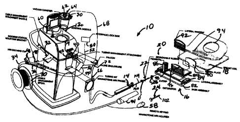
sequential mounting of separate components. First the depth stop 26, then the
sleeve 22
(as in FIG. 1 ), and then the biopsy tool 14 is slid onto the single targeting
rail 98.
Alternatively as depicted in FIGS. 2-3, the single targeting rail 98 may
receive the depth
stop 26 and then an MRI biopsy device 14 is used without a separate sleeve 22.
The
maximum depth of penetration into the patient's breast is preset by the
location of the
depth stop 26 on the single targeting rail 98. An engagement mechanism between
the
holster 44 and the single targeting rail 98 (not shown) and/or an engagement
mechanism
formed by a catch, is depicted as an upwardly projecting pin 110, on an upper
rail-
gripping arm{xe "0112 upper rail-gripping arm"} 112 of the depth stop 26 and a
13
CA 02508100 2005-05-20
downwardly spring-biased rocker latch{xe "0114 downwardly spring-biased rocker
latch"} 114 that snaps onto the upwardly projecting pin 110, preventing
inadvertent
retraction of the MRI biopsy device 14. The holster 44 may be disengaged by
downward
pressure on a proximal actuating arm{xe "0116 proximal actuating arm"} 116 of
the
rocker latch 114.
~oos61 The single targeting rail 98 may be longitudinally sized to be
proximally
extending sufficient that the MRI biopsy device 14 engages the single
targeting rail 98
prior to the probe 100 contacting the patient's skin. The single targeting
rail 98 is also
sized to not extend proximally so far as to preclude use in a closed bore MRI
machine
(not shown). Such an MRI compatible biopsy system 10 is believed to minimize
the
procedure turn-around time to less than 45 minutes as described above. Despite
this
expeditious turn-around, a radiologist may position the probe 100 accurately
to within 2
mm (5 mm maximum) of the lesion center. Further, the radiologist may maximize
access
to both breasts (left or right) during a procedure (both sides of the table)
with minimal
repositioning of the patient. Further, a minimal amount of force is required
to penetrate
tissue, such as less than 4 lbs. Although the depth stop 26 serves to prevent
overshooting,
features for repositioning the depth stop 26 prior to further insertion of the
probe 100
allow clinical flexibility in targeting another location.
~oos~l In FIG. 4, an alternative guidance assembly{xe "0200 alternative
guidance
assembly"} 200 for the MRI compatible biopsy system 10 incorporates a
cradle{xe "0202
cradle"} 202 that attaches to a targeting rail{xe "0204 targeting rail"} 204
and provides a
biopsy rail{xe "0206 biopsy rail"} 206 for supporting the MRI biopsy device,
both rails
204, 206 aligned to the Z axis. 'The targeting rail 204 is attached to the
positioning pillar
86 (not shown in FIG. 4) and vertically adjusted to a desired Y-position. A
circular
attachment point{xe "0208 circular attachment point"} 208 may form a
rotational
engagement to the positional pedestal 86 to allow an angled targeting guide.
X00881 A lateral face{xe "0210 lateral face"} 210 of the targeting rail 204
includes an
upper flange{xe "0212 upper flange"} 212 and a lower flange{xe "0214 lower
flange"}
214, each having an L-shaped cross section for slidingly receiving a sleeve
mount{xe
"0216 sleeve mount"} 216. Vertical rows of laterally projecting ridges{xe
"0218 vertical
rows of laterally projecting ridges"} 218 in each flange 212, 214 serve as a
locking
14
CA 02508100 2005-05-20
surface for the sleeve mount 216. Between the flanges 212, 214, a side
channel{xe "0220
side channel"} 220 is recessed therein. The sleeve mount 216 guides a
sleeve{xe "0222
sleeve"} 222 by having its sleeve hub{xe "0224 sleeve hub"} 224 proximally
received in a
hub receptacle{xe "0225 hub receptacle of the sleeve mount"} 225 of the sleeve
mount
216 and distally positioned and constrained by a depth stop{xe "0226 depth
stop"} 226.
toos9l The depth stop 226 includes a slide member{xe "0228 slide member of
depth
stop"} 228 that engages the side channel 220. A depth stop housing{xe "0230
depth stop
housing"} 230 attaches thereto, terminating in a reticule{xe "0232 reticule"}
232. A
locking lever{xe "0234 locking lever"} 234 is vertically pinned within a
distally open
recess (not shown) defined in the depth stop 226 with a lateral portion{xe
"0236 lateral
portion"} 236 spring biased away therefrom such that distally projecting
feet{xe "0238
distally projecting feet"} 238 pivot against and engage the ridges 218,
especially against a
proximal movement. Depressing the lateral portion 236 proximally against the
distally
open recess of the depth stop housing 230 releases the distally projecting
feet 238 to
allow repositioning the depth stop 226 distally.
(00901 An axis of penetration of the biopsy device 10 is aligned with the axes
defined by
the targeting rail 204 and the biopsy rail 206, which are laterally and
vertically
orthogonally offset therefrom, respectively. Extending a horizontal plane from
the
targeting rail 204 and extending a vertical plane from the biopsy rail 206
intersect at a
common centerline that is the axis of penetration. Having the biopsy rail 206
vertically
aligned and parallel to the axis of penetration advantageously provides
support for the
weight of the biopsy device 14 with a minimum of torsion loads that may
otherwise
create deflections of an inserted distal end. Thereby, even for a relatively
heavy and
elongated device, positioning and maintaining its distal end is achievable
within 5 mm,
and even 2 mm, of a desired insertion point. Thereby, a "hands free" procedure
may be
performed, and the inconvenience or the impracticability of penetration in the
illustrative
version may be replaced by one vertically displaced above the axis of
penetration. In
particular, having a cradle that may be engaged to either side of the
targeting rail 204
would provide further vertical symmetry to take full advantage of the space
afforded by
the breast coil 18.
CA 02508100 2005-05-20
~oo9y While a "hands free" capability is advantageous for a single insertion /
multiple
sample biopsy device, it should be appreciated that such penetration guidance
with a
preset depth stop as described herein has application to even light-weight
biopsy devices
that employ a core needle biopsy with a single insertion per single sample. In
particular,
correct placement need not be conditional on continuous imaging. Over
penetration
during insertion and inadvertent displacement is avoided when hands are free.
A bottom dovetail channel{xe "0240 bottom dovetail channel"} 240 in the
targeting rail 204 receives a top dovetail extension{xe "0242 top dovetail
extension"} 242
on the cradle 202, which is slid therein. It should be appreciated that
mounting is shown
herein on the right side of the positioning pedestal 86 when viewed
proximally, but that
the guidance assembly 200 advantageously comprises symmetric parts that allow
mounting and use on either side of the positioning pedestal 86 to increase
flexibility in
positioning the probe 100. Thus, a horizontal base{xe "0244 horizontal base of
the
cradle"} 244 of the cradle 202 forms the biopsy rail 206 as a biopsy guide
channel{xe
"0246 biopsy guide channel"} 246 flanked by a first and second pair of monocle
receptacles{xe "0248, 0250 first and second pair of monocle receptacles"} 248,
250 so
that a pair of locking hooks{xe "0252 locking hooks"} 252 on a monocle{xe
"0254
monocle"} 254 may be inserted in either pair of monocle receptacles 248, 250,
depending
on which is closer to the patient. Rather than mounting the cradle 202 to the
targeting rail
204 as depicted, the cradle may be directly attached to the positioning
pedestal 86 (not
shown). The cradle 202 is mechanically robust and can support the gross weight
of the
MRI biopsy device 14. Since the MRI biopsy device 14 does not share the cradle
202, the
cradle 202 may be optimized to support the MRI biopsy device 14 when either
shallow or
deep lesions need to be accessed.
~oo9s~ A guide bushing{xe "0256 guide bushing"} 256 inserted in a monocle
reticule{xe
"0258 monocle reticule"} 258 guides a marking instrument and/or a scoring
scalpel (not
shown) as an initial step in locating and preparing an insertion point. The
monocle 254
may be removed thereafter or left in place to guide the sleeve 222 in addition
to the
reticule 232 of the depth stop 226, the latter which may also hold a guide
bushing{xe
"0260 guide bushing"} 260 for guiding the sleeve 222. Removing the guide
bushings 256,
260 allows for the reticules 258, 232 of the monocle 254 and depth stop 226 to
guide a
16
CA 02508100 2005-05-20
larger component, such as a fiducial 262 used for locating a suspicious lesion
relative to
the guidance assembly 200.
~oo9a~ 'The alignment of the sleeve 222 is maintained by first passing through
the hub
receptacle 225 of the sleeve mount 216, which receives the sleeve hub 224. In
the
illustrative version, the sleeve 222 has an open ended shaft{xe "0266 open
ended shaft"}
266 for receiving an introducer obturator{xe "0268 introducer obturator"} 268
that
includes a piercing tip (e.g., flat blade){xe "0270 piercing tip (e.g., flat
blade)"} 270 at a
distal end of solid obturator shaft{xe "0272 solid obturator shaft"} 272. A
beveled
recess{xe "0276 beveled recess"} 276 into the solid obturator shaft 272 is
aligned with a
sleeve side aperture{xe "0278 sleeve side aperture"} 278 of the sleeve 222,
and thus
ultimately of the probe 100 (FIGS. 1-3). The materials of the obturator 268
may be
selected to aid in locating the sleeve side aperture 276 of the sleeve 222,
which otherwise
may be more difficult to visualize and locate in an MRI scan slice.
~oo9s~ The sleeve hub 224 has its proximal cylindrical edge{xe "0280 proximal
cylindrical edge"} 280 attached to a guidance thumbwheel{xe "0282 guidance
thumbwheel"} 282 that proximally extends from the hub receptacle 225 of the
sleeve
mount 216 for rotating the sleeve 222 to position its sleeve side aperture 278
with
reference to a visual mark, depicted as a locking slot{xe "0284 locking slot"}
284, on the
thumbwheel 282 corresponding thereto. The thumbwheel 282 includes a central
through
hole{xe "0286 thumbwheel central through hole"} 286 sealed by a wiper seal{xe
"0288
wiper seal"} 288 and a duckbill seal{xe "0290 duckbill seal"} 290 trapped
between the
thumbwheel 282 and the proximal cylindrical edge 280 of the sleeve hub 224.
'Thus
insertion of the obturator 268, which includes a locking tab{xe "0292 locking
tab"} 292
that enters the locking slot 284, closes the central through hole 286 and
forms a dynamic
seal against the wiper seal 288.
After removing the obturator 268, a stylet{xe "0298 stylet"} 298 may be
inserted
into the sleeve 222 so that a proximally presented hose nib{xe "0300 stylet
hose nib"} 300
of the stylet 298 may be used to insufflate the surgical site or used for
other purposes such
as draining bodily fluids or inserting therapeutic or diagnostic agents
through a stylet
shaft{xe "0302 stylet shaft"} 302 of the stylet 298 to a stylet side
aperture{xe "0304 stylet
17
CA 02508100 2005-05-20
side aperture"} 304 that is aligned with the side aperture 278 of the sleeve
222. The stylet
298 also includes a locking tab{xe "0306 stylet locking tab"} 306.
~009~1 The sleeve mount 216 includes a downwardly spring-biased rocker
latch{xe "0308
downwardly spring-biased rocker latch"} 308 that snaps onto a camped catch{xe
"0310
camped catch"} 310 on the depth stop 226, preventing inadvertent retraction of
the sleeve
222. The sleeve mount 216 may be disengaged by downward pressure on a proximal
actuating arm{xe "0312 proximal actuating arm"} 312 of the rocker latch 308.
An
upwardly spring-based rocker latch{xe "0314 upwardly spring-based rocker
latch"} 314
attached to the bottom of the sleeve mount 216 similarly engages the depth
stop 226.
Thus, after the depth stop 226 is set on the targeting rail 204 to a desired
depth of
insertion, the sleeve mount 216 may be distally advanced without overshooting
and
subsequently be held in place when removing implements therefrom such as the
obturator
268, stylet 298, and MRI biopsy device 14.
(oo9s~ In FIG. 5, a lateral fence supported pedestal{xe "0320 lateral fence
supported
pedestal"} 320 provides an alternative support for spatially positioning a
primary
targeting rail{xe "0322 primary targeting rail"} 322 that in turn guides
insertion of the
sleeve 22 or other piercing biopsy devices (not shown in FIG. 5). The primary
targeting
rail 322 includes an attachment axle{xe "0324 attachment axle"} 324 that
received in
either a left or right side axle hub (not shown) of a (Y-axis) height yoke{xe
"0326 (Y-
Axis) height yoke"} 326 that is vertically adjustable upon a pedestal{xe "0328
pedestal"}
328 that in turn is laterally adjustable upon a lateral fence{xe "0330 lateral
fence"} 330.
The pedestal 328 includes a proximal upright rectangular column{xe "0332
proximal
upright rectangular column"} 332 with a thinner wall{xe "0334 thinner wall"}
334
projecting from its distal side that flares laterally outward (defining left
and right vertical
rectangular slots 336, 338{xe "0336, 0338 left and right vertical rectangular
slots 336"})
as part of a bracket 340 with top and bottom hanger arms{xe "0344, 0346 top
and bottom
hanger arms"} 344, 346 that slide laterally respectively on a top track{xe
"0348 top
track"} 348 and a bottom track{xe "0350 bottom track"} 350 formed in the
lateral fence
330. A lateral (X-axis) adjustment lever{xe "0351 lateral (X-axis) adjustment
lever"} 351
may be raised to lift the pedestal 328 and thus the hanger arms 344, 346 out
of
engagement to the tracks 348, 350 as the lateral adjustment lever 351 is
repositioned to
18
CA 02508100 2005-05-20
the left or right to a desired location with reference to a lateral
measurement guide (not
shown).
~0099~ The height yoke 326 is a rectangular cuff interrupted in a mid-portion
of a distal
side to form locking left and right hands{xe "0352 locking left and right
hands"} 352
respectively ride vertically in the left and right vertical rectangular slots
336. The locking
left and right hands 352 have respective ridged proximal surfaces (not shown)
that are
selectively drawn proximally into locking engagement by a height locking
lever{xe "0356
height locking lever"} 356 with a ridged surface{xe "0358 ridged surface"} 358
on a
proximal side of each vertical rectangular slot 336. Lifting the height
locking lever 356
unlocks the height yoke 326 for height adjustment. Proximal top surface of the
height
yoke 326 serves as a sight{xe "0360 sight"} 360 to read a height measurement
scale{xe
"0362 height measurement scale"} 362 presented on a proximal surface of the
pedestal
328. Raising the height locking lever 356 takes the height yoke 326 out of
locking
engagement to the pedestal 328 as the height yoke 326 is vertically
respositioned.
~ooiool Symmetrical mounting provisions for the primary targeting rail 322
allows for use
on either side of pedestal 328 so that full access may be made to the lateral
fence 330.
The attachment axle 324 allows rotation so that an axis of penetration may
include an
upward or downward trajectory. In the illustrative version proximal corners of
the height
yoke 326 includes angle detents{xe "0364 angle detents"} 364 (e.g., -
15°, 0°, +15°) that
are selectable by an angle lock lever{xe "0366 angle lock lever"} 366. The
primary
targeting rail 322 includes a distal detent{xe "0347 distal detent"} 347 that
serves as a
home reference for a fiducial holder or monocle, examples of which are
described herein
but not shown in FIG. 5.
~ooioy In FIG. 6, a further alternative guidance assembly{xe "0400 further
alternative
guidance assembly"} 400, that may be attached to the lateral fence supported
pedestal 320
of FIG. 5, includes a cradle{xe "0402 cradle"} 402 that engages a bottom
channel{xe
"0403 bottom channel"} 403 of the primary targeting rail 322. To provide
additional
guidance to the MRI biopsy device 14 of FIGS. 1-3, a secondary targeting
rail{xe "0406
secondary targeting rail"} 406 includes a lateral channel{xe "0408 lateral
channel"} 408
that is guided along a longitudinal guide tab{xe "0410 longitudinal guide
tab"} 410 of the
primary targeting rail 322. When fully engaged thereon, a pawl{xe "0412 pawl"}
412
19
CA 02508100 2005-05-20
pivoting under urging of a pawl spring{xe "0414 pawl spring"} 414 about a
vertical pawl
pin{xe "0416 vertical pawl pin"} 416 in a lateral window{xe "0418 lateral
window"} 418
proximally positioned in the secondary targeting rail 406 drops into a
proximal detent{xe
"0420 proximal detent"} 420 proximally positioned on the primary targeting
rail 322.
(ooloZl A sleeve{xe "0422 sleeve"} 422 includes a hollow shaft (or cannula){xe
"0423
hollow shaft (or cannula)"} 423 that is proximally attached to a cylindrical
hub{xe "0424
cylindrical hub"} 424 and has a lateral aperture{xe "0426 lateral aperture"}
426 proximate
to an open distal end{xe "0428 distal end"} 428. The cylindrical hub 424 has
an exteriorly
presented thumbwheel{xe "0430 thumbwheel"} 430 for rotating the lateral
aperture 426.
The cylindrical hub 424 has an interior recess{xe "0432 interior recess"} 432
that
encompasses a duckbill seal{xe "0434 duckbill seal"} 434, wiper seal{xe "0436
wiper
seal"} 436 and a seal retainer{xe "0438 seal retainer"} 438 to provide a fluid
seal when the
shaft 423 is empty and for sealing to an inserted introducer obturator{xe
"0440 introducer
obturator"} 440.
(ooios( The introducer obturator 440 advantageously incorporates a number of
components with corresponding features. A hollow shaft{xe "0442 hollow shaft"}
442
includes a fluid lumen{xe "0444 fluid lumen"} 444 that communicates between an
imagable side notch{xe "0446 imagable side notch"} 446 and a proximal port{xe
"0448
proximal port"} 448. The hollow shaft 442 is longitudinally sized to extend
when fully
engaging a piercing tip{xe "0449 piercing tip"} 449 out of the distal end 428
of the sleeve
422. An obturator thumbwheel cap{xe "0450 obturator thumbwheel cap"} 450
encompasses the proximal port 448 and includes a locking feature{xe "0452
locking
feature"} 452, which includes a visible angle indicator{xe "0454 visible angle
indicator"}
454, that engages the sleeve thumbwheel 430 to ensure that the imagable side
notch 446
is registered to the lateral aperture 426 in the sleeve 422. An obturator seal
cap{xe "0456
obturator seal cap"} 456 may be engaged proximally into the obturator
thumbwheel cap
450 to close the fluid lumen 444. The obturator seal cap 456 includes a
locking feature{xe
"0458 locking feature"} 458 that includes a visible angle indicator{xe "0460
visible angle
indicator"} 460 that corresponds with the visible angle indicator 454 on the
obturator
thumbwheel cap 430.
CA 02508100 2005-05-20
~ooioa~ In FIGS. 7-1 l, the introduces obturator 440 is shown in greater
detail. The
obturator 440 has the hollow shaft 442 that provides the mufti-function fluid
lumen 444.
In FIG. 8, the piercing tip 449 is formed by a flat blade 441 that is attached
within a
vertical slot{xe "0455 vertical slot"} 445 formed between two distal ramped
triangular
supports{xe "0447, 449 distal ramped triangular supports"} 447, 449. The
proximal port
448 of the hollow shaft 442 forms a hose nib (e.g., leur fitting) for using
the lumen 444
for pneumatic or fluid transfers to the imagable side notch 446, which serves
as an
imagable side notch and is proximate to the flat blade 441. In FIGS. 7, 9,
exterior
engagement features on the proximal port 448 include a circumferential raised
ring{xe
"0451 circumferential raised ring"} 451 proximal to a circumferential ring
slot{xe "0453
circumferential ring slot"} 453. In FIG. 9, a vent hole{xe "0455 vent hole"}
455 through
an opposite lateral side to the imagable side notch 446 allows equalization of
pressure
within a sleeve or the use of a vacuum lumen in the sleeve (not shown in FIGS.
7-11). In
FIGS. 10, 11, a top guide slot{xe "0457 top guide slot"} 457 passes
longitudinally down
the proximal port 448 of the hollow shaft 442 so that engagement with a sleeve
may be
keyed to align the imagable side notch 446 with a side aperture in the sleeve.
In FIGS. 7,
9, rounded leading and trailing edges{xe "0459, 0461 rounded leading and
trailing
edges"} 459, 461 of the imagable side notch 446 minimize tissue trauma.
Alternatively,
the top guide slot 457 may allow visual indexing so that confinmation may be
confirmed
that the imagable side notch 446 is rotated out of alignment with a side
aperture during
penetration to prevent tissue entering the image side notch 446. Thereafter,
the imagable
side notch 446 may be rotated into alignment for imaging confirmation and/or
use of the
mufti-function lumen 444.
~ooios~ It should be appreciated that various other sleeve, obturator, stylet
and/or probes
may advantageously be used, such as described in the U.S. nonprovisional
patent
application entitled LOCALIZATION MECHANISM FOR AN MRI COMPATIBLE
BIOPSY DEVICE to Hibner et al., Serial No. 10/171,330, filed on 23 April 2002,
and
published on 23 October 2003 as Pub. No. US 2003/0199785, and the U.S.
nonprovisional patent application filed on even day herewith entitled "MRI
BIOPSY
APPARATUS INCORPORATING A SLEEVE AND MULTI-FUNCTION
OBTURATOR" to Tsonton et al, Ser. No. , the disclosures of both of
which are hereby incorporated by reference in their entirety.
21
CA 02508100 2005-05-20
~ooio6~ With reference to FIGS. 6 and 12, the sleeve 422 is guided during
penetration of
tissue by a sleeve mount{xe "0460 sleeve mount"} 460 having a sleeve hub{xe
"0462
sleeve hub"} 462 that receives the cylindrical hub 424 of the sleeve 422. The
sleeve
mount 460 has a lateral sleeve hub channel{xe "0464 lateral sleeve hub
channel"} 464 that
slides along top and bottom guide flanges{xe "0466, 468 top and bottom guide
flanges"}
466, 468 of the secondary targeting rail 406, each having an aligned and
recess ridged,
ratcheting surface{xe "0470 ridged, ratcheting surface"} 470 that interacts
with a
respective top and bottom ratcheting feature{xe "0472,474 top and bottom
ratcheting
feature"} 472, 474 on respective top and bottom rail lock rocker latches{xe
"0476, 0478
top and bottom rail lock rocker latches"} 476, 478 that are engaged by
respective top and
bottom latch pins{xe "0480, 482 top and bottom latch pins"} 480, 482 in
respective sides
of the sleeve mount 460. The ratcheting features 472, 474 are proximally
ramped such as
to allow distal movement. Distal portions of each rail lock rocker latches
478, 480 are
biased away from the sleeve mount 460 by respective rail lock compression
springs{xe
"0484, 0486 rail lock compression springs"} 484, 486 to bias the ratcheting
features 472,
474 into contact with the ridges surfaces 470 of the guide flanges 466, 468.
Simultaneous
depression of the rail lock rocker latches 476, 478 allow the sleeve mount 460
to be
drawn proximally, withdrawing any sleeve 422 supported therein, until the
sleeve mount
460 reaches a proximal end of the secondary targeting rail 406, whereupon the
sleeve
mount 460 rotates the pawl 412 clockwise (as viewed from the top) and is thus
engaged to
the secondary targeting rail 406 as the secondary targeting rail 406 is
unlocked from the
primary targeting rail 322 causing removal therefrom with continued proximal
movement.
(ooio~l Before mounting the secondary targeting rail 406 onto the primary
targeting rail
322 in the first place, the sleeve mount 460 is advantageously adjustably
positioned on
the secondary targeting rail 406 to set a desired depth of penetration. In
particular, a depth
guide{xe "0490 depth guide"} 490 is formed by a crescent-shaped depth
indicator{xe
"0492 crescent-shaped depth indicator"} 492 having a lateral channel{xe "0496
lateral
channel"} 496 shaped to engage the top and bottom guide flanges 466, 468.
Forward
ramped surfaces{xe "0498 forward ramped surfaces"} 498 on the top and bottom
of the
lateral channel 496 are positioned to engage the ridged ratcheting surfaces
470 on the
secondary targeting rail 406 allowing assembly by inserting the depth
indicator 492 from
22
CA 02508100 2005-05-20
a distal end of the secondary targeting rail 406. Frictional engagement
thereafter resists
further proximal movement and strongly opposes any distal movement, especially
from a
depth lead screw{xe "0499 depth lead screw"} 499 of the depth guide 490 whose
distal
end{xe "0501 distal end"} 501 rotates within an outboard hole{xe "0503
outboard hole"}
503 in the depth indicator 492 and whose proximal end deflects laterally as a
depth
actuator lever{xe "0505 depth actuator lever"} 505 used to rotate and
longitudinally
position the depth lead screw 499 therein. A mid portion of the depth lead
screw 499 is
received in a longitudinal through hole{xe "0509 longitudinal through hole"}
509 formed
in the sleeve mount 460 outboard to its lateral channel 408. For coarse depth
adjustment,
outer lead threads{xe "0507 outer lead threads"} 507 on the depth lead screw
499
selectively engage the sleeve mount 460 until top and bottom coarse ("quick")
adjust
buttons{xe "0511, 513 top and bottom coarse adjust buttons"} 51 l, 513 are
inwardly
depressed into the sleeve mount 460, compressing respective top and bottom
coarse
adjust compression springs{xe "0515, 0517 top and bottom coarse adjust
compression
springs"} 515, 517. Each coarse adjust button 51 l, 513 includes a respective
vertically
elongate aperture{xe "0519, 0521 vertically elongate aperture"} 519, 521 whose
inward
surface presents a worm gear segment{xe "0523, 0525 worm gear segment"} 523,
525 to
engage the outer lead threads 507 on the depth lead screw 499 when urged into
engagement by relaxed coarse adjust compression springs 515, 517.
tooiosl In FIG. 13, a localization fixture{xe "0502 localization fixture"} 502
for use with
the breast coil 18 of FIG. 1 advantageously includes a base assembly{xe "0504
base
assembly"} 504 having a top recess{xe "0506 top recess"} 506 sized to receive
a
detachable lateral assembly (precision tray){xe "0508 detachable lateral
assembly
(precision tray)"} 508 that contains a pedestal{xe "0510 pedestal"} 510 that
slides when a
cam-type lock{xe "0512 cam-type lock for pedestal"} 512 is released within a
horizontal
window{xe "0514 horizontal window"} 514 defined in the detachable lateral
assembly
508. The pedestal 510 in turn defines a vertical window{xe "0516 vertical
window"} 516
within which a y-axis mount{xe "0518 y-axis mount"} 518 slides up and down
with a
cam-type lock 519 to vertically (y-axis) position a z-axis track{xe "0520 z-
axis track"}
520 with folding front mount monocle {xe "0522 folding front mount monocle
"}522.
~ooio9l In FIGS. 13-14, it should be appreciated that the folding front mount
monocle 522
is advantageously placed near to the lateral window 92. Thus, even if the z-
stop (not
23
CA 02508100 2005-05-20
shown) is positioned proximally or not used, a reference point is provided
near to the
patient's breast for marking and scoring. In addition, the monocle 522 may be
rotated
away so as to not interfere with other components.
~oomo~ The base assembly 504 includes downwardly open left and right
channels{xe
"0524, 526 left and right channels"} 524, 526 that engage features on an
underlying
medial plate (not shown) that may be disengaged by left and right side
levers{xe "0528,
530 left and right side levers"} 528, 530. An open track{xe "0532 opens track
in top
recess"} 532, defined in the top recess 506, receives one or more downwardly
projecting
features (not shown) from the detachable lateral assembly 508 for engagement
to the
track 532. Once fully positioned, these features would engage the track 532 to
provide a
tactile confirmation to a clinician that the detachable lateral assembly 508
is fully
inserted. A push button{xe "0534 push button on lateral assembly"} 534
proximally
positioned to the detachable lateral assembly 508 allows disengagement. It is
advantageous in many instances that the engagement and disengagement of the
various
components of the localization fixture 502 provide positive tactile and visual
confirmation that assembly and engagement has been achieved while producing a
minimum of noise that may be disconcerting to a patient.
~ooml An advantage afforded by the detachable lateral assembly 508 is that a
clinician
may preset the desired coordinates for sleeve/probe insertion without the
inconvenience
of making these settings at the MRI machine. In particular, the pedestal 512
and z-axis
track 520 may be adjusted within their respective windows 514, 516 and locked
into
place. A z-stop (not shown) may similarly be positioned accurately upon the z-
axis track
520.
~oomz~ In FIGS. 15-16, an alternative lateral plate{xe "0550 lateral plate
with adjustable
bars"} 550 for a localization fixture{xe "0551ocalization fixture 551"} 551
advantageously includes positionable and/or removable bars{xe "0552
positionable and/or
removable bars"} 552. It should be appreciated that the bars 552 are
illustrated as being
vertically assembled to a frame{xe "0554 frame"} 554 but may alternatively be
horizontally assembled in some applications. By being able to offset or remove
any given
bar 552, an inconvenient situation of having a desired insertion point being
behind a bar
552 is avoided.
24
CA 02508100 2005-05-20
~oom31 In FIG. 16, the localization fixture 551 advantageously allows a
significant
amount of adjustment to be accomplished so that localization and guidance
components
may be centered around the patient's breast, rather than centering the
patient's breast
within a device. In particular, a precision tray{xe "0556 precision tray"} 556
is provided
that is guided into position on a top recess{xe "0558 top recess"} 558 of a
base
assembly{xe "0560 base assembly"} 560 by a pair of left and right open
tracks{xe "0562,
564 left and right open tracks"} 562, 564 therein that engage left and right
downwardly
projecting T-shaped features{xe "0566, 568 left and right, downwardly project
T-shaped
features"} 566, 568. A click stop{xe "0570 click stop"} 570 snaps into a
distal aperture{xe
"0572 distal aperture"} 572 of a center track{xe "0574 center track"} 574
~ooma~ The base assembly 560 in turn has downwardly open left and right
dovetail
channels{xe "0576, 578 downwardly open left and right dovetail channels"} 576,
578 that
slidingly engage upwardly extending dovetail rails{xe "0580, 582 upwardly
extending
dovetail rails"} 580, 582 of a medial plate 584. A cam lock{xe "0586 cam lock
on base
assembly"} 586 on the base assembly 560 causes the dovetail rails 580, 582 to
be locked
in respective dovetail channels 576, 578.
~oomsl In FIG. 1 S, a narrow lateral channel{xe "0588 narrow lateral channel"}
588 across
the precision tray 556 defines lateral (x-axis) positioning for a tower
pedestal{xe "0590
tower pedestal"} 590, which is depicted in FIG. 34. In FIG. 17, the tower
pedestal 590
includes a pedestal body{xe "0592 pedestal body"} 592 with a base{xe "0594
base"} 594
sufficiently wide to span across the narrow lateral channel 588. A threaded
post{xe
"0596"} 596 downwardly extends through the narrow lateral channel 588 to
engage a
threaded hole{xe "0598 threaded hole"} 598 in a lock down member{xe "0600 lock
down
member"} 600 that slides along an undersurface of the precision tray 556.
Selective
engagement of the tower pedestal 590 is achieved by rotating a lock down
knob{xe "0602
lock down knob"} 602 on the top of the pedestal body 592, which rotates the
threaded
post 596 to space apart or clamp the base 594 and lock down member 600.
~oom6~ Vertical (y-axis) positioning of a molded z-axis rail{xe "0604 molded z-
axis rail"}
604 is provided by a male friction member 606 that is constrained within a
vertical
channel 608 in the pedestal body 592 and is clamped by a cam lock{xe "0610 cam
lock"}
610. The z-axis rail 604 includes a female friction clamp{xe "0612 female
friction
CA 02508100 2005-05-20
clamp"} 612 to engage the male friction member 606. A monocle mount{xe "0614
monocle mount"} 614 is pivotally attached to a distal end of the z-axis rail
604 and is
remotely pivoted by a proximal monocle flip lever{xe "0616 monocle flip
lever"} 616 on
a proximal end of the z-axis rail 604.
~oom~ In FIG. 18, breast localizing portions of a one-rail localization
fixture{xe "0630
one-rail localization fixture"} 630 are depicted that advantageously allow a
single datum
to enhance accuracy. All tightening to lock rear fence{xe "0632 rear fence"}
632 and front
fence{xe "0634 front fence"} 634 occurs in the same direction against a common
surface
of a rail{xe "0636 rail"} 636 that is mounted over a molded-in coil mount{xe
"0638
molded in coil mount"} 638 of a base plate{xe "0640 base plate"} 640. A
lateral assembly
(not shown) would advantageously engage and lock to the same rail 636.
~oomsl The localization and guidance features described herein provide a great
deal of
accuracy. It would be further desirable to avoid inadvertent contact to these
portions that
cause, for instance, an inserted sleeve or probe to be displaced. In
particular, with an MRI
biopsy device mounted to a localization fixture, a clinician may inadvertently
bump into
the proximally extending holster, overcoming the locking of the guidance
components.
To that end, in FIG 19, a localization fixture{xe "0670 localization fixture"}
670 may
advantageously incorporate a manually or spring-opened lever{xe "0672 manually
or
spring opened lever"} 672 that extends proximally from a main base{xe "0674
main
base"} 674. A spring-loaded pop-up lock 676 engages when the lever 672 is
fully
extended.
~oom9~ Alternatively, in FIG. 20, a localization fixture{xe "0690 localization
fixture"} 690
includes left and right telescoping arms{xe "0692, 694 left and right
telescoping arms"}
692, 694 distally connected by a bar{xe "0696 bar"} 696 that are manually,
compressed-
gas, or spring-biased to extend outwardly.
toomo~ In FIGS. 21-24, a localization fixture{xe "0700 localization fixture"}
700
advantageously facilitates centering the patient breast between a rear
fence{xe "0702
flexible, disposable rear fence"} 702 and lateral plate{xe "0704 lateral
plate"} 704 while
creating an even pressure to avoid discomfort, eliminating the need to reach
under the
patient to adjust a medial plate{xe "0706 medial plate"} 706 that supports the
rear fence
702, creating a two-step sequential lock down mechanism. In particular, the
locks are
26
CA 02508100 2005-05-20
positive to provide tactile and visual feedback to the clinician but are
noiseless for patient
comfort. Keeping the patient comfortable has a benefit of making her less
likely to move.
In particular, the flexible, disposable rear fence 702 is centrally
supported{xe "0709
centrally supported disposable rear fence"} at 709, allowing its lateral
portions{xe "0708,
710 lateral portions"} 708, 710 to flex to the patient's needs. The lateral
plate 704 is
curved to also enhance comfort and to assist in centering the breast. A cam
lock{xe "0701
cam lock"} 701 cams against a right proximal portion{xe "0730b right proximal
portion"}
730b of a right guide surface{xe "0728b right guide surface"} 728b to thereby
urge the
lateral plate 704 into locking engagement with a corner surface{xe "0703
corner surface"}
703 (FIG. 40) of a left proximal portion{xe "0730a proximal portion of the
guide
surface"} 730a of a left guide surface{xe "0728a guide surface"} 728 of a
supporting base
plate{xe "0705 supporting base plate"} 705 of the localization fixture 700.
Left and right
distal portion{xe "0732a-b distal portions of guide surface"} 732 respectively
of the left
and right guide surfaces 728a, 728b of the base plate 705 guides the medial
plate 706. In
particular, resilient guide members{xe "0733, 734 resilient guide members"}
733, 734
extend laterally from the medial plate 706 for centering respectively against
distal
portions 732a, 732b.
~ooi2y In FIGS 23-24, a circular lock control{xe "0712 circular lock control"}
712
includes a control knob 720 which rotates 90° within a knob recess{xe
"0721 knob
recess"} 721 formed in the lateral plate 704 communicating via a shaft{xe
"0722 shaft"}
722 to a sliding member{xe "0723 sliding member"} 723 that slides within a
slot{xe
"0724 slot in the medial plate"} 724 the medial plate 706 by including
caroming
surfaces{xe "0735 caroming surfaces"} 735 of the knob 720 that force
downwardly
projecting ribbing{xe "0726 downwardly projecting ribbing"} 726 of the lateral
plate 704
into contact with the medial plate 706 for stabilization and accuracy. Insofar
as these
plates 704, 706 are not required to accurately position instruments, this
flexibility is
satisfactory. These two controls 712, 701 advantageously lock into position
the plates
704, 706 that thus compress and localize the patient's breast without having
to reach
under the patient.
~ooiz2~ In FIG. 25, a localization fixture{xe "0750 localization fixture"} 750
advantageously includes a disposable flexible and curved medial pad{xe "0752
disposable
flexible and curved medial pad"} 752 formed of a material known for use in
prosthetic
27
CA 02508100 2005-05-20
devices due to its flexibility for low pressures and its ability to be quickly
warmed for
comfort mounted to a medial fence{xe "0751 medial fence"} 751 and medial
plate{xe
"0753 medial plate"} 753. Accordion-like vertical bars{xe "0754, 756 accordion-
like
vertical bars"} 754, 756 contact the top and bottom of the patient's breast to
assist in
providing equal compression about the patient's breast. Thickened lateral
edges{xe "0758
thickened lateral edges"} 758 of a lateral fence{xe "0760 lateral fence"} 760
increase
strength and stability. Proximal controls advantageously include a palm
rest{xe "0759
palm rest"} 759 extending from a lateral plate{xe "0757 lateral plate"} 757
presenting an
aperture{xe "0755 aperture"} 755 for the fingers of the user to wrap around a
medial
grip{xe "0745 medial grip"} 745 that is attached to the median plate 753,
allowing a
convenient hand squeeze control{xe "0751 hand squeeze control"} 751 to draw
the lateral
fence 760 toward the medial fence 751 to compress the patient's breast
whereupon the
user uses his free hand to actuate a cam lock{xe "0747 cam lock"} 747 to lock
the lateral
and median plates 757, 753 together.
~ooiZ31 In FIG. 26, an alternative patient support{xe "0800 patient support"}
800 similar
to massage tables for full support incorporates an hour-glass shaped arched
upper
portion{xe "0802 hour glass shaped arched upper portion"} 802 with left and
right lateral
cutouts{xe "0804, 806 left and right lateral cutouts"} 804, 806 to allow the
patient's breast
to hang pendulously for the localization fixture (not shown in FIG. 26). Below
and
connecting to the top and bottom of the upper portion 802 is a guide
surface{xe "0808
guide surface"} 808 having corresponding left and right lateral cutouts{xe
"0810, 812 left
and right lateral cutouts"} 810, 812, exposing a table surface{xe "0814 table
surface"} 814
that becomes the y-axis reference.
~oor2a~ In FIGS. 27-28, guidance components of a localization fixture{xe "0900
localization fixture"} 900 advantageously incorporate slides, locks and
magnifying bubble
gauges reminiscent of carpentry table saws for having everything outside on
the outer
edges of the device, enhancing visibility of measurements. Locking levers are
semantically obvious with respect to position. A large cross sectional area of
a lower rail
provides superior support.
~ooris~ In FIG. 29, guidance components of a localization fixture{xe "1000
localization
fixture"} 1000 advantageously separate targeting and support for a distal end
of an MRI
28
CA 02508100 2005-05-20
biopsy device 14 with a proximal support arm{xe "1002 proximal support arm"}
1002,
that may be rotated out of the way when the MRI biopsy device 14 is not
present, as
shown in FIG. 30. The proximal support arm 1002 supports the weight of the MRI
biopsy
device 14 and has a secondary locking mechanism for accurate and secure
positioning
(not shown). A distal end (i.e., probe) of the MRI biopsy device 14 is
supported and
aligned by a separate structure, illustrated by a telescoping targeting
ring{xe "1004
telescoping targeting ring"} 1004, and is depicted in FIG. 30.
~oolz6~ In FIG. 31, an X-Y alignment fixture{xe "1006 X-Y alignment fixture"}
1006 is
depicted as an approach to aligning the telescoping targeting ring 1004. A
base plate{xe
"1008 base plate"} 1008 of the localization fixture 1000 includes a lateral
channel{xe
"1009 lateral channel"} 1009 that guides a horizontal support{xe "1010
horizontal
support"} 1010 of the X-Y alignment fixture 1006. Scale marks (not shown) may
be read
from a scale window{xe "1012 scale window"} 1012 with a locking mechanism (not
shown) to maintain this lateral (X) position. A vertical guide 1014 (FIG. 31 )
of the X-Y
alignment fixture 1006 may be advantageously formed of transparent material
and placed
proximate to a lateral plate{xe "1016 lateral plate"} 1016. This vertical
guide 1014 is also
advantageously hinged to the horizontal support 1010 such that it may be
flipped down
when desired. The vertical guide 1014 includes a vertical slot{xe "1018
vertical slot"}
1018 centered within a vertical channel{xe "1020 vertical channel"} 1020. A
distal end of
the telescoping targeting ring 1004 includes locking cam ridges{xe "1022
locking cam
ridges"} 1022 that may be brought into engagement with the vertical channel
1020 to lock
the telescoping targeting ring 1004 at a selected vertical (Y) position. This
adjustment
may be performed prior to assembling this portion of components to the MRI
breast coil
(not shown in FIGS. 31-33). In FIGS. 31-33, the telescoping targeting ring
1004 is shown
to have a septum{xe "1024 septum"} 1024 that is pierced by a sleeve or probe,
which
assists in preventing inadvertent retraction and the forming of a pneumatic
seal. The
telescoping targeting ring 1004 also includes a side port{xe "1026 side port"}
1026 that
may be used for a drain and other purposes.
~ooiz~~ In FIGS. 34-36, a guide rail member{xe "1100 guide rail member"} 1100
of a
localization fixture{xe "1102 localization fixture"} 1102 is depicted that has
enhanced
strength, button actuated needle rotation and an enhanced sample collection
window.
Cam ridges{xe "1104 cam ridges"} 1104 of a probe-to-rail interface{xe "1106
probe-to-
29
CA 02508100 2005-05-20
a
rail interface"} 1106 increase the registration surface area to improve
support and
strength. A button{xe "1108 button"} 1108 that mechanically communicates
through a
rotation mechanism{xe "1110 rotation mechanism"} 1110 allows remote rotation
(e.g., 10
degrees per actuation), obviating the need to reach in and manually rotate. A
sample
collection window{xe "1112 transparent sample collection window"} formed of
transparent material enhances visibility.
~ooi2sl In FIGS. 37-38, a honeycomb lateral plate{xe "1200 honeycomb lateral
plate"}
1200 with an integral distal targeting fixture{xe "1202 integral distal
targeting fixture"}
1202 may be used with one of the afore-mentioned localization fixtures (not
shown in
FIGS. 37-38). The integral distal targeting fixture 1202 includes a vertically
sliding, door
hinged attachment{xe "1204 vertically sliding, door hinged attachment"} 1204
to a right-
side channel{xe "1206 right-side channel"} 1206 of the lateral plate 1202. A
right
adjustment screw{xe "1208 right adjustment screw"} 1208 locks the vertically
sliding,
door hinged attachment 1204 to a particular vertical (~ coordinate. A
horizontal arm{xe
"1210"} 1210 of the integral distal targeting fixture 1202 includes a
reticule{xe "1212
reticule"} 1212 that is horizontally slidingly engaged to a top track{xe "1214
top track"}
1214 and locks at a selected lateral (X) location by a middle locking screw{xe
"1216
middle locking screw"} 1216. At a leftmost end of the horizontal arm 1210, a
latching
mechanism{xe "1218 latching mechanism"} 1218 is formed by a grooved end{xe
"1220
grooved end"} 1220 that engages a left-side vertical channel{xe "1222 left
side vertical
channel"} 1222 of the lateral plate 1200 that is held in position by a left
locking screw{xe
"1224 left locking screw"} 1224, as shown in FIG. 38, which also shows a
sleeve{xe
"1226 sleeve"} 1226 and obturator 1228 inserted through the reticule 1212 to
interface
with an MRI biopsy device (not shown in FIGS. 54-56).
~ooia9l In FIG. 39, a localization fixture{xe "1300 localization fixture"}
1300
advantageously includes shaped, soft, medial and lateral elastomeric pads{xe
"1302
medial elastomeric pad"}{xe "1304 lateral elastomeric pad"} 1302, 1304 (e.g.,
disposable
gel) inwardly projecting respectively from a medial fence{xe "1306 medial
fence"} 1306
and lateral plate{xe "1308 lateral plate"} 1308. Thereby, conformability and
patient
comfort are enhanced. Increased posterior flexibility is anatomically correct,
improving
support. A top portion of the lateral plate 1308 advantageously tips
outwardly, relying
upon a thicker portion{xe "1310 thicker portion of upper lateral pad"} 1310 of
the lateral
CA 02508100 2005-05-20
~r
elastomeric pad 1304 for patient support, which helps accommodate a more
diverse
population of patients.
~ooi3ol In FIGS. 40-41, a tower pedestal{xe "1400 tower pedestal"} 1400 on a
lateral
plate{xe "1402 lateral plate"} 1402 forms a lateral assembly{xe "1404 lateral
assembly"}
1404 and includes ratcheting jack style ridges{xe "1406 ratcheting jack style
ridges"}
1406 and a minor axis locking lever{xe "1408 minor axis locking lever"} 1408
along a
minor axis (vertical, Y) to position a hinged z-axis guide rail{xe "1410
hinged z-axis
guide rail"} 1410, which is hinged at its proximal end to a cuff{xe "1412
cuff'} 1412 that
encompasses the tower pedestal 1400. Thus, a monocle{xe "1412 monocle"} 1412
at a
distal end of the guide rail 1410 may be rotated up out of the way when
desired while
maintaining a set Y-coordinate. Similarly, the lateral plate 1402 incorporates
a major axis
cam lock{xe "1414 major axis cam lock"} 1414 that is attached to the tower
pedestal 1400
for locking at a particular lateral point (~.
~ooi3il In FIG. 42, a lateral plate{xe "1500 lateral plate2"} 1500 for a
localization fixture
includes molded horizontal grid members{xe "1502 horizontal grid members"}
1502. To
enhance the ability to select insertion points that would ordinarily lie
behind one of the
grid members 1502, each grid member 1502 includes a left and right undulating
portion{xe "1504, 1506 left and right undulating portions"} 1504, 1506 on each
side of a
horizontal portion{xe "1508 horizontal portion"} 1508. Thus, when using the
sleeve or
probe to move a particular grid member 1502 either up or down, these
undulating
portions 1504, 1506 horizontally extend to allow greater displacement of the
horizontal
portion 1508.
~ooi3z~ In FIGS. 43-47, an alternative lateral fence{xe "1600 alternative
lateral fence"}
1600 for a localization fixture includes vertical guide vanes{xe "1602
vertical guide
vanes"} 1602 that are advantageously detachable and laterally repositionable
so as to not
obscure possible insertion points. Further, each vertical guide vane 1602
includes a
distally outwardly bowed shaped{xe "1604 distally outwardly bowed shaped"}
1604
presenting upwardly camped surface{xe "1606 upwardly camped surface"} 1606 to
a
downwardly camped lower bar{xe "1608 downwardly camped lower bar"} 1608 and an
upwardly camped surface{xe "1610 upwardly camped surface"} 1610 to a
downwardly
camped upper bar{xe "1612 downwardly camped upper bar"} 1612 of a lateral
fence
31
CA 02508100 2005-05-20
frame{xe "1614 lateral fence frame"} 1614, thus reacting distal pressure upon
each
vertical guide vane 1602 into the frame 1614 through a strong edge-on portion
of the
guide vane 1602. Left, center and right fixed vertical supports{xe "1601,
1603, 1605 left,
center and right fixed vertical supports"} 1601, 1603, 1605 provide rigidity
to the frame
1614. A top pin 1616{xe "1616 top pin"} projects horizontally and distally
away from a
top end of each guide vane 1602. An upwardly curved cross bar{xe "1618
upwardly
curved cross bar"} 1618 is attached at its aft midpoint on top of the top pin
1616.
Similarly, a bottom pin{xe "1620 bottom pin"} 1620 projects horizontally and
distally
away from a bottom end of each guide vane 1602. An upwardly curved cross
bar{xe
"1622 upwardly curved cross bar"} 1622 is attached at its aft midpoint on top
of the
bottom pin 1620. A series of horizontal and rectangular slots{xe "1624 upper
horizontal
and rectangular slots"} 1624 are spaced along the upper bar 1612 and a series
of
horizontal and rectangular slots{xe "1626 lower horizontal and rectangular
slots"} 1626
are spaced along the lower bar 1608. A pair of vertically aligned slots 1624,
1626 receive
the respective curved cross bars 1618, 1622 of a guide vane 1602. Each cross
bar 1618,
1622 is resiliently deformed into a more straight configuration during
insertion and
thereafter resists withdrawal. Each slot 1624, 1626 is sufficiently elongate
to allow some
lateral adjustment even without detachment.
[ooi33~ In FIGS. 48-49, an alternative box localization fixture{xe "1700
alternative box
localization fixture"} 1700 for the MRI biopsy system of FIG. 1 includes left
and right
flanges{xe "1702, 1704 left and right flanges"} 1702, 1704 shaped for
insertion into a
breast coil (not shown). Bridged at the base of the flanges 1702, 1704, a
lateral fence{xe
"1706 lateral fence"} 1706 is downwardly inserted into frame channels{xe "1708
frame
channels"} 1708 for compressing the lateral side of a patient's breast, top
and bottom. The
lateral fence 1706 is slightly curved on top and the corners are more "human".
Adjustment knobs{xe "1709 adjustment knobs"} 1709 on the bottom of each
vertical
guide rod{xe "1713 vertical guide rod"} 1713 in the lateral frame 1708 allow
for lateral
adjustment to reach otherwise obscured locations. The lateral fence 1706
advantageously
pivots by twisting PC around a center point{xe "1711 center point"} 1711 (FIG.
49) for
compliance with the breast. The flanges 1702, 1704 are integral to a carrier
frame{xe
"1710 carrier frame"} 1710 that includes an upper bar{xe "1712 upper bar"}
1712 spaced
away from the lateral fence 1706 and having a top edge that opens up for
increased
32
CA 02508100 2005-05-20
visibility. In FIG. 49, an X-plate guide{xe "1714 X-plate guide"} 1714 is
vertically
oriented resting upon a lower surface{xe "1716 lower surface"} 1716 of the
carrier frame
1710 with a tablesaw-type lock{xe "1718 tablesaw-type lock"} 1718 about its
proximal
edge. A lateral measurement scale{xe "1720 lateral measurement scale"} 1720
(FIG. 48)
assists in lateral position. Further support to the X-plate guide 1714 is
received from a
proximally open hook on a top, distal corner of the X-plate guide 1714 that
hooks onto
the upper bar 1712. It should be appreciated that the front edge of the Garner
frame 1710
is gas assist with side pockets of gas on L-legs that attach to the breast
coil, providing
strength and accurate hanging of the system from the breast coil. Right and
left locking
cams{xe "1722, 1724 right and left locking cams"} 1722, 1724 lock the entire
carrier
frame 1710 into the upper bar 1712 to remove any tolerance build-up. A Y-
height
fence{xe "1726 Y-height fence"} 1726 grips the parallel proximal and distal
vertical
edges of the X-plate guide 1714 for vertical height adjustment, with a
proximal tablesaw-
type lock{xe "1728 tablesaw-type lock"} 1728.
~ooi3a~ In FIGS. 50-53, an alternative fiducial holder{xe "1800 alternative
fiducial
holder"} 1800 for the localization fixture of FIG. 1 is depicted. A threaded
hub{xe "1802
threaded hub"} 1802 receives a fiducial as depicted in FIG. 4 or as described
below. A
proximal channel arm{xe "1804 proximal channel arm"} 1804 engages a primary
targeting rail with a distal locking channel{xe "1806 distal locking channel"}
1806 that
grips the primary targeting rail until a pair of release arms{xe "1808, 1810
pair of release
arms"} 1808, 1810 spread the distal locking channel 1806.
(oo~3s1 In FIG. 54, a short fiducial instrument{xe "3200a short fiducial
instrument"}
3200a is an example of the fudicial 262 in FIG. 4 used with a localization
fixture to locate
a coordinate at an external point on the patient's skin. A clear polycarbonate
body{xe
"3202a clear polycarbonate body"} 3202a is assembled from a valve body{xe
"3204a
valve body"} 3204a attached to a hollow snout{xe "3206a hollow snout"} 3206a.
An
imaging lumen{xe "3208a imaging lumen"} 3208a passes longitudinally from a
proximal
fill spout{xe "3210a proximal fill spout"} 3210a proximally extending from the
valve
body 3204a, through a one-way valve chamber{xe "3212a one-way valve chamber"}
3212a into an elongate cavity{xe "3214a elongate cavity"} 3214a in the hollow
snout
3206a whose distal end is partially sealed by a porous plug{xe "3216a porous
plug"}
3216a. Examples of materials for the porous plug 3216a include porous PTFE,
porous
33
CA 02508100 2005-05-20
polyethylene, porous polypropoylene, polystyrene, and glass frit. External
threads{xe
"3218a external threads"} 3218a on a proximal end of the hollow snout 3206a
allow for
engagement to a holder, such as monocle or sleeve mount. In use, imagable
fluid, such as
but not limited to those materials described herein, are inserted into the
proximal fill
spout 3210a by inserting a syringe needle (not shown) through a septum{xe
"3217a
septum"} 3217a that seals the proximal fill spout 3210a, causing a seal{xe
"3220a seal"}
3220a to unseat in the valve chamber 3212a compressing valve spring{xe "3222a
valve
spring"} 3222a as the fluid enters the elongate chamber 3214a as depicted by
arrow{xe
"3224a fluid arrow"} 3224a while air evacuates through porous plug 3216a as
depicted by
arrow{xe "3226a air arrow"} 3226a. The end user continues to fill until
evidently filled, as
viewed through a clear polycarbonate body 3202a, when resistance is felt to
forcing in
more fluid, when the fill spout 3210a appears full, or when fluid begins to
ooze through
the porous plug 3216a. It should be appreciated that a two-way valve may be
included
that would allow an over-pressure to release fluid or for a user to withdraw
fluid. In
addition, the septum 3217a may suffice to hold fluid in the short fiducial
instrument
3200a.
[00136] In FIG. 55, a long fiducial instrument{xe "3200b short fiducial
instrument"}
3200b is an example of an imaging obturator or stylet or alternate features
for a fiducial
used external to the patient. Although not shown in FIG. 55 for clarity, a
second open
lumen may be included for inserting a tool. A piercing tip may also be
included for use as
an introducer obturator with an open ended sleeve. A clear polycarbonate
body{xe "3202b
clear polycarbonate body"} 3202b has an integral valve body portion{xe "3204b
valve
body portion"} 3204b formed with a hollow snout portion{xe "3206b hollow snout
portion"} 3206b. An imaging lumen{xe "3208b imaging lumen"} 3208b passes
longitudinally from a proximal pipe fitting{xe "3210b proximal pipe fitting"}
3210b
proximally extending from the valve body portion 3204b, through a one-way
valve
chamber{xe "3212b one-way valve chamber"} 3212b into an elongate cavity{xe
"3214b
elongate cavity"} 3214b in the hollow snout 3206b whose distal end is
partially sealed by
a small vent hole{xe "3216a vent hole"} 3216b. External threads{xe "3218b
external
threads"} 3218b on a proximal end of the hollow snout 3206b allow for
engagement to a
holder, such as a sleeve hub.
34
CA 02508100 2005-05-20
[0013~~ In use, imagable fluid, such as but not limited to those materials
described herein,
are inserted into the proximal pipe fitting 3210b, causing a seal{xe "3220b
seal"} 3220b
to unseat in the valve chamber 3212b compressing closure valve spring{xe
"3222b valve
spring"} 3222b as the fluid enters the elongate chamber 3214b as depicted by
arrow{xe
"3224b fluid arrow"} 3224b while air evacuates through vent hole 3216b as
depicted by
arrow{xe "3226b air arrow"} 3226b. After filling, surface tension of the
liquid prevents
loss of fluid through the vent hole 3216b.
[oo13s1 While the present invention has been illustrated by description of
several
embodiments and while the illustrative embodiments have been described in
considerable
detail, it is not the intention of the applicant to restrict or in any way
limit the scope of the
appended claims to such detail. Additional advantages and modifications may
readily
appear to those skilled in the art. For example, other imaging modalities may
benefit from
aspects of the present invention. As another example, a fiduciary marker
separate from
the lateral plate may be positioned to a specific point on the exterior of the
patient's breast
as part of a guidance assembly.
[00139] What is claimed is: