Note: Descriptions are shown in the official language in which they were submitted.
CA 02508165 2011-12-06
- 1 -
PYRIDINES FOR TREATING INJURED MAMMALIAN NERVE TISSUE
FIELD OF THE INVENTION
The invention provides novel pyridines, pharmaceutical compositions comprising
such pyridines, and methods of using such compositions in treating injured
mammalian nerve
tissue, including but not limited to an injured spinal cord. In one
embodiment, the
compounds, compositions, and methods of the instant invention treat a
mammalian nerve
tissue injury by restoring action potential or nerve impulse conduction though
a nerve tissue
lesion. Significantly, in vivo application of compounds of the instant
invention established,
on the basis of SSEP testing, that the compounds provide longer lasting
effects at lower
concentrations than comparable treatment with the known agent 4-aminopyridine
(4 AP).
The methods of this invention can be used to promote repair of neuronal damage
caused by
disease or physical trauma.
BACKGROUND OF THE INVENTION
The biological basis for functional loss after spinal cord injury is the
elimination of
nerve impulse transmission "up and down" the spinal cord. The basis for a
partial functional
recovery, independent of how old the injury is, is the restoration of such
nerve impulses - in
the case of the instant invention, by pharmacological means.
Mechanical damage to the nervous system of mammals results in sometimes
irreversible functional deficits. Most functional deficits associated with
trauma to both the
Peripheral Nervous System (PNS) or Central Nervous System (CNS) result from
damage to
the nerve fiber or axon, blocking the flow of nerve impulse traffic along the
nerve fiber. This
may be due to a physical discontinuity in the cable produced by axotomy. The
blockage may
also occur where the membrane no longer functions as an ionic fence, and/or
becomes focally
demyelinated [Honmou, O. and Young, W. (1995) Traumatic injury to the spinal
axons
(Waxman, S.G., Kocsis, J.D., Stys, P.K., Eds.): The Axon, New York: Oxford UP,
pp 480-
CA 02508165 2005-06-03
WO 2004/052291
PCT/US2003/038834
- 2 -
503; Maxwell, W.L. (1996): Histopathological changes at central nodes of
ravier after
stretch-injury, Microscopy Research and Technique, 34:522-535; Maxwell, W.L.,
Watt, C.,
Graham, DI., Gennarelli, LA. (1993): Ultrastructural evidence of axonal
shearing as a result
of lateral acceleration of the head in non-human primates, Acta Neuropathol,
86:136-144;
Maxwell, W.L., Graham, D.I. (1997): Loss of axonal microtubules and
neurofilaments after
stretch-injury to guinea pig optic nerve fibers, J Neurotrauma, 14:603-614;
Blight, AR.
(1993): Remyelination, Revascularization, and Recovery of Function in
Experimental Spinal
Cord Injury (Seil, F.J., Ed.): Advances in Neurobiology: Neural Injury and
Regeneration,
Vol. 59, New York, Raven Press, pp. 91-103]. In either case, functional
deficits occur
because of the break in nerve impulse conduction. Even the severe behavioral
deficits
associated with spinal cord injury is now understood to be largely due to the
initial
mechanical damage to white matter [Blight, A.R.: Morphometric analysis of a
model of
spinal cord injury in guinea pigs, with behavioral evidence of delayed
secondary pathology, J.
Neurolog. Sci., 103:156-171, 1991]. Delayed but progressive episodes of so-
called
"secondary injury" [Honmou and Young, W. (1995): Traumatic injury to the
spinal axons
(Waxman, S.G., Kocsis, J.D., Stys, P.K., Eds.): The Axon, New York: Oxford UP
pp 480-
503; Young, W. (1993): Secondary injury mechanisms in acute spinal cord
injury, .J. Emerg.
Med., 11:13-22.] subsequently enlarge the lesion leading to the typical
clinical picture of a
cavitated contused spinal cord, and intractable behavioral loss.
Spinal cord injury is a compression injury to the cord even in clinical
injuries
experienced by humans. The popular notion that the spinal cord is "severed" is
largely
incorrect, as true anatomical transection of the spinal cord is quite rare in
human injuries.
After the injury, there is a variable amount ¨ or "rind" ¨ of spinal cord
white matter left
intact. However, this region of anatomically intact nerve fibers does not
function. In
particular, this local region (usually less than 1 vertebral segment in
extent) does not conduct
nerve impulses through the region of damage. This is believed to be due to
demyelination, as
well as other factors. The loss, or the reduced thickness of myelin, which
insulates the nerve
process, causes conduction blockage at the Nodes of Ranvier. This is because
so-called
"voltage gated" fast potassium channels are localized at paranodal regions in
myelinated
nerve fibers underneath an insulating layer of myelin. When myelin retracts or
is lost after
injury, the clusters of potassium channels are exposed to extracellular fluids
and are also
deprived of their electrical insulation. Potassium loss though these naked
channels both
CA 02508165 2005-06-03
WO 2004/052291
PCT/US2003/038834
-3 -
increases the extracellular concentration of potassium, and helps extinguish a
nerve impulse
(actually a depolarization of this local nerve membrane). Indeed, it is well
known that the
extracellular microenvironment near a spinal injury is rich in potassium,
which by itself
dampens the ability of nervous tissue to function normally. Eidelberg, et al.,
(1975),
Immediate consequences of spinal cord injury: Possible role of potassium in
axonal
conduction block, Surg Neurol 3:317-321.
Moreover, the loss of the electrical insulating capacity of myelin facilitates
short
circuit potassium current that aids in extinguishing the nerve impulse before
it can begin to
cross the nodal region. Blight A.R. (1993), "Remyelination, revascularization,
and recovery
of function in experimental spinal cord injury", Seil F.J. (ed) Advances in
neurobiology:
Neural injury and regeneration (vol) 59 pp 91-103. Drugs that block this
exodus of potassium
from inside the nerve fiber to the outside milieu (so called channel blockers)
are believed to
be the biological basis for the restoration of action potential (or nerve
impulse) conduction
through spinal lesions associated with variable recoveries of functions in
human patients.
Hayes K.C., et al. (1993) Pre-clinical trial of 4-Aminopyridine in patients
with chronic spinal
cord injury, Paraplegia 31:216-224; Hayes K.C. (1994) 4-Aminopyridine and
spinal cord
injury: A review, Restor Neurol Neurosci 6:259-270; Hansebout R.R., Blight et
al. (1993) 4-
Aminopyridine in chronic spinal cord injury: A controlled, double-blind,
crossover study in
eight patients. J Neurotrauma 10:19-24. The only drug of this type, 4-
Aminopyridine (the
"time release" form of the drug is called Fampridine), has shown promise in
restoring nerve
function in paralyzed persons. However, clinically meaningful recoveries of
function only
occur in about 30% of the treated population, and in the balance, these
recoveries are
associated with numerous unwanted side effects that occur at the
concentrations of the drug
required. Such unacceptable side effects include dizziness mid loss of balance
at one end of a
scale ¨ to the possibility of seizures at the other.
This problem is of such magnitude that infusions of 4 AP directly into the
cerebrospinal fluid have been applied in dogs, Pratt K., et al., (1995) Plasma
and cerebral
spinal fluid concentrations of 4-Aminopyridine following intravenous injection
and metered
intrathecal delivery in canines, J Neurotrauma 12:23-39, and has been recently
tried in six
human patients. Halter J.A., et al. (2000) Intrathecal administration of 4-
Aminopyridine in
chronic spinal injured patients, Spinal Cord 12:7828-232. This would
theoretically provide
high concentrations of the drug directly at the spinal cord lesion,
eliminating high
CA 02508165 2011-12-06
- 4 -
concentrations in the blood. While such intrathecal administration is
possible, it requires
extensive and complicated surgery to implant special pumps and to cannulate
the damaged
spinal cord. The need exists, therefore, for improved compounds,
pharmaceutical
compositions, and methods that are useful in the treatment of spinal injury
and that do not
suffer from the aforementioned drawbacks. In particular, there is a need for
compounds,
compositions, and methods which will reduce the damaging effect of a traumatic
injury to
mammalian CNS tissue, especially spinal tissue, by in vivo treatment thereof.
OBJECTS OF ASPECTS OF THE INVENTION
It is an object of an aspect of the instant invention to provide novel
compounds,
pharmaceutical compositions, and methods useful in treating injured mammalian
nerve tissue,
including but not limited to an injured spinal cord.
It is a further object of an aspect of the instant invention to provide novel
compounds,
pharmaceutical compositions and methods that are useful in treating injured
mammalian
nerve tissue, including but not limited to an injured spinal cord and that
restore action
potential or nerve impulse conduction through lesions.
It is a further object of an aspect of the instant invention to provide
compounds,
compositions, and methods which will reduce the damaging effect of a traumatic
injury to
mammalian nerve tissue, especially spinal tissue, by in vivo treatment
thereof.
It is a further object of an aspect of the instant invention to provide
compounds,
compositions, and methods which will stimulate growth or proliferation of
nerve tissue.
It is a still further object of an aspect of the instant invention to provide
novel
compounds, pharmaceutical compositions and methods that are useful in treating
injured
mammalian nerve tissue, including but not limited to an injured spinal cord,
that are free of
unwanted side effects, and that can be readily administered to a subject in
need.
SUMMARY OF THE INVENTION
In accordance with the above-stated objects, of aspects, the instant invention
provides
novel substituted pyridines, pharmaceutical compositions comprising such
pyridines, and
methods of using such pyridines in treating injured mammalian nerve tissue,
including but
not limited
CA 02508165 2005-06-03
WO 2004/052291 PCT/US2003/038834
- 5 -
to an injured spinal cord. In one embodiment, the compounds, compositions, and
methods of
the instant invention treat a mammalian nerve tissue lesion by restoring
action potential or
nerve impulse conduction through the nerve tissue lesion. Significantly, in
vivo application of
compounds of the instant invention revealed, on the basis of SSEP testing
defined hereinafter,
that the compounds provide longer lasting effects at a lower concentration
than comparable
treatment with the known agent 4 AP. The compounds, upon in vivo
administration, reduced
the deleterious effect of traumatic CNS tissue injury though restoration of
nerve impulse
conduction through nerve tissue lesions.
The compounds, compositions, and methods of the instant invention are
relatively
free of unwanted side effects and can be readily administered to a subject in
need. Substituted
pyridines of the instant invention show a previously unrecognized potassium
channel
blocking activity, can be safely delivered to mammals, work at lower
concentrations than the
known agent 4 AP, and are effective in a single dosage application for an
extended period of
dine. Examples of pyridines of the instant invention are provided by the
following formula
(I). It should be appreciated that pharmaceutically acceptable salts,
solvates, and
polymorphs of the pyridines of the present invention are also contemplated.
Ri R2
N
R8 R6
1
I
'.
R ' N R7
where RI is H or a CI-CI alkyl group;
CA 02508165 2005-06-03
WO 2004/052291
PCT/US2003/038834
- 6 -
0 0
11 11
R2 is a -C-R3 group, a ¨P-R4 group or an OR group;
1
R5
0
11
R3 is H, a CI-Cm alkyl group, an OR group, an alkylene ester group -(CH2)C-
OR10, an amine
group -NR11,-6x12 or a ¨(CH2).= group where in is 1-3 and forms a ring with
R6, R is a C1-C20
alkyl group (preferably a C1-C6 alkyl group), an aryl (preferably phenyl)
group or an alkylene
aryl group (where the alkylene group is a Ci-C20 alkylene group, preferably a
Ci-C3 alkylene
group, and the aryl group is preferably a phenyl group), R1 is a C,-C,0 alkyl
group
(preferably, a C1-C3 alkyl group), n is 1 to 20 (preferably 1 to 3), R11 is
selected from H, Ci-
C4 alkyl, aryl, alkylene aryl (wherein the alkylene group is up to 20 carbon
units in length and
the aryl group is preferably phenyl) or an alkylene ester group as described
above, and R12 is
selected from H, Cl-C4 alkyl, aryl, alkylene aryl (wherein the alkylene group
is up to 20
carbon units in length and the aryl group is preferably phenyl) or an alkylene
ester group as
described above or is a -(CH2),-group where z is 0 to 2, such that R12 forms a
ring with R6,
and preferably wherein when one of R" and R12 is other than H, the other of
Ril or R12 is H;
R6 is H, CI-C4 alkyl, F, Cl, Br, I, NO2 or a NR13R14 group where R13 is H or a
C1-C3 alkyl
group and R14 is a -(CH2)m- group where m is 0 to 3 and forms a ring with the
0
11
-C-R3 group when R3 is absent; and each of R7, R8 and R9 is independently
selected
from H, CI-C4 alkyl, F, CI, Br, I or NO2, preferably, at least two, and more
preferably three of
R7, R8 and R9 are H.
The present invention includes the pharmaceutically acceptable acid addition
salts of
compounds of the formula (I). The acids which are used to prepare the
pharmaceutically
acceptable acid addition salts of the aforementioned base compounds of this
invention are
those which form non-toxic acid addition salts, i.e., salts containing
pharmacologically
acceptable anions, such as the hydrochloride, hydrobromide, hydroiodide,
nitrate, sulfate,
bisulfate, phosphate, acid phosphate, acetate, lactate, citrate, acid citrate,
tartrate, bitartrate,
succinate, maleate, fumarate, gluconate, saccharate, benzoate,
methanesulfonate,
CA 02508165 2005-06-03
WO 2004/052291
PCT/US2003/038834
- 7 -
ethanesulfonate, benzqnesulfonate, p-toluenesulfonate and pamoate [i.e., 1,1 t-
methylene-
bis(2-hydroxy-3 naphthoate)] salts.
The invention also includes base addition salts of formula (I). The chemical
bases that
may be used as reagents to prepare pharmaceutically acceptable base salts of
those
compounds of formula (I) that are acidic in nature are those that form non-
toxic base salts
with such compounds. Such non-toxic base salts include, but are not limited to
those derived
from such pharmacologically acceptable cations such as alkali metal cations
(e.g., potassium
and sodium) and alkaline earth metal cations (e, calcium and magnesium),
ammonium or
water-soluble amine addition salts such as N-methylglucamine-(meglumine), and
the lower
alkanolammonium and other base salts of pharmaceutically acceptable organic
amines.
The compounds of this invention include all stereoisomers (i.e., cis and trans
isomers)
and all optical isomers of compounds of the formula (I) (e.g., R and S
enantiomers), as well
as racemic, diastereomeric and other mixtures of such isomers, as well as all
polymorphs of
the compounds.
The compounds of this invention may contain olefin-like double bonds. When
such
bonds are present, the compounds of the invention exist as cis and trans
configurations and as
mixtures thereof.
Unless otherwise indicated, the alkyl and alkenyl groups referred to herein,
as well as
the alkyl moieties of other groups referred to herein (e.g., alkoxy), may be
linear or branched,
and they may also be cyclic (e.g., cyclopropyl, cyclobutyl, cyclopentyl, or
cyclohexyl) or be
linear or branched and contain cyclic moieties. Unless otherwise indicated,
halogen includes
fluorine, chlorine, bromine, and iodine.
The present invention also relates to a method for treating injured mammalian
nerve
tissue, especially injured human nerve tissue, including reducing the
deleterious effect of
CNS or PNS tissue injury, comprising administering to a mammal, preferably a
human,
requiring such treatment an effective amount of a compound of the formula (I)
or a
pharmaceutically acceptable salt, solvate, or polymorph thereof.
CA 02508165 2005-06-03
WO 2004/052291
PCT/US2003/038834
- 8 -
The present invention also relates to a pharmaceutical composition for
treating injured
mammalian nerve tissue, especially injured human nerve tissue, including
reducing the
deleterious effect of CNS or PNS tissue injury, comprising:
(a) a pharmaceutically acceptable carrier; and
(b) a compound of the formula (I) or a pharmaceutically acceptable salt,
solvate, or
polymorph thereof.
wherein the amount of the active compound (i.e., the compound of formula (I),
or a
pharmaceutically acceptable salt, solvate, or polymorph thereof) is such that
the composition
is effective in treating injured mammalian nerve tissue ,including reducing
the deleterious
effects of CNS or PNS tissue injury.
The present invention also relates to a method of reducing the deleterious
effect of
CNS or PNS tissue injury, and treating an associated nerve tissue lesion, by
restoring action
potential or nerve impulse conduction through the nerve tissue lesion by in
vivo
administration of an effective amount of a compound of the formula (I), or a
pharmaceutically acceptable salt, solvate, or polymorph thereof.
The compounds and pharmaceutical compositions of the instant invention may be
applied as neurotrophic factors. The term "neurotrophic factor", as used
herein, refers to
compounds which are capable of stimulating growth or proliferation of nervous
tissue. In this
regard, they may be administered alone or with known neurotrophic factors
including, but are
not limited to, nerve growth factor (NGF), insulin growth factor (IGF-1) and
its active
truncated derivatives such as gIGF-1, acidid and basic fibroblast growth
factor (aFGF and
bFGF, respectively), platelet-derived growth factors (PDGF), brain-derived
neurotrophic
factor (BDNF), ciliary neurotrophic factors (CNTF), glial cell line-derived
neurotrophic
factor (GDNF), neurotrophin-3 (NT-3) and neurotrophin 4/5 (NT-4/5).
In vitro testing of compounds of the instant invention has demonstrated that
they
exhibit not only potassium channel blockade properties, but serve to
specifically excite neural
circuits in general. Further, in vivo testing established the effectiveness of
these compounds
in restoring nerve impulse conduction through damaged regions of the spinal
cord white
matter (comprised solely of nerve fibers). Somatosensory evoked potential
testing (SSEP),
CA 02508165 2011-12-06
- 9 -
which reveals the ability of the spinal cord to propagate ascending evoked
volleys of nerve
impulses through a lesion, which are then recorded at the brain (as explained
further
hereinafter), established in vivo recovery of SSEP exceeding 20% of the normal
SSEP (pre-
injury), as well as exceeding the magnitude of recovery of a similar SSEP
recovery induced
by 4 AP. Significantly, in vivo application of compounds of the instant
invention revealed,
on the basis of SSEP testing, that these compounds, in only one application,
provide longer
lasting effects at a lower concentration than comparable 4 AP treatment.
In accordance with another aspect, there is provided a pharmaceutical
composition for
the treatment of injured mammalian nerve tissue, comprising a pharmaceutically
acceptable
carrier and a compound selected from the group consisting of: N-(4-Pyridyl) t-
Butyl
Carbamate; N-(4-Pyridyl) Ethyl Carbamate; N-(4-Pyridyl) Methyl Carbamate; and
N-(4-
Pyridyl) Isopropyl Carbamate.
1 5 In accordance with a further aspect, there is provided a use of a
pharmaceutical
composition, or pharmaceutically acceptable salt or solvate thereof for
treating a mammal
suffering from injured mammalian nerve tissue, the pharmaceutical composition,
or
pharmaceutically acceptable salt or solvate thereof, comprising a compound
selected from the
group consisting of:
N-(4-Pyridyl) t-Butyl Carbamate;
N-(4-Pyridyl) Ethyl Carbamate;
N-(4-Pyridyl) Methyl Carbamate; and
N-(4-Pyridyl) Isopropyl Carbamate.
In accordance with another aspect, there is provided a use of an effective
dose of a
pharmaceutical composition comprising a N-(4-Pyridyl) Carbamate or
pharmaceutical
compound or a pharmaceutically acceptable salt or solvate thereof for treating
a mammal
having a spinal cord injury, wherein the effective dose for the pharmaceutical
composition is
lower than a therapeutic dose of 4-aminopyridine in the same mammal for the
same injury;
CA 02508165 2013-11-12
- 9a -
and the N-4 Pyridyl Carbamate displays activity in restoration of action
potential conduction
through a spinal cord lesion when administered to a spinal cord tissue in
vitro.
In accordance with another aspect, there is provided a use of an effective
dose of a
pharmaceutical composition comprising a N-(4-Pyridyl) Carbamate or a
pharmaceutically
acceptable salt thereof for treating a mammal having a spinal cord injury,
wherein the
effective dose for the pharmaceutical composition is lower than an effective
dose of 4-
aminopyridine in the same mammal for the same injury; and the N-4 Pyridyl
Carbamate
displays activity in restoration of action potential conduction through a
spinal cord lesion
when administered to a spinal cord tissue in vitro; and wherein the N-4-
Pyridyl Carbamate is
N-(4-Pyridyl) t-Butyl Carbamate, N-(4-Pyridyl) Ethyl Carbamate, N-(4-Pyridyl)
Methyl
Carbamate; or N-(4-Pyridyl) Isopropyl Carbamate.
Additional aspects of the instant invention are presented in the following
detailed
description.
BRIEF DESCRIPTION OF THE DRAWINGS
FIGURE 1 illustrates (i) a double sucrose gap isolation and recording chamber
used to test
compound action potential propagation through a standardized crush lesion to
strips of guinea
pig spinal cord white matter in organ culture (section A), (ii) a single
elicited CAP (section
B), (iii) a number of repetitive CAPs (section C), (iv) CAP conduction through
the cord after
being subjected to standardized crush injury (section D), and (v) a CAP
beginning to reappear
after pharmacological intervention.
FIGURE 2 illustrates a SSEP testing protocol used to test compounds of the
instant invention
in vivo and reflects normal recordings in intact animals, the elimination of
sensory impulses
after a mid-thoracic spinal cord injury, and the importance of a median nerve
control
procedure.
CA 02508165 2013-11-12
- 9b -
FIGURE 3 illustrates an actual, unedited electrical record of a normal SSEP,
and shows rapid
establishment of a nearly normal cortical potential (P 1) in response to the
in vivo
administration of N- (4 Pridyl) Methyl carbamate, a compound of the instant
invention.
FIGURE 4 illustrates the structural formulae of certain examples of pyridine
derivatives of
the instant invention.
FIGURE 5 illustrates the structural formulae of additional examples of certain
pyridine
derivatives of the instant invention.
CA 02508165 2005-06-03
WO 2004/052291
PCT/US2003/038834
- 10 -
FIGURE 6 illustrates a double sucrose gap isolation and recording chamber used
to test
compound action potential propagation through a standardized crush lesion to
strips of guinea
pig spinal cord white matter in organ culture.
FIGURE 7 illustrates the measurement of SSEP's in a sedated guinea pig.
FIGURE 8 illustrates responses of recovered compound nerve impulses to in the
presence of
compounds of the present invention.
DETAILED DESCRIPTION OF THE INVENTION
As used herein, the following terms have the following respective meanings. "4
AP"
means 4 ¨ aminopyridine.
"Double sucrose gap isolation and recording chamber" is a novel means used in
testing
compound action potential propagation through a standardized crush lesion to
strips of guinea
pig spinal cord white matter in organ culture. The biological basis for the
loss of behavior
after spinal cord injury is the interruption of nerve impulse "traffic"
ascending the cord to the
brain from nerve "inputs" from the body, and the reverse ¨ loss of impulse
traffic arising in
the brain "descending" the spinal cord to targets in the body. Thus, this test
vehicle is a first
evaluation of the crucial and relevant biological basis for paralysis. The
double sucrose gap
chamber is an exceptional means to "prescreen" numerous pharmacological
interventions
prior to the more arduous and time consuming means of testing similar
functioning in the
"whole" animal independent of other practical considerations such as the best
route of
administration (for example intravenous or oral administration) that can only
be evaluated in
animal testing.
As shown in FIG. 6, the isolated spinal cord (stippled band) is shown mounted
in the
chamber, with the central well continuously perfused with oxygenated Krebs
solution (similar
to extracellular fluid). The test drugs were added to this chamber. The two
ends of the spinal
cord lie in separate wells filled with isotonic KC1 (similar to intracellular
fluid) divided from
the central well by narrow gaps filled with flowing, isotonic sucrose
solution. Electrodes
were formed from Ag/AgC1 wires. Action potentials were generated through a
pair of
electrodes at the left hand sucrose gap, conducted through the central part of
the spinal cord
and recorded by another pair of electrodes in the right-hand gap by
conventional bridge
CA 02508165 2011-12-06
- 11 -
amplification techniques. A compression to the cord is performed at its
approximate mid-
point, within the central Krebs solution containing chamber. This then
interferes with
conduction of compound action potentials through the cord to the pair of
recording electrodes
on the far side.
As indicated above, a strip of isolated spinal cord white mailer (obtained
from adult
guinea pigs), approximately 35-40 mm in length, is placed in the chamber and
continuously
superfused with oxygenated Krebs' solution (c. 2 ml/mm) by means of a
peristaltic pump.
The free ends of the spinal cord strip are placed across the sucrose gap
channels to side
compartments filled with isotonic (120 mM) potassium chloride. The temperature
of the
chamber is maintained with a Peltier unit in the base, thermostatically
controlled with a
thermistor system (Cambion Instruments). The axons are stimulated and compound
action
potentials, as well as compound membrane potential (in the form of gap
potential) are
recorded at opposite ends of the strip of white matter by silver/silver
chloride wire electrodes
positioned within the side chambers and the central bath. The central bath is
connected to
instrument ground. Stimuli are delivered through stimulus isolation units and
are usually in
the form of 0.1 msec constant current unipolar pulses. Recordings are made
using a bridge
amplifier and Neurocorder (both from Neurodata Instruments Inc.) for digital
data storage on
videotape. Subsequent analysis are performed using custom Labviewe software
(National
Instruments) on a Macintosh Power PC G3 computer. These procedures, along with
construction of the double sucrose gap chamber are described in Moriarty,
L.J.; Duerstock,
B.S.; Bajaj, C.L.; Lin, K.; Borgens, R.B. Two and Three Dimensional Computer
Graphic
Evaluation of the Subacute Spinal Cord Injury. J. Neurologic. Sci. 1998, 155,
121 - 137;
Borgens, R.B.; Shi, R.; Bohner, T.D. Behavioral Recovery from Spinal Cord
Injury
Following Delayed Application of Polyethylene Glycol. Journal of Experimental
Biology
2002, 205,1 - 12.
"Effective amount" when used herein with reference to a novel pyridine of the
instant
invention denotes a quantity of the compound which, when administered to a
patient or
subject, is sufficient to result in a measurable improvement in electrical
and/or behavioral
function of a nerve which has been so damaged or injured that normal
functioning is not
possible. As discussed below, the efficacy of the treatment may be determined
in a variety of
ways, including methods which detect restoration of nerve function. With
respect to the use
of the term "effective amount" with other agents, for example, 4 AP, that term
is used to
CA 02508165 2005-06-03
WO 2004/052291
PCT/US2003/038834
- 12 -
describe an amount of an agent effective within the context of that agent's
use in the present
invention.
"Nerve tissue" as used herein refers to any vertebrate nerve tissue,
particularly including
mammalian cells of the central nervous system (CNS) and peripheral nervous
system (PNS).
More particularly, nerve tissue includes spinal cord neuronal structures,
peripheral nervous
system nerves, and even nerve cells of the brain.
"Nerve tissue injury", "injured mammalian nerve tissue", or "CNS or PNS nerve
tissue
injury" include any damage to relevant nerve tissu. e irrespective of cause,
e.g., injuries
attributable to trauma including but not limited to nerve tissue lesions,
traumatically-induced
compression, tumors, hemorrhage, infectious processes, spinal stenosis, or
impaired blood
supply.
"Treating injured mammalian nerve tissue" includes, but is not limited, to the
in vivo
administration of compounds, compositions, and methods of the instant
invention to restore
action potential or nerve impulse conduction through a nerve tissue lesion.
The term may also
include such administration in an effort to reduce the damaging effects of any
injury to
mammalian nerve tissue, whether through restoration of action potential or
nerve impulse
conduction, by stimulating growth or proliferation of nervous tissue, by
ameliorating
unwanted conditions in the extracellular microenvironment near an injury, or
otherwise.
"Neurotrophic factor", as used herein, refers to compounds which are capable
of stimulating
growth or proliferation of nervous tissue, including compounds of the instant
invention and
known neurotrophic factors described previously herein.
"Somatosensory evoked potential testing (SSEP)" is a means of measuring
ascending nerve
impulse "traffic" in a "whole" animal. SSEP reveals the ability of the spinal
cord to propagate
ascending evoked volleys of nerve impulses through a lesion, which are then
recorded at the
brain. These volleys of evoked potentials are initiated by electrical
stimulation of the tibial
nerve of the hind leg of an animal, which induces ascending sensory volleys of
nociceptive
impulses to the contralateral sensory cortex of the brain in the intact
animal. A severe spinal
cord injury eliminates the ability of evoked potentials to cross the lesion
and thus SSEPs are
not recorded at the brain after lesioning. Quantitative evaluation of the
recovery of SSEP
CA 02508165 2005-06-03
WO 2004/052291
PCT/US2003/038834
- 13 -
conduction through the whole animal provides a more relevant indicator of the
feasibility of
these compounds to be useful in reversing behavioral loss after specific,
localized, and
standardized CNS injury. To eliminate the possibility that the failure to
record SSEPs could
be due to other factors, a control recording is carried out using the median
nerve of the
foreleg. Since this neural circuit is completely intact (that is ¨ "above" the
level of the spinal
injury) normal SSEPs must be obtained after such "control" stimulation. FIGURE
2 is a
diagram of this testing protocol and provides examples of normal recordings in
intact
animals, the elimination of sensory impulses after a mid-thoracic spinal cord
injury, and
explains these issues in detail, as well as, emphasizing the importance of a
median nerve
control procedure.
"Tracks", "Long Tracks", and "CAPS" have the following meaning. In the spinal
cord
(independent of the brain), long unbroken bundles (called "tracts") of nerve
fibers cany nerve
impulses sometimes over the entire length of the cord, both ascending and
descending it
("long tracts"). The nerve impulses carried in such "long tracts" (such as the
dorsal column
pathway, the spinothalamic tract, etc.) are CAPs. In the whole animal, nerve
impulses
stimulated, for example by a painful stimuli applied to the foot, ascend the
cord all the way to
the brain ¨ to the somatosensory region of the brain's Sensory Cortex. While
the CAPS
traveling up the cord to the hind brain may not have synapses mediating
conduction from one
nerve to the next nerve ¨ getting from the hindbrain to the Cortex is a very
different matter ¨
where hundreds of synapses "relay" nerve impulse from one brain neuron to the
next in its
various regions to end at the sensory cortex. Here is where the painful
stimulus applied at the
foot is finally appreciated in consciousness. These ascending CAPS together
with synaptic
transmission through the brains complex neural circuits are called "Evoked
Potentials" in
contrast to CAPs. CAPs are measured through limited regions of spinal cord
white matter,
while evoked potentials represent conduction though the entire animal. A
spinal cord injury
interrupts CAP conduction through the cord (Figure 1) but of course, it also
eliminates
conduction through the spinal cord in the entire animal.
I. Synthesis of Novel Pyridines
The synthesis of representative compounds of the instant invention, the
structural
formulae of which are illustrated in FIGS. 4 and 5, was accomplished as set
forth in detail
CA 02508165 2011-12-06
- 14 -
below. However, it will be appreciated that other derivatives of, and
substitutions to, novel
pyridines of formula (I) are also within the scope of this invention.
The starting materials for these representative compounds of formula (I) are
either
commercially available or known in the art. For example, 4-
(Methylamino)pyridine was
purchased from Lancaster Synthesis. 4-(Dimethylamino)pyridine was purchased
from
Sigma-Aldrich, Milwaukee, WI. The following amides and ureas were synthesized
using
conventional methods, for example, described in Ghosh, S.; Krishnan, A.; Das,
P.K.
Ramakrishnan, S. Determination of Critical Micelle Concentration by Hyper-
Rayleigh
Scattering. J. Anz. Chenz. Soc. 2003, 125, 1602 ¨ 1606: Kato, T.; Yamamoto,
Y.; Talceda, S.
Ketene and Its Derivatives. IV. Reaction of Primary Amine with Ketene Acetals.
Yalcugalcu
Zasshi 1973, 93(8), 1034 - 1042: Meanwell, N.A; Sit, S.Y.; Gao, J.; Wong,
H.S.; Gao, O.;
St. Laurent, D.R.; Balasubramanian, N. Regiospecific Functionalization of 1,3-
Dihydro-2H-
benziinidazol-2-one and Structurally Related Cyclic Urea Derivatives. J. Org.
Chem. 1995,
60, 1565 - 1582: Kovalenko, A.L.; Serov, Y.V.; Nikonov, A.A.; Tselinskii, I.V.
Aminomethylation of N,N'- and N,N,Ar-Substituted Ureas with N-Methylene-tert-
butylamine.
Zhur. Org. Khinz. 1991, 27(10), 2074 - 2077.
0 0
H H
\N H Me
\N
CA 02508165 2005-06-03
WO 2004/052291 PCT/US2003/038834
- 15 -
0
\N
c5N H
N 0
11-1
The following carbamates were synthesized from 4-AP.
0
H
\N 0/R
R = Me, Et, or t-Bu
4-AP was purchased from Richman Chemical Co., Lower Gwynedd, PA. All
reagents were used as received without further purification. Melting points
were determined
in capillary tubes using a Thomas Hover melting point apparatus and are
uncorrected. NMR
spectra were obtained on a Bruker ARX-300 instrument using the indicated
solvent.
CA 02508165 2005-06-03
WO 2004/052291
PCT/US2003/038834
- 16 -
Synthesis of N- (4-Pyridyl) t-Butyl Carbamate (1). To a solution of 4-
aminopyridine (50.0
g, 531 mmol) in triethylamine/CH2C12 (1:1, 200 mL) at 0 C was slowly added a
solution of
di-t-butyl-dicarbonate (116 g, 531 mmol) in CH2C12 (150 mL). The resulting
mixture was
allowed to warm to rt overnight then was concentrated. The crude product was
taken up in
hot Et0Ac, filtered and precipitated with hexanes. The precipitate was
collected by filtration,
washed with hexanes and dried under vacuum to give 91.0 g (88% yield) of pure
t-butyl
carbamate. Mp = 147 - 148 C. 1H NMR (300 MHz, CDC13) 8 8.97 (s, 1 H, N-H);
8.39 (d,
J = 5.2 Hz, 2 H, Ar); 7.41 (d, J = 5.2 Hz, 2 H, Ar); 1.46 (s, 9 H, t-Bu). 13C
NMR (75 MHz,
CDC13) 8 153.1 (C=0); 150.3 (Ar); 147.3 (Ar); 113.0 (Ar); 81.5 (0-C-R3); 28.6
(Me).
Anal. Calc'd for C10li14N202 (MW = 194.23): C = 61.84, H = 7.27, N = 14.42.
Found: C =
61.60, H = 7.05, N = 14.50.
Synthesis of N- (4-Pyridyl) Ethyl Carbamate (2). To a solution of 4-
aminopyridine (20.0
g, 212 mmol) in CH2C12 (200 mL) at 0 C was added triethylamine (30.0 mL, 212
mmol) and
ethyl chloroformate (20.3 mL, 212 mmol). The resulting mixture was allowed to
warm to rt
overnight then was concentrated. The solid products were slurried with
saturated aqueous
NaHCO3, stirred for 1 h, concentrated and dried under vacuum. The crude
product was
slurried with hot Me0H (500 mL) for 1 h, filtered and the filtrate was
concentrated. The crude
carbamate was recrystallized from toluene/hexanes to give 31.8 g (90% yield)
of pure ethyl
carbamate. Mp = 127 - 128 C. 1H NMR (300 MHz, CDC13) 8 10.02 (s, 1 H, N-H);
8.56
(d, J= 6.2 Hz, 2 H, Ar); 7.63 (d, J= 6.2 Hz, 2 H, Ar); 4.33 (q, J = 7.1 Hz, 2
H, OCH2); 1.38
(t, J = 7.1 Hz, 3 H, Me). 13C NMR (75 MHz, CDC13) 8 154.3 (C=0); 150.2 (Ar);
147.3
(Ar); 113.3 (Ar); 61.8 (OCH2); 14.8 (Me). Anal. Calc'd for C8HioN202 (MW =
166.18): C
= 57.82, H = 6.07, N = 16.86. Found: C = 58.01, H = 5.92, N = 16.62.
Synthesis of N- (4-Pyridyl) Methyl Carbamate (3). To a solution of 4-
aminopyridine (20.0
g, 212 mmol) in CH2C12 (200 mL) at 0 C was added triethylamine (30.0 mL, 212
mmol) and
methyl chloroformate (16.4 mL, 212 mmol). The resulting mixture was allowed to
warm to rt
overnight then was concentrated. The solid products were slurried with
saturated aqueous
NaHCO3, stirred for 1 h, concentrated and dried under vacuum. The crude
product was then
slurried with hot Me0H (500 mL) for 1 h, filtered and the filtrate was
concentrated. The crude
carbamate was recrystallized from toluene/hexanes to give 15.5 g (48% yield)
of pure methyl
carbamate.
CA 02508165 2005-06-03
WO 2004/052291
PCT/US2003/038834
- 17 -
The following procedure may also be used to synthesize N- (4-Pyridyl) Methyl
Carbamate (3): To a solution of 4-aminopyridine (15.0 g, 160 mmol) in CH2C12
(200 mL) at
0 C was added triethylamine (30.0 mL, 212 mmol) then methyl chloroformate
(15.0 mL, 192
mmol). The crude carbamate was recrystallized from water to give 15.9 g (66%
yield) of
pure methyl carbamate 12. Mp = 168 - 170 C. 1H NMR (300 MHz, Me0H-d4) 6 8.49
(d, J
= 6.5 Hz, 2 H, Ar); 7.67 (d, J = 6.5 Hz, 2 H, Ar); 5.22 (br s, 1 H, N-H); 3.94
(s, 3 H, OMe).
13C NMR (75 MHz, Me0H-d4) 6 156.0 (C=0); 151.0 (Ar); 149.2 (Ar); 114.3 (Ar);
53.3
(0Me). Anal. Calc'd for C7H8N202 (MW = 152.15): C = 55.26, H = 5.30, N =
18.41.
Found: C = 55.29, H = 5.31, N = 18.20.
Synthesis of N- (4-Pyridyl) Isopropyl Carbamate (4). To a solution of 4-
aminopyridine
(2.00 g, 21.2 mmol) in CH2C12 (50 mL) at 0 C was added triethylamine (3.50 mL,
25.0
mmol) and isopropyl chloroformate (22.0 mL, 1.0 M in toluene). The ice bath
was removed
and the resulting mixture was allowed to warm to rt over 2 h. The reaction
mixture was
filtered through a plug of silica gel (Et0Ac) then concentrated. The crude
product was
recrystallized from Et0Ac to give 1.90 g (50% yield) of pure isopropyl
carbamate.
Synthesis of N- (4-Pyridyl) Dodecyl Carbamate (5). To a solution of 4-
aminopyridine
(3.00 g, 31.9 mmol) in CH2C12 (50 mL) at 0 C was added triethylamine (5.60 mL,
40.0
mmol) and dodecyl chloroformate (8.70 g, 35.0 mmol). The ice bath was removed
and the
resulting mixture was allowed to warm to rt over 3 h before being poured into
saturated
aqueous NaHCO3. The product was extracted with CH2C12, washed with brine,
dried
(MgSO4) and concentrated. The crude product was recrystallized from
Et0Ac/hexanes to
give 8.80 g (90% yield) of pure dodecyl carbamate.
Synthesis of N- (4-Pyridyl) Benzyl Carbamate (6). To a solution of 4-
aminopyridine (2.00
g, 21.2 mmol) in CH2C12 (30 mL) at 0 C was added triethylamine (3.50 mL, 25.0
nu-nol) and
benzyl chloroformate (3.10 mL, 22.0 mmol). The ice bath was removed and the
resulting
mixture was allowed to warm to rt overnight before being poured into saturated
aqueous
NaHCO3. The product was extracted with CH2C12, dried (Na2SO4) and
concentrated. The
crude product was recrystallized from Et0Ac to give 2.90 g (60% yield) of pure
benzyl
carbamate.
CA 02508165 2005-06-03
WO 2004/052291
PCT/US2003/038834
- 18 -
Synthesis of N- (4-Pyridyl) Benzamide (7). To a solution of 4-aminopyridine
(3.00 g, 31.9
mmol) in CH2C12 (50 mL) at 0 C was added triethylamine (6.70 mL, 47.9 mmol)
and benzoyl
chloride (3.70 mL, 31.9 mmol). The ice bath was removed and the resulting
mixture was
allowed to warm tort over 1.5 h. The reaction was quenched with saturated
aqueous NaHCO3,
extracted with CH2C12, washed with brine, dried (MgSO4) and concentrated. The
crude
product was recrystallized from Et0Acihexanes to give 4.73 g (75% yield) of
pure
benzamide.
Synthesis of N- (4-Pyridyl) Acetamide (8). To a solution of 4-aminopyridine
(2.00 g, 21.2
mmol) in CH2C12 (30 mL) at 0 C was added triethylamine (3.60 mL, 25.6 mmol)
and acetic
anhydride (2.20 mL, 23.2 mmol). The ice bath was removed and the resulting
mixture was
allowed to warm to rt overnight before being poured into saturated aqueous
NaHCO3. The
mixture was stirred for 15 min then the layers were separated. The aqueous
layer was
extracted with CH2C12 twice more in the same fashion. The combined organic
layers were
dried (Na2SO4) and concentrated. The crude product was recrystallized from
Et0Ac to give
1.60 g (56% yield) of pure acetamide.
Synthesis of N- (4-Pyridyl) Propionamide (9). To a solution of 4-aminopyridine
(3.00 g,
31.9 mmol) in CH2C12 (30 mL) at 0 C was added triethylamine (6.70 mL, 47.8
mmol) and
propionyl chloride (3.30 mL, 38.2 mmol). The ice bath was removed and the
resulting
mixture was allowed to warm to rt over 4 h before being poured into saturated
aqueous
NaHCO3. The mixture was stirred for 45 min and was filtered. The filter cake
was
recrystallized from Et0Ac to give 1.65 g (50% yield) of pure propionamide.
Synthesis of N- (4-Pyridyl) Trimethylacetamide (10). To a solution of 4-
aminopyridine
(2.00 g, 21.2 mmol) in CH2C12 (30 mL) at 0 C was added triethylamine (3.50 mL,
25.0
mmol) and pivaloyl chloride (2.70 mL, 23.2 mmol). The ice bath was removed and
the
resulting mixture was allowed to warm to rt overnight before being poured into
saturated
aqueous NaHCO3. The mixture was stirred for 5 min then the layers were
separated. The
aqueous layer was extracted with CH2C12 twice more in the same fashion. The
combined
organic layers were dried (Na2SO4) and concentrated. The crude product was
recrystallized
from Et0Ac to give 1.40 g (37% yield) of pure trimethylacetamide.
CA 02508165 2005-06-03
WO 2004/052291
PCT/US2003/038834
- 19 -
Synthesis of N - (4-Pyridyl) Ethyl Succinamate (11). To a solution of 4-
aminopyridine
(1.50 g, 16.0 mmol) in CH2C12 (50 mL) was added triethylamine (3.50 mL, 25.0
mmol) and
ethyl-4-oxo-4-chlorobutyrate (2.25 mL, 16.0 mmol). The resulting mixture was
allowed to
stir for 1.5 h before being poured into saturated aqueous NaHCO3. The mixture
was stirred
for 5 min then the layers were separated. The aqueous layer was extracted
twice more with
CH2C12. The combined organic layers were dried (Na2SO4) and concentrated. The
crude
product was recrystallized from Et0Ac/hexanes to give 590 mg (17% yield) of
pure
succinamate.
Synthesis of N, N' - (4-Pyridyl) Urea (12). A mixture of 4-aminopyridine (3.00
g, 31.9
mmol) and carbonyldiimidazole (5.17 g, 32.0 mmol) in benzene (50 mL) was
refluxed for
5 h. The reaction mixture was concentrated and the crude product was
recrystallized from
H20 to give 1.60 g(47% yield) of pure urea.
Synthesis of IV, N' - (3,4-Pyridyl) Urea (13). A mixture of 3,4-
diaminopyridine (2.50 g, 23.0
mmol) and carbonyldiimidazole (7.50 g, 46.0 mmol) in benzene (50 mL) was
refluxed for 4 h
before being poured into H20. The resulting mixture was acidified with 1.0 M
HC1 and the
layers were separated. The pH of the aqueous layer was adjusted to -40 wit
saturated aqueous
NaHCO3 then the solution was concentrated. The solid products were dried under
high
vacuum for 16 h then were slurried with hot Me0H. The slurried mixture was
filtered and the
filtrated was concentrated. The crude product was recrystallized from H20 to
give 2.63 g
(85% yield) of pure urea.
Synthesis of P, P-Diphenyl N- (4-Pyridyl) Phosphinamide (14). To a solution of
4-
aminopyridine (1.00 g, 10.6 mmol) in CH2C12 (30 mL) was added triethylamine
(1.70 mL,
12.0 mmol) and diphenylphosphinic chloride (2.00 mL, 10.6 mmol). The reaction
mixture
was allowed to stir for 6 h before being poured into saturated aqueous NaHCO3.
The resulting
mixture was stirred for 15 min then the layers were separated. The aqueous
layer was
extracted twice more with CH2C12. The combined organic layers were dried
(Na2SO4) and
concentrated. The crude product was recrystallized from Me0H/H20 to give 2.69
g (86%
yield) of pure phosphinamide.
Synthesis of 4-Pyridinyl Phosphoramidic acid, Diphenyl Ester (15). To a
solution of 4-
aminopyridine (1.00 g, 10.6 mmol) in CH2C12 (30 mL) was added triethylamine
(2.20 mL,
CA 02508165 2005-06-03
WO 2004/052291
PCT/US2003/038834
- 20 -
16.0 mmol) and diphenylchlorophosphate (2.30 mL, 11.0 mmol). The reaction
mixture was
allowed to stir for 3 h before being poured into saturated aqueous NaHCO3. The
resulting
mixture was stirred for 15 min then the layers were separated. The aqueous
layer was
extracted twice more with CH2C12. The combined organic layers were dried
(Na2SO4) and
concentrated. The crude product was recrystallized from Me0H/H20 to give 1.36
g (39%
yield) of pure phosphoramidic acid diester.
Unless indicated otherwise, the pressure of each of the above reactions is not
critical.
Generally, the reactions will be conducted at a pressure of about one to about
three
atmospheres, preferably at ambient pressure (about one atmosphere).
Compounds of the formula (I) which are basic in nature are capable of forming
a wide
variety of different salts with various inorganic and organic acids. Although
such salts must
be pharmaceutically acceptable for administration to animals, it is often
desirable in practice
to initially isolate a compound of the formula (I) from the reaction mixture
as a
pharmaceutically unacceptable salt and then simply convert the latter back to
the free base
compound by treatment with an alkaline reagent, and subsequently convert the
free base to a
pharmaceutically acceptable acid addition salt. The acid addition salts of the
base compounds
of this invention are readily prepared by treating the base compound with a
substantially
equivalent amount of the chosen mineral or organic acid in an aqueous solvent
medium or in
a suitable organic solvent such as methanol or ethanol. Upon careful
evaporation of the
solvent, the desired solid salt is obtained.
The acids which are used to prepare the pharmaceutically acceptable acid
addition
salts of the base compounds of this invention are those which form non-toxic
acid addition
salts, i.e., salts containing pharmacologically acceptable anions, such as
hydrochloride,
hydrobromide, hydroiodide, nitrate, sulfate or bisulfate, phosphate or acid
phosphate, acetate,
lactate, citrate or acid citrate, tartrate or bitartrate, succinate, maleate,
fumarate, gluconate,
saccharate, benzoate, methanesulfonate and pamoate [i.e., 1,1"-methylene-bis-
(2-hydroxy-3-
naphthoate)] salts.
Those compounds of the formula (I) which are also acidic in nature are capable
of
forming base salts with various pharmacologically acceptable cations. Examples
of such salts
include the alkali metal or alkaline-earth metal salts and particularly, the
sodium and
CA 02508165 2005-06-03
WO 2004/052291
PCT/US2003/038834
-21 -
potassium salts. These salts are all prepared by conventional techniques. The
chemical bases
which are used as reagents to prepare the pharmaceutically acceptable base
salts of this
invention are those which form non-toxic base salts with the herein described
acidic
compounds of formula (I). These non-toxic base salts include those derived
from such
pharmacologically acceptable cations as sodium, potassium, calcium and
magnesium, etc.
These salts can easily be prepared by treating the corresponding acidic
compounds with an
aqueous solution containing the desired pharmacologically acceptable cations,
and then
evaporating the resulting solution to dryness, preferably under reduced
pressure.
Alternatively, they may also be prepared by mixing lower alkanolic solutions
of the acidic
compounds and the desired alkali metal alkoxide together, and then evaporating
the resulting
solution to dryness in the same manner as before. In either case,
stoichiometric quantities of
reagents are preferably employed in order to ensure completeness of reaction
and maximum
product yields.
The compositions of the present invention may be formulated in a conventional
manner using one or more pharmaceutically acceptable carriers.
Pharmaceutically acceptable
earners that may be used in these pharmaceutical compositions include, but are
not limited to,
ion exchangers, alumina, aluminum stearate, lecithin, serum proteins, such as
human serum
albumin, buffer substances such as phosphates, glycine, sorbic acid, potassium
sorbate,
partial glyceride mixtures of saturated vegetable fatty acids, water, salts or
electrolytes, such
as prolamine sulfate, disodium hydrogen phosphate, potassium hydrogen
phosphate, sodium
chloride, zinc salts, colloidal silica, magnesium trisilicate, polyvinyl
pyrrolidone, cellulose-
based substances, polyethylene glycol, sodium carboxymethylcellulose,
polyacrylates, waxes,
polyethylene-polyoxypropylene-block polymers, polyethylene glycol and wool
fat.
The compositions of the present invention may be administered orally,
parenterally,
by inhalation spray, topically, rectally, nasally, buccally, vaginally or via
an implanted
reservoir. The term "parenteral" as used herein includes subcutaneous,
intravenous,
intramuscular, intra-articular, intra-synovial, intrasternal, intrathecal,
intrahepatic,
intralesional and intracranial injection or infusion techniques. Preferably,
the compositions
are administered orally, intraperitoneally, or intravenously.
Sterile injectable forms of the compositions of this invention may be aqueous
or
oleaginous suspension. These suspensions may be formulated according to
techniques known
CA 02508165 2005-06-03
WO 2004/052291
PCT/US2003/038834
- 22 -
in the art using suitable dispersing or wetting agents and suspending agents.
The sterile
injectable preparation may also be a sterile injectable solution or suspension
in a non-toxic
parenterally-acceptable diluent or solvent, for example as a solution in 1, 3-
butanediol.
Among the acceptable vehicles and solvents that may be employed are water,
Ringer's
solution and isotonic sodium chloride solution. In addition, sterile, fixed
oils are
conventionally employed as a solvent or suspending medium. For this purpose,
any bland
fixed oil may be employed including synthetic mono- or di-glycerides. Fatty
acids, such as
oleic acid and its glyceride derivatives are useful in the preparation of
injectables, as are
natural pharmaceutically-acceptable oils, such as olive oil or castor oil,
especially in their
polyoxyothylated versions. These oil solutions or suspensions may also contain
a long-chain
alcohol diluent or dispersant, such as Ph. Hely or similar alcohol.
The pharmaceutical compositions of this invention may be orally administered
in any
orally acceptable dosage form including, but not limited to, capsules,
tablets, aqueous
suspensions or solutions. In the case of tablets for oral use, carriers which
are commonly used
include lactose and corn starch. Lubricating agents, such as magnesium
stearate, are also
typically added. For oral administration in a capsule form, useful diluents
include lactose and
dried corn starch. When aqueous suspensions are required for oral use, the
active ingredient is
combined with emulsifying and suspending agents. If desired, certain
sweetening, flavoring
or coloring agents may also be added.
Alternatively, the pharmaceutical compositions of this invention may be
administered
in the form of suppositories for rectal administration. These can be prepared
by mixing the
agent with a suitable non-irritating excipient which is solid at room
temperature but liquid at
rectal temperature and therefore will melt in the rectum to release the drug.
Such materials
include cocoa butter, beeswax and polyethylene glycols.
The pharmaceutical compositions of this invention may also be administered
topically, especially when the target of treatment includes areas or organs
readily accessible
by topical application, including diseases of the eye, the skin, or the lower
intestinal tract.
Suitable topical formulations are readily prepared for each of these areas or
organs.
Topical application for the lower intestinal tract can be effected in a rectal
suppository
formulation (see above) or in a suitable enema formulation. Topically-
transdermal patches
may also be used.
CA 02508165 2005-06-03
WO 2004/052291
PCT/US2003/038834
-23 -
For topical applications, the pharmaceutical compositions may be formulated in
a
suitable ointment containing the active component suspended or dissolved in
one or more
carriers. Carriers for topical administration of the compounds of this
invention include, but
are not limited to, mineral oil, liquid petrolatum, white petrolatum,
propylene glycol,
polyoxyethylene, polyoxypropylene compound, emulsifying wax and water.
Alternatively,
the pharmaceutical compositions can be formulated in a suitable lotion or
cream containing
the active components suspended or dissolved in one or more pharmaceutically
acceptable
carriers. Suitable carriers include, but are not limited to, mineral oil,
sorbitan monostearate,
polysorbate 60, cetyl esters wax, cetearyl alcohol, 2-octyldodecanol, benzyl
alcohol and
water.
For ophthalmic use, the pharmaceutical compositions may be formulated as
micronized suspensions in isotonic, pH adjusted sterile saline, or,
preferably, as solutions in
isotonic, pH adjusted sterile saline, either with our without a preservative
such as
benzylalkonium chloride. Alternatively, for ophthalmic uses, the
pharmaceutical
compositions may be formulated in an ointment such as petrolatum.
The pharmaceutical compositions of this invention may also be administered by
nasal
aerosol or inhalation. Such compositions are prepared according to techniques
well-known in
the art of pharmaceutical formulation and may be prepared as solutions in
saline, employing
benzyl alcohol or other suitable preservatives, absorption promoters to
enhance
bioavailability, fluorocarbons, and/or other conventional solubilizing or
dispersing agents.
The amount of novel pyridine of the instant invention that may be combined
with the
carrier materials to produce a single dosage form will vary depending upon the
host treated,
the particular mode of administration. Preferably, the compositions should be
formulated so
that a dosage of between about 5-100 mg/kg, more preferably about 10-50 mg/kg
body
weight/day of the novel pyridine can be administered to a patient receiving
these
compositions.
It should also be understood that a specific dosage and treatment regimen for
any
particular patient will depend upon a variety of factors, including the
activity of the specific
compound employed, the age, body weight, general health, sex, diet, time of
administration,
rate of excretion, drug combination, and the judgment of the treating
physician and the
severity of the particular disease or injury being treated.
CA 02508165 2005-06-03
WO 2004/052291
PCT/US2003/038834
- 24 -
According to an alternate embodiment, the invention provides a method of
administering a novel pyridine compound and a neurotrophic agent in a single
dosage form or
in separate, multiple dosage forms. If separate dosage forms are utilized,
they may be
administered concurrently, consecutively or within less than about 12 hours,
more preferably
within less than about 8 hours of one another, depending upon the
bioavailability and
pharmacokinetics of the agents.
Preferably, the methods of this invention are used to restore nerve impulse
conduction
through nerve tissue lesions in a patient.
The methods and compositions of this invention may be used to treat nerve
damage
caused by a wide variety of diseases or physical traumas. These include, but
are not limited
to, Alzheimer's disease, Parkinson's disease, ALS, stroke and ischemia
associated with
stroke, neural paropathy, other neural degenerative diseases, motor neuron
diseases, sciatic
crush, spinal cord injuries and facial nerve crush. The compounds of the
invention may be
administered alone or as part of a combination therapy. If a combination of
active agents is
administered, then they may be administered simultaneously, separately, or
sequentially.
11. In Vitro Testing for Recovery of Nerve Impulse Conduction in
Response to the
Novel Pyridines
In vitro screening of novel pyridines of the instant invention for potential
therapeutic
value demonstrated their effectiveness in restoring nerve impulse conduction
through
damaged regions of the spinal cord white matter (comprised solely of nerve
fibers). To merit
advancement to in vivo testing, any compound in aqueous solution applied to
the cord
damaged in isolation should reveal such partial recovery of compound action
potential
conduction within at least 15 minutes after application Shi R, Pryor JD
(2002), Pathological
changes of isolated spinal cord axons in response to mechanical stretch,
Neuroscience
110:765-77. Referring to FIGS. 1 and 6, in vitro testing of the novel
pyridines using the
double sucrose gap isolation and recording chamber (defined previously)
satisfied this
criterion. Such compound effectiveness in restoring nerve impulse conduction
was
determined by testing compound action potential propagation through a
standardized crush
lesion to strips of guinea pig spinal cord white matter in organ culture.
CA 02508165 2005-06-03
WO 2004/052291
PCT/US2003/038834
- 25 -
As previously disclosed, the biological basis for the loss of behavior after
spinal cord
injury is the interruption of nerve impulse "traffic" ascending the cord to
the brain from nerve
"inputs" from the body, and the reverse ¨ loss of impulse traffic arising in
the brain
"descending" the spinal cord to targets in the body. Thus, this test vehicle
is a first evaluation
of the crucial and relevant biological basis for paralysis.
Double Sucrose Gap In Vitro Recording: Now referring to Figure 6, this figure
shows the double sucrose gap recording and isolation chamber. The entire
spinal cord of a
guinea pig is quickly dissected from the deeply anesthetized animal (see
below) and placed in
buffered, aerated Krebs solution until use. Prior to mounting in the chamber,
the cord is
usually split lengthwise to isolate a long strip (ca. 38 ¨ 40 mm) of
predominately white matter
which is then mounted in the chamber. There are three large compartments which
contain
different bathing media: a central one in which oxygenated Krebs solution is
continuously
pumped and withdrawn (by aspiration) and two end chambers filled with isotonic
KC1. The
length of the spinal cord spans all three chambers along with two small
reservoirs located in-
between the chambers. These small reservoirs contain sucrose which is
continuously pumped
and aspirated. The sucrose helps to electrically isolate the ends of the cord
from the center
section in a physiological solution. Stimulation at one end of the spinal cord
produces
compound action potentials (CAPs) that are conducted through the white matter
to be
recorded at the other end of the chamber. It is the electrical isolation of
the ends, permitted
by the flowing sucrose, and the fact that the ends of the cord segment are
closer to
intracellular potential than the center (at extracellular potential) that
provides unexcelled
resolution of CAPs. Furthermore, continuous monitoring of the compound
membrane
potential (gap potential) can also be followed during the course of each
experiment.
Typically the spinal cord is allowed to stabilize in the recording chamber for
about 1 h
in order to produce a characteristic response to electrical stimulation.
Subsequently the cord
is stretched in its center (in the central chamber) using an impactor at about
1.5 m/s. This
stretches the spinal cord in a standardized fashion. The stretching induces a
transitory loss in
CAP propagation across the lesion, which improves with time. Once spontaneous
recovery
produces a stable "recovery CAP", drug is added to the medium bathing the
central chamber.
Recording of CAPs is continuous, however about 0.5 h is required for the drug
induced
changes in amplitude to stabilize. This response is reported as an increase
(or decrease) in
the "pre-drug" recovered potential (which is normalized to 100%). Subsequently
the drug is
CA 02508165 2005-06-03
WO 2004/052291
PCT/US2003/038834
- 26 -
washed out of the chamber, and the cord's lesion exposed to fresh aerated
Krebs solution for
approximately 1 h prior to obtaining "post-drug" electrical recordings.
Still referring to the double sucrose gap recording and isolation chamber,
FIG. 1,
Section A, illustrates features of a slightly different embodiment of this
apparatus. In
particular, a schematic 3D drawing at the top right of FIG. 1 shows its
dimensions ¨ the
chamber is fashioned from clear Plexiglas. Below the schematic 3D drawing the
four
compartments are diagrammed and labeled according to the solutions that are
pumped into
them. These solutions are drawn off by aspiration, producing a modest, but
continuous, flow
of media using a capillary pump (not shown). The outside compartments are at
or near
intracellular potential while the middle chamber (containing a balanced
physiological organ
culture solution) is at extracellular potential. Therefore, the inversion of
the membrane
potential during a nerve impulse episode is directly measured in a similar
manner to "single"
unit intracellular electrophysiological recordings ¨ producing an increase in
resolution of the
Compound Action Potential (CAP).
The CAP is produced by the synchronous firing of individual nerve fibers
(called
axons) numbering in the tens of thousands spanning the length of the guinea
pig spinal cord.
The cord is stimulated to synchronously "fire" CAPs on one side of the chamber
(in the
example shown, the left side) and the arrival of the CAPs are recorded on the
right end.
Mixing of the two different solutions is greatly reduced by a swiftly flowing
boundary of
sucrose through the indicated chambers, which also electrically isolates the
ends of the spinal
cord segment which spans the entire width of the chamber. The physiological
solution
(Krebs') is also pH stabilized and oxygenated in the reservoir that provides
the media
pumped though the middle chamber. This increases, and ensures, the viability
of the cord
during physiological measurements. Carefully dissected ca. 40 mm long segments
of cord can
be maintained in the chamber ¨ functioning normally (as shown by nerve impulse
conduction) for up to two days. A single elicited CAP is shown in Section B,
and a number of
repetitive CAPs (produced by a train of repeated stimulations) in Section C.
Such records are
indicative of intracellular, single nerve fiber recordings, and this level of
resolution is
approximately 100 times better than conventional means of measuring CAPS by
extracellular
recording techniques.
CA 02508165 2005-06-03
WO 2004/052291
PCT/US2003/038834
- 27 -
Compound nerve impulse (CAP) conduction though the cord following a
standardized
crush injury to the cord in its, middle ¨ that is - in the central
physiological compartment is
shown in Section D. The passage of the nerve impulses stimulated on the left
side reaches the
injury and is blocked ¨ failing to traverse the chamber to be recorded on the
right side.
Referring to FIG. 1, Section F, a CAP is beginning to reappear after
pharmacological
intervention: the compound of the instant invention was introduced into the
physiological
solution bathing the injury site in the middle compartment, i.e. the
compartment containing
Kreb's solution. The most precise means of comparing the quantitative aspects
of recovery of
nerve impulse conduction is to compute the derivative of the magnitude and
duration of the
III. In Vivo Testing
During live animal testing using guinea pigs, novel pyridine derivatives of
the instant
invention were administered through a gastric tube to large adult guinea pigs
which had
Prior to spinal cord injury, fully adult guinea pigs (Hartly strain, around
400 g) were
Subsequently, a dorsal laminectomy surgical procedure was carried out exposing
the
dorsal (posterior) region of the mid-thoracic spinal cord within the vertebral
column, the
CA 02508165 2011-12-06
- 28 -
standardized technique (constant displacement compression. Blight AR (1991a),
Morphometric analysis of a model of spinal cord injury in guinea pigs, with
behavioral
evidence of delayed secondary pathology, J Neurol Sci 103:156-171,; Borgens
RB, Shi R,
Bohnert D (2002), Behavioral recovery from spinal cord injury following
delayed application
of polyethylene glycol, J Exp Biol 205:1-12.
Within 5-10 minutes of this procedure, another set of SSEP records were taken
¨
demonstrating in every case the complete elimination of SSEPs while also
revealing a
positive control procedure (median nerve stimulation) This "flatline" record
proved the
injury, to the cord, to be severe and to compromise all ascending functions
revealed by the
elimination of the SSEP.
All animals were allowed to recover, heal, and were not used again for
surgical
experiments until 2 months post injury. However, periodically physiological
records were
taken to monitor their status ¨ and a final record at around 2 months post
injury to confirm
the chronic loss of ascending SSEP conduction through the mid-thoracic lesion.
After 8 weeks had elapsed and a "pre-drug application" electrical record
obtained, the
guinea pigs were lightly sedated and a test compound was administered directly
into the
stomach by a gastric tube (compounds were undissolved during introduction to
the stomach,
however, the gastric tube and any remnants of the compounds were flushed into
the stomach
by secondary introduction of less than 1 ml of distilled water).
Electrical recordings of SSEP were begun thereafter, and were taken every hour
for a
5 to 6 hour period post- treatment.
Blood samples were obtained at these various times and were frozen for later
determination of the precise plasma level of the compounds by HPLC detection
techniques.
(This last step allows a more knowledgeable development of compounds for
clinical use).
95 In most instances, drugs were directly administered to the animal's
stomach by a
gastric tube inserted in the mouth of sedated a/finials.
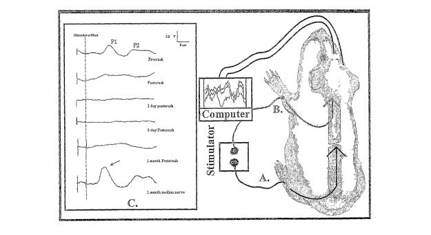
Referring to FIGURE 2, a normal functioning neural circuit is outlined by the
pathway of nerve impulses stimulated in the medial nerve of the foreleg ¨ into
the spinal
cord, ascending the spinal cord to terminate in evoked potentials measured in
the contralateral
CA 02508165 2005-06-03
WO 2004/052291
PCT/US2003/038834
- 29 -
sensory motor cortex of the brain. Stimulation of the tibial nerve of the leg
in an intact
(undamaged or normal animal) would produce a similar barrage of evoked
potentials arriving
at the brain. This is shown as Peak (P) 1 and 2; early and late arriving
evoked potentials the
top of the actual electrical recordings to the left of the guinea pig.
Crushing the spinal cord in
between the brain and the hind leg eliminates the transmission of these
potentials up the
spinal cord. The injury eliminated the appreciation of painful stimuli applied
to the foot by
the Cortex of the Brain by interrupting the SSEP.
The actual somatosensory evoked potential recording, shown in FIGURE 2 on the
left, illustrated the complete elimination of evoked potential immediately
after such a crush
was made (postcrush record). This state of blocked conduction persisted for
one continuous
month of testing in this animal. The bottom record shows a median nerve
control procedure
performed at one month in this spinal cord injured animal proving that evoked
potentials
could be measured at the brain had they not been eliminated by the injury.
The neural circuit, simplified in the insert in the right of FIGURE 2, shows
an
electrical stimulation applied to the hindpaw ¨ actually to the tibial nerve ¨
which would
normally produce evoked potentials ascending the foot and cord to the brain.
This was
interrupted by a spinal cord injury represented as a break in the cord. It can
be established in
the laboratory that the elimination of ascending SSEPs recorded from
electrodes located at
the brain is not an artifact attributable to a control procedure. In the
experiments that
generated the data reflected in FIGURE 2, the forepaw was stimulated (actually
the median
nerve of the foreleg) producing SSEPs that ascended the cord to the brain-
because this neural
circuit was not interrupted by the local spinal cord damage (i.e. it was
"above" the level of
the injury).
On the left of FIGURE 2, actual physiological records of SSEPs are shown (in
the
inset marked C). Each waveform is an average of 200 repetitive stimulations of
the relevant
nerves in the hind or forepaw. At the top a normal SSEP is shown ¨ recorded at
the sensory
cortex ¨ in response to stimulation of the hind paw's tibial nerve. It should
be noted that there
are two SSEP peaks shown (P 1 and P 2). P 1 is an early arriving evoked
potential (recorded
ca. 24 msec after stimulation), and P 2 is later arriving (about 60 msec after
stimulation). This
is because fast and slow conducting fibers and synapses segregate into peaks
of different
latencies as they ascend the spinal cord to the brain. A crush to the mid-
thoracic spinal cord
CA 02508165 2005-06-03
WO 2004/052291
PCT/US2003/038834
- 30 -
eliminated recording of the SSEP (post crush record) ¨ which did not recover
during 1 month
of continuous monitoring in this individual animal. The bottom record shows a
control SSEP
(median nerve stimulation) proving that the inability to record a SSEP was not
due to any
other variable but the damage to the spinal cord.
Now turning to a slightly different protocol, large (>350 gm) adult female
guinea pigs
were sedated (sodium pentobarbital, see below) and a normal regimen of
somatosensory
evoked potential (SSEP) testing was carried out in the intact animal. This
included recording
evoked potentials from the sensorimotor cortex in response to stimulation of
first (usually)
the median nerve of the contralateral fore limb and the median nerve of the
contralateral hind
limb. After these baseline records were obtained, animals were deeply
anesthetized, the
spinal cord surgically exposed and crushed by a standardized technique (see
Surgery below).
Within an hour, a second set of electrical records was obtained, demonstrating
the loss of
conduction of evoked potential conduction through the fresh lesion. It was
important to first
establish that the neural circuit above the level of the crush injury was
intact and normal
SSEPs could be recorded subsequent to medial nerve stimulation in this animal,
which
constitutes an internal control on SSEP recording procedures (see Fig. 7).
Referring to FIG. 7, record A is an unedited electrical record of early
arriving
evoked potentials ascending the spinal cord in response to stimulation of the
tibial
nerve (peak 1; P 1). The bottom three traces are individual trains of the 200
stimulations. The single trace above, along with records B ¨ D, is the
averaged
waveform. This record was obtained from a sedated guinea pig prior to spinal
cord
compression. In B, the waveform, taken one hour after spinal cord compression,
shows a control stimulation of the medial nerve of the forelimb in the same
animal.
Note the large amplitude of the early arriving EP. In C, the complete loss of
SSEP
conductance through the spinal cord lesion is revealed subsequent to
stimulation of
the tibial nerve of the hind limb at the same time and in the same animal.
This
complete loss in the ability of ascending volleys of compound action
potentials to be
conducted through damaged spinal cord white matter is still obvious 1 week
later in
this animal (D). Such records are characteristic of all animals used in this
report.
CA 02508165 2011-12-06
- 31 -
Similar to FIG. 2 the drawing of the guinea pig in FIG. 7 shows the control
circuit
activated by stimulation of the medial nerve. The neural circuit activated by
stimulation of
the tibial nerve of the hind limb is shown to be interrupted by damage to the
spinal cord.
After these records were obtained, the animals recovered from anesthesia and
were
maintained for 1 week. Another set of evoked potentials were recorded to
establish the extent
of the remaining conduction deficit ¨ 1 week post injury. After these records
were taken,
animals were administered either 4-AP, which was used as a standard, or one of
the
derivatives by gavage. The effect that the drugs had on conduction was then
monitored at t =
30 min, 1 hour, and then hourly up to 4 hours. After the period of recording
was concluded,
the animals were allowed to recover and then were returned to their cages. A
final set of
records was obtained the next day (-18 h later) then the animal was
euthanized.
Surgery: A conventional constant displacement injury is favored over a
constant
impact as a means to reduce the variability of behavioral and anatomical
consequences of the
injury. This conventional method is described in Borgens, R.B.; Shi, R.;
Bohner, T.D.
Behavioral Recovery from Spinal Cord Injury Following Delayed Application of
Polyethylene Glycol. Journal of Experimental Biology 2002, 205,1 - 12: Blight,
A.R.
Morphometric analysis of a model of spinal cord injury in guinea pigs, with
behavioral
evidence of delayed secondary pathology. J. Neurolog. Sci. 1991, 103, 156-171.
Briefly: Adult guinea pigs (>' 350 g; Hartley Strain) were anesthetized by an
intramuscular injection of 100 mg/kg ketamine HC1 and 20 mg/kg xylazine. A
dorsal
laminectomy procedure exposed the spinal cord at about the 12th thoracic
vertebral level
(T12) to the first lumbar level (L1). The exposed cord was crushed using a
specially modified
forceps possessing a détente. To immobilize animals for electrical records, a
more gentle
sedation was produced by intramuscular injection (0.1 cc sodium pentobarbital,
50 mg/ml).
At the end of the study, while the animals were under anesthesia, the guinea
pigs were
euthanized by increasing the anesthetic dose significantly, followed by
perfusion/fixation
(glutaraldehyde in phosphate buffered Ringer's solution).
CA 02508165 2011-12-06
- 32 -
SSEPs: It is the loss of nerve impulse conduction through the white matter of
the
spinal cord lesion that is associated with the catastrophic deficits in
behavior observed in SCI.
These volleys of compound nerve impulses ascending and descending the spinal
cord are
associated with numerous axons and synapses, and are referred to as "evoked
potentials" (EP)
when stimulated synchronously by electrical activation of a compound nerve of
the lower or
upper limbs (in the SSEP) or activation of the cortex (during motor evoked
potential
recording, or MEPs, not performed here). This form of stimulation of largely
ascending
impulses - then recording them at the contralateral sensorimotor cortex of the
brain is referred
to as somatosensory evoked potential testing (SSEP).
Stimulation of the tibial nerve of the hind limb was accomplished using a pair
of
needle electrodes inserted near the nerve at the popliteal notch of the knee.
Similar SSEPs
were evoked by stimulation of the median nerve of the forelimb with pair of
stimulation
electrodes. Recording electrodes were located in the scalp covering the
contralateral cortex
region, with an indifferent electrode usually located in the pinna of the ear.
A complete electrical record for one period of investigation involved
stimulating the
nerve 200 times in a train (2 mA square wave, 200 sec duration at 3 Hz; see
section A of
FIG. 7). Three to four sets of these records were then averaged to produce a
single waveform
for quantitative study as shown in sections A and B of FIG. 7. Recording and
stimulation
used a Nihon Kohden Neuropak 4 and a Power Mac G-3 computer.
Quantitative evaluation involved measurements of latency from the initiation
of
stimulation (noted by the stimulus artifact) to the initiation of the EP,
however, the most
reliable and informative comparative data involved a determination of the area
beneath the
EP in pixels using IPLabTM spectrum software (Scanalytics, Farifax, VA). These
areas
beneath the peak waveform (that is above baseline) were normalized by dividing
the post
injury (or post treatment) EP by the area of the initial EP recorded in the
normal animal. If
all (100 %) nerve fibers normally fired in synchrony by the stimulation
regimen before injury
were theoretically recruited into conduction after a treatment, the average
SSEPs should thus
reach unity (1.0).
CA 02508165 2005-06-03
WO 2004/052291
PCT/US2003/038834
- 33 -
Drug Administration: Drugs were administered by gavage using the following
methods. All tested drugs were introduced in solution (see below) into the
guinea pig
stomach using a round tipped feeding needle (either 18 gauge, 55 mm length or
16 gauge, 75
mm length; Fine Science Tools). Gavage is only carried out on the sedated
animal. In guinea
pigs, the soft palate is continuous with the base of the tongue with only a
small opening to
pass the tube. The feeding needle is advanced between the incisors and the
beginning of the
molars, which initiates a swallowing or gag reflex, facilitating advancement
of the feeding
needle to the stomach. Prior to this operation, the correct needle (sized
relative to the size of
the guinea pig) is marked by placing its end adjacent the animal's last rib
and marking the
proximal portion of the needle near the tip of the nose. This provides an
estimate of the
required length to advance to the stomach during gavage. The needle can be
connected to
either a syringe or an aspirator allowing material to be introduced into, or
withdrawn from,
the stomach.
Dosage and weight data: A starting dosage for guinea pigs was estimated by
working
within the range of 4-AP given in clinical cases of paraplegia in dogs (0.5 ¨
1.0 mg/kg body
weight). In the case of an approx. 500 g guinea pig, this would result in a
total dosage of
about 0.25 g. A stock solution for gavage was prepared where 0.2 cc of an
aqueous solution
contained 0.2 mg 4-AP. This allowed the relative standardization of total
concentration given
to animals, and was facilitated by the lack of significant variation in their
weights (for the ten
animals that were tested with 4-AP the mean weight was 421.5 + 20.9 g; for
methyl
carbamate 3 the mean weight was 411.6 + 23 g; P > 0.5). The effective dosage
for 4-AP was
sufficient to produce an improvement in the electrophysiological record equal
to 0.47 + 0.04
mg/kg. By comparison, the effective dosage for carbamate 3 appeared to be
slightly lower
(0.37 + 0.2 mg/kg) but this difference was not statistically different given
the small sample
size (P >0.05).
IV. Results
In vitro Testing of Conduction through Guinea Pig Spinal Cord
The (methylamino)pyridines, amides and ureas, shown below, all failed to
produce
any enhancement in the recovery CAP amplitude at the same effective
concentration as 4-AP.
CA 02508165 2005-06-03
WO 2004/052291
PCT/US2003/038834
- 34 -
Me H Me Me
\.,
N N
I I
N N
0
0
\N
N N
0
H
<
NI 0
6/
N
H I 1
1 NN.
I I
N H H
Three compounds, N-(4-pyridyl) methyl carbamate (3); N-(4-pyridyl) ethyl
carbamate
(2) and N-(4-pyridyl) t-butyl carbamate (1), all revealed varying ability to
restore conduction
through injured spinal cord. Pilot trials with these three carbamates
established that working
concentrations equal to or below that of 4 AP (100 p,M) were capable of
producing a
reproducible recovery of conduction in the double sucrose gap recording
chamber. Methyl
carbamate (3) and ethyl carbamate (2) were found to behave similarly to 4-AP,
producing
recovery of CAP conduction at a concentration of approximately 100 M in the
absence of
CA 02508165 2005-06-03
WO 2004/052291
PCT/US2003/038834
- 35 -
toxicity. Toxicity in these in vitro trials was defined as the significant
reduction of the CAP.
t-Butyl carbamate (1), on the other hand, was found to be toxic to white
matter at 100 jiM
when added to the bathing solution. It was found, however, that carbamate (1)
reproducibly
enhanced CAP conduction through the lesion, in the absence of toxicity, at a
much lower
effective concentration (1 M) than is seen with 4-AP. For methyl carbamate
(3), an
effective concentration of 100 [tM produced an increase in the amplitude of
the recovered
CAP averaging 30.6 + 11.7 % (n = 4). For ethyl carbamate (2), the increase was
18.6 + 8.7
% (n = 8) at the same concentration. This increase in amplitude was
statistically significant
(P < 0.04). After a 1 h wash, the enhanced CAP fell in amplitude to pre-drug
levels. t-Butyl
carbamate (1) increased the recovery CAP amplitude by 15.4 + 3.4 % at l[tM (n
= 5). This
increase was very significant relative to the pre-drug CAPs (P < 0.002).
Figure 8 provides
examples of electrical records of CAP enhancement for each of these three
drugs and
graphically displays these data. In particular, in panel A, the first
electrical record shows the
recovered compound action potential (CAP) prior to the addition of ethyl
carbamate (2) (the
initial small wave form is the stimulus artifact). The second record was taken
about 1/2 hour
after the addition of carbamate (2). Note the ¨ 20 % increase in the amplitude
of the CAP.
The third record shows that the amplitude of this enhanced CAP has fallen back
to pre-drug
levels after removal of the drug and an ¨ 30 min wash. Panel B shows a similar
set of
electrical records obtained after introduction of methyl carbamate (3). Again,
the increase in
amplitude in the presence of the drug is obvious. This enhancement was only
sustained
during the administration of the drug and approximate pre-drug levels were
measured after a
washing out of the drug. Panel C depicts a similar set of records to A and B,
using t-butyl
carbamate (1). This drug was effective at enhancing CAP amplitudes at much
lower
concentrations (1 tiM). Panel D provides a histogram of all data. Carbamates
(3) and (2)
statistically enhanced CAP amplitudes at 100 Iµ,4 concentration (P ( 0.04;
one asterisk),
while carbamate (1) statistically improved the CAP at 1 ,M compared to pre-
drug amplitudes
(P ( 0.002; two asterisks).
In Vivo Testing in Guinea Pig
Post surgery, SSEPs had been eliminated in all animals by the time electrical
records
could be accomplished (< 1 hour). In all but two animals these 'flat-line"
recordings were
characteristic of the 1 week recordings as well, indicating there was little
evidence for
CA 02508165 2005-06-03
WO 2004/052291
PCT/US2003/038834
- 36 -
spontaneous recovery of conduction in any of the guinea pigs one week after
surgery. In the
two exceptions, a small and very early arriving Evoked Potential (EP;
approximately 25 msec
latency) was noted, however, the magnitude of this peak was barely detectable
above baseline
and it was not dependably evoked. Therefore, gavage and further recordings
were carried out
on these animals and their data are pooled with the rest. Gavage of 4-AP and
carbamates (3)
and (2) was uneventful; however, we were unable to carry out this procedure
using the t-butyl
carbamate (1). Crystalline carbamate (1) was not tolerated and the initial
attempts at
producing a solution of known concentration for oral insertions were
problematic. As a
result, provided herein are only the preliminary in vitro data below for
comparison purposes.
4-AP produced a strong and dependable enhancement of EPs within 30 min to 1
hour
after insertion as observed by a return of an early arriving EP (over
baseline). Two of the ten
animals failed to respond to 4-AP by 4 hours after gavage. There were no
failures to respond
when using the carbamates (1) and (2).
Note that even though the sample size was small, an apparent enhancement of EP
recovery by methyl carbamate (1) was demonstrated (see Fig. 3). FIG. 3 shows
the,
Recovery of spinal cord conduction subsequent to oral administration of N-(4-
pyridyl)
methyl carbamate (1). In FIG. 3 an unedited electrical record of a normal SSEP
is shown in
section A. The waveforms were recorded by electrodes located over the sedated
guinea pig's
sensory cortex after electrical stimulation of the tibial nerve of the hind
paw. The bottom 3
records, still in section A, are individual traces of 200 repetitive
stimulations of the nerve.
The dotted line (SA) marks the stimulus artifact. An early arriving cortical
potential is
shown, approximately 30 ms after stimulation (marked P 1) in the uninjured
guinea pig (A).
In section B, responses to the oral ingestion of the test compound are shown
in the
same animal providing the record in section A. The top record of section B was
made 1 hour
after a crush injury to the midthoracic spinal cord as described herein. Note
the complete
elimination of the cortical potential after stimulation of the tibial nerve of
the hind paw. One
week later (second trace in section B) this failure to conduct impulses
through the spinal cord
injury has changed little. The bottom record was made 1 hour after gastric
tube
administration of N-(4-pyridyl) methyl carbamate (1). Note the rapid
establishment of a
nearly normal cortical potential (P 1).
CA 02508165 2005-06-03
WO 2004/052291
PCT/US2003/038834
- 37 -
Even though the sample size was small, an apparent enhancement of EP recovery
by
methyl carbamate (1) is demonstrated (Fig. 3). Thus, some statistical
comparison between
these groups is provided. All data were normalized by dividing the area
(expressed as pixels)
beneath recovered EP by the same pre-injury data.
The data shows that carbamate (1) significantly enhanced the recovery of EP
compared to 4-AP at 1 week post injury. Both two-way ANOVA and a straight
forward
Student's T test confirmed a significant increase (ANOVA = 0.02; two- tailed
Student's T =
0.47). Given that the latter test is more conservative relative to the small
sample size only
this comparison is provided in Table 1 below. Note the enhancement of EPs by
the methyl
carbamate (1) compared to the structurally similar ethyl carbamate (2) (Table
1). The latter
carbamate did not meet significance compared to 4-AP data.
Table 1
Test Compound n Mean $D SEM Range Statistic
1 4-AP 10 40.0 31.0 9.8 0 ¨ 100 1 vs. 2, P = 0.47
2 Methyl carbamate (1) 5 78.0 14.2 5.8 58 - 100 2 vs. 3, P = 0.003
3 Ethyl carbamate (2) 4 41.5 13.2 6.6 27 - 55
1 vs. 3, P = 0.93
The three carbamate derivatives of 4-AP (1, 2, and 3) were tested. All three
compounds induced an increase in CAP above the pre-drug level, without any
indication of
toxicity at the concentrations studied. The methyl and ethyl carbamates (3)
and (2) were
effective at the same concentration as 4-AP (100 juM), although they induced a
slightly lower
recovery of CAP than 4-AP. The results from t-butyl carbamate (1) were also
interesting.
Using the concentration that was optimal for 4-AP, this compound proved to be
extremely
toxic to the spinal cord. However, at 1 pM, 1% of 4-AP's optimal
concentration, t-butyl
carbamate (1) showed an increase in recovery CAP that was equivalent to the
recovery
observed for carbamates (3) and (2).
CA 02508165 2013-02-21
-38-
Accordingly, it is clear from the present disclosure that three compounds,
methyl carbamate
(3), ethyl carbamate (2) and t-butyl carbamate (1) are all capable of
enhancing action potential
conductance in mechanically injured spinal cord segments. At 100 M,
carbamates (3) and (2)
enhanced CAP conduction significantly, whereas t-butyl carbamate, at 1 p.M was
able to enhance
CAP conduction significantly.
It is to be understood by those skilled in the art that the foregoing
description and examples
are merely illustrative of the present invention. The claims should not be
limited by the description
but should be given the broadest interpretation consistent with the
specification as a whole. Variations
of the detail presented herein may be made without departing from the scope of
the present invention
as defined by the following claims.