Note: Descriptions are shown in the official language in which they were submitted.
CA 02508199 2005-05-25
-1-
APPARATUS AND METHODS FOR OCCLUDING A HOLLOW
ANATOMICAL STRUCTURE
Field of the invention
[0001] The present invention relates generally to surgical methods and
apparatus for occluding a hollow tissue structure, such as when occluding
vessels, or pedunculated structures such as an appendix, gall bladder or
appendages on the heart. More specifically, the present invention relates to a
method and device for occluding the left atrial appendage of the heart in
either
an open surgical procedure or minimally invasive procedure.
Background of the Invention
[0002] Atrial fibrillation is a common cardiac rhythm disorder that affects
more than two million people each year. Until relatively recently, atrial
fibrillation
was thought to be a nuisance arrhythmia with few consequences. However,
recent medical research has uncovered some devastating complications
including cardiomyopathy, congestive heart failure and stroke.
[0003] During atrial fibrillation the upper part of the heart beats (quivers)
faster than the rest of the heart. This phenomenon is due to the generation of
erratic or extra electrical signals which cause the top part of the heart to
quiver
rapidly and irregularly (fibrillate) as many as 300-600 times a minute.
However,
the entire heart does not beat that fast. The heart is a muscular pump divided
into four chambers, two atria on the top of the heart and two ventricles on
the
bottom portion of the heart. Normally, the heartbeat starts in the right
atrium
when a special group of cells sends an electrical signal. These cells are
called
the sinoatrial or SA node, sinus node or the heart's "pacemaker". The signal
spreads throughout the atria and to the atrioventricular or AV node. The AV
CA 02508199 2005-05-25
-2-
node connects to a group of fibers in the ventricles that conduct the
electrical
signal. The electrical impulse travels via these specialized fibers to all
parts of
the ventricles. The specialized fibers are also known as the His-Purkinje
system. The electrical signal must follow this exact route for the heart to
pump
properly. Normally, the heart beats at 60-80 times per minute at rest. This
number represents the contractions of the lower heart or ventricles. During
atrial
fibrillation, electrical signals from other parts of the heart disrupt the
heart's
normal rhythm and cause the atria to quiver or beat too fast. However, only a
small number of these atrial beats make it through the AV node, which acts
like
a gate to the ventricles. This is fortunate, because a rapid ventricular
heartbeat
would be much more dangerous and potentially fatal. However, some atrial
fibrillation does make it through the AV node making the heart beat faster
than
normal. An atrial fibrillation attack is usually not life threatening. The
most
significant danger is stroke.
[0004] Blood usually moves completely through the chambers of the
heart. During atrial fibrillation, the heart is not pumping normally or
efficiently.
The blood begins to pool in the atria and this stagnation of blood can cause
the
blood to thicken and form clots. These clots are then ejected out of the heart
and into the bloodstream where they can lodge in the brain causing a stroke.
Atrial fibrillation can make stroke five times more likely than in the general
population. When the heart experiences atrial fibrillation there may not be
enough blood pumping to the brain or other organs. This can cause dizziness,
shortness of breath or organ failure. Untreated atrial fibrillation will also
weaken
the heart due to phenomenon known as remodeling. The heart, like the rest of
the body, adapts to changes. The fast abnormal rhythm in the atria causes
electrical changes, and this can enlarge the heart.
_
CA 02508199 2005-05-25
-3-
[0005] There are three major objectives in the treatment of atrial
fibrillation: the restoration of normal sinuous rhythm, control of ventricular
rate
during atrial fibrillation, and the prevention of blood clot formation. Some
methods of treatment for atrial fibrillation include pharmacological therapy,
pacemakers, and surgery.
[0006] For the prevention of blood clots, research has demonstrated that
the anticoagulation warfarin (e.g., Coumadin) is effective in reducing the
risk of
blood clot formation and stroke but it does not totally eliminate the risk. An
anticoagulant such a warfarin interferes with the body's natural clotting
mechanism. The dosage of warfarin is highly individualized and must be
carefully monitored with blood tests to ensure safety. While this
pharmacological treatment may significantly reduce the risk of stroke, it also
increases the risk of bleeding and may be inappropriate for many atrial
fibrillation patients.
[0007] As an alternative to pharmacological therapy, there are a few
surgical procedures that isolate the left atrial appendage from the blood's
circulatory system. The most common approach is to occlude the left atrial
appendage during open-heart surgery. In open heart surgery the patient is
placed on a heart-lung bypass machine and the heart is temporarily isolated
from the circulatory system while the surgeon operates on the heart. The left
atrial appendage is a small hollow extension (i.e., a pedunculated structure)
formed off the lateral wall of the left atrium. It has been referred to as a
small
windsock or a small, flat hollow finger-like protrusion. The left atrial
appendage
usually contracts with the rest of the left atrium during normal heart
function
thereby continually moving blood throughout the hollow extension. During
atrial
fibrillation, the left atrial appendage often fails to contract thereby
allowing the
_
CA 02508199 2005-05-25
-4-
blood to pool inside the appendage, becoming stagnated. As a result, the blood
becomes thicker and thrombus or clot formation begins. These clots can be
slowly ejected from the left atrial appendage into the left atrium and left
ventricle, and then released into the bloodstream thereby becoming an
obstruction in the brain or other vascular structures. For this reason, it is
advantageous to prevent these clots from forming and being dislodged into the
bloodstream. One method of preventing the occurrence of clots is to occlude
the appendage thus preventing blood from entering and forming clots. This
also prevents clots already formed in the appendage from escaping into the
bloodstream. Normally, the occlusion of the left atrial appendage is performed
in conjunction with other procedures such as a mitrel valve replacement or
coronary artery bypass procedure and not as the sole reason for the procedure.
[0008] There are several different methods being used today to occlude
the left atrial appendage. One method is percutaneous left atrial appendage
transcathether occlusion. A small occlusion device is deployed from a venous
access catheter into the left atrium and blocks the opening into the atrial
appendage. In order to access the left atrium from the vena cava's right
atrium,
the surgeon must go through the atrial wall. Many surgeons are uncomfortable
with making an opening in this wall without being able to repair it at the end
of
the procedure. There are also issues of placing an occlusion device inside the
heart. If the occlusion device becomes detached within the heart, the result
may be fatal.
[0009] Another method of occlusion is placing a loop around the left atrial
appendage and cinching it down in a manner similar to a garrote. When trying
to place a flaccid loop around an irregular pedunculated structure, it can be
difficult to make certain the loop is positioned at the base of the appendage.
CA 02508199 2005-05-25
-5-
When cinching the loop, it is very easy to over tighten the loop, and this can
result in severing the delicate atrial appendage. Even a partial tear can
create
problems in getting access to repair the tear. This method of occlusion may
not
always seal the opening between the appendage interior and the atrium. That
is, there may still be a partial opening due to the way the appendage wall
collapses during cinching of the loop. Such a partial opening could still
allow
some flow into and out of the atrial appendage, leading to the problems
mentioned above. In addition, transforming the relatively flat structure of
the
appendage onto a round hard mass, as does a cinching method, could lead to
other problems.
[0010] Another method of occlusion is to place a linear surgical stapler at
the base of the appendage and a left atrial wall and stapling the appendage
closed. Due to the limited access, the ability to visualize the entire atrial
appendage while placing the stapler in the correct location can be a problem.
It
is very difficult to make certain the staple line makes a complete occlusion
of
the appendage. Again, a partial occlusion of the appendage can still result in
the formation and dislodgement of clots.
[0011] For the aforementioned reasons, it would be desirable to provide
improved methods and devices to reliably occlude hollow anatomical structures,
and especially the left atrial appendage of the heart completely and safely.
Such methods may be performed during an open-heart surgical procedure such
as a valve replacement or coronary artery bypass. It would also be desirable
to
provide methods and devices that may be used in a minimally invasive
procedure while the heart is beating without placing the patient on a heart-
lung
bypass machine. A minimally invasive device would allow access through either
an intercostal space between the ribs or a supra and/or sub-xiphoid approach
to
_
CA 02508199 2005-05-25
-6-
gain access to the left atrial appendage. Such devices will allow complete
visualization of the left atrial appendage for the surgeon and permit minor
placement adjustments to be made before permanent installation is made. The
devices would also allow complete occlusion of the left atrial appendage,
eliminating the risk of clots forming in the appendage, traveling throughout
the
bloodstream, and possibly lodging in the brain causing a stroke.
Summary of the Invention
[0012] The present invention provides devices and methods for occluding
a hollow anatomical structure, such as the left atrial appendage of the heart.
Generally, the device comprises a clamp having at least first and second
clamping portions adapted to be placed on opposite sides of the hollow
anatomical structure. At least one of the first and second clamping portions
is
movable toward the other of the first and second clamping portions from an
open position into a clamping position to occlude the hollow anatomical
structure. The clamp comprises a closed, annular shape configured to
surround the hollow anatomical structure in the open position and then
assumes a flattened shape in the clamping position to occlude the hollow
interior of the hollow anatomical structure. The first and second clamping
portions can further comprise concave portions curved in opposite directions
to
form the clamp into a generally oval shape.
0013] In various embodiments, at least one of the first and second
clamping portions is spring biased toward the other of the first and second
clamping portions in the clamping position. In this regard, one clamping
portion
may be normally spring biased toward the other of the first and second
clamping portions when the first and second clamping portions are in the open
_ _
CA 02508199 2005-05-25
-7-
position. Upon release, the spring biased clamping portion moves toward the
other clamping portion into a clamping or occluding position. In another
embodiment, one of the first and second clamping portions is movable toward
the other of the first and second clamping portions to an over-center position
at
which a spring bias takes effect and moves the one clamping portion toward the
other clamping portion to the clamping position.
[0014] In another aspect of the invention, the first and second clamping
portions have tissue engaging surfaces for engaging the hollow anatomical
structure in the clamping position. The tissue engaging surfaces are roughened
and preferably adapted to promote tissue ingrowth. The tissue engaging
surfaces may be comprised of a fabric covering on at least one of the first
and
second clamping portions. Other manners of promoting tissue ingrowth may be
used on one or both clamping portions such as etching and other pore-creating
methods such as metal deposition. Pore size should preferably range from
200-400 microns. A protective overmold may be provided of, for example,
silicone to assist with traction if tissue ingrowth feature is not utilized on
one or
both clamping portions. Alternatively, and if necessary, silicone overmolding
or
other protective guards may be used to prevent irritation of surrounding
tissue,
while clamping areas of the device may be designed to promote traction and/or
tissue ingrowth such as described above.
[0015] In another aspect, the first and second clamping portions have
complementary shapes in cross section such that the complementary shapes fit
together in the clamping position. At least one of the first and second
clamping
portions may be convexly curved toward the other of the first and second
clamping portions in cross section. This feature may assist with providing
more
CA 02508199 2005-05-25
-8-
uniform force distribution and/or more sealing force along the length of the
clamp.
[0016] In another aspect, projections may be provided on at least one of
the first and second clamping portions. The projections are configured to
engage and, optionally, pass through the hollow anatomical structure when the
clamp is in the clamping position. The projections thereby assist with
retention
of the clamp on the tissue. To further assist with clamp retention, receiving
elements may be provided on the opposing clamping portion to engage and
lock with the projections when the clamp is in the clamping position. Other
types of locking elements may be provided, such as ratchet elements,
undulations on tissue engaging areas, bands on the outside of the clamp or
other suitable structure.
[0017] The clamp may also have an actuating element configured to
move one of the first and second clamping portions toward the other of the
first
and second clamping portions. This may, for example, be one or more
magnetic elements on one or both clamping portions, or a mechanical actuation
element such as a rotating or sliding cam element, or any other suitable
actuation mechanism.
[0018] The invention also provides apparatus for occluding a hollow
anatomical structure which includes a clamp delivery and actuation device. In
the preferred embodiment, the delivery and actuation device includes first and
second jaws, and an actuator configured to move at least one of the first and
second jaws toward the other of the first and second jaws. The clamp delivery
and actuation device preferably includes a pistol grip with an actuating
member
configured to be manually depressed to move one of the first and second jaws
toward the other of the first and second jaws. The first and second clamping
CA 02508199 2005-05-25
-9-
portions are secured between the first and second jaws and may be moved
from the open position to the clamping position by moving at least one of the
first and second jaws. This allows the clamp to be repeatedly opened and
closed, as necessary for repositioning purposes, during the surgical
procedure.
The clamp may be secured to the jaws in any suitable manner, such as by
using suture or by using other types of gripping elements. The delivery and
actuation device preferably carries a mechanism to release the clamp, such as
a blade to cut the suture, or a tension member which may be pulled to release
the gripping elements or suture. In the case of using the suture, the tension
member may be used to untie the suture, and may be an end of the suture
itself.
[0019] The clamp is preferably coupled to the first and second jaws so as
to pivot about an axis generally transverse to its length. This pivoting
action
may take place passively or actively. To provide for active or selective
pivoting
of the clamp, as may be desired by a surgeon to more accurately position the
clamp, the delivery and actuation device includes a pivoting mechanism
coupled to the clamp and configured to pivot the clamp in opposite directions
about the axis. The surgeon may operate the pivoting mechanism at the
proximal or handle end of the device.
[0020] The invention further provides methods for occluding a hollow
anatomical structure with an annular clamp having at least first and second
clamping portions. Generally, the method comprises surrounding the hollow
anatomical structure with the annular clamp, and then moving at least one of
the first and second clamping portions toward the other of the first and
second
clamping portions to occlude the hollow anatomical structure. In the preferred
embodiment, the hollow anatomical structure is a pedunculated organ or portion
CA 02508199 2005-05-25
-10-
of an organ. Most specifically, it is the left atrial appendage of a heart.
Preferably, the method involves accessing the left atrial appendage of the
heart
by a mini-thoracotomy or by another minimally invasive approach.
[0021] The method preferably further comprises engaging a structure
configured to promote tissue ingrowth, such as a fabric covering, with the
hollow anatomical structure, and optionally also engaging the anatomical
tissue
with projections to promote tissue ingrowth after clamping has taken place.
The
clamp may be passively or actively pivoted with respect to the delivery device
prior to the step of moving at least one of the first and second clamping
portions
toward the other. In another aspect, a tissue gripper with flat, paddle-shaped
gripper elements is provided and used to gently grasp and pull the tissue
through the clamp when the clamp is in the open, annular configuration.
[0022] It will be appreciated that various additional aspects of the
methods carried out by the various embodiments of this invention will be
readily
apparent based on the use of the devices and components of the clamp and the
delivery and actuation device as described hereinabove and further below.
[0023] The present invention provides improved devices and methods for
occlusion of hollow tissue such as the left atrial appendage. One advantage of
various embodiments described herein is that the surgeon can open and close
the clamp if needed to change the position of the clamp for a better result
prior
to release of the clamp onto the tissue. The configuration of the delivery and
actuation device is such that the device can be used not only in an open
surgical procedure, but in a minimally invasive surgical procedure during
which,
for example, the device is placed between or under the patient's ribs for
access
to the left atrial appendage. The implantable clamp has a geometry which traps
CA 02508199 2012-07-27
-11-
appendage tissue within an annular opening thereby positively attaching the
clamp to the tissue.
[0024] In another embodiment, the invention contemplates an apparatus
for occluding a hollow anatomical structure comprising a clamp delivery and
actuation device including a hollow structure containing a clamp deploying
member. The apparatus further includes clamp having at least first and second
clamping portions adapted to be placed on opposite sides of the hollow
anatomical structure. At least one of the first and second clamping portions
is
movable toward and away from the other of the first and second clamping
portions between a closed position and an open position in which the clamp
assumes an annular shape. The clamp is carried within the hollow structure in
the closed position and is extendable out of the hollow structure by the
deploying member whereupon the clamp may be actuated to the open position
and clamped onto the hollow anatomical structure. This embodiment is
especially useful for minimally invasive surgical procedures.
[0024.1] According to one aspect of the present invention there is provided
a clamping device for implantation into a patient and occluding an appendage
of the heart of the patient, the device comprising a clamp having at least
first
and second clamping portions adapted to be placed on opposite sides of the
appendage, at least one of the first and second clamping portions being
movable toward the other of the first and second clamping portions from an
open position into a clamping position to occlude the appendage, the clamp
comprising an annular shaped structure configured to surround the appendage
in the open position and a flattened shape in the clamping position configured
to occlude the hollow interior of the appendage, wherein the first and second
clamping portions have tissue engaging surfaces for engaging the appendage
in the clamping position, and the first and second clamping portions are
surrounded by an annular fabric structure that promotes tissue ingrowth, the
annular fabric structure forming a closed continuous configuration lengthwise
around the annular shaped structure.
CA 02508199 2012-07-27
[0024.2] According to a further aspect of the present
invention there is-11a-
provided a clamping device for implantation into a patient and occluding an
appendage of the heart of the patient, the device comprising a clamp having at
least first and second concave clamping portions adapted to be placed on
opposite sides of the appendage to form a generally oval shape, at least one
of
the first and second clamping portions being movable under spring bias toward
the other of the first and second clamping portions from an open position into
a
clamping position and locked in place in the clamping position to occlude the
appendage, the clamp configured as an annular shared structure to surround
the appendage in the open position and assume a flattened shape in the
clamping position to occlude the hollow interior of the appendage, and an
annular fabric structure surrounding each of the first and second clamping
portions, wherein the fabric prevents the clamp from slipping on the appendage
and promotes ingrowth of tissue, the annular fabric structure forming a closed
continuous configuration lengthwise around the annular shared structure.
[0024.3] According to another aspect of the present
invention there is
provided a device for implantation into a patient and occluding a appendage of
the patient, the device comprising a clamp having at least first and second
concave clamping portions adapted to be placed on opposite sides of the
appendage to form a generally oval shape, at least one of the first and second
clamping portions being movable toward the other of the first and second
clamping portions from an open position into a clamping position and locked in
place in the clamping position to occlude the appendage, the clamp configured
as an annular shared structure to surround the appendage in the open position
and assume a flattened shape in the clamping position to occlude the
appendage, and the one of the first and second clamping portions being
movable toward the other of the first and second clamping portions to an over-
center position at which the one of the first and second clamping portions is
spring biased toward the other of the first and second clamping portions, and
an annular fabric structure surrounding each of the first and second clamping
portions to prevent the clamp from slipping on the appendage, and wherein the
CA 02508199 2012-07-27
fabric promotes tissue ingrowth, the annular fabric structure forming a closed
-11b-
continuous configuration lengthwise around the annular shaped structure.
[0024.4] According to a still further aspect of the present
invention there is
provide a clamping apparatus for implantation into a patient and occluding an
appendage of the heart of the patient, the apparatus comprising a clamp
delivery and actuation device including first and second jaws, and an actuator
configured to move at least one of the first and second jaws toward the other
of
the first and second jaws, and a clamp having at least first and second
clamping portions releasably secured between the first and second jaws and
adapted to be placed on opposite sides of the appendage, at least one of the
first and second clamping portions being movable toward the other of the first
and second clamping portions from an open position into a clamping position by
the one of the first and second jaws to occlude the appendage, the clamp
comprising an annular shaped structure configured to surround the appendage
in the open position and a flattened shape in the clamping position configured
to occlude the hollow interior of the appendage, wherein the first and second
clamping portions have tissue engaging surfaces for engaging the appendage
in the clamping position, and the tissue engaging surfaces formed by an
annular fabric structure that promotes tissue ingrowth, the annular fabric
structure forming a closed continuous configuration lengthwise around the
annular shared structure.
[0024.5] According to another aspect of the present
invention there is
provided a device for implantation into a patient and occluding a hollow
anatomical structure of the patient, the device comprising a clamp having at
least first and second clamping portions adapted to be placed on opposite
sides
of the hollow anatomical structure, at least one of the first and second
clamping
portions being movable toward the other of the first and second clamping
portions from an open position into a clamping position to occlude the hollow
anatomical structure, the clamp comprising a shape configured to surround the
hollow anatomical structure in the open position and a flattened shape in the
clamping position configured to occlude the hollow interior of the hollow
anatomical structure, wherein the first and second clamping portions have
CA 02508199 2012-07-27
-11c-
tissue engaging surfaces for engaging the hollow anatomical structure in the
clamping position, and wherein at least one of the tissue engaging surfaces
promotes tissue ingrowth.
[0024.6] According to a further aspect of the present invention there is
provided a clamping apparatus for implantation into a patient and occluding an
appendage of the heart of the patient, the apparatus comprising a clamp
delivery and actuation device including first and second jaws, and an actuator
configured to move at least one of the first and second jaws toward the other
of
the first and second jaws, and a clamp having at least first and second
clamping portions adapted to be placed on opposite sides of the hollow
anatomical structure, at least one of the first and second clamping portions
being movable toward the other of the first and second clamping portions from
an open position into a clamping position to occlude the hollow anatomical
structure, the clamp comprising a shape configured to surround the hollow
anatomical structure in the open position and a flattened shape in the
clamping
position configured to occlude the hollow interior of the hollow anatomical
structure, wherein the first and second clamping portions have tissue engaging
surfaces for engaging the hollow anatomical structure in the clamping
position,
and wherein at least one of the tissue engaging surfaces promotes tissue
ingrowth.
[0025] These and other features, objects and advantages of the
invention will become more readily apparent to those of ordinary skill in the
art
upon review of the following detailed description, taken in conjunction with
the
accompanying drawings.
BRIEF DESCRIPTION OF THE DRAWINGS
[0026] FIG. 1 is a side elevational view of an apparatus constructed in
accordance with the invention including a clamp and a delivery and actuation
device.
[0027] FIG. 2A is an enlarged side elevational view of the clamp and
jaws shown in FIG. 1, with the clamp in an open position.
CA 02508199 2011-10-07
appendage of the heart of the patient, the device comprising a clamp having at
-lid-
least first and second clamping portions adapted to be placed on opposite
sides
of the appendage, at least one of the first and second clamping portions being
movable toward the other of the first and second clamping portions from an
open position into a clamping position to occlude the appendage, the clamp
comprising an annular shaped structure configured to surround the appendage
in the open position and a flattened shape in the clamping position configured
to occlude the hollow interior of the appendage, wherein the first and second
clamping portions have tissue engaging surfaces for engaging the appendage
in the clamping position, and the first and second clamping portions are
surrounded by an annular fabric structure that promotes tissue ingrowth, the
annular fabric structure forming a closed continuous configuration lengthwise
around the annular shaped structure.
[0024.9] According to one aspect of the present invention
there is provided
a clamping device for implantation into a patient and occluding an appendage
of the heart of the patient, the device comprising a clamp having at least
first
and second concave clamping portions adapted to be placed on opposite sides
of the appendage to form a generally oval shape, at least one of the first and
second clamping portions being movable under spring bias toward the other of
the first and second clamping portions from an open position into a clamping
position and locked in place in the clamping position to occlude the
appendage,
the clamp configured as an annular shared structure to surround the
appendage in the open position and assume a flattened shape in the clamping
position to occlude the hollow interior of the appendage, and an annular
fabric
structure surrounding each of the first and second clamping portions, wherein
the fabric prevents the clamp from slipping on the appendage and promotes
ingrowth of tissue, the annular fabric structure forming a closed continuous
configuration lengthwise around the annular shared structure.
[0024.10] According to a further aspect of the present
invention there is
provided a device for implantation into a patient and occluding a appendage of
the patient, the device comprising a clamp having at least first and second
concave clamping portions adapted to be placed on opposite sides of the
CA 02508199 2011-10-07
-lie-
appendage to form a generally oval shape, at least one of the first and second
clamping portions being movable toward the other of the first and second
clamping portions from an open position into a clamping position and locked in
place in the clamping position to occlude the appendage, the clamp configured
as an annular shared structure to surround the appendage in the open position
and assume a flattened shape in the clamping position to occlude the
appendage, and the one of the first and second clamping portions being
movable toward the other of the first and second clamping portions to an over-
center position at which the one of the first and second clamping portions is
spring biased toward the other of the first and second clamping portions, and
an annular fabric structure surrounding each of the first and second clamping
portions to prevent the clamp from slipping on the appendage, and wherein the
fabric promotes tissue ingrowth, the annular fabric structure forming a closed
continuous configuration lengthwise around the annular shaped structure.
[0024.11] According to another aspect of the present invention there is
provide a clamping apparatus for implantation into a patient and occluding an
appendage of the heart of the patient, the device comprising a clamp delivery
and actuation device including first and second jaws, and an actuator
configured to move at least one of the first and second jaws toward the other
of
the first and second jaws, and a clamp having at least first and second
clamping portions releasably secured between the first and second jaws and
adapted to be placed on opposite sides of the appendage, at least one of the
first and second clamping portions being movable toward the other of the first
and second clamping portions from an open position into a clamping position by
the one of the first and second jaws to occlude the appendage, the clamp
comprising an annular shaped structure configured to surround the appendage
in the open position and a flattened shape in the clamping position configured
to occlude the hollow interior of the appendage, wherein the first and second
clamping portions have tissue engaging surfaces for engaging the appendage
in the clamping position, and the tissue engaging surfaces formed by an
annular fabric structure that promotes tissue ingrowth, the annular fabric
CA 02508199 2011-10-07
-11f-
structure forming a closed continuous configuration lengthwise around the
annular shared structure.
[0024.12] According to a still further aspect of the present invention there
is
provided a device for implantation into a patient and occluding a hollow
anatomical structure of the patient, the device comprising a clamp having at
least first and second clamping portions adapted to be placed on opposite
sides
of the hollow anatomical structure, at least one of the first and second
clamping
portions being movable toward the other of the first and second clamping
portions from an open position into a clamping position to occlude the hollow
anatomical structure, the clamp comprising a shape configured to surround the
hollow anatomical structure in the open position and a flattened shape in the
clamping position configured to occlude the hollow interior of the hollow
anatomical structure, wherein the first and second clamping portions have
tissue engaging surfaces for engaging the hollow anatomical structure in the
clamping position, and wherein at least one of the tissue engaging surfaces
promotes tissue ingrowth.
[0025] These and other features, objects and advantages of the invention
will become more readily apparent to those of ordinary skill in the art upon
review of the following detailed description, taken in conjunction with the
accompanying drawings.
BRIEF DESCRIPTION OF THE DRAWINGS
[0026] FIG. us a side elevational view of an apparatus constructed in
accordance with the invention including a clamp and a delivery and actuation
device.
[0027] FIG. 2A is an enlarged side elevational view of the clamp and jaws
shown in FIG. 1, with the clamp in an open position.
CA 02508199 2005-05-25
-12-
[0028] Fig. 2B is an enlarged side elevational view similar to Fig. 2A, but
illustrating the clamp in a closed or clamping position.
[0029] Fig. 20 is an enlarged view of encircled portion "2C" of Fig. 2A
with the fabric covering broken away.
[0030] Fig. 3 is a top view of the clamp and jaws shown in Figs. 2A and
2B, illustrating the pivotal action of the clamp.
[0031] Fig. 4A is a partially fragmented perspective view illustrating the
clamp being applied to the left atrial appendage of the heart.
[0032] Fig. 4B is a perspective view similar to Fig. 4A, but illustrating the
clamp in a closed position on the left atrial appendage.
[0033] Fig. 5 is a perspective view similar to Fig. 4A, but illustrating a
lateral approach of the clamp onto the left atrial appendage.
[0034] Fig. 6A is a cross sectional view illustrating the left atrial
appendage and a portion of the heart.
[0035] Fig. 6B is a cross sectional view similar to Fig. 6A, but illustrating
the application of a clamp to the left atrial appendage according to the
invention.
[0036] Fig. 6C is an enlarged view of the encircled portion 60 shown in
Fig. 6B.
[0037] Fig. 7 is a partially cross sectioned side elevational view of the
jaws and clamp shown in Fig. 1, partially sectioned to illustrate a clamp
release
feature.
[0038] Fig. 7A is an enlarged view of encircled portion 7A shown in Fig.
7.
CA 02508199 2005-05-25
-13-
[0039] Fig. 8A is a side elevational view of a clamp and alternative jaw
orientation for the clamp delivery and actuation device, with the clamp shown
in
an open position.
[0040] Fig. 8B is a side elevational view similar to Fig. 8A, but illustrating
the clamp in a closed or clamping position.
[0041] Fig. 9 is a disassembled perspective view of an alternative clamp
according to the invention.
[0042] Fig. 10 is a cross sectional view illustrating the clamp of Fig. 9
applied to the left atrial appendage.
[0043] Fig. 11 is a cross sectional view similar to Fig. 10, but illustrating
a
clamp portion having an alternative cross sectional shape.
[0044] Fig. 12 is a partially cross sectioned top view of the distal end of a
clamp delivery and actuation device, as well as a clamp, secured to the jaws
of
the device in one alternative manner.
[0045] Fig. 13 is a perspective view of the distal end of a clamp delivery
and actuation device, and a clamp, constructed in accordance with another
alternative embodiment.
[0046] Fig. 14 is a top view of the clamp and jaw assembly shown in Fig.
13.
[0047] Fig. 15 is a fragmented cross sectional view illustrating the clamp
of Figs. 13 and 14 in a closed or clamping position on the left atrial
appendage.
[0048] Fig. 16 is a perspective view of a clamp delivery and actuation
device distal end, as well as a clamp, having a clamp retaining and releasing
feature constructed in accordance with another embodiment.
[0049] Fig. 17A is a transverse cross sectional view illustrating the clamp
and gripping elements shown in Fig. 16.
CA 02508199 2005-05-25
-14-
[0050] Fig. 17B is a cross sectional view similar to Fig. 17A, but
illustrating the release of one of the gripping elements to thereby release
the
corresponding clamping portion into a closed or clamping position.
[0051] Fig. 18 is a perspective view illustrating yet another embodiment
of a clamp gripping element in accordance with the invention.
[0052] Figs. 18A and 18B illustrate respective top views of the clamp
gripping element in the closed and open positions.
[0053] Fig. 19 is a perspective view illustrating an alternative apparatus
including a clamp and delivery and actuation device.
[0054] Fig. 19A is a top view of the distal end of the device shown in Fig.
19.
[0055] Fig. 20 is a perspective view of the distal end of the device shown
in Figs. 19 and 19A.
[0056] Fig. 20A is a perspective view similar to Fig. 20 but illustrating an
alternative yoke design for the clamp pivoting mechanism.
[0057] Figs. 21A-21C are respective front elevational views illustrating
the operation of the clamp pivoting mechanism shown in Fig. 20.
[0058] Figs. 22A and 22B are side elevational views of another
alternative clamp constructed in accordance with the invention and,
respectively, shown in open and closed positions.
[0059] Fig. 23 is a side elevational view of another alternative clamp
using ratchet elements to achieve an adjustable closed or clamping position.
[0060] Fig. 24 is a side elevational view of another embodiment of a
clamp having a two piece construction and again using ratchet elements to
achieve an adjustable closed or clamping position.
CA 02508199 2005-05-25
-15-
[0061] Figs. 25A and 25B illustrate perspective views of another
alternative clamp in the open and closed positions, and using a rotatable cam
element to actuate the clamp into the closed position.
[0062] Figs. 26A and 26B illustrate side elevational views of another
alternative clamp utilizing magnetic elements to move the clamping portions
between the open and closed positions.
[0063] Figs. 27A and 27B illustrate another alternative clamp,
respectively, in the open and closed positions and using a linear tension
element to move the clamp from the open to the closed position.
[0064] Figs. 28A and 28B illustrate another alternative clamp,
respectively, in the open and closed positions and comprised of a rigid
clamping
portion and a leaf spring clamping portion.
[0065] Fig. 29 is a perspective view illustrating a gripper assembly used
to pull the hollow anatomical structure through a clamp constructed in
accordance with the invention.
[0066] Fig. 30A is a cross sectional view illustrating the distal end of an
alternative apparatus constructed in accordance with the invention including a
clamp and a delivery and actuation device which may initially contain and then
deploy and actuate the clamp.
[0067] Fig. 30B is a cross sectional view similar to Fig. 30A, but
illustrating the clamp fully deployed into an open position around a hollow
anatomical structure.
[0068] Fig. 30C is a cross sectional view similar to Fig. 30B, but
illustrating the clamp actuating procedure employed by retracting the clamp
into
the delivery and actuation device.
_
CA 02508199 2005-05-25
-16-
[0069] Fig. 30D is a cross sectional view similar to Fig. 30C, but
illustrating the clamp in its fully damped or closed position on the hollow
anatomical structure.
[0070] Fig. 31 is a cross sectional view taken along line 31-31 of Fig.
30C.
[0071] Figs. 32A and 32B are perspective views illustrating respective
engaged and disengaged positions of the delivery and actuation device and the
clamp.
Detailed Description
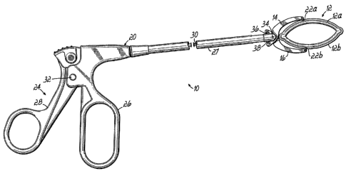
[0072] Referring initially to Figs. 1, 2A, 2B and 3, a first embodiment of
the invention includes an apparatus 10 comprising a clamp 12 having
respective first and second damping portions 12a, 12b secured between first
and second jaws 14, 16. A delivery and actuation device 20 carries first and
second jaws 14, 16 for actuating clamp 12 between open and dosed or
clamping positions as will be described further below. Clamp portions 12a, 12b
are secured to jaws 14, 16 by respective sutures 22a, 22b. Delivery and
actuation device 20 includes a pistol grip 24 having a stationary handle 26
coupled with an elongate jaw support member 27. A movable handle 28 is
coupled with an actuating bar 30 and pivots with respect to stationary handle
26
at a pivot member 32. When movable handle 28 is depressed toward
stationary handle 26, this action draws actuating bar 30 to the left as viewed
in
Fig. 1. Actuating bar 30 has a connecting portion 30a secured to respective
rigid wire members 36, 38. Wire members 36, 38 are secured to jaws 14, 16
such that when wire members 36, 38 are pulled by actuating bar 30, jaws 14,
16 pivot toward each other about pivot member 34. This moves clamping
CA 02508199 2005-05-25
-17-
portion 12a to an over-center position with respect to clamping portion 12b
whereupon clamping portion 12a snaps into the clamping position shown in Fig.
2B.
[0073] Thus, it will be appreciated that the clamp 12 changes from the
generally oval, annular (i.e., closed ring-shape) shape shown in Fig. 2A to
the
flattened, curved shape shown in Fig. 28 when moving from the open to the
closed position. It will also be appreciated with respect to this embodiment
and
others disclosed herein that clamp 12 may be repeatedly opened and closed by
device 20. This can allow repositioning of clamp 12, as necessary, during the
surgical procedure prior to release of clamp 12 from device 20. Fig. 3 further
illustrates that clamp 12 is pivotal about an axis extending transverse to the
length of clamp 12 in opposite directions as shown by arrows 40, 41. This
pivoting action is a passive pivoting action. That is, clamp 12 may freely
pivot
from the generally straight orientation shown in solid lines to the respective
oppositely angled orientations shown in dash-dot lines.
[0074] Fig. 2C illustrates, in enlarged detail, an end portion of clamp 12
with a fabric covering 60 partially removed to reveal leaf spring members 56,
58, which operate as will be discussed below. As shown in Fig. 2C, leaf spring
member 56 includes a rounded end portion 56a which is designed to protect the
patient from irritation which may have otherwise been caused by exposed sharp
edges of leaf springs 56, 58. Fig. 2C also illustrates an end portion 58a of
leaf
spring member 58 which is angled relative to the remaining portion of leaf
spring member 58 and is positioned inside of rounded end portion 56a. A stop
tab 61 is also formed in rounded end portion 56a, such as through a stamping
operation. As leaf spring member 58 moves from the open position (shown in
solid lines) to the closed position (shown in dash-dot lines), angled end
portion
CA 02508199 2005-05-25
. .
58a will rotate against and finally stop behind tab 61 to lock leaf spring
member -18-
58a in the closed position.
[0075] Figs. 4A and 4B partially illustrate the chest
anatomy of a patient
comprising ribs 44 and a heart 48 including a left atrial appendage 50. In one
approach using the present invention, clamp 12 may be delivered medially
between respective ribs 44 through, for example, a thoracotomy and intercostal
space. In this regard, a relatively small incision (not shown) is made between
ribs 44 and clamp 12, jaws 14, 16 and elongate jaw support member 27 are
directed between ribs 44 through the incision. The opened clamp 12 may be
placed around left atrial appendage 50 such that clamping portions 12a, 12b,
which form an annular shape, surround left atrial appendage 50 as shown in
Fig. 4A. As further shown in Fig. 4B, when jaws 14, 16 are actuated to a
closed
position as previously described, clamping portions 12a, 12b move together
essentially as shown to clamp and close off or occlude left atrial appendage
50.
[0076] Fig. 5 illustrates one of several other
approaches which may be
used with the present invention. In this regard, clamp 12, jaws 14, 16, and
elongate jaw support member 27 may be introduced in an intercostal space
between a patient's ribs 44 using a lateral approach to thereby access the
left
atrial appendage 50. After suitably angling clamp 12 and surrounding left
atrial
appendage 50 with clamping portions 12a, 12b, jaws 14, 16 may be actuated as
previously described to bring clamping portions 12a, 12b together and close
off
left atrial appendage 50. It will be appreciated that a sub-xiphoid approach
may
also be used, as well as several other approaches, such as open surgical
approaches used during open heart surgery.
[0077] Figs. 6A, 6B and 6C illustrate schematic cross
sections of a
portion of heart 48. In particular, left atrial appendage 50 is shown in cross
CA 02508199 2005-05-25
-19-
section to illustrate its hollow interior 52 which communicates with the left
atrium
54 of heart 48. Fig. 6A illustrates the normal configuration of left atrial
appendage 52. Fig. 6B illustrates clamp 12 in place, with Fig. 6C illustrating
the
same in enlarged detail. As shown in Figs. 6B and 6C, clamping portions 12a,
12b respectively comprise clamp members 56, 58, at least one of which acts as
a leaf spring member, and having a fabric covering 60 thereon. Fabric
coverings may be treated with collagen, albumin, etc., to promote tissue
ingrowth 64, 66. Fabrics such as DACRON polyester or expanded
polytetrafluoroethylene may be used in this regard to promote inflammatory
response and tissue ingrowth. Such tissue ingrowth 64, 66 will then assist
with
retaining clamp 12 in place. Clamp 12 may be placed extremely close to the
outer surface 54a of left atrium 54 to ensure that there is very little void
space at
junction 68 (Fig. 6C). Elimination of void space is important, for example, to
ensure that blood clots do not form from stagnant blood.
[0078] Figs. 7 and 7A illustrate one embodiment of a mechanism used
for releasing clamp 12 from jaws 14, 16. Specifically, sutures 22a, 22b may be
tied through apertures 70a, 70b. Respective tension members 72a, 72b are
coupled to blades 74a, 74b. When tension members 72a, 72b are pulled
proximally (see arrow 76 in Fig. 7A), blades 74a, 74b move past apertures 70a,
70b and cut sutures 22a, 22b. Optionally, sutures 22a, 22b may be formed as a
single suture such that a single blade may be used to release the clamp 12 and
the entire suture may be then carried out of the patient with one of the jaws
14,
16. In the embodiment shown, in which both sutures 22a, 22b are cut, the
sutures 22a, 22b may, for example, remain tied to clamp portions 12a, 12b.
[0079] Figs. 8A and 8B respectively illustrate the open and closed
positions of an alternative apparatus 10' constructed in accordance with the
CA 02508199 2005-05-25
-20-
invention. In this embodiment, like reference numerals are used to indicate
like
components of the first embodiment described above. Therefore, these like
components need no additional explanation. Components having reference
numerals with prime marks (') indicate components which have been slightly
modified in this embodiment as will be apparent. In this regard, all
components
of apparatus 10' may be as described previously, except that jaws 14', 16' are
angled such that they hold clamp 12 at an acute angle a relative to the axis
of
elongate jaw support member 27. That is, the length of clamp 12 extends along
an axis 78 which forms an angle of in the range of approximately 200 - 30
with
respect to axis 79 of elongate jaw support member 27. This angled delivery
orientation of clamp 12 has been found to enable easier application of clamp
12
to the left atrial appendage 50 (Fig. 4A). An additional angle y may also be
utilized as viewed from the top and discussed relative to Fig. 19A below. In
all
other respects, the operation of apparatus 10' may be the same as described
above.
[0080] Figs. 9 and 10 illustrate one alternative embodiment of a clamp 80
constructed in accordance with the invention. In this regard, clamp 80 may be
comprised of two separate clamping portions 80a, 80b, at least one of which
acts as a leaf spring member. Clamping portion 80a includes slots 82, 84 which
receive respective connecting tabs 86, 88 of clamping portion 80b. In this
manner, a generally oval annular shape is obtained when clamping portion 80a
is connected to clamping portion 80b. Clamping portions 80a, 80b may be
covered with a fabric or other suitable coating for promoting tissue ingrowth
as
previously described and/or for traction purposes. Fig. 9 further illustrates
undulating, stamped or molded side edges 85, 87 which also may be
considered projections to prevent clamp movement or migration. This may be
CA 02508199 2005-05-25
-21-
combined with a tissue ingrowth feature as mentioned herein. Alternatively, or
in addition, the clamps of this invention may have a resilient polymeric
coating
on one or both clamping portions to promote traction. For example, the
polymeric material may be silicone. As illustrated in Fig. 10, clamping
portion
80a is flat in cross section, while clamping portion 80b is circular in cross
section. This provides for more uniform and efficient force distribution along
the
length of clamp 80 in the closed position as shown in Fig. 10 when clamping
off
the left atrial appendage from the left atrium as previously described.
Silicone
coatings 81a, 81b are used for traction, i.e., to prevent slippage of clamp
80.
The interior 52 of left atrial appendage 50 is thereby closed off completely
from
the left atrium 54 such that residual pockets which can trap stagnant blood
are
minimized or eliminated.
[0081] Fig. 11 illustrates a cross section similar to Fig. 10 but showing an
alternative clamping portion 80a' which has been slightly modified to have a
concave surface in cross section facing the convex outer surface of clamping
portion 80b. This design can promote a better fit between clamping portion
80a'
and clamping portion 80b to ensure better sealing and potentially less void
space at junction 68.
[0082] Fig. 12 illustrates a top, partial cross sectioned view of an
alternative apparatus 90 constructed in accordance with the invention.
Apparatus 90 may be constructed the same as the first described embodiment
in all respects except for the manner of securing clamp 12 to jaws 14, 16
(only
one shown in Fig. 12). In this regard, a suture 92 is tied with a suitable
slip knot
94 such that the ends 92a of suture 92 may be pulled to release clamp 12 from
jaws 14, 16. It will be appreciated that a slip knot similar to slip knot 94
may be
used to secure each clamping portion, although only one slip knot 94 and
CA 02508199 2005-05-25
-22-
clamping portion 12a are shown in Fig. 12. Alternatively, suture 92 may be
secured by only one slip knot 94 with a portion of the suture 92 extending
around and coupling suitably with the opposite clamping portion 12b (not
shown).
[0083] Figs. 13-15 illustrate another alternative embodiment of an
apparatus 100, again only showing the distal end of apparatus 100 in Figs. 13
and 14. The portions not shown may be constructed and operated similarly to
the previously described embodiments. In this embodiment, apparatus 100
includes a clamp 102 with first and second clamping portions 102a, 102b, which
may or may not be covered with fabric, but which are illustrated as curved
leaf
spring members in Figs. 13 and 14, without a fabric covering for clarity. In
this
embodiment, suture material 104 extends through respective curved slots 106a,
106b, 108a, 108b in each clamping portion 102a, I 02b with the curved slots
106a, 106b, 108a, 108b thereby allowing for pivoting action in the jaws 107,
109
as previously described with respect to jaws 14, 16 of the first embodiment
and
as shown best in Fig. 14. Also in this embodiment, a plurality of projections
110
are provided on one of the clamping portions 102b and are received by
respective aligned apertures 112 formed in the opposite clamping portion 102a
when in the closed or clamping position as shown in Fig. 15. Collapsible
sleeves 114 may be placed around the projections 110 so as to prevent
snagging on tissue during delivery and application of the clamp 102 to the
tissue such as the left atrial appendage 50. In this embodiment, to release
the
clamp 102 from the jaws 107, 109, the suture material 104 may simply be cut at
the proximal end (not shown) and then carried out with the apparatus 100 after
application of the clamp 102 to the tissue (appendage 50). As shown in Fig.
15,
the projections 110 will extend through the fabric covering 102c, the tissue,
and
CA 02508199 2005-05-25
-23-
the receiving element or aperture 112 in this case when the clamp 102 is in
the
closed or clamping position. This not only assists with securing the clamp 102
in the closed position, but also further promotes tissue ingrowth as a small
amount of bleeding will occur because of the penetration of the projection 110
and this bleeding can promote tissue ingrowth into the fabric covering 102c.
[0084] Figs. 16, 17A and 17B illustrate another alternative apparatus 120 =
constructed in accordance with the invention. In this embodiment, an
alternative mechanism is provided for securing and releasing a clamp 122 to
and from the jaws 124, 126. In this regard, gripping elements 128, 130 are
provided in the form of spring loaded fingers which are normally biased to the
open position shown in the upper portion of Fig. 17B. A cam-type recess 132,
134 receives each gripping element 128, 130 such that the fingers are drawn
together around the respective clamping portions 122a, 122b as shown in Fig.
17A. Small diameter rods 136, 138 are placed through respective eyelets 140,
142 and 144, 146 to hold the fingers together. When rod 136 is removed from
the corresponding eyelets 140, 142 as shown in the upper portion of Fig. 17B,
the eyelets 140, 142 spread apart and the gripping element 128 biases itself
out
of the cam-type recess 132 into an open position. In this embodiment, this
release may then allow the normally closed clamp 122 to assume its closed
position around the tissue 148 through biased movement of clamping portion
122a toward portion 122b. Once the tissue 148 has been clamped as shown in
Fig. 17B, the opposite gripping element 130 may be released from clamping
portion 122b in the same manner, whereupon the apparatus 120 may be
withdrawn from the patient.
[0085] Figs. 18, 18A and 18B illustrate another alternative clamp portion
gripping element 150 having a pair of fingers 152, 154 which engage the
CA 02508199 2005-05-25
-24-
clamping portion 156 in a manner similar to the gripping elements disclosed in
Figs. 16, 17A and 17B. As with the embodiment of Figs. 16, 17A, and 17B, the
gripping elements are carried as separate pieces on, or formed as part of, the
jaws (e.g., jaws 14, 16). Fingers 152, 154 are normally closed as shown in
Fig.
18A to firmly hold the clamping portion 156 therebetween, but may be opened
by drawing a tension member 160 and ball or wedge member 162 rearward as
shown in Fig. 18B. In this manner, one clamp portion may be released at a
time in a manner similar to that described in connection with Figs. 16, 17A
and
17B. If the jaws are actuated to move one of the damping portions to an over
center position relative to the other clamping portion as previously
described,
then the release mechanism shown in Figs. 18, 18A and 18B may release both
clamping portions at the same time after the damping operation has taken
place.
[0086] Referring now to Figs. 19, 19A and 20, an alternative embodiment
of an apparatus 200 is shown in which like reference numerals refer to like
components of the first embodiment and reference numerals having prime (')
marks refer to components which have been slightly modified relative to the
corresponding components in the first embodiment, as will be apparent.
Apparatus 200 comprises a clamp delivery and actuation device 202 having an
elongate jaw support member 27' with a clamp pivoting mechanism 204 at one
end, including a rotatable actuating member 206. Rotatable actuating member
206 serves to rotate a rod (Fig. 20) back and forth via a suitable gear
arrangement (not shown) or direct coupling to thereby rotate a yoke 210 back
and forth. Yoke 210 is coupled with one end of clamp 12 and, therefore,
rotation of yoke 210 back and forth pivots clamp 12 back and forth through a
CA 02508199 2005-05-25
-25-
desired angle as shown in Figs. 21A-21C. This angle may, for example, be in
the range of about 100 to about 40 .
[0087] Apparatus 200, and specifically delivery and actuation device 202,
includes a pistol grip handle 24' having a stationary handle portion 26' and a
movable handle portion 28 coupled to stationary handle portion 26' by a pivot
32. Stationary handle portion 26' is coupled to the proximal side of pivoting
mechanism 204. A stationary jaw 216 and a movable jaw 218 are coupled to
the distal end of elongate jaw support member 27'. Clamp 12 is secured to
jaws 216, 218 by suture material 220, 222 using a slip knot configuration as
previously described such that when the exposed ends of suture material 220,
222 are pulled at the proximal end of apparatus 200, clamp 12 is released from
jaws 216, 218. A link 230 is pivotally coupled to jaw 218 at a pivot 232, and
jaw
218 is further pivotally coupled to elongate jaw support member 27' at a pivot
234. An actuation bar or rod 236 is pulled proximally when the surgeon
squeezes handle portion 28 toward stationary handle portion 26'. This causes
jaw 218 to pivot upwardly relative to stationary jaw 216 to close clamp 12 as
previously described. Jaw 218 may likewise be moved away from stationary
jaw 216 by moving handle portion 28 away from stationary handle portion 26' to
thereby open clamp 12 if, for example, necessary to reposition clamp 12 on the
tissue (not shown). As further shown in Fig. 19A, jaws 216, 218 are angled
relative to the longitudinal axis of elongate jaw support member 27' by an
angle
y as viewed from the top. This assists with positioning clamp 12 relative to
the
left atrial appendage. This may also be coupled with the upward angle as
shown, and as more specifically described in connection with Figs. 8A and 8B
above.
CA 02508199 2005-05-25
-26-
[0088] Fig. 20A illustrates an alternative embodiment which is the same
as Fig. 20 but uses a yoke 210' in the shape of a closed loop instead of a
forked
yoke 210.
[0089] Figs. 22A-28B illustrate various alternative clamps constructed
according to the invention. More specifically, Figs. 22A and 22B illustrate
respective first and second clamping portions 1200, 1202 which may be
actuated from an open position as shown in Fig. 22A, to a closed position, as
shown in Fig. 22B, by a sliding cam elements 1204, 1206 moving in the
direction of arrows 1208 and locked in recesses 1211, 1213. This may be done
by pulling tension members 1205, 1207. Fig. 23 illustrates a one piece clamp
1210 which may be moved from an open position as shown in solid lines to a
closed position as shown in dash-dot lines and locked in place by respective
ratchets 1212 at an appropriate clamping position. Fig. 24 is similar to Fig.
23,
but illustrates a two piece clamp 1220 having first and second clamping
portions
1220a, 1220b each locked in place on the other clamping portion by respective
ratchets 1222a, 1222b. Figs. 25A and 25B illustrate a clamp 1230 having first
and second clamping portions 1230a, 1230b movable from the open position
shown in Fig. 25A to the closed position shown in Fig. 25B. Rotatable cam
elements 1232, 1234 are pivotally connected to the clamping portions 1230a,
1230b and are engageable with containment members 1236, 1238 coupled with
clamping portions and having surfaces engaged with the cam elements 1232,
1234 during rotation thereof. Rotation of cam elements 1232, 1234 against
containment members 1236, 1238 by pulling tension members 1233, 1235
forces the flexible clamping portions 1230a, 1230b together as shown in Fig.
25B. Figs. 26A and 26B illustrate a clamp 1240 with respective first and
second
clamping portions 1240a, 1240b movable together by magnetic attraction which
CA 02508199 2005-05-25
-27-
may, for example, be brought about by permanent magnets 1242, 1244 as
shown, or by an electromagnetic device (not shown). In addition, one or both
clamping portions 1240a, 1240b may act as a leaf spring as previously
described. Figs. 27A and 27B illustrate a clamp 1250 having first and second
clamping portions 1250a, 1250b and activated by drawing respective tension
member portions 1252, 1254 against raised elements 1256, 1258 secured to
each clamping portion 1250a, 1250b. Ratchet type locking elements 1255,
1257 may be used to retain tension member portions in the clamping positions
shown in Fig. 27B. Figs. 28A and 288 illustrate another clamp 1260 comprised
of a leaf spring clamping portion 1260a and a rigid clamping portion 1260b.
Leaf spring 1260a may be depressed relative to rigid member 1260b
whereupon it snaps into place to clamp the tissue therebetween.
[0090] Fig. 29 illustrates the distal end of a paddle type gripper device
1300 which may be used to pull tissue, such as the left atrial appendage,
through a clamp as described herein. More specifically, device 1300 includes a
paddle type pivoting gripper 1302 at the distal end thereof. Gripper 1302
includes an elongate support member 1304 with first and second gripper
members 1306, 1308 at the distal end, at least one of which moves toward the
other to grip tissue (not shown) therebetween. In the embodiment shown, these
flat paddle like gripper elements 1306, 1308 include knobbed tissue engaging
surfaces 1310, 1312 to gently but firmly enable gripping of delicate tissue,
such
as tissue of the left atrial appendage. Gripper elements 1306, 1308 are
actuated toward each other to a closed position in a manner similar to the
jaws
disclosed above in the first embodiment of apparatus 10. More specifically,
gripper elements 1306, 1308 include proximal end portions 1314, 1316 pivoted
in a scissor-type fashion to elongate support member 1304 at a pivot 1315. An
CA 02508199 2005-05-25
-28-
actuating rod 1318 is pulled proximally, such as through the use of a pistol
grip
construction as previously described, and is coupled to wires 1320 (only one
shown) which are respectively coupled to proximal end portions 1314, 1316. In
this manner, gripper elements 1306, 1308 may be repeatedly closed and
opened to gently grip and pull tissue through a clamp, such as disclosed
hereinabove.
[0091] Referring to Figs. 30A-30C, an apparatus 1400 is shown and
includes a clamp delivery and actuation device 1402 configured to internally
carry, deploy and then actuate a clamp 1404 onto a hollow anatomical structure
1405. Clamp 1404 includes respective clamping portions 1404a, 1404b having
respective rails 1406, 1408 carried thereon, such as by being integrally
molded
therewith or otherwise secured thereto. Rails 1406, 1408 ride on respective
guide members 1410, 1412, as best shown in Fig. 31, for purposes as will be
described. Device 1402 includes a tube 1420 which may have a diameter sized
for minimally invasive surgery (e.g., 8 mm) and which carries a first rod or
clamp deployment member 1422 preferably in the form of a piston-type member
which reciprocates within the interior of tube 1420. 0-rings 1424, 1426 or
similar elements may be used to provide some frictional resistance and better
control to the reciprocating motion of rod 1422. A gripper 1430 is carried for
reciprocating movement with rod 1422 and is used to grasp clamp 1404 as
shown in Figs. 30A-30C, as well as in Figs. 31 and 32A. A tube 1432 is also
carried by rod 1422 and holds gripper 1430. Tube 1432 may be used to open
and close gripper elements 1430a, 1430b (Figs. 31, 32A-B) and also to push
clamp 1404 out of tube 1420 during deployment of clamp 1404 as described
below. As further shown in Fig. 32B, when tube 1432 is retracted, or pulled to
the left with respect to gripper 1430, gripper elements 1430a, 1430b will
spring
_
CA 02508199 2005-05-25
-29-
apart into their normally biased open or disengaged position. At this point,
clamp 1404 may be disengaged from delivery and actuation device 1402.
Pushing tube 1432 in the opposite direction into the position shown in Fig.
32A
will close gripper elements 1430a, 1430b. It will be appreciated that other
manners of securing clamp 1404 for movement with respect to device 1402
may be used instead.
[0092] More specifically referring to Fig. 30A, clamp 1404 may be initially
fully contained within tube 1420, in a closed position, although Fig. 30A
shows
clamp 1404 partially deployed. As clamp 1404 is pushed entirely out of the
distal end of tube 1420, the clamp will be opened as shown in Fig. 30B since
the path along which the rails 1406, 1408 move along guides 1410, 1412 forces
clamping portion 1404a past an over-center position at which clamping portion
1404a will snap into the open position shown. The combined delivery and
actuation device 1402 and clamp 1404 may be initially delivered in a compact
state through a small incision in the patient. Once the distal end of
apparatus
1400 is inserted in this fashion, deployment may take place as shown in Fig.
30B and described above. Once the hollow anatomical structure 1405 is
positioned between clamping portions 1404a, 1404b as shown in Fig. 30B,
clamp 1404 may be withdrawn into tube 1420. A compression member 1434
will deform clamping portion 1404a past an over-center position toward the
closed position whereupon clamping portion 1404a will snap into the closed
position shown in Fig. 30D. At this point, tube 1432 may be retracted to the
left
as shown in Figs. 30D and 32B thereby releasing gripper 1430. As slots 1436,
1438 (Fig. 31) are formed on opposite sides of tube 1420, the hollow
anatomical structure 1405 may be initially retracted into tube 1420 during the
clamping process. As rails 1406, 1408 disengage their respective guide
CA 02508199 2011-10-07
-30-
members 1410, 1412 as shown in Fig. 30D, delivery and actuation device 1402
may be withdrawn from the patient leaving the clamp 1404 and hollow
anatomical structure 1405 in place. As further shown in dash-dot lines in
Figs.
32A and 32B, clamp 1404 may have a fabric covering 1440 which, preferably, is
adapted to promote tissue ingrowth as previously'discussed. This embodiment
is especially adapted for use in minimally invasive surgical procedures. For
such purposes, the maximum outer diameter of tube 1420 is preferably about
12 mm, although various cross sectional shapes may be used having outer
diameters from, for example, about 8 mm to about 12 mm.
[0093] While the present invention has been illustrated by a description
of various preferred embodiments and while these embodiments have been
described in some detail, it is not the intention of the Applicant to restrict
or in
any way limit the scope of the appended claims to such detail. Additional
advantages and modifications will readily appear to those skilled in the art.
The
various features of the invention may be used alone or in numerous
combinations depending on the needs and preferences of the user. This has
been a description of the present invention, along with the preferred methods
of
practicing the present invention as currently known.