Note: Descriptions are shown in the official language in which they were submitted.
CA 02508459 2005-06-02
WO 2004/050140 PCT/US2003/038317
MEDICAL DEVICES FOR DELIVERY OF
THERAPEUTIC AGENTS
FIELD OF THE INVENTION
[0001] The present invention relates to implantable or insertable medical
devices,
such as intraluminal stems, that release therapeutic agents. The medical
devices of the
present invention are particularly appropriate for the release of high
molecular weight
therapeutic agents, such as DNA.
BACKGROUND OF THE INVENTION
[0002] Percutaneous iransluminal coronary angioplasty ("PTCA" or
"angioplasty")
procedures have been performed for many years as an adjunct to correcting
vascular
disease in patients. Angioplasty procedures involve the insertion, through the
vascular
system, of a catheter having a balloon that is placed across a lesion or
blockage in a
coronary artery. The balloon is then inflated to compress the lesion or
blockage against
the arterial walls, thereby opening the artery for increased blood flow.
[0003] In some cases, however, the goal of the angioplasty procedure is
defeated at
least in part by a complete or partial reclosure of the artery at or near the
compressed
lesion or blockage. Two mechanisms are believed to be principally responsible
for
reclosure of the artery. The first mechanism is recoil, which is a mechanical
process
involving the elastic rebound of the compressed lesion or blockage. The second
mechanism is restenosis, which is believed to be caused by proliferation of
the smooth
muscle cells present in the artery walls near the lesion or blockage.
Restenosis can occur
over a period of several weeks or months after the PTCA procedure.
[0004) Many different methods have been employed to limit the effect of
restenosis,
including radiation treatments and various drug therapies, delivered locally
and
systemically, to slow proliferation of the smooth muscle cells. Recoil of the
arterial walls
can be prevented by using stems, which can be temporarily or permanently
deployed
within the artery to mechanically maintain patency of the artery. Stems are
very effective
CA 02508459 2005-06-02
WO 2004/050140 PCT/US2003/038317
at carrying out this task, but they may also irritate the contacting arterial
walls, which
may in turn encourage additional restenosis.
[0005] Gene therapy has been used for diverse medical purposes, including
slowing
proliferation of smooth muscle cells. Genes are usually delivered into a
patient's cells
through a vector, such as a retroviral vector, whose DNA is genetically
engineered to
include a desired DNA sequence. Alternatively, nonviral gene transfer methods
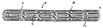
can be
used, such as plasmid DNA vectors, along with polymeric carriers, DNA
condensing
agents, lipofection and receptor mediated delivery vectors.
[0006] In connection with angioplasty, incorporation of appropriate DNA
molecules
into the coronary artery walls near the treatment site can be beneficial to
inhibit
restenosis. A polymer-coated stmt can be used as the delivery vehicle for the
DNA, in
addition to maintaining patency of the artery following PTCA.
[0007] However, effective delivery of high-molecular-weight therapeutic
agents,
such as DNA and any associated vector, can entail large amounts of therapeutic
agent and
long delivery times. Large amounts of polymeric material provided as a coating
on the
stent may, therefore, be required to adequately incorporate the therapeutic
agent and
ensure controlled and extended release of the therapeutic agent over a
required period of
time. Consequently, the polymeric coating may become relatively thick,
increasing the
susceptibility, during expansion of the stmt, to cracking of the coating. Such
cracking
can reduce the effectiveness of the coating to deliver the therapeutic agent
therefrom,
among other consequences. Moreover, because some medical devices such as stems
have
limited surface areas for disposition of a polymer coating, it would be
desirable to provide
a coating that actually enhances the uptake of the therapeutic agent by the
tissue of
interest.
[0008] The manufacture of medical devices with high-molecular-weight
therapeutic
agents in polymer matrices can also present processing difficulties. For
example,
relatively high shear stresses are commonly encountered while processing a
mixture of a
polymeric material and a therapeutic agent. In the case of certain high-
molecular-weight
therapeutic agents such as polynucleotides (e.g., plasmids), for example,
these shear
stresses can, in turn, disrupt the conformational and/or structural integrity
of the
therapeutic agent. .
[0009] Moreover, certain biostable polymers that are highly biocompatible
(e.g.,
CA 02508459 2005-06-02
WO 2004/050140 PCT/US2003/038317
polystyrene-polyisobutylene copolymers) may in some cases provide insufficient
mass
transport therethrough of high-molecular-weight therapeutic agents after
deployment,
limiting their utility in medical devices that deliver such agents.
[0010] Accordingly, there is a need for coatings for stems and other medical
devices
that release high-molecular-weight therapeutic agents in a controlled fashion
over a
period of time and do not suffer from the foregoing and other disadvantages.
The
coatings should, therefore, contain a therapeutically effective amount of high-
molecular-
weight therapeutic agent and provide adequate control of the release of that
therapeutic
agent. In addition, in the case of expandable medical devices such as stems
and balloons,
the coatings should resist cracking that may occur during expansion of the
medical
device. Moreover, the conformational and structural integrity of high-
molecular-weight
therapeutic agents such as DNA should be preserved to the greatest extent
possible during
manufacture of the medical device.
SUMMARY OF THE INVENTION
(0011] These and other needs are met by the present invention.
[0012] According to one aspect of the present invention, a medical device is
provided, at least a portion of which is insertable or implantable into the
body of a patient.
The medical device comprises: (a) a plasmid DNA layer, which comprises plasmid
DNA;
and (b) a polymeric covering layer disposed over the plasmid DNA layer.
[0013] Examples of implantable or insertable medical devices include
catheters,
balloons, filters, coils, clips, slings, and intraluminal stems, for instance,
vascular stems.
[0014] The plasmid DNA layer may be applied in a number of ways, for example,
by
dipping at least a portion of the medical device into a solution comprising
the plasmid
DNA.
[0015] The polymeric covering layer can be, for example, a biostable polymeric
covering layer or a biodisintegrable polymeric covering layer.
[0016] Examples of biostable polymeric covering layers include those that
comprise
one or more of the following: polyolefm polymers and copolymers; ethylenic
copolymers; polyurethane polymers and copolymers; metallocene catalyzed
polyethylene
polymers and copolymers; ionomers; polyester-ether polymers and copolymers;
polyamide-ether polymers and copolymers; and silicone polymers and copolymers.
CA 02508459 2005-06-02
WO 2004/050140 PCT/US2003/038317
[0017] The biostable polymeric covering layer can comprise, for example, a
block
copolymer comprising at least two polymeric blocks A and B, wherein A is a
polyolefin
block and B is a vinyl aromatic block. For example, A can be a polyolefin
block of the
general formula -(CRR'-CHz)"-, where R and R' are linear or branched aliphatic
groups
or cyclic aliphatic groups and B can be is a vinyl aromatic polymer block. As
another
example, A can be a polyolefm block that comprises one or more monomers
selected
from ethylene, butylene and isobutylene, and B can be a vinyl aromatic polymer
block
that comprises one or more monomers selected from styrene and a-methylstyrene.
[0018] Examples of biodisintegrable polymeric covering layers include those
that
comprise one or more of the following: lactic acid polymers and copolymers,
glycolic
acid polymers and copolymers, trimethylene carbonate polymers and copolymers,
caprolactone polymers and copolymers, hyaluronic acid polymers and copolymers,
hydroxybutyrate polymers and copolymers, and tyrosine-based polymers and
copolymers.
[0019] The biodisintegrable polymeric covering layer can comprise, for
example, (a)
hyaluronic acid polymers, (b) copolymers of lactic acid and glycolic acid,
and/or (c)
tyrosine-derived polycarbonates.
[0020] According to another aspect of the present invention, a medical device
is
provided, at least a portion of which is insertable or implantable into the
body of a patient.
The medical device comprises (a) a therapeutic agent containing layer. which
comprises a
high-molecular-weight therapeutic agent; and (b) a polymeric covering layer
disposed
over the high-molecular-weight-therapeutic-agent layer. The polymeric covering
layer
comprises one or more polymers selected from (i) a block copolymer comprising
at least
two polymeric blocks A and B, wherein A is a polyolefm block and wherein B is
a vinyl
aromatic block, (ii) a polymer or copolymer of lactic acid, (iii) a polymer or
copolymer of
glycolic acid, and (iv) a tyrosine-based polymer or copolymer.
[0021] The therapeutic agent containing layer may be applied in a number of
ways,
for example, by dipping at least a portion of the medical device into a
solution comprising
the high-molecular-weight therapeutic agent.
[0022] Examples of high-molecular-weight therapeutic agents include: (a)
polysaccharide therapeutic agents having a molecular weight greater than
1,000; (b)
polypeptide therapeutic agents having a molecular weight greater than 10,000;
and (c)
4
CA 02508459 2005-06-02
WO 2004/050140 PCT/US2003/038317
polynucleotides having a molecular weight greater than 2,000, for instance,
plasmid
DNA.
[0023] According to another aspect of the present invention, a medical device
is
provided, at least a portion of which is insertable or implantable into the
body of a patient.
The medical device comprises (a) polymeric layer comprising a removable
component as
well as (i) a block copolymer comprising at least two polymeric blocks A and
B, wherein
A is a polyolefin block and B is a vinyl aromatic block and/or (ii) a tyrosine-
based
polymer or copolymer; and (b) a high-molecular-weight therapeutic agent
disposed below
or within the polymeric layer.
[0024] The removable component can be, for example, a leachable material, such
as
polyethylene glycols, polyalkylene oxides (e.g., polyethylene oxide and
copolymers of
polyethylene oxide and polypropylene oxide), polyhydroxyethylmethacrylates,
polyvinylpyrrolidones, polyacrylamide and its copolymers, liposomes, proteins,
peptides,
salts, sugars, polysaccharides, polylactides, cationic lipids, detergents,
polygalactides,
polyanhydrides, polyorthoesters and their copolymers, and soluble cellulosics.
[0025] According to another aspect of the present invention, a medical device
is
provided, at least a portion of which is insertable or implantable into the
body of a patient.
The medical device comprises (a) a polymeric layer comprising a polymer and a
plasticizer; and (b) a high-molecular-weight polynucleotide therapeutic agent
(e.g.,
plasmid DNA) disposed below or within the polymeric layer.
[0026] The plasticizer can be, for example, glycerol, triacetyl glycerin,
ethylene
glycol, triethylene glycol, polyethylene glycol, propylene glycol,
polyalkylene oxides
(e.g., polyethylene oxide and copolymers of polyethylene oxide and
polypropylene
oxide), citric acid esters, sebacic acid esters, phthalic acid esters, and
silicone fluid.
[0027] According to another aspect of the present invention, a medical device
is
provided, at least a portion of which is insertable or implantable into the
body of a patient.
The medical device comprises a multi-layer coating that covers at least a
portion of the
medical device. The multi-layer coating further comprises (a) one or more
therapeutic
agent containing layers comprising a therapeutic agent and (b) one or more
polymeric
layers comprising a polymer, wherein the one or more polymeric layers have a
composition gradient in a direction normal to the surface of the coating.
[0028] The therapeutic agent can be, for example, a high-molecular-weight
CA 02508459 2005-06-02
WO 2004/050140 PCT/US2003/038317
therapeutic agent.
(0029] The one or more therapeutic agent containing layers can be disposed,
for
example, beneath the one or more polymeric layers. In an alternative
embodiment, a
plurality of therapeutic agent containing layers are disposed in an
alternating
configuration with a plurality of polymer layers.
[0030] In some embodiments, the composition gradient is provided within a
single
polymeric layer. In others, the composition gradient is provided within a
plurality of
polymeric layers (e.g., 2, 3, 4, 5 or more polymeric layers).
[0031] The composition gradient can comprise, for example, (a) a gradient in
porosity, (b) a gradient in polymer composition, for example, a gradient in
the relative
proportions of two or more monomer species within a copolymer or a gradient in
the
relative proportions of two or more polymers within a polymer blend (for
example, the
relative proportions of a hydrophobic polymer, such as styrene-isobutylene
copolymer,
and a hydrophilic polymer, such as a styrene-ethylene oxide copolymer), (c) a
gradient in
the composition of a teachable species, (d) a gradient in the composition of
an acidic
species, (e) a gradient in the composition of a basic species and/or (f) a
gradient in the
composition of an ionic species.
[0032] One advantage of the present invention is that polymer coated medical
devices such as stems, containing therapeutic agents, including high-molecular-
weight
therapeutic agents such as DNA, can be provided in which the rate of release
of the
therapeutic agents is adequately regulated so as to provide a therapeutically
effective
amount of such agent over a desired period of time.
[0033] Another advantage of the present invention is that polymer coated
medical
devices, such as stems, containing therapeutic agents, including high-
molecular-weight
therapeutic agents such as DNA, can be provided in which the polymer resists
cracking
upon expansion of the medical device.
[0034] Another advantage of the present invention is that medical devices such
as
stems, containing therapeutic agents, including high-molecular-weight
therapeutic agents
such as DNA, can be provided wherein the structural integrity of the
therapeutic agent is
not substantially disrupted during medical device manufacture.
[0035] Yet another advantage of the present invention is that medical devices
such as
CA 02508459 2005-06-02
WO 2004/050140 PCT/US2003/038317
stems, containing therapeutic agents, and particularly high-molecular-weight
therapeutic
agents such as DNA, can be provided in which the uptake of the therapeutic
agent by the
targeted tissue is enhanced.
[0036] These and other embodiments and advantages of the invention will become
apparent from the following detailed description, and the accompanying
drawings, which
illustrate by way of example the features of the invention.
BRIEF DESCRIPTION OF THE DRAWINGS
[0037] Fig. 1 is a schematic diagram of a stmt with a polymer coating,
according to
an embodiment of the invention.
[0038] Fig. 2 is a schematic diagram of a stmt with a polymer coating,
according to
an embodiment of the invention.
[0039] Fig. 3 is a schematic diagram of a stmt with a polymer coating,
according to
an embodiment of the invention.
(0040] Fig. 4 is a graph of DNA release as a function of time for
biodisintegrable
coatings, according to an embodiment of the invention.
[0041] Fig. 5 is a graph of DNA release as a function of time for
biodisintegrable
coatings, according to an embodiment of the invention.
[0042] Fig. 6 is a graph of coating dissolution as a function of time for
biodisintegrable coatings, according to an embodiment of the invention.
[0043] Fig. 7 is a graph of coating dissolution as a function of time for -
biodisintegrable coatings, according to an embodiment of the invention.
[0044] Fig. 8 is a graph of coating dissolution as a function of time for
biodisintegrable coatings, according to an embodiment of the invention.
[0045] Fig. 9 is a graph of DNA release as a function of time for
biodisintegrable
coatings, according to an embodiment of the invention.
[0046] Fig. 10 is a graph of DNA release as a function of time for
biodisintegrable
coatings, according to an embodiment of the invention.
[0047] Fig. 11 is a graph of DNA adsorption as a function of DNA
concentration,
according to an embodiment of the invention.
[0048] Fig. 12 is a graph of DNA release as a function of time for
biodisintegrable
coatings, according to an embodiment of the invention.
7
CA 02508459 2005-06-02
WO 2004/050140 PCT/US2003/038317
[0049] Fig. 13 is a photograph of a stmt after implantation in a rabbit iliac
artery,
according to an embodiment of the invention.
[0050] Fig. 14 is a graph of DNA release as a function of time for a biostable
coating, according to an embodiment of the invention.
[0051] Fig. 15 is a graph of dextran release as a function of time for
biostable
coatings, according to an embodiment of the invention.
[0052] Fig. 16 is a graph of dextran release as a function of time for
biostable
coatings, according to an embodiment of the invention.
[0053] Fig. 17 is a photograph of a biostable coating material after NaCI
extraction
in PBS, according to an embodiment of the invention.
[0054] Fig. 18 is a graph of DNA release as a function of time for biostable
coatings,
according to an embodiment of the invention.
DETAILED DESCRIPTION OF THE INVENTION
[0055] The various embodiments of the present invention are directed to
implantable
or insertable medical devices in which a polymer coating layer is used to
regulate local
delivery of a therapeutic agent, and typically a high-molecular-weight
therapeutic agent,
as defined below.
[0056] Localized delivery of a therapeutic agent from an implantable or
insertable
medical device is advantageous, because higher local concentrations of the
therapeutic
agent and/or more regulated delivery thereof can be achieved than with
systemic
administration. Consequently, increased cellular uptake of the therapeutic
agent and
therapeutic efficacy can be achieved with localized delivery, as opposed to
systemic
delivery of the therapeutic agent.
[0057] For example, systemic administration of several doses of therapeutic
agent
typically results in peaks and troughs in the level of concentration received
by the tissue.
In some cases, the peaks may be higher than a maximum desired level, leading
to
undesirable side effects, for example, and the troughs may be lower than a
minimum
effective level for the therapeutic agent. On the other hand, local
administration of the
therapeutic agent, for example, via a coated stent, can provide a
concentration level of
delivered agent that remains within a therapeutically effective range for a
longer period of
time.
CA 02508459 2005-06-02
WO 2004/050140 PCT/US2003/038317
[0058] The present invention is applicable to implantable or insertable
medical
devices of any shape or configuration. Examples of medical devices appropriate
for the
practice of the present invention include intraluminal catheters (including
vascular
catheters such as balloon catheters), guide wires, balloons, filters (e.g.,
vena cava filters),
stems, stmt grafts, cerebral stems, cerebral aneurysm filler coils (including
metal coils
and GDC--Guglilmi detachable coils), clips, slings, vascular grafts,
myocardial plugs,
pacemaker leads and heart valves.
[0059] More specific examples of medical devices for the practice of the
present
invention include intraluminal stems such as endovascular, biliary, tracheal,
gastrointestinal, urethral, ureteral, esophageal and coronary vascular stems.
The
intraluminal stems of the present invention may be, for example, balloon-
expandable or
self expandable. Thus, although certain embodiments of the present invention
will be
described herein with reference to vascular stents, the present invention is
applicable to
other medical devices, including other types of stems.
[0060] In general, stents for use in connection with the present invention
typically
comprise a plurality of apertures or open spaces between metallic filaments
(including
fibers and wires), segments or regions. Typical structures include: an open-
mesh network
comprising one or more knitted, woven or braided metallic filaments; an
interconnected
network of articulable segments; a coiled or helical structure comprising one
or more
metallic filaments; and, a patterned tubular metallic sheet (e.g., a laser cut
tube).
[0061] Figs. l and 3 illustrate two embodiments of polymer coated endovascular
stents 10 according to the present invention. Figure 2 shows a detailed
enlargement of a
portion of a polymer-coated stmt that is similar in design to that shown in
Figure 1. Each
stmt 10 can be, for example, a coronary stmt sized to fit in the blood vessel
of a patient,
which is formed from a plurality of structural elements 12. The construction
of each stent
permits the stmt 10 to be introduced into the vascular system in a collapsed
configuration, minimizing the diameter of the stmt 10. Each stmt 10 can then
expand to
an expanded position at the desired location within the blood vessel of the
patient. The
structural elements 12 of each stmt 10 form a conventional frame, such as
tubular shape,
and permits the stent 10 to self expand or to expand to the desired shape
after an
expansive force is applied, for example, by the expansion of a balloon within
the stmt.
(0062] A coating 16 is applied on the surface of each stmt 10. According to
the
9
CA 02508459 2005-06-02
WO 2004/050140 PCT/US2003/038317
present invention, coating 16 can include either a biostable or
biodisintegrable polymer as
described more fully below, which contains, or is provided as a coating over,
a
therapeutic agent. The therapeutic agent is released in a controlled manner
after
introduction of the stmt 10 into the body of the patient. As one specific
example, in the
case of high-molecular-weight therapeutic agent such as plasmid DNA, a typical
coronary
stent can have a uniform coating of approximately 1,000 micrograms in weight
or more,
which contains up to 100 micrograms of plasmid DNA or more.
[0063) The structural elements 12 of each stmt 10 form windows 14 such that
the
stmt 10 does not have a continuous outer shell. Windows 14 are generally
present in most
stmt configurations, although the specific details of the shape of structural
elements 12
and the construction of stmt 10 can vary as can be seen, for example, from
Figs. 1-3.
Each stent 10 can thus be coated with polymeric coating 16 such that windows
14 remain
free of coating. Alternatively, each stmt 10 can be covered by coating 16 such
that a
layer or web of coating (not shown) also covers the windows 14 between
elements 12.
For certain embodiments, it is beneficial that the windows 14 be left free of
a covering.
The unobstructed windows: (a) allow a freer exchange of nutrients between the
inner
walls of the vessel and the fluid flowing through the vessel, such as blood
flowing in an
artery and (b) do not block flow to vessel side-branches. In alternate
embodiments, the
material filling the windows is sufficiently porous to allow exchange of
nutrients and
oxygen.
[0064] Various embodiments of the invention can be implemented by dipping a
medical device of interest into a solution (e.g., a solution containing a
polymer and a
high-molecular-weight therapeutic agent). In such embodiments, it may be
desirable to
employ a stmt holder, such as those known in the art, which facilitates
placing the stem in
solution and subsequently removing and spinning the stmt to remove excess
solution.
[0065] Typical sites for placement of the medical devices of the present
invention
include the coronary and peripheral vasculature (collectively referred to
herein as the
vasculature), esophagus, trachea, colon, gastrointestinal tract, biliary
tract, urinary tract,
prostate, brain and surgical sites. Where the medical device is inserted into
the
vasculature, for example, the therapeutic agent is may be released to a blood
vessel wall
adjacent the device, and may also be released to downstream vascular tissue as
well.
[0066] After the medical devices of the present invention are deployed at a
suitable
CA 02508459 2005-06-02
WO 2004/050140 PCT/US2003/038317
site, the therapeutic agent is released and delivered locally to tissue
adjacent the medical
device. Depending upon the application, various release profiles can be
provided in
accordance with the present invention including: (a) 50% release (i.e., SO% of
the total
release from the medical device that occurs over the prescribed course of
implantation/insertion) occurring during a period of 15-60 minutes after
implantation/insertion, (b) 50% release occurring over a period of 1-6 hours,
(b) 50%
release occurring over a period of 6-24 hours, (c) 50% release occurring over
a period of
24-96 hours (4 days), (d) 50% release occurring over a period of 4-14 days,
(e) 50%
release occurring over a period of 2-8 weeks, (f) SO% release occurring over a
period of
8-32 weeks.
[0067] Typical subjects (also referred to herein as "patients") are vertebrate
subjects
(i.e., members of the subphylum cordata), including, mammals such as cattle,
sheep, pigs,
goats, horses, dogs, cats and humans.
[0068] "Therapeutic agents", "pharmaceutically active agents",
"pharmaceutically
active materials", "drugs" and other related terms may be used interchangeably
herein and
include genetic therapeutic agents, non-genetic therapeutic agents, and cells.
[0069] Exemplary non-genetic therapeutic agents include: (a) anti-thrombotic
agents
such as heparin, heparin derivatives, urokinase, and PPack
(dextrophenylalanine proline
arginine chloromethylketone); (b) anti-inflammatory agents such as
dexamethasone,
prednisolone, corticosterone, budesonide, estrogen, sulfasalazine and
mesalamine; (c)
antineoplastic/antiproliferative/anti-mitotic agents such as paclitaxel, 5-
fluorouracil,
cisplatin, vinblastine, vincristine, epothilones, endostatin, angiostatin,
angiopeptin,
monoclonal antibodies capable of blocking smooth muscle cell proliferation,
and
thymidine kinase inhibitors; (d) anesthetic agents such as lidocaine,
bupivacaine and
ropivacaine; (e) anti-coagulants such as D-Phe-Pro-Arg chloromethyl ketone, an
RGD
peptide-containing compound, heparin, hirudin, antithrombin compounds,
platelet
receptor antagonists, anti-thrombin antibodies, anti-platelet receptor
antibodies, aspirin,
prostaglandin inhibitors, platelet inhibitors and tick antiplatelet peptides;
(f) vascular cell
growth promoters such as growth factors, transcriptional activators, and
translational
promotors; (g) vascular cell growth inhibitors such as growth factor
inhibitors, growth
factor receptor antagonists, transcriptional repressors, translational
repressors, replication
inhibitors, inhibitory antibodies, antibodies directed against growth factors,
bifunctional
11
CA 02508459 2005-06-02
WO 2004/050140 PCT/US2003/038317
molecules consisting of a growth factor and a cytotoxin, bifunctional
molecules
consisting of an antibody and a cytotoxin; (h) protein kinase and tyrosine
kinase
inhibitors (e.g., tyrphostins, genistein, quinoxalines); (i) prostacyclin
analogs; (j)
cholesterol-lowering agents; (k) angiopoietins; (1) antimicrobial agents such
as triclosan,
cephalosporins, aminoglycosides and nitrofurantoin; (m) cytotoxic agents,
cytostatic
agents and cell proliferation affectors; (n) vasodilating agents; and
(o)agents that
interfere with endogenous vasoactive mechanisms.
[0070] Exemplary genetic therapeutic agents include anti-sense DNA and RNA,
oligo decoys, as well as DNA coding for: (a) anti-sense RNA, (b) tRNA or rRNA
to
replace defective or deficient endogenous molecules, (c) angiogenic factors
including
growth factors such as acidic and basic fibroblast growth factors, vascular
endothelial
growth factor, epidermal growth factor, transforming growth factor a and /3,
platelet-
derived endothelial growth factor, platelet-derived growth factor, tumor
necrosis factor a,
hepatocyte growth factor and insulin-like growth factor, (d) cell cycle
inhibitors including
CD inhibitors, and (e) thymidine kinase ("TK") and other agents useful for
interfering
with cell proliferation. Also of interest is DNA encoding for the family of
bone
morphogenic proteins ("BMP's"), including BMP-2, BMP-3, BMP-4, BMP-5, BMP-6
(Vgr-1), BMP-7 (OP-1), BMP-8, BMP-9, BMP-10, BMP-11, BMP-12, BMP-13, BMP-
14, BMP-1 S, and BMP-16. Currently beneficial BMP's are any of BMP-2, BMP-3,
BMP-4, BMP-5, BMP-6 and BMP-7. These dimeric proteins can be provided as
homodimers, heterodimers, or combinations thereof, alone or together with
other
molecules. Alternatively, or in addition, molecules capable of inducing an
upstream or
downstream effect of a BMP can be provided. Such molecules include any of the
"hedgehog" proteins, or the DNA's encoding them.
(0071] Cells include cells of human origin (autologous or allogeneic),
including stem
cells and platelets, or from an animal source (xenogeneic), which can be
genetically
engineered if desired to deliver proteins of interest.
[0072] Numerous therapeutic agents, not necessarily exclusive of those listed
above,
have been identified as candidates for vascular treatment regimens, for
example, as agents
targeting restenosis. Such agents are appropriate for the practice of the
present invention
and include one or more of the following: (a) Ca-channel blockers including
benzothiazapines such as diltiazem and clentiazem, .dihydropyridines such as
nifedipine,
12
CA 02508459 2005-06-02
WO 2004/050140 PCT/US2003/038317
amlodipine and nicardapine, and phenylalkylamines such as verapamil, (b)
serotonin
pathway modulators including: 5-HT antagonists such as ketanserin and
naftidrofuryl, as
well as 5-HT uptake inhibitors such as fluoxetine, (c) cyclic nucleotide
pathway agents
including phosphodiesterase inhibitors such as cilostazole and dipyridamole,
adenylate/guanylate cyclase stimulants such as forskolin, as well as adenosine
analogs,
(d) catecholamine modulators including a-antagonists such as prazosin and
bunazosine,
(3-antagonists such as propranolol and a/(3-antagonists such as labetalol and
carvedilol, (e)
endothelin receptor antagonists, (f) nitric oxide donors/releasing molecules
including
organic nitrateslnitrites such as nitroglycerin, isosorbide dinitrate and amyl
nitrite,
inorganic nitroso compounds such as sodium nitroprusside, sydnonimines such as
molsidomine and linsidomine, nonoates such as diazenium diolates and NO
adducts of
alkanediamines, S-nitroso compounds including low molecular weight compounds
(e.g.,
S-nitroso derivatives of captopril, glutathione and N-acetyl penicillamine)
and high
molecular weight compounds (e.g., S-nitroso derivatives of proteins, peptides,
oligosaccharides, polysaccharides, synthetic polymers/oligomers and natural
polymers/oligomers), as well as C-nitroso-compounds, O-nitroso-compounds, N-
nitroso-
compounds and L-arginine, (g) ACE inhibitors such as cilazapril, fosinopril
and enalapril,
(h) ATII-receptor antagonists such as saralasin and losartin, (i) platelet
adhesion
inhibitors such as albumin and polyethylene oxide, (j) platelet aggregation
inhibitors
including aspirin and thienopyridine (ticlopidine, clopidogrel) and GP
IIb/IIIa inhibitors
such as abciximab, epitifibatide and tirofiban, (k) coagulation pathway
modulators
including heparinoids such as heparin, low molecular weight heparin, dextran
sulfate and
[3-cyclodextrin tetradecasulfate, thrombin inhibitors such as hirudin,
hirulog, PPACK(D-
phe-L-propyl-L-arg-chloromethylketone) and argatroban, FXa inhibitors such as
antistatin and TAP (tick anticoagulant peptide), Vitamin K inhibitors such as
warfarin, as
well as activated protein C, (1) cyclooxygenase pathway inhibitors such as
aspirin,
ibuprofen, flurbiprofen, indomethacin and sulfinpyrazone, (m) natural and
synthetic
corticosteroids such as dexamethasone, prednisolone, methprednisolone and
hydrocortisone, (n) lipoxygenase pathway inhibitors such as
nordihydroguairetic acid and
caffeic acid, (o) leukotriene receptor antagonists, (p) antagonists of E- and
P-selectins, (q)
inhibitors of VCAM-1 and ICAM-1 interactions, (r) prostaglandins and analogs
thereof
including prostaglandins such as PGE1 and PGI2 and prostacyclin analogs such
as
13
CA 02508459 2005-06-02
WO 2004/050140 PCT/US2003/038317
ciprostene, epoprostenol, carbacyclin, iloprost and beraprost, (s) macrophage
activation
preventers including bisphosphonates, (t) HMG-CoA reductase inhibitors such as
lovastatin, pravastatin, fluvastatin, simvastatin and cerivastatin, (u) fish
oils and omega-3-
fatty acids, (v) free-radical scavengers/antioxidants such as probucol,
vitamins C and E,
ebselen, traps-retinoic acid and SOD mimics, (w) agents affecting various
growth factors
including FGF pathway agents such as bFGF antibodies and chimeric fusion
proteins,
PDGF receptor antagonists such as trapidil, IGF pathway agents including
somatostatin
analogs such as angiopeptin and ocreotide, TGF-(3 pathway agents such as
polyanionic
agents (heparin, fucoidin), decorin, and TGF-(i antibodies, EGF pathway agents
such as
EGF antibodies, receptor antagonists and chimeric fusion proteins, TNF-a
pathway
agents such as thalidomide and analogs thereof, Thromboxane A2 (TXA2) pathway
modulators such as sulotroban, vapiprost, dazoxiben and ridogrel, as well as
protein
tyrosine kinase inhibitors such as tyrphostin, genistein and quinoxaline
derivatives, (x)
MMP pathway inhibitors such as marimastat, ilomastat and metastat, (y) cell
motility
inhibitors such as cytochalasin B, (z) antiproliferative/antineoplastic agents
including
antimetabolites such as purine analogs (e.g., 6-mercaptopurine or cladribine,
which is a
chlorinated purine nucleoside analog), pyrimidine analogs (e.g., cytarabine
and S-
fluorouracil) and methotrexate , nitrogen mustards, alkyl sulfonates,
ethylenimines,
antibiotics (e.g., daunorubicin, doxorubicin), nitrosoureas, cisplatin, agents
affecting
microtubule dynamics (e.g., vinblastine, vincristine, colchicine, paclitaxel
and
epothilone), caspase activators, proteasome inhibitors, angiogenesis
inhibitors (e.g.,
endostatin, angiostatin and squalamine), raparnycin, cerivastatin,
flavopiridol and
suramin, (aa) matrix deposition/organization pathway inhibitors such as
halofuginone or
other quinazolinone derivatives and tranilast, (bb) endothelialization
facilitators such as
VEGF and RGD peptide, (cc) blood rheology modulators such as pentoxifylline,
and (dd)
endothelial-cell specific mitogens.
[0073] Further therapeutic agents appropriate for the practice of the present
invention, again not necessarily exclusive of those listed above, are also
disclosed in U.S.
Patent No. 5,733,925 assigned to NeoRx Corporation, the entire disclosure of
which is
incorporated by reference.
[0074] The present invention is especially useful in delivering high-molecular-
14
CA 02508459 2005-06-02
WO 2004/050140 PCT/US2003/038317
weight therapeutic agents, which are defined herein to include therapeutic
agents having a
molecular weight greater than 500, typically greater than 1,000, more
typically greater
than 2,000, or agents which contain one or more components having such
molecular
weights. Examples are polysaccharide therapeutic agents having a molecular
weight
greater than 1,000; polypeptide therapeutic agents having a molecular weight
greater than
10,000; polynucleotides, including antisense polynucleotides, having a
molecular weight
greater than 2,000, gene-encoding polynucleotides, including plasmids, having
a
molecular weight greater than 500,000; viral and non-viral particles having a
diameter
greater than about 50 nanometers, and cells.
[00'75] A "polynucleotide" is a nucleic acid polymer. A polynucleotide can
include
both double- and single-stranded sequences, and can include naturally derived
and
synthetic DNA sequences. The term also includes sequences that include any of
the
known base analogs of DNA and RNA, and includes modifications, such as
deletions,
additions and substitutions (generally conservative in nature) to native
sequences. In
some embodiments of the invention, the polynucleotide can be, for example, an
antisense
polynucleotide. In others, polynucleotide can be, for example, of transfection
unit length,
which is typically on the order of about 1 kb or greater.
[0076] Typical polynucleotide therapeutic agents include the genetic
therapeutic
agents specifically listed above, and more generally include DNA encoding for
various
polypeptide and protein products including those previously listed. Some
additional
examples of polynucleodde therapeutic agents include DNA encoding for the
following:
cytokines such as colony stimulating factors (e.g., granulocyte-macrophage
colony-
stimulating factor), tumor necrosis factors (e.g., fas ligand) and
interleukins (e.g., IL-10,
an anti-inflammatory interleukin), as well as protease inhibitors,
particularly serine
protease inhibitors (e.g., SERP-1), tissue inhibiting metalloproteinases
(e.g., TIMP-1,
TIMP-2, TIMP-3, TIMP-4), monocyte chemoattractant proteins (e.g., MCP-1),
protein
kinase inhibitors including cyclin-dependent kinase inhibitors (e.g., p27, p21
),
endogenous and inducible nitric oxide synthase, CO-generating enzymes, such as
hemoxygenases, which catalyze the oxidation of heme into the biologically
active
molecules iron biliverdin arid CO (e.g., HOI-1), antiproliferative compounds,
such as
hKIS in a transdominant mutant peptide form, which are capable of interfering
with the
CA 02508459 2005-06-02
WO 2004/050140 PCT/US2003/038317
ability of endogenous hKIS to phosphorylate p27 thereby enhancing cell cycle
arrest, as
well as derivatives of the foregoing.
[0077] Vectors of interest for delivery of polynucleotide therapeutic agents
include
(a) viral vectors such as adenovirus, adenoassociated virus and lentivirus,
and (b) non-
viral vectors such as DNA plasmid, along with condensing agents, receptor
mediated
delivery vectors, polymeric carriers, lipids (including cationic lipids), and
liposomes.
[0078] The term "polypeptide" refers to a polymer of amino acid residues. Both
full-
length proteins and fragments thereof are encompassed by the definition. The
terms also
include modifications, such as deletions, additions and substitutions
(generally
conservative in nature), to native sequence. Exemplary polypeptides include
any of the
polypeptides/proteins listed in the preceding paragraphs.
[0079] The term "polysaccharide" refers to a polymer of monosaccharide
residues.
Typical polysaccharides include any of the polysaccharides listed in the
preceding
paragraphs. Low and high molecular weight heparin and dextran, including
derivatives of
the same, for example, dextran sulfate salts and dextran-metal complexes such
as dextran-
iron complex, are some exemplary polysaccharides.
[0080] Hybrids of the above high-molecular-weight therapeutics (e.g.,
DNA/protein
hybrids and polysaccharide/protein hybrids) are also within the scope of the
present
invention.
[OOSI] Some specific classes of high-molecular-weight therapeutic agents are
anti-
proliferative agents, anti-inflammatory agents, anti-thrombotic agents, lipid
mediators,
vasodilators, anti-spasm agents, remodeling agents, endothelial-cell specific
mitogens, as
well as nucleotide sequences (which may further include an associated delivery
vector)
encoding for therapeutic agents having any one or combination of these
therapeutic
effects. Examples include plasmids that encode an antiproliferative protein
within the
arterial walls to help prevent a recurring blockage due to restenosis, anti-
inflammatory
proteins and anti-thrombotic polysaccharides designed to prevent blood
clotting.
[0082] It is noted that multiple therapeutic agents can be used simultaneously
in
connection with the present invention. Moreover, even in embodiments centered
on the
use of high-molecular-weight therapeutic agents, the medical device may
optionally
contain other therapeutic agents that are suitable for localized delivery from
imptantable
or insertable medical devices, even though these optional therapeutic agents
are riot high-
16
CA 02508459 2005-06-02
WO 2004/050140 PCT/US2003/038317
molecular-weight therapeutic agents. Numerous examples of such other
therapeutic
agents are described above.
[0083] The amount of therapeutic agent that is provided in connection with the
various embodiments of the present invention is readily determined by those of
ordinary
skill in the art and ultimately depends upon the condition to be treated, the
nature of the
therapeutic agent itself, the avenue by which the medical device is
administered to the
intended subject, and so forth.
[0084] In some embodiments of the present invention, the therapeutic agent is
incorporated within a polymer layer provided as a coating on the medical
device. The
polymer layer hence acts as a depot for the therapeutic agent, releasing the
therapeutic
agent in a controlled manner once the medical device has been positioned
within the
patient's body.
[0085] In other embodiments, a polymer layer acts as a barrier layer to
control the
passage of the therapeutic agent. In such embodiments, the therapeutic agent
is
positioned under the barrier layer. As an example, the barner layer can be
disposed over
a layer of therapeutic agent which has been disposed directly onto the surface
of the
medical device or onto the surface of a polymeric coating layer previously
applied onto
the surface of the medical device. As another example, the barrier layer can
be disposed
over a layer that contains a material in addition to the therapeutic agent,
for example, a
polymer matrix layer within which the therapeutic agent is incorporated.
[0086] Polymers appropriate for the practice of the present invention include
a
variety of biocompatible polymers known in the art to be suitable for use in
implantable
or insertable medical devices. The biocompatible polymer may be biostable or
biodisintegrable. By "biostable" is meant a polymer that does not
substantially
disintegrate (i.e., deteriorate) in vivo. Thus, a biostable polymer is one
that maintains its
structural integrity, i.e., is substantially inert, in the presence of a
physiological
environment. "Biodisintegrable" polymers are those that undergo substantial
deterioration in vivo, and include soluble polymers, bioerodable polymers and
biodegradable polymers.
[0087] Exemplary biocompatible biostable polymers include numerous
thermoplastic
and elastomeric polymeric materials that are known in the art. Polyolefins
such as
metallocene catalyzed polyethylenes, polypropylenes, and polybutylenes and
copolymers
17
CA 02508459 2005-06-02
WO 2004/050140 PCT/US2003/038317
thereof; ethylenic polymers such as polystyrene; ethylenic copolymers such as
ethylene
vinyl acetate (EVA), ethylene-methacrylic acid and ethylene-acrylic acid
copolymers
where some of the acid groups have been neutralized with either zinc or sodium
ions
(commonly known as ionomers); polyacetals; chloropolymers such as
polyvinylchloride
(PVC); fluoropolymers such as polytetrafluoroethylene (PTFE); polyesters such
as
polyethylene terephthalate (PET); polyester-ethers; polysulfones; polyarnides
such as
nylon 6 and nylon 6,6; polyamide ethers; polyethers; elastomers such as
elastomeric
polyurethanes and polyurethane copolymers; silicones; polycarbonates; and
mixtures and
block or random copolymers of any of the foregoing are non-limiting examples
of
biostable biocompatible polymers useful for manufacturing the medical devices
of the
present invention.
[00$$] Additional exemplary biocompatible biostable polymers, which are not
necessarily exclusive of those listed in the prior paragraph, are described in
U.S. Patent
No. 6,153,252, the disclosure of which is incorporated by reference. These
polymeis
include polyurethanes, silicones, poly(meth)acrylates, polyesters,
polyalkylene oxides
such as polyethylene oxide, polyvinyl aicohols, polyethylene glycols and
polyvinyl
pyrrolidone; hydrogels such as those formed from crosslinked polyvinyl
pyrrolidone and
polyesters could also be used. Other polymers include polyolefms,
polyisobutylene and
ethylene-alphaolefin copolymers; acrylic polymers (including methacrylic
polymers) and
copolymers, vinyl halide polymers and copolymers, such as polyvinyl chloride;
polyvinyl
ethers, such as polyvinyl methyl ether; polyvinylidene halides such as
polyvinylidene
fluoride and polyvinylidene chloride; polyacrylonitrile, polyvinyl ketones;
polyvinyl
aromatics such as polystyrene; polyvinyl esters such as polyvinyl acetate;
copolymers of
vinyl monomers with each other and olefins, such as ethylene-methyl
methacrylate
copolymers, acrylonitrile-styrene copolymers, ABS resins and ethylene-vinyl
acetate
copolymers; polyamides, such as nylon 6,6 and polycaprolactam; alkyd resins;
polycarbonates; polyoxymethylenes; polyimides; polyethers; epoxy resins;
rayon; rayon-
triacetate, cellulose, cellulose acetate, cellulose acetate butyrate;
cellophane; cellulose
nitrate; cellulose propionate; cellulose ethers (i.e. carboxymethyl cellulose
and
hydroxyalkyl celluloses); and combinations thereof. Polyamides for the purpose
of this
application would also include polyamides of the form --NH--(CHZ)"-CO-- and NH-
-
(CHZ)X--NH--CO--(CHZ)y--CO, wherein n is typically an integer in from 6 to 13;
x is an
18
CA 02508459 2005-06-02
WO 2004/050140 PCT/US2003/038317
integer in the range of form 6 to 12; and y is an integer in the range of from
4 to 16.
Mixtures and block or random copolymers of any of the foregoing are also
useful in the
present invention.
[00$9] Among particularly beneficial biostable polymeric materials are
polyolefms,
polyolefin-polyvinylaromatic copolymers including polystyrene-polyisobutylene
copolymers and butadiene-styrene copolymers, ethylenic copolymers including
ethylene
vinyl acetate copolymers (EVA) and copolymers of ethylene with acrylic acid or
methacrylic acid; elastomeric polyurethanes and polyurethane copolymers;
metallocene
catalyzed polyethylene (mPE), mPE copolymers; ionomers; polyester-ethers;
polyamide-
ethers; silicones; and mixtures and copolymers thereof.
[0090] Also among particularly beneficial biostable polymeric materials are
block
copolymers having at least two polymeric blocks A and B. Examples of such
block
copolymers include the following: (a) BA (linear diblock), (b) BAB or ABA
(linear
triblock), (c) B(AB)n or A(BA)" (linear alternating block), or (d) X-(AB) " or
X-(BA) ~
(includes diblock, triblock and other radial block copolymers), where n is a
positive
whole number and X is an initiator molecule (also sometimes referred to as a
starting seed
molecule). One specific group of polymers have X-(AB)" structures, which are
frequently referred to as diblock copolymers and triblock copolymers where n=1
and n=2,
respectively (this terminology disregards the presence of the initiator
molecule, for
example, treating A-X-A as a single A block with the triblock therefore
denoted as BAB).
Where n=3 or more, these structures are commonly referred to as star-shaped
block
copolymers.
[0091] The A blocks are typically soft elastomeric components which are based
upon
one or more polyolefms, for example, a polyolefinic block having alternating
quaternary
and secondary carbons of the general formulation: -(CRR'-CHZ)~-, where R and
R' are
linear or branched aliphatic groups such as substituted or unsubstituted
methyl, ethyl,
propyl, isopropyl, butyl, isobutyl and so forth, or substituted or
unsubstituted cyclic
aliphatic groups such as cyclohexane, cyclopentane, and the like. Specific
examples
CH3
HZC=
include blocks of based on isobutylene, c"3, (i.e., polymers where R and R'
are
the same and are methyl groups) and blocks based on ethylene and butylene. .
[0092] The B blocks are typically hard thermoplastic blocks that, when
combined
19
CA 02508459 2005-06-02
WO 2004/050140 PCT/US2003/038317
with the soft A blocks, are capable of, inter alia, altering or adjusting the
hardness of the
resulting copolymer to achieve a desired combination of qualities. Beneficial
B blocks
are polymers of methacrylates or polymers of vinyl aromatics. More beneficial
B blocks
~CHZ
are (a) made from monomers of styrene , styrene derivatives (e.g., a-
methylstyrene, ring-alkylated styrenes or ring-halogenated styrenes) or
mixtures of the
same or are (b) made from monomers of methylmethacrylate, ethylmethacrylate
hydroxyethyl methacrylate or mixtures of the same.
[0093] Typical initiator molecules are those known in the art and include tent-
ester,
tent-ether, tert-hydroxyl or tent-halogen containing compounds, for example,
cumyl esters
of hydrocarbon acids, alkyl cumyl ethers, cumyl halides and cumyl hydroxyl
compounds
as well as hindered versions of the above.
[0094] Particular polymers within this category include (a) copolymers of
polyisobutylene with polystyrene or polymethylstyrene, for example,
polystyrene-
polyisobutylene-polystyrene (SIBS) triblock copolymers; these polymers are
described,
for example, in U.S. Patent No. 5,741,331, U.S. Patent No. 4,946,899 and
United States
Patent Application 20020107330, each of which is hereby incorporated by
reference in its
entirety; and (b) a copolymer containing one or more blocks of polystyrene and
one or
more random blocks of ethylene and butylene, for example, a polystyrene-
polyethylene/butylene-polystyrene (SEBS) copolymer, available as KratonTM G
series
polymers available from Kraton Polymers.
[0095] Typical biodisintegrable polymers include, but are not limited to,
polylactic
acid, polyglycolic acid and copolymers and mixtures thereof such as poly(L-
lactide)
(PLLA), poly(D,L-lactide), polyglycolic acid (polyglycolide), poly(L-lactide-
co-D,L-
lactide), poly(L-lactide-co-glycolide), poly(D, L-lactide-co-glycolide),
poly(glycolide-co-
trimethylene carbonate), poly(D,L-lactide-co-caprolactone), poly(glycolide-co-
caprolactone), polyethylene oxide (PEO), polydioxanone, polypropylene
fumarate,
poly(ethyl glutamate-co-glutamic acid), poly(tert-butyloxy-carbonylmethyl
glutamate),
polycaprolactone, polycaprolactone co-butylacrylate, polyhydroxybutyrate and
copolymers of polyhydroxybutyrate, poly(phosphazene), polyphosphate ester),
poly(amino acid) and poly(hydroxy butyrate), polydepsipeptides, malefic
anhydride
24
CA 02508459 2005-06-02
WO 2004/050140 PCT/US2003/038317
copolymers, polyphosphazenes, polyiminocarbonates, poly[(97.5% dimethyl-
trimethylene
carbonate)-co-(2.S% trimethylene carbonate)], cyanoacrylate,
hydroxypropylmethylcellulose, polysaccharides such as hyaluronic acid,
chitosan and
regenerate cellulose, tyrosine-based polymers (e.g., tyrosine-derived
polycarbonates such
as the TyrosorbTM Synthetic Polymers available from Integra LifeSciences and
those
described in U.S. Patent No. 6,120,491), and proteins such as gelatin and
collagen and
genetically engineered variants thereof (e.g., collagen engineered to include
thrombin
cleavage sites), as well as mixtures and copolymers of the above, among
others.
[0096] Additional biodisintegrable polymers, which are not necessarily
exclusive of
those listed in the prior paragraph, are described in U.S. Patent No.
6,153,252, the
disclosure of which is incorporated by reference. These polymers include
aliphatic
polyesters, poly(amino acids), copoly(ether-esters), polyalkylene oxalates,
polyamides,
poly(iminocarbonates), polyorthoesters, polyoxaesters, polyamidoesters,
polyoxaesters
containing amido groups, poly(anhydrides), polyphosphazenes, biomolecules, and
blends
thereof. For the purpose of this invention, aliphatic polyesters include
homopolymers and
copolymers of lactide (which includes lactic acid d-,1- and meso lactide),
epsilon-
caprolactone, glycolide (including glycolic acid), hydroxybutyrate,
hydroxyvalerate, para-
dioxanone, trimethylene carbonate (and its alkyl derivatives), 1,4-dioxepan-2-
one, 1,S-
dioxepan-2-one, 6,6-dimethyl-1,4-dioxan-2-one and polymer blends thereof Among
poly(iminocarbonate)s useful in the present invention include those described
by
Kemnitzer and Kohn, in the Handbook of Biodegradable Polymers, edited by Domb,
Kost
and Wisemen, Hardwood Academic Press, 1997, pages 2S1-272. Among copoly(ether-
esters) useful in the present invention include those copolyester-ethers
described in
Journal of Biomaterials Research, Vol. 22, pages 993-1009, 1988 by Cohn and
Younes
and Cohn, Polymer Preprints (ACS Division of Polymer Chemistry) Vol. 30(1),
page 498,
1989 (e.g. PEO/PLA). Among polyalkylene oxalates useful in the present
invention
include those described in U.S. Pat. Nos. 4,208,51 l; 4,141,087; 4,130,639;
4,140,678;
4,105,034; and 4,205,399 (incorporated by reference herein). Among
polyphosphazenes,
co-, ter- and higher order mixed monomer based polymers made from L-lactide,
D,L-
lactide, lactic acid, glycolide, glycolic acid, para-dioxanone, trimethylene
carbonate and
epsilon-caprolactone useful in the present invention include those described
by Allcock in
The Encyclopedia of Polymer Science, Vol. 13, pages 31-41, Wiley
Intersciences, John
21
CA 02508459 2005-06-02
WO 2004/050140 PCT/US2003/038317
Wiley & Sons, 1988 and by Vandorpe, Schacht, Dejardin and Lemmouchi in the
Handbook of Biodegradable Polymers, edited by Domb, Kost and Wisemen, Hardwood
Academic Press, 1997, pages 161-182 (which are hereby incorporated by
reference
herein). Polyanhydrides from diacids of the form HOOC--C6H4--O--(CH2)m -O--
C6Ha--
COOH where m is an integer in the range of from 2 to 8 and copolymers thereof
with
aliphatic alpha-omega diacids of up to 12 carbons are also useful in the
present invention.
Among polyoxaesters, polyoxaamides and polyoxaesters containing amines and/or
amido
groups useful in the present invention include those described in one or more
of the
following U.S. Pat. Nos. 5,464,929; 5,595,751; 5,597,579; 5,607,687;
5,618,552;
5,620,698; 5,645,850; 5,648,088; 5,698,213 and 5,700,583 (which are
incorporated herein
by reference). Polyorthoesters include those described by Heller in Handbook
of
Biodegradable Polymers, edited by Domb, Kost and Wisemen, Hardwood Academic
Press, 1997, pages 99-118 (hereby incorporated herein by reference).
Biodisintegrable
polymers also include naturally occurnng materials that may be enzymatically
degraded
in the human body or are hydrolytically unstable in the human body such as
fibrin,
fibrinogen, collagen, elastin, and absorbable biocompatible polysaccharides
such as
chitosan, starch, fatty acids (and esters thereof), glucoso-glycans and
hyaluronic acid.
Mixtures and block or random copolymers of any of the foregoing are also
contemplated.
[0097) A layer of the polymer can be provided upon the medical device using
essentially any technique known in the art. For example, where the polymer can
be
applied as a liquid (e.g., where monomer is applied as a liquid and
subsequently
polymerized; where the polymer is dissolved or dispersed in a solvent or
Garner liquid
and the solvent or Garner liquid subsequently removed; or, where the polymer
is a
thermoplastic material that can be heated to above its melting point, applied
and
subsequently cooled), a number of techniques are available for application,
including
casting, spin coating, web coating, spray coating, dip coating, fluidized bed
coating,
positive displacement coating, ink jet techniques and so forth. Where the
polymer is of a
thermoplastic character, a variety of additional standard thermoplastic
processing
techniques can also be used including compression molding, injection molding,
blow
molding, spinning, vacuum forming and calendaring, as well as extrusion.
[0098] In general, it is desirable to control the release of the therapeutic
agent from
22
CA 02508459 2005-06-02
WO 2004/050140 PCT/US2003/038317
the medical device such that therapeutic agent remains available for release
after the
device is fully deployed at the treatment site. This typically means that no
more than
about 50%, and more typically no more than about 10%, of the therapeutic agent
is
released prior to full deployment of the medical device.
[0099] For restenosis treatment, it is desirable that the release be initiated
before or at
the time at which cell proliferation occurs, which generally begins
approximately three
days after the injury to the artery wall by the PTCA procedure. Of course, the
release
profile will be tailored to the condition that is being treated. For example,
where an anti-
inflammatory or anti-thrombotic effect is desired, release is typically
initiated sooner.
Moreover, in instances where DNA is used that has an expression half life that
is shorter
than the time period desired for administration of the therapy, release of the
DNA from
the device is typically regulated such that it occurs over a time period
longer than the
half life of the DNA expression, thus allowing new copies of DNA to be
introduced over
time and thereby extending the time of gene expression.
[0100] The performance of the medical devices of the invention can be
evaluated in
vitro in a number of ways, including investigation of the release kinetics of
the
therapeutic agent, as well as the integrity of the therapeutic agent that is
released. For
instance, in the case where the therapeutic agent is a high-molecular-weight
therapeutic
agent such as DNA, the conformational and structural integrity of the DNA can
be
investigated.
[OlOI] In many embodiments of the invention, high-molecular-weight therapeutic
agents are incorporated during the processing of the polymer material that
forms a coating
on a surface of the medical device. However, the therapeutic agents may not be
stable
under the conditions required for such processing. For instance, high-
molecular-weight
therapeutic agents such as polynucleotides and polypeptides, and especially
polynucleotides in the form of plasmid DNA, may be subjected to substantial
shear
stresses when they are mixed with a polymer and applied to a medical device as
described
above. This is especially true where an organic solvent is used to process the
polymer. In
these cases, polynucleotides such as plasmid DNA are commonly insoluble in
these
organic solvents. Although water/oil emulsions can be prepared to facilitate
dispersion
of the plasmid DNA, the use of high speed mechanical mixing to achieve
effective
23
CA 02508459 2005-06-02
WO 2004/050140 PCT/US2003/038317
emulsification can result in shearing of the polynucleotide, ultimately
reducing
transfection efficiency.
[0102] In accordance with certain embodiments of the present invention, this
obstacle is addressed by first precipitating or depositing the polynucleotide
(or other high-
molecular-weight therapeutic agent) on the surface of the medical device.
Subsequently,
a polymeric barrier layer is provided over the polynucleotide layer. In this
way, shear
stresses upon the polynucleotide can be controlled, and the release of the
polynucleotide
can be regulated. The rate of release of the active polynucleotide can be
controlled by
the type and construction of the polymeric barrier layer. At the same time,
the
polynucleotide is protected by the polymer from rapid degradation within the
patient.
[0103] Various methods are available for forming a precipitated layer of
polynucleotide upon the medical device. For example, a solution of the
polynucleotide
can first be provided. Then, the medical article can be dipped into the
polynucleotide
solution, followed by drying. Alternatively, the poIynucleotide solution can
be applied to
the medical article by other coating techniques such as those previously
discussed (e.g.,
solvent casting, spin coating, web coating, spray coating, fluidized bed
coating, positive
displacement coating, and ink jet techniques), so long as the shear stresses
are kept to
tolerable levels. Dipping the medical device in an aqueous solution of
polynucleotide is
an example of a method of forming the precipitated layer.
[0104] The precipitated layer of potynucleotide is subsequently covered with a
layer
of polymer, such as those discussed above, which acts as a barrier for the
release of the
polynucleotide. The polymer layer can be applied using any of the coating
techniques
previously discussed and can either be biostable, in which case the
polynucleotide will be
transported through the layer, or biodisintegrable, in which case the
polynucleotide is
released by transport through the layer, by disintegration (e.g.,
biodegradation, bioerosion
and/or dissolution) of the layer, or both.
[0105] Regardless of whether the therapeutic agent is disposed within the
polymer or
whether the polymer acts as a barner layer, it may be difficult in some
instances to
achieve adequate transport of the therapeutic agent through the polymer to
effect
significant release, especially where the therapeutic agent is a high-
molecular-weight
therapeutic agent. For example, this difficulty is observed on occasion for
various
biostable polymers, including block copolymers comprising polymer blocks of
olefin
24
CA 02508459 2005-06-02
WO 2004/050140 PCT/US2003/038317
molecules and polymer blocks of vinyl aromatic molecules, for example, block
copolymers of polyisobutylene and polystyrene (or of polyisobutylene and
polystyrene
derivatives such as poly a-methylstyrene).
[0106] In certain embodiments of the present invention, these transport issues
are
addressed by combining the polymer with a removable material. Without wishing
to be
bound by theory, it is believed that, by providing a removable material
according to the
above embodiments of the invention, a more porous polymer is provided,
increasing the
transport of high-molecular-weight therapeutic agent through the polymer.
[0107j In some embodiments, the removable materials are Ieachable materials
(i.e.,
materials that can be extracted by exposure to a solvent or other agent that
causes removal
of the teachable material). In these embodiments, the teachable material and
the polymer
are combined and associated with the medical device, typically by applying the
combination to the medical device surface. Subsequently, the teachable
material is
removed either in vitro (i.e., before insertion or implantation) or in vivo
(i.e., after
insertion or implantation). Where the teachable material is removed in vitro,
the solvent
may be selected such that the teachable material is removed from the polymer,
while the
high-molecular-weight therapeutic agent that is present is not substantially
removed (for
example, in the case where DNA is selected as the therapeutic agent, a
teachable material
can be selected that is removable upon solvent exposure, while the DNA remains
undissolved in the solvent). The teachable material can be removed in vivo for
example,
upon exposure of the teachable material to a physiological fluid, which
dissolves, erodes
or degrades the teachable material.
[0108] For example, the polymer may comprise a biostable polymer having
regions
of teachable material dispersed therein. The teachable material can be removed
from the
remaining bulk of biostabte polymer by mechanisms such as dissolution, erosion
or
degradation, It is also effective to utilize a biodisintegrable polymer having
Ieachable
regions dispersed therein, so long as the time frame within which the
teachable regions
are removed is substantially shorter than the time frame within which the
remaining bulk
of the polymer disintegrates. These regions will, therefore, degrade more
quickly,
providing, as discussed below, means to increase transport of the high-
molecular-weight
therapeutic agent through the remaining biodisintegrable polymer.
[0109] Typical teachable materials include the following: polyethylene glycol
(also
CA 02508459 2005-06-02
WO 2004/050140 PCT/US2003/038317
la~own as polyoxyethylene) , polyalkylene oxides including polyethylene oxide
and
polyethylene oxide/polypropylene oxide copolymers (also known as poloxamers),
polyhydroxyethylmethacrylate, polyvinytpyrrolidone, polyacrylamide and its
copolymers,
polylactides, polyglycolides, polyanhydzides, polyorthoesters and their
copolymers,
proteins including albumin, peptides, liposomes, cationic lipids, ionic or
nonionic
detergents, salts including potassium chloride, sodium chloride and calcium
chloride,
sugars including galactose, glucose and sucrose, polysaccharides including
soluble
celluloses, heparin, cyclodextrins and dextran, and blends of the same.
Further teachable
materials can be found among the biodisintegrable polymers listed above.
[0110] Where a polynucleotide is used as the high-molecular-weight therapeutic
agent, teachable components that are further known to improve transfection
efficacy, such
as potyalkylene oxides, cationic lipids, liposomes and cyclodextrins, are
particularly
beneficial.
[0111] Moreover where the polymer that is selected contains hydrophobic
elements,
for example, biostable copolymers having blocks of polyisobutylene and
polystyrene, the
Ieachable component is ideally amphiphilic to assist with the formulation of
the polymer
(e.g., where an water-in-oil or oil-in-water emulsion is formed during
formulation).
Typical amphiphilic teachable components include polyalkylene oxides and ionic
or
nonionic detergents.
[0112] Thus, teachable components such as polyalkylene oxides are particularly
beneficial for the practice of the invention, because they can (1) like other
teachable
components, enhance therapeutic agent transport upon deployment of the medical
device,
(2) provide stable emulsions due to their amphiphilic properties, particularly
during
matrix formation with polymers containing hydrophobic elements, and (3)
enhance
cellular uptake of polynucleotides due to their transfection-enhancing
characteristics.
[0113] In other embodiments, the removable materials are evaporable. In some
of
these embodiments, the evaporable materials may comprise evaporable salts such
as
ammonium salts (e.g., ammonium bicarbonate). Alternatively, they may comprise
the oil
phase and/or the water phase in a water-in-oil or oil-in-water emulsion of the
polymer and
the high-molecular-weight therapeutic agent. Typical emulsifying agents for
this purpose
include polyalkylene oxides and detergents. Typical oil phase materials
include toluene,
tetrahydrofuran, butyl acetate, chloroform, and methyIene chloride. In this
embodiment,
26
CA 02508459 2005-06-02
WO 2004/050140 PCT/US2003/038317
the emulsion is typically applied to a surface of the medical device, after
which the
volatile or evaporabIe phases (typically the water and oil) are removed, for
example, by
applying heat under vacuum conditions. It is also noted that, in many
instances, the
emulsifying agent will also elute from the polymer once the medical device is
inserted or
implanted as discussed above, removing further material from the polymer.
Moreover,
where a polynucleotide is used as the high-molecular-weight therapeutic agent,
emulsifying agents that are also known to improve transfection efficacy, such
as
polyalkylene oxides and cationic lipids, are particularly beneficial.
[0114] In general, it is desirable to tailor the release profile of
therapeutic agent from
the medical devices of the present invention. According to certain embodiments
of the
invention, release can be tailored by providing a medical device that has a
mufti-layer
coating covering at least a portion of the medical device. The mufti-layer
coating
includes: (a) one or more therapeutic agent containing layers and (b) one or
more
polymeric layers. The one or more polymeric layers provide a polymer
composition
gradient in a direction normal to the surface of the coating (i.e., a gradient
in the polymer
composition is observed as one proceeds deeper into the coating). Although
these
embodiments may be used with all sizes of therapeutic agents, high-molecular-
weight
therapeutic agents are particularly beneficial.
[OlIS] Such a polymer composition gradient can be provided in a number of
ways.
As an example, a single polymeric layer can be provided, which has a
composition
gradient over its thickness. As another example, multiple polymeric layers of
differing
composition can be disposed over one another to collectively provide a polymer
composition gradient in a direction normal to the surface of the coating.
[0116] In some embodiments, the therapeutic agent containing layers are
disposed
beneath the polymeric layers. In other embodiments, the therapeutic agent
containing
layers are interspersed between the polymeric layers, typically in an
alternating
configuration. In either case, the therapeutic agent release profile that is
associated with
the medical device is shaped by the composition gradient that is established
within the
polymeric portion of the mufti-layer coating. Moreover, the therapeutic agent
release
profile can be tuned by varying the shape of this gradient.
[0117] One way to establish a polymer layer composition gradient is to vary
the
27
CA 02508459 2005-06-02
WO 2004/050140 PCT/US2003/038317
composition of the polymer material itself. For example, the relative amounts
of two or
more monomers within a copolymer can be varied to establish such a gradient.
Alternatively, the relative amounts of two or more polymers (including
copolymers)
within a polymer blend can be varied.
[011$] As a specific example, the relative amounts of a hydrophobic polymer
(for
example, a polystyrene-polyisobutylene copolymer such as the polystyrene-
polyisobutylene-polystyrene block copolymers discussed above) and a
hydrophilic
polymer (for example, a styrene-ethyleneoxide copolymer such as a polystyrene-
polyethylene oxide-polystyrene triblock copolymer) can be varied within a
polymer blend
to create a hydrophobicity gradient within the coating.
[0119] As a more specific example, a layer containing a hydrophilic
therapeutic
agent such as plasmid DNA is deposited on a medical device such as a stmt.
Subsequently, multiple layers, each containing a blend of hydrophilic and
hydrophobic
polymers, are deposited over the therapeutic agent-containing layer. The
innermost
deposited layer is provided with the greatest relative amount of hydrophobic
polymer,
with each subsequently deposited layer containing higher and higher relative
amounts of
hydrophilic polymer. As a result, a hydrophobicity gradient is established.
(0120] Conversely, with a hydrophobic therapeutic agent such as paclitaxel,
the
innermost layer is provided with the greatest relative amount of hydrophilic
polymer,
with subsequent layers containing relatively greater amounts of hydrophobic
polymer.
[0121] Another way to establish a polymer layer composition gradient is to
vary the
porosity within the polymer layers. Polymer porosity can be established, for
example,
during the course of polymer formation or subsequent to polymer formation. For
example, polymer porosity can be established by providing the polymer with a
removable
component, such as those discussed above. Upon removal of the removable
component
(e.g., either in vitro or in vivo), a porous structure is established. (As
discussed further
below, where the teachable component is dissolved upon implantation or
insertion of the
medical device in vivo, an osmotic gradient is also established, which can
also influence
therapeutic agent release.)
[0122] Another way to establish a polymer layer composition gradient is to
vary the
concentration of one or more additional species the polymer layers. For
example, by
varying the concentration of an acidic or basic species within the one or more
polymer
28
CA 02508459 2005-06-02
WO 2004/050140 PCT/US2003/038317
layers, a pH gradient can be provided. Examples of basic and acidic species
include
polylysine polymers and polyacrylic acids (e.g., carbopol).
(0123] As another example, an osmotic gradient can be provided by varying the
concentration of a soluble species within the one or more polymer layers.
Examples of
soluble species include the soluble teachable species listed above.
[0124] As another example, a charge gradient can be provided by varying the
concentration of an ionic species within the one or more polymer layers.
Examples of
ionic species for this purpose include potassium metaphosphates.
[0125] In many embodiments of the present invention, it is desirable to apply
a
polymer to an expandable medical device, such as a stmt or a balloon catheter,
for
example, by providing a coating of the polymer on the device. This can occur,
for
example, in the case where a high-molecular-weight therapeutic agent is
disposed within
the polymer (i.e., within a polymer matrix) or where the polymer acts as a
barrier layer
for a high-molecular-weight therapeutic agent. With many polymer materials,
however,
polymer cracking can occur upon expansion of the medical device. Moreover,
where
large amounts of polymer coating are used (e.g., in response to the need for
large
amounts of therapeutic agent), cracking difficulties upon implantation or
insertion of the
medical device can be exacerbated.
[OI26] To address such issues, the polymer in some embodiments of the
invention is
admixed with a plasticizer to improve the polymer's resistance to cracking,
thus avoiding,
for example, uncontrolled release, embolism risks and unsuccessful therapeutic
outcomes.
[0127] The plasticizer can also be selected to modify the rate at which the
high-
molecular-weight therapeutic agent is released from the polymer, for example,
by
influencing the diffusivity of the high-molecular-weight therapeutic agent
within the
polymer or by influencing the degradation rate of a biodisintegrable polymer.
[0128] Typical plasticizers include for example: glycerol (glycerin USP),
triacetyl
glycerin (triacetin), ethylene glycol, triethylene glycol, polyethylene
glycol, propylene
glycol, polyalkylene oxides including polyethylene oxide and polyethylene
oxidelpolypropylene oxide copolymers, citric acid esters, sebacic acid esters,
phthalic
acid esters, silicone fluids, and analogs and derivatives and mixtures
thereof.
[0129] In some embodiments, the plasticizer functions both to provide
resistance to
29
CA 02508459 2005-06-02
WO 2004/050140 PCT/US2003/038317
cracking of the polymer and as a teachable material, which as discussed above
is believed
to provide a more porous polymer network, facilitating transfer of the high-
molecular-
weight therapeutic agent through the polymer. Examples of plasticizers that
provide this
dual functionality include, but are not limited to, ethylene glycol,
triethylene glycol,
polyethylene glycol, propylene glycol, polyalkylene oxides including
polyethylene oxide
and polyethylene oxide/polypropylene oxide copolymers.
[0130] Where a polynucleotide is used as the high-molecular-weight therapeutic
agent, plasticizers that are also known to improve transfection efficacy, such
as
polyalkylene oxides and cationic lipids, are particularly beneficial.
E~~AMPLES
Example 1. Materials
(0131] Biodisintegrable polymers: (a) poly(lactic-co-glycolic acid) (50 mol%
lactic
acid-SO mol% glycolic acid) having acid end groups available from MediSorb,
hereinafter
referred to as "PLGA (acid end groups"), (b) low molecular weight poly(lactic-
co-
glycolic acid) (SO moI% lactic acid-SO mol% glycolic acid) available from
MediSorb,
hereinafter referred to as "PLGA (low molecular weight), (c) collagen type I
(available
from Sigma), (d) gelatin type A (available from Sigma), (e) gelatin type B
(available from
Sigma), and (f) hyaluronic acid (available from Anika Therapeutics) (HA).
[0132] Plasticizers: (a) triacetin (Sigma), (b) sebacic acid dibutyl ester
(Sigma), (c)
glycerol (Sigma), (d) polyethylene glycol 3350 (PEG) (Union Carbide) and (fJ
silicon oil
(Dow Corning).
[0133] A 4-kilobase reporter plasmid pNGVL2 (University of Michigan) encoding
beta-galactosidase was isolated by cationic affinity chromatography and
purified by CsCI
gradient centrifugation for use herein.
Example 2. Stent Coating
[0134] PLGA (low molecular weight) and PLGA (acid end groups) were
homogenized (2 minutes, highest level) separately into a stable emulsion with
plasmid
DNA (18.6 mg/ml) in a (3:1) (mg:ul) ratio. NIR stems (7/9 mm) were dipped into
the two
different solutions for 15 seconds and then spun to remove excess coating from
the
CA 02508459 2005-06-02
WO 2004/050140 PCT/US2003/038317
windows of the stmt. The coated stems were then dried in a vacuum oven at
40°C
overnight before testing.
[0135) Collagen was formulated with poly(acrylic acid) (PAA) (available from
Aldrich) as a model for DNA (10 mg/ml) in a (5:1) (mg:mg) ratio and sprayed
onto the
stmt. The spraying parameters were then adjusted to produce the optimal level
of
coating.
[0136] Stents were coated with both types of gelatin in the same procedure as
collagen. In addition, the glycerol and PEG plasticizers were used in the
formulation at
different concentrations (5 to 30 wt% for the PEG and 12.5 to 25 wt% for the
glycerol) to
help prevent cracking.
[0137] Stents were coated with hyaluronic acid in the same procedure as
collagen.
The plasticizers PEG, triacetin, sebacic acid dibutyl ester, and polyethylene
glycol 3350
(PEG) were used in the formulation to determine their effect on cracking, and
the
formulation with the best properties was used to encapsulate DNA. For example,
22 wt%
silicon oil (Si) and 78 wt% hyaluronic acid (HA) are first homogenized for
five minutes
into a stable emulsion. An Si-HA:DNA emulsion is then made by adding DNA
(typically
18.6 mg/ml) in a (1:1) (mg:ul) ratio homogenizing for two additional minutes.
Stems
were dipped for 10 seconds and spun at a high rpm to remove excess coating. (
1:1 )
(mg:ul) HA:DNA samples were also made by homogenizing for two minutes at the
highest level and then following the same dipping arid spinning procedure as
above.
Example 3. Conformational Analysis of Released DNA
[0138] Released DNA was assessed through 1% agarose gel electrophoresis (70 V,
1
h) in the presence of ethidium bromide and compared to un-encapsulated plasmid
DNA to
determine the structural integrity and purity of released DNA.
Example 4. Analysis of Coating Solubility and DNA release
(0139] The various coatings were evaluated for solubility by dipping the
coated
stems in PBS with a pH of 7.4 for predetermined intervals, and by measuring
the amount
of coating dissolved during each time span. Similarly, in vitro release of
plasmid DNA
was evaluated for each stent by immersing the stent in PBS of 7.4 pH, and by
measuring
the concentration of released plasmid DNA in the solution at 280 nm.
31
CA 02508459 2005-06-02
WO 2004/050140 PCT/US2003/038317
Example 5. Evaluation of Coating Mechanical InteQrity
[0140] The mechanical integrity of the coatings was evaluated by viewing the
stems
under an optical microscope to determine the extent of webbing over windows of
the
stmt, and by viewing the coating under a scanning electron microscope (SEM) to
determine cracking and surface characteristics of the polymer before and after
expansion
of the stmt.
Example 6. Mechanical Inte city of Coatin~~, DNA Release and DNA Inte rity
Associated with PLGA Polymers.
[0141] Table 1 shows the ratio PLGA to DNA for several stmt samples. Table 1
also shows the average coating weight for the coating of the polymer on a
stmt, which is
representative of the thickness of the coating, and indicates whether the
coating cracked
or not when the stmt was expanded.
[0142] Under optical microscopy, stents coated from both types of PLGA were
shown to exhibit webbing and filled windows. However, PLGA (acid end groups)
has
approximately twice the amount of coating by weight on the stmt than PLGA (low
molecular weight) as seen from Table 1 below. When analyzed by SEM, PLGA (acid
end groups) did not crack upon expansion of the stmt even when the windows
were filled
with the polymer. However, PLGA (low molecular weight) does exhibit cracking
upon
expansion of the stem when analyzed by SEM.
Table 1: Characteristics of PLGA
PLGA:DNA Ave. Coating# of Cracking
Ratio Weight Samples
PLGA (low molecular(3:1) 2500 p.g 3 Yes
weight)
PLGA (acid end (3:1 ) 4700 p.g 3 No
groups)
PLGA (acid end (1:1) _ 3 No
groups) 3500 ~,g
[0143] DNA release curves were generated from the release kinetics of DNA from
stents coated with both types of PLGA (presented in Figures 4 and 5 with two
different
time scales). The percent cumulative release of PLGA (acid end groups) was
significantly
32
CA 02508459 2005-06-02
WO 2004/050140 PCT/US2003/038317
higher than PLGA (low molecular weight) at all time points. More than 25% of
the
plasmid DNA within the PLGA (acid ends groups) was released within 20 days,
while
PLGA (low molecular weight) only released 12% within that same time period.
Both
types of PLGA show a burst of release of DNA within the first two hours. p,g
[0144] Contrary to what was expected, the percent cumulative release rate of
(3:1)
PLGA:DNA coated samples was higher than the (1:1) PLGA:DNA coated samples
(Fig.
5). This could be due to the initial burst of DNA release within the first few
minutes for
the (3:1) PLGA:DNA samples. Also, the amount of coating achieved on the stems
was
less for the (1:1) samples than the {3:1) samples, as there was less plugging
of the
windows of the stmt. SEM pictures of released stems show that most of the PLGA
coating has been removed from the stent after approximately 70 days, and only
some
residual coating remains.
[0145] Plasmid DNA released from both types of PLGA was analyzed to determine
the conformation of the DNA. The control plasmid DNA was primarily in
supercoiled
form (4 kb band) while the released PLGA/DNA shows conversion to the open
circular
form (6 kb band). The bands formed from the PLGAIDNA samples are low in
intensity.
Example 7. Mechanical Inte~,rity of Coating, DNA Release and DNA Inte~rity
Associated with Gelatin, CollagYen and Hyaluronic Acid Polymers.
[0146] A summary of the characteristics for the additional biodisintegrable
polymers
is shown in Table 2. Table 2 indicates the average coating weight for various
combinations, whether the coating cracked upon expansion of the stmt, and the
average
dissolution rate for certain of the combinations.
Table 2: Characteristics of Additional Polymers
Polymer Ave. Coating CrackingAve. Dissolution
Weight Rate
Hyaluronic Acid 300 pg Yes 8 wg/min
(HA)
Gelatin Type B 500 p.g Yes 43 ~.g/min
Gelatin Type A 350 ~.g Y_es 44 ~.g/min
Colla~il Type 650 p.g Yes 43 pglmin
I
Gel B w/ 12 2000 p,g No
% Glycerol
.5
_ 700 pg No
_
Gel B w/ 22% G~cerol
33
CA 02508459 2005-06-02
WO 2004/050140 PCT/US2003/038317
HA w/ 5% PEG 1000 ~,g Some
HA w/ 10% PEG 1600 ~,g Some
HA w/ 10% Si 450 ~,g No
HA w/ 22% Si 700 ~.g No
HA w/ 30% Si 700 ~g No
Si-HA:DNA 1300 gg No
HA-DNA 1 SO wg Yes
[0147] A significant amount of collagen coating was achieved upon the stent-
typically greater than the amount of coatings for the other polymers without
plasticizes.
Collagen was found to have a high solubility, and initial formulations of
collagen with
PAA resulted in the precipitation of PAA. Stents coated with this polymer also
exhibited
extensive cracking upon expansion.
[0148] Both types of gelatin exhibited moderately high solubility, a high
initial
degradation rate and cracking upon expansion of the stent. PAA was also shown
to
precipitate readily from formulations with gelatin type A.
[0149] Incorporation of glycerol as a plasticizes into both types of gelatin
resulted in
the elimination of cracking of the polymer coating, as seen from SEM
photographs and
also resulted in a 400% increase in the amount of coating on the stmt. With
this addition,
some increase in the solubility of gelatin in PBS can be seen (Fig. 6). There
is a initial
burst of dissolution within the first 1 ~/Z minutes, where at least 80% of the
coating with
the addition of glycerol is dissolved, as opposed to just 60% of pure gelatin
type B,
making glycerol an unattractive plasticizes for certain longer-term
applications.
[0150] PEG was found to separate out of solution when combined with gelatin,
and
was not pursued further.
[0151] Turning now to hyaluronic acid, the generated solubility curves ofFig.
7
indicate that hyaluronic acid coatings have a lower solubility than coatings
of both types
of gelatin and collagen.
[0152] Studies of hyaluronic acid with the plasticizers triacetin and sebacic
acid
dibutyl ester showed that, even if the solution is homogenized into an
emulsion, the
plasticizers separate out of solution within one-half hour. The plasticizes
PEG was found
to be stable in solution however, and to greatly reduce the amount of cracking
of the
polymer coated stent when viewed by SEM, as well as increase the amount of
coating on
the stmt by up to 500% (see Table 2). Incorporation of silicon oil (Si) into
the hyaluronic
34
CA 02508459 2005-06-02
WO 2004/050140 PCT/US2003/038317
acid plasticizer helped to eliminate cracking of the polymer-coated stmt when
viewed by
SEM, and to increase the amount of coating achieved upon the stmt by over 200%
(Table
2).
[0153] Solubility tests PBS indicate that PEG, presumably because it is a
hydrophilic
compound, incxeases the solubility of hyaluronic acid. Figure 8 shows that
there is a 95%
initial burst phase of dissolution within the first five minutes where PEG is
used. Almost
all of the PEG/HA coating is dissolved within the first four minutes as
compared to only
35% of a pure hyaluronic acid sample. On the other hand, solubility tests of
hyaluronic
acid and silicon oil show that the incorporation of silicon lowers the
solubility of
hyaluronic acid. There is a direct correlation between the increase in
concentration of
silicon oil and decrease in solubility of hyaluronic acid.
[0154] Only approximately 150 ug of coating was achieved on the HA/DNA coated
stems, while the Si-HA/DNA samples had an average of 1300 p.g (Table 2). SEM
photographs show a uniform coating for both types of DNA containing polymers
upon
the stmt. It was also found that essentially no cracking occurs upon expansion
of stents
coated with Si-HA/DNA, while stems coated with HA/DNA show cracking both
before
and after (to a greater extent) expansion.
[0155] DNA release curves (Fig. 9) of hyaluronic acid show that most of the
DNA,
over 25 pg, is released within 20 minutes. The Si-HA samples show a 30 p.g
cumulative
release during the first 45 minutes, followed by a gradual elution of up to 46
~.g
cumulative release over a period of 1'/2 days. In terms of DNA release in
percentages,
both HA and Si-HA samples release up to approximately 35%, but the HA samples
release this amount twenty-three times faster than the Si-HA samples, with
more than
22% released within the first five minutes (Fig. 10). SEM analysis and the
mass weight
of released samples indicate that some coating remains on the Si-HA stmt even
after the
stmt stops releasing DNA.
[0156] Hence, coatings for a stmt containing HA (which is a biocompatible, non-
toxic polymer) can be used to release plasmid DNA in a controlled and
sustained manner.
Since the coating fully dissolves within a few days, no residual coating is
left on the stmt
after delivery of the plasmid DNA. As shown in Figure 9, coatings containing
silicon,
HA and DNA release the bulk of DNA between five minutes to an hour and a half
after
insertion. T'he silicon and HA polymer coating also allows a large increase in
the amount
CA 02508459 2005-06-02
WO 2004/050140 PCT/US2003/038317
of coating on the stmt, without clogging of the stent windows or cracking of
the polymer
film.
[0157] The DNA samples released from the HA and Si-HA coated stems show two
bands, possibly three, of unequal intensity upon the gel. For the Si-HA
samples, the
lower band corresponds to the original pure supercoiled form of DNA (4 kb),
while the
more intense band (6 kb) corresponds to the nicked, open circular form of DNA,
suggesting that the majority of the DNA is in the nicked, open circular form.
A possible,
very faint third band also seems to be apparent on the gel that could
represent the linear,
degraded form of DNA. The presence of the less desirable open circular form of
the
DNA is apparently due to homogenization of the mixture, during which the large
plasmid
molecules is mechanically sheared into the smaller molecules.
[0158] The HA-DNA samples show two bands of equal intensity corresponding to
the supercoiled and open circular form of DNA, suggesting a roughly equal
concentration
of both forms. Similarly, a third band corresponding to the linear form of DNA
might be
represented on the gel. In both types of HA samples, intensity of the band
corresponds
positively to the concentration of DNA present in the released samples.
Example 8. Precipitation of DNA onto a Stent.
[0159] For this example, stents were immersed in vials of DNA solutions (0.04
to 4.9
mg DNA/ml deionized water) and stored at -20°C overnight. The vials
were slowly
brought to room temperature and stems were removed. Stems were placed on the
mandrels and dried for 1 hour at 37°C.
[0160] From Fig. 11, it is seen that by increasing the concentration of DNA in
the
starring solution, the amount of DNA that is adsorbed to the stem likewise
increases in a
predictable manner.
Example 9. Polymer Overcoats.
[0161] DNA coatings were prepared as described above in Example 8 (4.8 mg/ml
DNA solution was used for this procedure). DNA films covered the stmt windows
and
remained intact after coverage with the overcoats. A 1 % solution of PEG-PLGA
( 1000
molecular weight PEG; 70 mol% lactic acid-30 mol% glycolic acid; inherent
viscosity
0.45) in chloroform was sprayed over the DNA undercoat.
36
CA 02508459 2005-06-02
WO 2004/050140 PCT/US2003/038317
[0162] Upon examination under SEM, it was apparent that the stems with block
copolymer overcoats of polyethylene glycol 1000/70:30 poly (DL-lactide-co-
glycolide)
(PEG-PLGA) became porous upon stmt expansion. It is believed that the
resulting
porosity may enhance DNA diffusion, while also ensuring side branches of the
vasculature do not become blocked. Figure 12 graphically illustrates the
release profile of
DNA from PEG-PLGA coated stems at various DNA and polymer overcoat loadings.
As
can be seen from this figure, the PEG/PLA overcoats modulated DNA release.
Increasing
coating weights of PEG-PLGA were shown to decrease DNA release.
[0163] Fig. 13 is a photograph of a stent after seven days of implantation in
a rabbit
iliac artery. The stmt was coated with plasmid DNA (containing a LacZ reporter
gene)
and subsequently provided with an overcoat of polyethylene glycol 1000/70:30
PEG-
PLGA using procedures like those described above. (3-galactosidase expression
was used
to assess transduction. Photographed from the adventitial surface, the dark
staining
observed around the stmt struts in Fig. 13 is considered areas of cell
transduction.
[0164] Figure 14 graphically illustrates the release profile of DNA overcoated
with a
polystyrene-polyisobutylene-polystyrene triblock copolymer (SIBS) using
procedures like
those described above. The SIBS is produced using procedures like those
described in
U.S. Patent No. 4,946,899 and United States Patent Application 20020107330.
Incremental DNA release was observed over period of about 20 minutes. Although
not
illustrated, increasing coating weights of SIBS was shown to decrease DNA
release.
Example 10. Addition of Polyethylene Glycol to Modulate Release of Dextran
from a
Polystyrene-Polyisobutylene-Polystyrene Triblock Copolymer Matrix.
[0165] To determine the effect of PEG molecular weight on FITC-dextran
(fluorescein-isothiocyanate-dextran, MW 70,000, available from Sigma) elution
from a
SIBS coating, three formulations were studied, as seen in Fig. 15.
Formulations included
4.9% SIBS with 0.1 % PEG (either 900,000 MW, 100,000 MW, or 8,000 MW available
from Polysciences) prepared with 10% FITC-dextran (based on the weight of the
SIBS/PEG solids). The suspension was pipetted onto coupons and dried at room
temperature for 3 hours. Coated coupons were immersed in PBS at 37°C
and release
profiles were obtained using spectrofluorometric detection of the release of
FITC-dextran.
37
CA 02508459 2005-06-02
WO 2004/050140 PCT/US2003/038317
The amount of FITC-dextran released was derived from a calibration curve
plotted with
known concentrations of FITC-dextran.
[0166] Fig. 15 illustrates cumulative dextran release from a SIBS polymex
matrix as
a function of time for PEG of various molecular weights (i.e., 8,000, 100,000
and
900,000), As can be seen from this Figure, dextran release was substantially
limited with
the lower molecular weight PEGs (i.e., the 8,000 and 100,000 molecular weight
PEGs),
but not for the higher molecular weight PEG (i.e., the 900,000 molecular
weight PEG).
[0167] The methods associated with Fig. 1 S were also used in connection with
Figure 16. In this case, FITC-dextran release was assessed as a function of
the amount of
PEG 900,000 MW used. 'The addition of SIBS was varied to achieve an overall
solids
content of S%.
(0168] Fig. 16 illustrates cumulative dextran release as a function of time
for various
ratios of SIBS and PEG900K (i.e., 900,000 molecular weight PEG) within the
SIBS
polymer matrix. As can be seen from Fig. 16, The 2.5%SIBS/2.5%PEG900K
formulations shows a large burst of release over a relatively short time
period, whereas
the 4.99%SIBS/0.01%PEG900K demonstrated much lower release over an extended
time
period.
Example 11. Addition of Micronized Sodium Chloride to Increase Porosity of a
Pol~st~rene-Polyisobutylene-Polystyrene Triblock Copolymer Matrix.
(0169] A SIBSINaCI suspension in chloroform was prepared (using 16% SIBS, 84%
micronized NaCl) and immersed in a sonicator at maximum power for S minutes to
facilitate dispersion. Coupons were dipped in the dispersion and dried at
70°C under
vacuum for 2 hours. The salt was then extracted from the coating by immersing
the
coupon in PBS for 22 hrs.
[0170] Figure 17 is a micrograph of the 16% SIBS/84% micronized NaCI coating
following 22 hours of extraction in PBS. The interconnected pores averaged 7
um in
diameter.
38
CA 02508459 2005-06-02
WO 2004/050140 PCT/US2003/038317
Example 12. Addition of DNA and Poloxamer to a Pol~ ene-Pol 'sobutylene-
Polystyrene Triblock Copolymer Matrix.
[0171] 9000 ppm of poloxamer (P104) available from BASF was used to stabilize
a
water-in-oil emulsion. The coating formulation was prepared by mixing 9000 ppm
P 104
in toluene with SIBS. DNA (20 mg/rnl stock solution) was added dropwise to a
final
organic:aqueous ratio of 3:1 (final formulation: 9000 ppm P104, 7.5% SIBS and
0.5%
DNA). Stems were dipped into the emulsion and spun at 5000 rpm for 16 seconds.
Samples were dried at 50°C for 1 hour.
[0172] The use of 9000 ppm of poloxamer allowed a stable water-in-oil emulsion
to
be readily created with minimal mechanical mixing. In addition to assisting
with
emulsion formation, the use of the poloxamer further assisted in the formation
of a porous
polymer network. Moreover, as noted above, literature studies have
demonstrated
enhanced DNA transfection in the presence of poloxamers.
[0173] Fig. 18 is a graph of percent cumulative release as a function of time
for a
stmt coated with a matrix of SIBS, DNA and poloxamer. As can be seen from this
figure, 50% of the DNA release occurred between 0 and 20 minutes.
[0174) It will be apparent to those skilled in the art that various
modifications and
variations can be made in the structure and the methodology of the present
invention,
without departing from the spirit or scope of the invention, thus it is
intended that the
present invention covers the modifications and variations of this invention
provided they
come within the scope of the appended claims and their equivalents.
39