Note: Descriptions are shown in the official language in which they were submitted.
CA 02509345 2000-01-13
77513-6D
SPECIFICATION
BACTERIAL CELL COMPONENT-UNRESPONSIVE MODEL MOUSE
This is a divisional application of Canadian
Patent Application No. 2,368,217 filed January 13, 2000.
The subject matter claimed in the parent
application is restricted to TLR2 knockout mice, whereas the
subject matter of this divisional application is restricted
to MyD88 knockout mice. However, it should be borne in mind
that the expression "the present invention" or the like
throughout this specification encompasses the subject
matters of both the parent and divisional applications.
Technical Field
The present invention relates to model non-human
animals being unresponsive to a lipoprotein/lipopeptide,
which is a cell component of bacteria that belong to
Mycoplasma, Spirochaeta, Escherichia or the like, the
bacterial cell components including peptidoglycan, which is
a cell wall fraction of Gram-positive bacteria, and
endotoxin, which is a cell wall fraction of Gram-negative
bacteria, and further relates to a screening method and the
like of a suppressor or a promoter of bacterial infection
and an agonist or an antagonist for TLR2 with the bacterial
cell component-unresponsive model non-human animals.
Background Art
Cytokines are intracellular signal transmitters
which play an important role in an immune response, a
response upon infection, hematopoiesis, inhibition of virus
infection and tumor cells. Among them, a cytokine which
transmits signals between lymphocytes is called interleukin
1
CA 02509345 2000-01-13
77513-6D
(hereinafter, "IL"). Among ILs, IL-1 is a cytokine which
mediates various immune responses and inflammatory
responses, and is involved in maintenance of homeostasis of
living organisms and produced from various cells such as
monocytes, macrophages, keratinocytes, vascular endothelial
cells and the like when the living organisms get infected or
hurt. It has been known that there are two kinds of IL-1,
IL-1a and IL-1R, both of which
la
CA 02509345 2000-01-13
77513-6D
bind to the same receptor. It has been also known that IL-1
exerts its function simultaneously with the activation by an
antigen to T cell and by mitogens, makes T cells release IL-2,
and enhances the expression of IL-2 receptors to induce T cell
proliferation, and that it acts on monocytes and macrophages
in order to induce the production of TNF-at, IL-1, IL-6.
IL-i has two kinds of IL-1 receptors (hereinafter
"IL-1R"), and both of the IL-iRs, type I and type II, have three
immunogloblin-like domains in their extracellular domains.
Type I receptors express in T cells and connective tissue, and
type II receptors express in splenic B cells, myeloids and the
like, and it has been known that type I receptors induce NF-
KB in nuclei. It has been also known that there is an IL-i
receptor antagonist (hereinafter "IL-Ira') which shows no
bioactivity in spite that it binds to IL-1R with the affinity
similar to that of IL-1 a and IL-1 0, and that it prevents IL-1
from binding to IL-1R competitively.
IL-18 is known to promote the production of interferon-
7 (hereinafter ' IFN- 7 ") , to enhance the activation of natural
killer cells, to induce the production of IFN-7 from T cells
in cooperation of IL-12, and to act an important role in a Thl
(IL-2 producting helper T cells) response. Further, it is known
that IL-18 has no structural similarity to IL-12 in spite that
it has a functional similarity, and has a structural similarity
to IL-1 . Moreover, it has been also known that IL-18 is produced
as an inactive precursor that requires cleavage by IL-i0 -
converting enzyme (ICE) /caspasel for its maturation, as in the
case of IL-10, and that IL-18 activates IL-1R-associated
kinase (IRAK) and NF-KB.
A plurality of molecules showing homology to IL-1R have
2
CA 02509345 2000-01-13
77513-6D
been identified so far, and signal pathways mediated by IL-
1R family is being studied intensively now. It has been known
that MyD88 is a cytoplasmic protein comprised of an IL-1R
homologous domain and a Death domain, and functions as an
adaptor molecule which activates NF-KB by recruiting IRAK to
IL-1R complex after IL-1 stimulation, and that MyD88 gene was
originally separated as a myeloid differentiation primary
response gene, which rapidly induces M1 myeloleukemic cells to
macrophages by IL-6-stimulated differentiation.
Toxins in bacterial cells being comprised of
lipopolysaccharide, which is a major structural component of
the outer membrane encompassing peptidoglycan on the surface
of Gram-negative bacteria, are called endotoxin, and it has been
known that lipopolysaccharide is comprised of lipid called
lipid A and various kinds of saccharide which covalently bind
to the lipid A. It has been also known that this endotoxin has
a bioactivity mainly involved in fever, decrease of leukocytes
and platelet, hemorrhagic necrosis of bone marrow cells,
hypoglycemia, induction of IFN, activation of B limphocyte
(immune response cell derived from marrow), and the like.
It has been known that a Toll gene is required to control
dorsoventral patterning during the embryonic development of
Drosophila (Cell 52, 269-279,1988, Annu. Rev. Cell Dev. Biol.
12, 393-416, 1996), and for antifungal immune responses in adult
fly (Cell 86, 973-983, 1996). It has been clarified that the
Toll is a type I transmembrane receptor with an extracellular
domain containing leucine-rich repeat (LRR) and that its
cytoplasmic domain shows high homology to that of mammalian
interleukin-Irecepter (IL-1R) (Nature 351, 355-356, 1991, Annu.
Rev. Cell Dev. Biol. 12, 393-416, 1996, J. Leukoc. Biol. 63,
3
CA 02509345 2000-01-13
77513-6D
650-657, 1998). It has been also clarified that another Toll
family member, 18-wheeler, participates in the antibacterial
host defense but not in the antifungal immune response, and that
particular pathogens induce specific antimicrobial immune
responses in Drosophila through the selective activation of the
Toll pathways (Proc.Natl. Acad. Sci. USA 94, 14614-14619, 1997,
EMBO J. 16, 6120-6130, 1997, Curr. Opin. Immunol. 11, 13-18,
1999).
Recently, mammalian homologs of Toll, designated as
Toll-like receptors (TLRs), have been identified, and so far,
six families including TLR2 and TLR4 have been reported (Nature
388, 394-397, 1997, Proc. Natl. Acad. Sci. USA 95, 588-593, 1998,
Blood 91, 4020-4027, 1998, Gene 231, 59-65, 1999). It has been
known that the TLR families, as in the case of the IL-1R, recruit
IL-1R-associated kinase (IRAK) through the adaptor protein
MyD88 as a signal transmitter and activate TRAF 6, and then
activate NF-KB in the downstream (J. Exp. Med. 187, 2097-2101,
1998, Mol. Cell 2, 253-258, 1998, Immunity 11, 115-122, 1999).
Further, the role of the TLR families in mammals is also believed
to participate in innate immune recognition as pattern
recognition receptors (PRRs), which recognize bacterial cell
common structures (Cell 91, 295-298, 1997).
It has been reported that one of such pathogen-associated
molecular patterns (PAMPs) to be recognized by the PRRs is
lipopolysaccharide (LPS), a major component of the outer
membrane of Gram-negative bacteria (Cell 91, 295-298, 1997),
that the LPS stimulates host cells and makes them produce
various proinflammatory cytokines including TNF- a, IL-1, and
IL-6 (Adv. Tmmunol. 28, 293-450, 1979, Annu. Rev. Immunol. 13,
437-457, 1995), and that the LPS captured by LPS-binding protein
4
CA 02509345 2000-01-13
77513-6D
(LBP) is delivered to CD14 on the cell surface (Science 249,
1431-1433, 1990, Annu. Rev. Immunol. 13, 437-457, 1995).
However, since CD14 is a glycosyiphosphatidylinositol
(GPI)-anchored protein without a transmembrane domain, the
existence of a bona fide signaling receptor of LPS has been
believed.
TLR4, which belongs to the TLR family, is a signaling
molecule of LPS, which is a bacterial cell component of
Gram-negative bacteria, and transfection of the TLR4 leads to
a low constitutive activation of NF-KB (J. Exp. Med. 188,
2091-2097, 1998, Nature 395,284-288,1998). On the other hand,
as TLR2 transmits LPS signal when overexpressed in human
embryonic kidney 293 cells in vitro, TLR2 has been thought to
be a candidate for the LPS receptor. In addition, Godawski's
group has reported that human TLR2 could interact with CD14 to
form the LPS receptor complex (J. Immunol. 163, 639-643, 1999).
Stimulation treatment with LPS leads to oligomerization of
receptors and to subsequent recruitment of IRAK to the receptor
complex. In contrast, groups of Poltorak and Qureshi have
reported that TLR4 is the causative gene of the LPS
hyporesponsiveness of C3H/HeJ mice, that is, the Lps gene,
according to positional cloning (Science 282, 2085-2088, 1998,
J. Exp. Med. 189, 615-625, 1999).
The inventors of the present invention have found by
generation of TLR4-deficient mice that TLR4 is actually
involved in LPS signaling (J. Immunol. 162, 3749-3752, 1999).
The findings may be attributed to species-specific differences
in the primary structure of TLR, in other words, LPS signaling
could be mediated by TLR4 in mice and by TLR2 in humans. However,
there is a report showing that mouse TLR2 also activated NF-
5
CA 02509345 2000-01-13
77513-6D
KB in response to LPS (J. Immunol. 162, 6971-6975, 1999). In
addition, Chow et al. have reported that they obtained the
result showing that human TLR4 activated NF-KB-mediated gene
expression by stimulation to LPS/CD14 in a dose-dependent or
a time-dependent manner, which is consistent with the
observation of C3H/HeJ mice, whereas they obtained the result
conflicting with that of Kirschning's group when human 293 cells
were used, and they have speculated that the differences of
outcome may be due to differences in the lot of 293 cells as
well (J. Biol. Chem. 274, 10689-10692, 1999).
Recently, it has been reported that TLR2 may not be
involved exclusively in responsiveness to LPS derived from
Gram-negative bacteria (J. Immunol. 162, 6971-6975, 1999) but
may also act as a signaling receptor for peptidoglycan (PGN)
and lipoteichoic acid (LTA) from Gram-positive bacteria, which
have another common bacterial structural pattern (J. Biol. Chem.
274, 17406-17409, 1999, J. Immunol. 163, 1-5, 1999). Further,
it has been also reported that whole Gram-positive bacteria,
soluble PGN, and LTA induced the activation of NF-KB in 293 cells
expressing TLR2, but not induced the activation of NF-KB in the
cells expressing TLRl or TLR4 (J. Biol. Chem. 274, 17406-17409,
1999). Still further, it has been also reported that Chinese
hamster ovary (CHO) fibroblast cells which express human TLR2
but not TLR4 were activated similarly by heat-killed
Staphylococcus aureus and Streptococcus pneumoniae, and PGN
derived from Staphylococcus aureus (J. Immunol. 163, 1-5,
1999).
Mycoplasmas, known as pathogens in animals and humans,
are wall-ltmss bacteria, yet they are capable of activating
macrophages. A number of reports have identified this
6
CA 02509345 2000-01-13
77513-6D
macrophage-activating material as lipoproteins/lipopeptides,
and one of these lipopeptides, the 2kD macrophage-activating
lipopeptide MALP-2 derived from Mycoplasma fermentans, was
biochemically fully characterized and has become available by
synthesis (J. Exp. Med. 185:1951, 1997). It is known that the
lipid moiety has 2 asymmetric C atoms, and that the synthetic
MALP-2 comprised of the S, R racemate had a specific activation
similar to the natural compound action at picomolar
concentrations in vitro. Little is known about the signal
pathways or the cell-surface receptors for MALP-2, except that
MALP-2 activates NF-,B.
It is reported that lipoproteins/lipopeptides from
mycobacterium and Borrelia burgdorferi induced the activation
of host cells through TLR2 in vitro (Science 285, 736-739, 1999,
Science 285, 732-736, 1999). Nevertheless, the conclusions
obtained from overexpression experiments do not necessarily
reflect the function of TLR family in vivo. It is also reported
that the results of analysis of the responsiveness based on NF-
KB activation are not related to biological responses mediated
by these stimuli (Infect. Immun. 66. 1638-1647, 1998).
In addition, it is known that the function of a specific
gene can be analyzed in individual level by using transgenic
mice in which genes are artificially introduced and expressed,
and gene-deficient mice generated by gene targeting in which
specific genes on genomes are artificially transformed by
homologous recombination with embryonic stem cells
(hereinafter "ES cells"). In general, gene-deficient mice are
called knockout mice, and TLR2 knockout mice and MyD88 knockout
mice have not been known, and moreover, it has not been known
that TLR2 knockout mice and MyD88 knockout mice are unresponsive
7
CA 02509345 2000-01-13
77513-6D
to bacterial cell components, either.
Summary of the Invention
Though in vivo responses to bacterial cell
components are expected to vary depending on the difference
of expression levels of each TLR on the cell surface, the
contribution of individual members of the TLR family and
MyD88, the adaptor protein of the TLR family, to signaling
by bacterial cell components' stimuli in vivo remains to be
elucidated. An object of the present invention is to
provide model non-human animals being unresponsive to a
lipoprotein/lipopeptide, a cell component of bacteria which
belong to Mycoplasma, Spirochaeta, Escherichia or the like,
bacterial cell components including peptidoglycan, which is
a cell wall fraction of Gram-positive bacteria, and
endotoxin, which is a cell wall fraction of Gram-negative
bacteria and the like, for example, non-human animals whose
function of TLR2 and MyD88 genes is deficient on their
chromosomes, which are useful for elucidating the
contribution of individual members of the TLR family and
MyD88, the adaptor protein of the TLR family, to signaling
by bacterial cell components' stimuli in vivo in particular,
the role of TLR2 and MyD88 in vivo, and to provide a
screening method and the like of a suppressor or a promoter
of bacterial infection and an agonist or an antagonist for
TLR2 with the bacterial cell component-unresponsive model
non-human animals.
8
CA 02509345 2000-01-13
77513-6D
The inventors of the present invention have
conducted intensive study for attaining the object and
achieved the present invention.
A first aspect of the present invention relates to
a model non-human animal being unresponsive to bacterial
cell components characterized in being unresponsive to a
lipoprotein/lipopeptide, which is a bacterial cell
component. The lipoprotein/lipopeptide is preferably a
macrophage-activating lipopeptide derived from bacteria
which belong to Mycoplasma. Preferably, the model non-human
animal is unresponsive to peptidoglycan, which is a
bacterial cell component. Preferably, the model non-human
animal is hyporesponsive to a cell wall fraction of Gram-
positive bacteria. Also preferably, the model non-human
animal is unresponsive to endotoxin, which is a bacterial
cell component. Also preferably, the model non-human animal
is unresponsive to lopoteichoic acid, which is a bacterial
cell component. The model non-human animal may be
unresponsive to Mycobacterium tuberculosis lysate, which is
a bacterial cell component. The model non-human animal may
be a non-human animal whose function of TLR2 gene is
deficient on its chromosome. The model non-human animal may
be a non-human animal whose function of MyD88 gene is
deficient on its chromosome. The non-human animal may be a
rodent, especially a mouse.
9
CA 02509345 2000-01-13
77513-6D
A second aspect of the present invention relates
to a screening method of a suppressor or a promoter of
responsiveness to bacterial cell components.
According to a first major embodiment of this
aspect, the method comprises:
bringing a macrophage or splenocyte obtained from
the non-human animal being unresponsive to bacterial cell
components mentioned above and a subject material into
contact in vitro;
culturing the macrophage or splenocyte in the
presence of the bacterial cell components; and
measuring a macrophage or splenocyte activity
level of the macrophage or splenocyte.
According to a second major embodiment of this
aspect, the method comprises:
bringing a macrophage or splenocyte obtained from
the non-human animal being unresponsive to bacterial cell
components mentioned above and bacterial cell components
in vitro;
culturing the macrophage or splenocyte in the
presence of a subject material; and
measuring a macrophage or splenocyte activity
level of the macrophage or splenocyte.
CA 02509345 2000-01-13
77513-6D
According to a third major embodiment of this
aspect, the screening method comprises:
administering a subject material to the non-human
animal being unresponsive to bacterial cell components
mentioned above;
culturing a macrophage or splenocyte obtained from
the non-human animal in the presence of the bacterial cell
components; and
measuring a macrophage or splenocyte activity
level of the macrophage or splenocyte.
According to a fourth major embodiment of this
aspect, the method comprises:
administering a subject material to the non-human
animal being unresponsive to bacterial cell components
mentioned above;
infecting the non-human animal with bacteria; and
measuring a macrophage or splenocyte activity
level of a macrophage or splenocyte obtained from the non-
human animal.
According to a fifth major embodiment of this
aspect, the screening method comprises:
infecting the non-human animal being unresponsive
to bacterial cell components mentioned above with bacteria;
11
CA 02509345 2000-01-13
77513-6D
culturing a macrophage or splenocyte obtained from
the non-human animal in the presence of a subject material;
and
measuring a macrophage or splenocyte activity
level of the macrophage or splenocyte.
According to a sixth major embodiment of this
aspect, the screening method comprises:
infecting the non-human animal being unresponsive
to bacterial cell components mentioned above, with bacteria;
administering a subject material to the non-human
animal; and
measuring macrophage or activity level of a
macrophage or splenocyte obtained from the non-human animal.
According to a seventh major embodiment of this
aspect, the screening method comprises:
administering a subject material to the non-human
animal being unresponsive to bacterial cell components
mentioned above;
infecting the non-human animal with bacteria; and
measuring a macrophage or splenocyte activity
level of a macrophage or splenocyte in the non-human animal.
12
CA 02509345 2000-01-13
77513-6D
According to an eighth major embodiment of this
aspect, the screening method comprises:
infecting the non-human animal being unresponsive
to bacterial cell components with bacteria;
administering a subject material to the non-human
animal; and
measuring a macrophage or splenocyte activity
level of a macrophage or splenocyte in the non-human animal.
The macrophage or splenocyte activity level may be
assessed in comparison to a measured value of a wild type
non-human animal as control, which is the same species as
the non-human animal being unresponsive to bacterial cell
components. Preferably, the measurement of the macrophage
activity level may be conducted by measuring a production
amount of cytokine and/or nitrous ion in the macrophage.
Preferably, the measurement of the splenocyte activity level
may be conducted by measuring an expression amount of MHC
class II in the splenocyte. The bacterial cell component
may be a lipoprotein/lipopeptide, which may be derived from
cell components of bacteria which belong to Mycoplasma,
Spirochaeta, Escherichia or the like. The bacterial cell
component may also be peptidoglycan. The bacterial cell
component may be endotoxin. The bacterial cell component
may be lipoteichoic acid. The bacterial cell component may
be Mycobacterium tuberculosis lysate. The suppressor or the
promoter of responsiveness to bacterial cell components may
be a suppressor or a promoter of bacterial infection. The
13
CA 02509345 2000-01-13
77513-6D
suppressor or the promoter of responsiveness to bacterial
cell components may be an agonist or an antagonist of TLR2.
The suppressor or the promoter of responsiveness to
bacterial cell components may be a suppressor or a promoter
of interleukin-1 activity. The suppressor or the promoter
of responsiveness to bacterial cell components may be a
suppressor or a promoter of interleukin-18 activity. The
suppressor or the promoter of responsiveness to bacterial
cell components may be a suppressor or a promoter of IFN-y
activity. The suppressor or the promoter of responsiveness
to bacterial cell components may be a suppressor or a
promoter of TNF-a activity.
A third aspect of the present invention relates to
a suppressor or a promoter of responsiveness to bacterial
cell components that is obtainable by the screening method
mentioned above. The suppressor or the promoter of
responsiveness to bacterial cell components may be a
suppressor or a promoter of bacterial infection. The
suppressor or the promoter of responsiveness to bacterial
cell components may be an agonist or an antagonist of TLR2.
A fourth aspect of the present invention relates
to a method of assessing a subject material.
According to a first major embodiment of this
aspect, the method comprises:
14
CA 02509345 2000-01-13
77513-6D
administering the subject material to the non-
human animal being unresponsive to bacterial cell components
mentioned above; and
assessing bioactivity of the subject material.
According to a second major embodiment of this
aspect, the method comprises:
administering the subject material to the non-
human animal being unresponsive to bacterial cell components
mentioned above and to a wild-type non-human animal
respectively; and
assessing and comparing bioactivity of each
subject material.
The bioactivity may be an endotoxin activity, an
interleukin-1 activity or an interleukin-18 activity.
A fifth aspect of the present invention relates to
a method of detecting bacterial cell components.
According to a first major embodiment of this
aspect, the method comprises:
administering a subject material to the non-human
animal being unresponsive to bacterial cell components
mentioned above; and
detecting bacterial cell components in the subject
material.
CA 02509345 2000-01-13
77513-6D
According to a second major embodiment of this
aspect, the method comprises:
administering a subject material to the non-human
animal being unresponsive to bacterial cell components
mentioned above and to a wild-type non-human animal
respectively; and
detecting bacterial cell components in the subject
material.
The bacterial cell component may be a
lipoprotein/lipopeptide which is preferably derived from
cell components of bacteria which belong to Mycoplasma,
Spirochaeta or Escherichia. The bacterial cell component
may be peptidoglycan, endotoxin, or lipoteichoic acid.
A sixth aspect of the present invention relates to
a process for obtaining a TLR2 knockout mouse. The process
comprises the steps of: constructing a targeting vector by
replacing a whole or a part of a gene fragment of an exon
region containing a cytoplasmic region of TLR2 gene obtained
by screening a mouse genomic library with a probe derived
from a mouse EST clone with a plasmid having a poly A signal
and a marker gene; linearizing and then introducing the
targeting vector into an embryonic stem cell; generating
chimeric mice by microinjecting the targeting ES cells whose
function of TLR2 gene is deficient into the blastocysts of
16
CA 02509345 2011-02-28
77513-6D
mice; generating heterozygous mice by mating the chimeric mice
and wild-type mice; intercrossing the heterozygous mice.
A seventh aspect of the invention relates to a
process for obtaining an MyD88 knockout mouse. The process
comprises the steps of: constructing a targeting vector by
replacing a whole or a part of a gene fragment of two exon
regions encoding a C-terminal portion of MyD88 gene region
obtained by screening a mouse genomic library with a probe
derived from a mouse EST clone with a plasmid having a poly A
signal and a marker gene; linearizing and then introducing the
targeting vector into the embryonic stem cell; generating
chimeric mice by microinjecting the targeting ES cells whose
function of MyD88 gene is deficient into the blastocysts of
mice; generating heterozygous mice by mating the chimeric mice
and wild-type mice; intercrossing the heterozygous mice.
Specifically, one aspect of the invention relates to
a screening method of a subject material to determine whether
the subject material is a suppressor or a promoter of
responsiveness of a macrophage or a splenic B cell to a MALP-2,
lipopolysaccharide (LPS), peptidoglycan (PGN) derived from cell
wall of Gram-positive bacteria, lipoteichoic acid (LTA) derived
from cell wall of Gram-positive bacteria or Mycobacterium
tuberculosis lysate which method comprises: providing a
macrophage or splenic B cell obtained from an MyD88 knockout
mouse that (i) is unresponsive to a lipopeptide which is a cell
component of Mycoplasma, (ii) shows lower responsiveness to LPS
than C3H/HeJ mice, (iii) is unresponsive to PGN derived from
cell wall of Gram-positive bacteria, LTA derived from cell wall
of Gram-positive bacteria and Mycobacterium tuberculosis lysate
and (iv) is responsive to interleukin-4 (IL-4) and interferon-y
17
CA 02509345 2012-02-01
77513-6D
(IFN-y); bringing the macrophage or splenic B cell and the
MALP-2, LPS, PGN derived from cell wall of Gram-positive
bacteria, LTA derived from cell wall of Gram-positive bacteria,
or Mycobacterium tuberculosis lysate into contact with each
other in vitro; culturing the macrophage or splenic B cell in
the presence of the subject material; measuring an activity
level of the macrophage or splenic B cell, and comparing the
activity level of the macrophage or splenic B cell with a
corresponding measured value of a cultured macrophage or
splenic B cell obtained from a wild-type mouse used as a
control, wherein the activity level of the macrophage is
measured by measuring a production amount of a TNF-a or a
nitrous ion in the macrophage and the activity level of the
splenic B cell is measured by measuring an expression amount of
major histocompatibility complex (MHC) class II (I-Ab) molecule
in the splenic B cell.
Another aspect of the invention relates to a
screening method of a subject material to determine whether the
subject material is a suppressor or a promoter of
responsiveness of a macrophage or a splenic B cell to a MALP-2,
lipopolysaccharide (LPS), peptidoglycan (PGN) derived from cell
wall of Gram-positive bacteria, lipoteichoic acid (LTA) derived
from cell wall of Gram-positive bacteria, or Mycobacterium
tuberculosis lysate, which method comprises: providing an
MyD88 knockout mouse that (i) is unresponsive to a lipopeptide
which is a cell component of Mycoplasma, (ii) shows lower
responsiveness to LPS than C3H/HeJ mice, (iii) is unresponsive
to PGN derived from cell wall of Gram-positive bacteria,
17a
CA 02509345 2012-02-01
77513-6D
LTA derived from cell wall of Gram-positive bacteria and
Mycobacterium tuberculosis lysate and (iv) is responsive to
interleukin-4 (IL-4) and interferon-y (IFN-y); administering
the subject material to the MyD88 knockout mouse; infecting the
MyD88 knockout mouse with the bacteria; measuring an activity
level of a macrophage or splenic B cell obtained from the
MyD88 knockout mouse, and comparing the activity level of the
macrophage or splenic B cell with a corresponding measured
value of a macrophage or splenic B cell obtained from a
wild-type mouse used as a control, wherein the activity level
of the macrophage is measured by measuring a production amount
of a TNF-a or a nitrous ion in the macrophage and the activity
level of the splenic B cell is measured by measuring an
expression amount of major histocompatibility complex (MHC)
class II (I-Ab) molecule in the splenic B cell.
Brief Explanation of Drawings
Fig. 1 is a graph showing gene maps of the
MyD88 knockout mice and the wild-type mice of the present
invention.
Fig. 2 is a graph showing survival indices of the
MyD88 knockout mice and the wild-type mice of the present
invention having an injection of LPS derived from Escherichia
coli.
17b
CA 02509345 2000-01-13
77513-6D
Fig. 3 is a graph showing the results of T cell
proliferation mediated by IL-1 in the MyD88 knockout mice
and the wild-type mice of the present invention.
Fig. 4 is a graph showing the results of
IL-1-induced TNF-a and IL-6 levels in blood in the MyD88
knockout mice and the wild-type mice of the present
invention.
Fig. 5 is a graph showing the results of NK cell
activation mediated by IL-18 in the MyD88 knockout mice and
the wild-type mice of the present invention.
Fig. 6 is a graph showing the results of the
production
18
CA 02509345 2000-01-13
77513-6D
of IFN- 7 stimulated by IL-12 and IL-18 in the MyD88 knockout
mice and the wild-type mice of the present invention.
Fig. 7 is a graph showing that the mutation of dominant
negative MyD88 is involved in IL-18-induced NF-,,B activity and
AP-1 activity.
Fig. 8 is a graph.showing the results of responsiveness
of macrophages and splenic B cells of the MyD88. knockout mice,
the wild-type mice and the TLR4 knockout mice of the present
invention to Salmonella minnesota Re-595.
Fig. 9 is a graph showing the results of responsiveness
of macrophages and splenic B cells of the MyD88 knockout mice,
the wild-type mice and the TLR4 knockout mice of the present
invention to IL-4 and interferon-7 .
Fig. 10 is a graph showing the results of responsiveness
of macrophages and splenic B cells of the MyD88 knockout mice,
the wild-type mice and the TLR4 knockout mice of the present
invention to Porphyromonas gingivalis.
Fig. 11 is a graph showing the results of responsiveness
of macrophages and splenic B cells of the MyD88 knockout mice,
the wild-type mice and the TLR4 knockout mice of the present
invention to Escherichia cola 055:B5.
Fig. 12 is a graph showing the results of responsiveness
of macrophages and splenic B cells of the MyD88 knockout mice,
the wild-type mice and the TLR4 knockout mice of the present
invention to peptidoglycan.
Fig. 13 is a graph showing the results of responsiveness
of macrophages and splenic B cells of the MyD88 knockout mice,
the wild-type mice and the TLR4 knockout mice of the present
invention to lipoteichoic acid.
Fig. 14 is a graph showing the results of responsiveness
19
CA 02509345 2000-01-13
77513-6D
of macrophages and splenic B cells of the MyD88 knockout mice,
the wild-type mice and the TLR4 knockout mice of the present
invention to whole cell lysates of Mycobacterium tuberculosis.
Fig. 15 is a graph showing gene maps of the TLR2 knockout
mice and the wild-type mice of the present invention.
Fig. 16 is a graph showing survival indices of the TLR2
knockout mice and the wild-type mice of the present invention
having an injection of LPS derived from Escherichia coll.
Fig. 17 is a graph showing lipid A- or LPS-induced
production amount of IL-6, TNF-a or N02 in the TLR2 knockout
mice, the wild-type mice and the TLR4 knockout mice of the
present invention.
Fig. 18 is a graph showing the results of responsiveness
of splenic B cells of the TLR2 knockout mice, the wild-type mice
and the TLR4 knockout mice of the present invention to LPS
derived from Salmonella minnesota Re-595.
Fig. 19 is a graph showing the results of responsiveness
of peritoneal macrophages of the TLR2 knockout mice, the
wild-type mice and the TLR4 knockout mice of the present
invention to cell wall fractions of Gram-positive bacteria.
Fig. 20 is a graph showing PGN- or LTA-induced production
amount of IL-6, NO.- or TNF-a in the TLR2 knockout mice, the
wild-type mice and the TLR4 knockout mice of the present
invention.
Fig. 21 is a graph showing the results of LPS- or
PGN-stimulated in vitro kinase assay, Western blot analysis and
electrophoretic mobility shift assay in the TLR2 knockout mice,
the wild-type mice and the TLR4 knockout mice of the present
invention.
Fig. 22 is a graph showing the results of responsiveness
CA 02509345 2000-01-13
77513-6D
of peritoneal macrophages of CH3/HeJ mice to lipopeptide
MALP-2.
Fig. 23 is a graph showing the results of responsiveness
of human monocytes to lipopeptide MALP-2.
Fig. 24 is a graph showing the results of responsiveness
of peritoneal macrophages of the TLR2 knockout mice, the
wild-type mice, the TLR4 knockout mice and the MyD88 knockout
mice of the present invention to lipopeptide MALP-2.
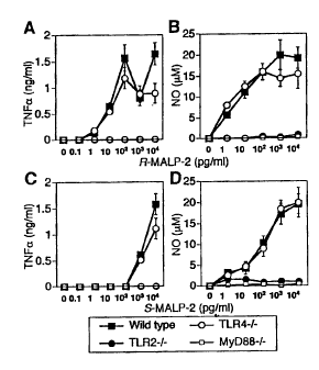
Fig. 25 is a graph showing the results of lipopeptide
MALP-2-stimulated in vitro kinase assay, Western blot analysis
and electrophoretic mobility shift assay in the TLR2 knockout
mice, the wild-type mice, the TLR4 knockout mice and the MyD88
knockout mice of the present invention.
Best Mode for Carrying out the Invention
Examples of bacterial cell components of the present
invention include: a lipoprotein/lipopeptide, which is a cell
component of bacteria which belong to Mycoplasma, Spirochaeta,
Escherichia; peptidoglycan comprised by combining repeating
polysaccharides of N-acetylglucosamine and N-acetylmuramic
acid, which is a skeletal structure of bacterial cell wall, and
relatively short peptide chain; lipopolysaccharide (LPS) which
exists mainly as an outer membrane component of Gram-negative
bacteria and is also called endotoxin; lipoteichoic acid (LTA),
which is a cell wall component of Gram-positive bacteria;
Mycobacterium tuberculosis lysate; and a cell wall fraction of
Gram-positive bacteria. Further, in the present invention,
carriers which carry the above mentioned bacterial cell
components, and the bacterial cell themselves are expediently
included in the examples of the bacterial cell components.
21
CA 02509345 2000-01-13
77513-6D
In the present invention, "unresponsiveness to bacterial
cell components" means that living organisms, or cells, tissue,
or organs which comprise living organisms show low reactivity
or almost no reactivity to the stimuli of the bacterial cell
components, and "hyporesponsiveness" means low reactivity to
the stimuli. Therefore, a model non-human animal being
unresponsive to bacterial cell components in the present
invention means a non-human animal such as a mouse, a rat, a
rabbit or the like, where living organisms, or cells, tissue,
or organs which comprise living organisms show low reactivity
or almost no reactivity to the stimuli of the bacterial cell
components. Examples of the stimuli of the bacterial cell
components include an in vivo stimulus where a bacterial cell
component is administered to a living organism and an in vitro
stimulus where a bacterial cell component is brought into
contact with cells separated from a living organism. As a
example of a model non-human animal being unresponsive to
bacterial cell components, a non-human animal unresponsive to
bacterial cell components such as a lipoprotein/lipopeptide,
peptidoglycan, a cell wall fraction of Gram-positive bacteria,
endotoxin, lipoteichoic acid, Mycobacterium tuberculosis
lysate and the like is exemplified, and specifically, a
non-human animal whose function of TLR2 gene is deficient on
its chromosome, such as a TLR2 knockout mouse and the like, and
a non-human animal whose function of MyD88 gene is deficient
on its chromosome, such as a MyD88 knockout mouse and the like
are exemplified.
In the present invention, "deficiency of MyD88 or TLR2
gene function on a chromosome" means that a part of or a whole
of MyD88 or TLR2 gene on a chromosome is deficient and the
22
CA 02509345 2000-01-13
77513-6D
function to express MyD88 or TLR2, which is expressed in
wild-types, is lost. Examples of a non-human animal whose
function of MyD88 or TLR2 gene is deficient on its chromosome
include a rodent such as a rat or the like whose function of
MyD88 or TLR2 gene is deficient other than MyD88 or TLR2 knockout
mice.
The term "a wild-type non-human animal" in the present
invention means a non-human animal being the same species of
the non-human animal whose function of MyD88 or TLR2 gene is
deficient on its chromosome. For example, in case of mice, it
means MyD88- or TLR2-nondeficient type mice of same species
among F2 mice generated at the expected Mendelian ratio. When
the deficient type and the wild-type mice of these F2 mice, in
particular, the wild-type littermates are used for experiments
simultaneously, it becomes possible to conduct precise
comparative experiments at individual level. With an example
of knockout mice which have deficiency in MyD88 or TLR2, a
generating method of the non-human animal whose function of
MyD88 or TLR2 gene is deficient on its chromosome will now be
explained.
MyD88 or TLR2 gene can be cloned by amplifying a mouse
genomic library by PCR or other methods with a probe derived
from a mouse EST clone or the like. By DNA recombination
technique, a part of or a whole of this cloned MyD88 or TLR2
gene, for example, a part or a whole of an exon region containing
a cytoplasmic region of MyD88 or TLR2 gene is replaced with
a poly A signal and a marker gene such as a neomycin resistance
gene or the like, a targeting vector is constructed by inducing
gene such as diphtheria toxin A fragment (DT-A) gene or herpes
simplex virus thymidine kinase (HSV-tk) gene or the like into
23
CA 02509345 2000-01-13
77513-6D
5'-terminal side, this constructed targeting vector is
linearized, and introduced into embryonic stem cells (ES cells)
by electroporation method or the like, then cultured, and.
subsequently ES cells achieving homologous recombination by
G418, ganciclovir (GANC) or other such antibiotics are selected.
It is preferable to confirm whether these selected ES cells are
the object. recombinants by Southern blot analysis or the like.
Chimeric mice can be obtained by microinjecting the
recombined ES cells into blastocysts of mice, and put the
blastocysts back into uteri of recipient mice. Under high
chimeric ratio, there will be born much more male chimeric mice
than female ones. In such case, heterologous recombinant mice
(+/-: Fl) are generated by intercrossing the chimeric mice with
female wild-type mice, and the homologous recombinant mice [F2;
wild-type mice (+/+), MyD88 or TLR2 knockout mice (-/-)] can
be obtained by mating the heterologous recombinant male mice
and female mice. All of these mice are generated at the expected
Mendelian ratio. As the method of confirming whether MyD88 or
TLR2 knockout mice of the present invention are born, for
example, the method wherein RNA is isolated from peritoneal
macrophages of mice obtained by the above-stated method, and
is examined by Northern blot analysis or the like, and the method
wherein the expression of MyD88 or TLR2 in the mice is examined
by Western blot analysis or the like are exemplified.
It is possible to confirm that the generated MyD88
knockout mice are unresponsive to cell wall components of
bacteria, for example, by contacting a lipoprotein/lipopeptide,
which is a cell component of bacteria which belong to Mycoplasma,
Spirochaeta, Escherichia or the like with macrophages of MyD88
knockout mice or human monocytes in vitro, and then measuring
24
CA 02509345 2000-01-13
77513-6D
the production amount of TNF- a or N02_ in the cells; by injecting
LPS, which is a cell wall component of Gram-negative bacteria
into MyD88 knockout mice by intravenous injection or the like,
and then measuring bioactivity of endotoxin such as fever, shock,
decrease of leukocytes or platelet, hemorrhagic necrosis of
bone marrow cells, hypoglycemia, induction of IFN, activation
of B limphocyte (immune response cell derived from marrow) or
the like; by measuring the induction of IFN, proliferative
response of splenic B cells, the expression'of MHC class II
antigen on the surface of splenic B cells, in macrophages or
splenic B cells of MyD88 knockout mice, in the presence of LPS
derived from bacteria, or peptidoglycan, which is a cell
component of Gram-positive bacteria, lipotheichoic acid,
Mycobacterium tuberculosis lysate or the like.
The MyD88 knockout mice of the present invention are
unresponsive to a lipoprotein/lipopeptide, which is a bacterial
cell component, and show lower responsiveness to endotoxin than
C3H/HeJ mice, which have been known as being hyporesponsive to
endotoxin so far, and no shock symptom has been observed.
Moreover, macrophages and splenic B cells of MyD88 knockout mice
are unresponsive not only to endotoxin but also to peptidoglycan
being a cell wall component of Gram-positive bacteria,
lipotheichoic acid, Mycobacterium tuberculosis lysate and the
like, while they are responsive to IL-4 and IFN- T . Therefore,
the knockout mice being unresponsive to bacterial cell
components can be used as useful model for elucidating action
mechanisms of a lipoprotein/lipopeptide, endotoxin,
peptidoglycan, lipotheichoic acid or the like, and for
establishing a treatment method for endotoxin shock.
Further, the generated TLR2 knockout mice can be
CA 02509345 2000-01-13
77513-6D
confirmed to be unresponsive to cell wall fractions of bacteria,
for example, by contacting a lipoprotein/lipopeptide, which is
a cell component of bacteria which belong to Mycoplasma,
Spirochaeta, Escherichia or the like with macrophages of TLR2
knockout mice or human monocytes in vitro, and then measuring
the production amount of TNF- a or N02 in the cells; by measuring
the induction of TNF, proliferative response of splenocytes,
the expression of MHC class II antigen on the surface of splenic
B cells and the like, in macrophages or splenic B cells of TLR2
knockout mice, in the presence of cell wall fractions of
Gram-positive bacteria, peptidoglycan, which is a cell wall
component of Gram-positive bacteria or the like. The TLR2
knockout mice of the present invention are unresponsive to a
lipoprotein/lipopeptide, which is a bacterial cell component,
and peptidoglycan, and hyporesponsive to cell wall fractions
of Gram-positive bacteria, and responsive to LPS, LTA and IL-4.
Therefore, the TLR2 knockout mice can be used as useful model
for elucidating action mechanisms of a lipoprotein/lipopeptide,
peptidoglycan, cell wall fractions of Gram-positive bacteria
or the like.
The non-human animals being unresponsive to bacterial
cell components of the, present invention can be used for
screening of a suppressor or a promoter of bacterial infection,
a suppressor or a promoter of responsiveness to bacterial cell
components such as an agonist or an antagonist to TLR2, for
assessment of bioactivity of various subject materials, for
detection of bacterial cell components, and the like, other than
for elucidating action mechanisms of bacterial cell components.
A screening method of a suppressor or a promoter of bacterial
infection or a suppressor or a promoter of responsiveness to
26
CA 02509345 2000-01-13
77513-6D
bacterial cell components such as an agonist or an antagonist
to TLR2 will be explained below with examples of the screening
method of a suppressor or a promoter of bacterial infection.
Followings are exemplified as examples. The screening
method of preventives and immune response
restoratives /promoters and the like in bacterial infection
comprising the steps of: macrophages, splenocytes or the like
obtained from the non-human animal being unresponsive to
bacterial cell components of the present invention and a subject
material are brought into contact in advance in vitro; the
macrophages or the splenocytes are cultured in the presence of
bacterial cell components; the macrophage activity level or the
splenocyte activity level of the macrophages or of the
splenocytes is measured and assessed, and the screening method
of remedies, symptom improvers and the like for bacterial
infection comprising the steps of: macrophages or splenocytes
obtained from the non-human animal being unresponsive to
bacterial cell components of the present invention and
bacterial cell components are brought into contact in advance
in vitro; the macrophages or the splenocytes are cultured in
the presence of a subject material; the macrophage activity
level or the splenocyte activity level of the macrophages or
of the splenocytes is measured and assessed.
In addition, the examples include the screening method
of preventives and immune response restoratives /promoters and
the like for bacterial infection comprising the steps of: a
subject material is administered in advance to the non-human
animal being unresponsive to bacterial cell components of the
present invention; macrophages or splenocytes obtained from the
non-human animal are cultured in the presence of bacterial cell
27
CA 02509345 2000-01-13
77513-6D
components; the macrophage activity level or the splenocyte
activity level of the macrophages or of the splenocytes is
measured and assessed, and the screening method of preventives
and immune response restoratives/promoters and the like for
bacterial infection comprising the steps of: a subject material
is administered in advance to the non-human animal being
unresponsive to bacterial cell components of the present
invention; the non-human animal is made to be infected with
bacteria; the macrophage activity level or the splenocyte
activity level of the macrophages or of the splenocytes obtained
from the non-human animal is measured and assessed.
Further, the screening method of remedies, symptom
improvers and the like for bacterial infection comprising the
steps of: the non-human animal being unresponsive to bacterial
cell components of the present invention is made to be infected
with bacteria in advance; macrophages or splenocytes obtained
from the non-human animal are cultured in the presence of a
subject material; the macrophage activity level or the
splenocyte activity level of the macrophages or of the
splenocytes is measured and assessed, and the screening method
of remedies, symptom improvers and the like for bacterial
infection comprising the steps of: the non-human animal being
unresponsive to bacterial cell components of the present
invention is made to be infected with bacteria in advance; a
subject material is administered to the non-human animal; the
macrophage activity level or the splenocyte activity level of
the macrophages or of the splenocytes obtained from the
non-human animal is measured and assessed are exemplified.
Furthermore, the screening method of preventives and
immune response restoratives/promoters and the like for
28
CA 02509345 2000-01-13
77513-6D
bacterial infection comprising the steps of: a subject material
is administered in advance to the non-human animal being
unresponsive to bacterial cell components of the present
invention; the non-human animal is made to be infected with
bacteria; the macrophage activity level or the splenocyte
activity level in the non-human animal is measured and assessed,
and the screening method of remedies, symptom improvers and the
like for bacterial infection comprising the steps of: the
non-human animal being unresponsive to bacterial cell
components of the present invention is made to be infected with
bacteria in advance; a subject material is administered to the
non-human animal; the macrophage activity level or the
splenocyte activity level in the non-human animal is measured
and assessed are exemplified.
As a method of measuring and assessing the macrophage
activity level, the method of measuring and assessing the
production amount of cytokine and/or nitrous ion in the
macrophage is exemplified, and as a method of measuring and
assessing the splenocyte activity level, a method of measuring
and assessing the expression amount of MHC class II in the
splenocyte is exemplified. In the measurement and the
assessment of the macrophage activity level or the splenocyte
activity level, it is preferable to assess the levels in
comparison to the measured value of a wild type non-human animal,
in particular, a littermate wild type non-human animal of the
non-human animal being unresponsive to bacterial cell
components as control, because there will be no dispersion
caused by individual differences . This can be applied to the
assessment of bioactivity of various subject materials and
detection of bacterial cell components and the like, in which
29
CA 02509345 2000-01-13
77513-6D
the non-human animal being unresponsive to bacterial cell
components of the present invention is used.
Examples of a suppressor or a promoter, which is the
object of the screening methods of the present invention,
include a suppressor or a promoter of responsiveness to
bacterial cell components such as a lipoprotein/lipopeptide
derived from a cell component of bacteria which belong to
Mycoplasma, Spirochaeta or Escherichia, peptidoglycan,
endotoxin, lipoteichoic acid, Mycobacterium tuberculosis
lysate and the like, and a suppressor or a promoter of
interleukin-1 activity, interleukin-18 activity, IFN- 'activity, TNF- a
activity and the like, other than the
suppressor or the promoter of bacterial infection, or the
agonist or the antagonist to TLR2.
Though the screening of an agonist or an antagonist to
TLR2 can be performed in the same manner as the screening of
the suppressor or the promoter of bacterial infection as
aforementioned, it is also possible to use TLR4 knockout mice
together. In other words, it is possible to conduct the
screening of the agonist or the antagonist to TLR2 and/or TLR4
by administering a subject material to each of TLR2 and TLR4
knockout mice, and to wild-type mice if necessary, and by
comparing and assessing the activity levels of macrophages or
splenocytes derived from the TLR2 knockout mice and the TLR4
knockout mice.
The assessing method of a subject material of the present
invention is characterized by that the subject material is
administered to the non-human animal being unresponsive to
bacterial cell components of the present invention and then the
bioactivity of the subject material is assessed. The
CA 02509345 2000-01-13
77513-6D
bioactivity of the subject material, for example, endotoxin
activity, interleukin-1 activity, interleukin-18 activity and
the like can be assessed by the assessing method of the subject
materials of the present invention. For instance, by precisely
assessing endotoxin activity of a subject material with MyD88
knockout mice of the present invention, it becomes possible to
obtain useful information for developing antagonists to
endotoxin or other such pharmaceuticals which can suppress the
shock or fever caused by endotoxin.
The relationship between IL-1 and the illness in disease
model mice can be examined by precisely assessing IL-1 activity
of a subject material with MyD88 knockout mice of the present
invention. It becomes possible to obtain useful information
for developing pharmaceuticals which can cure diseases such as
rheumatoid arthritis caused by overexpression of IL-1, a
graft-versus-host disease, asthma and the like by precisely
assessing IL-1 activity of a subject material and by analyzing
the involvement of IL-1 in disease model mice. Examples of IL-1
activity as an object of assessment include mitogens such as
phytohemagglutinin (PHA), concanavalin A (Con A) and the like,
proliferation inducing activity of T cells caused by co-
stimulation with IL-2 at a low concentration, and activity which
induces the production of TNF- a, IL-i and IL-6 by working on
monocytes and macrophages.
Moreover, by precisely assessing IL-1 activity of a
subject material with MyD88 knockout mice of the present
invention, it becomes possible to obtain useful information for
developing pharmaceuticals which can cure diseases caused by
overproduction of IL-18, such as I type diabetes, a graft-
versus-host disease and the like. Examples of IL-18 activity
31
CA 02509345 2000-01-13
77513-6D
as an object of assessment include activity which promotes
production of IFN-7, activity which enhances activity of NK
cells, activity which induces production of IFN- 7 from T cells
in cooperation with IL-12, and action which activates IRAK or
NF-KB.
With the method of detecting the bacterial cell
components of the present invention, it is possible to detect
insubstantial amount of bacterial cell components contained in
subject materials in the non-human animal being unresponsive
to bacterial cell components of the present invention after the
subject material has been administered to the non-human animal.
The examples of such bacterial cell components include; a
lipoprotein/lipopeptide derived from a cell component of
bacteria which belong to Mycoplasma, Spirochaeta, Escherichia
and the like; endotoxin derived from Escherichia coli,
Klebsiella pneumoniae, Pseudomonas aeruginosa, Salmonella
typhimurium, Serratia marcescens, Shigella flexneri, Vibrio
cholerae, Salmonella minnesota, Porphyromonas gingivalis and
the like; peptidoglycan derived from Staphylococcus aureus,
Corynebacterium diphtheriae, Nocardia coeliaca and the like;
lipoteichoic acid derived from Streptococcus pneumoniae and the
like; and whole cell lysates of Mycobacterium tuberculosis.
The present invention will be explained more specifically
with examples below, but the technological scope of the present
invention is not limited to these examples.
Example 1 (Generation of MyD88 knockout mice)
A MyD88 gene was screened from a 129/SvJ mouse genomic
library (Stratagene), subcloned into pBluescript vector
(Stratagend), and characterized by restriction enzyme mapping
and DNA sequencing. A targeting vector was constructed by
32
CA 02509345 2000-01-13
77513-6D
replacing the 1.0 kb genomic fragment of the wild-type allele
with a neomycin resistance gene from pMCl-neo (Stratagene).
The replaced genomic fragment contained 2 exons encoding the
domain that resembles the cytoplasmic domain of the IL-1RAcP
(receptor accessory protein). The neomycin resistance gene
was flanked by the 1.l.kb 5' genomic fragment and the 5.2 kb
3' fragment. Then, an HSV-tk cassette was introduced into the
3' end of the genomic fragment. E14.1 ES cells were transfected
with the linearized targeting vector and selected with G418 and
gancyclovir. Doubly resistant 176 clones were screened for
homologous recombination by PCR and 33 clones were verified by
Southern blot analysis using the probe indicated in Fig. 1.
Three independently identified targeted ES clones were
microinjected into the blastocysts of C57BL/6 mice. Thus
obtained chimeric mice were mated with C57BL/6 female mice to
produce heterozygous mice. The Heterozygous mice were
intercrossed to obtain homozygotes, and MyD88-deficient were
born at the expected Mendelian ratios (+/+:+/-:-/- = 52:93:53)
from the intercross. The MyD88 knockout mice of the present
invention grew healthy and showed no obvious abnormalities
until 20 weeks of age. Northern blot analysis was performed
to confirm that the inactivation of the MyD88 gene was caused
by mutation. MyD88 mRNA could not be detected in the liver and
the spleen of the MyD88-deficient mice. Flow cytometric
analysis of CD3, B220, CD4, and CD8 in thymus, spleen, and lymph
node showed that lymphocyte composition was not altered in the
MyD88 knockout mice in comparison with wild-type mice.
Example 2 (Unresponsiveness of MyD88 knockout mice to
Endotoxin)
1mg of LPS derived from Escherichia coli (055:B5) was
33
CA 02509345 2000-01-13
77513-6D
administered to 10 MyD88 knockout mice of the present invention,
and endotoxin-unresponsiveness was examined through the
survival ratio of the mice. 10 wild-type littermates were used
as control. The results are shown in Fig. 2. It is confirmed
by Fig. 2 that though the wild-type mice have responded to LPS
and all of them died within 4 days after administration, none
of the MyD88 knockout mice of the present invention have died
within 4 days after LPS administration, and that the mice are
endotoxin-unresponsive.
Example 3 (Impaired IL-1-mediated functions in MyD88 knockout
mice)
1 x 10⁵ of thymocytes of the MyD88 knockout mice of the
present invention were cultured in 96-well plates for 72 hours
with mixtures containing 2 pg/ml of phytohemagglutinin (PHA),
which is a costimulant of IL-1 for T cell proliferation, 2.5
pg/ml of concanavalin A (ConA), or 2 ng/ml of IL-2 respectively,
and 100 U/ml of IL-1 0 (Genzyme) , and T cells were proliferated.
Proliferation of T cells were examined by measuring [3H] amount
of [3 H] thymidine taken into the cells. Asa result, thymocytes
of wild-type littermates displayed enhanced proliferation when
cultured with PHA, ConA or IL-2 in the presence of IL- 0 , however,
thymocytes of the MyD88 knockout mice of the present invention
show almost no enhanced proliferation (see Fig. 3) . It has been
found that similar results could be obtained even when splenic
B cells were used instead of thymocytes.
Further, thymocytes of MyD88 knockout mice of the present
invention were cultured with 10 ng/ml of phorbol 12-myristate
13-acetate paramethoxyamphetamine (PMA) or 2.5 pg/ml of Con A
in the presence of 20 ng/ml of IL-2 (Genzyme) in a same manner
as above-mentioned, and enhancement of proliferation was
34
CA 02509345 2000-01-13
77513-6D
examined. There was no difference between thymocytes of MyD88
knockout mice of the present invention and of wild-type
littermates in their proliferation as to the reaction of IL-2
and PMA or Con A (see Fig. 3). These results indicate that
IL-1-mediated growth signal of T cells was impaired in the
thymocytes of MyD88 knockout mice of the present invention.
MyD88 knockout mice of the present invention were
intravenously injected with 1 jig of IL- S (Genzyme), and 2 hours
later liver and sera were taken. Total RNA was extracted from
the liver using Trizol reagent (GIBCO). This RNA (10 g) was
electrophoresed and transferred to a nylon membrane, then
Northern blot analysis was conducted with 32P-labelled cDNA for
acute phase proteins such as serum amyloid A (SAA-I), serum
amyloid P(SAP), and haptoglobin (HP). In comparing IL-i-
induced increase of mRNA expression in wild-type littermates
and in MyD88 knockout mice of the present invention, increase
of induction was observed in wild-type mice, but not observed
in MyD88 knockout mice.
Because IL-1 induces production of acute phase proteins
such as tumor necrosis factor (TNF- a ) or IL-6, and
proinflammatory cytokines, increase of TNF- a and IL-6
concentrations in serum taken from MyD88 knockout mice of the
present invention and wild-type littermates by the above-stated
method were measured by ELISA. As a result, TNF-a and IL-6
concentrations increased by IL-10 in wild-type mice, while
neither TNF-a nor IL-6 concentration increased by IL-118 in
MyD88 knockout mice (see Fig.4).
Thus, IL-1-mediated major biological functions has been
found to be severely impaired in MyD88 knockout mice of the
present invention.
CA 02509345 2000-01-13
77513-6D
Example 4 (Impaired IL-18-mediated functions in MyD88 knockout
mice)
It has been well known that IL-18 enhances lytic activity
of NK cells. Splenic B cells from MyD88 knockout mice of the
present invention and wild-type littermates were cultured in
the presence or absence of 20 ng/ml of IL-18 (Hayashibara
Biochemical Laboratories, Inc.) for 24 hours with 51Cr -labelled
mouse lymphoma cells (hereinafter "YAC-1") targeting cells. 4
hours later, released51Cr in supernatants were counted by a
gamma counter. As a result, when splenic B cells were cultured
in the presence of IL-18 in vitro, lytic activity to YAC-1
targeting cells was dramatically enhanced in wild-type mice,
but it was not enhanced in MyD88 knockout mice. When IL-2 was
used instead of IL-18, lytic activity was also enhanced in
splenic B cells of MyD88 knockout mice of the present invention
(see Fig.5).
Further, splenic B cells of MyD88 knockout mice of the
present invention and their wild-type littermates were
stimulated by 20 ng/ml of IL-18 and cultured for 24 hours in
vitro, then production of IFN-7 in culture supernatants was
measured by ELISA. As a result, production of IFN- 7 was
induced in wild-type mice, however, production of IFN-7 was
not observed in MyD88 knockout mice of the present invention
(see Fig.5).
Splenic T cells of MyD88 knockout mice of the present
invention and their wild-type littermates, which were purified
to 95% or over, were cultured on anti-CD3 antibody (20
pg/ml)(PharMingen)-coated plates in the presence of 2 ng/ml
IL-12. 4 days after the onset of culture, cells were harvested
and washed with Hanks' balanced salt solution. The washed cells
36
CA 02509345 2000-01-13
77513-6D
(2 x 10⁵) were stimulated and cultured again on anti-CD3 antibody
(20 pg/ml) -coated 96-well plates for 24 hours with 20 ng/ml of
IL-18 or 2 ng/ml of IL-12. Concentration of IFN-'r in culture
supernatants was determined by ELISA and compared. The result
indicates that Splenic T cells of MyD88 knockout mice of the
present invention cannot enhance IL-18-responsive production
of IFN-7 (see Fig.6).
MyD88 knockout mice of the present invention and their
wild-type littermates were intraperitoneally injected with 500
pg of heat-killed Propionibacterium acnes (P. acnes). Seven
days after injection, T cells were purified from spleen, then
cultured and stimulated on anti-CD3 antibody (20 pg/ml) -coated
96-well plates for 24 hours in the presence or the absence of
ng/ml of IL-18. Concentration of IFN- 7 in culture
supernatants was determined by ELISA. MyD88 knockout mice of
the present invention and their wild-type littermates were
intravenously injected with 2 mg of Bacillus Calmette-Guerin
(BCG) (Kyowa) . 14 days after injection, T cells were purified
from spleen, then cultured and stimulated for 24 hours, as
20 described above, subsequently concentration of IFN- 7 was
measured. As a result, in both cases, high level of IFN-7
production in response to IL-18 was observed in wild-type mice,
but production level of IFN-7 could not be enhanced in the
presence of IL-18 in MyD88 knockout mice of the present
invention (see Fig.6).
These results demonstrate that MyD88 knockout mice of the
present invention are defective in Thl cell development in vivo
as is the case with IL-18-deficient mice, and that their major
biological activities mediated by IL-18 were completely
abolished.
37
CA 02509345 2000-01-13
77513-6D
Next, it was examined whether the dominant negative MyD88
mutant blocked IL-18-induced NF-KB activation as well. COS-7
cells were transiently transfected with MyD88 (amino acid
152-296) expression vector together with NF- KB-dependent
luciferase reporter gene, and luciferase activity after IL-
18 treatment was measured. Coexpression of MyD88 blocked
IL-18-induced activation almost completely (see Fig. 7).
Because IL-18 activates AP- 1 -dependent gene information,
whether MyD88 (amino acid 152-296) also acted as a dominant
negative mutant of IL-18-induced AP-1 activation was
investigated. Stimulation with IL-18 induced an approximately
3- to 4-fold increase in AP-1 activity, and this activation was
blocked by coexpression of MyD88 (amino acid 152-296) (see Fig.
7). These results show that MyD88 is involved in IL-18-induced
activation of both NF-,B and AP-l.
Further, whether IL-18-induced activation of NF-KB was
observed in MyD88-deficient cells was examined. Splenic T
cells cultured in the presence of IL-12 and anti-CD3 antibody
for 4 days were starved for 3 hours and then stimulated with
IL-18. Nuclei extracted from the stimulated cells were
analyzed by a gel mobility shift assay using a specific probe
containing NF- KB binding site. IL-18-induced NF- KB DNA
binding activity was detected in the nuclear extracts from
wild-type cells but not from MyD88-deficient cells. On the
other hand, treatment of wild-type or MyD88-deficient
thymocytes with TNF-a resulted in almost the same levels of
NF-KB DNA binding activity, demonstrating that the impaired
IL-18-induced NF-KB activity in MyD88-deficient cells was not
due to the abnormal function or impairment of regulating ability
of NF-KB.
38
CA 02509345 2000-01-13
77513-6D
In addition to induction of NF-KB activation, IL-1 is also
known to activate c-Jun N-terminal kinase (JNK). To test
whether IL-18 induces JNK activation, an in vitro kinase assay
was carried out using GST-c-Jun-fusion protein as a substitute.
Treatment with IL-18 induced JNK activation in Th1-developing
cells of wild-type mice. However, IL-18-induced JNK
activation was not observed in MyD88-deficient cells. By
contrast, normal activation of JNK was observed in MyD88-
deficient cells treated with TNF-a. IL-18-induced NF-KB and
JNK activation was impaired in MyD88-deficient mice. These
results demonstrate that MyD88 is essential for IL-18-induced
activation of both NF-,B and JNK.
Example 5 (Unresponsiveness of macrophages and splenic B cells
of MyD88 knockout mice to bacterial cell wall components)
5-1 (Generation of TLR4-deficienct mice)
It has recently been reported that C3H/HeJ mice are
hyporesponsive to LPS because of a missense point mutation in
the Toll-like receptor(TLR)-4 gene (Science 282, 2085-8, 1998,
J. Exp. Med. 189, 615-625, 1999, J. Immunol. 162, 3749-3752,
1999), and the inventors have demonstrated that macrophages and
splenic B cells of TLR4-deficient mice are hyporesponsive to
LPS, and that TLR4 gene, is essential for LPS signaling (J.
Immunol. 162, 3749-3752, 1999). In order to compare the
responsiveness of macrophages and splenic B cells of TLR4- and
MyD88-deficient mice to bacterial cell wall components,
TLR4-deficient mice (F2 interbred from 129/OlaXC57BL/6) were
generated by gene targeting as described previously (J. Immunol.
162, 3749-3752, 1999). Age-matched groups of wild-type, TLR4-,
and MyD88-deficient mice were used for the following examples.
5-2 (Preparation of bacterial cell wall components)
39
CA 02509345 2000-01-13
77513-6D
LPS of Escherichia coli Serotype 055:B5 (Sigma),
Klebsiella pneumoniae (Sigma), Pseudomonas aeruginosa
Serotype 10 (Sigma), Salmonella typhimurium (Sigma), Serratia
marcescens (Sigma), Shigella flexneri Serotype 1A (Sigma) and
Vibrio cholerae Serotype Inaba 569B (Sigma) and the like were
purchased. They were prepared by phenol extraction and
purified by gel filtration. LPS from Salmonella minnesota
Re-595 prepared by phenol-chloroform-petroleum ether
extraction procedure was also purchased (Sigma). LPS and Lipid
A of Porphyromonas gingivalis 381 was prepared by the method
as described previously (FEBS Lett. 332, 1994, 197-201). Whole
cell lysates of Mycobacterium tuberculosis was prepared by the
following process: Mycobacterium tuberculosis Aoyama B strain
(NIHJ 1635) was cultured in Dubos broth (DIFCO) for 1 month;
cells were collected and resuspended with phosphate buffered
saline (PBS); cells were sonicated.
5-3 (Preparation of peritoneal macrophages)
2 ml of 4% thioglycollate was intraperitoneally injected
into the generated wild-type, TLR4- and MyD88-deficient mice
respectively. Three days later, peritoneal exudate cells were
isolated from the peritoneal cavity and washed with ice-cold
Hank's buffered salt solution (HBSS), then peritoneal cells
were obtained. The cells were made to float in RPMI1640 medium,
then put in plastic plates separatedly, and cultured for 2 hours
at 37 C and washed with Hank's buffered salt solution to remove
nonadherent cells. Adherent cells were used as peritoneal
macrophages in the experiments bellow.
5-4 (Unresponsiveness to LPS of Salmonella minnesota Re-595)
CA 02509345 2000-01-13
77513-6D
Responsiveness of each peritoneal macrophage of the
wild-type (wild-type), TLR4-deficient (TLR4-/-), MyD88-
deficient (MyD88-/-) mice and the like to LPS were examined with
LPS of Salmonella minnesota Re-595. Peritoneal macrophages
from each mouse were cultured for 24 hours in the presence of
various concentrations (0, 0.01, 0.1, 1, 10 or 100 ig/ml) of
LPS and stimulated, then concentration of tumor necrosis factor
(TNF-a) released from LPS-responsive macrophages was measured
by ELISA (see Fig. 8A). By these results, it has been found
that production of TNF-a increases in response to LPS in a
dose-dependent manner in macrophages of wild-type mice, by
contrast, no production of TNF- a is observed in TLR4- or
MyD88-deficient mice even when they receive LPS stimuli at a
concentration of 100 pg/ml, and that these mice are LPS-
unresponsive.
Further, responsiveness of splenic B cells to LPS of
Salmonella minnesota Re-595 was examined. Splenic B cells (1
x 105) of each of the wild-type, TLR4- and MyD88-deficient mice
were isolated, cultured in 96-well plates and stimulated by
various concentrations (0, 0.01, 0.1, 1, 10 or 100 pg/ml) of
LPS. 1 I1Ci of [ 3H ] -thymidine (DuPont) was added 40 hours after
onset of the culture, then the cells were cultured for another
8 hours, and [3H] uptake was measured by a $ scintillation
counter (Packard) (see Fig. 8B). As a result, LPS-induced
proliferative response was promoted in response to LPS in a
dose-dependent manner in splenic B cells of wild-type mice, by
contrast, no LPS-induced proliferative response was observed
in splenic B cells of both TLR4- and MyD88-deficient mice.
41
CA 02509345 2000-01-13
77513-6D
The expression of major histocompatibility complex (MHC)
class II (I-Ab molecule) on the surface of splenic B cells in
response to Re-595 LPS was examined by flow cytometry. Splenic
B cells (1 x 10⁶) from each of the wild-type, MyD88- and
TLR4-deficient mice were cultured for 48 hours in the presence
of various concentrations (0, 0.01, 0.1, 1, 10 or 100 pg/ml)
of LPS. After the culture, the cells were collected and then
stained by combining I-Ab molecule on the surface of the cells
and FITC-labelled antibody which is constructed by combining
phycoerythrin (PE; PharMingen) -conjugated anti-B220 antibody
or biotinylated anti-mouse I-Ab antibody (PharMingen) and
fluorescein isocyanate (FITC; PharMingen)-conjugated
streptavidin. The stained cells were analyzed on
fluorescence-activated cell sorter Calibur (FACS Calibur)
using CELLQuest software (Becton Dickinson). As a result,
Re-595 LPS caused an increase in the expression of I-Ab molecule
on the surface of splenic B cells of wild-type mice. In contrast,
Re-595 LPS did not enhance I-Ab molecule expression in splenic
B cells of either TLR4- or MyD88-deficient mice, even when
stimulated with high concentration of LPS (100 pg/ml) (see Fig.
8C). The above-mentioned results indicate that both TLR4- and
MyD88-deficient mice are unresponsive to LPS of Salmonella
minnesota Re-595.
5-5 (Responsiveness of TLR4- and MyD88-deficient mice to IL-4
and IFN-7')
In order to examine whether splenic B cells of TLR4- and
MyD88-defidient mice are unresponsive to all stimuli, the
42
CA 02509345 2000-01-13
77513-6D
responsiveness of spienic B cells of TLR4- and MyD88-deficient
mice to other stimuli were investigated. The investigation
demonstrates that there was no impairment as to their
responsiveness to the stimuli as described below, and that these
mice were specifically defective in their response to LPS.
Splenic B cells (1 x 10⁵) from each of the wild-type,
MyD88- and TLR4-deficient mice were isolated, cultured for 40
hours in the presence of both IL-4 (Genzyme) and anti-IgM
antibody, or in the presence of anti-CD40 antibody, then
[3H]-thymidine (DuPont) was added and the cells were cultured
for another 8 hours, and [3H] uptake was measured by a S
scintillation counter (see Fig. 9A). As a result, splenic B
cells of both TLR4- and MyD88-deficient mice showed same
reaction as splenic B cells of wild-type mice with regard to
the response to IL-4 and the mixture of anti-IgM antibody, or
to the anti-CD40 antibody.
Next, Splenic B cells (1 x 10⁶) from each of the wild-type,
MyD88- and TLR4-deficient mice were cultured for 48 hours in
the presence or absence of 100 U/ml of IL-4, and then stimulated.
Subsequently, the cells were stained by combining I-Abmolecule
on the surface of the splenic B cells and PE-conjugated
anti-B220 antibody or FITC-conjugated anti-mouse I-A b antibody.
The cell proliferation was measured on fluorescence-activated
cell sorter Calibur using CELLQuest software (see Fig. 9B). As
a result, splenic B cells of both TLR4- and MyD88-deficient mice
showed same reaction as those of wild-type mice with regard to
the response to IL-4 as well.
43
CA 02509345 2000-01-13
77513-6D
Each of wild-type, MyD88- and TLR4-deficient mice were
intraperitoneally injected with 5000 U of IFN- T (Genzyme) or
PBS. Three days after injection, peritoneal macrophages were
collected and stained by combining I-Ab molecule on the surface
of the macrophage membranes and FITC-conjugated anti-mouse I -A b
antibody, then analyzed by fluorescence-activated cell sorter
Calibur using CELLQuest software (see Fig. 9C). The result
indicated that the expression of I-Ab molecule in peritoneal
macrophages, in other words, blockage level of IFN- T -induced
cell proliferation was comparative among wild-type, MyD88- and
TLR4-deficient mice.
5-6 (Analysis of phagocytosis)
Macrophages of wild-type, MyD88- and TLR4-deficient mice
added with 0.025 % of fluorescent latex beads (0.75 pm)
(Polyscience) were cultured for 2 hours at 37 C in a CO2
incubater. Then the culture materials were washed vigorously
three times with PBS to remove non-phagocytosed beads and
incubated with PBS containing 2.5 % of formaldehyde for 20
minutes, and fixed with formaldehyde. Visualization of these
fixed cells with Axiophoto microscope (Carl Zeiss, Inc. ) showed
that peritoneal macrophages of both TLR4- and MyD88-deficient
mice phagocytosed the latex particles, and therefore, that
phagocytic ability of the macrophages of TLR4- and MyD88-
deficient mice were not impaired by these other stimuli.
5-7 (Responsiveness to LPS of Porphyromonas gingivalis)
44.
CA 02509345 2000-01-13
77513-6D
As LPS of Porphyromonas gingivalis shows some reaction
in its ability to activate cells of LPS-hyporesponsive C3H/HeJ
mice (J. Immunol. 158, 1997, 4430-6), responsiveness of each
mouse to LPS of Porphyromonas gingivalis was examined as in the
case with Salmonella minnesota Re-595. In macrophages of
wild-type mice, TNF-a was induced in response to LPS of
Porphyromonas gingivalis in a dose-dependent manner. However,
macrophages of TLR4-deficient mice were hyporesponsive like
those of C3H/HeJ mice, and only showed the TNF- a producibility
which was about one third of that of wild-type mice macrophages.
In contrast, macrophages of MyD88-deficient mice did not
produce any detectable TNF- a, even when stimulated with high
concentration of LPS (see Fig. 10A).
Splenic B cells of TLR4-deficient mice exhibited low
level proliferative response to LPS of Porphyromonas gingivalis
381, and enhanced the expression of I-Ab molecule of spienic
B cells, however, splenic B cells of MyD88-deficient mice did
not exhibit proliferative response and the expression of I-
Ab molecule could not confirmed (see Fig. 10B and C) . Further,
the same results were obtained with lipid A of Porphyromonas
gingivalis 381. This indicates that TLR4-deficient mice are
hyporesponsive and MyD88-deficient mice are unresponsive to LPS
of Porphyromonas gingivalis. In addition, it has been found
that MyD88 is essential for the signaling induced by LPS of
Porphyromonas gingivalis, whereas TLR4 shows partial
contribution.
5-8 (Responsiveness to LPS of Escherichia coli 055:B5)
CA 02509345 2000-01-13
77513-6D
Responsiveness to LPS of Escherichia cola 055:B5 was
examined as in the case with Salmonella minnesota Re-595. The
responsiveness to LPS of Escherichia colt (055:B5)was impaired
in peritoneal macrophages of both TLR4- and MyD88-deficient
mice, compared with those of wild-type mice (Fig. 11A). However,
when stimulated with high concentration of LPS, macrophages of
TLR4-deficient mice produced a small amount of TNF-a. In
contrast, macrophages of MyD88-deficient mice did not produce
TNF-a even when stimulated with high concentration of LPS.
Similar tendencies were observed in proliferative
responses in splenic B cells of these mice (see Fig. 11B).
Furthermore, when stimulated with LPS at a concentration over
10 pg/ml, splenic t cells of TLR4-deficient mice showed a
certain expression level of I-A' molecule similar to the level
shown by splenic B cells of wild-type mice. In contrast,
splenic B cells of MyD88-deficient mice did not show I-Ab
molecule expression even when stimulated with LPS at a
concentration of 100 pg/mi (see Fig. 11C). As in the case of
stimuli with LPS of Porphyromonas gingivalis, these results
indicate that TLR4-deficient mice are hyporesponsive and
MyD88-deficient mice are unresponsive to LPS of Escherichia
colt (055:B5).
5-9 (Responsiveness to peptidoglycan)
It has been reported that Peptidoglycan (PGN), which is
a major cell wall component of Gram-positive bacteria,
activates macrophages (J. Immunol. 155, 1995, 2620-30, Infect.
Immun. 62, 1994, 2715-21). Therefore, responsiveness to PGN
46
CA 02509345 2000-01-13
77513-6D
of Staphylococcus aureus (Fluka) was examined as in the case
with Salmonella minnesota Re-595. When stimulated with PGN,
peritoneal macrophages of TLR4-deficient mice produced TNF-
a in a dose-dependent manner to almost the same extent as
macrophages of wild-type mice. In contrast, macrophages of
MyD88-deficient mice did not produce TNF-a even when
stimulated with high concentration of PGN (see Fig. 12A).
When stimulated with PGN of Staphylococcus aureus,
splenic B cells of wild-type mice displayed proliferative
responses, and the proliferative response was severely impaired
in peritoneal macrophages of MyD88-deficient mice compared with
those of wild-type mice, but in TLR4-deficient mice, the
proliferative response was not severely impaired as in
MyD88-deficient mice (see Fig. 12B). Further, when stimulated
with PGN at a concentration over 10 pg/mi, splenic B cells of
TLR4-deficient and wild-type mice showed enhancement of I-Ab
molecule expression. In contrast, splenic B cells of
MyD88-deficient mice did not show enhancement of I-Ab molecule
expression even when stimulated with PGN at a concentration of
100 pg/ml (see Fig. 12C). Thus, TLR4-deficient mice showed
almost the same response to PGN of Staphylococcus aureus as
wild-type mice, while MyD88-deficient mice showed no
responsiveness.
5-10 (Responsiveness to lipoteichoic acid)
As lipoteichoic acid (LTA) is a cell wall component of
Gram-positive bacteria and induces activation of monocytes and
macrophages (Infect. Immun. 62, 1994, 2715-21), responsiveness
47
CA 02509345 2000-01-13
77513-6D
to LTA of Streptococcus pneumoniae was examined as in the case
with Salmonella minnesota Re-595. Peritoneal macrophages of
wild-type mice increased production of TNF-C1 in response to
LTA in a dose-dependent manner. In contrast, macrophages of
MyD88-deficient mice did not produce TNF-CL even when
stimulated with high concentration of LTA. In comparison with
peritoneal macrophages of wild-type mice, TNF- a production was
also impaired in those of TLR4-deficient mice, however, TNF-
Cr was induced when stimulated with 100 pg/ml of LTA (see Fig.
13A).
Next, proliferative responses and enhancement of I-A
molecule expression in splenic B cells of these mice in response
to stimulation from LTA of Streptococcus pneumoniae was
analyzed (see Fig. 13B). The results indicated that splenic
B cells of wild-type mice enhanced their response to LTA in a
dose-dependent manner, whereas splenic B cells of MyD88-
deficient mice exhibited a severely defective proliferative
response to LTA. Though splenic B cells of TLR4-deficient mice
also exhibited an impaired proliferative response, they
exhibited proliferative response when stimulated with high
concentration of LTA. Further, in splenic B cells of wild-
type and TLR4-deficient mice, enhancement of I-An molecule
expression was also observed on the cell surface, whereas no
enhancement was observed in those of MyD88-deficient mice (see
Fig. 13C). This indicates that MyD88-deficient mice are
unresponsive to stimulation from LTA of Streptococcus
pneumoniae.
48
CA 02509345 2000-01-13
77513-6D
5-11 (Responsiveness to whole cell lysates of Mycobacterial
tuberculosis)
As cell wall components of Mycobacterial tuberculosis,
especially lipoarabinomannan, are known to induce activation
of myeloid cells (J. Immunol. 149, 1992, 541-7, J. Clin. Invest.
91, 1993, 2076-83), responsiveness to whole cell lysates of
Mycobacterial tuberculosis was examined as in the case with
Salmonella minnesota Re-595. Macrophages of wild-type mice
produced TNF-a in response to the whole lysates in a dose-
dependent manner. Macrophages of TLR4-deficient mice also
exhibited TNF-a production though the production amount was
smaller than those of wild-type mice. However, macrophages of
MyD88-deficient mice did not produce TNF- a in response to the
whole cell lysates of Mycobacterial tuberculosis at a high
concentration (see Fig. 14A).
Next, responsiveness of these mice to stimulation from
whole cell lysates of Mycobacterial tuberculosis was examined.
Splenic B cells of wild-type mice exhibited enhancement of
proliferative responses and I-Ab molecule expression on the
surface of the cells in response to the whole cell lysates in
a dose-dependent manner. Splenic B cells of TLR4-deficient
mice also showed proliferative responses and I-Ab molecule
expression, although these responses were lower than those of
splenic B cells of wild-type mice. In contrast, splenic B cells
of MyD88-deficient mice displayed severely impaired
proliferative responses and enhancement of I-Ab molecule
49
CA 02509345 2000-01-13
77513-6D
expression, indicating that they are unresponsive to the whole
cell lysates (see Fig. 14B and C).
5-12 (Responsiveness to other bacterial cell wall components)
Responsiveness of wild-type, TLR4- and MyD88-deficient
mice to other bacterial cell wall components [LPSs of Klebsiella
pneumoniae, Pseudomonas aeruginosa 10, Salmonella typhimurium,
Shigella flexneri, Vibrio cholerae and the like, and PGN of
Staphylococcus epidermidis, which is provided from Shigeo
Kawata of Dainippon Pharmaceutical Co. ] was examined in a same
manner as aforementioned. The results are shown in Table 1.
This Table 1 shows that MyD88-deficient mice are unresponsive
to all bacterial cell components.
CA 02509345 2000-01-13
77513-6D
Table 1
Sample Responsiveness of mice
LPS wild-type TLR4-/- MyD88-/-
Escherichia coli 055:B5 ++ + -
Klebsiella pneumoniae ++ - -
Porphyromonas gingivalis ++ + -
Pseudomonas aeruginosa ++ + -
Salmonella minnesota Re595 ++ - -
Salmonella typhimurium ++ + -
Serratia marcescens ++ + -
Shigella flexneri ++ + -
Vibrio cholerae ++ + -
PGN
Staphylococcus aureus ++ ++ -
Staphylococcus epidermidis ++ + -
LTA
Streptococcus faecalis ++ + -
whole cell lysates of Mycobacterium tuberculosis
Mycobacterium tuberculosis ++ + -
It has been found that LPS can be classified into two
types : one type includes LPSs which utilize TLR4 as their unique
signaling receptor and show unresponsiveness (LPSs of
Salmonella minnesota Re595, Klebsiella pneumoniae and the
like); another type includes LPSs which show hyporesponsiveness
to TLR4-deficient mice (LPSs of Porphyromonas gingivalis,
51
CA 02509345 2000-01-13
77513-6D
Escherichia cols 055:B5, Pseudomonas aeruginosa, Shigella
f lexneri, Salmonella typhimurium, Vibrio cholerae and the like).
Since MyD88-deficient mice show no responsiveness to these
latter LPSs, it is presumed that the recognition and signaling
of these LPSs are mediated by both TLR4 and other TLRs, and/or
by TLR-related receptors that use MyD88 as an adaptor molecule.
Example 6 (Generation of TLR2 knockout mice)
TLR2 gene was screened from 129/SvJ mouse genomic library
(Stratagene) using a probe derived from a mouse EST clone
(accession number D77677) similar to human TLR2 gene, and
subcloned into pBluescript vector (Stratagene), then
characterized by restriction enzyme mapping and DNA sequencing.
A targeting vector was constructed by replacing a gene fragment
at an exon region 1.3 kb containing cytoplasmic domain of TLR2
gene with pMC1-neo (Stratagene) having Poly A signal. The
targeting vector was flanked by a 4.8 kb 5' genomic fragment
and a 1.0 kb 3' fragment and contained an HSV-tk cassette at
the 5' terminal. The targeting vector was linearized with Sall
and electroporated into E14.1 embryonic stem cells (ES cells).
120 clones resistant to G418 and gancyclovir were screened for
homologous recombination by PCR and 9 clones were confirmed by
Southern blot analysis using the probe indicated in Figure 15A.
Chimeric mice were generated by microinjection of 3
targeted ES clones containing a homologously recombined mutant
TLR2 allele into blastocysts of C57BL/6 mice. Male chimeric
mice were bred to C57BL/6 females to produce heterozygous mice.
The heterozygous mice were intercrossed to obtain homozygotes
52
CA 02509345 2000-01-13
77513-6D
(Fig 15B) . TLR2-deficient mice of the present invention could
be generated at the expected Mendelian ratio, and did not show
any obvious abnormality until 20 weeks.
To confirm that the homologous recombination caused
inactivation of the TLR2 gene, total RNA (15 pg) was extracted
from peritoneal macrophages (5 x 10⁶) of wild-type (+/+) and
TLR2 knockout (-/-) mice and then electrophoresed, transferred
to a nylon membrane, and Northern blot analysis was conducted
using cDNA specific to [32P]-labelled TLR2, or cDNA specific
to GAPDH (glycelaldehyde-3-phosphate dehydrogenase) as the
method previously described (Immunity 9, 143-150, 1998). As
a result, TLR2 mRNA was not detected in peritoneal macrophages
of TLR2-deficient mice (Fig 15C). In addition, it was shown
that the expressions of CD3, B220, CD4, and CD8 in thymocytes
and splenocytes of TLR2 knockout mice were not different from
those of wild-type mice (data not shown).
Example 7 (Responsiveness of TLR2 knockout mice to endotoxin)
1 mg of LPS derived from Escherichia coli (055:B5) was
injected into each of TLR2 knockout mice (n-5), TLR4 knockout
mice (n=5) and wild-type mice (n=5), and LPS
unresponsiveness was examined by their survival rate.
The results are shown in Fig. 16. Fig. 16 confirms that
though TLR2 knockout mice (TLR2-/-) and wild-type mice
responded to LPS and almost all of them died within
4 days after injection, none of TLR4 knockout mice (TLR4-/-)
died even after 6 days after injection, and that TLR4 knockout
mice are unresponsive to endotoxin.
Example 8 (Responsiveness of TLR2 knockout mice to cell
components of Gram-negative bacteria)
53
CA 02509345 2000-01-13
77513-6D
Each of TLR2 knockout (TLR2-/-), TLR4 knockout (TLR4-
/-) and wild-type (wild-type) mice were intraperitoneally
injected with 2 ml of 4% thioglycollate medium (DIFCO). Three
days later, peritoneal exudate cells were isolated from the
peritoneal cavity of each mouse. These cells were cultured in
RPMI1640 medium (GIBCO) supplemented with 10% fetal bovine
serum (GIBCO) for 2 hours at 37 C and washed with ice-cold Hank's
buffered salt solution (HBSS; GIBCO) to remove nonadherent
cells. Adherent cells were used as peritoneal macrophages for
following experiments.
Each of obtained peritoneal macrophages were cultured for
24 hours with 1.0 ng/ml of synthetic lipid A derived from
Escherichia coli (compound 506; Daiichi Pure Chemicals) or LPS
derived from Salmpnella minnesota Re-595 (Sigma) in the
presence or absence of IFN- 7 (30 unit/ml) . Synthetic lipid A,
which was soluble in endotoxin-free water and containing 0.025%
of triethylamine, was used as said synthetic lipid A. After
the culture, production amounts of IL-6 (Fig. 17A), TNF- a (Fig.
17B) and NO,- (Fig. 17C) in culture supernatants were measured.
Production amount of IL-6 was measured by enzyme-linked
immunosorbent assay (ELISA; ENDOGEN), and that of TNF-a was
measured by ELSIA, according to manufacturer(Genzyme)'s
instructions, and that of N02 was measured by the Greiss method
using NO./N03 Assay Kit (Dojindo Laboratories).
These results indicate that macrophages of wild-type mice
and TLR2 knockout mice showed similar responsiveness to LPS and
lipid A, and produced IL-6 and TNF- a, and it was confirmed that
production of TNF- a would be further increased when IFN- 7 was
added to LPS or lipid A before the culture. By contrast,
macrophages of TLR4 knockout mice produced neither IL- 6 nor TNF-
54
CA 02509345 2000-01-13
77513-6D
(x . Further, production of N02- was confirmed by culturing
macrophages of wild-type and TLR2 knockout mice with IFN-T
-added lipid A or LPS. The obtained results were same as those
aforementioned even in the case the injection amount of lipid
A or LPS was arranged to be 1 pg/ml (data not shown).
Next, each of peritoneal macrophages of wild-type, TLR2
knockout and TLR4 knockout mice were cultured in the presence
of LPS derived from Salmonella minnesota Re-595 at various
concentrations shown in Fig. 17D, and production of TNF- a was
measured. The results indicate that macrophages of wild-type
mice and TLR2 knockout mice showed similar tendency to increase
in response to LPS in a dose-dependent manner, while macrophages
of TLR4 knockout mice produced no TNF-a in response to any
concentration of LFS.
Example 9 (Responsiveness to LPS of Salmonella minnesota
Re-595)
Responsiveness of splenocytes of various mice (wild-type,
TLR2-/- and TLR4-/-) to LPS of Salmonella minnesota Re-595 were
examined. Splenocytes (1 x 10³) of each mouse were isolated
and then cultured and stimulated in 96-well plates with various
concentrations of LPS shown in Fig. 18A. 1 pCi of [3H] -thymidine
(DuPont) was added 40 hours after the onset of the culture, and
cells were further cultured for 8 hours, then [ 3H ] uptake was
measured by a 0 scintillation counter (Packard) (Fig. 18A).
As a result, the cell proliferative response was promoted in
response to LPS in a dose-dependent manner in splenocytes of
wild- type and TLR2 knockout mice as well. By contrast, whatever
the concentration of LPS as a stimulus would be, no LPS-induced
cell proliferative response was observed in splenocytes of
TLR4-deficient mice.
CA 02509345 2000-01-13
77513-6D
In addition, the expression of major histocompatibility
complex (MHC) class II (I-Ab) on the surface of B cells in
response to Re-595 LPS was examined by flow cytometry. Splenic
B cells (1 x 10⁵) of each of wild-type, TLR2 knockout (2-/-)
and TLR4 knockout (4-/-) mice were isolated and cultured for
48 hours in 96-well plates with various concentrations (0, 101,
102, 103, 104 or 105 ng/ml) of LPS or 100 U/ml of IL-4. After
the culture, the cells were collected and stained by combining
I-Ab molecule on the surface of the cells and FITC-labelled
antibody which is constructed by combining phycoerythrin (PE;
PharMingen)-conjugated anti-B220 antibody or biotinylated
anti-mouse I-Ab antibody (PharMingen) and fluorescein
isocyanate (FITC; PharMingen) -conjugated streptavidin. The
stained cells were analyzed on fluorescence-activated cell
sorter Calibur (FACS Calibur) using CELLQuest software (Becton
Dickinson) (Fig. 18B). As a result, Re-595 LPS enhanced the
expression of I-Ab molecule on the surface of splenic B cells
of wild-type and TLR2 knockout mice. In contrast, I -Ab molecule
expression in splenic B cells of TLR4-deficient mice were not
enhanced by Re-595 LPS, even when stimulated with high
concentration of LPS (105ng/ml). The above-mentioned results
indicate that TLR2 knockout mice show responsiveness to LPS as
wild-type mice did. When stimulated with IL-4, each knockout
mice show normal expression of I-Ab molecule on the surface of
splenic B cells.
Example 10 (Unresponsiveness of macrophages of TLR2 knockout
mice to cell wall components derived from Gram-positive
bacteria)
Responsiveness of each peritoneal macrophages of said
wild-type (wild-type), TLR2 knockout (TLR2-/-), TLR4 knockout
56
CA 02509345 2000-01-13
77513-6D
(TLR4 - / - ) mice and the like to cell wall components derived from
Gram-positive bacteria were examined with prepared cell wall
specimens of S. aureus, C. diphtheriae and N. coeliaca. The
cell specimens were prepared in accordance with the method
previously described (Biken J. 18, 77-92, 1975, Infect. Immun.
38, 817-824, 1982), that is, bacterial cells grown under
appropriate cultural conditions were disrupted with either a
Braun mechanical cell homogenizer (model MSK; B. Braun
Apparatebau) or a Dyno-Mill (type KDL; Willy A, Biochofen
Manufactureing Engineers). A crude cell wall fraction
obtained by differential centrifugation of a disrupted cell
suspension was purified and prepared by removal of components
noninherent in cell walls with protease treatment.
Peritoneal macrophages of each mouse were cultured for
24 hours in the presence of various concentrations (0, 0. 1, 1,
10 or 100 pg/ml) of said preparations and stimulated, then
concentration of tumor necrosis factor (TNF- Cr) released from
each macrophage was measured by ELISA (Fig. 19). By these
results, it has been found that production of TNF- a in response
to cell wall components derived from Gram-positive bacteria was
more impaired in macrophages of TLR2 knockout mice than in those
of wild-type and TLR4 knockout mice.
Example 11 (Responsiveness of TLR2 knockout mice to cell wall
components of Gram-positive bacteria)
Next, it was investigated that which cell wall component
of Gram-positive bacteria activated macrophages via TLR2. As
it has been reported that both peptidoglycan, which is a cell
wall component of Gram-positive bacteria, and lipoteichoic acid
(LTA) actuate monocytes / macrophages (Infect. Immun. 60,
3664-3672, 1992, Immunity 1, 509-516, 1994, J. Biol. Chem. 271,
57
CA 02509345 2000-01-13
77513-6D
23310-23316, 1996, Infect. Immun. 64, 1906-1912, 1996),
production amounts of IL-6 and NO2- in response to peritoneal
macrophages of various kinds of mouse were measured in
accordance with the same method as in example 8, with 10 pg/mi
of Staphylococcus aureus PGN (Fluka; Fig. 20A) or 10 pg/ml of
Staphylococcus aureus LTA (Sigma; Fig. 20C). Further,
production of TNF- a in peritoneal macrophages of various kinds
of mouse in response to PGN (Fig. 20B) or LTA (Fig. 20D) were
measured in accordance with the same method as in example 10.
The results shown in Fig. 20A indicates that: peritoneal
macrophages of wild-type and TLR4 knockout mice produced IL-6
in response to PGN, in contrast, those of TLR2 knockout mice
produced no IL-6; NO2- was produced when peritoneal macrophages
of wild-type and TLR4 knockout mice were cultured with PGN in
the presence of IFN- 7 , in contrast, no NO2- was produced when
those of TLR2 knockout mice were used; IL-6 was produced in
peritoneal macrophages of wild-type and TLR2 knockout mice in
response to LTA, in contrast, no IL-6 was produced in those of
TLR4 knockout mice (Fig. 20C); N02- was produced when
peritoneal macrophages of wild-type aqd TLR2 knockout mice were
cultured with LTA in the presence of IFN-7, in contrast, no
NO2" was produced when those of TLR4 knockout mice were used
(Fig. 20C).
As shown in Fig. 20B, peritoneal macrophages of TLR4
knockout mice, as well as those of wild-type mice, increased
production of TNF-a in response to PGN in a dose-dependent
manner, in contrast, those of TLR2 knockout mice showed
substantial impairment in production of TNF- a, indicating that
TLR2 knockout mice were unresponsive to PGN. As shown in Fig.
20D, peritoneal macrophages of TLR2 knockout mice, as well as
58
CA 02509345 2000-01-13
77513-6D
those of wild-type mice, induced production of TNF- a' in
response to LTA in a dose-dependent manner, in contrast, no TNF-
a was produced in those of TLR4 knockout mice, indicating that
TLR4 knockout mice were unresponsive to LTA. These results
demonstrate that PGN, which is a cell wall component of
Gram-positive bacteria, activates macrophages via TLR2, and
that LTA activates macrophages via TLR4.
Example 12 (LPS or PGN-stimulated in vitro kinase assay and
Western blot analysis)
TLR family members are known as intracellular signaling
molecules which activate a serine-threonine kinase IRAK via an
adapter protein MyD88, and subsequently activate rel-type
transcription factor, NF-xB (Mol. Cell 2, 253-258, 1998, J. Exp.
Med. 187, 2097-2101, 1998, Immunity 11, 115-122, 1999).
Whether LPS and PGN activate the intracellular signaling
molecules was examined as follows: peritoneal macrophages (1
X 106) of various kinds of mouse were stimulated with 1 ng/ml
of LPS of Salmonella minnesota Re-595 or 1 pg/ml of PGN of
Staphylococcus aureus for the period indicated in Fig. '21; these
bacterial cell components were lysed in lysis buffer (buffer
containing Triton X-100 at a final concentration of 1.0 %, 137
mM of NaCl, 20 mM of Tris-HC1,. 5 mM of EDTA, 10 % of glycerol,
1 mM of PMSF, 20 pg/ml of Aprotinin, 20 pg/mi of Leupeptin, 1
mM of Na3VO4, and 10 mM of Q -glycerophosphate; pH 8.0) ; the cells
were immunoprecipitated with anti-IRAK antibody (Hayashibara
Biochemical Laboratories, Inc.); in vitro kinase assay were
conducted as previously described (Biochem. Biophys. Res.
Commun. 234, 183-196, 1998, Immunity 11, 115-122, 1999);
autophosphorylation of IRAK were measured (Auto shown in Fig.
21A and B).
*Trade-mark
59
CA 02509345 2000-01-13
77513-6D
The lysates were dissolved by SDS-polyacrylamide gel
electrophoresis and transferred to a nitrocellulose membrane.
The membrane was blotted with anti-IRAK antibody (Transduction
Laboratories) and visualized by using the enhanced
chemiluminescence system (DuPont) (WB in Fig. 21A and B). These
results show that IRAK activation in response to LPS was
observed in wild-type (wild-type) and TLR2 knockout (TLR2-/-)
mice, but not observed in TLR4 knockout (TLR4-/-) mice. In
contrast, IRAK activation in response to PGN was observed only
in wild-type and TLR4 knockout mice. Thus indicates that LPS
is recognized via TLR4, and that PGN is recognized via TLR2
respectively.
NF- K B activation in response to LPS or PGN was also
investigated. Macrophages of various kinds of mouse were
stimulated with said LPS or PGN, then nuclear extracts from the
macrophages were purified and incubated with a probe specific
to DNA binding site of NF-KB, and visualized by electrophoretic
mobility shift assay as described previously (Immunity 9,
143-150, 1998). The results are shown in Fig. 21C and D.
Arrows in Fig. 21C and D indicate toe position of a complex
comprised of NF-KB and the specific probe, and arrowheads
indicate the position of specific probe only. As a result, DNA
binding activity of NF-KB in response to LPS was detected in
nuclear extracts from macrophages of wild-type and TLR2
knockout mice, but not in those of TLR4 knockout mice. In
contrast, NF-KB activation in response to PGN was observed in
macrophages of wild-type and TLR4 knockout mice but not in those
of TLR2 knockout mice. Thus indicates that TLR4 is essential
for LPS-induced NF-rB activation, and that TLR2 is essential
for PGN-induced NF-,B activation.
CA 02509345 2000-01-13
77513-6D
Example 13 (Stereospecific lipopeptide synthesis and HPLC
purification of R- and S-MALP-2)
The stereoisomers of S-(2,3-dihydroxypropyl)-L-cystein
were synthesized as described previously (Int. J. Peptide
Protein. Res. 38, 545, 1991) using two reagents, (S)-(-)-
glycidol and (R)-(+)-glycidol (Sigma-Aldrich), which contain
enantiomers purified to 99 % or over respectively, as starting
materials. The isomers of N a -fluorenylmethoxycarbonyl-
protected S-[2(S), 3-bis(palmitoyloxy)propyl]-L-cystein and
S-[2(R), 3-bis(palmitoyloxy) propyl]-L-cystein were
synthesized respectively from these steroisomers and coupled
according to the previously described method, and a
carrier-bound f luorenylmethoxycarbonyl -protected peptide was
obtained. 10 mg of = crude MALP- 2 was further purified in batch
treatment by reversed phase HPLC using SP 250/10 Nucleosil 300-7
C8 column (Macherey & Nagel) and was eluted at 400 C with a linear
gradient of water/2-propanol containing 0.1% trifluoroacetic
acid. Elution of active material was monitored by the NO
release assay. The final product was characterized by mass
spectroscopy and amino acid analysis if or determination of the
exact peptide content. MALP-2 was prepared to be a
concentration of 1 mg/ml using a solution of water/2-propanol
1:1 (v/v) and stored at 4 C.
Example 14 (Responsiveness of peritoneal macrophages of CH3/Hej
mice to lipoprotein/lipopeptide)
PEC (peritoneal exudate cells) were isolated from
endotoxin-hyporesponsive mice derived from CH3/HeJ, and these
PEC (6 x 10⁵) were cultured overnight at 37 C in 24-well cell
culture plates having 1.25 ml of Dulbecco MEM medium (DMEM)
which contained 5 % of FCS and 25 pM of 2-mercaptoethanol.
61
CA 02509345 2000-01-13
77513-6D
Peritoneal macrophages were prepared by removing nonadherent
cells from the cultured material and exchanging the culture
liquid for fresh one. The peritoneal macrophages were cultured
in the presence of both various concentrations (0.1, 1, 10, 102,
103, 104, 105 or 106 pg/ml) of R-MALP-2 or S-MALP-2 obtained by
the method described in example 8 and recombined interferon-
7 (rIFN-7) at a concentration of 30 unit/ml, and production
amounts of N02-, TNF- a and IL-6 in the culture supernatants were
measured (Fig. 22). TNF- a was measured by ELISA (Genzyme) at
3 hours after the onset of the culture, IL-6 was measured by
ELISA (ENDOGEN) at 21 hours after the onset of the culture, and
N02" was measured by Greiss method using N02/NO3 assay kit
(Do j indo Laboratories) at 46 hours after the onset of the
culture. These results indicate that R-MALP-2 shows higher
specific activity to peritoneal macrophages than S-MALP-2.
Example 15 (Responsivenss of human monocytes to a
lipoprotein/lipopeptide)
Human monocytes from healthy volunteers were washed and
used for experiments. With various concentrations (0.1, 1, 10,
102, 103, 104, or 105 pg/ml) of R-MALP-2 or S-MALP-2 obtained
by the method in example 13, human monocytes (7. 5 x 10⁵) were
stimulated for 20 hours. After stimulation, production
amounts of IL-8, MCP-1 and TNF- a were measured by ELISA (Fig.
23). The results indicate that R-MALP-2 shows higher specific
activity to human monocytes which have not yet differentiated
to macrophages or the like than S-MALP-2 as shown in macrophages
derived from the mice in example 14.
Example 16 (Unresponsiveness of TLR2 knockout mice to a
lipoprotein/lipopeptide)
Responsiveness of each peritoneal macrophage of wild-
62
CA 02509345 2000-01-13
77513-6D
type (wild-type), TLR2 knockout (TLR2-/-), TLR4 knockout
(TLR4-/-), and MyD88 knockout (MyD88-/-) mice to a
lipoprotein/lipopeptide was examined with MALP-2 derived from
mycoplasma. Peritoneal macrophages of each mouse were
isolated by the method described in example 14, and each of
peritoneal macrophages were cultured for 24 hours with various
concentrations (0, 0.1, 1, 10, 102, 103, or 104 pg/ml) of R-
MALP- 2 or S-MALP-2 obtained in example 13, in the presence (Fig.
24B and D) or absence (Fig. 24 A and C) of rINF T (30 unit/ml).
After the culture, production amounts of TNF- a and N02- in the
culture supernatants were measured (Fig. 24).
The results indicate that production of TNF-a and N02-
increased in response to R-MALP-2 in a dose-dependent manner
in peritoneal macrophages of wild-type and TLR4-deficient mice,
whereas neithor TNF- CL nor N02- was produced in those of TLR2-
and MyD88-deficient mice (Fig. 24 A and B). Similar results
were obtained with S-MALP-2 as well (Fig. 24 C and D) . Further,
it has been confirmed that peritoneal macrophages of TLR2- and
MyD88-deficient mice were unresponsive to R-MALP-2- or S-
MALP-2-stimulated IL-6 production data not shown). Thus
indicates that a lipoprotein/lipopeptide derived from
mycoplasma, such as R-MnP-2 or the like, activates macrophages
via TLR2 and MyD88.
Example 17 (lipoprotein/lipopeptide-stimulated in vitro
kinase assay and Western blot analysis)
In order to examine whether a lipoprotein/lipopeptide
activates intracellular signaling molecules with the results
of example 16, peritoneal macrophages of the 4 kinds of mouse
(1 x 10⁶) were stimulated with 0.3 ng/ml of R-MALP-2 for 10
minutes, and in vitro kinase assay (Auto in Fig. 25A), Western
63
CA 02509345 2000-01-13
77513-6D
blot analysis (WB in Fig. 25A), electrophoretic mobility shift
assay (Fig. 25B) were conducted with anti-IRAK antibody as in
example 12. In addition, in vitro kinase assay (Auto in Fig.
25C) and Western blot analysis (WB in Fig. 25C) with anti-JNK1
antibody were also conducted. As a result, activation of IRAK,
NF-,,B and JNK in response to MALP could not confirmed in
macrophages of TLR2 and MyD88 knockout mice. These results
indicate that the lipoprotein/lipopeptide derived from
mycoplasma causes vital reaction via TLR2 and MyD88 signaling
pathway.
Industrial Applicability
The MyD88 knockout mouse, which is the bacterial cell
component-unresponsive model animal of the present invention,
is unresponsive to endotoxin derived from Gram-negative
bacteria, peptidoglycan derived from Gram-positive bacteria,
lipoteichoic acid, mycobacterium tuberculosis lysate and other
such cell wall components of Gram-positive bacteria, a
lipoprotein/lipopeptide and the like, and the TLR2 knockout
mouse is unresponsive to peptidoglycan which is a cell wall
component of Gram-positive bacteria and the like, a
lipoprotein/lipopeptide, and the like, and hyporesponsive to
cell wall fractions of Gram-positive bacteria. Therefore, by
using these knockout mice, it becomes possible to obtain useful
information of signaling receptors of selective components such
as peptidoglycan which is a cell wall component of Gram-positive
bacteria, a lipoprotein/lipopeptide and the like, to conduct
screenings of a promoter or a suppressor of bacterial infection,
a promoter or a suppressor of responsiveness to bacterial cell
components such as an agonist, an antagonist to TLR2 or the like,
64
CA 02509345 2000-01-13
77513-6D
to evaluate endotoxin activity, IL-1 activity and IL-18
activity in subject materials, and to detect bacterial cell
components in subject materials, and consequently, it becomes
possible to obtain useful information for development of
medicines for diseases caused by excessive production of
bacterial cell wall components such as endotoxin and the like,
IL-1, IL-18 or receptors of these materials and the like, and
for elucidating molecular mechanism in a process of infection
by bacteria such as Mycoplasma, Spirochaeta and the like, and
for development of new remedies for infections.