Note: Descriptions are shown in the official language in which they were submitted.
CA 02510250 2005-06-16
WO 2004/066808 PCT/US2003/040547
s Glycan Markers for Dia nosing and Monitoring Disease
CLAIM OF PRIORITY
This application claims priority under 3s USC ~119(e) to U.S. Patent
Application Serial No. 60/43s,s86 filed on December 20, 2002, the entire
contents of
which are hereby incorporated by reference.
TECHNICAL FIELD
This invention relates to diag~losing and monitoring disease, and more
particularly to diag~zosing and monitoring cancer.
BACKGROUND
The importance of carbohydrates in the physiology of living organisms has
1 s been recognized. Beyond their crucial role in metabolism, sugars play a
role in
almost every physiological process. For instance, linear sugars found on cell
surfaces
and attached to proteins and lipids provide characteristic cellular
signatures, mediate
cell-cell communications, and actively orchestrate intracellular signal
transduction.
Branched and linear sugars found on the surfaces of proteins and other
biopolymers
provide characteristic protein signatures, mediate protein localization and
targeting,
and actively modulate protein function and efficacy, stabilize
pharmacokinetics, and
can affect therapeutic (clinical) potency
Although changes in the regulation and processing of sugars have been
correlated to a number of abnormal physiologic states, a laclc of sufficiently
sensitive
2s detection methods has limited the usefulness of these markers to conditions
under
which there are gross changes in carbohydrates, which generally correlate with
extremely advanced disease states. The present invention provides novel
methods
having increased sensitivity, which allows for the detection of more subtle
sugar
changes which may be associated with earlier as well as later disease stages.
SUMMARY
The present invention is based on the discovery of ultra-sensitive diagnostic
methods for detecting changes in glycosylation that are correlated with pre-
cancerous,
CA 02510250 2005-06-16
WO 2004/066808 PCT/US2003/040547
early cancerous, or cancerous states, e.g., changes correlated with cell
transformation
or metastasis.
In one aspect, the present invention provides a method for evaluating a
subject
by providing a sample comprising a pre-selected target glycoprotein, for
example, a
marlcer for cancer, such as for example prostate specific antigen (PSA), alpha-
fetoprotein (AFP), or carcinoembryonic antigen (CEA). The sample can be any
bodily fluid or tissue from a subject, including but not limited to urine,
blood, serum,
semen, saliva, feces, or tissue, and the sample can be unconcentrated or
concentrated
using routine methods. The glycoprofile of the target glycoprotein is
determined
using a method that is sufficiently sensitive to detect a target glycoprotein
in amounts
less than about 1000 ng/ml e.g., less then about 500, 250, 100, 75,, 50, 25,
20, 10, 5, 4;
3, 2, l, 0.5, 0.1 ng/ml of target glycoprotein in the sample, for example,
from about
0.1 ng/ml to 1 ~.g/ml of target glycoprotein. In some embodiments, the sample
has a
greater amount of the target glycoprotein than the limit of detection of the
method
used to determine the glycoprofile, e.g., has greater than 1000 ng/ml of the
target
glycoprotein. Assuming an average mass of about 20,000 Da, this is equivalent
to
about 5 pM-50 nM or about 5 femtomoles/ml to about 50 picomoles/mL of target
glycoprotein. In some embodiments, the glycoprofile indicates that the subject
has a
predefined clinical status, for example, one of a set of stages, such as
stages which
correspond to progressive stages of a disorder, e.g., cancer, a precancerous
condition,
a benign condition, or no condition ("no condition" as used herein means that
the
subject does not have any benign, precancerous or cancerous condition
associated
with the preselected target glycoprotein).
In a second aspect, the present invention provides a method for evaluating a
subject by providing a sample from the subject. In some embodiments, the
sample
can comprise any of the following: about 0.1 ng/ml to 1 ~,g/ml; about 5 pM-
SOnM,
e.g., about 5 femtomoles/ml to about 50 picomoles/ml; less than about 1 ~,g;
or less
than about 50 pmols of a pre-selected target glycoprotein. The glycoprofile of
the
target molecule is then determined, and in some embodiments, the glycoprofile
indicates that the subject has a predefined clinical status, e.g., one of a
set of stages
which correspond to progressive stages of a disorder, e.g., that the subject
has cancer,
a precancerous condition, a benign condition, or no condition.
2
CA 02510250 2005-06-16
WO 2004/066808 PCT/US2003/040547
In a third aspect, the invention features a method for evaluating the clinical
status of a subject by providing a sample from the subject, isolating a
preselected
target glycoprotein from the sample, e.g., by immunopurification; and
contacting the
target protein with an enzyme. The enzyme can be an immobilized enzyme, e.g.,
an
enzyme bound to a bead. The enzyme can be bound to the bead using any method
l~nown in the art, such as chemically crossliu~ing the antibody to the bead
using a
bifunctional crosslinlcer, including but not limited to
bis(sulfosuccinimidyl)suberate
and/or dimethyl adipimidate. Then, the glycoprofile of the target protein is
determined. In some embodiments, the glycoprofile indicates that the subject
has a
predefined clinical status, e.g., one of a set of stages which correspond to
progressive
stages of a disorder, e.g., that the subject has cancer, a precancerous
condition, a
benign condition, or no condition.
In one embodiment, determining the glycoprofile of a target glycoprotein can
include removing one or more pre-selected glycans from said target molecule;
e.g.,
enzyrnatically (using, for example, PNGase F, PNGase A, EndoH, EndoF, O-
glycanase, and/or one or more proteases, e.g., trypsin, or LysC) or chemically
(e.g.,
using anhydrous hydrazine (N) or reductive or non-reductive beta-elimination
(O)).
In another embodiment, one or more experimental constraints can be applied
to the glycan, such as enzyme or chemical digestion.
W some embodiments, one or more of the method steps can be repeated. This
repetition can be done before, during and/or after administration of a
treatment to the
subject, to monitor the effectiveness of the treatment.
In some embodiments, the sample can comprise less than about 50 pmol of the
target glycoprotein; less than about 10 pmol of the target glycoprotein; less
than about
1.0 pmol of the target glycoprotein; less than about 0.5 pmol of the target
glycoprotein; less than about 0:1 pmol of the of the target glycoprotein; less
than
about 0.05 pmol of the target glycoprotein; less than about 0.01 pmol of the
target
glycoprotein; or less than about 0.005 pmol of the target glycoprotein.
In a further embodiment, determining the glycoprofile comprises determining
one or more of: the presence, concentration, percentage, composition, or
sequence of
one or more glycans associated with the target molecule. The glycoprofile can
be
CA 02510250 2005-06-16
WO 2004/066808 PCT/US2003/040547
determined by a method selected from CE, e.g., CE/LIF, NMR, mass spectrometry
(both MALDI and ESI), and HPLC with fluorescence detection.
In some embodiments, determining the glycoprofile comprises detecting
alterations in one or more of sialylation, modification of sialic acids,
including
sulfation, branching, presence or absence of a bisecting N-acetylglucosamine,
or
changes in the number of glycosylation sites. In some embodiments, determinng
the
glycoprofile comprises detecting alterations in (31-6 branching structures,
e.g., of N-
linked and/or O-linlced oligosaccharides. In some embodiments, determining the
glycoprofile comprises detecting alterations in Lewis antigens, e.g., Lewis
antigen
levels, sialylation, and/or fucosylation, ihte~° alia.
In some embodiments, the subject is suspected of having a cellular
proliferative and/or differentiative disorder, such as cancer, e.g.,
caxcinoma, sarcoma,
metastatic disorders or hematopoietic neoplastic disorders, e.g., leulcemias.
W some
embodiments, the glycopro~le indicates that the subject has cancer; has a pre-
disorder
condition, e.g., a precancerous condition; or has a benign condition, such as
a benign
tumor, benign hyperplasia, e.g., BPH; or has no condition, i.e., is normal. In
some
embodiments, the presence, concentration, percentage, composition, or sequence
of
one or more glycans indicates that the subject has cancer; has a pre-disorder
condition, e.g., a precancerous condition; or has a benign condition, such as
a benign
tumor, benign hyperplasia, e.g., BPH. In some embodiments, the cancer is
breast
carcinoma, lung carcinoma, colon carcinoma, prostate cancer or hepatocellular
carcinoma.
In some embodiments, the presence, concentration, percentage, composition or
sequence of one or more glycans further indicates the stage of the cancer
and/or the
growth rate of the cancer, and/or the prognosis.
In some embodiments, the subject does not have cancer and/or has one or
more benign hyperplasias, such as benign prostatic hyperplasia, or a
precancerous
condition e.g., a condition that is lil~ely to progress to cancer.
In some embodiments, the subject has a PSA level of about 0-4 ng/mL, about
4-10 ng/mL or about 10-20 ng/mL or more.
In some embodiments, the subject is being screened for a disorder
characterized by changes in the glycoprofile of a target protein, e.g., a
cellular
CA 02510250 2005-06-16
WO 2004/066808 PCT/US2003/040547
proliferative and/or differentiative disorder, e.g., cancer. In some
embodiments, the
subject has previously tested negative for the disease by another, non-sugar-
based
diagnostic method, e.g., physical examination, irnmunodiagnostic test;
detection of
protein levels, e.g., in blood or urine; imaging, e.g., x=ray, MRI, CAT,
ultrasound; or
biopsy In some embodiments, a second, non-glycoprofile diagnostic test is also
performed, e.g., before, concurrently with, or after the glycoprofile
determination.
In some embodiments, the method can also include providing a reference
glycoprofile, such as a reference glydoprofile correlated with l~nown normal,
benign,
precancerous, or cancerous states, and comparing the glycoprofile of the
target
glycoprotein to the reference. The reference can be included in a database as
described herein. Comparing the glycoprofile can include comparing any data
determined by the methods of the present invention, including but not limited
to the
presence, concentration, percentage, composition or sequence of one or more
selected
glycans of the target glycoprotein, to the reference. This comparison allows
diagnosis, staging, prognosis, or monitoring.
In a fourth aspect, the invention provides a method for monitoring a subject
by
providing a sample from the subject comprising a target protein;
immunopurifying the
target protein; contacting the target protein with immobilized enzyme;
determining the
glycoprofile of the target protein; and, optionally, repeating the prior steps
one or
more times. The repetition of steps can be done after administration of a
treatment to
the subject.
In a fifth aspect, the invention provides methods for determining the
metastatic
potential of a tumor by providing a sample from the subj ect; isolating a
target protein
by immunopurification; contacting the target protein with one or more
immobilized
enzyme; and determining the glycoprofile of the target protein, wherein the
glycoprofile indicates the metastatic potential of the tumor.
In a sixth aspect, the invention provides a database comprising a plurality of
records. Each record can include one or more of the following:
data on the glycoprofile of a target glycoprotein associated with a disorder
isolated from a sample from a subject;
CA 02510250 2005-06-16
WO 2004/066808 PCT/US2003/040547
data on the status of the subject, e.g., whether the subject has cancer, a pre-
cancerous condition, a benign condition; or no condition, and any clinical
outcome
data, e.g., metastasis, recurrence, remission, recovery, or death;
data on any treatment administered to the subject;
data on the subject's response to treatment, e.g., the efficacy of the
treatment;
personal data on the subject, e.g., age, gender, education, etc. and/or
environmental data, such as the presence of a substance in the environment,
residence in a preselected geographic area, and performing a preselected
occupation.
In some embodiments, the database is created by entering data resulting from
determining the glycoprofile of a target glycoprotein in a sample from a
subject using
a method described herein.
In a seventh aspect, the invention provides a method of evaluating a subject
by
providing a sample from the subject; irninunopurifying a target protein from
the
sample; and determining the glycoprofile of the target protein in the sample,
wherein
the glycoprofile of the target protein in the sample indicates that the
subject has
cancer, a precancerous condition, or a benign condition.
In an eighth aspect, the invention provides a method of evaluating a subject,
such as a subject suspected of having prostate cancer, the method comprising
providing a sample from said subject, immunopurifying PSA from the sample, and
determining the glycoprofile of the PSA in the sample, wherein the
glycoprofile of the
PSA in the sample indicates that the subject has prostate cancer, metastatic
cancer,
prostatitis, benign prostate hyperplasia, or no condition. In some
embodiments, the
glycoprofile includes one or more of: a higher degree of branching as well as
sialic
acid; (2) different fucosylated structures; and/or (3) different chain length
of
antennary arms, which indicate that the subject has prostate cancer, or is at
rislc for
developing prostate cancer. In some embodiments, the glycoprofile indicates
the
presence of high molecular weight glycans that are not present in a normal or
reference subject, which indicates that the subject has prostate cancer, or is
at risl~ for
developing prostate cancer. In some embodiments, the glycoprofile includes the
presence of a glycan of about 3300 molecular weight that is not present in a
normal or
reference subject, which indicates that the subject has prostate cancer, or is
at risl~ for
6
CA 02510250 2005-06-16
WO 2004/066808 PCT/US2003/040547
developing prostate cancer. The subject can have serum PSA levels of about 0-4
ng/mL; about 4-10 ng/mL; about 10-20 ng/ml; or 20 ng/mL.
In a ninth aspect, the invention provides a method of evaluating a subject,
such
as a subject suspected of having liver cancer, by providing a sample from said
subject,
imrnunopurifying AFP from the sample, and determining the glycoprofile of the
AFP,
wherein the glycoprofile of the AFP indicates that the subject has cirrhosis
or HCC or
no condition. The subj ect can have serum AFP levels of about 0-20 ng/mL;
about 20-
1000 ng/mL; or >1000 ng/mL.
In a tenth aspect, the invention provides a method of evaluating a subject,
e.g.,
a subject suspected of having one or more tumors thought to arise from
entodermal
tissues (including cancers of the colon, stomach, lung, pancreas, liver,
breast, and
esophagus), by providing a sample from said subject, immunopurifying CEA from
the
sample, and determining the glycoprofile of the CEA, wherein the glycoprofile
of the
CEA indicates that the subject has or does not have has or does not have
cirrhosis,
inflammatory bowel disease, chronic lung disease, pancreatitis, or a cancer of
the
colon, stomach, lung, pancreas, liver, breast, or esophagus. The subject can
have
serum or plasma AFP levels of about 0-5 ng/mL; about 5-10 ng/mL; or >10 ng/mL.
In an eleventh aspect, the invention provides a method of evaluating the
status
of a subject by providing a sample from the subject, immunopurifying a pre-
selected
target protein from the sample using antibodies bound to magnetic beads,
contacting
the purified target protein with immobilized enzyme, and determining the
glycoprofile
of the target protein, wherein the glycoprofile indicates the status of the
subject.
In a twelfth aspect, the invention provides a method for identifying candidate
reagents capable of detecting glycoprofile differences between a first
glycoprotein
having a first glycoprofile and a second glycoprotein having a second
glycoprofile, by
contacting the first glycoprotein with one or more candidate reagents, e.g.,
lectins,
antibodies, and/or polysaccharide-binding peptides (for instance isolated
through
phage display); optionally contacting the second glycoprotein with the one or
more
candidate reagents, e.g., lectins, antibodies, and/or polysaccharide-binding
peptides;
and evaluating the ability of the candidate reagents to detect glycoprofile
differences
between the first and second glycoproteins. In some embodiments, the
glycoprofile of
the first glycoprotein and/or the second glycoprotein can also be determined.
In some
CA 02510250 2005-06-16
WO 2004/066808 PCT/US2003/040547
embodiments, the first and second glycoproteins are obtained from subjects
having
different clinical statuses, e.g., normal, benign hyperplastic, precancerous,
cancerous,
metastatic, etc. In some embodiments, the first a~ld second glycoproteins have
the
same protein core.
In a thirteenth aspect, the invention provides a method for identifying
glycoprotein changes correlated with patient status, for example, different
stages of a
diseases, with different prognoses or clinical outcomes, etc., the method
comprising
providing samples from a plurality of subjects, e.g., subjects having the same
stage of
a disease and/or subjects having different stages of a disease (the stages can
be
determined by standard methods); determining the glycoprofile of a target
glycoprotein, e.g., a preselected target glycoprotein marlcer for the disease;
and
comparing the glycoprofile of one subject with the glycoprofile of another.
The
glycoprofile information obtained can then be correlated to patient status.
The
method may also comprise repetition of the steps, e.g., to monitor the
progress of a
disease in an individual and/or a number of individuals. hz some embodiments,
the
method includes monitoring the status of an individual, e.g., monitoring the
rate of
growth of a cancer, the efficacy of treatment, etc. The method may further
include
entering the information into a database as described herein.
As used herein, the term "sample" refers to any bodily fluid or tissue from a
subject, including but not limited to urine, blood, serum, semen, saliva,
feces, or
tissue. A sample as used herein can be wzconcentrated or can be concentrated
using
standard methods.
As used herein, the term "glycoprofile" refers to one or more properties of
the glycans
of a glycoprotein; for example, the glycoprofile can include, but is not
limited to, one
or more of the following: number or placement of glycans; number or placement
of
N-linlced glycans; number or placement of O-linl~ed glycans; sequence of one
or more
attached glycans; tertiary structure of one or more glycans, e.g., branching
pattern,
e.g., biantennary, triantennary, tetrantennary, and so on; number or placement
of
Lewis antigens; number or placement of fucosyl or sialyl groups; molecular
weight or
mass of the intact glycoprotein; molecular weight or mass of the glycoprotein
following the application of one or more experimental constraints, e.g.,
digestion
(enzymatic or chemical); molecular weight or mass of some or all of the
glycans after
CA 02510250 2005-06-16
WO 2004/066808 PCT/US2003/040547
being released from the glycoprotein, e.g., enzymatically or chemically;
molecular
weight or mass of some or all of the glycans after being released from the
glycoprotein and following the application of one or more experimental
constraints;
mass signature; or charge. In one embodiment, the glycoprofile is determined
by a
method other than one which involves determining if the glycoprotein binds one
or
more lectins or antibodies.
As used herein, "target protein" or "target glycoprotein" refers to a
glycoprotein which demonstrates one or more changes in glycoprofile that can
be
correlated with the onset, state, progression, or prognosis of a disorder,
e.g., a
proliferative and/or differentiative disorder. The amino acid, e.g., rion-
sugar, part of
1 S the glycoprotein is referred to as the "core protein." The target
glycoprotein can be
preselected, for example, on the basis of a risk factor, e.g., environmental
or genetic
risk factor, for a particular disorder, or on the basis of a previous test,
e.g., a non-sugar
based test, a blood test, biopsy, physical examination, etc., indicating the
possibility
that the subject has a particular disorder. Then the glycoprotein target
associated with
that disorder can be selected and the glycoprofile determined as described
herein.
Examples of proliferative and/or differentiative disorders include cancer,
e.g.,
carcinomas, sarcomas, metastatic disorders or hematopoietic neoplastic
disorders,
e.g., leukemias, as well as proliferative slcin disorders, e.g., psoriasis or
hyperkeratosis. Other myeloproliferative disorders include polycythemia vera,
myelofibrosis, chronic myelogenous (myelocytic) leukemia, and primary
thrombocythaemia, as well as acute leulcemia, especially erythroleulcemia, and
paroxysmal nocturnal haemoglobinuria. Metastatic tumors can arise from a
multitude
of primary tumor types, including but not limited to those of prostate, colon,
lung,
breast and liver origin.
As used herein, the terms "cancer," "hyperproliferative" and "neoplastic"
refer
to cells having the capacity for autonomous growth, i.e., an abnormal state or
condition characterized by rapidly proliferating cell growth.
Hyperproliferative and
neoplastic disease states may be categorized as pathologic, i.e.,
characterizing or
constituting a disease state, or may be categorized as non-pathologic, i.e., a
deviation
from normal but not associated with a disease state. The term is meant to
include all
types of cancerous growths or oncogenic processes, metastatic tissues or
malignantly
CA 02510250 2005-06-16
WO 2004/066808 PCT/US2003/040547
transformed cells, tissues, or organs, irrespective of histopathologic type or
stage of
invasiveness. "Pathologic hyperproliferative"'cells occur in disease states
characterized by malignant tumor growth. "Benign hyperproliferative" cells can
include non-malignant tumor cells, such as are associated with benign
prostatic
hyperplasias, hepatocellular adenomas, hemangiomas, focal nodular
hyperplasias,
angiomas, dysplastic nevi, lipomas, pyogenic granulomas, seborrheic
lceratoses,
dennatofibromas, keratoacanthomas, keloids, and the lilce.
The terms "cancer" or "neoplasms" include malignancies of the vaxious organ
systems, such as affecting lung, breast, thyroid, lymphoid, gastrointestinal,
and
genitourinary tract, as well as adenocarcinomas which include malignancies
such as
most colon cancers, renal-cell carcinoma, prostate cancer and/or testicular
tumors,
non-small cell carcinoma of the lung, cancer of the small intestine and cancer
of the
esophagus.
The term "carcinoma" is art recognized and refers to malignancies of
epithelial or endocrine tissues including respiratory system carcinomas,
gastrointestinal system carcinomas, genitourinary system carcinomas,
testicular
carcinomas, breast carcinomas, prostatic carcinomas, endocrine system
carcinomas,
and melanomas. Exemplary carcinomas include those forming from tissue of the
cervix, lung, prostate, breast, head and neck, colon and ovary. The term also
includes
carcinosarcomas, e.g., which include malignant tumors composed of
carcinomatous
and sarcomatous tissues. An "adenocarcinoma" refers to a carcinoma derived
from
glandular tissue or in which the tumor cells form recognizable glandular
structures.
The term "sarcoma" is art recognized and refers to malignant tumors of
mesenchyrnal derivation.
Additional examples of proliferative disorders include hematopoietic
neoplastic disorders. As used herein, the term "hematopoietic neoplastic
disorders"
includes diseases involving hyperplastic/neoplastic cells of hematopoietic
origin, e.g.,
arising from myeloid, lymphoid or erythroid lineages, or precursor cells
thereof.
Preferably, the diseases arise from poorly differentiated acute leukemias,
e.g.,
erythroblastic leulcemia and acute megalcaryoblastic leukemia. Additional
exemplary
myeloid disorders include, but are not limited to, acute promyeloid leukemia
(APML),
acute myelogenous leukemia (AML) and chronic myelogenous leukemia (CML)
CA 02510250 2005-06-16
WO 2004/066808 PCT/US2003/040547
(reviewed in Vaickus, L., Ball, E.D., Foon, K.A. (1991) Immune nzarhe>"s in
lzematologic malignancies. Crit Rev. in Oncol./Hemotol. 11:267-97); lymphoid
malignancies include, but are not limited to acute lymphoblastic leukemia
(ALL)
which includes B-lineage ALL and T-lineage ALL, chronic lymphocytic leukemia
(CLL), prolymphocytic leukemia (PLL), hairy cell leukemia (HLL) and
Waldenstrom's macroglobulinemia (WM). Additional forms of malignant lymphomas
include, but are not limited to non-Hodgkin lymphoma and variants thereof,
peripheral T cell lymphomas, adult T cell leukemia/lynphoma (ATL), cutaneous T-
cell lymphoma (CTCL), large granular lymphocytic leukemia (LGF), Hodgkin's
disease and Reed-Sternberg disease.
As used herein the term "pre-cancerous" refers to a condition that is likely
to
develop into cancer if left untreated. Pre-cancerous conditions in general may
be
associated with, for example, atypical hyperplasia, atypical proliferation,
dysplasia,
carcinoma in situ, or intraepithelial neoplasia, inter alia, but are generally
not
associated with metastatic disease.
As used herein "early cancer" refers to a condition that is cancerous but has
not significantly progressed, e.g., is in an early stage. In general, early
stage cancer
has not significantly metastasized, or has not metastasized at all.
The present invention has a number of advantages. For instance, the methods
described herein allow the identification of changes in glycosylation that are
associated with transformation and/or metastasis. The present methods allow
this
identification to be made at a much earlier stage than previously possible.
Further, the
present invention provides methods for diagnosing patients at a much earlier
stage,
thus enhancing the efficacy of, and aiding in the selection and monitoring of,
treatments. The present methods also provide for the screening of individuals
who are
not even suspected of having cancer, including individuals who are at risk for
cancer
due to, for example, genetic or environmental factors.
Unless otherwise defined, all technical and scientific terms used herein have
the same meaning as commonly understood by one of ordinary skill in the art to
which this invention belongs. Although methods and materials similar or
equivalent
to those described herein can be used in the practice or testing of the
present
invention, suitable methods and materials are described below. All
publications,
11
CA 02510250 2005-06-16
WO 2004/066808 PCT/US2003/040547
patent applications, patents, and other references mentioned herein are
incorporated
by reference in their entirety. W case of conflict, the present specification,
including
definitions, will control. In addition, the materials, methods, and examples
are
illustrative only and not intended to be limiting.
Other features and advantages of the invention will be apparent from the
following detailed description, and from the claims.
DESCRIPTION OF DRAWINGS
FIG 1 is a drawing of the glycan structure present on normal prostate serum
antigen
(PSA).
FIG 2 is a drawing of the basic branching patterns of N-linked sugars.
FIG 3 is an illustration of mass-identity relationships for the branching
patterns of
PSA. '
FIG 4 is a photograph of a gel showing the results of PAGE analysis of
oligosaccharides derived from normal and transformed PSA (from LNCaP cells).
ANTS labeled samples were separated by gel electrophoresis. Lane l, dextran
standard (Glyko); lane 2, asialobiantennary oligosaccharide without fucose;
lane 3,
asialobiantennary oligosaccharide with fucose; lane 4, asialotriantennary
oligosaccharide marker (2,2,6); lane 5, oligosaccharides from normal PSA
treated
with sialidase; lane 6, oligosaccharide released from transformed PSA.
FIG 5 is a mass spectrogram of whole PSA from normal human serum.
FIG 6A is a mass spectrogram of intact glycans purified from PSA.
FIG 6B is a mass spectrogram of sialidase-treated glycans purified from PSA
FIG 6C is a mass spectrogram of galactosidase-treated glycans purified from
PSA
FIG 6D is a mass spectrogram of hexosaaninidase-treated glycans purified from
PSA
FIG. 7 is an illustration of the structure of the glycans of PSA, as
determined from the
mass spectrometry profiles as seen in FIG. 6A-6D.
FIG. 8A is a flowchart illustrating a method for purifying PSA from blood.
FIG. 8B is a mass spectrogram of glycans isolated from PSA from cancer
patients.
12
CA 02510250 2005-06-16
WO 2004/066808 PCT/US2003/040547
DETAILED DESCRIPTION
The present invention provides ultra-sensitive methods for detecting changes
in glycosylation that are correlated with pre-cancerous, early cancerous, or
cancerous
states, e.g., changes that accompany cell traalsformation or metastasis.
Because the
chance of complete recovery is increased with earlier detection of cancer, the
present
invention provides therapeutically useful methods of early detection,
diagnosis,
staging and prognostication.
Aberrant glycosylation occurs in essentially all types of experimental and
human cancer. Among others, changes in (316 GIcNAc branching structure and
order of N-linked glycans, changes in sialation of O-linlced TN-antigen and
Thomsen-Friedenriech or T antigen structures, and changes in expression levels
of
sialated and unsialated Lewis factors (sialyl-Lex, sialyl-Lea, and Ley) have
all been
correlated to tumor progression.
In general, the carbohydrate moiety of any N-linked glycoprotein can be
placed in one of three major categories on the basis of the structure and
location of the
monosaccharide added to this trimannosyl core: high mannose, hybrid or
complex.
For all of these structures, the linlc to the protein is through the amino
acid asparagine
(N-linked). In N-linked sugars the reducing terminal core is strictly
conserved
(Man3GlcNAc2) and the glycosylamine linkage is always via a GIcNAc residue.
The
large diversity of N-linked oligosaccharides arises from variations in the
oligosaccharide chain beyond the core motif. First, there can be differential
extension
of the biantemiary arms of the core. Second, variation can arise from
increased
branching resulting in tri- and tetra~itermary structures. In this case,
several N-
acetylglucosaminyl transferases can act on the biantennary structure to form
more
highly branched oligosaccharides. Finally, other residues can be added to the
nascent
glycan chain including al-~6 fucosylation of the core N-acetylglucosamine
residue,
and cxl->3 fucosylation of antennary N-acetylglucosamine residues.
O-linked glycans attach to proteins by an O-glycosidic bond to serine or
threonine on the peptide chain. Unlike N-linked sugars, O-linlced sugars are
based on
a number of different cores, giving rise to great structural diversity. O-
linked glycans
are generally smaller than N-linked, and there is no consensus motif for
locating O-
linlced glycosylation on the protein.
13
CA 02510250 2005-06-16
WO 2004/066808 PCT/US2003/040547
Changes in glycosylation patterns are known to alter the specificity and/or
structure of proteins acid as a consequence their function, and changes in
glycosylation have been long thought to be markers of tumor progression.
Changes in
mucin structure have been exploited as general tumor markers for diagnosis,
immunotherapy and development of potential cancer vaccines (Syrigos et al.,
Anticancer Res 19:5239-44 (1999); Graham et al., Cancer Imrnunol Immunother
42:71-80 (1996)). Several experiments have pointed to an increased number of
(316
branchings of N-linked sugars in tumor cells and in metastases of marine
melanomas
and fibrosarcomas (I~awano et al., Glycobiology 1:375-385(1991); Bruyneel et
al., J.
Cell. Sci. 95:279-86 (1990)). Furthermore, the biological regulation of
branched sugar
formation appears to be altered in several cancerous cells resulting in a
shift towards
higher branched sugars (Takano et al., Glycobiology 4:665-74 (1994); Dennis et
al.,
Semin. Cancer Biol. 2:411-20 (1991)). Many cancer types produce or overexpress
enzymes, such as N-acetylglucosaminyl transferases IV and V, to form tri- and
tetrantemlary "aberrant" structures (Mori et al., J Gastroenterol. Hepatol.
13:610-9
(1998); Naitoh et al., J. Gastroenterol. Hepatol. 14:436-45 (1999); Guo et
al., J. Cell.
Biochem. 79:370-85 (2000)). It should be noted that the glycosylation
differences can
either be dramatic (as in changes in the number of branches on the sugar chain
i.e. bi-
antennary to tri and tetra-antennary chains) or subtle variations in terminal
or internal
residues.
Among the glycoproteins that have been investigated for use as diagnostic
markers of cancer are a-fetoprotein (AFP) for hepatocellular carcinoma (HCC),
mucin-1 (MUC1) for breast cancer, prostate specific antigen (PSA) for prostate
cancer, and carcinoembryonic antigen (CEA) for tumors thought to arise from
entodennal tissues, including cancers of the colon, stomach, lung, pancreas,
liver,
breast, and esophagus. However, to date, these methods of diagnosis have been
limited by the technology available for evaluating the marlcers. For instance,
although
generally elevated PSA levels (above about 4 ng/ml) can be indicative of
prostate
cancer, increased PSA (about 4-10 ng/ml) can be the result of non-malignant
conditions including prostatitis and benign prostate hyperplasia, or BPH. The
fact
that both benign and malignant prostatic growth leads to increases in plasma
levels of
PSA confounds its use as an indicator of cancer initiation, progression, and
stage.
14
CA 02510250 2005-06-16
WO 2004/066808 PCT/US2003/040547
Thus, while the PSA test has revolutionized the detection of prostate cancer
and has
provided a tool to estimate the efficacy of cancer treatments, it leads to a
large number
of false positives and is most likely the single most important factor in the
unnecessary treatment of many in the population.
Like many proteins, PSA is a glycoprotein, with a molecular weight range
from about 26,000 to 34,000 Da depending on the technique used to characterize
the
protein as well as the procedure used to isolate it. PSA typically contains
one N-
linlced carbohydrate chain attached to asparagine 45 of the polypeptide chain.
A
majority of PSA isolated from normal human seminal fluid appears to contain a
complex bi-antennary carbohydrate chain (carbohydrate chain with one branched
structure) that is terminally capped by sialic acid and contains a fucose
linked 1-~6 to
a core N-acetylglucosamine, as shown in Fig. 1 ( Belanger et al., Prostate
27:187-97
(1995)). As such, human PSA is composed of 7 to 12% (by mass) carbohydrate on
average. However, it has been observed that several isoforms of PSA exist in
serum
that differ only in the structure of the carbohydrate chain attached to
asparagines (Guo
et al., J. Cell. Biochem. 79:370-85 (2000)). The differences in the structure
of the
carbohydrate may be correlated to changes in disease status from benign to
malignant
(Prakash and Robbins, Glycobiology 10(2):174-176 (2000)).
a-fetoprotein (AFP) is a normal fetal serum glycoprotein synthesized by the
liver, yollc sac, and gastrointestinal tract of the developing fetus with
sequence
homology to albumin. Although it is a major component of fetal plasma, AFP
clears
rapidly from the circulation after birth, and in healthy adults less than 10
~g/L is
found in the circulation. AFP is elevated in normal pregnancy and in benign
liver
disease such as hepatitis and cirrhosis, as well as in cancer, particularly
hepatocellular
and germ cell (nonseminoma) carcinoma and testicular germ cell tumors, and
less
cormnonly in other malignancies such as pancreatic cancers, gastric cancers,
colonic
cancers, and bronchogenic cancers; lilce PSA, AFP levels cal be used to
grossly
distinguish between benign and malignant conditions; elevations up to about
500
ng/ml are generally not associated with malignancies. AFP is in use as a
diagnostic
and therapeutic tool for use in HCC. 'Differences in sialation and
fucosylation ofAFP
have been detected that correlate with the presence of malignancy (Naitoh et
al., J.
Gastroent. Hep. 14:436-445 (1999)).
CA 02510250 2005-06-16
WO 2004/066808 PCT/US2003/040547
Carcinoembryonic antigen (CEA) is a complex immunoglobulin-life
glycoprotein of about 201~D that is associated with the plasma membrane of
tumor
cells, from which it may be released into the blood. Although it was first
identified in
colon cancer, elevated CEA blood levels are not specific for colon cancer or
for
malignancy in general; elevated CEA levels are detected in a variety of
cancers other
than colonic, including pancreatic, gastric, lung, and breast, as well as
benign
conditions including ciiThosis, inflammatory bowel disease, chrouc lung
disease, and
pancreatitis. Confounding the issue, CEA was found to be elevated in up to 19
percent of smokers and in 3 percent of a healthy control population, making
simple
CEA levels not useful for diagnostic purposes. Importantly, differences have
been
observed not only in the carbohydrate composition of CEA in normal versus
cancerous colon tissues (Garcia et al., Cancer Res. 51(20):5679-86 (1991)),
but also in
CEA from different tumor sources, both in total % carbohydrate, and mole % of
the
individual sugars (DeYoung et al., Aust J Exp Biol Med Sci. 56(3):321-31
(1978)).
A number of other proteins have been described which have altered
glycosylation patterns that male them potentially useful marlcers for
malignancy,
including a-1-antitypsin and transferrin, which demonstrate altered
fucosylation in
HCC (Naitoh et al., supra). Other potential markers include insulin-life
growth
factor-1 (IGF-1); human chromic gonadotropin (HCG), particularly the beta
subunit;
CA125, a marker for some breast cancers; guanylyl cyclase-C (GC-C), a marker
for
some colorectal, bladder, and stomach cancers; Nuclear matrix proteins NMP 22
and
48, NMP22 for bladder cancers and NMP48 for prosate cancers; alpha-methylacyl-
CoA racemase (AMACR), a marlcer for some prostate cancers; and CA19-9
(pancreatic and gastrointestinal, e.g., stomach cancers), CA242 (pancreatic
and lung
cancers), CA72-4 (colorectal and ovarian cancers) and CA50 (pancreatic and
bladder
cancers)(see Carpelan-Holmstrom et al., Anticancer Res. 22(4):2311-6 (2002);
Chang
et al., J. Natl. Cancer Inst. 94(22):1697-703 (2002); Sedlaczek et al., Cancer
95(9):1886-93 (2002); Bubley et al., J. Urol. 168(5):2249-52 (2002); Louhimo
et al.,
Int. J. Cancer 101(6):545-8 (2002); Rodriguez et al., Cancer 95(3):670-1
(2002);
Lahme et al., Urol. Int. 66(2):72-7 (2001)).
Until now, all of these potentially useful markers have been limited to use in
cases of extremely advanced cancers or in non-physiologic in vitro systems due
to the
16
CA 02510250 2005-06-16
WO 2004/066808 PCT/US2003/040547
laclc of sensitivity of the detection methods of the prior art.
Chromatographic and
electrophoretic techniques, in combination with enzymatic or chemical
cleavage, have
been developed to identify and quantify the monomeric saccharide composition
of
oligosaccharide chains (Chen et al., Glycobiology 8:1045-52 (1998); Raju et
al.,
Glycobiology 10:477-86 (2000)). Fluorophore Assisted Carbohydrate Analysis
(FACE), as the name suggests, involves labeling the oligosaccharide with a
fluorescent probe and subsequent separation of glycan structures on a
polyacrylamide
gel electrophoresis (Frado et al., Electrophoresis 21:2296-308 (2000); Yang et
al.,
Biotechnol Prog 16:751-9 (2000)). While the FACE and HPLC techniques are very
powerful, a serious limitation is the need for microgram amounts of material
for
characterization. Furthermore, the labeling protocols to detect
oligosaccharide
structures and the gel/HPLC separation techniques are lab intensive. Thus,
there is a
clear need for a method that is applicable to small quantities of sample
material. The
present invention, requiring only pico- to femtomoles of material, provides
such a
method.
In some embodiments, the methods of the present invention can include
determining the glycoprofile of a glycoprotein. The properties can be
determined by
analyzing the glycans of the intact glycoprotein, by releasing the glycans
from the
glycoprotein before analysis, or by digesting the intact glycoprotein and
analyzing the
glycans attached to one or more of the resulting glycopeptide fragments.
Properties of
the glycans which can be determined include: the mass of part or all of the
saccharide
structure, the charges of the chemical units of the saccharide, identities of
the
chemical units of the saccharide, confirmations of the chemical units of the
saccharide, total charge of the saccharide, total number of sulfates of the
saccharide,
total number of acetates, total number of phosphates, presence and number of
carboxylates, presence and number of aldehydes or l~etones, dye-binding of the
saccharide, compositional ratios of substituents of the saccharide,
compositional ratios
of anionic to neutral sugars, presence of uronic acid, enzymatic sensitivity,
linl~ages
between chemical units of the saccharide, charge, branch points, number of
bra~lches,
nu~.nber of chemical units in each branch, core structure of a branched or
unbranched
saccharide, the hydrophobicity and/or charge/charge density of each branch,
absence
or presence of GIcNAc and/or fucose in the core of a branched saccharide,
number of
17
CA 02510250 2005-06-16
WO 2004/066808 PCT/US2003/040547
mannose in an extended core of a branched saccharide, presence or absence or
sialic
acid on a branched chain of a saccharide, the presence or absence of galactose
on a
branched chain of a saccharide.
A property of a glycan can be identified by any means known in the art. The
procedure used to identify a property may depend on the type of property;
methods
include, but are not limited to, capillary electrophoresis (CE), NMR, mass
spectrometry (both MALDI and ESI), and HPLC with fluorescence detection. For
example, molecular weight can be determined by several methods including mass
spectrometry. The use of mass spectrometry for determining the molecular
weight of
glycans is well known in the art. Mass spectrometry has been used as a
powerful tool
to characterize polymers such as glycans because of its accuracy (~1 Dalton)
in
reporting the masses of fragments generated (e.g., by enzymatic cleavage), and
also
because only pM sample concentrations are required. For example, matrix-
assisted
laser desorption ionization mass spectrometry (MALDI-MS) has been described
for
identifying the molecular weight of polysaccharide fragments in publications
such as
Rhomberg, et al., PNAS USA 95, 4176-4181 (1998); Rhomberg, et al., PNAS USA
95, 12232-12237 (1998); and Ernst, et al. PNAS USA 95, 4182-4187 (1998). Other
types of mass spectrometry known the art, such as electron spray-MS, fast atom
bombardment mass spectrometry (FAB-MS) and collision-activated dissociation
mass
spectrometry (CAD) can also be used to identify the molecular weight of the
glycan
or glycan fragments. The compositional ratios of substituents or chemical
units
(quantity and type of total substituents or chemical units) can be determined
using
methodology known in the art, such as capillary electrophoresis. A glycan can
be
subjected to an experimental constraint such as enzymatic or chemical
degradation to
separate each of the chemical units of the glycans, or fragments of the
glcyans. These
units then can be separated using capillary electrophoresis to determine the
quantity
and type of substituents or chemical units present in the glycan.
Mass spectrometry data is a valuable tool to ascertain information about the
glycan fragment sizes after the glycan has undergone degradation with enzymes
or
chemicals. After a molecular weight of a glycan is identified, it can be
compared to
molecular weights of other known glycans. Because masses obtained from the
mass
spectrometry data are accurate to one Dalton (1D), the size of one or more
glycan
18 '
CA 02510250 2005-06-16
WO 2004/066808 PCT/US2003/040547
fragments obtained by enzymatic digestion can be precisely determined, and a
number
of substituents (i.e., sulfates and acetate groups present) can be determined.
One
technique for comparing molecular weights is to generate a mass line and
compare the
molecular weight of the uz~l~nov~ni glycan to the mass line to determine a
subpopulation of glycans which have the same molecular weight. A "mass line"
as
used herein is an information database, preferably in the form of a graph or
chart
which stores information for each possible type of glycan having a unique
sequence
based on the molecular weight of the glycan. Thus, a mass line can describe a
number
of glycans having a particular molecular weight. For example, a two-unit
polysaccharide (i.e., disaccharide) has 32 possible polymers at a molecular
weight
corresponding to two saccharides. Thus, a mass line can be generated by
uniquely
assigning a particular mass to a particular length of a given fragment (all
possible di,
tetra, hexa, octa, up to a hexadecasaccharide), and tabulating the results.
W addition to molecular weight, other properties can be determined using
methods known in the art. The compositional ratios of substituents or chemical
wits
(quantity and type of total substituents or chemical units) can be determined
using
methodology known in the art, such as capillary electrophoresis. A glycan can
be
subjected to an experimental constraint such as enzymatic or chemical
degradation to
separate each of the chemical units of the glycans. These units then can be
separated
using capillary electrophoresis to determine the quantity and type of
substituents or
chemical units present in the glycan. Additionally, a number of substituents
or
chemical units can be determined using calculations based on the molecular
weight of
the glycan. A number of experimental constraints can be applied to aid in the
determination of the glycoprofile; for instance, the sugar can be degraded or
modified
by enzyrnatically removing one or more chemical units) of the polysaccharide,
e.g.,
one or more of a sialic acid, fucose, galactose, glucose, xylose, GlcNAc,
and/or a
GaINAc can be removed from the polysaccharide moiety. Examples of enzymes
which can be used to remove a chemical unit from the polysaccharide moiety
include:
a-galactosidase to cleave a al-~3 glycosidic linl~age after a galactose, (3-
galactosidase
to cleave a (31-4 linkage after a galactose, an a2-~3 sialidase to cleave a
a2~3
glycosidic linkage after a sialic acid, an a2~6 sialidase to cleave after an
a2-~6
linlcage after a sialic acid, an al-~2 fucosidase to cleave a a1-~2 glycosidic
linkage
19
CA 02510250 2005-06-16
WO 2004/066808 PCT/US2003/040547
after a fucose, a al-~3 fucosidase to cleave a a1~3 glycosidic linl~age after
a fucose,
an al-~4 fucosidase to cleave a al-~4 glycosidic linlcage after a fucose, azi
al-~6
fucosidase to cleave an a1-~6 glycosidic linlcage after a fucose, a N-
acetylglucosaminidase to cleave a ail-~2, a (314 or (31-6 linlcage after a
GIcNAc.
The structure and composition of the saccharide moiety can be analyzed, for
example, by enzymatic degradation. For each type of monosaccharide and the
various
types of linkages between a particular monosaccharide and a polysaccharide
chain,
there exists a modifying enzyme. For example, galactosidases can be used to
cleave
glycosidic linkages after a galactose. Galactose can be present in a
polysaccharide
chain through an al-~3 glycosidic linl~age or a (31-4 linl~age. a-
Galactosidase can
be used to cleave al-~3 glycosidic linl~ages after a galactose and (3-
galactosidase can
be used to cleave a (31-4 linkage after a galactose. Sources of (3-
galactosidase
include S. pheumon.iae. In addition, various sialidases can be used to
specifically
cleave an a2-~3, an a2-~6, an a2~8, or an a2-~9 linkage after a sialic acid.
For
example, sialidase from A. u~efaciens cleaves all sialic acids whereas other
enzymes
show a preference for linkage position. Sialidase (S. pheunaof~iae) cleaves a2-
~3
linkages almost exclusively whereas Sialidase II (G pe~r~ifzgefas) cleaves
a2~3 and
a2-~6 linkages only. Fucose can be linked to a polysaccharide by any of an
a1~2,
al-~3, a1~4, and al-~6 glycosidic linkage, and fucosidases which cleave each
of
these liucages after a fucose can be used. a-Fucosidase II (X. manila~tis)
cleaves only
a1~2 linkages after fucose whereas a-fucosidase from bovine kidney cleaves
only
al-~6 linkages. GlcNAc can form three different types of linkages with a
polysaccharide chain. These are a ~i1~2, a ~31-~4 and a al-~6 linkages.
Various N-
acetylglucosa~ninidase can be used to cleave GIcNAc residues in a
polysaccharide
chain. (3-N-Acetylhexosaminidase from Jaclc Bean can be used to cleave non-
reducing terminal (31-X2,3,4,6 linlced N-acetylglucosamine, and N-
acetylgalactosamine from oligosaccharides whereas alpha-N-
Acetylgalactosaminidase
(Chicken liver) cleaves terminal alpha 1-~3 linlced N-acetylgalactosamine from
glycoproteins. Other enzymes such as aspartyl-N-acetylglucosaminidase can be
used
to cleave at a beta linlcage after a GIcNAc in the core sequence of N-linked
oligosaccharides.
CA 02510250 2005-06-16
WO 2004/066808 PCT/US2003/040547
Enzymes for degrading a polysaccharide at other specific monosaccharides
such as mannose, glucose, xylose and N-acetylgalactosamine (GaINAc) are also
known.
Degrading enzymes are also available which can be used to determine
branching identity, i.e., is a polysaccharide mono-, bi-, tri- or
tetrantennary. Various
endoglycans are available which cleave polysaccharides having a certain number
of
branches but do not cleave polysaccharides having a different number of
branches.
For example, EndoF2 is an endoglycan that clips only biamtennary structures.
Thus, it
can be used to distinguish biantennary structures from tri- and tetrantennary
structures.
In addition, modifying enzymes can be used to determine the presence and
number of substituents of a chemical unit. For example, enzymes can be used to
determine the absence or presence of sulfates using, e.g., a sulfatase to
remove a
sulfate group or a sulfotransferase to add a sulfate group.
Glucuronidase and iduronidase can also be used to cleave at the glycosidic
linkages after a glucuronic acid and an iduronic acid, respectively. W a
similar
manner, enzynes exist that cleave galactose residues in a lincage specific
manner and
enzymes that cleave mannose residues in a linkage specific manner.
The property of the glycan that is detected by this method can also be any
structural property of a glycan or unit. For instance, the property of the
glycan can be
the molecular mass or length of the glycan. In other embodiments the property
can be
the compositional ratios of substituents or units, type of basic building
block of a
polysaccharide, hydrophobicity, enzymatic sensitivity, hydrophilicity,
secondary
structure and conformation (i.e., position of helices), spatial distribution
of
substituents, linlcages between chemical units, number of branch points, core
structure
of a branched polysaccharide, ratio of one set of modifications to another set
of
modifications (i.e., relative amounts of sulfation, acetylation or
phosphorylation at the
position for each), and binding sites for proteins.
Methods of identifying other types of properties are easily identifiable to
those
of skill in the art and generally can depend on the type of property and the
type of
glycan; such methods include, but are not limited to capillary electrophoresis
(CE),
NMR, mass spectrometry (both MALDI and ESI), and HPLC with fluorescence
21
CA 02510250 2005-06-16
WO 2004/066808 PCT/US2003/040547
detection. For example, hydrophobicity can be determined using reverse-phase
high-
pressure liquid chromatography (RP-HPLC). Enzymatic sensitivity can be
identified
by exposing the glycan to an enzyme and determining a number of fragments
present
after such exposure. The chirality can be determined using circular dichroism.
Protein binding sites can be determined by mass spectrometry, isothermal
calorimetry
and NMR. Linkages can be determined using NMR and/or capillary
electrophoresis.
Enzymatic modification (not degradation) can be determined in a similar mamzer
as
enzymatic degradation, i.e., by exposing a substrate to the enzyme and using
MALDI-
MS to determine if the substrate is modified. For example, a sulfotransferase
can
transfer a sulfate group to an oligosaccharide chain having a concomitant
increase of
80Da. Conformation can be determined by modeling and nuclear magnetic
resonance
(NMR). The relative amounts of sulfation can be determined by compositional
analysis or approximately determined by raman spectroscopy.
Methods for identifying the charge and other properties of polysaccharides
have been described in Veu{ataraman, G., et al., Scieyace, 286, 537-542
(1999), and
U.S. Patent Applications Serial Nos. 09/557,997 and 09/558,137, both filed on
April
24, 2000, which are hereby incorporated by reference. Other suitable methods
for use
as described here are lcnown to those spilled in the art. See, for example,
Keiser, et
al., Nature Medicine 7(1), 1-6 (January 2001); Venl~ataraman, et al., Science
286,
537-542 (1999). See also, U.S. Patent No. 6,190,522 to Haro, 5,340,453 to
Jaclcson,
and 6,048,707 to Kloclc, for specific techniques that can be utilized.
In the method of capillary gel-electrophoresis, reaction samples can be
analyzed by small-diameter, gel-filled capillaries. The small diameter of the
capillaries (50 microns) allows for efficient dissipation of heat generated
during
electrophoresis. Thus, high field strengths can be used without excessive
Joule
heating (400 V/m), lowering the separation time to about 20 minutes per
reaction run,
therefore increasing resolution over conventional gel electrophoresis.
Additionally,
many capillaries can be analyzed in parallel, allowing amplification of
generated
glycan information. In particular, capillary electrophoresis coupled with
Laser
W duced Fluorescence detection (CE-LIF) can be used to achieve accurate
structural
determinations. (Krylov et al., J. Chromatogr. B741:31-35 (2000); Song et al.,
Axial.
Biochem. 304(1):126-9 (2002); Monsarrat et al., Glycobiology 9(4):335-42
(1999)).
22
CA 02510250 2005-06-16
WO 2004/066808 PCT/US2003/040547
In one aspect, the present method can include the construction and use of a
database comprising a plurality of records containing data regarding known
glycan
molecules having known properties, when analyzed using one or more techniques
for
analysis, e.g., as described in U.S. Patent Application No. 10/244,805. For
example,
the known glycans can be target glycoproteins, saccharides, oligosaccharides
or
polysaccharides of known composition, structure and molecular weight. The
properties can be the data obtained using a technique such as capillary
electrophoresis, high pressure liquid chromatography (HPLC), gel permeation
and/or
ion exchange chromatography, nuclear magnetic resonance (NMR), modification
with
an enzyme such as digestion with an exoenzyme or endoenzyme, chemical
digestion,
or chemical modification, inter alia. The process can be performed for the
entire
molecule or a portion thereof. The results can also be further quantitated.
Each
record in the database can include one or more of the following: data on the
status of
the subjects from whom the known glycans were isolated, e.g., normal,
cancerous,
pre-cancerous, benign; data on the correlation of one or more properties of
the glycan
to the subjects' status; prognostic data; therapeutic data (such as the
administration of
a given compound and the subsequent effect of the compound); data on the
growth
rate of any cancers, etc. In some embodiments, the record can include data on
one or
more of the presence of a treatment (e.g., the administration of a compound
e.g., a
drug (e.g., a hormone), vitamin, food or dietary supplement); the presence of
an
environmental factor (e.g., the presence of a substance in the environment);
the
presence of a genetic factor or physical factor such as age.
The database can be any lcind of storage system capable of storing the various
data for each of the records as described herein. For example, the database
can be a
flat file, a relational database, a table in a database, an object in a
computer readable
volatile or non-volatile memory, data accessible by computer program, such as
data
stored in a resource fork of an application program file on a computer
readable
storage medium. Preferably, the database is in a computer readable medium
(e.g., a
computer memory or storage device).
Once the ultrasensitive methods of the present invention have been used to
determine the nature of the changes in glycosylation that accompany the
23
CA 02510250 2005-06-16
WO 2004/066808 PCT/US2003/040547
transformation process, the information derived can be used to develop other
diagnostic tools, such as kits based on ELISA and/or lectin-binding
techniques. Thus
the information derived using the methods described herein could be used to
provide
the information for the development of other accurate assays of glycosylation
changes
with the onset of cancer. In addition, the methods of the present invention
can be used
to correlate the mass and identity of the glycans on a target protein with a
given
disease state or stage, thus allowing for rapid staging using only a simple
mass
determination. This information is useful to physicians, for example in
selecting
treatments, e.g., directing a physician to choose a particular treatment
course, and/or
allowing the physician to monitor the progress of a selected treatment course.
For
example, if the glycopro~le of the target glycoprotein indicates that a cancer
is
unlikely to become metastatic, the physician can choose not to use
chemotherapy or
radiation therapy.
EXAMPLES
The invention is further described in the following examples, which do not
limit the scope of the invention described in the claims.
Materials and Methods.
Characterization of immunopurified target protein:
Target protein and glycan purity was examined by Western blotting followed
by silver staining (to detect protein) and/or by glycoprotein ECL
chemiluminescence
(to detect carbohydrates) (Amersham). In the latter assay, carbohydrate
residues are
oxidized with periodate and then linked to a biotin hydrazide. The signal was
developed as in other chemiluminescence detection systems according to the
manufacturer's directions. Proteins that are not glycosylated give no signal.
These
detection systems are suited to examination of the eluates from immobilized
antibody
colum~.ls, and will provide information needed for further characterization.
Once a
clean protein band was detected in the material isolated, we proceeded
directly to MS
sequencing. Immunopurification is typically sufficient for glycotyping.
24
CA 02510250 2005-06-16
WO 2004/066808 PCT/US2003/040547
Carbohydrate Structure Determination by MALDI-MS of Intact Proteins or
Peptide Fra~nents:
Once the protein recovered in the step above was determined to be relatively
pure, the intact protein was then examined by MALDI-MS directly. In addition,
peptides derived from using suitable proteolytic enzymes can be analyzed; a
small
peptide containing a carbohydrate moiety which is produced by a suitable
proteolytic
enzyme (e.g. clostripain or chymotrypsin) can be isolated and examined by MS.
These glycopeptides could be about 9-13 amino acids long and thus have a
molecular
weight in the range of about 1000-4500 Da, a region where mass spectrometric
data
can be obtained more easily, accurately and with high sensitivity (requiring
less than a
picomole of material). As stated earlier, MALDI-MS is very sensitive and
requires
only a few picomoles or less of material. The mass accuracy was in the order
of about
0.1-0.01%. In the case of glycopeptides, the analysis is typically completed
in the
positive mode using either 2,5-dihydroxybenzoic acid or (a-cyano-4-
hydroxycinnamic acid). Then, accelerating voltage and grid voltage of the
machine
are systematically changed to maximize the signal-to-noise ratio.
Preparation of oli~osaccharides for MS analysis or sequencing;
N-linl~ed glycans were released from affinity purred proteins by incubation
with PNGase F (New England Biolabs). Using PNGase F covalently bonded to
amine-derivatized magnetic beads (Pierce), approximately 1-10 ~,g or more of
glycoprotein was digested to yield 50 ng -1 ~,g of polysaccharides. (Smaller
or
larger amowzts can also be used, and other enzymes can also be bound to beads,
e.g.,
by chemically crossliucing to the bead using a bifunctional crosslinl~er, such
as
bis(sulfosuccinimidyl)suberate or dimethyl adipimidate). The protein was first
denatured for 10 minutes at 95° C, then incubated with PNGase F
overnight at 37° C.
A 3X volume of cold ethanol was then added to the sample and incubated on ice
for 1
hour to precipitate the protein, leaving the released glycans in solution.
After
centrifuging for 5 minutes, the supernatant was collected and dried on a
SpeedVac.
Dried glycans were then resuspended in water and purified on a GlycoClean H
activated carbon cartridge (Glyko). The eluted sample was then lyophilized to
dryness, and resuspended in 100 ~,1 of water for sequencing or MALDI analysis,
to
give a final concentration of approximately 10 .MIL.
CA 02510250 2005-06-16
WO 2004/066808 PCT/US2003/040547
Carbohydrate Structure Determination by MALDI-MS of Isolated Glycans~
N-linleed glycans were analyzed using a 2,5-dihydroxybenzoic acid matrix
with 300 mM spermine in water. One microliter (1 ~,l) of a glycan sample, of
approximately 50 femtomoles -100 pmoles, generally in the range of 5-20
pmoles,
was applied to the MALDI-MS plate, inmnediately followed by 1 ~,1 of saturated
matrix solution. The sample was then allowed to dry prior to analysis (Mechref
and
Novotny, Journal of the American Society for Mass Spectrometry 9:1293-1302
(1998); Mechref and Novotny, Analytical Chemistry 70:455-463 (1998)).
Alternatively, saccharide complexation with a peptide can be used
(Venkataraman et
al., Science 286:537-42. (1999); Rhomberg et al, Proc. Natl. Acad. Sci. USA
95:4176-
81 (1998)). For sequence analysis, the appropriate glycosidase was added (for
example, sialidase, (3-galactosidase or N-acetylhexosamidase) in sodium
acetate
buffer according to manufacturer's instructions (Glyko, Inc.) and the mass of
the
saccharide structures was measured after appropriate incubation procedures.
Sequencing of N-linked oligosaccharides from serum-derived PSA with MALDI-MS
involves the following strategy: an array of glycosidases can be used to read
the
sequence from the terminal non-reducing end to the N-acetylglucosamine N-
liu~ed to
the asparagine residue.
Determination of Mass-Identity Relationsh~s:
Once the mass of the glycans on the target proteins in the samples has been
determined, this mass is then associated with the identity of those glycans
using
methods lmown in the art, see for example U.S. Patent No. 5,607,859, USSN
09/558,137 WO 00/65521. As one example, the mass-identity relationship for
normal
PSA would be determined as follows.
Shown in Table 1 are the molecular weights of the different building bloclcs
of
an oligosaccharide chain typically found on N-linlced glycosylation sites. As
one
example, PSA derived from normal tissue has these building blocks arranged in
a
specific sequence, i.e., as shown in Figure 1. If the biochemical pathways of
branched sugar formation are different in the tumor cells, then additional
branches can
be added to the PSA oligosaccharide core. The introduction of an additional
branch
(i.e., formation of a triantennary structure in correlation with the onset of
malignancy)
will generally result in a mass change, e.g., a mass change of approximately
657 Da
26
CA 02510250 2005-06-16
WO 2004/066808 PCT/US2003/040547
above that of the PSA oligosaccharide derived from normal PSA. Similarly, the
mass
of a tetrantennary saccharide will generally increase by 1,022 Da compared to
the
normal biantennary saccharide structure present on PSA. The mass differences
of the
oligosaccharides can be easily monitored using a MALDI-MS teclnuque as
described
herein.
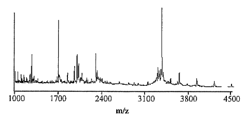
As one example, PSA isolated from serum (normal) generally has a
predominant glycosylation of the biantennary type with a mass of 2370.2 Da.
However, PSA isolated form cancer cells (e.g., LnCaP cells) generally has the
2370.2
Da biantennary structure, plus additional species corresponding to
triantennary
(3026.8 Da) and tetrantennary (3392.1 Da) saccharides. This characteristic
difference
in the mass spectrum of PSA from normal and cancer cells can be used to
establish a
"mass-identity" correlate, as shown in Figure 3. This can be done for any
target
protein. It is important to note that while each of the peals in this mass-
identity
spectrum represents a class of molecules (bi-, tri- or tetra-antennary),
subtle variations
within each of these groups can result in the further splitting of these
peaks. For
~ example, the masses mentioned above were calculated including the presence
of
terminal sialic acid residues for each of the chains. This may or may not
always be
the case. For instance, only two (instead of three) of the chains in a
triantennary
structure might have terminal sialic acids. In this case, the mass will
correspondingly
change, and such changes are readily detected using the MS methods described
herein. A mass sig~iature of the oligosaccharide representing the 'normal'
target
glycoprotein, e.g., PSA, as compared to target glycoprotein, e.g., PSA, from
tumor
cells can be easily obtained from this analysis. Reproducible differences
corresponding to systematic changes in glycan metabolism within cancer cells,
e.g.,
prostate cancer cells, e.g., LNCaP cells, will be identifiable using the
present methods.
27
CA 02510250 2005-06-16
WO 2004/066808 PCT/US2003/040547
Table 1: Table of common monomers found in N linked glycoproteins and their
molecular weights.
IDENTITY OF MONOMER MASS
Glucose 180.2
Galactose 180.2
Mannose 180.2
Fucose 164.2
N-Acetyl-Glucosamine 221..2
N-Acetyl Galactosamine 221.2
Xylose 150.1
N-Acetyl Neuraminic Acid 309.3
Correlation of Mass-Identity Relationships with Disease State or Stage:
Samples from subjects with different known disease states and stages are
analyzed, e.g., samples obtained from a bank of samples, e.g., IMPATH
(BioClinical
Partners, Inc, Franklin, MA). Generally, subjects with known medical history
are
chosen. Once the mass of the glycans on the target proteins in the samples has
been
determined and associated with the identity of those glycans, a correlation is
made
between the mass-identity of the glycans and the state or stage of the
disease. As one
example, changes in glycosylation may be correlated with disease state,
including but
not limited to the following: non-cancerous normal, non-cancerous hyperplastic
(e.g.,
benign prostate hyperplasia (BPH)), non-cancerous inflammatory (e.g.,
prostatitis,
proliferative inflammatory atrophy (PIA)), pre-cancerous (e.g., prostate
intraepithelial
neoplasia (PIN)), or cancerous (e.g., prostate cancer (PCa)). Changes in
glycosylation
may also be correlated with disease stage, for example using a system such as
the
TNM (tumor only (T), spread to a node (I~, or metastatic (M)) or other grading
system (including but not limited to the Gleason Grade/Gleason Score or other
grading system. Taking prostate cancer as one example, which is not meant to
be
limiting, the following grading system may be useful: Stage I (A) cancer can't
be felt
on digital rectal exam (DRE), causes no symptoms, and has not spread outside
the
prostate; Stage II (B) cancer can be felt on DRE or increased PSA, but has not
spread
outside the prostate; Stage III (c) cancer has spread outside the prostate to
nearby
tissues; Stage IV (D) cancer has spread to lymph nodes or to other parts of
the body.
28
CA 02510250 2005-06-16
WO 2004/066808 PCT/US2003/040547
Any other system of staging disease, e.g., clinically or pathologically, that
is known in
the art can be used.
Example 1: Glycotyain~ PSA in LNCaP cells
Isolation of PSA from LnCaP cells:
LnCaP cells were plated in RPMI 1640 medium containing 10% FBS for 48-
72 hours, and the cultures were washed with warm HBSS after which new medimn
was added. Culture supernatants were collected 24- 48 hours later and frozen
at -
20°C. PSA measurements were made on thawed supernatants using a
commercially
available mouse anti-human PSA monoclonal antibody (TandemE PSA
T_m_m__unoenzyrnatic Assay; Hybritech, Sa~l Diego, CA). The results are
generally
expressed as ng/ml of PSA/10~ cells. The limit of sensitivity of this assay is
approximately 0.2 ng/ml (Ballangrud et al., Clin Cancer Res 5:3171s-3176s
(1999);
Corey et al., Prostate 35:135-43 (1998); Gau et al., Cancer Res 57:3830-4
(1997);
Hedlund et al., Prostate 41:154-65 (1999); Nagasaki et al., Clin Chem 45:486-
96
(1999)).
Briefly, PSA from the media was purified by use of anti-PSA antibody linked
gel. A polyclonal rabbit anti human PSA antibody (Dorm et al., Prostate 14,
237-49
(1989)) (AXL 685, Accurate Chemical & Scientific Corporation) was linked to
Protein G Sepharose using an Irmnunopure crosslinlcing kit (Pierce, Rockford,
IL).
Before crosslinlcing, protein G Sepharose was equilibrated with hnmunopure
binding
buffer and then mixed with anti PSA IgG at a concentration of 3-4 mg IgG/ml of
gel.
The solution was mixed by gentle inversion at room temperature. After 30-60
minutes, the gel was washed with buffer and the antibody bound using a
solution of
DMP (Dimethyl pimelimidate) for 1-2 hours at room temperature; the remaining
active sites was bloclced using immunopure bloclcing buffer. Unbound IgG was
eluted with glycine-HCl (pH 2.5), the gel was washed and then stored in PBS
containing 0.02% sodium azide. For immunopurification, medium containing PSA
was incubated with washed anti-PSA bound gel. After incubation at room
temperature for 30 to 60 minutes, the'unbound fraction was withdrawn and the
gel
was washed 3-4x with PBS. Bound PSA was then eluted in a batchwise procedure
using an equal volume of 100mM acetic acid. Resulting fractions (3 or 4) were
29
CA 02510250 2005-06-16
WO 2004/066808 PCT/US2003/040547
collected and concentrated using a Speed Vac. Concentrated fractions were then
resolved by SDS-PAGE to confirm purity and 'molecular size, as is shown in
Figure 4.
In some cases, the fractions eluted were placed in tubes containing SOmI of
Tris-HCn
(pH 8.5) and used for estimating concentrations of recovered PSA (Hybritech
lcit)
(Qian et al., Clin. Chem. 43:352-9. (1997)).
Following isolation, the PSA is analyzed as described herein.
Example 2: Gl~yping PSA in human serum
Isolation of PSA from human serum:
A method of solid-phase affinity capture has been developed that is estimated
to purify greater than 90% of the PSA present in serum samples (Hurst et an.,
Anal.
Chem. 71:4727-33. (1999)). All reactions were carried out in sterile, low
retention,
1.5 mL microcentrifuge tubes (VWR). Amino-polystyrene beads (3-3.4 mm, 5%w/v;
Spherotech, Inc.) were treated with 0.5% glutaraldehyde in sodium carbonate
buffer.
After washing to remove excess reagent, a rabbit anti-human PSA antibody
(Accurate
Chemical & Scientific Corporation) in carbonate buffer was allowed to bind for
several hours at room temperature. After washing the beads, l0mg/mL sodium
cyanoborohydride was allowed to react for 1 hour to covalently lock the
a~ztibody in
place. The derivatized beads were mixed with human serum for 2 hrs. at room
temperature with gentle rocking. After capture, the samples were washed Sx
with
PBS and eluted with 1:3:2 Formic Acid/Water/Acetonitrile.
Following isolation, the intact PSA was analyzed as described herein. Figure
5 shows that PSA isolated from normal human serum and analyzed by the present
methods is a relatively pure, single entity, with an empirically determined
mass of
28,478.3 Da, which is in very close agreement with the theoretical molecular
mass of
the primary PSA polypeptide with a single fucosylated, biantemiary sugar
structure.
.These results illustrate that the present methods are applicable to PSA
isolated from
human serum. Similar methods can be used to isolate any target marker protein
of
choice, for instance, AFP or CEA.
Examine 3: MALDI-MS based sequencing of N-linked ~lycans from PSA
Normal PSA was obtained from Calbiochem or purified from serum samples
of healthy male volunteers (obtained from a clinical organization called
1MPATH)
CA 02510250 2005-06-16
WO 2004/066808 PCT/US2003/040547
S and the glycans were separated from the protein as described herein.
Briefly, the
glyca~l structure of PSA was isolated after PNGase F digestion and directly
analyzed
via MALDI-MS. As is shown in Figure 6A, analysis of the intact glycan
structure
yielded a mass of 2369.5, which is consistent with a biantemlary structure
with a core
fucose and two terminal sialic acids (theoretical mass of 2370.2; Figure 7).
Treatment
with sialidase resulted in a decrease in mass of 582.6, consistent with the
loss of two
sialic acid residues (Figure 6B and Figure 7). The addition of galactosidase
to the
asialo sample resulted in a further mass decrease of 324.1, resulting from the
cleavage
of two galactose residues from the nascent chain (Figure 6C and Figure 7).
Finally,
treatment of the sample with N-acetylglucosaminidase resulted in a mass
decrease
consistent with the loss of two N-acetylglucosamine residues(Figure 6D and
Figure
7).
These results demonstrate that the present methods are applicable to the
determination of the composition of N-liuced glycans from PSA. In addition,
based
on the enzyme specificity as well as the mass shift observed upon enzymatic
treatment, the sequence of the unknown oligosaccharide can be determined,
i.e., an
unambiguous oligosaccharide structure can be assigned to unl~nown samples.
These
results confirms that the present methods are useful for providing the
structural
information required to assign mass identity relationships. In addition, this
experiment was completed on submicrogram amounts of material, amounts
available
from in vivo samples, demonstrating the applicability of these methods to
physiological samples. Similar methods can be used to determine the glycotype
of
any target protein, e.g., AFP and CEA.
Example 4: Comparison of PSA Glycotypes from Normal Individuals and Cancer
Patients
PSA from individuals suffering from prostate cancer was isolated from 1 mL
serum samples as outlined in Figure 8A. Briefly, PSA was captured on magnetic
beads (Millipore Corp.) that were coated with a low affinity polyclonal
antibody
(Scripps Labs San Diego, CA). PSAvVas eluted with a 100% acetonitrile/0.1% TFA
solution, and either analyzed as is or the glycan was analyzed separately
after
digestion. Typical yields after immunopurification were 60-80% as measured by
aaz
31
CA 02510250 2005-06-16
WO 2004/066808 PCT/US2003/040547
anti-PSA ELISA (Table 2). In some experiments, the PSA protein + glycosylation
was analyzed directly via MALDI MS as outlined in Example 2. In these
experiments, PSA from cancer patients consisted of multiple entities, many of
which
possessed a molecular mass greater than 28.5 kDa. Alternatively, the
glycosylation of
PSAwas cleaved using either enzymatic (using PNGase, as described in Example
3)
or chemical (using hydrazinolysis, substantially as described in Wolff et al.
Prep
Biochem Biotechnol. 29(1):1-21 (1999)) means.
The results of analysis after digestion with PNGase are shown in Figure 8B.
Direct analysis of the N-linked glycan of PSA in samples from individuals with
cancer indicated that it is not the same as that found in normal PSA (i.e.,
PSA in
samples from normal individuals that don't have cancer, compare figures 6A and
8B),
possessing species with a higher degree of branching as well as other
modifications
(i.e., see peak at about 3300 in Fig. 8B, which is not present in samples from
normal
individuals). Examination of both the mass of the intact PSA glycoprotein as
well as
the isolated glycan revealed that the glycoforms of PSA from cancer samples
have
several key differences, including (1) the PSA glycofonns in cancer possess a
lugher
degree of branching as well as sialic acid; (2) the PSA glycoforms in cancer
possess
different fucosylated structures; and (3) chain length of anteimary arms in
PSA from
cancer is distinct from that in normal individuals. Some samples from cancer
patients
also display improperly processed, lower molecular weight glycans as well
(Fig. 8C).
Of note is the fact that the two analyses, of intact and isolated glycans,
gave separate
but complimentary information.
Table 2. Typical yields of PSA from the serum of cancer patients.
Ab (m s) PSA remain (u PSA elute (n % Recover
) )
25 0 717 71.7
25 0 753 75.3
50 ~0 681 68.1
It is to be understood that while the invention has been described in
conjunction with the detailed description thereof, the foregoing description
is intended
to illustrate and not limit the scope of the invention, which is defined by
the scope of
32
CA 02510250 2005-06-16
WO 2004/066808 PCT/US2003/040547
the appended claims. Other aspects, advantages, and modifications are within
the
scope of the following claims.
33