Note: Descriptions are shown in the official language in which they were submitted.
DEMANDES OU BREVETS VOLUMINEUX
LA PRESENTE PARTIE DE CETTE DEMANDE OU CE BREVETS
COMPREND PLUS D'UN TOME.
CECI EST LE TOME DE _2
NOTE: Pour les tomes additionels, veillez contacter le Bureau Canadien des
Brevets.
JUMBO APPLICATIONS / PATENTS
THIS SECTION OF THE APPLICATION / PATENT CONTAINS MORE
THAN ONE VOLUME.
THIS IS VOLUME 1 OF 2
NOTE: For additional volumes please contact the Canadian Patent Office.
CA 02510893 2010-08-25
72249-172
BINDING AGENTS WHICH INHIBIT MYOSTATIN
FIELD OF THE INVENTION
The invention relates to growth factors and in particular to the growth factor
myostatin
and agents which bind myostatin and inhibit its activity.
BACKGROUND
Myostatin, also known as growth/differentiation factor 8 (GDF-8), is a
transforming
growth factor-B (TGF-B) family member known to be involved in regulation of
skeletal muscle
mass. Mostmembers of the TGF-B-GDF family are expressed non-specifically in
many tissue
types and exert a variety of pleitrophic actions. However, myostatin is
largely expressed in the
cells of developing and adult skeletal muscle tissue and plays an essential
role in negatively
controlling skeletal muscle growth (McPherron et al. Nature (London) 387, 83-
90 (1997)).
Recent studies, however, indicate that low levels of myostatin expression can
be measured in
cardiac, adipose and pre-adipose tissues.
The myostatin protein has been highly conserved evolutionarily (McPherron et
al. PNAS
USA 94:12457-12461 (1997)). The biologically active C-terminal region of
myostatin has 100
percent sequence identity between human, mouse, rat, cow, chicken, and turkey
sequences. The
function of myostatin also appears to be conserved across species as well.
This is evident from
the phenotypes of animals having a mutation in the myostatin gene. Two breeds
of cattle, the
Belgian Blue (Hanset R., Muscle Hypertrophy of Genetic Origin and its Use to
Improve Beef
Production, eds, King, J.W.G. & Menissier, F. (Nijhoff, The Hague, The
Netherlands) pp. 437-
449) and the Piedmontese (Masoero, G. & Poujardieu, B, Muscle Hypertrophy of
Genetic Origin
and its Use to Improve Beef Production., eds, King, J.W.G. & Menissier, F.
(Nijhoff, The
Hague,The Netherlands) pp. 450-459) are characterized by a "double muscling"
phenotype and
increase in muscle mass. These breeds were shown to contain mutations in the
coding region of
the myostatin gene (McPherron et al.(1997) supra). In addition, mice
containing a targeted
deletion of the gene encoding myostatin (Mstn) demonstrate a dramatic increase
in muscle mass
without a corresponding increase in fat. Individual muscles of Mstn "'" mice
weigh approximately
100 to 200 percent more than those of control animals as a result of muscle
fiber hypertrophy and
hyperplasia (Zimmers et al. Science 296, 1486 (2002)).
1
CA 02510893 2005-06-17
WO 2004/058988 PCT/US2003/040781
Administration of myostatin to certain strains of mice has been shown to
create a
condition similar to muscle wasting disorders found associated with cancer,
AIDS, and muscular
dystrophy, for example. Myostatin administered as myostatin-producing CHO
cells to athymic
nude mice resulted in a wasting effect with a high degree of weight loss, a
decrease of as much as
50% of skeletal muscle mass in addition to fat wasting, and severe
hypoglycemia (Zimmers et al.
supra).
Loss of myostatin appears to result in the retention of muscle mass and
reduction in fat
accumulation with aging. It has been shown that age-related increases in
adipose tissue mass and
decrease in muscle mass were proportional to myostatin levels, as determined
by a comparison of
fat and muscle mass in Mstn +i+ when compared with Mstn --adult knockout mice
(McFerron et
al. J. Clin. Invest 109, 595 (2002)). Mstn __ mice showed decreased fat
accumulation with age
compared with Mstn "'mice.
In addition myostatin may play a role in maintaining blood glucose levels and
may
influence the development of diabetes in certain cases. It is known that, for
example, skeletal
muscle resistance to insulin-stimulated glucose uptake is the earliest known
manifestation of non-
insulin-dependent (type 2) diabetes mellitus (Corregan et al. Endocrinology
128:1682 (1991)). It
has now been shown that the lack of myostatin partially attenuates the obese
and diabetes
phenotypes of two mouse models, the agouti lethal yellow (AY) (Yen et al.
FASEB J. 8:479
(1994)), and obese (Lep b"b). Fat accumulation and total body weight of the
AY~a, Mstn "" double
mutant mouse was dramatically reduced compared with the AYa Mstn "'mouse
(McFerron et al.,
(2002) supra). In addition, blood glucose levels in the AY/a, Mstn -" mice was
dramatically lower
than in AY1a Mstn +/+ mice following exogenous glucose load, indicating that
the lack of myostatin
improved glucose metabolism. Similarly Lepb"b Mstn _i_ mice showed decreased
fat
accumulation when compared with the Lepb' b Mstn++phenotype.
Therefore, there is considerable evidence from the phenotypes of over-
expressing and
knockout animals that myostatin may play a role in contributing to a number of
metabolic
disorders including disorders resulting in muscle wasting, diabetes, obesity
and hyperglycemia.
SUMMARY OF THE INVENTION
The present invention is directed to binding agents which bind myostatin and
inhibit its
activity. The binding agents comprise at least one peptide capable of binding
myostatin. The
myostatin-binding peptides are preferably between about 5 and about 50 amino
acids in length,
more preferably between about 10 and 30 amino acids in length, and most
preferably between
about 10 and 25 amino acids in length. In one embodiment the myostatin-binding
peptide
comprises the amino acid sequence WMCPP (SEQ ID NO: 633). In another
embodiment the
2
CA 02510893 2005-06-17
WO 2004/058988 PCT/US2003/040781
myostatin binding peptides comprise the amino acid sequence Ca1a Wa3WMCPP (SEQ
ID NO:
352), wherein a1, a2 and a3 are selected from a neutral hydrophobic, neutral
polar, or basic amino
acid. In another embodiment the myostatin binding peptide comprises the
sequence
Cb1b Wb3WMCPP (SEQ ID NO: 353), wherein b1 is selected from any one of the
amino acids T,
I, or R; b2 is selected from any one of R, S, Q; b3 is selected from any one
of P, R and Q, and
wherein the peptide is beween 10 and 50 amino acids in length, and
physiologically acceptable
salts thereof. In another embodiment, the myostatin binding peptide comprises
the formula:
c1c2c3c4c5C6Cc7c3WC9WMCPPcioc11ci2ci3 (SEQ ID NO: 354), wherein:
c1 is absent or any amino acid;
c2 is absent or a neutral hydrophobic, neutral polar, or acidic amino acid;
c3 is absent or a neutral hydrophobic, neutral polar, or acidic amino acid;
c4 is absent or any amino acid;
c5 is absent or a neutral hydrophobic, neutral polar, or acidic amino acid;
c6 is absent or a neutral hydrophobic, neutral polar, or basic amino acid;
c7 is a neutral hydrophobic, neutral polar, or basic amino acid;
c$ is a neutral hydrophobic, neutral polar, or basic amino acid;
c9 is a neutral hydrophobic, neutral polar or basic amino acid; and
c10 to c13 is any amino acid; and wherein the peptide is between 20 and 50
amino acids in
length, and physiologically acceptable salts thereof.
A related embodiment the myostatin binding peptide comprises the formula:
d1d2d3d4d5d6Cd7d8Wd9WMCPP d10d11d12d13 (SEQ ID NO: 355), wherein
d1 is absent or any amino acid;
d2 is absent or a neutral hydrophobic, neutral polar, or acidic amino acid;
d3 is absent or a neutral hydrophobic, neutral polar, or acidic amino acid;
d4 is absent or any amino acid;
d5 is absent or a neutral hydrophobic, neutral polar, or acidic amino acid;
d6 is absent or a neutral hydrophobic, neutral polar, or basic amino acid;
d7 is selected from any one of the amino acids T, I, or R;
d8 is selected from any one of R, S, Q;
d9 is selected from any one of P, R and Q, and
d10 to d13 is selected from any amino acid,
and wherein the peptide is between 20 and 50 amino acids in length, and
physiologically
acceptable salts thereof.
Additional embodiments of binding agents comprise at least one of the
following
peptides:
(1) a peptide capable of binding myostatin, wherein the peptide comprises the
sequence
WYeje Ye3G, (SEQ ID NO: 356)
wherein e1 is P, S or Y,
e2 is C or Q, and
3
CA 02510893 2005-06-17
WO 2004/058988 PCT/US2003/040781
e3 is G or H, wherein the peptide is between 7 and 50 amino acids in length,
and
physiologically acceptable salts thereof;
(2) a peptide capable of binding myostatin, wherein the peptide comprises the
sequence
f1EMLf,SLf3f4LL, (SEQ ID NO: 455),
wherein f1 is M or I,
f2 is any amino acid,
f3 is L or F,
f4 isE,QorD;
and wherein the peptide is between 7 and 50 amino acids in length, and
physiologically
acceptable salts thereof;
(3) a peptide capable of binding myostatin wherein the peptide comprises the
sequence
L91g2LLg3g4L, (SEQ ID NO: 456), wherein
g1 is Q, D or E,
92 is S, Q, D or E,
g3 is any amino acid,
g4 is L, W, F, or Y, and wherein the peptide is between 8 and 50 amino acids
in length,
and physiologically acceptable salts thereof;
(4) a peptide capable of binding myostatin, wherein the peptide comprises the
sequence
h1h2h3h4h5h6h7h$h9 (SEQ ID NO: 457), wherein
h1 is R or D,
h2 is any amino acid,
h3 is A,TSorQ,
h4isLorM,
h5isLorS,
h6 is any amino acid,
h7 is F or E,
h$ is W, F or C,
h9 is L, F, M or K, and wherein the peptide is between 9 and 50 amino acids in
length,
and physiologically acceptable salts thereof.
In one embodiment, the binding agents of the present invention further
comprise at least
one vehicle such as a polymer or an Fc domain, and may further comprise at
least one linker
sequence. In this embodiment, the binding agents of the present invention are
constructed so that
at least one myostatin-binding peptide is attached to at least one vehicle.
The peptide or peptides
are attached directly or indirectly through a linker sequence, to the vehicle
at the N-terminal, C-
terminal or an amino acid sidechain of the peptide. In this embodiment, the
binding agents of the
present invention have the following generalized structure:
(X').-F'_(X2)b, or multimers thereof;
wherein F1 is a vehicle; and X1 and X2 are each independently selected from
-(L').- P';
-(L1)r-P1-(L 2)a -P2;
4
CA 02510893 2005-06-17
WO 2004/058988 PCT/US2003/040781
-(L1).-Pl-(L2)d-P2-(L3)e-P3;
and -(L'),-P'-(L 2)a-P2-(L3)e -P3-(L4)i-P4;
wherein P1, P2, P3, and P4 are peptides capable of binding myostatin; and
L', L2, L3, and L4 are each linkers; and a, b, c, d, e, and f are each
independently 0 or 1,
provided that at least one of a and b is 1, and physiologically acceptable
salts thereof.
In various embodiments of binding agents having this generalized structure,
the peptides
Pl, P2, P3, and P4 can be independently selected from one or more of any of
the peptides
comprising the sequences provided above. Pl, P2, P3, and P4 are independently
selected from one
or more peptides comprising any of the following sequences: SEQ ID NO: 633,
SEQ ID NO:
352, SEQ ID NO: 353, SEQ ID NO: 354, SEQ ID NO: 355, SEQ ID NO: 356, SEQ ID
NO: 455,
SEQ ID NO: 456, or SEQ ID NO: 457.
In a further embodiment, the binding agents comprise peptides fused to an Fe
domain,
either directly or indirectly, thereby providing peptibodies. The peptibodies
of the present
invention display a high binding affinity for myostatin and can inhibit the
activity of myostatin as
demonstrated both in vitro using cell based assays and in animals.
The present invention also provides nucleic acid molecules comprising
polynucleotides
encoding the peptides, peptibodies, and peptide and peptibody variants and
derivatives of the
present invention.
The present invention provides pharmaceutically acceptable compositions
comprising one
or more binding agents of the present invention.
The binding agents of the present invention inhibit myostatin activity in
vitro and in vivo.
The binding agents of the present invention increase lean muscle mass in a
treated animal and
decreases fat mass as a percentage of body weight of the animal. The myostatin
binding agents of
the present invention increase muscular strength in treated animal models.
The present invention provides methods of inhibiting myostatin activity in
animals
including humans by administering an effective dosage of one or more binding
agents to the
subject. The present invention provides methods of increasing lean muscle mass
in animals
including humans by administering an effective dosage of one or more binding
agents. The
present invention further provides methods of treating myostatin- related
disorders by
administering an therapeutically effective dosage of one or more myostatin
binding agents in a
pharmaceutically acceptable composition to a subject. The present invention
provides methods of
treating muscle wasting disorders including muscular dystrophy, muscle wasting
due to cancer,
AIDS, rheumatoid arthritis, renal failure, uremia, chronic heart failure, age-
related sarcopenia,
prolonged bed-rest, spinal chord injury, stroke, bone fracture. The present
invention also provides
5
CA 02510893 2011-07-28
76322-29
methods of treating metabolic disorders including obesity,
diabetes, hyperglycemia, and bone loss.
The present invention also provides a method of
increasing muscle mass in food animals by administering an
effective dosage of one or more myostatin binding agents to
the animal.
The present invention provides assays utilizing
one or more myostatin binding agents to identify and
quantitate myostatin in a sample. The assays may be
diagnostic assays for measuring or monitoring myostatin
levels in individuals with a myostatin related disorder or
disease.
Specific aspects of the invention include:
- a binding agent comprising at least one peptide or
a physiologically acceptable salt thereof capable of binding
myostatin, wherein the peptide comprises the amino acid
sequence Cala2Wa3WMCPP (SEQ ID NO: 352) , wherein a1, a2 and a3
are a neutral hydrophobic, neutral polar, or basic amino acid,
and wherein the peptide is between 10 and 50 amino acids in
length;
- a binding agent comprising at least one peptide or
a physiologically acceptable salt thereof capable of binding
myostatin, wherein the peptide comprises the sequence
c1c2c3c4c5c6Cc7c8Wc9WMCPPc1oc11c12c13 (SEQ ID NO: 354) , wherein: cl
is absent or any amino acid; c2 is absent or a neutral
hydrophobic, neutral polar, or acidic amino acid; c3 is absent
or a neutral hydrophobic, neutral polar, or acidic amino acid;
c4 is absent or any amino acid; c5 is absent or a neutral
6
CA 02510893 2011-07-28
76322-29
hydrophobic, neutral polar, or acidic amino acid; c6 is absent
or a neutral hydrophobic, neutral polar, or basic amino acid;
c-, is a neutral hydrophobic, neutral polar, or basic amino
acid; c8 is a neutral hydrophobic, neutral polar, or basic
amino acid; c9 is a neutral hydrophobic, neutral polar or basic
amino acid.; and clo to c13 is any amino acid; and wherein the
peptide is between 20 and 50 amino acids in length;
- a binding agent comprising at least one peptide or
a physiologically acceptable salt thereof capable of binding
myostatin, wherein the peptide comprises the sequence
d,d7d3d4d5d6Cd7d8Wd9WMCPP d1od11d12d,3 (SEQ ID NO: 355), wherein
d1 is absent or any amino acid; d2 is absent or a neutral
hydrophobic, neutral polar, or acidic amino acid; d3 is absent
or a neutral hydrophobic, neutral polar, or acidic amino acid;
d4 is absent or any amino acid; d5 is absent or a neutral
hydrophobic, neutral polar, or acidic amino acid; d6 is absent
or a neutral hydrophobic:, neutral polar, or basic amino acid;
d7 is any one of the amino acids T, I, or R; d8 is any one of
R, S, or Q; d9 is any one of P, R or Q, and dlo to d13 is any
amino acid, and wherein the peptide is between 20 and 50 amino
acids in length;
- a binding agent wherein said agent has the
structure: (X'),,-Fl- (X2) b, or a multimer thereof or a
physiologically acceptable salt of said structure or said
multimer; wherein F1 is a vehicle; and X1 and X2 are each
independently - (L1) c- P1; -(L'),-P'-(L 2 )d -P2; - (L') c-P1- (L2) d-Pz-
(L3) e-P3; or - (L1) -P1- (L2) a-PZ- (L3) e -P3- (L4) f-P4; wherein P1, P`,
P3, and P4 are peptides capable of binding myostatin, wherein
each peptide comprises the amino acid sequence Cala2Wa3WMCPP
(SEQ ID NO: 352), wherein al, a2 and a3 are a neutral
6a
CA 02510893 2011-07-28
76322-29
hydrophobic, neutral polar, or basic amino acid, wherein the
peptide is between 10 and 50 amino acids in length, and
wherein L1, L2, L3, and L4 are each linkers; and a, b, c, d, e,
and f are each independently 0 or 1, provided that at least
one of a and b is 1;
- a binding agent wherein said agent has the
structure: (XI),-F 1_ (X2) b, or a multimer thereof or a
physiologically acceptable salt of said structure or said
multimer; wherein F1 is a vehicle; and X1 and X2 are each
independently - (L1) c- P1; - (L`) c-P1- (L2) d -P2; - (L1) --PI- (L`) d-P2-
(L3) e-P3; and - (L') c-Pl- (L2) d-Pl- (L') e -P3- (L4) f-P4; wherein PI, P2,
P3, and P4 are peptides capable of binding myostatin, wherein
each peptide comprises the amino acid sequence Cb1b2Wb3WMCPP
(SEQ ID NO: 353), wherein b, is any one of the amino acids T,
I, or R; b2 is any one of R, S, or Q; b3 is any one of P, R or
Q, wherein the peptide is between 10 and 50 amino acids in
length, and wherein L1, L2, L3, and L4 are each linkers; and a,
b, c, d, e, and f are each independently 0 or 1, provided that
at least one of a and b is 1;
- a myostatin binding agent wherein the binding
agent has the structure F'-(L')- P1, wherein the vehicle F1 is a
human IgG Fc, wherein the linker L1 is (Gly)5, and wherein the
peptide P1 is selected from any one of SEQ ID NO: 311, SEQ ID
NO: 325, SEQ ID NO: 326, SEQ ID NO: 336, and SEQ ID NO: 337;
- a binding agent that binds myostatin, wherein the
binding agent has the structure F1-(L')- P1, wherein the vehicle
F1 is a human IgG Fc, wherein the linker L1 is (Gly)5 followed
by AQ, and wherein the peptide P comprises SEQ ID NO: 311;
6b
CA 02510893 2011-07-28
76322-29
an isolated nucleic acid molecule comprising a
polynucleotide sequence encoding the binding agent as
described herein;
- an expression vector comprising the nucleic acid
molecule as described herein;
- a host cell comprising the expression vector as
described herein;
- a pharmaceutical composition comprising an
effective amount of the binding agent as described herein in
admixture with a pharmaceutically acceptable carrier
thereof;
- use of an effective amount of the binding agent
as described herein for inhibiting myostatin activity in a
subject in need thereof;
- use of a therapeutically effective amount of the
composition as described herein for increasing the ratio of
lean muscle mass to fat in a subject in need thereof;
- use of a therapeutically effective amount of the
composition as described herein for treating muscle wasting
in a subject suffering from a disease or condition selected
from the group consisting of muscular dystrophy; amyotrophic
lateral sclerosis; congestive obstructive pulmonary disease;
chronic heart failure; cancer; AIDs; renal failure; uremia;
rheumatoid arthritis; age-related sarcopenia; and
muscle-wasting due to prolonged bedrest, spinal cord injury,
stroke, bone fracture, or aging;
6c
CA 02510893 2011-07-28
76322-29
use of a therapeutically effective amount of the
composition as described herein for reducing fat accumulation
and lowering blood glucose in a subject suffering from a
metabolic disorder selected from the group consisting of
diabetes, obesity, hyperglycemia, and bone loss;
- a method of detecting myostatin in a sample
ex vivo comprising contacting the sample with the binding
agent as described herein, and detecting the bound complex;
- a method of measuring myostatin in a sample
ex vivo comprising contacting the sample with the binding
agent as described herein, and measuring the bound complex;
- a method of diagnosing a myostatin related
disorder in a subject comprising contacting a sample taken
from the subject with the binding agent as described herein,
and detecting the bound complex, wherein the myostatin
related disorder is selected from the group consisting of
muscular dystrophy; amyotrophic lateral sclerosis;
congestive obstructive pulmonary disease; chronic heart
failure; cancer; AIDs; renal failure; uremia; rheumatoid
arthritis; age-related sarcopenia; muscle-wasting due to
prolonged bedrest, spinal cord injury, stroke, bone
fracture, or aging; diabetes; obesity; hyperglycemia; and
bone loss;
- a commercial package comprising the composition
as described herein together with instructions for use for
inhibiting myostatin activity in a subject;
6d
CA 02510893 2011-07-28
76322-29
a commercial package comprising the composition
as described herein together with instructions for use for
increasing lean muscle mass in a subject;
- a commercial package comprising the composition
as described herein together with instructions for use for
increasing the ratio of lean muscle mass to fat in a
subject;
- a commercial package comprising the composition
as described herein together with instructions for use for
treating muscle wasting in a subject suffering from a
disease or condition selected from the group consisting of
muscular dystrophy; amyotrophic lateral sclerosis;
congestive obstructive pulmonary disease; chronic heart
failure; cancer; AIDs; renal failure; uremia; rheumatoid
arthritis; age-related sarcopenia; and muscle-wasting due to
prolonged bedrest, spinal cord injury, stroke, bone
fracture, or aging; and
- a commercial package comprising the composition
as described herein together with instructions for use for
reducing fat accumulation and lowering blood glucose in a
subject suffering from a metabolic disorder selected from
the group consisting of diabetes, obesity, hyperglycemia,
and bone loss.
6e
CA 02510893 2010-08-25
72249-172
BRIEF DESCRIPTION OF THE FIGURES
Figure 1 shows myostatin activity as measured by
expressed luciferase activity (y-axis) vrs. Concentration
(x-axis) for the TN8-19 peptide QGHCTRWPWMCPPY
(Seq ID No: 32) and the TN8-19 peptibody (pb) to determined
the IC50 for each using the C2C12 pMARE luciferase assay
described in the Examples below. The peptibody has a lower
IC50 value compared with the peptide.
Figure 2 is a graph showing the increase in total
body weight for CD1 nu/nu mice treated with increasing
dosages of the lx mTN8-19-21 peptibody over a fourteen day
period compared with mice treated with a huFc control, as
described in Example 8.
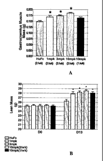
Figure 3A shows the increase in the mass of the
gastrocnemius muscle mass at necropsy of the mice treated in
Figure 2 (Example 8). Figure 3B shows the increase in lean
mass as determined by NMR on day 0 compared with day 13 of
the experiment described in Example 8.
Figure 4 shows the increase in lean body mass as
for CD1 nu/nu mice treated with biweekly injections of
increasing dosages of lx mTN8-19-32 peptibody as determined
by NMR on day 0 and day 13 of the experiment described in
Example 8.
Figure 5A shows the increase in body weight for
CD1 nu/nu mice treated with biweekly injections of
lx mTN8-19-7 compared with 2x mTN8-19-7 and the control
animal for 35 days as described in Example 8. Figure 5B
shows the increase in lean carcass weight at necropsy for
the lx and 2x versions at 1 mg/kg and 3 mg/kg compared with
the animals receiving the vehicle (huFc)(controls).
6f
CA 02510893 2010-08-25
72249-172
Figure 6A shows the increase in lean muscle mass
vrs. body weight for aged mdx mice treated with either
affinity matured 1x mTN8-19-33 peptibody or huFc vehicle at
mg/kg subcutaneously every other day for three months.
5 Figure 6B shows the change in fat mass compared to body
weight as determined by NMR for the same mice after 3 months
of treatment.
6g
CA 02510893 2005-06-17
WO 2004/058988 PCT/US2003/040781
DETAILED DESCRIPTION OF THE INVENTION
The present invention provides binding agents capable of binding myostatin and
inhibiting its activity. The myostatin binding agents can be used in assays,
to identify, quantitate,
or monitor the level of myostatin in an animal. The myostatin binding agents
of the present
invention reduce myostatin activity. The myostatin binding agents of the
present invention
increase lean muscle mass in animals, decrease fat mass as a percentage of
body weight, and
increase muscle strength. The myostatiri binding agents of the present
invention can be used to
treat a variety of metabolic disorders in which myostatin plays a role,
including muscle wasting
disorders such as muscular dystrophies, muscle wasting due to cancer, AIDS,
rheumatoid arthritis,
renal failure, uremia, chronic heart failure, prolonged bed-rest, spinal chord
injury, stroke, and
age-related sarcopenia as well as other metabolic disorders including
diabetes, obesity,
hyperglycemia, and bone loss, by administering a therapeutic dosage of one or
more binding
agents in a pharmaceutically acceptable composition to a subject.
Myostatin
Myostatin, a growth factor also known as GDF-8, is known to be a negative
regulator of
skeletal muscle tissue. Myostatin is synthesized as an inactive preproprotein
which is activated
by proteolyic cleavage (Zimmers et al., supra (2002)). The precurser protein
is cleaved to
produce an NH2-terminal inactive prodomain and an approximately 109 amino acid
COOH-
terminal protein in the form of a homodimer of about 25 kDa, which is the
mature, active form
(Zimmers et al, supra (2002)). It is now believed that the mature dimer
circulates in the blood as
an inactive latent complex bound to the propeptide (Zimmers et al, supra
(2002).
As used herein the term "full-length myostatin" refers to the full-length
human
preproprotein sequence described in McPherron et al. supra (1997), as well as
related full-length
polypeptides including allelic variants and interspecies homologs which are
also described in
McPherron et al. (1997). As used herein, the term "prodomain" or "propeptide"
refers to the
inactive NH2-terminal protein which is cleaved off to release the active COOH-
terminal protein.
As used herein the term "myostatin" or "mature myostatin" refers to the
mature, biologically
active COOH-terminal polypeptide, in monomer, dimer, multimeric form or other
form.
"Myostatin" or "mature myostatin" also refers to fragments of the biologically
active mature
myostatin, as well as related polypeptides including allelic variants, splice
variants, and fusion
peptides and polypeptides. The mature myostatin COOH-terminal protein has been
reported to
have 100% sequence identity among many species including human, mouse,
chicken, porcine,
turkey, and rat (Lee et al., PNAS 98, 9306 (2001)). Myostatin may or may not
include additional
7
CA 02510893 2010-08-25
72249-172
terminal residues such as targeting sequences, or methionine and lysine
residues and /or tag or
fusion protein sequences, depending on how it is prepared.
As used herein the term "capable of binding to myostatin" or "having a binding
affinity
for myostatin" refers to a binding agent or peptide which binds to myostatin
as demonstrated by
as the phage ELISA assay, the BIAcore or KinExATM assays described in the
Examples below.
As used herein, the term "capable of modifying myostatin activity" refers to
the action of
an agent as either an agonist or an antagonist with respect to at least one
biological activity of
myostatin. As used herein, "agonist" or "mimetic"activity refers an agent
having biological
activity comparable to a protein that interacts with the protein of interest,
as described, for
example, in International application WO 01/83525, filed May 2, 2001.
As used herein, the term "inhibiting myostatin activity" or "having antagonist
activity"refers to the ability of a peptide or binding agent to reduce or
block myostatin activity or
signaling as demonstrated or in vitro assays such as, for example, the pMARE
C2C12 cell-based
myostatin activity assay or by in vivo animal testing as described below.
Structure of Myostatin Binding Agents
In one embodiment, the binding agents of the present invention comprise at
least one
myostatin binding peptide covalently attached to at least one vehicle such as
a polymer or an Fc
domain. The attachment of the myostatin-binding peptides to at least one
vehicle is intended to
increase the effectiveness of the binding agent as a therapeutic by increasing
the biological
activity of the agent and/or decreasing degradation in vivo, increasing half-
life in vivo, reducing
toxicity or immunogenicity in vivo. The binding agents of the present
invention may further
comprise a linker sequence connecting the peptide and the vehicle. The peptide
or peptides are
attached directly or indirectly through a linker sequence to the vehicle at
the N-terminal, C-
terminal or an amino acid sidechain of the peptide. In this embodiment, the
binding agents of the
present invention have the following structure:
(X')a F'-(X2)b, or multimers thereof;
wherein F' is a vehicle; and X' and X2 are each independently selected from
-(L') - P';
-(L')C-P1-(L2)d -P2;
-(L')c P'-L2)d-P2-(L,3)e P3;
and -(L')eP'-(L2)d-P2-(L3)e P3-(L4)r-P4;
wherein P', P2, P3, and P4 are peptides capable of binding myostatin; and
8
CA 02510893 2010-08-25
72249-172
L', L2, L3, and L4 are each linkers; and a, b, c, d, e, and f are each
independently 0 or 1,
provided that at least one of a and b is 1.
Any peptide containing a cysteinyl residue may be cross-linked with another
Cys-
containing peptide, either or both of which may be linked to a vehicle. Any
peptide having more
than one Cys residue may form an intrapeptide disulfide bond, as well.
In one embodiment, the vehicle is an Fc domain, defined below. This embodiment
is
referred to as a "peptibody". As used herein, the term "peptibody" refers to a
molecule
comprising an antibody Fc domain attached to at least one peptide. The
production of peptibodies
is generally described in PCT publication WO 00/24782, published May 4, 2000.
Exemplary peptibodies are provided as lx and 2x configurations with
one copy and two copies of the peptide (attached in tandem) respectively, as
described in the
Examples below.
Peptides
As used herein the term "peptide" refers to molecules of about 5 to about 90
amino acids
linked by peptide bonds. The peptides of the present invention are preferably
between about 5 to
about 50 amino acids in length, more preferably between about 10 and 30 amino
acids in length,
and most preferably between about 10 and 25 amino acids in length, and are
capable of binding to
the myostatin protein.
The peptides of the present invention may comprise part of a sequence of
naturally
occuring proteins, may be randomized sequences derived from naturally occuring
proteins, or
may be entirely randomized sequences. The peptides of the present invention
may be generated
by any methods known in the art including chemical synthesis, digestion of
proteins, or
recombinant technology. Phage display and RNA-peptide screening, and other
affinity screening
techniques are particularly useful for generating peptides capable of binding
myostatin.
Phage display technology is described, for example, in Scott et al. Science
249: 386
(1990); Devlin et al., Science 249: 404 (1990); U.S. Patent No. 5,223,409,
issued June 29, 1993;
U.S. Patent No. 5,733,731, issued March 31, 1998; U.S. Patent No. 5,498,530,
issued March 12,
1996; U.S. Patent No. 5,432,018, issued July 11, 1995; U.S. Patent No.
5,338,665, issued August
16, 1994; U.S. Patent No. 5,922,545, issued July 13, 1999; WO 96/40987,
published December
19, 1996; and WO 98/15833, published April 16, 1998.
Using phage libraries, random peptide sequences are displayed by fusion with
coat
proteins of filamentous phage. Typically, the displayed peptides are affinity-
eluted either
specifically or non-specifically against the target molecule. The retained
phages may be enriched
9
CA 02510893 2005-06-17
WO 2004/058988 PCT/US2003/040781
by successive rounds of affinity purification and repropagation. The best
binding peptides are
selected for further analysis, for example, by using phage ELISA, described
below, and then
sequenced. Optionally, mutagenesis libraries may be created and screened to
further optimize the
sequence of the best binders (Lowman, Ann Rev Biophys Biomol Struct 26:401-24
(1997)).
Other methods of generating the myostatin binding peptides include additional
affinity
selection techniques known in the art. A peptide library can be fused in the
carboxyl terminus of
the lac repressor and expressed in E.coli. Another E. coli-based method allows
display on the
cell's outer membrane by fusion with a peptidoglycan-associated lipoprotein
(PAL). Hereinafter,
these and related methods are collectively referred to as "E. coli display."
In another method,
translation of random RNA is halted prior to ribosome release, resulting in a
library of
polypeptides with their associated RNA still attached. Hereinafter, this and
related methods are
collectively referred to as "ribosome display." Other methods employ chemical
linkage of
peptides to RNA. See, for example, Roberts and Szostak, Proc Natl Acad Sci
USA, 94: 12297-
303 (1997). Hereinafter, this and related methods are collectively referred to
as "RNA-peptide
screening." Yeast two-hybrid screening methods also may be used to identify
peptides of the
invention that bind to myostatin. In addition, chemically derived peptide
libraries have been
developed in which peptides are immobilized on stable, non-biological
materials, such as
polyethylene rods or solvent-permeable resins. Another chemically derived
peptide library uses
photolithography to scan peptides immobilized on glass slides. Hereinafter,
these and related
methods are collectively referred to as "chemical-peptide screening." Chemical-
peptide screening
may be advantageous in that it allows use of D-amino acids and other
analogues, as well as non-
peptide elements. Both biological and chemical methods are reviewed in Wells
and Lowman,
Curr Opin Biotechnol 3: 355-62 (1992).
Additionally, selected peptides capable of binding myostatin can be further
improved
through the use of "rational design". In this approach, stepwise changes are
made to a peptide
sequence and the effect of the substitution on the binding affinity or
specificity of the peptide or
some other property of the peptide is observed in an appropriate assay. One
example of this
technique is substituting a single residue at a time with alanine, referred to
as an "alanine walk" or
an "alanine scan". When two residues are replaced, it is referred to as a
"double alanine walk".
The resultant peptide containing amino acid substitutions are tested for
enhanced activity or some
additional advantageous property.
In addition, analysis of the structure of a protein-protein interaction may
also be used to
suggest peptides that mimic the interaction of a larger protein. In such an
analysis, the crystal
structure of a protein may suggest the identity and relative orientation of
critical residues of the
protein, from which a peptide may be designed. See, for example, Takasaki et
al., Nature Biotech
CA 02510893 2005-06-17
WO 2004/058988 PCT/US2003/040781
15:1266 (1977). These methods may also be used to investigate the interaction
between a
targeted protein and peptides selected by phage display or other affinity
selection processes,
thereby suggesting further modifications of peptides to increase binding
affinity and the ability of
the peptide to inhibit the activity of the protein.
In one embodiment, the peptides of the present invention are generated as
families of
related peptides. Exemplary petides are represented by SEQ ID NO: 1 through
132. These
exemplary peptides were derived through an selection process in which the best
binders generated
by phage display technology were further analyzed by phage ELISA to obtain
candidate peptides
by an affinity selection technique such as phage display technology as
described herein.
However, the peptides of the present invention may be produced by any number
of known
methods including chemical synthesis as described below.
The peptides of the present invention can be further improved by the process
of "affinity
maturation". This procedure is directed to increasing the affinity or the
activity of the peptides
and peptibodies of the present invention using phage display or other
selection technologies.
Based on a consensus sequence, directed secondary phage display libraries, for
example, can be
generated in which the "core" amino acids (determined from the consensus
sequence) are held
constant or are biased in frequency of occurrence. Alternatively, an
individual peptide sequence
can be used to generate a biased, directed phage display library. Panning of
such libraries under
more stringent conditions can yield peptides with enhanced binding to
myostatin, selective
binding to myostatin, or with some additional desired property. However,
peptides having the
affinity matured sequences may then be produced by any number of known methods
including
chemical synthesis or recombinantly. These peptides are used to generate
binding agents such as
peptibodies of various configurations which exhibit greater inhibitory
activity in cell-based assays
and in vivo assays.
Example 6 below describes affinity maturation of the "first round" peptides
described
above to produce affinity matured peptides. Exemplary affinity matured
peptibodies are presented
in Tables IV and V. The resultant lx and 2x peptibodies made from these
peptides were then
further characterized for binding affinity, ability to neutralize myostatin
activity, specificity to
myostatin as opposed to other TNFB family members, and for additional in vitro
and in vivo
activity, as described below. Affinity-matured peptides and peptibodies are
referred to by the
prefix "m" before their family name to distinguish them from first round
peptides of the same
family.
Exemplary first round peptides chosen for further affinity maturation
according to the
present invention included the following peptides: TN8-19 QGHCTRWPWMCPPY (SEQ
ID
NO: 33), and the linear peptides Linear-2 MEMLDSLFELLKDMVPISKA (SEQ ID NO:
104),
11
CA 02510893 2005-06-17
WO 2004/058988 PCT/US2003/040781
Linear-15 HHGWNYLRKGSAPQWFEAWV (SEQ ID NO: 117), Linear-17,
RATLLKDFWQLVEGYGDN (SEQ ID NO: 119), Linear-20 YREMSMLEGLLDVLERLQHY
(SEQ ID NO: 122), Linear-21 HNSSQMLLSELIMLVGSMMQ (SEQ ID NO: 123), Linear-24
EFFHWLHNHRSEVNHWLDMN (SEQ ID NO: 126). The affinity matured families of each
of
these is presented below in Tables IV and V.
The peptides of the present invention also encompass variants and derivatives
of the
selected peptides which are capable of binding myostatin. As used herein the
term "variant"
refers to peptides having one or more amino acids inserted, deleted, or
substituted into the original
amino acid sequence, and which are still capable of binding to myostatin.
Insertional and
substitutional variants may contain natural amino acids as well as non-
naturally occuring amino
acids. As used herein the term "variant" includes fragments of the peptides
which still retain the
ability to bind to myostatin. As used herein, the term "derivative" refers to
peptides which have
been modified chemically in some manner distinct from insertion, deletion, and
substitution
variants. Variants and derivatives of the peptides and peptibodies of the
present invention are
described more fully below.
Vehicles
As used herein the term "vehicle" refers to a molecule that may be attached to
one or
more peptides of the present invention. Preferably, vehicles confer at least
one desired property
on the binding agents of the present invention. Peptides alone are likely to
be removed in vivo
either by renal filtration, by cellular clearance mechanisms in the
reticuloendothelial system, or by
proteolytic degradation. Attachment to a vehicle improves the therapeutic
value of a binding
agent by reducing degradation of the binding agent and/or increasing half-
life, reducing toxicity,
reducing immunogenicity, and/or increasing the biological activity of the
binding agent.
Exemplary vehicles include Fc domains; linear polymers such as polyethylene
glycol
(PEG), polylysine, dextran; a branched chain polymer (see for example U.S.
Patent No. 4,289,872
to Denkenwalter et al., issued September 15, 1981; U. S. Patent No. 5,229,490
to Tam, issued
July 20, 1993; WO 93/21259 by Frechet et al., published 28 October 1993); a
lipid; a cholesterol
group (such as a steroid); a carbohydrate or oligosaccharide; or any natural
or synthetic protein,
polypeptide or peptide that binds to a salvage receptor.
In one embodiment, the myostatin binding agents of the present invention have
at least
one peptide attached to at least one vehicle (Fl, F2) through the N-terminus,
C-terminus or a side
chain of one of the amino acid residues of the peptide(s). Multiple vehicles
may also be used;
such as an Fc domain at each terminus or an Fc domain at a terminus and a PEG
group at the
other terminus or a side chain.
12
CA 02510893 2010-08-25
72249-172
An Fc domain is one preferred vehicle. As used herein, the term 'Pc domain"
encompasses native Fc and Fc variant molecules and sequences as defined below.
As used herein
the term "native Fe' refers to a non-antigen binding fragment of an antibody
or the amino acid
sequence of that fragment which is produced by recombinant DNA techniques or
by enzymatic or
chemical cleavage of intact antibodies. A preferred Fc is a fully human Fc and
may originate
from any of the immunoglobulins, such as IgG1 and IgG2. However, Fc molecules
that are
partially human, or originate from non-human species are also included herein.
Native Fc
molecules are made up of monomeric polypeptides that may be linked into
dimeric or multimeric
forms by covalent (i.e., disulfide bonds) and non-covalent association. The
number of
intermolecular disulfide bonds between monomeric subunits of native Fc
molecules ranges from 1
to 4 depending on class (e.g., IgG, IgA, IgE) or subclass (e.g., IgG1, IgG2,
IgG3, IgAl, IgGA2).
One example of a native Fc is a disulfide-bonded dimer resulting from papain
digestion of an IgG
(see Ellison et al. (1982), Nucl Acids Res 10: 4071-9). The term "native Fc"
as used herein is
used to refer to the monomeric, dimeric, and multimeric forms.
As used herein, the term "Fc variant" refers to a modified form of a native Fc
sequence
provided that binding to the salvage receptor is maintained, as described, for
example, in WO
97/34631 and WO 96/32478. Fc variants may
be constructed for example, by substituting or deleting residues, inserting
residues or truncating
portions containing the site. The inserted or substituted residues may also be
altered amino acids,
such as peptidomimetics or D-amino acids. Fc variants may be desirable for a
number of reasons,
several of which are described below. Exemplary Fc variants include molecules
and sequences in
which:
1. Sites involved in disulfide bond formation are removed. Such removal may
avoid
reaction with other cysteine-containing proteins present in the host cell used
to produce the
molecules of the invention. For this purpose, the cysteine-containing segment
at the N-terminus
may be truncated or cysteine residues may be deleted or substituted with other
amino acids (e.g.,
alanyl, seryl). Even when cysteine residues are removed, the single chain Fc
domains can still
form a dimeric Fc domain that is held together non-covalently.
2. A native Fc is modified to make it more compatible with a selected host
cell. For
example, one may remove the PA sequence near the N-terminus of a typical
native Fc, which may
be recognized by a digestive enzyme in E. coli such as proline iminopeptidase.
One may also add
an N-terminal methionyl residue, especially when the molecule is expressed
recombinantly in a
bacterial cell such as E. coli.
13
CA 02510893 2005-06-17
WO 2004/058988 PCT/US2003/040781
3. A portion of the N-terminus of a native Fc is removed to prevent N-terminal
heterogeneity when expressed in a selected host cell. For this purpose, one
may delete any of the
first 20 amino acid residues at the N-terminus, particularly those at
positions 1, 2, 3, 4 and 5.
4. One or more glycosylation sites are removed. Residues that are typically
glycosylated
(e.g., asparagine) may confer cytolytic response. Such residues may be deleted
or substituted
with unglycosylated residues (e.g., alanine).
5. Sites involved in interaction with complement, such as the Clq binding
site, are
removed. For example, one may delete or substitute the EKK sequence of human
IgGl.
Complement recruitment may not be advantageous for the molecules of this
invention and so may
be avoided with such an Fc variant.
6. Sites are removed that affect binding to Fc receptors other than a salvage
receptor. A
native Fc may have sites for interaction with certain white blood cells that
are not required for the
fusion molecules of the present invention and so may be removed.
7. The ADCC site is removed. ADCC sites are known in the art. See, for
example,
Molec Immunol 29 (5):633-9 (1992) with regard to ADCC sites in IgGl. These
sites, as well, are
not required for the fusion molecules of the present invention and so may be
removed.
8. When the native Fc is derived from a non-human antibody, the native Fc may
be
humanized. Typically, to humanize a native Fc, one will substitute selected
residues in the non-
human native Fc with residues that are normally found in human native Fc.
Techniques for
antibody humanization are well known in the art.
The term "Fc domain" includes molecules in monomeric or multimeric form,
whether
digested from whole antibody or produced by other means. As used herein the
term "multimer"
as applied to Fc domains or molecules comprising Fc domains refers to
molecules having two or
more polypeptide chains associated covalently, noncovalently, or by both
covalent and non-
covalent interactions. IgG molecules typically form dimers; IgM, pentamers;
IgD, dimers; and
IgA, monomers, dimers, trimers, or tetramers. Multimers may be formed by
exploiting the
sequence and resulting activity of the native Ig source of the Fc or by
derivatizing such a native
Fc. The term "dimer" as applied to Fc domains or molecules comprising Fc
domains refers to
molecules having two polypeptide chains associated covalently or non-
covalently.
Additionally, an alternative vehicle according to the present invention is a
non-Fc domain
protein, polypeptide, peptide, antibody, antibody fragment, or small molecule
(e.g., a
peptidomimetic compound) capable of binding to a salvage receptor. For
example, one could use
as a vehicle a polypeptide as described in U.S. Patent No. 5,739,277, issued
April 14, 1998 to
Presta et al. Peptides could also be selected by phage display for binding to
the FcRn salvage
receptor. Such salvage receptor-binding compounds are also included within the
meaning of
14
CA 02510893 2010-08-25
72249-172
"vehicle"and are within the scope of this invention. Such vehicles should be
selected for
increased half-life (e.g., by avoiding sequences recognized by proteases) and
decreased
immunogenicity (e.g., by favoring non-immunogenic sequences, as discovered in
antibody
humanization).
In addition, polymer vehicles may also be used to construct the binding agents
of the
present invention. Various means for attaching chemical moieties useful as
vehicles are currently
available, see, e.g., Patent Cooperation Treaty ("PCT") International
Publication No. WO
96/11953, entitled "N-Terminally Chemically Modified Protein Compositions and
Methods,"
herein incorporated by reference in its entirety. This PCT publication
discloses, among other
things, the selective attachment of water soluble polymers to the N-terminus
of proteins.
A preferred polymer vehicle is polyethylene glycol (PEG). The PEG group may be
of
any convenient molecular weight and may be linear or branched. The average
molecular weight
of the PEG will preferably range from about 2 kDa to about 100 kDa, more
preferably from about
5 kDa to about 50 kDa, most preferably from about 5 kDa to about 10 kDa. The
PEG groups will
generally be attached to the compounds of the invention via acylation or
reductive alkylation
through a reactive group on the PEG moiety (e.g., an aldehyde, amino, thiol,
or ester group) to a
reactive group on the inventive compound (e.g., an aldehyde, amino, or ester
group). A useful
strategy for the PEGylation of synthetic peptides consists of combining,
through forming a
conjugate linkage in solution, a peptide and a PEG moiety, each bearing a
special functionality
that is mutually reactive toward the other. The peptides can be easily
prepared with conventional
solid phase synthesis as known in the art. The peptides are "preactivated"
with an appropriate
functional group at a specific site. The precursors are purified and fully
characterized prior to
reacting with the PEG moiety. Ligation of the peptide with PEG usually takes
place in aqueous
phase and can be easily monitored by reverse phase analytical HPLC. The
PEGylated peptides
can be easily purified by preparative HPLC and characterized by analytical
HPLC, amino acid
analysis and laser desorption mass spectrometry.
Polysaccharide polymers are another type of water soluble polymer which may be
used
for protein modification. Dextrans are polysaccharide polymers comprised of
individual subunits
of glucose predominantly linked by al-6 linkages. The dextran itself is
available in many
molecular weight ranges, and is readily available in molecular weights from
about 1 kDa to about
70 kDa. Dextran is a suitable water-soluble polymer for use in the present
invention as a vehicle
by itself or in combination with another vehicle (e.g., Fc). See, for example,
WO 96/11953 and
WO 96/05309. The use of dextran conjugated to therapeutic or diagnostic
immunoglobulins has
been reported; see, for example, European Patent Publication No. 0 315 456.
CA 02510893 2010-08-25
72249-172
Dextran of about 1 kDa to about 20 kDa is preferred when dextran is
used as a vehicle in accordance with the present invention.
Linkers
The binding agents of the present invention may optionally further comprise a
"linker"
group. Linkers serve primarily as a spacer between a peptide and a vehicles or
between two
peptides of the binding agents of the present invention. In one embodiment,
the linker is made up
of amino acids linked together by peptide bonds, preferably from 1 to 20 amino
acids linked by
peptide bonds, wherein the amino acids are selected from the 20 naturally
occurring amino acids.
One or more of these amino acids may be glycosylated, as is understood by
those in the art. In
one embodiment, the 1 to 20 amino acids are selected from glycine, alanine,
proline, asparagine,
glutamine, and lysine. Preferably, a linker is made up of a majority of amino
acids that are
sterically unhindered, such as glycine and alanine. Thus, exemplary linkers
are polyglycines
(particularly (Gly)5, (Gly)8), poly(Gly-Ala), and polyalanines. As used
herein, the designation
"g" refers to a glycine homopeptide linkers. As shown in Table II, "gn" refers
to a 5x gly linker
at the N terminus, while "gc" refers to 5x gly linker at the C terminus.
Combinations of Gly and
Ala are also preferred. One exemplary linker sequence useful for constructing
the binding agents
of the present invention is the following: gsgsatggsgstassgsgsatg (Seq ID No:
305). This linker
sequence is referred to as the "k" or 1k sequence. The designations "kc", as
found in Table If,
refers to the k linker at the C-terminus, while the designation "kn", refers
to the k linker at the N-
terminus.
The linkers of the present invention may also be non-peptide linkers. For
example, alkyl
linkers such as -NH-(CH2)s-C(O)-, wherein s = 2-20 can be used. These alkyl
linkers may further
be substituted by any non-sterically hindering group such as lower alkyl
(e.g., C1-C6) lower acyl,
halogen (e.g., Cl, Br), CN, NH2, phenyl, etc. An exemplary non-peptide linker
is a PEG linker,
and has a molecular weight of 100 to 5000 kDa, preferably 100 to 500 kDa. The
peptide linkers
may be altered to form derivatives in the same manner as above.
Exemplary Binding Agents
The binding agents of the present invention comprise at least one peptide
capable of
binding myostatin. In one embodiment, the myostatin binding peptide is between
about 5 and
about 50 amino acids in length, in another, between about 10 and 30 amino
acids in length, and in
another, between about 10 and 25 amino acids in length. In one embodiment the
myostatin
binding peptide comprises the amino acid sequence WMCPP (SEQ ID NO: 633). In
other
embodiment, the myostatin binding peptide comprises the amino acid sequence
16
CA 02510893 2005-06-17
WO 2004/058988 PCT/US2003/040781
Cala2Wa3WMCPP (SEQ ID NO: 352), wherein a1, a2 and a3 are selected from a
neutral
hydrophobic, neutral polar, or basic amino acid. In another embodiment the
myostatin binding
peptide comprises the amino acid sequence Cb1b2Wb3WMCPP (SEQ ID NO: 353),
wherein b1 is
selected from any one of the amino acids T, I, or R; b2 is selected from any
one of R, S, Q; b3 is
selected from any one of P, R and Q, and wherein the peptide is beween 10 and
50 amino acids in
length, and physiologically acceptable salts thereof.
In another embodiment, the myostatin binding peptide comprises the formula:
c1c2c3c4c5c6Cc7c8Wc9WMCPPc10c11c12c13 (SEQ ID NO: 354), wherein:
c1 is absent or any amino acid;
c2 is absent or a neutral hydrophobic, neutral polar, or acidic amino acid;
c3 is absent or a neutral hydrophobic, neutral polar, or acidic amino acid;
c4 is absent or any amino acid;
c5 is absent or a neutral hydrophobic, neutral polar, or acidic amino acid;
c6 is absent or a neutral hydrophobic, neutral polar, or basic amino acid;
c7 is a neutral hydrophobic, neutral polar, or basic amino acid;
c$ is a neutral hydrophobic, neutral polar, or basic amino acid;
c9 is a neutral hydrophobic, neutral polar or basic amino acid; and
c10 to c13 is any amino acid; and wherein the peptide is between 20 and 50
amino acids in
length, and physiologically acceptable salts thereof.
A related embodiment the myostatin binding peptide comprises the formula:
d1d2d3d4d5d6Cd7d8Wd9WMCPP d10d11d12d13 (SEQ ID NO: 355), wherein
d1 is absent or any amino acid;
d2 is absent or a neutral hydrophobic, neutral polar, or acidic amino acid;
d3 is absent or a neutral hydrophobic, neutral polar, or acidic amino acid;
d4 is absent or any amino acid;
d5 is absent or a neutral hydrophobic, neutral polar, or acidic amino acid;
d6 is absent or a neutral hydrophobic, neutral polar, or basic amino acid;
d7 is selected from any one of the amino acids T, I, or R;
ds is selected from any one of R, S, Q; ,
d9 is selected from any one of P, R and Q, and
d10 to d13 is selected from any amino acid,
and wherein the peptide is between 20 and 50 amino acids in length, and
physiologically
acceptable salts thereof.
Additional embodiments of binding agents comprise at least one of the
following
peptides:
(1) a peptide capable of binding myostatin, wherein the peptide comprises the
sequence
WYe1e Ye3G, (SEQ ID NO: 356)
wherein e1 is P, S or Y,
e2 is C or Q, and
17
CA 02510893 2005-06-17
WO 2004/058988 PCT/US2003/040781
e3 is G or H, wherein the peptide is between 7 and 50 amino acids in length,
and
physiologically acceptable salts thereof.
(2) a peptide capable of binding myostatin, wherein the peptide comprises the
sequence
f1EMLf2SLf3f4LL, (SEQ ID NO: 455),
wherein f, is M or I,
f2 is any amino acid,
f3isLorF,
f4 is E,QorD;
and wherein the peptide is between 7 and 50 amino acids in length, and
physiologically
acceptable salts thereof.
(3) a peptide capable of binding myostatin wherein the peptide comprises the
sequence
kgig23g4L, (SEQ ID NO: 456), wherein
g, is Q, D or E,
g2 is S, Q, D or E,
g3 is any amino acid,
g4 is L, W, F, or Y, and wherein the peptide is between 8 and 50 amino acids
in length,
and physiologically acceptable salts thereof.
(4) a peptide capable of binding myostatin, wherein the peptide comprises the
sequence
h,h2h3h4h5h6h7h8h9 (SEQ ID NO: 457), wherein
h1 is R or D,
h2 is any amino acid,
h3 is A, T S or Q,
h4 is L or M,
h5 is L or S,
h6 is any amino acid,
h7 is F or E,
h8 is W, F or C,
h9 is L, F, M or K, and wherein the peptide is between 9 and 50 amino acids in
length,
and physiologically acceptable salts thereof.
In one embodiment, the binding agents of the present invention further
comprise at least
one vehicle such as a polymer or an Fc domain, and may further comprise at
least one linker
sequence. In this embodiment, the binding agents of the present invention are
constructed so that
at least one myostatin-binding peptide is covalently attached to at least one
vehicle. The peptide
or peptides are attached directly or indirectly through a linker sequence, to
the vehicle at the N-
terminal, C-terminal or an amino acid sidechain of the peptide. In this
embodiment, the binding
agents of the present invention have the following generalized structure:
(X1)a Fl-(X2)b, or multimers thereof;
wherein F' is a vehicle; and X' and X2 are each independently selected from
-(L')c- P';
-(L'),-P'-(L 2)d -P2;
18
CA 02510893 2005-06-17
WO 2004/058988 PCT/US2003/040781
-(L1)eP1-(L2)d-P2-(L)eP3;
and -(Ll)eP1-(L2)a-P2-(L3)e -P3-(L4)rP4;
wherein P1, P2, P3, and P4 are peptides capable of binding myostatin; and
L1, L2, L3, and L4 are each linkers; and a, b, c, d, e, and f are each
independently 0 or 1,
provided that at least one of a and b is 1.
In one embodiment of the binding agents having this generalized structure, the
peptides
P1, P2, P3, and P4 can be selected from one or more of any of the peptides
comprising the
sequences provided above. Peptides P1, P2, P3, and P4 can be selected from one
or more peptides
comprising any of the following sequences: SEQ ID NO: 633, SEQ ID NO: 352, SEQ
ID NO:
353, SEQ ID NO: 354, SEQ ID NO: 355, SEQ ID NO: 356, SEQ ID NO: 455, SEQ ID
NO:
456, or SEQ ID NO: 457.
In a further embodiment, the vehicles of binding agents having the general
formula above
are Fc domains. The peptides are therefore fused to an Fc domain, either
directly or indirectly,
thereby providing peptibodies. The peptibodies of the present invention
display a high binding
affinity for myostatin and can inhibit the activity of myostatin as
demonstrated by in vitro assays
and in vivo testing in animals provided herein.
The present invention also provides nucleic acid molecules comprising
polynucleotides
encoding the peptides, peptibodies, and peptide and peptibody variants and
derivatives of the
present invention. Exemplary nucleotides sequences are given below.
Variants and Derivatives of Peptides and Peptibodies
The binding agents of the present invention also encompass variants and
derivatives of
the peptides and peptibodies described herein. Since both the peptides and
peptibodies of the
present invention can be described in terms of their amino acid sequence, the
terms "variants" and
"derivatives" can be said to apply to a peptide alone, or a peptide as a
component of a peptibody.
As used herein, the term "peptide variants" refers to peptides or peptibodies
having one or more
amino acid residues inserted, deleted or substituted into the original amino
acid sequence and
which retain the ability to bind to myostatin and modify its activity. As used
herein, fragments of
the peptides or peptibodies are included within the definition of "variants".
It is understood that any given peptide or peptibody may contain one or two or
all three
types of variants. Insertional and substitutional variants may contain natural
amino acids, as well
as non-naturally occuring amino acids or both.
Peptide and peptibody variants also include mature peptides and peptibodies
wherein
leader or signal sequences are removed, and the resulting proteins having
additional amino
terminal residues, which amino acids may be natural or non-natural.
Peptibodies with an
19
CA 02510893 2005-06-17
WO 2004/058988 PCT/US2003/040781
additional methionyl residue at amino acid position -1 (Mef 1-peptibody) are
contemplated, as are
peptibodies with additional methionine and lysine residues at positions -2 and
-1 (Met2-Lys 1-).
Variants having additional Met, Met-Lys, Lys residues (or one or more basic
residues, in general)
are particularly useful for enhanced recombinant protein production in
bacterial host cells.
Peptide or peptibody variants of the present invention also includes peptides
having
additional amino acid residues that arise from use of specific expression
systems. For example,
use of commercially available vectors that express a desired polypeptide as
part of glutathione-S-
transferase (GST) fusion product provides the desired polypeptide having an
additional glycine
residue at amino acid position-1 after cleavage of the GST component from the
desired
polypeptide. Variants which result from expression in other vector systems are
also
contemplated, including those wherein histidine tags are incorporated into the
amino acid
sequence, generally at the carboxy and/or amino terminus of the sequence.
In one example, insertional variants are provided wherein one or more amino
acid
residues, either naturally occurring or non-naturally occuring amino acids,
are added to a peptide
amino acid sequence. Insertions may be located at either or both termini of
the protein, or may be
positioned within internal regions of the peptibody amino acid sequence.
Insertional variants with
additional residues at either or both termini can include, for example, fusion
proteins and proteins
including amino acid tags or labels. Insertional variants include peptides in
which one or more
amino acid residues are added to the peptide amino acid sequence or fragment
thereof.
Insertional variants also include fusion proteins wherein the amino and/or
carboxy termini
of the peptide or peptibody is fused to another polypeptide, a fragment
thereof or amino acids
which are not generally recognized to be part of any specific protein
sequence. Examples of such
fusion proteins are immunogenic polypeptides, proteins with long circulating
half lives, such as
immunoglobulin constant regions, marker proteins, proteins or polypeptides
that facilitate
purification of the desired peptide or peptibody, and polypeptide sequences
that promote
formation of multimeric proteins (such as leucine zipper motifs that are
useful in dimer
formation/stability).
This type of insertional variant generally has all or a substantial portion of
the native
molecule, linked at the N- or C-terminus, to all or a portion of a second
polypeptide. For
example, fusion proteins typically employ leader sequences from other species
to permit the
recombinant expression of a protein in a heterologous host. Another useful
fusion protein
includes the addition of an immunologically active domain, such as an antibody
epitope, to
facilitate purification of the fusion protein. Inclusion of a cleavage site at
or near the fusion
junction will facilitate removal of the extraneous polypeptide after
purification. Other useful
CA 02510893 2005-06-17
WO 2004/058988 PCT/US2003/040781
fusions include linking of functional domains, such as active sites from
enzymes, glycosylation
domains, cellular targeting signals or transmembrane regions.
There are various commercially available fusion protein expression systems
that may be
used in the present invention. Particularly useful systems include but are not
limited to the
glutathione-S-transferase (PST) system (Pharmacia), the maltose binding
protein system (NEB,
Beverley, MA), the FLAG system (IBI, New Haven, CT), and the 6xHis system
(Qiagen,
Chatsworth, CA). These systems are capable of producing recombinant peptides
and/or
peptibodies bearing only a small number of additional amino acids, which are
unlikely to
significantly affect the activity of the peptide or peptibody. For example,
both the FLAG system
and the 6xHis system add only short sequences, both of which are known to be
poorly antigenic
and which do not adversely affect folding of a polypeptide to its native
conformation. Another N-
terminal fusion that is contemplated to be useful is the fusion of a Met-Lys
dipeptide at the
N-terminal region of the protein or peptides. Such a fusion may produce
beneficial increases in
protein expression or activity.
Other fusion systems produce polypeptide hybrids. where it is desirable to
excise the
fusion partner from the desired peptide or peptibody. In one embodiment, the
fusion partner is
linked to the recombinant peptibody by a peptide sequence containing a
specific recognition
sequence for a protease. Examples of suitable sequences are those recognized
by the Tobacco
Etch Virus protease (Life Technologies, Gaithersburg, MD) or Factor Xa (New
England Biolabs,
Beverley, MA).
The invention also provides fusion polypeptides which comprise all or part of
a peptide or
peptibody of the present invention, in combination with truncated tissue
factor (tTF). tTF is a
vascular targeting agent consisting of a truncated form of a human coagulation-
inducing protein
that acts as a tumor blood vessel clotting agent, as described U.S. Patent
Nos.: 5,877,289;
6,004,555; 6,132,729; 6,132,730; 6,156,321; and European Patent No. EP
0988056. The fusion
of tTF to the anti-myostatin peptibody or peptide, or fragments thereof
facilitates the delivery of
anti-myostatin antagonists to target cells, for example, skeletal muscle
cells, cardiac muscle cells,
fibroblasts, pre-adipocytes, and possibly adipocytes.
In another aspect, the invention provides deletion variants wherein one or
more amino
acid residues in a peptide or peptibody are removed. Deletions can be effected
at one or both
termini of the peptibody, or from removal of one or more residues within the
peptibody amino
acid sequence. Deletion variants necessarily include all fragments of a
peptide or peptibody.
In still another aspect, the invention provides substitution variants of
peptides and
peptibodies of the invention. Substitution variants include those peptides and
peptibodies wherein
one or more amino acid residues are removed and replaced with one or more
alternative amino
21
CA 02510893 2005-06-17
WO 2004/058988 PCT/US2003/040781
acids, which amino acids may be naturally occurring or non-naturally
occurring. Substitutional
variants generate peptides or peptibodies that are "similar" to the original
peptide or peptibody, in
that the two molecules have a certain percentage of amino acids that are
identical. Substitution
variants include substitutions of 1, 2, 3, 4, 5, 6, 7, 8, 9, 10, 15, and 20
amino acids within a
peptide or peptibody, wherein the number of substitutions may be up to ten
percent of the amino
acids of the peptide or peptibody. In one aspect, the substitutions are
conservative in nature,
however, the invention embraces substitutions that are also non-conservative
and also includes
unconventional amino acids.
Identity and similarity of related peptides and peptibodies can be readily
calculated by
known methods. Such methods include, but are not limited to, those described
in Computational
Molecular Biology, Lesk, A.M., ed., Oxford University Press, New York (1988);
Biocomputing:
Informatics and Genoine Projects, Smith, D.W., ed., Academic Press, New York
(1993);
Computer Analysis of Sequence Data, Part 1, Griffin, A.M., and Griffin, H.G.,
eds., Humana
Press, New Jersey (1994); Sequence Analysis in Molecular Biology, von Heinje,
G., Academic
Press (1987); Sequence Analysis Primer, Gribskov, M. and Devereux, J., eds.,
M. Stockton Press,
New York (1991); and Carillo et al., SIAM J. Applied Math., 48:1073 (1988).
Preferred methods to determine the relatedness or percent identity of two
peptides or
polypeptides, or a polypeptide and a peptide, are designed to give the largest
match between the
sequences tested. Methods to determine identity are described in publicly
available computer
programs. Preferred computer program methods to determine identity between two
sequences
include, but are not limited to, the GCG program package, including GAP
(Devereux et al., Nucl.
Acid. Res., 12:387 (1984); Genetics Computer Group, University of Wisconsin,
Madison, WI,
BLASTP, BLASTN, and FASTA (Altschul et al., J. Mol. Biol., 215:403-410
(1990)). The
BLASTX program is publicly available from the National Center for
Biotechnology Information
(NCBI) and other sources (BLAST Manual, Altschul et al. NCB/NLM/NIH Bethesda,
MD 20894;
Altschul et al., supra (1990)). The well-known Smith Waterman algorithm may
also be used to
determine identity.
Certain alignment schemes for aligning two amino acid sequences may result in
the
matching of only a short region of the two sequences, and this small aligned
region may have very
high sequence identity even though there is no significant relationship
between the two full-length
sequences. Accordingly, in certain embodiments, the selected alignment method
will result in an
alignment that spans at least ten percent of the full length of the target
polypeptide being
compared, i.e., at least 40 contiguous amino acids where sequences of at least
400 amino acids are
being compared, 30 contiguous amino acids where sequences of at least 300 to
about 400 amino
22
CA 02510893 2005-06-17
WO 2004/058988 PCT/US2003/040781
acids are being compared, at least 20 contiguous amino acids where sequences
of 200 to about
300 amino acids are being compared, and at least 10 contiguous amino acids
where sequences of
about 100 to 200 amino acids are being compared. For example, using the
computer algorithm
GAP (Genetics Computer Group, University of Wisconsin, Madison, WI), two
polypeptides for
which the percent sequence identity is to be determined are aligned for
optimal matching of their
respective amino acids (the "matched span", as determined by the algorithm).
In certain
embodiments, a gap opening penalty (which is typically calculated as 3X the
average diagonal;
the "average diagonal" is the average of the diagonal of the comparison matrix
being used; the
"diagonal" is the score or number assigned to each perfect amino acid match by
the particular
comparison matrix) and a gap extension penalty (which is usually 1/10 times
the gap opening
penalty), as well as a comparison matrix such as PAM 250 or BLOSUM 62 are used
in
conjunction with the algorithm. In certain embodiments, a standard comparison
matrix (see
Dayhoff et al., Atlas of Protein Sequence and Structure, 5(3)(1978) for the
PAM 250 comparison
matrix; Henikoff et al., Proc. Natl. Acad. Sci USA, 89:10915-10919 (1992) for
the BLOSUM 62
comparison matrix) is also used by the algorithm.
In certain embodiments, for example, the parameters for a polypeptide sequence
comparison can be made with the following: Algorithm: Needleman et al., J.
Mol. Biol., 48:443-
453 (1970); Comparison matrix: BLOSUM 62 from Henikoff et al., supra (1992);
Gap Penalty:
12; Gap Length Penalty: 4; Threshold of Similarity: 0, along with no penalty
for end gaps.
In certain embodiments, the parameters for polynucleotide molecule sequence
(as
opposed to an amino acid sequence) comparisons can be made with the following:
Algorithm:
Needleman et al., supra (1970); Comparison matrix: matches = +10, mismatch =
0; Gap Penalty:
50: Gap Length Penalty: 3
Other exemplary algorithms, gap opening penalties, gap extension penalties,
comparison
matrices, thresholds of similarity, etc. may be used, including those set
forth in the Program
Manual, Wisconsin Package, Version 9, September, 1997. The particular choices
to be made will
be apparent to those of skill in the art and will depend on the specific
comparison to be made,
such as DNA-to-DNA, protein-to-protein, protein-to-DNA; and additionally,
whether the
comparison is between given pairs of sequences (in which case GAP or BestFit
are generally
preferred) or between one sequence and a large database of sequences (in which
case FASTA or
BLASTA are preferred).
Stereoisomers (e.g., D-amino acids) of the twenty conventional (naturally
occuring)
amino acids, non-naturally occuring amino acids such as a-, a-disubstituted
amino acids, N-alkyl
amino acids, lactic acid, and other unconventional amino acids may also be
suitable components
23
CA 02510893 2005-06-17
WO 2004/058988 PCT/US2003/040781
for peptides of the present invention. Examples of non-naturally occuring
amino acids include, for
example: aminoadipic acid, beta-alanine, beta-aminopropionic acid,
aminobutyric acid,
piperidinic acid, aminocaprioic acid, aminoheptanoic acid, aminoisobutyric
acid, aminopimelic
acid, diaminobutyric acid, desmosine, diaminopimelic acid, diaminopropionic
acid, N-
ethylglycine, N-ethylaspargine, hyroxylysine, a110-hydroxylysine,
hydroxyproline, isodesmosine,
allo-isoleucine, N-methylglycine, sarcosine, N-methylisoleucine, N-
methylvaline, norvaline,
norleucine, orithine, 4-hydroxyproline, y-carboxyglutamate, F,-N,N,N-
trimethyllysine, 8-N-
acetyllysine, O-phosphoserine, N-acetylserine, N-formylmethionine, 3-
methylhistidine, 5-
hydroxylysine, o-N-methylarginine, and other similar amino acids and amino
acids (e.g., 4-
hydroxyproline).
Naturally occurring residues may be divided into (overlapping) classes based
on common
side chain properties:
1) neutral hydrophobic: Met, Ala, Val, Leu, Ile, Pro, Tip, Met, Phe;
2) neutral polar: Cys, Ser, Thr, Asn, Gln, Tyr, Gly;
3) acidic: Asp, Glu;
4) basic: His, Lys, Arg;
5) residues that influence chain orientation: Gly, Pro; and
6) aromatic: Tip, Tyr, Phe.
Substitutions of amino acids may be conservative, which produces peptides
having
functional and chemical characteristics similar to those of the original
peptide. Conservative
amino acid substitutions involve exchanging a member of one of the above
classes for another
member of the same class. Conservative changes may encompass unconventional
amino acid
residues, which are typically incorporated by chemical peptide synthesis
rather than by synthesis
in biological systems. These include peptidomimetics and other reversed or
inverted forms of
amino acid moieties.
Non-conservative substitutions may involve the exchange of a member of one of
these
classes for a member from another class. These changes can result in
substantial modification in
the functional and/or chemical characteristics of the peptides. In making such
changes, according
to certain embodiments, the hydropathic index of amino acids may be
considered. Each amino
acid has been assigned a hydropathic index on the basis of its hydrophobicity
and charge
characteristics. They are: isoleucine (+4.5); valine (+4.2); leucine (+3.8);
phenylalanine (+2.8);
cysteine/cystine (+2.5); methionine (+1.9); alanine (+1.8); glycine (-0.4);
threonine (-0.7); serine
24
CA 02510893 2005-06-17
WO 2004/058988 PCT/US2003/040781
(-0.8); tryptophan (-0.9); tyrosine (-1.3); proline (-1.6); histidine (-3.2);
glutamate (-3.5);
glutamine (-3.5); aspartate (-3.5); asparagine (-3.5); lysine (-3.9); and
arginine (-4.5).
The importance of the hydropathic amino acid index in conferring interactive
biological
function on a protein is understood in the art. Kyte et al., J. Mol. Biol.,-
157:105-131 (1982). It is
known that certain amino acids may be substituted for other amino acids having
a similar
hydropathic index or score and still retain a similar biological activity. In
making changes based
upon the hydropathic index, in certain embodiments, the substitution of amino
acids whose
hydropathic indices are within 2 is included. In certain embodiments, those
which are within 1
are included, and in certain embodiments, those within 0.5 are included.
It is also understood in the art that the substitution of like amino acids can
be made
effectively on the basis of hydrophilicity, particularly where the
biologically functional peptibody
or peptide thereby created is intended for use in immunological embodiments,
as in the present
case. In certain embodiments, the greatest local average hydrophilicity of a
protein, as governed
by the hydrophilicity of its adjacent amino acids, correlates with its
immunogenicity and
antigenicity, i.e., with a biological property of the protein.
The following hydrophilicity values have been assigned to these amino acid
residues:
arginine (+3.0); lysine (+3.0); aspartate (+3.0 1); glutamate (+3.0 1);
serine (+0.3); asparagine
(+0.2); glutamine (+0.2); glycine (0); threonine (-0.4); proline (-0.5 1);
alanine (-0.5); histidine
(-0.5); cysteine (-1.0); methionine (-1.3); valine (-1.5); leucine (-1.8);
isoleucine (-1.8); tyrosine (-
2.3); phenylalanine (-2.5) and tryptophan (-3.4). In making changes based upon
similar
hydrophilicity values, in certain embodiments, the substitution of amino acids
whose
hydrophilicity values are within 2 is included, in certain embodiments, those
which are within
1 are included, and in certain embodiments, those within 0.5 are included.
One may also
identify epitopes from primary amino acid sequences on the basis of
hydrophilicity. These
regions are also referred to as "epitopic core regions."
Exemplary amino acid substitutions are set forth in Table 1 below.
Amino Acid Substitutions
Original Residues Exemplary Substitutions Preferred Substitutions
Ala Val, Leu, Ile Val
Arg Lys, Gln, Asn Lys
Asn Gln, Glu, Asp Gln
Asp Glu, Gln, Asp Glu
Cys Ser, Ala Ser
CA 02510893 2005-06-17
WO 2004/058988 PCT/US2003/040781
Gln Asn, Glu, Asp Asn
Glu Asp, Gln, Asn Asp
Gly Pro, Ala Ala
His Asn, Gln, Lys, Arg Arg
Ile Leu, Val, Met, Ala, Phe, Norleucine Leu
Leu Norleucine, Ile, Val, Met, Ala, Phe Ile
Lys Arg, 1,4 Diamino-butyric Acid, Gln, Asn Arg
Met Leu, Phe, Ile Leu
Phe Leu, Val, Ile, Ala, Tyr Leu
Pro Ala Gly
Ser Thr, Ala, Cys Thr
Thr Ser Ser
Trp Tyr, Phe Tyr
Tyr Tip, Phe, Thr, Ser Phe
Val Ile, Met, Leu, Phe, Ala, Norleucine Leu
One skilled in the art will be able to produce variants of the peptides and
peptibodies of
the present invention by random substitution, for example, and testing the
resulting peptide or
peptibody for binding activity using the assays described herein.
Additionally, one skilled in the art can review structure-function studies or
three-
dimensional structural analysis in order to identify residues in similar
polypeptides that are
important for activity or structure. In view of such a comparison, one can
predict the importance
of amino acid residues in a protein that correspond to amino acid residues
which are important for
activity or structure in similar proteins. One skilled in the art may opt for
chemically similar
amino acid substitutions for such predicted important amino acid residues. The
variants can then
be screened using activity assays as described herein.
A number of scientific publications have been devoted to the prediction of
secondary
structure. See Moult J., Curr. Op. in Biotech., 7(4):422-427 (1996), Chou et
al., Biochemistry,
13(2):222-245 (1974); Chou et al., Biochemistry, 113(2):211-222 (1974); Chou
et al., Adv.
Enzymol. Relat. Areas Mol. Biol., 47:45-148 (1978); Chou et al., Ann. Rev.
Biochem., 47:251-276
and Chou et al., Biophys. J., 26:367-384 (1979). Moreover, computer programs
are currently
available to assist with predicting secondary structure. One method of
predicting secondary
structure is based upon homology modeling. For example, two polypeptides or
proteins which
26
CA 02510893 2005-06-17
WO 2004/058988 PCT/US2003/040781
have a sequence identity of greater than 30%, or similarity greater than 40%
often have similar
structural topologies. The recent growth of the protein structural database
(PDB) has provided
enhanced predictability of secondary structure, including the potential number
of folds within a
protein's structure. See Holm et al., Nucl. Acid. Res., 27(1):244-247 (1999).
It has been
suggested (Brenner et al., Curr. Op. Struct. Biol., 7(3):369-376 (1997)) that
there are a limited
number of folds in a given protein and that once a critical number of
structures have been
resolved, structural prediction will become dramatically more accurate.
Additional methods of predicting secondary structure include "threading"
(Jones, D.,
Curr. Opin. Struct. Biol., 7(3):377-87 (1997); Sippl et al., Structure,
4(1):15-19 (1996)), "profile
analysis" (Bowie et al., Science, 253:164-170 (1991); Gribskov et al., Meth.
Enzym., 183:146-159
(1990); Gribskov et al., Proc. Nat. Acad. Sci., 84(13):4355-4358 (1987)), and
"evolutionary
linkage" (See Holm, supra (1999), and Brenner, supra (1997)).
In certain embodiments, peptide or peptibody variants include glycosylation
variants
wherein one or more glycosylation sites such as a N-linked glycosylation site,
has been added to
the peptibody. An N-linked glycosylation site is characterized by the
sequence: Asn-X-Ser or
Asn-X-Thr, wherein the amino acid residue designated as X may be any amino
acid residue
except proline. The substitution or addition of amino acid residues to create
this sequence
provides a potential new site for the addition of an N-linked carbohydrate
chain. Alternatively,
substitutions which eliminate this sequence will remove an existing N-linked
carbohydrate chain.
Also provided is a rearrangement of N-linked carbohydrate chains wherein one
or more N-linked
glycosylation sites (typically those that are naturally occurring) are
eliminated and one or more
new N-linked sites are created.
The invention also provides "derivatives" of the peptides or peptibodies of
the present
invention. As used herein the term "derivative" refers to modifications other
than, or in addition
to, insertions, deletions, or substitutions of amino acid residues which
retain the ability to bind to
myostatin.
Preferably, the modifications made to the peptides of the present invention to
produce
derivatives are covalent in nature, and include for example, chemical bonding
with polymers,
lipids, other organic, and inorganic moieties. Derivatives of the invention
may be prepared to
increase circulating half-life of a peptibody, or may be designed to improve
targeting capacity for
the peptibody to desired cells, tissues, or organs.
The invention further embraces derivative binding agents covalently modified
to include
one or more water soluble polymer attachments, such as polyethylene glycol,
polyoxyethylene
27
CA 02510893 2005-06-17
WO 2004/058988 PCT/US2003/040781
glycol, or polypropylene glycol, as described U.S. Patent Nos.: 4,640,835;
4,496,689; 4,301,144;
4,670,417; 4,791,192; and 4,179,337. Still other useful polymers known in the
art include
monomethoxy-polyethylene glycol, dextran, cellulose, or other carbohydrate
based polymers,
poly-(N-vinyl pyrrolidone)-polyethylene glycol, propylene glycol homopolymers,
a
polypropylene oxide/ethylene oxide co-polymer, polyoxyethylated polyols (e.g.,
glycerol) and
polyvinyl alcohol, as well as mixtures of these polymers. Particularly
preferred are peptibodies
covalently modified with polyethylene glycol (PEG) subunits. Water-soluble
polymers may be
bonded at specific positions, for example at the amino terminus of the
peptibodies, or randomly
attached to one or more side chains of the polypeptide. The use of PEG for
improving the
therapeutic capacity for binding agents, e.g. peptibodies, and for humanized
antibodies in
particular, is described in US Patent No. 6, 133, 426 to Gonzales et al.,
issued October 17, 2000.
The invention also contemplates derivatizing the peptide and/or vehicle
portion of the
myostatin binding agents. Such derivatives may improve the solubility,
absorption, biological
half-life, and the like of the compounds. The moieties may alternatively
eliminate or attenuate
any undesirable side-effect of the compounds and the like. Exemplary
derivatives include
compounds in which:
1. The derivative or some portion thereof is cyclic. For example, the peptide
portion may
be modified to contain two or more Cys residues (e.g., in the linker), which
could cyclize by
disulfide bond formation.
2. The derivative is cross-linked or is rendered capable of cross-linking
between
molecules. For example, the peptide portion may be modified to contain one Cys
residue and
thereby be able to form an intermolecular disulfide bond with a like molecule.
The derivative may
also be cross-linked through its C-terminus.
3. One or more peptidyl [-C(O)NR-] linkages (bonds) is replaced by a non-
peptidyl
linkage. Exemplary non-peptidyl linkages are -CH2-carbamate [-CH2-OC(O)NR-],
phosphonate, -
CH2-sulfonamide [-CH2-S(O)2NR-], urea [-NHC(O)NH-], -CH2-secondary amine, and
alkylated
peptide [-C(O)NR6- wherein R6 is lower alkyl].
4. The N-terminus is derivatized. Typically, the N-terminus may be acylated or
modified
to a substituted amine. Exemplary N-terminal derivative groups include -NRRI
(other than -NH2),
-NRC(O)R1, -NRC(O)ORI, -NRS(O)2R1, -NHC(O)NHRI, succinimide, or
benzyloxycarbonyl-
NH- (CBZ-NH-), wherein R and RI are each independently hydrogen or lower alkyl
and wherein
the phenyl ring may be substituted with 1 to 3 substituents selected from the
group consisting of
C1-C4 alkyl, C1-C4 alkoxy, chloro, and bromo.
5. The free C-terminus is derivatized. Typically, the C-terminus is esterified
or amidated.
For example, one may use methods described in the art to add (NH-CH2-CH2-NH2)2
to
28
CA 02510893 2005-06-17
WO 2004/058988 PCT/US2003/040781
compounds of this invention at the C-terminus. Likewise, one may use methods
described in the
art to add -NH2, (or "capping" with an -NH2 group) to compounds of this
invention at the C-
terminus. Exemplary C-terminal derivative groups include, for example, -C(O)R2
wherein R2 is
lower alkoxy or -NR3R4 wherein R3 and R4 are independently hydrogen or Cl-C3
alkyl (preferably
Cl-C4 alkyl).
6. A disulfide bond is replaced with another, preferably more stable, cross-
linking moiety
(e.g., an alkylene). See, e.g., Bhatnagar et al., JMed Chem 39: 3814-9 (1996),
Alberts et al.,
Thirteenth Am Pep Symp, 357-9 (1993).
7. One or more individual amino acid residues is modified. Various
derivatizing agents
are known to react specifically with selected side chains or terminal
residues, as described in
detail below.
Lysinyl residues and amino terminal residues may be reacted with succinic or
other
carboxylic acid anhydrides, which reverse the charge of the lysinyl residues.
Other suitable
reagents for derivatizing alpha-amino-containing residues include imidoesters
such as methyl
picolinimidate; pyridoxal phosphate; pyridoxal; chloroborohydride;
trinitrobenzenesulfonic acid;
O-methylisourea; 2,4 pentanedione; and transaminase-catalyzed reaction with
glyoxylate.
Arginyl residues may be modified by reaction with any one or combination of
several
conventional reagents, including phenylglyoxal, 2,3-butanedione, 1,2-
cyclohexanedione, and
ninhydrin. Derivatization of arginyl residues requires that the reaction be
performed in alkaline
conditions because of the high pKa of the guanidine functional group.
Furthermore, these
reagents may react with the groups of lysine as well as the arginine epsilon-
amino group.
Specific modification of tyrosyl residues has been studied extensively, with
particular
interest in introducing spectral labels into tyrosyl residues by reaction with
aromatic diazonium
compounds or tetranitromethane. Most commonly, N-acetylimidizole and
tetranitromethane are
used to form O-acetyl tyrosyl species and 3-nitro derivatives, respectively.
Carboxyl side chain groups (aspartyl or glutamyl) may be selectively modified
by
reaction with carbodiimides (R'-N=C=N-R') such as 1-cyclohexyl-3-(2-
morpholinyl-(4-ethyl)
carbodiimide or 1-ethyl-3-(4-azonia-4,4-dimethylpentyl) carbodiimide.
Furthermore, aspartyl and
glutamyl residues may be converted to asparaginyl and glutaminyl residues by
reaction with
ammonium ions.
Glutaminyl and asparaginyl residues may be deamidated to the corresponding
glutamyl
and aspartyl residues. Alternatively, these residues are deamidated under
mildly acidic
conditions. Either form of these residues falls within the scope of this
invention.
29
CA 02510893 2005-06-17
WO 2004/058988 PCT/US2003/040781
Cysteinyl residues can be replaced by amino acid residues or other moieties
either to
eliminate disulfide bonding or, conversely, to stabilize cross-linking. See,
e.g., Bhatnagar et al.,
(supra).
Derivatization with bifunctional agents is useful for cross-linking the
peptides or their
functional derivatives to a water-insoluble support matrix or to other
macromolecular vehicles.
Commonly used cross-linking agents include, e.g., 1, 1 -bis(diazoacetyl)-2-
phenylethane,
glutaraldehyde, N-hydroxysuccinimide esters, for example, esters with 4-
azidosalicylic acid,
homobifunctional imidoesters, including disuccinimidyl esters such as 3,3'-
dithiobis(succinimidylpropionate), and bifunctional maleimides such as bis-N-
maleimido-1,8-
octane. Derivatizing agents such as methyl-3-[(p-
azidophenyl)dithio]propioimidate yield
photoactivatable intermediates that are capable of forming crosslinks in the
presence of light.
Alternatively, reactive water-insoluble matrices such as cyanogen bromide-
activated
carbohydrates and the reactive substrates described in U.S. Patent Nos.
3,969,287; 3,691,016;
4,195,128; 4,247,642; 4,229,537; and 4,330,440 are employed for protein
immobilization.
Carbohydrate (oligosaccharide) groups may conveniently be attached to sites
that are
known to be glycosylation sites in proteins. Generally, O-linked
oligosaccharides are attached to
serine (Ser) or threonine (Thr) residues while N-linked oligosaccharides are
attached to
asparagine (Asn) residues when they are part of the sequence Asn-X-Ser/Thr,
where X can be any
amino acid except proline. X is preferably one of the 19 naturally occurring
amino acids other
than proline. The structures of N-linked and O-linked oligosaccharides and the
sugar residues
found in each type are different. One type of sugar that is commonly found on
both is N-
acetylneuraminic acid (referred to as sialic acid). Sialic acid is usually the
terminal residue of
both N-linked and O-linked oligosaccharides and, by virtue of its negative
charge, may confer
acidic properties to the glycosylated compound. Such site(s) may be
incorporated in the linker of
the compounds of this invention and are preferably glycosylated by a cell
during recombinant
production of the polypeptide compounds (e.g., in mammalian cells such as CHO,
BHK, COS).
However, such sites may further be glycosylated by synthetic or semi-synthetic
procedures known
in the art.
Other possible modifications include hydroxylation of proline and lysine,
phosphorylation
of hydroxyl groups of seryl or threonyl residues, oxidation of the sulfur atom
in Cys, methylation
of the alpha-amino groups of lysine, arginine, and histidine side chains [see,
for example,
Creighton, Proteins: Structure and Molecule Properties (W. H. Freeman & Co.,
San Francisco),
pp. 79-86 (1983)].
Compounds of the present invention may be changed at the DNA level, as well.
The DNA
sequence of any portion of the compound may be changed to codons more
compatible with the
CA 02510893 2005-06-17
WO 2004/058988 PCT/US2003/040781
chosen host cell. For E. coli, which is the preferred host cell, optimized
codons are known in the
art. Codons may be substituted to eliminate restriction sites or to include
silent restriction sites,
which may aid in processing of the DNA in the selected host cell. The vehicle,
linker and peptide
DNA sequences may be modified to include any of the foregoing sequence
changes.
Additional derivatives include non-peptide analogs that provide a stabilized
structure or
lessened biodegradation, are also contemplated. Peptide mimetic analogs can be
prepared based
on a selected inhibitory peptide by replacement of one or more residues by
nonpeptide moieties.
Preferably, the nonpeptide moieties permit the peptide to retain its natural
confirmation, or
stabilize a preferred, e.g., bioactive, confirmation which retains the ability
to recognize and bind
myostatin. In one aspect, the resulting analog/mimetic exhibits increased
binding affinity for
myostatin. One example of methods for preparation of nonpeptide mimetic
analogs from peptides
is described in Nachman et al., Regul Pept 57:359-370 (1995). If desired, the
peptides of the
invention can be modified, for instance, by glycosylation, amidation,
carboxylation, or
phosphorylation, or by the creation of acid addition salts, amides, esters, in
particular C-terminal
esters, and N-acyl derivatives of the peptides of the invention. The
peptibodies also can be
modified to create peptide derivatives by forming covalent or noncovalent
complexes with other
moieties. Covalently-bound complexes can be prepared by linking the chemical
moieties to
functional groups on the side chains of amino acids comprising the
peptibodies, or at the N- or C-
terminus.
In particular, it is anticipated that the peptides can be conjugated to a
reporter group,
including, but not limited to a radiolabel, a fluorescent label, an enzyme
(e.g., that catalyzes a
colorimetric or fluorometric reaction), a substrate, a solid matrix, or a
carrier (e.g., biotin or
avidin). The invention accordingly provides a molecule comprising a peptibody
molecule,
wherein the molecule preferably further comprises a reporter group selected
from the group
consisting of a radiolabel, a fluorescent label, an enzyme, a substrate, a
solid matrix, and a carrier.
Such labels are well known to those of skill in the art, e.g., biotin labels
are particularly
contemplated. The use of such labels is well known to those of skill in the
art and is described in,
e.g., U.S. Patent Nos.3,817,837; 3,850,752; 3,996,345; and 4,277,437. Other
labels that will be
useful include but are not limited to radioactive labels, fluorescent labels
and chemiluminescent
labels. U.S. Patents concerning use of such labels include, for example, U.S.
Patent Nos.
3,817,837; 3,850,752; 3,939,350; and 3,996,345. Any of the peptibodies of the
present invention
may comprise one, two, or more of any of these labels.
Methods of Making Peptides and Peptibodies
31
CA 02510893 2010-08-25
72249-172
The peptides of the present invention can be generated using a wide variety of
techniques
known in the art. For example, such peptides can be synthesized in solution or
on a solid support
in accordance with conventional techniques. Various automatic synthesizers are
commercially
available and can be used in accordance with known protocols. See, for
example, Stewart and
Young (supra); Tam et al., J Am Chem Soc, 105:6442, (1983); Merrifield,
Science 232:341-347
(1986); Barany and Merrifield, The Peptides, Gross and Meienhofer, eds,
Academic Press, New
York, 1-284; Barany et al., Int J Pep Protein Res, 30:705-739 (1987); and U.S.
Patent No.
5,424,398.
Solid phase peptide synthesis methods use a copoly(styrene-divinylbenzene)
containing
0.1-1.0 mM amines/g polymer. These methods for peptide synthesis use
butyloxycarbonyl (t-
BOC) or 9-fluorenylmethyloxy-carbonyl(FMOC) protection of alpha-amino groups.
Both
methods involve stepwise syntheses whereby a single amino acid is added at
each step starting
from the C-terminus of the peptide (See, Coligan et al., Curr Prot Immunol,
Wiley Interscience,
1991, Unit 9). On completion of chemical synthesis, the synthetic peptide can
be deprotected to
remove the t-BOC or FMOC amino acid blocking groups and cleaved from the
polymer by
treatment with acid at reduced temperature (e.g., liquid HF-10% anisole for
about 0.25 to about 1
hours at 0 C). After evaporation of the reagents, the peptides are extracted
from the polymer with
1% acetic acid'solution that is then lyophilized to yield the crude material.
This can normally be
purified by such techniques as gel filtration on Sephadex G-15 using 5% acetic
acid as a solvent.
Lyophilization of appropriate fractions of the column will yield the
homogeneous peptides or
peptide derivatives, which can then be characterized by such standard
techniques as amino acid
analysis, thin layer chromatography, high performance liquid chromatography,
ultraviolet
absorption spectroscopy, molar rotation, solubility, and quantitated by the
solid phase Edman
degradation.
Phage display techniques can be particularly effective in identifying the
peptides of the
present invention as described above. Briefly, a phage library is prepared
(using e.g. ml 13, fd, or
lambda phage), displaying inserts from 4 to about 80 amino acid residues. The
inserts may
represent, for example, a completely degenerate or biased array. Phage-bearing
inserts that bind
to the desired antigen are selected and this process repeated through several
cycles of reselection
of phage that bind to the desired antigen. DNA sequencing is conducted to
identify the sequences
of the expressed peptides. The minimal linear portion of the sequence that
binds to the desired
antigen can be determined in this way. The procedure can be repeated using a
biased library
containing inserts containing part or all of the minimal linear portion plus
one or more additional
degenerate residues upstream or downstream thereof. These techniques may
identify peptides of
the invention with still greater binding affinity for myostatin than agents
already identified herein.
32
CA 02510893 2005-06-17
WO 2004/058988 PCT/US2003/040781
Regardless of the manner in which the peptides are prepared, a nucleic acid
molecule
encoding each such peptide can be generated using standard recombinant DNA
procedures. The
nucleotide sequence of such molecules can be manipulated as appropriate
without changing the
amino acid sequence they encode to account for the degeneracy of the nucleic
acid code as well as
to account for codon preference in particular host cells.
The present invention also provides nucleic acid molecules comprising
polynucleotide
sequences encoding the peptides and peptibodies of the present invention.
These nucleic acid
molecules include vectors and constructs containing polynucleotides encoding
the peptides and
peptibodies of the present invention, as well as peptide and peptibody
variants and derivatives.
Exemplary nucleic acid molecules are provided in the Examples below.
Recombinant DNA techniques also provide a convenient method for preparing full
length
peptibodies and other large polypeptide binding agents of the present
invention, or fragments
thereof.. A polynucleotide encoding the peptibody or fragment may be inserted
into an
expression vector, which can in turn be inserted into a host cell for
production of the binding
agents of the present invention. Preparation of exemplary peptibodies of the
present invention are
described in Example 2 below.
A variety of expression vector/host systems may be utilized to express the
peptides and
peptibodies of the invention. These systems include but are not limited to
microorganisms such
as bacteria transformed with recombinant bacteriophage, plasmid or cosmid DNA
expression
vectors; yeast transformed with yeast expression vectors; insect cell systems
infected with virus
expression vectors (e.g., baculovirus); plant cell systems transfected with
virus expression vectors
(e.g., cauliflower mosaic virus, CaMV; tobacco mosaic virus, TMV) or
transformed with bacterial
expression vectors (e.g., Ti or pBR322 plasmid); or animal cell systems. One
preferred host cell
line is E. coli strain 2596 (ATCC # 202174), used for expression of
peptibodies as described
below in Example 2. Mammalian cells that are useful in recombinant protein
productions include
but are not limited to VERO cells, HeLa cells, Chinese hamster ovary (CHO)
cell lines, COS cells
(such as COS-7), W138, BHK, HepG2, 3T3, RIN, MDCK, A549, PC12, K562 and 293
cells.
The term "expression vector" refers to a plasmid, phage, virus or vector, for
expressing a
polypeptide from a polynucleotide sequence. An expression vector can comprise
a transcriptional
unit comprising an assembly of (1) a genetic element or elements having a
regulatory role in gene
expression, for example, promoters or enhancers, (2) a structural or sequence
that encodes the
binding agent which is transcribed into mRNA and translated into protein, and
(3) appropriate
transcription initiation and termination sequences. Structural units intended
for use in yeast or
eukaryotic expression systems preferably include a leader sequence enabling
extracellular
33
CA 02510893 2005-06-17
WO 2004/058988 PCT/US2003/040781
secretion of translated protein by a host cell. Alternatively, where
recombinant protein is
expressed without a leader or transport sequence, it may include an amino
terminal methionyl
residue. This residue may or may not be subsequently cleaved from the
expressed recombinant
protein to provide a final peptide product.
For example, the peptides and peptibodies may be recombinantly expressed in
yeast using
a commercially available expression system, e.g., the Pichia Expression System
(Invitrogen, San
Diego, CA), following the manufacturer's instructions. This system also relies
on the pre-pro-
alpha sequence to direct secretion, but transcription of the insert is driven
by the alcohol oxidase
(AOX1) promoter upon induction by methanol. The secreted peptide is purified
from the yeast
growth medium using the methods used to purify the peptide from bacterial and
mammalian cell
supernatants.
Alternatively, the cDNA encoding the peptide and peptibodies may be cloned
into the
baculovirus expression vector pVL1393 (PharMingen, San Diego, CA). This vector
can be used
according to the manufacturer's directions (PharMingen) to infect Spodoptera
frugiperda cells in
sF9 protein-free media and to produce recombinant protein. The recombinant
protein can be
purified and concentrated from the media using a heparin-Sepharose column
(Pharmacia).
Alternatively, the peptide or peptibody may be expressed in an insect system.
Insect
systems for protein expression are well known to those of skill in the art. In
one such system,
Autographa californica nuclear polyhedrosis virus (AcNPV) can be used as a
vector to express
foreign genes in Spodoptera frugiperda cells or in Trichoplusia larvae. The
peptide coding
sequence can be cloned into a nonessential region of the virus, such as the
polyhedrin gene, and
placed under control of the polyhedrin promoter. Successful insertion of the
peptide will render
the polyhedrin gene inactive and produce recombinant virus lacking coat
protein coat. The
recombinant viruses can be used to infect S. frugiperda cells or Trichoplusia
larvae in which the
peptide is expressed (Smith et al., J Virol 46: 584 (1983); Engelhard et al.,
Proc Nat Acad Sci
(USA) 91: 3224-7 (1994)).
In another example, the DNA sequence encoding the peptide can be amplified by
PCR
and cloned into an appropriate vector for example, pGEX-3X (Pharmacia). The
pGEX vector is
designed to produce a fusion protein comprising glutathione-S-transferase
(GST), encoded by the
vector, and a protein encoded by a DNA fragment inserted into the vector's
cloning site. The
primers for PCR can be generated to include for example, an appropriate
cleavage site. Where the
fusion moiety is used solely to facilitate expression or is otherwise not
desirable as an attachment
to the peptide of interest, the recombinant fusion protein may then be cleaved
from the GST
portion of the fusion protein. The pGEX-3X/specific binding agent peptide
construct is
transformed into E. coli XL-1 Blue cells (Stratagene, La Jolla CA), and
individual transformants
34
CA 02510893 2005-06-17
WO 2004/058988 PCT/US2003/040781
isolated and grown. Plasmid DNA from individual transformants can be purified
and partially
sequenced using an automated sequencer to confirm the presence of the desired
specific binding
agent encoding nucleic acid insert in the proper orientation.
The fusion protein, which may be produced as an insoluble inclusion body in
the bacteria,
can be purified as follows. Host cells are collected by centrifugation; washed
in 0.15 M NaCl, 10
mM Tris, pH 8, 1 mM EDTA; and treated with 0.1 mg/ml lysozyme (Sigma, St.
Louis, MO) for
minutes at room temperature. The lysate can be cleared by sonication, and cell
debris can be
pelleted by centrifugation for 10 minutes at 12,000 X g. The fusion protein-
containing pellet can
be resuspended'in 50 mM Tris, pH 8, and 10 mM EDTA, layered over 50% glycerol,
and
10 centrifuged for 30 min. at 6000 X g. The pellet can be resuspended in
standard phosphate
buffered saline solution (PBS) free of Mg++ and Ca++. The fusion protein can
be further purified
by fractionating the resuspended pellet in a denaturing SDS-PAGE (Sambrook et
al., supra). The
gel can be soaked in 0.4 M ICI to visualize the protein, which can be excised
and electroeluted in
gel-running buffer lacking SDS. If the GST/fusion protein is produced in
bacteria as a soluble
15 protein, it can be purified using the GST Purification Module (Pharmacia).
The fusion protein may be subjected to digestion to cleave the GST from the
peptide of
the invention. The digestion reaction (20-40 mg fusion protein, 20-30 units
human thrombin
(4000 U/mg, Sigma) in 0.5 ml PBS can be incubated 16-48 hrs at room
temperature and loaded on
a denaturing SDS-PAGE gel to fractionate the, reaction products. The gel can
be soaked in 0.4 M
ICI to visualize the protein bands. The identity of the protein band
corresponding to the expected
molecular weight of the peptide can be confirmed by amino acid sequence
analysis using an
automated sequencer (Applied Biosystems Model 473A, Foster City, CA).
Alternatively, the
identity can be confirmed by performing HPLC and/or mass spectometry of the
peptides.
Alternatively, a DNA sequence encoding the peptide can be cloned into a
plasmid
containing a desired promoter and, optionally, a leader sequence (Better et
al., Science 240:1041-
43 (1988)). The sequence of this construct can be confirmed by automated
sequencing. The
plasmid can then be transformed into E. coli strain MC1061 using standard
procedures employing
CaC12 incubation and heat shock treatment of the bacteria (Sambrook et al.,
supra). The
transformed bacteria can be grown in LB medium supplemented with
carbenicillin, and
production of the expressed protein can be induced by growth in a suitable
medium. If present,
the leader sequence can effect secretion of the peptide and be cleaved during
secretion.
Mammalian host systems for the expression of recombinant peptides and
peptibodies are
well known to those of skill in the art. Host cell strains can be chosen for a
particular ability to
process the expressed protein or produce certain post-translation
modifications that will be useful
in providing protein activity. Such modifications of the protein include, but
are not limited to,
CA 02510893 2005-06-17
WO 2004/058988 PCT/US2003/040781
acetylation, carboxylation, glycosylation, phosphorylation, lipidation and
acylation. Different
host cells such as CHO, HeLa, MDCK, 293, W138, and the like have specific
cellular machinery
and characteristic mechanisms for such post-translational activities and can
be chosen to ensure
the correct modification and processing of the introduced, foreign protein.
It is preferable that transformed cells be used for long-term, high-yield
protein
production. Once such cells are transformed with vectors that contain
selectable markers as well
as the desired expression cassette, the cells can be allowed to grow for 1-2
days in an enriched
media before they are switched to selective media. The selectable marker is
designed to allow
growth and recovery of cells that successfully express the introduced
sequences. Resistant
clumps of stably transformed cells can be proliferated using tissue culture
techniques appropriate
to the cell line employed.
A number of selection systems can be used to recover the cells that have been
transformed for recombinant protein production. Such selection systems
include, but are not
limited to, HSV thymidine kinase, hypoxanthine-guanine
phosphoribosyltransferase and adenine
phosphoribosyltransferase genes, in tk-, hgprt- or aprt- cells, respectively.
Also, anti-metabolite
resistance can be used as the basis of selection for dhfr which confers
resistance to methotrexate;
gpt which confers resistance to mycophenolic acid; neo which confers
resistance to the
aminoglycoside G418 and confers resistance to chlorsulfuron; and hygro which
confers resistance
to hygromycin. Additional selectable genes that may be useful include trpB,
which allows cells to
utilize indole in place of tryptophan, or hisD, which allows cells to utilize
histinol in place of
histidine. Markers that give a visual indication for identification of
transformants include
anthocyanins,13-glucuronidase and its substrate, GUS, and luciferase and its
substrate, luciferin.
Purification and Refolding of Binding Agents
In some cases, the binding agents such as the peptides and/or peptibodies of
this invention
may need to be "refolded" and oxidized into a proper tertiary structure and
disulfide linkages
generated in order to be biologically active. Refolding can be accomplished
using a number of
procedures well known in the art. Such methods include, for example, exposing
the solubilized
polypeptide agent to a pH usually above 7 in the presence of a chaotropic
agent. The selection of
chaotrope is similar to the choices used for inclusion body solubilization,
however a chaotrope is
typically used at a lower concentration. Exemplary chaotropic agents are
guanidine and urea. In
most cases, the refolding/oxidation solution will also contain a reducing
agent plus its oxidized
form in a specific ratio to generate a particular redox potential which allows
for disulfide shuffling
to occur for the formation of cysteine bridges. Some commonly used redox
couples include
cysteine/cystamine, glutathione/dithiobisGSH, cupric chloride, dithiothreitol
DTT/dithiane DTT,
36
CA 02510893 2005-06-17
WO 2004/058988 PCT/US2003/040781
and 2-mercaptoethanol (bME)/dithio-bME. In many instances, a co-solvent may be
used to
increase the efficiency of the refolding. Commonly used cosolvents include
glycerol,
polyethylene glycol of various molecular weights, and arginine.
It may be desirable to purify the peptides and peptibodies of the present
invention.
Protein purification techniques are well known to those of skill in the art.
These techniques
involve, at one level, the crude fractionation of the proteinaceous and non-
proteinaceous fractions.
Having separated the peptide and/or peptibody from other proteins, the peptide
or polypeptide of
interest can be further purified using chromatographic and electrophoretic
techniques to achieve
partial or complete purification (or purification to homogeneity). Analytical
methods particularly
suited to the preparation of peptibodies and peptides or the present invention
are ion-exchange
chromatography, exclusion chromatography; polyacrylamide gel electrophoresis;
isoelectric
focusing. A particularly efficient method of purifying peptides is fast
protein liquid
chromatography or even HPLC.
Certain aspects of the present invention concern the purification, and in
particular
embodiments, the substantial purification, of a peptibody or peptide of the
present invention. The
term "purified peptibody or peptide" as used herein, is intended to refer to a
composition,
isolatable from other components, wherein the peptibody or peptide is purified
to any degree
relative to its naturally-obtainable state. A purified peptide or peptibody
therefore also refers to a
peptibody or peptide that is free from the environment in which it may
naturally occur.
Generally, "purified" will refer to a peptide or peptibody composition that
has been
subjected to fractionation to remove various other components, and which
composition
substantially retains its expressed biological activity. Where the term
"substantially purified" is
used, this designation will refer to a peptide or peptibody composition in
which the peptibody or
peptide forms the major component of the composition, such as constituting
about 50%, about
- 25 60%, about 70%, about 80%, about 90%, about 95% or more of the proteins
in the composition.
Various methods for quantifying the degree of purification of the peptide or
peptibody
will be known to those of skill in the art in light of the present disclosure.
These include, for
example, determining the specific binding activity of an active fraction, or
assessing the amount
of peptide or peptibody within a fraction by SDS/PAGE analysis. A preferred
method for
assessing the purity of a peptide or peptibody fraction is to calculate the
binding activity of the
fraction, to compare it to the binding activity of the initial extract, and to
thus calculate the degree
of purification, herein assessed by a "-fold purification number." The actual
units used to
represent the amount of binding activity will, of course, be dependent upon
the particular assay
technique chosen to follow the purification and whether or not the peptibody
or peptide exhibits a
detectable binding activity.
37
CA 02510893 2005-06-17
WO 2004/058988 PCT/US2003/040781
Various techniques suitable for use in purification will be well known to
those of skill in
the art. These include, for example, precipitation with ammonium sulphate,
PEG, antibodies
(immunoprecipitation) and the like or by heat denaturation, followed by
centrifugation;
chromatography steps such as affinity chromatography, (e.g., Protein-A-
Sepharose), ion exchange,
gel filtration, reverse phase, hydroxylapatite and affinity chromatography;
isoelectric focusing;
gel electrophoresis; and combinations of such and other techniques. As is
generally known in the
art, it is believed that the order of conducting the various purification
steps may be changed, or
that certain steps may be omitted, and still result in a suitable method for
the preparation of a
substantially purified binding agent.
There is no general requirement that the binding agents of the present
invention always be
provided in their most purified state. Indeed, it is contemplated that less
substantially purified
binding agent products will have utility in certain embodiments. Partial
purification may be
accomplished by using fewer purification steps in combination, or by utilizing
different forms of
the same general purification scheme. For example, it is appreciated that a
cation-exchange
column chromatography performed utilizing an HPLC apparatus will generally
result in a greater
"-fold" purification than the same technique utilizing a low-pressure
chromatography system.
Methods exhibiting a lower degree of relative purification may have advantages
in total recovery
of the peptide or peptibody, or in maintaining binding activity of the peptide
or peptibody.
It is known that the migration of a peptide or polypeptide can vary, sometimes
significantly, with different conditions of SDS/PAGE (Capaldi et al., Biochem
Biophys Res
Comm, 76: 425 (1977)). It will therefore be appreciated that under differing
electrophoresis
conditions, the apparent molecular weights of purified or partially purified
binding agent
expression products may vary.
Activity of Myostatin Binding Agents
After the construction of the binding agents of the present invention, they
are tested for
their ability to bind myostatin and inhibit or block myostatin activity. Any
number of assays or
animal tests may be used to determine the ability of the agent to inhibit or
block myostatin
activity. Several assays used for characterizing the peptides and peptibodies
of the present
invention are described in the Examples below. One assay is the C2C12 pMARE-
luc assay which
makes use of a myostatin-responsive cell line (C2C12 myoblasts) transfected
with a luciferase
reporter vector containing myostatin/activin response elements (MARE).
Exemplary peptibodies
are assayed by pre-incubating a series of peptibody dilutions with myostatin,
and then exposing
the cells to the incubation mixture. The resulting luciferase activity is
determined, and a titration
curve is generated from the series of peptibody dilutions. The IC50 (the
concentration of
38
CA 02510893 2005-06-17
WO 2004/058988 PCT/US2003/040781
peptibody to achieve 50% inhibition of myostatin activity as measured by
luciferase activity) was
then determined. A second assay described below is a BlAcore assay to
determine the kinetic
parameters ka (association rate constant), kd (dissociation rate constant),
and KD (dissociation
equilibrium constant) for the myostatin binding agents. Lower dissociation
equilibrium constants
(KD, expressed in nM) indicated a greater affinity of the peptibody for
myostatin. Additional
assays include blocking assays, to determine whether a binding agent such as a
peptibody is
neutralizing (prevents binding of myostatin to its receptor), or non-
neutralizing (does not prevent
binding of myostatin to its receptor); selectivity assays, which determine if
the binding agents of
the present invention bind selectively to myostatin and not to other TGFB
family members; and
KinEx ATM assays or solution-based equilibrium assays, which also determine KD
and are
considered to be more sensitive in some circumstances. These assays are
described in Example 3.
Figure 1 shows the IC50 of a peptide compared with the IC50 of the peptibody
form of the
peptide. This demonstrates that the peptibody is significantly more effective
at inhibiting
myostatin activity than the peptide alone. In addition, affinity-matured
peptibodies generally
exhibit improved IC50 and KD values compared with the parent peptides and
peptibodies. The
IC50 values for a number of exemplary affinity matured peptibodies are shown
in Table VII,
Example 7 below. Additionally, in some instances, making a 2x version of a
peptibody, where
two peptides are attached in tandem, increase the activity of the peptibody
both in vitro and in
vivo.
In vivo activities are demonstrated in the Examples below. The activities of
the binding
agents include anabolic acitivity increasing lean muscle mass in animal
models, as well as
decreasing the fat mass with respect to total body weight in treated animal
models, and increasing
muscular strength in animal models.
Uses of the Myostatin Binding Agents
The myostatin binding agents of the present invention bind to myostatin and
block or
inhibit myostatin signaling within targeted cells. The present invention
provides methods and
reagents for reducing the amount or activity of myostatin in an animal by
administering an
effective dosage of one or more myostatin binding agents to the animal. In one
aspect, the present
invention provides methods and reagents for treating myostatin-related
disorders in an animal
comprising administering an effective dosage of one or more binding agents to
the animal. These
myostatin-related disorders include but are not limited to various forms of
muscle wasting, as well
as metabolic disorders such as diabetes and related disorders, and bone
degenerative diseases such
as osteoporosis.
39
CA 02510893 2005-06-17
WO 2004/058988 PCT/US2003/040781
As shown in the Example 8 below, exemplary peptibodies of the present
invention
dramatically increases lean muscle mass in the CD1 nu/nu mouse model. This in
vivo activity
correlates to the in vitro binding and inhibitory activity described below for
the same peptibodies.
Muscle wasting disorders include dystrophies such as Duchenne's muscular
dystrophy,
progressive muscular dystrophy, Becker's type muscular dystrophy, Dejerine-
Landouzy muscular
dystrophy, Erb's muscular dystrophy, and infantile neuroaxonal muscular
dystrophy. For
example, blocking myostatin through use of antibodies in vivo improved the
dystrophic phenotype
of the mdx mouse model of Duchenne muscular dystrophy (Bogdanovich et al,
Nature 420, 28
(2002)). The peptibodies of'the present invention increase lean muscle mass as
a percentage of
body weight and decreases fat mass as percentage of body weight when
administered to an aged
mdx mouse model.
Additional muscle wasting disorders arise from chronic disease such as
amyotrophic
lateral sclerosis, congestive obstructive pulmonary disease, cancer, AIDS,
renal failure, and
rheumatoid arthritis. For example, cachexia or muscle wasting and loss of body
weight was
induced in athymic nude mice by a systemically administered myostatin (Zimmers
et al., supra).
In another example, serum and intramuscular concentrations of myostatin-
immunoreactive.protein
was found to be increased in men exhibiting AIDS-related muscle wasting and
was inversely
related to fat-free mass (Gonzalez-Cadavid et al., PNAS USA 95: 14938-14943
(1998)).
Additional conditions resulting in muscle wasting may arise from inactivity
due to disability such
as confinement in a wheelchair, prolonged bedrest due to stroke, illness,
spinal chord injury, bone
fracture or trauma, and muscular atrophy in a microgravity environment (space
flight). For
example, plasma myostatin immunoreactive protein was found to increase after
prolonged bedrest
(Zachwieja et al. J Gravit Physiol. 6(2):11(1999). It was also found that the
muscles of rats
exposed to a microgravity environment during a space shuttle flight expressed
an increased
amount of myostatin compared with the muscles of rats which were not exposed
(Lalani et al.,
J.Endocrin 167 (3):417-28 (2000)).
In addition, age-related increases in fat to muscle ratios, and age-related
muscular atrophy
appear to be related to myostatin. For example, the average serum myostatin-
immunoreactive
protein increased with age in groups of young (19-35 yr old), middle-aged (36-
75 yr old), and
elderly (76-92 yr old) men and women, while the average muscle mass and fat-
free mass declined
with age in these groups (Yarasheski et al. JNutrAging 6(5):343-8 (2002)). It
has also been
shown that myostatin gene knockout in mice increased myogenesis and decreased
adipogenesis
(Lin et al., Biochein Biophys Res Coininun 291(3):701-6 (2002), resulting in
adults with increased
muscle mass and decreased fat accumulation and leptin secretion. Exemplary
peptibodies
improve the lean muscle mass to fat ratio in aged mdx mice as shown below.
CA 02510893 2005-06-17
WO 2004/058988 PCT/US2003/040781
In addition, myostatin has now been found to be expressed at low levels in
heart muscle
and expression is upregulated after cardiomyocytes after infarct (Sharma et
al., J Cell Physiol. 180
(1): 1-9 (1999)). Therefore, reducing myostatin levels in the heart muscle may
improve recovery
of heart muscle after infarct.
Myostatin also appears to influence metabolic disorders including type 2
diabetes,
noninsulin-dependent diabetes mellitus, hyperglycemia, and obesity. For
example, lack of
myostatin has been shown to improve the obese and diabetic phenotypes of two
mouse models
(Yen et al. supra). It has been demonstrated in the Examples below that
decreasing myostatin
activity by administering the inhibitors of the present invention will
decreases the fat to muscle
ratio in an animal, including aged animal models. Therefore, decreasing fat
composition by
administering the inhibitors of the present invention will improve diabetes,
obesity, and
hyperglycemic conditions in animals.
In addition, increasing muscle mass by reducing myostatin levels may improve
bone
strength and reduce osteoporosis and other degenerative bone diseases. It has
been found, for
example, that myostatin-deficient mice showed increased mineral content and
density of the
mouse humerus and increased mineral content of both trabecular and cortical
bone at the regions
where the muscles attach, as well as increased muscle mass (Hamrick et al.
Calcif Tissue Int
71(1):63-8 (2002)).
The present invention also provides methods and reagents for increasing muscle
mass in
food animals by administering an effective dosage of the myostatin binding
agent to the animal.
Since the mature C-terminal myostatin polypeptide is identical in all species
tested, myostatin
binding agents would be expected to be effective for increasing muscle mass
and reducing fat in
any agriculturally important species including cattle, chicken, turkeys, and
pigs.
The binding agents of the present invention may be used alone or in
combination with
other therapeutic agents to enhance their therapeutic effects or decrease
potential side effects. The
binding agents of the present invention possess one or more desirable but
unexpected combination
of properties to improve the therapeutic value of the agents. These properties
include increased
activity, increased solubility, reduced degradation, increased half-life,
reduced toxicity, and
reduced immunogenicity. Thus the binding agents of the present invention are
useful for
extended treatment regimes. In addition, the properties of hydrophilicity and
hydrophobicity of
the compounds of the invention are well balanced, thereby enhancing their
utility for both in vitro
and especially in vivo uses. Specifically, compounds of the invention have an
appropriate degree
of solubility in aqueous media that permits absorption and bioavailability in
the body, while also
having a degree of solubility in lipids that permits the compounds to traverse
the cell membrane to
a putative site of action, such as a particular muscle mass.
41
CA 02510893 2010-08-25
72249-172
The binding agents of the present invention are useful for treating a
"subject" or any
animal, including humans, when administered in an effective dosages in a
suitable composition.
In addition, the mystatin binding agents of the present invention are useful
for detecting
and quantitating myostatin in a number of assays. These assays are described
in more detail
below.
In general, the binding agents of the present invention are useful as capture
agents to bind
and immobilize myostatin in a variety of assays, similar to those described,
for example, in Asai,
ed., Methods in Cell Biology, 37, Antibodies in Cell Biology, Academic Press,
Inc., New York
(1993). The binding agent may be labeled in some manner or may react with a
third molecule
such as an anti binding agent antibody which is labeled to enable myostatin to
be detected and
quantitated. For example, a binding agent or a third molecule can be modified
with a detectable
moiety, such as biotin, which can then be bound by a fourth molecule, such as
enzyme-labeled
streptavidin, or other proteins. (Akerstrom, J Immunol 135:2589 (1985);
Chaubert, Mod Pathol
10:585 (1997)).
Throughout any particular assay, incubation and/or washing steps may be
required after
each combination of reagents. Incubation steps can vary from about 5 seconds
to several hours,
preferably from about 5 minutes to about 24 hours. However, the incubation
time will depend
upon the assay format, volume of solution, concentrations, and the like.
Usually, the assays will
be carried out at ambient temperature, although they can be conducted over a
range of
temperatures.
Non-competitive binding assays:
Binding assays can be of the non-competitive type in which the amount of
captured
myostatin is directly measured. For example, in one preferred "sandwich"
assay, the binding
agent can be bound directly to a solid substrate where it is immobilized.
These immobilized
agents then bind to myostatin present in the test sample. The immobilized
myostatin is then
bound with a labeling agent, such as a labeled antibody against myostatin,
which can be detected.
In another preferred "sandwich" assay, a second agent specific for the binding
agent can be added
which contains a detectable moiety, such as biotin, to which a third labeled
molecule can
specifically bind, such as streptavidin. (See, Harlow and Lane, Antibodies, A
Laboratory Manual,
Ch 14, Cold Spring Harbor Laboratory, NY (1988)).
Competitive Binding Assays:
Binding assays can be of the competitive type. The amount of myostatin present
in the
sample is measured indirectly by measuring the amount of myostastin displaced,
or competed
away, from a binding agent by the myostatin present in the sample. In one
preferred competitive
42
CA 02510893 2005-06-17
WO 2004/058988 PCT/US2003/040781
binding assay, a known amount of myostastin, usually labeled, is added to the
sample and the
sample is then contacted with the binding agent. The amount of labeled
myostastin bound to the
binding agent is inversely proportional to the concentration of myostastin
present in the sample.
(following the protocols found in, for example Harlow and Lane, Antibodies, A
Laboratory
Manual, Ch 14, pp. 579-583, supra).
In another preferred competitive binding assay, the binding agent is
immobilized on a
solid substrate. The amount of myostastin bound to the binding agent may be
determined either
by measuring the amount of myostatin present in a myostatintbinding agent
complex, or
alternatively by measuring the amount of remaining uncomplexed myostatin.
Other Binding Assays
The present invention also provides Western blot methods to detect or quantify
the
presence of myostatin in a sample. The technique generally comprises
separating sample proteins
by gel electrophoresis on the basis of molecular weight and transferring the
proteins to a suitable
solid support, such as nitrocellulose filter, a nylon filter, or derivatized
nylon filter. The sample is
incubated with the binding agents or fragments thereof that bind myostatin and
the resulting
complex is detected. These binding agents may be directly labeled or
alternatively may be
subsequently detected using labeled antibodies that specifically bind to the
binding agent.
Diagnostic Assays
The binding agents or fragments thereof of the present invention may be useful
for the
diagnosis of conditions or diseases characterized by increased amounts of
myostatin. Diagnostic
assays for high levels of myostatin include methods utilizing a binding agent
and a label to detect
myostatin in human body fluids, extracts of cells or specific tissue extracts.
For example, serum
levels of myostatin may be measured in an individual over time to determine
the onset of muscle
wasting associated with aging or inactivity, as described, for example, in
Yarasheski et al., supra.
Increased myostatin levels were shown to correlate with average decreased
muscle mass and fat-
free mass in groups of men and women of increasing ages (Yarasheski et al.,
supra). The binding
agents of the present invention may be useful for monitoring increases or
decreases in the levels
of myostatin with a given individual over time, for example. The binding
agents can be used in
such assays with or without modification. In a preferred diagnostic assay, the
binding agents will
be labeled by attaching, e.g., a label or a reporter molecule. A wide variety
of labels and reporter
molecules are known, some of which have been already described herein. In
particular, the
present invention is useful for diagnosis of human disease.
43
CA 02510893 2005-06-17
WO 2004/058988 PCT/US2003/040781
A variety of protocols for measuring myostatin proteins using binding agents
of myostatin
are known in the art. Examples include enzyme-linked immunosorbent assay
(ELISA),
radioimmunoassay (RIA) and fluorescence activated cell sorting (FACS).
For diagnostic applications, in certain embodiments the binding agents of the
present
invention typically will be labeled with a detectable moiety. The detectable
moiety can be any
one that is capable of producing, either directly or indirectly, a detectable
signal. For example,
the detectable moiety may be a radioisotope, such as 3H, 14C, 32P, 35S, or
125I, a fluorescent or
chemiluminescent compound, such as fluorescein isothiocyanate, rhodamine, or
luciferin; or an
enzyme, such as alkaline phosphatase, (3galactosidase, or horseradish
peroxidase (Bayer et al.,
Meth Enz, 184: 138 (1990)).
Pharmaceutical Compositions
Pharmaceutical compositions of myostatin binding agents such as peptibodies
described
herein are within the scope of the present invention. Such compositions
comprise a
therapeutically or prophylactically effective amount of a myostatin binding
agent, fragment,
variant, or derivative thereof as described herein, in admixture with a
pharmaceutically acceptable
agent. In a preferred embodiment, pharmaceutical compositions comprise
antagonist binding
agents that inhibit myostatin partially or completely in admixture with a
pharmaceutically
acceptable agent. Typically, the myostastin binding agents will be
sufficiently purified for
administration to an animal.
The pharmaceutical composition may contain formulation materials for
modifying,
maintaining or preserving, for example, the pH, osmolarity, viscosity,
clarity, color, isotonicity,
odor, sterility, stability, rate of dissolution or release, adsorption or
penetration of the
composition. Suitable formulation materials include, but are not limited to,
amino acids (such as
glycine, glutamine, asparagine, arginine or lysine); antimicrobials;
antioxidants (such as ascorbic
acid, sodium sulfite or sodium hydrogen-sulfite); buffers (such as borate,
bicarbonate, Tris-HC1,
citrates, phosphates, other organic acids); bulking agents (such as mannitol
or glycine), chelating
agents (such as ethylenediamine tetraacetic acid (EDTA)); complexing agents
(such as caffeine,
polyvinylpyrrolidone, beta-cyclodextrin or' hydroxypropyl-beta-cyclodextrin);
fillers;
monosaccharides; disaccharides and other carbohydrates (such as glucose,
mannose, or dextrins);
proteins (such as serum albumin, gelatin or immunoglobulins); coloring;
flavoring and diluting
agents; emulsifying agents; hydrophilic polymers (such as
polyvinylpyrrolidone); low molecular
weight polypeptides; salt-forming counterions (such as sodium); preservatives
(such as
benzalkonium chloride, benzoic acid, salicylic acid, thimerosal, phenethyl
alcohol,
methylparaben, propylparaben, chlorhexidine, sorbic acid or hydrogen
peroxide); solvents (such
44
CA 02510893 2005-06-17
WO 2004/058988 PCT/US2003/040781
as glycerin, propylene glycol or polyethylene glycol); sugar alcohols (such as
mannitol or
sorbitol); suspending agents; surfactants or wetting agents (such as
pluronics, PEG, sorbitan
esters, polysorbates such as polysorbate 20, polysorbate 80, triton,
tromethamine, lecithin,
cholesterol, tyloxapal); stability enhancing agents (sucrose or sorbitol);
tonicity enhancing agents
(such as alkali metal halides (preferably sodium or potassium chloride,
mannitol sorbitol);
delivery vehicles; diluents; excipients and/or pharmaceutical adjuvants.
(Remington's
Pharmaceutical Sciences, 18`h Edition, A.R. Gennaro, ed., Mack Publishing
Company, 1990).
The optimal pharmaceutical composition will be determined by one skilled in
the art
depending upon, for example, the intended route of administration, delivery
format, and desired
dosage. See for example, Remington's Pharmaceutical Sciences, supra. Such
compositions may
influence the physical state, stability, rate of in vivo release, and rate of
in vivo clearance of the
binding agent.
The primary vehicle or carrier in a pharmaceutical composition may be either
aqueous or
non-aqueous in nature. For example, a suitable vehicle or carrier may be water
for injection,
physiological saline solution or artificial cerebrospinal fluid, possibly
supplemented with other
materials common in compositions for parenteral administration. Neutral
buffered saline or saline
mixed with serum albumin are further exemplary vehicles. Other exemplary
pharmaceutical
compositions comprise Tris buffer of about pH 7.0-8.5, or acetate buffer of
about pH 4.0-5.5,
which may further include sorbitol or a suitable substitute therefore. In one
embodiment of the
present invention, binding agent compositions may be prepared for storage by
mixing the selected
composition having the desired degree of purity with optional formulation
agents (Remington's
Pharmaceutical Sciences, supra) in the form of a lyophilized cake or an
aqueous solution.
Further, the binding agent product may be formulated as a lyophilizate using
appropriate
excipients such as sucrose.
The pharmaceutical compositions can be selected for parenteral delivery.
Alternatively,
the compositions may be selected for inhalation or for enteral delivery such
as orally, aurally,
opthalmically, rectally, or vaginally. The preparation of such
pharmaceutically acceptable
compositions is within the skill of the art.
The formulation components are present in concentrations that are acceptable
to the site
of administration. For example, buffers are used to maintain the composition
at physiological pH
or at slightly lower pH, typically within a pH range of from about 5 to about
8.
When parenteral administration is contemplated, the therapeutic compositions
for use in
this invention may be in the form of a pyrogen-free, parenterally acceptable
aqueous solution
comprising the desired binding agent in a pharmaceutically acceptable vehicle.
A particularly
suitable vehicle for parenteral injection is sterile distilled water in which
a binding agent is
CA 02510893 2010-08-25
72249-172
formulated as a sterile, isotonic solution, properly preserved. Yet another
preparation can involve
the formulation of the desired molecule with an agent, such as injectable
microspheres, bio-
erodible particles, polymeric compounds (polylactic acid, polyglycolic acid),.
beads, or liposomes,
that provides for the controlled or sustained release of the product which may
then be delivered
via a depot injection. Hyaluronic acid may also be used, and this may have the
effect of
promoting sustained duration in the circulation. Other suitable means for the
introduction of the
desired molecule include implantable drug delivery devices.
In another aspect, pharmaceutical formulations suitable for parenteral
administration may
be formulated in aqueous solutions, preferably in physiologically compatible
buffers such as
Hanks' solution, ringer's solution, or physiologically buffered saline.
Aqueous injection
suspensions may contain substances that increase the viscosity of the
suspension, such as sodium
carboxymethyl cellulose, sorbitol, or dextran. Additionally, suspensions of
the active compounds
may be prepared as appropriate oily injection suspensions. Suitable lipophilic
solvents or vehicles
include fatty oils, such as sesame oil, or synthetic fatty acid esters, such
as ethyl=oleate,
triglycerides, or liposomes. Non-lipid polycationic amino polymers may also be
used for
delivery. Optionally, the suspension may also contain suitable stabilizers or
agents to increase the
solubility of the ompounds and allow for the preparation of highly
concentrated solutions.ln
another embodiment, a pharmaceutical composition may be formulated for
inhalation. For
example, a binding agent may be formulated as a dry powder for inhalation.
Polypeptide or
nucleic acid molecule inhalation solutions may also be formulated with a
propellant for aerosol
delivery. In yet another embodiment, solutions may be nebulized. Pulmonary
administration is
further described in PCT publication WO 94/020069, which describes pulmonary
delivery of chemically modified proteins.
It is also contemplated that certain formulations may be administered orally.
In one
embodiment of the present invention, binding agent molecules that are
administered in this
fashion can be formulated with or without those carriers customarily used in
the compounding of
solid dosage forms such as tablets and capsules. For example, a capsule may be
designed to
release the active portion of the formulation at the point in the
gastrointestinal tract when
bioavailability is maximized and pre-systemic degradation is minimized.
Additional agents can
be included to facilitate absorption of the binding agent molecule. Diluents,
flavorings, low
melting point waxes, vegetable oils, lubricants, suspending agents, tablet
disintegrating agents,
and binders may also be employed.
Pharmaceutical compositions for oral administration can also be formulated
using
pharmaceutically acceptable carriers well known in the art in dosages suitable
for oral
46
CA 02510893 2010-08-25
72249-172
administration. Such carriers enable the pharmaceutical compositions to be
formulated as tablets,
pills, dragees, capsules, liquids, gels, syrups, slurries, suspensions, and
the like, for ingestion by
the patient.
Pharmaceutical preparations for oral use can be obtained through combining
active
compounds with solid excipient and processing the resultant mixture of
granules (optionally, after
grinding) to obtain tablets or dragee cores. Suitable auxiliaries can be
added, if desired. Suitable
excipients include carbohydrate or protein fillers, such as sugars, including
lactose, sucrose,
mannitol, and sorbitol; starch from corn, wheat, rice, potato, or other
plants; cellulose, such as
methyl cellulose, hydroxypropylmethyl-cellulose, or sodium
carboxymethylcellulose; gums,
including arabic and tragacanth; and proteins, such as gelatin and collagen.
If desired,
disintegrating or solubilizing agents may be added, such as the cross-linked
polyvinyl
pyrrolidone, agar, and alginic acid or a salt thereof, such as sodium
alginate.
Dragee cores may be used in conjunction with suitable coatings, such as
concentrated
sugar solutions, which may also contain gum arabic, talc,
polyvinylpyrrolidone, carbopol gel,
polyethylene glycol, and/or titanium dioxide, lacquer solutions, and suitable
organic solvents or
solvent mixtures. Dyestuffs or pigments may be added to the tablets or dragee
coatings for
product identification or to characterize the quantity of active compound,
i.e., dosage.
Pharmaceutical preparations that can be used orally also include push-fit
capsules made
of gelatin, as well as soft, sealed capsules made of gelatin and a coating,
such as glycerol or
sorbitol. Push-fit capsules can contain active ingredients mixed with fillers
or binders, such as
lactose or starches, lubricants, such as talc or magnesium stearate, and,
optionally, stabilizers. In
soft capsules, the active compounds may be dissolved or suspended in suitable
liquids, such as
fatty oils, liquid, or liquid polyethylene glycol with or without stabilizers.
Another pharmaceutical composition may involve an effective quantity of
binding agent
in a mixture with non-toxic excipients that are suitable for the manufacture
of tablets. By
dissolving the tablets in sterile water, or other appropriate vehicle,
solutions can be prepared in
unit dose form. Suitable excipients include, but are not limited to, inert
diluents, such as calcium
carbonate, sodium carbonate or bicarbonate, lactose, or calcium phosphate; or
binding agents,
such as starch, gelatin, or acacia; or lubricating agents such as magnesium
stearate, stearic acid, or
talc.
Additional pharmaceutical compositions will be evident to those skilled in the
art,
including formulations involving binding agent molecules in sustained- or
controlled-delivery
formulations. Techniques for formulating a variety of other sustained- or
controlled-delivery
means, such as liposome carriers, bio-erodible microparticles or porous beads
and depot
injections, are also known to those skilled in the art. See for example, WO
93/015722 that
47
CA 02510893 2005-06-17
WO 2004/058988 PCT/US2003/040781
describes controlled release of porous polymeric microparticles for the
delivery of pharmaceutical
compositions. Additional examples of sustained-release preparations include
semipermeable
polymer matrices in the form of shaped articles, e.g. films, or microcapsules.
Sustained release
matrices may include polyesters, hydrogels, polylactides (U.S. 3,773,919, EP
58,481),
copolymers of L-glutamic acid and gamma ethyl-L-glutamate (Sidman et al.,
Biopolymers,
22:547-556 (1983), poly (2-hydroxyethyl-methacrylate) (Langer et al., J.
Biomed. Mater. Res.,
15:167-277, (1981); Langer et al., Chem. Tech., 12:98-105(1982)), ethylene
vinyl acetate (Langer
et al., supra) or poly-D(-)-3-hydroxybutyric acid (EP 133,988). Sustained-
release compositions
also include liposomes, which can be prepared by any of several methods known
in the art. See
e.g., Eppstein et al.,PNAS (USA), 82:3688 (1985); EP 36,676; EP 88,046; EP
143,949.
The pharmaceutical composition to be used for in vivo administration typically
must be
sterile. This may be accomplished by filtration through sterile filtration
membranes. Where the
composition is lyophilized, sterilization using this method may be conducted
either prior to or
following lyophilization and reconstitution. The composition for parenteral
administration may
be stored in lyophilized form or in solution. In addition, parenteral
compositions generally are
placed into a container having a sterile access port, for example, an
intravenous solution bag or
vial having a stopper pierceable by a hypodermic injection needle.
Once the pharmaceutical composition has been formulated, it may be stored in
sterile
vials as a solution, suspension, gel, emulsion, solid, or a dehydrated or
lyophilized powder. Such
formulations may be stored either in a ready-to-use form or in a form (e.g.,
lyophilized) requiring
reconstitution prior to administration.
In a specific embodiment, the present invention is directed to kits for
producing a single-
dose administration unit. The kits may each contain both a first container
having a dried protein
and a second container having an aqueous formulation. Also included within the
scope of this
invention are kits containing single and multi-chambered pre-filled syringes
(e.g., liquid syringes
and lyosyringes).
An effective amount of a pharmaceutical composition to be employed
therapeutically will
depend, for example, upon the therapeutic context and objectives. One skilled
in the art will
appreciate that the appropriate dosage levels for treatment will thus vary
depending, in part, upon
30' the molecule delivered, the indication for which the binding agent
molecule is being used, the
route of administration, and the size (body weight, body surface or organ
size) and condition (the
age and general health) of the patient. Accordingly, the clinician may titer
the dosage and modify
the route of administration to obtain the optimal therapeutic effect. A
typical dosage may range
from about 0.1mg/kg to up to about 100 mg/kg or more, depending on the factors
mentioned
48
CA 02510893 2005-06-17
WO 2004/058988 PCT/US2003/040781
above. In other embodiments, the dosage may range from 0.1 mg/kg up to about
100 mg/kg; or 1
mg/kg up to about 100 mg/kg; or 5 mg/kg up to about 100 mg/kg.
For any compound, the therapeutically effective dose can be estimated
initially either in
cell culture assays or in animal models such as mice, rats, rabbits, dogs,
pigs, or monkeys. An
animal model may also be used to determine the appropriate concentration range
and route of
administration. Such information can then be used to determine useful doses
and routes for
administration in humans.
The exact dosage will be determined in light of factors related to the subject
requiring
treatment. Dosage and administration are adjusted to provide sufficient levels
of the active
compound or to maintain the desired effect. Factors that may be taken into
account include the
severity of the disease state, the general health of the subject, the age,
weight, and gender of the
subject, time and frequency of administration, drug combination(s), reaction
sensitivities, and
response to therapy. Long-acting pharmaceutical compositions may be
administered every 3 to 4
days, every week, or biweekly depending on the half-life and clearance rate of
the particular
formulation.
The frequency of dosing will depend upon the pharmacokinetic parameters of the
binding
agent molecule in the formulation used. Typically, a composition is
administered until a dosage
is reached that achieves the desired effect. The composition may therefore be
administered as a
single dose, or as multiple doses (at the same or different
concentrations/dosages) over time, or as
a continuous infusion. Further refinement of the appropriate dosage is
routinely made.
Appropriate dosages may be ascertained through use of appropriate dose-
response data.
The route of administration of the pharmaceutical composition is in accord
with known
methods, e.g. orally, through injection by intravenous, intraperitoneal,
intracerebral (intra-
parenchymal), intracerebroventricular, intramuscular, intra-ocular,
intraarterial, intraportal,
intralesional routes, intramedullary, intrathecal, intraventricular,
transdermal, subcutaneous,
intraperitoneal, intranasal, enteral, topical, sublingual, urethral, vaginal,
or rectal means, by
sustained release systems or by implantation devices. Where desired, the
compositions may be
administered by bolus injection or continuously by infusion, or by
implantation device.
Alternatively or additionally, the composition may be administered locally via
implantation of a membrane, sponge, or another appropriate material on to
which the desired
molecule has been absorbed or encapsulated. Where an implantation device is
used, the device
may be implanted into any suitable tissue or organ, and delivery of the
desired molecule may be
via diffusion, timed-release bolus, or continuous administration.
49
CA 02510893 2005-06-17
WO 2004/058988 PCT/US2003/040781
In some cases, it may be desirable to use pharmaceutical compositions in an ex
vivo
manner. In such instances, cells, tissues, or organs that have been removed
from the patient are
exposed to the pharmaceutical compositions after which the cells, tissues
and/or organs are
subsequently implanted back into the patient.
In other cases, a binding agent of the present invention such as a peptibody
can be
delivered by implanting certain cells that have been genetically engineered,
using methods such as
those described herein, to express and secrete the polypeptide. Such cells may
be animal or
human cells, and may be autologous, heterologous, or xenogeneic. Optionally,
the cells may be
immortalized. In order to decrease the chance of an immunological response,
the cells may be
encapsulated to avoid infiltration of surrounding tissues. The encapsulation
materials are
typically biocompatible, semi-permeable polymeric enclosures or membranes that
allow the
release of the protein product(s) but prevent the destruction of the cells by
the patient's immune
system or by other detrimental factors from the surrounding tissues.
Pharmaceutical compositions containing the binding agents of the present
invention are
administered to a subject to treat any myostatin-related disorders. These
include muscle-wasting
disorders including but not limited to muscular dystrophy, muscle wasting in
cancer, AIDS,
muscle atrophy, rheumatoid arthritis, renal failure/uremia, chronic heart
failure, prolonged bed-
rest, spinal chord injury, stroke, and aging related sarcopenia. In addition
these compositions are
administed to treat obesity, diabetes, hyperglycemia, and increase bone
density,
The invention having been described, the following examples are offered by way
of
illustration, and not limitation.
Example 1
Identification of myostatin -binding peptides
Three filamentous phage libraries, TN8-IX (5X10⁹ independent transformants),
TN12-I
(1.4X10⁹ independent transformants), and linear (2.3X10⁹ independent
transformants) (Dyax
Corp.) were used to select for myostatin binding phage. Each library was
incubated on myostatin-
coated surfaces and subjected to different panning conditions: non-specific
elution, and specific
elution using recombinant human activin receptor IIB/Fc chimera (R&D Systems,
Inc.,
Minneapolis, Minnesota), or myostastin propeptide elution as described below.
For all three
libraries, the phages were eluted in a non-specific manner for the first round
of selection, while
the receptor and promyostatin was used in the second and third rounds of
selection. The selection
procedures were carried out as described below.
CA 02510893 2005-06-17
WO 2004/058988 PCT/US2003/040781
Preparation of myostatin
Myostatin protein was produced recombinantly in the E.coli K-12 strain 2596
(ATCC #
202174) as follows. Polynucleotides encoding the human promyostatin molecule
were cloned
into the pAMG21 expression vector (ATCC No. 98113), which was derived from
expression
vector pCFM1656 (ATCC No. 69576) and the expression vector system described in
United
States Patent No. 4,710,473, by following the procedure described in published
International
Patent Application WO 00/24782. The polynucleotides encoding promyostatin were
obtained
from a mammalian expression vector. The coding region was amplified using a
standard PCR
method and the following PCR primers to introduce the restriction site for
NdeI and BamHI.
5' primer: 5'-GAGAGAGAGCATATGAATGAGAACAGTGAGCAAAAAG-3' (Seq ID No:
292)
3'primer: 5'-AGAGAGGGATCCATTATGAGCACCCACAGCGGTC-3' (Seq IDNo: 293)
The PCR product and vector were digested with both enzymes, mixed and ligated.
The
product of the ligation was transformed into E. coli strain #2596. Single
colonies were checked
microscopically for recombinant protein expression in the form of inclusion
bodies. The plasmid
was isolated and sequenced through the coding region of the recombinant gene
to verify genetic
fidelity.
Bacterial paste was generated from a 1OL fermentation using a batch method at
37 C.
The culture was induced with HSL at a cell density of 9.6 OD600 and harvested
six hours later at a
density of 104 OD600. The paste was stored at -80 C. E. coli paste expressing
promyostatin was
lysed in a microfluidizer at 16,000psi, centrifuged to isolate the insoluble
inclusion body fraction.
Inclusion bodies were resuspended in guanidine hydrochloride containing
dithiothreitol and
solubilized at room temperature. This was then diluted 30 fold in an aqueous
buffer. The
refolded promyostatin was then concentrated and buffer exchanged into 20mM
Tris pH 8.0, and
applied to an anion exchange column. The anion exchange column was eluted with
an increasing
sodium chloride gradient. The fractions containing promyostatin were pooled.
The promyostatin
produced in E.coli is missing the first 23 amino acids and begins with a
methionine before the
residue 24 asparagine. To produce mature myostatin, the pooled promyostatin
was enzymatically
cleaved between the propeptide and mature myostatin C terminal. The resulting
mixture was then
applied to a C4-rpHPLC column using a increasing gradient of acetonitrile
containing 0.1%
trifluoroacetic acid. Fractions containing mature myostatin were pooled and
dried in a speed-vac.
The recombinant mature myostatin produced from E. coli was tested in the
myoblast
C2C12 based assay described below and found to be fully active when compared
with
51
CA 02510893 2005-06-17
WO 2004/058988 PCT/US2003/040781
recombinant murine myostatin commercially produced in a mammalian cell system
(R&D
Systems, Inc., Minneapolis, Minnesota). The E.coli-produced mature myostatin
was used in the
phage-display and screening assays described below.
Preparation of Myostatin-Coated Tubes
Myostatin was immobilized on 5 ml Immuno TM Tubes (NUNC) at a concentration of
8 ug
of myostatin protein in 1 ml of 0.1M sodium carbonate buffer (pH 9.6). The
myostatin-coated
InnnunoTM Tube was incubated with orbital shaking for 1 hour at room
temperature. Myostatin-
coated Immuno TM Tube was then blocked by adding 5 ml of 2% milk-PBS and
incubating at
room temperature for 1 hour with rotation. The resulting myostatin-coated
Immuno TM Tube was
then washed three times with PBS before being subjected to the selection
procedures. Additional
Immuno TM Tubes were also prepared for negative selections (no myostatin). For
each panning
condition, five to ten ImmunoTM Tubes were subjected to the above procedure
except that the
Immuno TM Tubes were coated with lml of 2% BSA-PBS instead of myostatin
protein.
Negative Selection
For each panning condition, about 100 random library equivalents for TN8-IX
and TN12-
I libraries (5X10¹¹ pfu for TN8-IX, and 1.4X10¹¹ pfu for TN12-I) and about 10
random library
equivalents for the linear library (2.3X10¹⁰ pfu) were aliquoted from the
library stock and diluted
to 1 ml with PBST (PBS with 0.05% Tween-20). The 1 ml of diluted library stock
was added to
an ImmunoTM Tube prepared for the negative selection, and incubated for 10
minutes at room
temperature with orbital shaking. The phage supernatant was drawn out and
added to the second
ImmunoTM Tube for another negative selection step. In this way, five to ten
negative selection
steps were performed.
Selection for Myostatin Binding
After the last negative selection step above, the phage supernatant was added
to the
prepared myostatin coated ImmunoTM Tubes. The Immuno TM Tube was incubated
with orbital
shaking for one hour at room temperature, allowing specific phage to bind to
myostatin. After the
supernatant was discarded, the ImmunoTM Tube was washed about 15 times with 2%
milk-PBS,
10 times with PBST and twice with PBS for the three rounds of selection with
all three libraries
(TN8-IX, TN12-I, and Linear libraries) except that for the second round of
selections with TN8-
1X and TN12-I libraries, the Immuno TM Tube was washed about 14 times with 2%
milk-PBS,
twice with 2% BSA-PBS, 10 times with PBST and once with PBS.
Non-specific elution
After the last washing step, the bound phages were eluted from the Immuno TM
Tube by
adding 1 ml of 100 mM triethylamine solution (Sigma, St. Louis, Missouri) with
10-minute
52
CA 02510893 2005-06-17
WO 2004/058988 PCT/US2003/040781
incubation with orbital shaking. The pH of the phage containing solution was
then neutralized
with 0.5 ml of 1 M Tris-HC1(pH 7.5).
Receptor (Human Activin Receptor) elution of bound phage
For round 2 and 3, after the last washing step, the bound phages were eluted
from the
Immuno Tube by adding 1 ml of 1 M of receptor protein (recombinant human
activin receptor
IIB/Fc chimera, R&D Systems, Inc., Minneapolis, Minnesota) with a 1-hour
incubation for each
condition.
Propeptide elution of bound phage
For round 2 and 3, after the last washing step, the bound phages were eluted
from the
Immuno Tube by adding 1 ml of 1 gM propeptide protein (made as described
above) with a 1-
hour incubation for each condition.
Phage Amplification
Fresh Ecoli. (XL-1 Blue MRF') culture was grown to OD600 = 0.5 in LB media
containing 12.5 ug/ml tetracycline. For each panning condition, 20 ml of this
culture was chilled
on ice and centrifuged. The bacteria pellet was resuspended in 1 ml of the min
A salts solution.
Each mixture from different elution methods was added to a concentrated
bacteria sample
and incubated at 37 C for 15 minutes. 2 ml of NZCYM media (2x NZCYM, 50 ug/ml
Ampicillin) was added to each mixture and incubated at 37 C for 15 minutes.
The resulting 4 ml
solution was plated on a large NZCYM agar plate containing 50 ug/ml ampicillin
and incubated
overnight at 37 C.
Each of the bacteria/phage mixture that was grown overnight on a large NZCYM
agar
plate was scraped off in 35 ml of LB media, and the agar plate was further
rinsed with additional
35 ml of LB media. The resulting bacteria/phage mixture in LB media was
centrifuged to pellet
the bacteria away. 50 ul of the phage supernatant was transferred to a fresh
tube, and 12.5 ml of
PEG solution (20% PEG8000, 3.5M ammonium acetate) was added and incubated on
ice for 2
hours to precipitate phages. The precipitated phages were centrifuged down and
resuspended in 6
ml of the phage resuspension buffer (250 mM NaCl, 100 mM Tris pH8, 1 mM EDTA).
This
phage solution was further purified by centrifuging away the remaining
bacteria and precipitating
the phage for the second time by adding 1.5 ml of the PEG solution. After a
centrifugation step,
the phage pellet was resuspended in 400 ul of PBS. This solution was subjected
to a final
centrifugation to rid of remaining bacteria debris. The resulting phage
preparation was titered by
a standard plaque formation assay (Molecular Cloning, Maniatis et al., 3rd
Edition).
Additional rounds of selection and amplification
53
CA 02510893 2005-06-17
WO 2004/058988 PCT/US2003/040781
In the second round, the amplified phage (1011 pfu) from the first round was
used as the
input phage to perform the selection and amplification steps. The amplified
phage (1011 pfu) from
the second round in turn was used as the input phage to perform third round of
selection and
amplification. After the elution steps of the third round, a small fraction of
the eluted phage was
plated out as in the plaque formation assay above. Individual plaques were
picked and placed into
96 well microtiter plates containing 100 ul of TE buffer in each well. These
master plates were
incubated at 4 C overnight to allow phages to elute into the TE buffer.
Clonal Analysis
Phage ELISA
The phage clones were subjected to phage ELISA and then sequenced. The
sequences
were ranked as discussed below.
Phage ELISA was performed as follows. An E. Coli XL-1 Blue MRF'culture was
grown
until OD600 reached 0.5. 30 ul of this culture was aliquoted into each well of
a 96 well microtiter
plate. 10 ul of eluted phage was added to each well and allowed to infect
bacteria for 15 min at
room temperature. About 120 ul of LB media containing 12.5 ug/ml of
tetracycline and 50 ug/ml
of ampicillin were added to each well. The microtiter plate was then incubated
with shaking
overnight at 37 C. Myostatin protein (2 ug/ml in 0.1M sodium carbonate
buffer, pH 9.6) was
allowed to coat onto a 96 well MaxisorpTM plates (NUNC) overnight at 4 C. As a
control, a
separate MaxisorpTM plate was coated with 2% BSA prepared in PBS.
On the following day, liquid in the protein coated MaxisorpTM plates was
discarded,
washed three times with PBS and each well was blocked with 300 ul of 2% milk
solution at room
temperature for 1 hour. The milk solution was discarded, and the wells were
washed three times
with the PBS solution. After the last washing step, about 50 ul of PBST-4%
milk was added to
each well of the protein-coated MaxisorpTM plates. About 50 ul of overnight
cultures from each
well in the 96 well microtiter plate was transferred to the corresponding
wells of the myostatin
coated plates as well as the control 2% BSA coated plates. The 100 ul mixture
in the two kinds of
plates were incubated for 1 hour at room temperature. The liquid was discarded
from the
MaxisorpTMplates, and the wells were washed about three times with PBST
followed by two
times with PBS. The HRP-conjugated anti-M13 antibody (Amersham Pharmacia
Biotech) was
diluted to about 1:7,500, and 100 ul of the diluted solution was added to each
well of the
MaxisorpTM plates for 1 hour incubation at room temperature. The liquid was
again discarded and
the wells were washed about three times with PBST followed by two time with
PBS. 100 ul of
LumiGloTM Chemiluminescent substrate (KPL) was added to each well of the
MaxisorpTM plates
and incubated for about 5 minutes for reaction to occur. The chemiluminescent
unit of the
Maxisorp TM plates was read on a plate reader (Lab System).
54
CA 02510893 2010-08-25
72249-172
Sequencing of the phage clones
For each phage clone, the sequencing template was prepared by a PCR method.
The
following oligonucleotide pair was used to amplify a 500 nucleotide fragment:
primer #1: 5'-
CGGCGCAACTATCGGTATCAAGCTG-3' (Seq ID No: 294) and primer #2: 5'-
CATGTACCGTAACACTGAGTTTCGTC-3' (Seq ID No: 295). The following mixture was
prepared for each clone.
Reagents Volume (pL) /tube
distilled H2O 26.25
50% glycerol 10
lOX PCR Buffer (w/o MgC12) 5
25 mM MgC12 4
10mMdNTPmix 1
100 pM primer 1 0.25
100 p.M primer 2 0.25
Taq polymerase 0.25
Phage in TE (section 4) 3
Final reaction volume 50
A thermocycler (GeneAmp PCR System 9700, Applied Biosystem) was used to run
the
following program: [94 C for 5min; 94 C for 30 sec, 55 C for 30 sec, 72 C for
45 sec.] x30
cycles; 72 C for 7 min; cool to 4 C. The PCR product from each reaction was
cleaned up using
TM
the QlAquick Multiwell PCR Purification kit (Qiagen), following the
manufacturer's protocol.
The PCR cleaned up product was checked by running 10 ul of each PCR reaction
mixed with 1 ul
of dye (lOX BBXS agarose gel loading dye) on a 1% agarose gel. The remaining
product was
then sequenced using the AB1377 Sequencer (Perkin Elmer) following the
manufacturer
recommended protocol.
Sequence Ranking and Analysis
The peptide sequences that were translated from the nucleotide sequences were
correlated
to ELISA data. The clones that showed high chemiluminescent units in the
myostatin-coated
wells and low chemiluminescent units in the 2% BSA-coated wells were
identified. The
sequences that occurred multiple times were identified. Candidate sequences
chosen based on
these criteria were subjected to further analysis as peptibodies.
Approximately 1200 individual
clones were analyzed. Of these approximately 132 peptides were chosen for
generating the
peptibodies of the present invention. These are shown in Table I below. The
peptides having
CA 02510893 2005-06-17
WO 2004/058988 PCT/US2003/040781
SEQ ID NO: 1 to 129 were used to generate peptibodies of the same name. The
peptides having
SEQ ID NO: 130 to 141 shown in Table 1 comprise two or more peptides from SEQ
ID NO: 1 to
132 attached by a linker sequence. SEQ ID NO: 130 to 141 were also used to
generate
peptibodies of the same name.
Consensus sequences were determined for the TN-8 derived group of peptides.
These are
as follows:
KDXCXXWHWMCKPX (Seq ID No: 142)
WXXCXXXGFWCXNX (Seq ID No: 143)
IXGCXWWDXXCYXX (Seq ID No: 144)
XXWCVSPXWFCXXX (Seq ID No: 145)
XXXCPWFAXXCVDW (Seq ID No: 146)
For all of the above consensus sequences, the underlined "core sequences" from
each consensus
sequence are the amino acid which always occur at that position. "X" refers to
any naturally
occurring or modified amino acid. The two cysteines contained with the core
sequences were
fixed amino acids in the TN8-IX library.
TABLE I
PEPTIBODY NAME SEQ.ID No PEPTIDE SEQUENCE
Myostatin-TN8-Conl 1 KDKCKMWHWMCKPP
Myostatin-TN8-Con2 2 KDLCAMWHWMCKPP
Myostatin-TN8-Con3 3 KDLCKMWKWMCKPP
Myostatin-TN8-Con4 4 KDLCKMWHWMCKPK
Myostatin-TN8-Con5 5 WYPCYEFHFWCYDL
Myostatin-TN8-Con6 6 WYPCYEGHFWCYDL
Myostatin-TN8-Con7 7 IFGCKWWDVQCYQF
Myostatin-TN8-Con8 8 IFGCKWWDVDCYQF
Myostatin-TN8-Con9 9 ADWCVSPNWFCMVM
Myostatin-TN8-ConlO 10 HKFCPWWALFCWDF
Myostatin-TN8-1 11 KDLCKMWHWMCKPP
Myostatin-TN8-2 12 IDKCAIWGWMCPPL
Myostatin-TN8-3 13 WYPCGEFGMWCLNV
Myostatin-TN8-4 14 WFTCLWNCDNE
Myostatin-TN8-5 15 HTPCPWFAPLCVEW
Myostatin-TN8-6 16 KEWCWRWKWMCKPE
M ostatin-TN8-7 17 FETCPSWAYFCLDI
Myostatin-TN8-8 18 AYKCEANDWGCWWL
M ostatin-TN8-9 19 NSWCEDQWHRCWWL
Myostatin-TN8-10 20 WSACYAGHFWCYDL
Myostatin-TN8-11 21 ANWCVSPNWFCMVM
Myostatin-TN8-12 22 WTECYQQEFWCWNL
Mostatin-TN8-13 23 ENTCERWKWMCPPK
Myostatin-TN8-14 24 WLPCHQEGFWCMNF
Myostatin-TN8-15 25 STMCSQWHWMCNPF
56
CA 02510893 2005-06-17
WO 2004/058988 PCT/US2003/040781
Myostatin-TN8-16 26 IFGCHWWDVDCYQF
Myostatin-TN8-17 27 IYGCKWWDIQCYDI
Myostatin-TN8-18 28 PDWCIDPDWWCKFW
Myostatin-TN8-19 29 QGHCTRWPWMCPPY
Myostatin-TN8-20 30 WQECYREGFWCLQT
Myostatin-TN8-21 31 WFDCYGPGFKCWSP
Myostatin-TN8-22 32 GVRCPKGHLWCLYP
Myostatin-TN8-23 33 HWACGYWPWSCKWV
Myostatin-TN8-24 34 GPACHSPWWWCVFG
Myostatin-TN8-25 35 TTWCISPMWFCSQQ
Myostatin-TN8-26 36 HKFCPPWAIFCWDF
Myostatin-TN8-27 37 PDWCVSPRWYCNMW
Myostatin-TN8-28 38 VWKCHWFGMDCEPT
Myostatin-TN8-29 39 KKHCQIWTWMCAPK
Myostatin-TN8-30 40 WFQCGSTLFWCYNL
Myostatin-TN8-31 41 WSPCYDHYFYCYTI
Myostatin-TN8-32 42 SWMCGFFKEVCMWV
Myostatin-TN8-33 43 EMLCMIHPVFCNPH
Myostatin-TN8-34 44 LKTCNLWPWMCPPL
Myostatin-TN8-35 45 VVGCKWYEAWCYNK
Myostatin-TN8-36 46 PIHCTQWAWMCPPT
Myostatin-TN8-37 47 DSNCPWYFLSCVIF
Myostatin-TN8-38 48 HIWCNLAMMKCVEM
Myostatin-TN8-39 49 NLQCIYFLGKCIYF
Myostatin-TN8-40 50 AWRCMWFSDVCTPG
Myostatin-TN8-41 51 WFRCFLDADWCTSV
Myostatin-TN8-42 52 EKICQMWSWMCAPP
Myostatin-TN8-43 53 WFYCHLNKSECTEP
Myostatin-TN8-44 54 FWRCAIGIDKCKRV
Myostatin-TN8-45 55 NLGCKWYEVWCFTY
Myostatin-TN8-46 56 IDLCNMWDGMCYPP
Myostatin-TN8-47 57 EMPCNIWGWMCPPV
Myostatin-TN12-1 58 WFRCVLTGIVDWSECFGL
M ostatin-TN12-2 59 GFSCTFGLDEFYVDCSPF
Myostatin-TN12-3 60 LPWCHDQVNADWGFCMLW
Myostatin-TN12-4 61 YPTCSEKFWIYGQTCVLW
M ostatin-TN12-5 62 LGPCPIHHGPWPQYCVYW
Myostatin-TN12-6 63 PFPCETHQISWLGHCLSF
Myostatin-TN12-7 64 HWGCEDLMWSWHPLCRRP
Myostatin-TN12-8 65 LPLCDADMMPTIGFCVAY
Myostatin-TN12-9 66 SHWCETTFWMNYAKCVHA
Myostatin-TN12-10 67 LPKCTHVPFDQGGFCLWY
M ostatin-TN12-11 68 FSSCWSPVSRQDMFCVFY
Myostatin-TN12-13 69 SHKCEYSGWLQPLCYRP
Myostatin-TN12-14 70 PWWCQDNYVQHMLHCDSP
M ostatin-TN12-15 71 WFRCMLMNSFDAFQCVSY
Myostatin-TN12-16 72 PDACRDQPWYMFMGCMLG
Myostatin-TN12-17 73 FLACFVEFELCFDS
M ostatin-TN12-18 74 SAYCIITESDPYVLCVPL
57
CA 02510893 2005-06-17
WO 2004/058988 PCT/US2003/040781
Myostatin-TN12-19 75 PSICESYSTMWLPMCQHN
Myostatin-TN12-20 76 WLDCHDDSWAWTKMCRSH
Myostatin-TN12-21 77 YLNCVMMNTSPFVECVFN
Myostatin-TN12-22 78 YPWCDGFMIQQGITCMFY
Myostatin-TN12-23 79 FDYCTWLNGFKDWKCWSR
Myostatin-TN12-24 80 LPLCNLKEISHVQACVLF
Myostatin-TN12-25 81 SPECAFARWLGIEQCQRD
Myostatin-TN12-26 82 YPQCFNLHLLEWTECDWF
Myostatin-TN12-27 83 RWRCEIYDSEFLPKCWFF
Myostatin-TN12-28 84 LVGCDNVWHRCKLF
Myostatin-TN12-29 85 AGWCHVWGEMFGMGCSAL
Myostatin-TN12-30 86 HHECEWMARWMSLDCVGL
Myostatin-TN12-31 87 FPMCGIAGMKDFDFCVWY
Myostatin-TN12-32 88 RDDCTFWPEWLWKLCERP
Myostatin-TN12-33 89 YNFCSYLFGVSKEACQLP
Myostatin-TN12-34 90 AHWCEQGPWRYGNICMAY
Myostatin-TN12-35 91 NLVCGKISAWGDEACARA
Myostatin-TN12-36 92 HNVCTIMGPSMKWFCWND
Myostatin-TN12-37 93 NDLCAMWGWRNTIWCQNS
Myostatin-TN12-38 94 PPFCQNDNDMLQSLCKLL
Myostatin-TN12-39 95 WYDCNVPNELLSGLCRLF
Myostatin-TN12-40 96 YGDCDQNHWMWPFTCLSL
M ostatin-TN12-41 97 GWMCHFDLHDWGATCQPD
Myostatin-TN12-42 98 YFHCMFGGHEFEVHCESF
Myostatin-TN12-43 99 AYWCWHGQCVRF
Mostatin-Linear-1 100 SEHWTFTDWDGNEWWVRPF
Myostatin-Linear-2 101 MEMLDSLFELLKDMVPISKA
Myostatin-Linear-3 102 SPPEEALMEWLGWQYGKFT
Myostatin-Linear-4 103 SPENLLNDLYILMTKQEWYG
Myostatin-Linear-5 104 FHWEEGIPFHVVTPYSYDRM
Myostatin-Linear-6 105 KRLLEQFMNDLAELVSGHS
Myostatin-Linear-7 106 DTRDALFQEFYEFVRSRLVI
Myostatin-Linear-8 107 RMSAAPRPLTYRDIMDQYWH
Myostatin-Linear-9 108 NDKAHFFEMFMFDVHNFVES
Mostatin-Linear-10 109 QTQAQKIDGLWELLQSIRNQ
Myostatin-Linear-I l 110 MLSEFEEFLGNLVHRQEA
Myostatin-Linear-12 111 YTPKMGSEWTSFWHNRIHYL
Myostatin-Linear-13 112 LNDTLLRELKMVLNSLSDMK
Myostatin-Linear-14 113 FDVERDLMRWLEGFMQSAAT
Myostatin-Linear-15 114 HHGWNYLRKGSAPQWFEAWV
Myostatin-Linear-16 115 VESLHQLQMWLDQKLASGPH
Myostatin-Linear-17 116 RATLLKDFWQLVEGYGDN
Myostatin-Linear-18 117 EELLREFYRFVSAFDY
Myostatin-Linear-19 118 GLLDEFSHFIAEQFYQMPGG
Myostatin-Linear-20 119 YREMSMLEGLLDVLERLQHY
Myostatin-Linear-21 120 HNSSQMLLSELIMLVGSMMQ
Myostatin-Linear-22 121 WREHFLNSDYIRDKLIAIDG
Myostatin-Linear-23 122 QFPFYVFDDLPAQLEYWIA
58
CA 02510893 2005-06-17
WO 2004/058988 PCT/US2003/040781
Myostatin-Linear-24 123 EFFHWLHNHRSEVNHWLDMN
Myostatin-Linear-25 124 EALFQNFFRDVLTLSEREY
Myostatin-Linear-26 125 QYWEQQWMTYFRENGLHVQY
Myostatin-Linear-27 126 NQRMMLEDLWRIMTPMFGRS
Myostatin-Linear-29 127 FLDELKAELSRHYALDDLDE
Myostatin-Linear-30 128 GKLIEGLLNELMQLETFMPD
Myostatin-Linear-31 129 ILLLDEYKKDWKSWF
Myostatin-2xTN8-19 kc 130 QGHCTRWPWMCPPYGSGSATGGS
GSTASSGSGSATGQGHCTRWPWM
CPPY
Myostatin-2xTN8-con6 131 WYPCYEGHFWCYDLGSGSTASSG
SGSATGWYPCYEGHFWCYDL
Myostatin-2xTN8-5 kc 132 HTPCPWFAPLCVEWGSGSATGGSG
STASSGSGSATGHTPCPWFAPLCV
EW
Myostatin-2xTN8-18 kc 133 PDWCIDPDWWCKFWGSGSATGGS
GSTASSGSGSATGPDWCIDPDW W
CKFW
Myostatin-2xTN8-11 kc 134 ANWCVSPNWFCMVMGSGSATGG
SGSTASSGSGSATGANWCVSPNWF
CMVM
Myostatin-2xTN8-25 kc 135 PDWCIDPDWWCKFWGSGSATGGS
GSTASSGSGSATGPDWCIDPDWW
CKFW
Myostatin-2xTN8-23 kc 136 HWACGYWPWSCKWVGSGSATGG
SGSTASSGSGSATGHWACGYWPW
SCKWV
Myostatin-TN8-29-19 kc 137 KKHCQIWTWMCAPKGSGSATGGS
GSTASSGSGSATGQGHCTRWPWM
CPPY
Myostatin-TN8-19-29 kc 138 QGHCTRWPWMCPPYGSGSATGGS
GSTASSGSGSATGKKHCQIWTWM
CAPK
Myostatin-TN8-29-19 kn 139 KKHCQIWTWMCAPKGSGSATGGS
GSTASSGSGSATGQGHCTRWPWM
CPPY
Myostatin-TN8-29-19-8g 140 KKHCQIWTWMCAPKGGGGGGGG
QGHCTRWPWMCPPY
Myostatin-TN8-19-29-6gc 141 QGHCTRWPWMCPPYGGGGGGKK
HCQIWTWMCAPK
Example 2
Generating peptibodies
Construction of DNA encoding peptide-Fc fusion proteins
Peptides capable of binding myostatin were used alone or in combination with
each other
to construct fusion proteins in which a peptide was fused to the Fc domain of
human IgGl. The
59
CA 02510893 2010-08-25
72249-172
amino acid sequence of the Fc portion of each peptibody is as follows (from
amino terminus to
carboxyl terminus):
DKTHTCPPCPAPELLGGPSVFLFPPKPKDTLNIISRTPEVTCV V VDV
SHEDPEVKFNWYVDGVEVHNAKTKPREEQYNSTYRVVSVLTVL
HQDWLNGKEYKCKVSNKALPAP)EKTISKAKGQPREPQVYTLPPS
RDELTKNQV SLTCLVKGFYPSDIAVEWESNGQPENNYKTTPPVL
DSDGSFFLYSKLTVDKSRWQQGNVFSCSVMHEALHNHYTQKSLS
LSPGK (Seq ID No: 296)
The peptide was fused in the N configuration (peptide was attached to the N-
terminus of
the Fc region), the C configuration (peptide was attached to the C-terminus of
the Fc region), or
the N,C configuration (peptide attached both at the N and C terminus of the Fe
region). Separate
vectors were used to express N-terminal fusions and C-terminal fusions. Each
peptibody was
constructed by annealing pairs of oligonucleotides ("oligos") to the selected
phage nucleic acid to
generate a double stranded nucleotide sequence encoding the peptide. These
polynucleotide
molecules were constructed as ApaL to XhoI fragments. The fragments were
ligated into either
the pAMG2I Fc N-terminal vector for the N-terminal orientation, or the pAMG21-
Fc-C-terminal
vector for the C-terminal orientation which had been previously digested with
ApaLI and Xhol .
The resulting ligation mixtures were transformed by electroporation into E.
soli strain 2596 or
4167 cells (a hsdR- variant of strain 2596 cells) using standard procedures.
Clones were screened
for the ability to produce the recombinant protein product and to possess the
gene fusion having a
correct nucleotide sequence. A single such clone was selected for each of the
modified peptides.
Many of constructs were created using an alternative vector designated pAMG21-
2xBs-
N(ZeoR) Fc. This vector is simlar to the above-described vector except that
the vector digestion
was performed with BsmBI. Some constructs fused peptide sequences at both ends
of the Fc. In
those cases the vector was a composite of pAMG21-2xBs-N(ZeoR) Fc and pAMG21-
2xBs-C-Fc.
Construction of pAMG21
Expression plasmid pAMG21 (ATCC No. 98113) is derived from expression vector
pCFM1656 (ATCC No. 69576) and the expression vector system described in United
States
Patent No. 4,710,473, by following the procedure described in published
International Patent
Application WO 00/24782.
Fe N-terminal Vector
The Fc N-terminal vector was constructed using the pAMG21 Fc_Gly5_ Tpo vector
as a
template. A 5' PCR primer (below) was designed to remove the Tpo peptide
sequence in pAMG
Tpo GlyS and replace it with a polylinker containing ApaLI and XhoI sites.
Using this vector as a
template, PCR was performed with Expand Long Polymerase, using the following
5' primer and a
universal 3' primer:
CA 02510893 2005-06-17
WO 2004/058988 PCT/US2003/040781
5' primer: 5' -ACAAACAAACATATGGGTGCACAGAAAGCGGCCGCAAAAAAA
CTCGAGGGTGGAGGCGGTGGGGACA-3' (Seq ID No: 297)
3' primer: 5'-GGTCATTACTGGACCGGATC-3' (Seq ID No: 298)
The resulting PCR product was gel purified and digested with restriction
enzymes NdeI
and BsrGI. Both the plasmid and the polynucleotide encoding the peptide of
interest together
with its linker were gel purified using Qiagen (Chatsworth, CA) gel
purification spin columns.
The plasmid and insert were then ligated using standard ligation procedures,
and the resulting
ligation mixture was transformed into E. coli cells (strain 2596). Single
clones were selected and
DNA sequencing was performed. A correct clone was identified and this was used
as a vector
source for the modified peptides described herein.
Construction of Fe C-terminal Vector
The Fc C-terminal vector was constructed using pAMG21 Fc_Gly5_ Tpo vector as a
template. A 3' PCR primer was designed to remove the Tpo peptide sequence and
to replace it
with a polylinker containing ApaLI and XhoI sites. PCR was performed with
Expand Long
Polymerase using a universal 5' primer and the 3' primer.
5' Primer: 5'-CGTACAGGTTTACGCAAGAAAATGG-3' (Seq ID No: 299)
3' Primer: 5'-TTTGTTGGATCCATTACTCGAGTTTTTTTGCGGCCGCT
TTCTGTGCACCACCACCTCCACCTTTAC-3' (Seq ID No: 300)
The resulting PCR product was gel purified and digested with restriction
enzymes BsrGI
and BamHI. Both the plasmid and the polynucleotide encoding each peptides of
interest with its
linker were gel purified via Qiagen gel purification spin columns. The plasmid
and insert were
then ligated using standard ligation procedures, and the resulting ligation
mixture was
transformed into E. coli (strain 2596) cells. Strain 2596 (ATCC # 202174) is a
strain of E. coli K-
12 modified to contain the lux promoter and two lambda temperature sensitive
repressors, the
c1857s7 and the lac IQ repressor. Single clones were selected and DNA
sequencing was
performed. A correct clone was identified and used as a source of each
peptibody described
herein.
Expression in E. coli.
Cultures of each of the pAMG21-Fc fusion constructs in E. coli strain 2596
were grown
at 37 C in Terrific Broth medium (See Tartof and Hobbs, "Improved media for
growing plasmid
and cosmid clones", Bethesda Research Labs Focus, Volume 9, page 12, 1987,
cited in
aforementioned Sambrook et al. reference). Induction of gene product
expression from the luxPR
promoter was achieved following the addition of the synthetic autoinducer, N-
(3-oxohexanoyl)-
DL-homoserine lactone, to the culture medium to a final concentration of 20
nanograms per
milliliter (ng/ml). Cultures were incubated at 37 C for an additional six
hours. The bacterial
cultures were then examined by microscopy for the presence of inclusion bodies
and collected by
61
CA 02510893 2005-06-17
WO 2004/058988 PCT/US2003/040781
centrifugation. Refractile inclusion bodies were observed in induced cultures,
indicating that the
Fc-fusions were most likely produced in the insoluble fraction in E. coli.
Cell pellets were lysed
directly by resuspension in Laemmli sample buffer containing 10% (3-
mercaptoethanol and then
analyzed by SDS-PAGE. In most cases, an intense coomassie-stained band of the
appropriate
molecular weight was observed on an SDS-PAGE gel.
Folding and purifying potibodies
Cells were broken in water (1/10 volume per volume) by high pressure
homogenization (3
passes at 15,000 PSI) and inclusion bodies were harvested by centrifugation
(4000 RPM in J-6B
for 30 minutes). Inclusion bodies were solubilized in 6 M guanidine, 50 mM
Tris, 8 mM DTT,
pH 8.0 for 1 hour at a 1/10 ratio at ambient temperature. The solubilized
mixture was diluted 25
times into 4 M urea, 20% glycerol, 50 mM Tris, 160 mM arginine, 3 mM cysteine,
1 mM
cystamine, pH 8.5. The mixture was incubated overnight in the cold. The
mixture was then
dialyzed against 10 mM Tris pH 8.5, 50 mM NaCl, 1.5 M urea. After an overnight
dialysis the
pH of the dialysate was adjusted to pH 5 with acetic acid. The precipitate was
removed by
centrifugation and the supernatant was loaded onto a SP-Sepharose Fast Flow
column equilibrated
in 10 mM NaAc, 50 mM NaCl, pH 5, 4 C). After loading the column was washed to
baseline
with 10 mM NaAc, 50 mM NaCl, pH 5.2. The column was developed with a 20 column
volume
gradient from 50mM -500 mM NaCl in the acetate buffer. Alternatively, after
the wash to
baseline, the column was washed with 5 column volumes of 10 mM sodium
phosphate pH 7.0 and
the column developed with a 15 column volume gradient from 0-400 mM NaCl in
phosphate
buffer. Column fractions were analyzed by SDS-PAGE. Fractions containing
dimeric peptibody
were pooled. Fractions were also analyzed by gel filtration to determine if
any aggregate was
present.
A number of peptibodies were prepared from the peptides of Table I. The
peptides were
attached to the human IgG1 Fc molecule to form the peptibodies in Table II.
Regarding the
peptibodies in Table II, the C configuration indicates that the peptide named
was attached at the
C-termini of the Fc. The N configuration indicates that the peptide named was
attached at the N-
termini of the Fc. The N,C configuration indicates that one peptide was
attached at the N-termini
and one at the C-termini of each Fc molecule. The 2x designation indicates
that the two peptides
named were attached in tandem to each other and also attached at the N or the
C termini, or both
the N,C of the Fc, separated by the linker indicated. Two peptides attached in
tandem separated
by a linker, are indicated, for example, as Myostatin-TN8-29-19-8g, which
indicates that TN8-29
peptide is attached via a (gly)5 linker to TN8-19 peptide. The peptide(s) were
attached to the Fc
via a (g1y)5 linker sequence unless otherwise specified. In some instances the
peptide(s) were
attached via a k linker. The linker designated k or lk refers to the
gsgsatggsgstassgsgsatg (Seq ID
62
CA 02510893 2005-06-17
WO 2004/058988 PCT/US2003/040781
No: 301) linker sequence, with kc referring to the linker attached to the C-
terminus of the Fc, and
kn referring to the linker attached to the N-terminus of the Fc. In Table II
below, column 4 refers
to the linker sequence connecting the Fc to the first peptide and the fifth
column refers to the
configuration N or C or both.
Since the Fc molecule dimerizes in solution, a peptibody constructed so as to
have one peptide
will actually be a dimer with two copies of the peptide and two Fc molecules,
and the 2X version
having two peptides in tandem will actually be a dimer with four copies of the
peptide and two Fc
molecules.
Since the peptibodies given in Table II are expressed in E. coli, the first
amino acid
residue is Met (M). Therefore, the peptibodies in the N configuration are Met-
peptide-linker-Fc,
or Met-peptide-linker-peptide-linker-Fc, for example. Peptibodies in the C
configuration are
arranged as Met-Fc-linker-peptide or Met-Fc-linker-peptide-linker-peptide, for
example.
Peptibodies in the C,N configuration are a combination of both, for example,
Met-peptide-linker-
Fc-linker-peptide.
Nucleotide sequences encoding exemplary peptibodies are provided below in
Table II.
The polynucleotide sequences encoding an exemplary peptibody of the present
invention
includes a nucleotide sequence encoding the Fc polypeptide sequence such as
the following:
5'-GACAAAACTCACACATGTCCACCTTGCCCAGCACCTGAACTC
CTGGGGGGACCGTCAGTTTTCCTCTTCCCCCCAAAACCCAAGGACACCCTCA
TGATCTCCCGGACCCCTGAGGTCACATGCGTGGTGGTGGACGTGAGCCACG
AAGACCCTGAGGTCAAGTTCAACTGGTACGTGGACGGCGTGGAGGTGCATA
ATGCCAAGACAAAGCCGCGGGAGGAGCAGTACAACAGCACGTACCGTGTG
GTCAGCGTCCTCACCGTCCTGCACCAGGACTGGCTGAATGGCAAGGAGTAC
AAGTGCAAGGTCTCCAACAAAGCCCTCCCAGCCCCCATCGAGAAAACCATC
TCCAAAGCCAAAGGGCAGCCCCGAGAACCACAGGTGTACACCCTGCCCCCA
TCCCGGGATGAGCTGACCAAGAACCAGGTCAGCCTGACCTGCCTGGTCAAA
GGCTTCTATCCCAGCGACATCGCCGTGGAGTGGGAGAGCAATGGGCAGCCG
GAGAACAACTACAAGACCACGCCTCCCGTGCTGGACTCCGACGGCTCCTTCT
TCCTCTACAGCAAGCTCACCGTGGACAAGAGCAGGTGGCAGCAGGGGAACG
TCTTCTCATGCTCCGTGATGCATGAGGCTCTGCACAACCACTACACGCAGAA
GAGCCTCTCCCTGTCTCCGGGTAAA-3' (Seq ID No: 301)
In addition, the polynucleotides encoding the ggggg linker such as the
following are
included:
5'-GGTGGAGGTGGTGGT-3' (Seq ID No: 302)
The polynucleotide encoding the peptibody also includes the codon encoding the
methionine
ATG and a stop codon such as TAA.
63
CA 02510893 2005-06-17
WO 2004/058988 PCT/US2003/040781
Therefore, the structure of the first peptibody in Table II is TN8-Con 1 with
a C configuration
and a (91Y)5 linker is as follows: M-Fc-GGGGG-KDKCKMWHWMCKPP (Seq ID No: 303).
Exemplary polynucleotides encoding this peptibody would be:
5'- ATGGACAAAACTCACACATGTCCACCTTGCCCAGCACCTGAA
CTCCTGGGGGGACCGTCAGTTTTCCTCTTCCCCCCAAAACCCAAGGACACCC
TCATGATCTCCCGGACCCCTGAGGTCACATGCGTGGTGGTGGACGTGAGCC
ACGAAGACCCTGAGGTCAAGTTCAACTGGTACGTGGACGGCGTGGAGGTGC
ATAATGCCAAGACAAAGCCGCGGGAGGAGCAGTACAACAGCACGTACCGT
GTGGTCAGCGTCCTCACCGTCCTGCACCAGGACTGGCTGAATGGCAAGGAG
TACAAGTGCAAGGTCTCCAACAAAGCCCTCCCAGCCCCCATCGAGAAAACC
ATCTCCAAAGCCAAAGGGCAGCCCCGAGAACCACAGGTGTACACCCTGCCC
CCATCCCGGGATGAGCTGACCAAGAACCAGGTCAGCCTGACCTGCCTGGTC
AAAGGCTTCTATCCCAGCGACATCGCCGTGGAGTGGGAGAGCAATGGGCAG
CCGGAGAACAACTACAAGACCACGCCTCCCGTGCTGGACTCCGACGGCTCC
TTCTTCCTCTACAGCAAGCTCACCGTGGACAAGAGCAGGTGGCAGCAGGGG
AACGTCTTCTCATGCTCCGTGATGCATGAGGCTCTGCACAACCACTACACGC
AGAAGAGCCTCTCCCTGTCTCCGGGTAAAGGTGGAGGTGGTGGTAAGACAA
ATGCAAAATGTGGCACTGGATGTGCAAACCGCCG-3' (Seq ID No: 304)
TABLE II
Peptibody Name Peptide Nucleotide Sequence (Seq ID No)
Myostatin-TN8- KDKCKMWHWMCKPP AAAGACAAATGCAAAATGTGGCACTG 5 gly C
conl GATGTGCAAACCGCCG (Seq. ID No:
147)
Myostatin-TN8- KDLCAMWHWMCKPP AAAGACCTGTGCGCTATGTGGCACTG 5 gly C
con2 GATGTGCAAACCGCCG (Seq. ID No:
148)
Myostatin-TN8- KDLCKMWKWMCKPP AAAGACCTGTGCAAAATGTGGAAATG 5 gly C
con3 GATGTGCAAACCGCCG (Seq ID No:
149)
Myostatin-TN8- KDLCKMWHWMCKPK AAAGACCTGTGCAAAATGTGGCACTG 5 gly C
con4 GATGTGCAAACCGAAA (Seq ID No:
150)
Myostatin-TN8- WYPCYEFHFWCYDL TGGTACCCGTGCTACGAATTCCACTTC 5 gly C
cons TGGTGCTACGACCTG (Seq ID No: 151)
Myostatin-TN8- WYPCYEFHFWCYDL TGGTACCCGTGCTACGAATTCCACTTC 5 gly N
cons TGGTGCTACGACCTG (Seq ID No: 152)
Myostatin-TN8- WYPCYEGHFWCYDL TGGTACCCGTGCTACGAAGGTCACTT 5 gly C
con6 CTGGTGCTACGACCTG (Seq ID No: 153)
Myostatin-TN8- WYPCYEGHFWCYDL TGGTACCCGTGCTACGAAGGTCACTT 5 gly N
con6 CTGGTGCTACGACCTG (Seq ID No: 154)
Myostatin-TN8- IFGCKWWDVQCYQF ATCTTCGGTTGCAAATGGTGGGACGT 5 gly C
con? TCAGTGCTACCAGTTC (Seq ID No: 155)
Myostatin-TN8- IFGCKWWDVDCYQF ATCTTCGGTTGCAAATGGTGGGACGT 5 gly C
con8 TGACTGCTACCAGTTC (Seq ID No: 156)
Myostatin-TN8- IFGCKWWDVDCYQF ATCTTCGGTTGCAAATGGTGGGACGT 5 gly N
con8 TGACTGCTACCAGTTC (Seq ID No: 157)
Myostatin-TN8- ADWCVSPNWFCMVM GCTGACTGGTGCGTTTCCCCGAACTG 5 gly C
64
CA 02510893 2005-06-17
WO 2004/058988 PCT/US2003/040781
con9 GTTCTGCATGGTTATG (Seq ID No: 158)
Myostatin-TN8- HKFCPWWALFCWDF CACAAATTCTGCCCGTGGTGGGCTCT 5gly C
con10 GTTCTGCTGGGACTTC (Seq ID No: 159)
Myostatin-TN8-1 KDLCKMWHWMCKPP AAAGACCTGTGCAAAATGTGGCACTG 5 gly C
GATGTGCAAACCGCCG (Se ID No: 160
Myostatin-TN8-2 IDKCAIWGWMCPPL ATCGACAAATGCGCTATCTGGGGTTG 5 gly C
GATGTGCCCGCCGCTG (Se ID No: 161)
Myostatin-TN8-3 WYPCGEFGMWCLNV TGGTACCCGTGCGGTGAATTCGGTAT 5 gly C
GTGGTGCCTGAACGTT (Se ID No: 162)
Myostatin-TN8-4 WFTCLWNCDNE TGGTTCACCTGCCTGTGGAACTGCGA 5 gly C
CAACGAA (Se ID No: 163)
Myostatin-TN8-5 HTPCPWFAPLCVEW CACACCCCGTGCCCGTGGTTCGCTCC 5 gly C
GCTGTGCGTTGAATGG (Seq ID No:
164)
Myostatin-TN8-6 KEWCWRWKWMCKPE AAAGAATGGTGCTGGCGTTGGAAATG 5 gly C
GATGTGCAAACCGGAA (Seq ID No:
165)
Myostatin-TN8-7 FETCPSWAYFCLDI TTCGAAACCTGCCCGTCCTGGGCTTA 5 gly C
CTTCTGCCTGGACATC (Se ID No: 166)
Myostatin-TN8-7 FETCPSWAYFCLDI TTCGAAACCTGCCCGTCCTGGGCTTA 5 gly N
CTTCTGCCTGGACATC (Se ID No: 167)
Myostatin-TN8-8 AYKCEANDWGCWWL GCTTACAAATGCGAAGCTAACGACTG 5 gly C
GGGTTGCTGGTGGCTG (Seq ID No:
168)
Myostatin-TN8-9 NSWCEDQWHRCWWL AACTCCTGGTGCGAAGACCAGTGGCA 5 gly C
CCGTTGCTGGTGGCTG (Seq ID No:
169)
Myostatin-TN8-10 WSACYAGHFWCYDL TGGTCCGCTTGCTACGCTGGTCACTTC 5 gly C
TGGTGCTACGACCTG (Se ID No: 170)
Myostatin-TN8-11 ANWCVSPNWFCMVM GCTAACTGGTGCGTTTCCCCGAACTG 5 gly c
GTTCTGCATGGTTATG (Se ID No: 171)
Myostatin-TN8-12 WTECYQQEFWCWNL TGGACCGAATGCTACCAGCAGGAATT 5 gly C
CTGGTGCTGGAACCTG (Seq ID No:
172)
Myostatin-TN8-13 ENTCERWKWMCPPK GAAAACACCTGCGAACGTTGGAAATG 5 gly C
GATGTGCCCGCCGAAA (Seq ID No:
173)
Myostatin-TN8-14 WLPCHQEGFWCMNF TGGCTGCCGTGCCACCAGGAAGGTTT 5 gly C
CTGGTGCATGAACTTC (Se ID No: 174)
Myostatin-TN8-15 STMCSQWHWMCNPF TCCACCATGTGCTCCCAGTGGCACTG 5 gly C
GATGTGCAACCCGTTC (Seq ID No:
175)
Myostatin-TN8-16 IFGCHWWDVDCYQF ATCTTCGGTTGCCACTGGTGGGACGT 5 gly C
TGACTGCTACCAGTTC (Seq ID No:
176)
Myostatin-TN8-17 IYGCKWWDIQCYDI ATCTACGGTTGCAAATGGTGGGACAT 5 gly C
CCAGTGCTACGACATC (Seq ID No:
177)
Myostatin-TN8-18 PDWCIDPDWWCKFW CCGGACTGGTGCATCGATCCGGACTG 5 gly C
GTGGTGCAAATTCTGG (Seq ID No:
178)
Myostatin-TN8-19 QGHCTRWPWMCPPY CAGGGTCACTGCACCCGTTGGCCGTG 5 gly C
GATGTGCCCGCCGTAC (Seq ID No:
179)
Myostatin-TN8-20 WQECYREGFWCLQT TGGCAGGAATGCTACCGTGAAGGTTT 5 gly C
CTGGTGCCTGCAGACC (Se ID No: 180)l 1
CA 02510893 2005-06-17
WO 2004/058988 PCT/US2003/040781
Myostatin-TN8-21 WFDCYGPGFKCWSP TGGTTCGACTGCTACGGTCCGGGTTTC 5 gly C
AAATGCTGGTCCCCG (Se ID No: 181)
Myostatin-TN8-22 GVRCPKGHLWCLYP GGTGTTCGTTGCCCGAAAGGTCACCT 5 gly C
GTGGTGCCTGTACCCG (Se ID No: 182)
Myostatin-TN8-23 HWACGYWPWSCKWV CACTGGGCTTGCGGTTACTGGCCGTG 5 gly C
GTCCTGCAAATGGGTT (Se ID No: 183)
Myostatin-TN8-24 GPACHSPWWWCVFG GGTCCGGCTTGCCACTCCCCGTGGTG 5 gly C
GTGGTGCGTTTTCGGT (Se ID No: 184)
Myostatin-TN8-25 TTWCISPMWFCSQQ ACCACCTGGTGCATCTCCCCGATGTG 5 gly C
GTTCTGCTCCCAGCAG (Seq ID No:
185)
Myostatin-TN8-26 HKFCPPWAIFCWDF CACAAATTCTGCCCGCCGTGGGCTAT 5 gly N
CTTCTGCTGGGACTTC (Se ID No: 186)
Myostatin-TN8-27 PDWCVSPRWYCNMW CCGGACTGGTGCGTTTCCCCGCGTTG 5 gly N
GTACTGCAACATGTGG (Seq ID No:
187)
Myostatin-TN8-28 VWKCHWFGMDCEPT GTTTGGAAATGCCACTGGTTCGGTAT 5 gly N
GGACTGCGAACCGACC (Seq ID No:
188)
Myostatin-TN8-29 KKHCQIWTWMCAPK AAAAAACACTGCCAGATCTGGACCTG 5 gly N
GATGTGCGCTCCGAAA (Seq ID No:
189)
Myostatin-TN8-30 WFQCGSTLFWCYNL TGGTTCCAGTGCGGTTCCACCCTGTTC 5 gly N
TGGTGCTACAACCTG (Se ID No: 190)
Myostatin-TN8-31 WSPCYDHYFYCYTI TGGTCCCCGTGCTACGACCACTACTTC 5 gly N
TACTGCTACACCATC (Se ID No: 191)
Myostatin-TN8-32 SWMCGFFKEVCMWV TCCTGGATGTGCGGTTTCTTCAAAGA 5 gly N
AGTTTGCATGTGGGTT (Seq ID No:
192)
Myostatin-TN8-33 EMLCMIHPVFCNPH GAAATGCTGTGCATGATCCACCCGGT 5 gly N
TTTCTGCAACCCGCAC (Seq ID No:
193)
Myostatin-TN8-34 LKTCNLWPWMCPPL CTGAAAACCTGCAACCTGTGGCCGTG 5 gly N
GATGTGCCCGCCGCTG (Seq ID No:
194)
Myostatin-TN8-35 VVGCKWYEAWCYNK GTTGTTGGTTGCAAATGGTACGAAGC 5 gly N
TTGGTGCTACAACAAA (Seq ID No:
195)
Myostatin-TN8-36 PIHCTQWAWMCPPT CCGATCCACTGCACCCAGTGGGCTTG 5 gly N
GATGTGCCCGCCGACC (Seq ID No:
196)
Myostatin-TN8-37 DSNCPWYFLSCVIF GACTCCAACTGCCCGTGGTACTTCCT 5 gly N
GTCCTGCGTTATCTTC (Se ID No: 197)
Myostatin-TN8-38 HIWCNLAMMKCVEM CACATCTGGTGCAACCTGGCTATGAT' 5 gly N
GAAATGCGTTGAAATG (Seq ID No:
198)
Myostatin-TN8-39 NLQCIYFLGKCIYF AACCTGCAGTGCATCTACTTCCTGGG 5 gly N
TAAATGCATCTACTTC (Se ID No: 199)
Myostatin-TN8-40 AWRCMWFSDVCTPG GCTTGGCGTTGCATGTGGTTCTCCGAC 5 gly N
GTTTGCACCCCGGGT (Se ID No: 200)
Myostatin-TN8-41 WFRCFLDADWCTSV TGGTTTCGTTGTTTTCTTGATGCTGAT 5 gly N
TGGTGTACTTCTGTT (Se ID No: 201)
Myostatin-TN8-42 EKICQMWSWMCAPP GAAAAAATTTGTCAAATGTGGTCTTG 5 gly N
GATGTGTGCTCCACCA (Seq ID No:
202)
Myostatin-TN8-43 WFYCHLNKSECTEP TGGTTTTATTGTCATCTTAATAAATCT 5 gly N
66
CA 02510893 2005-06-17
WO 2004/058988 PCT/US2003/040781
GAATGTACTGAACCA (Seq ID No: 203)
Myostatin-TN8-44 FWRCAIGIDKCKRV TTTTGGCGTTGTGCTATTGGTATTGAT 5 gly N
AAATGTAAACGTGTT (Seq ID No: 204)
Myostatin-TN8-45 NLGCKWYEVWCFTY AATCTTGGTTGTAAATGGTATGAAGT 5 gly N
TTGGTGTTTTACTTAT (Se ID No: 205)
Myostatin-TN8-46 IDLCNMWDGMCYPP ATTGATCTTTGTAATATGTGGGATGGT 5 gly N
ATGTGTTATCCACCA (Se ID No: 206)
Myostatin-TN8-47 EMPCNIWGWMCPPV GAAATGCCATGTAATATTTGGGGTTG 5 gly N
GATGTGTCCACCAGTT (Seq ID No:
207)
Myostatin-TN12-1 WFRCVLTGIVDWSECF TGGTTCCGTTGCGTTCTGACCGGTATC 5 gly N
GL GTTGACTGGTCCGAATGCTTCGGTCT
G (Se ID No: 208)
Myostatin-TN12-2 GFSCTFGLDEFYVDCSP GGTTTCTCCTGCACCTTCGGTCTGGAC 5 gly N
F GAATTCTACGTTGACTGCTCCCCGTTC
(Se ID No: 209)
Myostatin-TN12-3 LPWCHDQVNADWGFC CTGCCGTGGTGCCACGACCAGGTTAA 5 gly N
MLW CGCTGACTGGGGTTTCTGCATGCTGT
GG (Se ID No: 210)
Myostatin-TN12-4 YPTCSEKFWIYGQTCV TACCCGACCTGCTCCGAAAAATTCTG 5 gly N
LW GATCTACGGTCAGACCTGCGTTCTGT
GG (Se ID No: 211)
Myostatin-TN12-5 LGPCPIHHGPWPQYCV CTGGGTCCGTGCCCGATCCACCACGG 5 gly N
YW TCCGTGGCCGCAGTACTGCGTTTACT
GG (Se ID No: 212)
Myostatin-TN12-6 PFPCETHQISWLGHCLS CCGTTCCCGTGCGAAACCCACCAGAT 5 gly N
F CTCCTGGCTGGGTCACTGCCTGTCCTT
C (Se ID No: 213)
Myostatin-TN12-7 HWGCEDLMWSWHPLC CACTGGGGTTGCGAAGACCTGATGTG 5 gly N
RRP GTCCTGGCACCCGCTGTGCCGTCGTC
CG (Se ID No: 214)
Myostatin-TN12-8 LPLCDADMMPTIGFCV CTGCCGCTGTGCGACGCTGACATGAT 5 gly N
AY GCCGACCATCGGTTTCTGCGTTGCTTA
C (Se ID No: 215)
Myostatin-TN12-9 SHWCETTFWMNYAKC TCCCACTGGTGCGAAACCACCTTCTG 5 gly N
VHA GATGAACTACGCTAAATGCGTTCACG
CT (Se ID No: 216)
Myostatin-TN12- LPKCTHVPFDQGGFCL CTGCCGAAATGCACCCACGTTCCGTT 5 gly N
WY CGACCAGGGTGGTTTCTGCCTGTGGT
AC (Se ID No: 217)
Myostatin-TN12- FSSCWSPVSRQDMFCV TTCTCCTCCTGCTGGTCCCCGGTTTCC 5 gly N
11 FY CGTCAGGACATGTTCTGCGTTTTCTAC
(Se ID No: 218)
Myostatin-TN12- SHKCEYSGWLQPLCYR TCCCACAAATGCGAATACTCCGGTTG 5 gly N
13 P GCTGCAGCCGCTGTGCTACCGTCCG
(Se ID No: 219)
Myostatin-TN12- PWWCQDNYVQHMLH CCGTGGTGGTGCCAGGACAACTACGT 5 gly N
14 CDSP TCAGCACATGCTGCACTGCGACTCCC
CG (Se ID No: 220)
Myostatin-TN12- WFRCMLMNSFDAFQC TGGTTCCGTTGCATGCTGATGAACTCC 5 gly N
VSY TTCGACGCTTTCCAGTGCGTTTCCTAC
(Se ID No: 221)
Myostatin-TN12- PDACRDQPWYMFMGC CCGGACGCTTGCCGTGACCAGCCGTG 5 gly N
16 MLG GTACATGTTCATGGGTTGCATGCTGG
GT (Se ID No: 222)
Myostatin-TN12- FLACFVEFELCFDS TTCCTGGCTTGCTTCGTTGAATTCGAA 5 gly N
67
CA 02510893 2005-06-17
WO 2004/058988 PCT/US2003/040781
17 CTGTGCTTCGACTCC (Seq ID No: 223)
Myostatin-TN12- SAYCIITESDPYVLCVP TCCGCTTACTGCATCATCACCGAATCC 5 gly N
18 L GACCCGTACGTTCTGTGCGTTCCGCTG
(Seq ID No: 224)
Myostatin-TN12- PSICESYSTMWLPMCQ CCGTCCATCTGCGAATCCTACTCCACC 5 gly N
19 HN ATGTGGCTGCCGATGTGCCAGCACAA
C (Se ID No: 225)
Myostatin-TN12- WLDCHDDSWAWTKM TGGCTGGACTGCCACGACGACTCCTG 5 gly N
20 CRSH GGCTTGGACCAAAATGTGCCGTTCCC
AC (Se ID No: 226)
Myostatin-TN12- YLNCVMMNTSPFVEC TACCTGAACTGCGTTATGATGAACAC 5 gly N
21 VFN CTCCCCGTTCGTTGAATGCGTTTTCAA
C (Se ID No: 227)
Myostatin-TN12- YPWCDGFMIQQGITCM TACCCGTGGTGCGACGGTTTCATGAT 5 gly N
22 FY CCAGCAGGGTATCACCTGCATGTTCT
AC (Se ID No: 228)
Myostatin-TN12- FDYCTWLNGFKDWKC TTCGACTACTGCACCTGGCTGAACGG 5 gly N
23 WSR TTTCAAAGACTGGAAATGCTGGTCCC
GT (Se ID No: 229)
Myostatin-TN12- LPLCNLKEISHVQACVL CTGCCGCTGTGCAACCTGAAAGAAAT 5 gly N
24 F CTCCCACGTTCAGGCTTGCGTTCTGTT
C (Se ID No: 230)
Myostatin-TN12- SPECAFARWLGIEQCQ TCCCCGGAATGCGCTTTCGCTCGTTGG 5 gly N
25 RD CTGGGTATCGAACAGTGCCAGCGTGA
C (Se ID No: 231)
Myostatin-TN12- YPQCFNLHLLEWTECD TACCCGCAGTGCTTCAACCTGCACCT 5 gly N
26 WF GCTGGAATGGACCGAATGCGACTGGT
TC (Se ID No: 232)
Myostatin-TN12- RWRCEIYDSEFLPKCW CGTTGGCGTTGCGAAATCTACGACTC 5 gly N
27 FF CGAATTCCTGCCGAAATGCTGGTTCTT
C (Se ID No: 233)
Myostatin-TN12- LVGCDNVWHRCKLF CTGGTTGGTTGCGACAACGTTTGGCA 5 gly N
28 CCGTTGCAAACTGTTC (Seq ID No:
234)
Myostatin-TN12- AGWCHVWGEMFGMG GCTGGTTGGTGCCACGTTTGGGGTGA 5 gly N
29 CSAL AATGTTCGGTATGGGTTGCTCCGCTCT
G (Se ID No: 235)
Myostatin-TN12- HHECEWMARWMSLD CACCACGAATGCGAATGGATGGCTCG 5 gly N
30 CVGL TTGGATGTCCCTGGACTGCGTTGGTCT
G (Se ID No: 236)
Myostatin-TN12- FPMCGIAGMKDFDFCV TTCCCGATGTGCGGTATCGCTGGTAT 5 gly N
31 WY GAAAGACTTCGACTTCTGCGTTTGGT
AC (Se ID No: 237)
Myostatin-TN12- RDDCTFWPEWLWKLC CGTGATGATTGTACTTTTTGGCCAGAA 5 gly N
32 ERP TGGCTTTGGAAACTTTGTGAACGTCC
A (Se ID No: 238)
Myostatin-TN12- YNFCSYLFGVSKEACQ TATAATTTTTGTTCTTATCTTTTTGGTG 5 gly N
33 LP TTTCTAAAGAAGCTTGTCAACTTCCA
(Seq ID No: 239)
Myostatin-TN12- AHWCEQGPWRYGNIC GCTCATTGGTGTGAACAAGGTCCATG 5 gly N
34 MAY GCGTTATGGTAATATTTGTATGGCTTA C
T (Se ID No: 240)
Myostatin-TN12- NLVCGKISAWGDEACA AATCTTGTTTGTGGTAAAATTTCTGCT 5 gly N
35 RA TGGGGTGATGAAGCTTGTGCTCGTGC
T (Seq ID No: 241)
Myostatin-TN12- HNVCTIMGPSMKWFC CATAATGTTTGTACTATTATGGGTCCA 5 gly N
68
CA 02510893 2005-06-17
WO 2004/058988 PCT/US2003/040781
36 WND TCTATGAAATGGTTTTGTTGGAATGAT C
(Seq ID No: 242)
Myostatin-TN12- NDLCAMWGWRNTIWC AATGATCTTTGTGCTATGTGGGGTTGG 5 gly N
37 QNS CGTAATACTATTTGGTGTCAAAATTCT C
(Seq ID No: 243)
Myostatin-TN12- PPFCQNDNDMLQSLCK CCACCATTTTGTCAAAATGATAATGA 5 gly N
38 LL TATGCTTCAATCTCTTTGTAAACTTCT
T (Seq ID No: 244)
Myostatin-TN12- WYDCNVPNELLSGLCR TGGTATGATTGTAATGTTCCAAATGA 5 gly N
39 LF ACTTCTTTCTGGTCTTTGTCGTCTTTTT
(Se ID No: 245)
Myostatin-TN12- YGDCDQNHWMWPFTC TATGGTGATTGTGATCAAAATCATTG 5 gly N
40 LSL GATGTGGCCATTTACTTGTCTTTCTCT C
T (Seq ID No: 246)
Myostatin-TN12- GWMCHFDLHDWGAT GGTTGGATGTGTCATTTTGATCTTCAT 5 gly N
41 CQPD GATTGGGGTGCTACTTGTCAACCAGA
T (Seq ID No: 247)
Myostatin-TN12- YFHCMFGGHEFEVHCE TATTTTCATTGTATGTTTGGTGGTCAT 5 gly N
42 SF GAATTTGAAGTTCATTGTGAATCTTTT C
(Se ID No: 248)
Myostatin-TN12- AYWCWHGQCVRF GCTTATTGGTGTTGGCATGGTCAATGT 5 gly N
43 GTTCGTTTT (Seq ID No: 249)
Myostatin-Linear- SEHWTFTDWDGNEW TCCGAACACTGGACCTTCACCGACTG 5 gly N
1 WVRPF GGACGGTAACGAATGGTGGGTTCGTC
CGTTC (Se ID No: 250)
Myostatin-Linear- MEMLDSLFELLKDMVP ATGGAAATGCTGGACTCCCTGTTCGA 5 gly N
2 ISKA ACTGCTGAAAGACATGGTTCCGATCT
CCAAAGCT (Se ID No: 251)
Myostatin-Linear- SPPEEALMEWLGWQY TCCCCGCCGGAAGAAGCTCTGATGGA 5 gly N
3 GKFT ATGGCTGGGTTGGCAGTACGGTAAAT
TCACC (Se ID No: 252)
Myostatin-Linear- SPENLLNDLYILMTKQ TCCCCGGAAAACCTGCTGAACGACCT 5 gly N
4 EWYG GTACATCCTGATGACCAAACAGGAAT
GGTACGGT (Se ID No: 253)
Myostatin-Linear- FHWEEGIPFHVVTPYS TTCCACTGGGAAGAAGGTATCCCGTT 5 gly N
YDRM CCACGTTGTTACCCCGTACTCCTACGA
CCGTATG (Se ID No: 254)
Myostatin-Linear- KRLLEQFMNDLAELVS AAACGTCTGCTGGAACAGTTCATGAA 5 gly N
6 GHS CGACCTGGCTGAACTGGTTTCCGGTC
ACTCC (Se ID No: 255)
Myostatin-Linear- DTRDALFQEFYEFVRS GACACCCGTGACGCTCTGTTCCAGGA 5 gly N
7 RLVI ATTCTACGAATTCGTTCGTTCCCGTCT
GGTTATC (Se ID No: 256)
Myostatin-Linear- RMSAAPRPLTYRDIMD CGTATGTCCGCTGCTCCGCGTCCGCTG 5 gly N
8 QYWH ACCTACCGTGACATCATGGACCAGTA
CTGGCAC (Se ID No: 257)
Myostatin-Linear- NDKAHFFEMFMFDVH AACGACAAAGCTCACTTCTTCGAAAT 5 gly N
9 NFVES GTTCATGTTCGACGTTCACAACTTCGT
TGAATCC (Se Id No: 258)
Myostatin-Linear- QTQAQKIDGLWELLQS CAGACCCAGGCTCAGAAAATCGACGG 5 gly N
IRNQ TCTGTGGGAACTGCTGCAGTCCATCC
GTAACCAG (Se ID No: 259)
Myostatin-Linear- MLSEFEEFLGNLVHRQ ATGCTGTCCGAATTCGAAGAATTCCT 5 gly N
11 EA GGGTAACCTGGTTCACCGTCAGGAAG
CT (Se ID No: 260)
Myostatin-Linear- YTPKMGSEWTSFWHN TACACCCCGAAAATGGGTTCCGAATG 5 gly N
69
CA 02510893 2005-06-17
WO 2004/058988 PCT/US2003/040781
12 RIHYL GACCTCCTTCTGGCACAACCGTATCC
ACTACCTG (Se ID No: 261)
Myostatin-Linear- LNDTLLRELKMVLNSL CTGAACGACACCCTGCTGCGTGAACT 5 gly N
13 SDMK GAAAATGGTTCTGAACTCCCTGTCCG
ACATGAAA (Se ID No: 262)
Myostatin-Linear- FDVERDLMRWLEGFM TTCGACGTTGAACGTGACCTGATGCG 5 gly N
14 QSAAT TTGGCTGGAAGGTTTCATGCAGTCCG
CTGCTACC (Se ID No: 263)
Myostatin-Linear- HHGWNYLRKGSAPQW CACCACGGTTGGAACTACCTGCGTAA 5 gly N
15 FEAWV AGGTTCCGCTCCGCAGTGGTTCGAAG
CTTGGGTT (Se ID No: 264)
Myostatin-Linear- VESLHQLQMWLDQKL GTTGAATCCCTGCACCAGCTGCAGAT 5 gly N
16 ASGPH GTGGCTGGACCAGAAACTGGCTTCCG
GTCCGCAC (Se ID No: 265)
Myostatin-Linear- RATLLKDFWQLVEGY CGTGCTACCCTGCTGAAAGACTTCTG 5 gly N
17 GDN GCAGCTGGTTGAAGGTTACGGTGACA
AC (Se ID No: 266)
Myostatin-Linear- EELLREFYRFVSAFDY GAAGAACTGCTGCGTGAATTCTACCG 5 gly N
18 TTTCGTTTCCGCTTTCGACTAC (Seq ID
No: 267)
Myostatin-Linear- GLLDEFSHFIAEQFYQ GGTCTGCTGGACGAATTCTCCCACTTC 5 gly N
19 MPGG ATCGCTGAACAGTTCTACCAGATGCC
GGGTGGT (Se ID No: 268)
Myostatin-Linear- YREMSMLEGLLDVLER TACCGTGAAATGTCCATGCTGGAAGG 5 gly N
20 LQHY TCTGCTGGACGTTCTGGAACGTCTGC
AGCACTAC (Se ID No: 269)
Myostatin-Linear- HNSSQMLLSELIMLVG CACAACTCCTCCCAGATGCTGCTGTC 5 gly N
21 SMMQ CGAACTGATCATGCTGGTTGGTTCCA
TGATGCAG (Se ID No: 270)
Myostatin-Linear- WREHFLNSDYLRDKLI TGGCGTGAACACTTCCTGAACTCCGA 5 gly N
22 AIDG CTACATCCGTGACAAACTGATCGCTA
TCGACGGT (Se ID No: 271)
Myostatin-Linear- QFPFYVFDDLPAQLEY CAGTTCCCGTTCTACGTTTTCGACGAC 5 gly N
23 WIA CTGCCGGCTCAGCTGGAATACTGGAT
CGCT (Se ID No: 272)
Myostatin-Linear- EFFHWLHNHRSEVNH GAATTCTTCCACTGGCTGCACAACCA 5 gly N
24 WLDMN CCGTTCCGAAGTTAACCACTGGCTGG
ACATGAAC (Se ID No: 273)
Myostatin-Linear- EALFQNFFRDVLTLSER GAAGCTCTTTTTCAAAATTTTTTTCGT 5 gly N
25 EY GATGTTCTTACTCTTTCTGAACGTGAA C
TAT (Se ID No: 274)
Myostatin-Linear QYWEQQWMTYFRENG CAATATTGGGAACAACAATGGATGAC 5 gly N
-26 LHVQY TTATTTTCGTGAAAATGGTCTTCATGT
TCAATAT (Se ID No: 275)
Myostatin-Linear- NQRMMLEDLWRIMTP AATCAACGTATGATGCTTGAAGATCT 5 gly N
27 MFGRS TTGGCGTATTATGACTCCAATGTTTGG C
TCGTTCT (Se ID No: 276)
Myostatin-Linear- FLDELKAELSRHYALD TTTCTTGATGAACTTAAAGCTGAACTT 5 gly N
29 DLDE TCTCGTCATTATGCTCTTGATGATCTT
GATGAA (Se ID No: 277)
Myostatin-Linear- GKLIEGLLNELMQLETF GGTAAACTTATTGAAGGTCTTCTTAAT 5 gly N
30 MPD GAACTTATGCAACTTGAAACTTTTATG C
CCAGAT (Se ID No: 278)
Myostatin-Linear- ILLLDEYKKDWKSWF ATTCTTCTTCTTGATGAATATAAAAAA 5 gly N
31 GATTGGAAATCTTGGTTT (Seq ID No:
279)
CA 02510893 2005-06-17
WO 2004/058988 PCT/US2003/040781
Myostatin- QGHCTRWPWMCPPYG CAGGGCCACTGTACTCGCTGGCCGTG lk N
2XTN8-19 kc SGSATGGSGSTASSGSG GATGTGCCCGCCGTACGGTTCTGGTT
SATGQGHCTRWPWMC CCGCTACCGGTGGTTCTGGTTCCACTG
PPY CTTCTTCTGGTTCCGGTTCTGCTACTG
GTCAGGGTCACTGCACTCGTTGGCCA
TGGATGTGTCCACCGTAT (Seq ID No:
280)
Myostatin- WYPCYEGHFWCYDLG TGGTATCCGTGTTATGAGGGTCACTTC 5 gly C
2XTN8-CON6 SGSTASSGSGSATGWY TGGTGCTACGATCTGGGTTCTGGTTCC
PCYEGHFWCYDL ACTGCTTCTTCTGGTTCCGGTTCCGCT
ACTGGTTGGTACCCGTGCTACGAAGG
TCACTTTTGGTGTTATGATCTG (Seq ID
No: 281)
Myostatin- HTPCPWFAPLCVEWGS CACACTCCGTGTCCGTGGTTTGCTCCG lk C
2XTN8-5 kc GSATGGSGSTASSGSGS CTGTGCGTTGAATGGGGTTCTGGTTCC
ATGHTPCPWFAPLCVE GCTACTGGTGGTTCCGGTTCCACTGCT
W TCTTCTGGTTCCGGTTCTGCAACTGGT
CACACCCCGTGCCCGTGGTTTGCACC
GCTGTGTGTAGAGTGG (Seq ID No:
282)
Myostatin- PDWCIDPDWWCKFWG CCGGATTGGTGTATCGACCCGGACTG 1k C
2XTN8-18 kc SGSATGGSGSTASSGSG GTGGTGCAAATTCTGGGGTTCTGGTTC
SATGPDWCIDPDWWC CGCTACCGGTGGTTCCGGTTCCACTG
KFW CTTCTTCTGGTTCCGGTTCTGCAACTG
GTCCGGACTGGTGCATCGACCCGGAT
TGGTGGTGTAAATTTTGG (Seq ID No:
283)
Myostatin- ANWCVSPNWFCMVM CCGGATTGGTGTATCGACCCGGACTG lk C
2XTN8-11 kc GSGSATGGSGSTASSGS GTGGTGCAAATTCTGGGGTTCTGGTTC
GSATGANWCVSPNWF CGCTACCGGTGGTTCCGGTTCCACTG
CMVM CTTCTTCTGGTTCCGGTTCTGCAACTG
GTCCGGACTGGTGCATCGACCCGGAT
TGGTGGTGTAAATTTTGG (Seq ID No;
284)
Myostatin- PDWCIDPDWWCKFWG ACCACTTGGTGCATCTCTCCGATGTG lk C
2XTN8-25 kc SGSATGGSGSTASSGSG GTTCTGCTCTCAGCAGGGTTCTGGTTC
SATGPDWCIDPDWWC CACTGCTTCTTCTGGTTCCGGTTCTGC
KFW AACTGGTACTACTTGGTGTATCTCTCC
AATGTGGTTTTGTTCTCAGCAA (Seq
ID No: 285)
Myostatin- HWACGYWPWSCKWV CACTGGGCATGTGGCTATTGGCCGTG 1k C
2XTN8-23 kc GSGSATGGSGSTASSGS GTCCTGCAAATGGGTTGGTTCTGGTTC
GSATGHWACGYWPWS CGCTACCGGTGGTTCCGGTTCCACTG
CKWV CTTCTTCTGGTTCCGGTTCTGCAACTG
GTCACTGGGCTTGCGGTTACTGGCCG
TGGTCTTGTAAATGGGTT (Seq ID No:
286)
Myostatin-TN8- KKHCQIWTWMCAPKG AAAAAACACTGTCAGATCTGGACTTG lk C
29-19 kc SGSATGGSGSTASSGSG GATGTGCGCTCCGAAAGGTTCTGGTT
SATGQGHCTRWPWMC CCGCTACCGGTGGTTCTGGTTCCACTG
PPY CTTCTTCTGGTTCCGGTTCCGCTACTG
GTCAGGGTCACTGCACTCGTTGGCCA
TGGATGTGTCCGCCGTAT (Seq ID No:
287)
Myostatin-TN8- QGHCTRWPWMCPPYG CAGGGTCACTGCACCCGTTGGCCGTG 1k C
19-29 kc SGSATGGSGSTASSGSG GATGTGCCCGCCGTACGGTTCTGGTT
SATGKKHCQIWTWMC CCGCTACCGGTGGTTCTGGTTCCACTG
71
CA 02510893 2005-06-17
WO 2004/058988 PCT/US2003/040781
APK CTTCTTCTGGTTCCGGTTCTGCTACTG
GTAAAAAACACTGCCAGATCTGGACT
TGGATGTGCGCTCCGAAA (Seq ID No:
288)
Myostatin-TN8- KKHCQIWTWMCAPKG AAAAAACACTGTCAGATCTGGACTTG lk N
29-19 kn SGSATGGSGSTASSGSG GATGTGCGCTCCGAAAGGTTCTGGTT
SATGQGHCTRWPWMC CCGCTACCGGTGGTTCTGGTTCCACTG
PPY CTTCTTCTGGTTCCGGTTCCGCTACTG
GTCAGGGTCACTGCACTCGTTGGCCA
TGGATGTGTCCGCCGTAT (Seq ID No:
289)
Myostatin-TN8- KKHCQIWTWMCAPKG AAAAAACACTGCCAGATCTGGACTTG 8 gly C
29-19-8g GGGGGGGQGHCTRWP GATGTGCGCTCCGAAAGGTGGTGGTG
WMCPPY GTGGTGGCGGTGGCCAGGGTCACTGC
ACCCGTTGGCCGTGGATGTGTCCGCC
GTAT (Se ID No: 290)
Myostatin-TN8- QGHCTRWPWMCPPYG CAGGGTCACTGCACCCGTTGGCCGTG 6 gly C
19-29-6gc GGGGGKKHCQIWTWM GATGTGCCCGCCGTACGGTGGTGGTG
CAPK GTGGTGGCAAAAAACACTGCCAGATC
TGGACTTGGATGTGCGCTCCGAAA
(Seq ID No: 291)
Example 3
In vitro Assays
C2C12 Cell Based Myostatin Activity
This assay demonstrates the myostatin neutralizing capability of the inhibitor
being tested
by measuring the extent that binding of myostatin to its receptor is
inhibited.
A myostatin-responsive reporter cell line was generated by transfection of
C2C12
myoblast cells (ATCC No: CRL-1772) with a pMARE-luc construct. The pMARE-luc
construct
was made by cloning twelve repeats of the CAGA sequence, representing the
myostatin/activin
response elements (Dennler et al. EMBO 17: 3091-3100 (1998)) into a pLuc-MCS
reporter vector
(Stratagene cat # 219087) upstream of the TATA box. The myoblast C2C12 cells
naturally
express myostatin/activin receptors on its cell surface. When myostatin binds
the cell receptors,
the Smad pathway is activated, and phosphorylated Smad binds to the response
element (Macias-
Silva et al. Cell 87:1215 (1996)), resulting in the expression of the lucerase
gene. Luciferase
activity is then measured using a commercial luciferase reporter assay kit
(cat # E4550, Promega,
Madison, WI) according to manufacturer's protocol. A stable line of C2C12
cells that had been
transfected with pMARE-luc (C2C12/pMARE clone #44) was used to measure
myostatin activity
according to the following procedure.
Equal numbers of the reporter cells (C2C12/pMARE clone #44) were plated into
96 well
cultures. A first round screening using two dilutions of peptibodies was
performed with the
myostatin concentration fixed at 4 nM. Recombinant mature myostatin was pre-
incubated for 2
72
CA 02510893 2005-06-17
WO 2004/058988 PCT/US2003/040781
hours at room temperature with peptibodies at 40 nM and 400 nM respectively.
The reporter cell
culture was treated with the myostatin with or without peptibodies for six
hours. Myostatin
activity was measured by determining the luciferase activity in the treated
cultures. This assay
was used to initially identify peptibody hits that inhibited the myostatin
signaling activity in the
reporter assay. Subsequently, a nine point titration curve was generated with
the myostatin
concentration fixed at 4 nM. The myostatin was preincubated with each of the
following nine
concentrations of peptibodies: 0.04 mM, 0.4 nM, 4 nM, 20 nM, 40 nM, 200 nM,
400 nM, 2 uM
and 4 uM for two hours before adding the mixture to the reporter cell culture.
The IC50 values
were for a number of examplary peptibodies are provided in Tables III and for
affinity matured
peptibodies, in Table VIII.
BlAcore assay
An affinity analysis of each candidate myostatin peptibody was performed on a
BlAcore 3000 (Biacore, Inc., Piscataway, NJ), apparatus using sensor chip CMS,
and 0.005
percent P20 surfactant (Biacore, Inc.) as running buffer. Recombinant mature
myostatin protein
was immobilized to a research grade CM5 sensor chip (Biacore, Inc.) via
primary amine groups
using the Amine Coupling Kit (Biacore, Inc.) according to the manufacturer's
suggested protocol.
Direct binding assays were used to screen and rank the peptibodies in order of
their
ability to bind to immobilized myostatin. Binding assays were carried by
injection of two
concentrations (40 and 400 nM) of each candidate myostatin-binding peptibody
to the
immobilized myostatin surface at a flow rate of 50 l/min for 3 minutes. After
a dissociation time
of 3 minutes, the surface was regenerated. Binding curves were compared
qualitatively for
binding signal intensity, as well as for dissociation rates. Peptibody binding
kinetic parameters
including ka (association rate constant), kd (dissociation rate constant) and
KD (dissociation
equilibrium constant) were determined using the BIA evaluation 3.1 computer
program (Biacore,
Inc.). The lower the dissociation equilibrium constants (expressed in nM), the
greater the affinity
of the peptibody for myostatin. Examples of peptibody KD values are shown in
Table III and
Table VI for affinity-matured peptibodies below.
Blocking assay on ActRIIB/Fc surface
Blocking assays were carried out using immobilized ActRIIB/Fc (R&D Systems,
Minneapolis, MN) and myostatin in the presence and absence of peptibodies with
the BlAcore
assay system. Assays were used to classify peptibodies as non-neutralizing
(those which did not
prevent myostatin binding to ActRIIB/Fc) or neutralizing (those that prevented
myostatin binding
73
CA 02510893 2005-06-17
WO 2004/058988 PCT/US2003/040781
to ActRIIB/Fc). Baseline myostatin-ActRIIB/Fc binding was first determined in
the absence of
any peptibody.
For early screening studies, peptibodies were diluted to 4 nM, 40 nM, and 400
nM in
sample buffer and incubated with 4 nM myostatin (also diluted in sample
buffer). The
peptibody: ligand mixtures were allowed to reach equilibrium at room
temperature (at least 5
hours) and then were injected over the immobilized ActRIIB/Fc surface for 20
to 30 minutes at a
flow rate of 10 uL/min. An increased binding response (over control binding
with no peptibody)
indicated that peptibody binding to myostatin was non-neutralizing. A
decreased binding
response (compared to the control) indicated that peptibody binding to
myostatin blocked the
binding of myostatin to ActRIIB/Fc. Selected peptibodies were further
characterized using the
blocking assay of a full concentration series in order to derive IC50 values
(for neutralizing
peptibodies) or EC50 (for non-neutralizing peptibodies). The peptibody samples
were serially
diluted from 200 nM to 0.05 mM in sample buffer and incubated with 4 mM
myostatin at room
temperature to reach equilibrium (minimum of five hours) before injected over
the immobilized
ActRIIB/Fc surface for 20 to 30 minutes at a flow rate of 10 uL/min. Following
the sample
injection, bound ligand was allowed to dissociate from the receptor for 3
minutes. Plotting the
binding signal vrs. peptibody concentration, the IC50 values for each
peptibody in the presence of
4 nM myostatin were calculated. It was found, for example, that the
peptibodies TN8-19, L2 and
L17 inhibit myostatin activity in cell-based assay, but binding of TN-8-19
does not block
myostatin/ActRIIB/Fc interactions, indicating that TN-8-19 binds to a
different epitope than that
observed for the other two peptibodies.
Epitope binning, for peptibodies
A purified peptibody was immobilized on a BIA.core chip to capture myostatin
before
injection of a second peptibody, and the amount of secondary peptibody bound
to the captured
myostatin was determined. Only peptibodies with distinct epitopes will bind to
the captured
myostatin, thus enabling the binning of peptibodies with similar or distinct
epitope binding
properties. For example, it was shown that peptibodies TN8-19 and L23 bind to
different epitopes
on myostatin.
Selectivity Assays
These assays were performed using BlAcore technology, to determine the
selectivity of
binding of the peptibodies to other TGF13 family members. ActRIIB/Fc,
TGFI3RIUFc and BMPR-
lA/Fc (all obtained from R & D Systems, Minneapolis, MN) were covalently
coupled to research
grade sensor chips according to manufacturer's suggested protocol. Because
BlAcore assays
74
CA 02510893 2005-06-17
WO 2004/058988 PCT/US2003/040781
detects changes in the refractive index, the difference between the response
detected with
injection over the immobilized receptor surfaces compared with the response
detected with
injection over the control surface in the absence of any peptibody represents
the actual binding of
Activin A, TGF(31, TGF(33, and BMP4 to the receptors, respectively. With pre-
incubation of
peptibodies and TGF(3 molecules, a change (increase or decrease) in binding
response indicates
peptibody binding to the TGF(3 family of molecules. The peptibodies of the
present invention all
bind to myostatin but not to Activin A, TGF(31, TGF(33, or BMP4.
KinEx ATM Equilibri um Assays
Solution-based equilibrium-binding assays using KinExATM technology (Sapidyne
Instruments, Inc.) were used to determine the dissociation equilibrium (KD) of
myostatin binding
to peptibody molecules. This solution-based assay is considered to be more
sensitive than the
BlAcore assay in some instances. Reacti-Ge1TM 6X was pre-coated with about 50
ug/ml
myostatin for over-night, and then blocked with BSA. 30pM and 100pM of
peptibody samples
were incubated with various concentrations (0.5 pM to 5 nM) of myostatin in
sample buffer at
room temperature for 8 hours before being run through the myostatin-coated
beads. The amount
of the bead-bound peptibody was quantified by fluorescent (Cy5) labeled goat
anti-human-Fc
antibody at 1 mg/ml in superblock. The binding signal is proportional to the
concentration of free
peptibody at equilibrium with a given myostatin concentration. KD was obtained
from the
nonlinear regression of the competition curves using a dual-curve one-site
homogeneous binding
model provided in the KinEx ATM software (Sapidyne Instruments, Inc.).
Example 4
Comparison of Myostatin Inhibitors
The ability of three exemplary first-round peptibodies to bind to (KD) and
inhibit (IC50)
were compared with the KD and IC50 values obtained for the soluble receptor
fusion protein
actRIIB/Fc (R &D Systems, Inc., Minneapolis, Minn.). The IC50 values were
determined using
the pMARE luc cell-based assay described in Example 3 and the KD values were
determined using
the Biacore assay described in Example 3.
TABLE III
Inhibitor IC50 (nM) KD (nM)
ActRIIB/Fc -83 -7
2xTN8-19-kc -9 -2
TN8-19 -23 -2
CA 02510893 2005-06-17
WO 2004/058988 PCT/US2003/040781
TN8-29 -26 -60
TN12-34 -30 -----
Linear-20 -11 The peptibodies have an IC50 that is improved over the
receptor/Fc inhibitor and binding
affinities which are comparable in two peptibodies with the receptor/Fc.
Example 5
Comparison of Ability of Peptide and Peptibody to Inhibit Myostatin
The following peptide sequence: QGHCTRWPWMCPPY (TN8-19) (SEQ ID NO: 33)
was used to construct the corresponding peptibody TN8-19(pb) according to the
procedure
described in Example 2 above. Both the peptide alone and the peptibody were
screened for
myostatin inhibiting activity using the C2C12 based assay described in Example
3 above. It can
be seen from Figure 1 the IC50 (effective concentration for fifty percent
inhibition of myostatin)
for the peptibody is significantly less than that of the peptide, and thus the
ability of the peptide to
inhibit myostatin activity has been substantially improved by placing it in
the peptibody
configuration.
Example 6
Generation of Affinity-Matured Peptides and Peptibodies
Several of the first round peptides used for peptibody generation were chosen
for affinity
maturation. The selected peptides included the following: the cysteine
constrained TN8-19,
QGHCTRWPWMCPPY (SEQ ID NO: 33), and the linear peptides Linear-2
MEMLDSLFELLKDMVPISKA (SEQ ID NO: 104); Linear-15
HHGWNYLRKGSAPQWFEAWV (SEQ. ID NO: 117); Linear-17
RATLLKDFWQLVEGYGDN (SEQ ID NO: 119); Linear-20 YREMSMLEGLLDVLERLQHY
(SEQ ID NO: 122), Linear-21 HNSSQMLLSELIMLVGSMMQ (SEQ ID NO: 123), Linear-24
EFFHWLHNHRSEVNHWLDMN (SEQ ID NO: 126). Based on a consensus sequence, directed
secondary phage display libraries were generated in which the "core"amino
acids (determined
from the consensus sequence) were either held constant or biased in frequency
of occurrence.
Alternatively, an individual peptide sequence could be used to generate a
biased, directed phage
display library. Panning of such libraries under more stringent conditions can
yield peptides with
enhanced binding to myostatin, selective binding to myostatin, or with some
additional desired
property.
Production of doped oligos for libraries
76
CA 02510893 2005-06-17
WO 2004/058988 PCT/US2003/040781
Oligonucleotides were synthesized in a DNA synthesizer which were 91% "doped"
at the
core sequences, that is, each solution was 91% of the represented base (A, G,
C, or T), and 3% of
each of the other 3 nucleotides. For the TN8-19 family, for example, a 91%
doped oligo used for
the construction of a secondary phage library was the following:
5'-CAC AGT GCA CAG GGT NNK NNK NNK caK ggK caK tgK acK cgK tgK
ccK tgK atK tgK ccK ccK taK NNK NNK NNK CAT TCT CTC GAG ATC A-3' (SEQ ID
NO: 634)
wherein "N" indicates that each of the four nucleotides A, T, C, and G were
equally represented,
K indicates that G and T were equally represented, and the lower case letter
represents a mixture
of 91% of the indicated base and 3% of each of the other bases. The family of
oligonucleotides
prepared in this manner were PCR amplified as described above, ligated into a
phagemid vectors,
for example, a modified pCES 1 plasmid (Dyax), or any available phagemid
vector according to
the protocol described above. The secondary phage libraries generated were all
91% doped and
had between 1 and 6.5x 10⁹ independent transformants. The libraries were
panned as described
above, but with the following conditions:
Round 1 Panning:
Input phage number: 1012 -1013 cfu of phagemid
Selection method: Nunc Immuno Tube selection
Negative selection: 2 X with Nunc Immuno Tubes coated with 2% BSA at 10 min.
each
Panning coating: Coat with 1 tg of Myostatin protein in 1 ml of 0.1M Sodium
carbonate buffer
(pH 9.6)
Binding time: 3 hours
Washing conditions: 6 X 2%-Milk-PBST; 6 X PBST; 2 X PBS
Elution condition: 100 mM TEA elution
Round 2 Panning:
Input phage number: 1011 cfu of phagemid
Selection method: Nunc Immuno Tube selection
Negative selection: 2 X with Nunc Immuno Tubes coated with 2% BSA at 30 min.
each
Panning coating: Coat with 1 gg of Myostatin protein in 1 ml of 0.1M Sodium
carbonate buffer
(pH 9.6)
Binding time: 1 hour
Washing conditions: 15 X 2%-Milk-PBST, 1 X 2%-Milk-PBST for 1 hr., 10 X 2%-BSA-
PBST,
1 X 2%-BSA-PBST for 1 hr., 10 X PBST and 3 X PBS
Elution condition: 100 mM TEA elution
Round 3 Panning:
Input phage number: 1010 cfu of phagemid
Selection method: Nunc Immuno Tube selection
Negative selection: 6 X with Nunc Immuno Tubes coated with 2% BSA at 10 min.
each
Panning coating: Coat with 0.1 g of Myostatin protein in 1 ml of 0.1M Sodium
carbonate buffer
(pH 9.6)
Binding time: 1 hour
77
CA 02510893 2005-06-17
WO 2004/058988 PCT/US2003/040781
Washing conditions: 15 X 2%-Milk-PBST, 1 X 2%-Milk-PBST for 1 hr., 10 X 2%-BSA-
PBST,
1 X 2%-BSA-PBST for 1 hr., 10 X PBST and 3 X PBS
Elution condition: 100 mM TEA elution
Panning of the secondary libraries yielded peptides with enhanced binding to
myostatin.
Individual selected clones were subjected phage ELISA, as described above, and
sequenced.
The following affinity matured TNS-19 family of peptides are shown in Table IV
below.
78
CA 02510893 2005-06-17
WO 2004/058988 PCT/US2003/040781
TABLE IV
Affinity- matured SEQ ID Peptide sequence
peptibody NO:
mTN8-19-1 305 VALHGQCTRWPWMCPPQREG
mTN8-19-2 306 YPEQGLCTRWPWMCPPQTLA
mTN8-19-3 307 GLNQGHCTRWPWMCPPQDSN
mTN8-19-4 308 MITQGQCTRWPWMCPPQPSG
mTN8-19-5 309 AGAQEHCTRWPWMCAPNDWI
mTN8-19-6 310 GVNQGQCTRWRWMCPPNGWE
mTN8-19-7 311 LADHGQCIRWPWMCPPEGWE
mTN8-19-8 312 ILEQAQCTRWPWMCPPQRGG
mTN8-19-9 313 TQTHAQCTRWPWMCPPQWEG
mTN8-19-10 314 VVTQGHCTLWPWMCPPQRWR
mTN8-19-11 315 IYPHDQCTRWPWMCPPQPYP
mTN8-19-12 316 SYWQGQCTRWPWMCPPQWRG
mTN8-19-13 317 MWQQGHCTRWPWMCPPQGWG
mTN8-19-14 318 EFTQWHCTRWPWMCPPQRSQ
mTN8-19-15 319 LDDQWQCTRWPWMCPPQGFS
mTN8-19-16 320 YQTQGLCTRWPWMCPPQSQR
mTN8-19-17 321 ESNQGQCTRWPWMCPPQGGW
mTN8-19-18 322 WTDRGPCTRWPWMCPPQANG
mTN8-19-19 323 VGTQGQCTRWPWMCPPYETG
mTN8-19-20 324 PYEQGKCTRWPWMCPPYEVE
mTN8-19-21 325 SEYQGLCTRWPWMCPPQGWK
mTN8-19-22 326 TFSQGHCTRWPWMCPPQGWG
mTN8-19-23 327 PGAHDHCTRWPWMCPPQSRY
mTN8-19-24 328 VAEEWHCRRWPWMCPPQDWR
mTN8-19-25 329 VGTQGHCTRWPWMCPPQPAG
mTN8-19-26 330 EEDQAHCRSWPWMCPPQGWV
mTN8-19-27 331 ADTQGHCTRWPWMCPPQHWF
mTN8-19-28 332 SGPQGHCTRWPWMCAPQGWF
mTN8-19-29 333 TLVQGHCTRWPWMCPPQRWV
mTN8-19-30 334 GMAHGKCTRWAWMCPPQSWK
mTN8-19-31 335 ELYHGQCTRWPWMCPPQSWA
mTN8-19-32 336 VADHGHCTRWPWMCPPQGWG
mTN8-19-33 337 PESQGHCTRWPWMCPPQGWG
mTN8-19-34 338 IPAHGHCTRWPWMCPPQRWR
mTN8-19-35 339 FTVHGHCTRWPWMCPPYGWV
mTN8-19-36 340 PDFPGHCTRWRWMCPPQGWE
mTN8-19-37 341 QLWQGPCTQWPWMCPPKGRY
mTN8-19-38 342 HANDGHCTRWQWMCPPQWGG
mTN8-19-39 343 ETDHGLCTRWPWMCPPYGAR
mTN8-19-40 344 GTWQGLCTRWPWMCPPQGWQ
mTN8-19 conl 345 VATQGQCTRWPWMCPPQGWG
mTN8-19 con2 346 VATQGQCTRWPWMCPPQRWG
79
CA 02510893 2005-06-17
WO 2004/058988 PCT/US2003/040781
mTN8 con6-1 347 QREWYPCYGGHLWCYDLHKA
mTN8 con6-2 348 ISAWYSCYAGHFWCWDLKQK
mTN8 con6-3 349 WTGWYQCYGGHLWCYDLRRK
mTN8 con6-4 350 KTFWYPCYDGHFWCYNLKSS
mTN8 con6-5 351 ESRWYPCYEGHLWCFDLTET
The consensus sequence derived from the affinity- matured TN-8-19- 1 through
Con2
(excluding the mTN8 con6 sequences) shown above is: Ca1a Wa3WMCPP (SEQ ID NO:
352).
All of these peptide comprise the sequence WMCPP (SEQ ID NO: 633). The
underlined amino
acids represent the core amino acids present in all embodiments, and a1, a2
and a3 are selected
from a neutral hydrophobic, neutral polar, or basic amino acid. In one
embodiment of this
consensus sequence, Cb1b2Wb3WMCPP (SEQ ID NO: 353), b1 is selected from any
one of the
amino acids T, I, or R; b2 is selected from any one of R, S, Q; and b3 is
selected from any one of
P, R and Q. All of the peptides comprise the sequence WMCPP (SEQ ID NO: 633).
A more
detailed analysis of the affinity matured TN8 sequences comprising SEQ ID NO:
352 provides the
following formula:
c1c2c3c4c5c6Cc7c8Wc9WMCPPc10c11c12c13 (SEQ ID NO: 354), wherein:
c1 is absent or any amino acid;
c2 is absent or a neutral hydrophobic, neutral polar, or acidic amino acid;
c3 is absent or a neutral hydrophobic, neutral polar, or acidic amino acid;
c4 is absent or any amino acid;
c5 is absent or a neutral hydrophobic, neutral polar, or acidic amino acid;
c6 is absent or a neutral hydrophobic, neutral polar, or basic amino acid;
c7 is a neutral hydrophobic, neutral polar, or basic amino acid;
c8 is a neutral hydrophobic, neutral polar, or basic amino acid;
c9 is a neutral hydrophobic, neutral polar or basic amino acid; and wherein
c 10 to C13 is any amino acid.
In one embodiment of the above formulation, b7 is selected from any one of the
amino
acids T, I, or R; b8 is selected from any one of R, S, Q; and b9 is selected
from any one of P, R
and Q. This provides the following sequence:
d1d2d3d4d5d6Cd7d8Wd9WMCPP d1od11d12d13 (SEQ ID NO: 355).
d1 is absent or any amino acid;
d2 is absent or a neutral hydrophobic, neutral polar, or acidic amino acid;
d3 is absent or a neutral hydrophobic, neutral polar, or acidic amino acid;
d4 is absent or any amino acid;
d5 is absent or a neutral hydrophobic, neutral polar, or acidic amino acid;
d6 is absent or a neutral hydrophobic, neutral polar, or basic amino acid;
d7 is selected from any one of the amino acids T, I, or R;
d8 is selected from any one of R, S, Q;
d9 is selected from any one of P, R and Q
CA 02510893 2005-06-17
WO 2004/058988 PCT/US2003/040781
and dlo through d13 are selected from any amino acid.
The consensus sequence of the mTN8 con6 series is WYeje Ye3G, (SEQ ID NO: 356)
wherein el is P, S or Y; e2 is C or Q, and e3 is G or H.
In addition to the TN-19 affinity matured family, additional affinity matured
peptides
were produced from the linear L-2, L-15, L-17, L-20, L-21, and L-24 first
round peptides. These
families are presented in Table V below.
Affinity ABLE V
matured SEQ ID Peptide Sequence
e tibod O:
L2 104 MLDSLFELLKDMVPISKA
mL2-Con 1 357 RMEMLESLLELLKEIVPMSKAG
mL2-Con2 358 RMEMLESLLELLKEIVPMSKAR
mL2-1 359 RMEMLESLLELLKDIVPMSKPS
mL2-2 360 GMEMLESLFELLQEIVPMSKAP
mL2-3 361 RMBMLESLLELLKDIVPISNPP
mL2-4 362 RIENLESLLELLQEIVPISKAE
mL2-5 363 RMEMLQSLLELLKDIVPMSNAR
mL2-6 364 RMEMLESLLELLKEIVPTSNGT
mL2-7 365 RMEMLESLFELLKEIVPMSKAG
mL2-8 366 RMEMLGSLLELLKEIVPMSKAR
mL2-9 367 QMELLDSLFELLKEIVPKSQPA
mL2-10 368 RMEMLDSLLELLKEIVPMSNAR
mL2-11 369 RMEMLESLLELLBEIVPMSQAG
mL2-12 370 QMEMLESLLQLLKEIVPMSKAS
mL2-13 371 RMEMLDSLLELLKDMVPMTTGA
mL2-14 372 RIEMLESLLELLKDMVPMANAS
mL2-15 373 RMEMLESLLQLLNEIVPMSRAR
mL2-16 374 RYMMLESLFDLLKELVPMSKGV
ml-2-17 375 RIEMLESLLELLKDIVPIQKAR
mL2-18 376 RMELLESLFELLKDMVPMSDSS
mL2-19 377 RMEMLESLLEVLQEIVPRAKGA
mL2-20 378 RMFMLDSLLQLLNEIVPMSHAR
mL2-21 379 RMEMLESLLELLKDIVPMSNAG
mL2-22 380 RMEMLQSLFELLKGMVPISKAG
mL2-23 381 RMEMLESLLELLKEIVPNSTAA
mL2-24 382 RMEMLQSLLELLKEIVPISKAG
mL2-25 383 IEMLDSLLELLNELVPMSKAR
L-15 117 GWNYLRKGSAPQWFEAWV
mLl5-conl 384 QVESLQQLLMWLDQKLASGPQG
mL15-1 385 RMEI-LESLFELLKEMVPRSKAV
.L15-2 386 QAVSLQHLLMWLDQKLASGPQH
81
CA 02510893 2005-06-17
WO 2004/058988 PCT/US2003/040781
mL15-3 387 EDSLQQLLMWLDQKLASGPQL
mL15-4 388 VASLQQLLIWLDQKLAQGPHA
mL15-5 389 VDELQQLLNWLDHKLASGPLQ
mL15-6 390 DVESLEQLLMWLDHQLASGPHG
mL15-7 391 QVDSLQQVLLWLEHKLALGPQV
mL15-8 392 GDESLQHLLMWLEQKLALGPHG
mL15-9 393 QIEMLESLLDLLRDMVPMSNAF
mL15-10 394 VDSLQQLLMWLDQKLASGPQA
ml-15-11 395 DESLQQLLIYLDKMLSSGPQV
mL15-12 396 AMDQLHQLLIWLDHKLASGPQA
mL15-13 397 IEMLESLLELLDEIALIPKAW
mL15-14 398 VVSLQHLLMWLEHKLASGPDG
mL15-15 399 GGESLQQLLMWLDQQLASGPQR
mL15-16 400 GVESLQQLLIFLDHMLVSGPHD
mL15-17 401 VESLEHLMMWLERLLASGPYA
mL15-18 402 QVDSLQQLLIWLDHQLASGPKR
mL15-19 403 VESLQQLLMWLEHKLAQGPQG
mL15-20 404 VDSLQQLLMWLDQKLAQGPHA
mL15-21 405 VDSLQQLLMWLDQQLASGPQK
mL15-22 406 GVEQLPQLLMWLEQKLASGPQR
mL15-23 407 GEDSLQQLLMWLDQQLAAGPQV
nil-15-24 408 ADDSLQQLLMWLDRKLASGPHV
mL15-25 409 VDSLQQLLIWLDQKLASGPQG
L-17 119 RATLLKDFWQLVEGYGDN
mL17-con1 410 DWRATLLKEFWQLVEGLGDNLV
mLl7-con2 411 QSRATLLKEFWQLVEGLGDKQA
mL17-1 412 DGRATLLTEFWQLVQGLGQKEA
mLl7-2 413 LARATLLKEFWQLVEGLGEKVV
mL17-3 414 GSRDTLLKEFWQLVVGLGDMQT
mL17-4 415 DARATLLKEFWQLVDAYGDRMV
mL17-5 416 NDRAQLLRDFWQLVDGLGVKSW
mL17-6 417 GVRETLLYELWYLLKGLGANQG
mL17-7 418 QARATLLKEFCQLVGCQGDKLS
mL17-8 419 QERATLLKEFWQLVAGLGQNMR
mL17-9 420 SGRATLLKEFWQLVQGLGEYRW
mL17-10 421 TMRATLLKEFWLFVDGQREMQW
mL17-11 422 GERATLLNDFWQLVDGQGDNTG
mL17-12 423 DERETLLKEFWQLVHGWGDNVA
mL17-13 424 GGRATLLKELWQLLEGQGANLV
mL17-14 425 TARATLLNELVQLVKGYGDKLV
mL17-15 426 GMRATLLQEFWQLVGGQGDNWM
mL17-16 427 STRATLLNDLWQLMKGWAEDRG
mL17-17 428 SERATLLKELWQLVGGWGDNFG
mL17-18 429 VGRATLLKEFWQLVEGLVGQSR
mL17-19 430 EIRATLLKEFWQLVDEWREQPN
82
CA 02510893 2005-06-17
WO 2004/058988 PCT/US2003/040781
mL17-20 431 QLRATLLKEFLQLVHGLGETDS
mL17-21 432 TQRATLLKEFWQLIEGLGGKHV
rnL17-22 433 HYRATLLKEFWQLVDGLREQGV
mL17-23 434 QSRVTLLREFWQLVESYRPIVN
mL17-24 435 LSRATLLNEFWQFVDGQRDKRM
mL17-25 436 WDRATLLNDFWHLMEELSQKPG
rL17-26 437 QERATLLKEFWRMVEGLGKNRG
mL17-27 438 NERATLLREFWQLVGGYGVNQR
L-20 122 YREMSMLEGLLDVLERLQHY
mL20-1 439 QRDMSMLWELLDVLDGLRQYS
mL20-2 440 TQRDMSMLDGLLEVLDQLRQQR
mL20-3 441 TSRDMSLLWELLEELDRLGHQR
mL20-4 442 MQHDMSMLYGLVELLESLGHQI
mL20-5 443 NRDMRMLESLFEVLDGLRQQV
mL20-6 444 GYRDMSMLEGLLAVLDRLGPQL
mL20 con 1 445 TQRDMSMLEGLLEVLDRLGQQR
mL20 con2 446 YRDMSMLEGLLEVLDRLGQQR
L-21 123 HNSSQMLLSELIMLVGSMMQ
mL2l-1 447 TQNSRQMLLSDFMMLVGSMIQG
mL21-2 448 QTSRHILLSEFMMLVGSIMHG
mL21-3 449 NSRQMLLSDLLHLVGTMIQG
mL21-4 450 MENSRQNLLRELIMLVGNMSHQ
mL21-5 451 QDTSRHMLLREFMMLVGEMIQG
mL21 conl 452 DQNSRQMLLSDLMILVGSMIQG
L-24 126 EFFHWLHNHRSEVNHWLDMN
mL24-1 453 1VFFQWVQKHGRVVYQWLDINV
mL24-2 454 FLQWLQNHRSEVEHWLVMDV
The affinity matured peptides provided in Tables IV and V are then assembed
into
peptibodies as described above and assayed using the in vivo assays.
The affinity matured L2 peptides comprise a consensus sequence of
f1EMLf,SLf3f4LL, (SEQ ID NO: 455), wherein f1 is M or I; f2 is any amino acid;
f3 is L or F; and
f4 isE,QorD.
The affinity matured L15 peptide family comprise the sequence Lg1g2LLg3g4L,
(SEQ ID
NO: 456), wherein gl is Q, D or E, g2 is S, Q, D or E, g3 is any amino acid,
and g4 is L, W, F, or
Y. The affinity matured L17 family comprises the sequence: h1h2h3h4h5h6h7h8h9
(SEQ ID NO:
457) wherein h1 is R or D; h2 is any amino acid; h3 is A, T S or Q; h4 is L or
M; h5 is L or S; h6 is
any amino acid; h7 is F or E; h8 is W, F or C; and h9 is L, F, M or K.
Consensus sequences may
also be determined for the mL20, mL21 and mL24 families of peptides shown
above.
Peptibodies were constructed from these affinity matured peptides as described
above,
using a linker attached to the Fc domain of human IgG1, having SEQ ID NO: 296,
at the N-
83
CA 02510893 2005-06-17
WO 2004/058988 PCT/US2003/040781
terminus (N configuration), at the C terminus (C configuration) of the Fc, or
at both the N and C
terminals (N,C configurations), as described in Example 2 above. The peptides
named were
attached to the C or N terminals via a 5 glycine (5G), 8 glycine or k linker
sequence. In the 2X
peptibody version the peptides were linked with linkers such as 5 gly, 8 gly
or k. Affinity
matured peptides and peptibodies are designated with a small "m" such as mTN8-
19-22 for
example. Peptibodies of the present invention further contain two splice sites
where the peptides
were spliced into the phagemid vectors. The position of these splice sites are
AQ-peptide-LE.
The peptibodies generally include these additional amino acids (although they
are not included in
the peptide sequences listed in the tables). In some peptibodies the LE amino
acids were removed
from the peptides sequences. These peptibodies are designated -LE.
Exemplary peptibodies, and exemplary polynucleotide sequences encoding them,
are
provided in Table VI below. This table includes examples of peptibody
sequences (as opposed to
peptide only), such as the 2x mTN8-19-7 (SEQ ID NO: 615) and the peptibody
with the LE
sequences deleted (SEQ ID NO: 617). By way of explanation, the linker
sequences in the 2x
versions refers to the linker between the tandem peptides. These peptibody
sequences contain the
Fc, linkers, AQ and LE sequences. The accompanying nucleotide sequence encodes
the peptide
sequence in addition to the AQ/LE linker sequences, if present, but does not
encode the
designated linker.
TABLE VI
Peptibody Name Peptide Nucleotide Sequence (SEQ ID No) Linker Term
-inus
mL2-Conl RMEMLESLLELL CGTATGGAAATGCTTGAATCTCTTC 5 gly N
KEIVPMSKAG TTGAACTTCTTAAAGAAATTGTTCC
AATGTCTAAAGCTGGT (SEQ ID NO:
458)
mL2-Con2 RMEMLESLLELL CGTATGGAAATGCTTGAATCTCTTC 5 gly N
KEIVPMSKAR TTGAACTTCTTAAAGAAATTGTTCC
AATGTCTAAAGCTCGT (SEQ ID NO:
459).
mL2-1 RMEMLESLLELL CGTATGGAAATGCTTGAATCTCTTC 5 gly N
KDIVPMSKPS TTGAACTTCTTAAAGATATTGTTCC
AATGTCTAAACCATCT (SEQ ID NO:
460)
mL2-2 GMEMLESLFELL GGTATGGAAATGCTTGAATCTCTTT 5 gly N
QEIVPMSKAP TTGAACTTCTTCAAGAAATTGTTCC
AATGTCTAAAGCTCCA - (SEQ ID NO:
461)
mL2-3 RMEMLESLLELL CGTATGGAAATGCTTGAATCTCTTC 5 gly N
KDIVPISNPP TTGAACTTCTTAAAGATATTGTTCC
AATTTCTAATCCACCA (SEQ ID NO:
462)
84
CA 02510893 2005-06-17
WO 2004/058988 PCT/US2003/040781
mL2-4 RIEMLESLLELLQ CGTATTGAAATGCTTGAATCTCTTC 5 gly N
EIVPISKAE TTGAACTTCTTCAAGAAATTGTTCC
AATTTCTAAAGCTGAA (SEQ ID NO:
463)
mL2-5 RMEMLQSLLELL CGTATGGAAATGCTTCAATCTCTTC 5 gly N
KDIVPMSNAR TTGAACTTCTTAAAGATATTGTTCC
AATGTCTAATGCTCGT (SEQ ID NO:
464)
mL2-6 RMEMLESLLELL CGTATGGAAATGCTTGAATCTCTTC 5 gly N
KEIVPTSNGT TTGAACTTCTTAAAGAAATTGTTCC
AACTTCTAATGGTACT (SEQ ID NO:
465)
mL2-7 R MMLESLFELL CGTATGGAAATGCTTGAATCTCTTT 5 gly N
KEIVPMSKAG TTGAACTTCTTAAAGAAATTGTTCC
AATGTCTAAAGCTGGT (SEQ ID NO:
466)
mL2-8 RMEMLGSLLELL CGTATGGAAATGCTTGGTTCTCTTC 5 gly N
KEIVPMSKAR TTGAACTTCTTAAAGAAATTGTTCC
AATGTCTAAAGCTCGT(SEQ ID NO:
467)
mL2-9 QMELLDSLFELL CAAATGGAACTTCTTGATTCTCTTT 5 gly N
KEIVPKSQPA TTGAACTTCTTAAAGAAATTGTTCC
AAAATCTCAACCAGCT (SEQ ID NO:
468)
mL2-10 RMEMLDSLLELL CGTATGGAAATGCTTGATTCTCTTC 5 gly N
KEIVPMSNAR TTGAACTTCTTAAAGAAATTGTTCC
AATGTCTAATGCTCGT (SEQ ID NO:
469)
mL2-11 RMEMLESLLELL CGTATGGAAATGCTTGAATCTCTTC 5 gly N
HEIVPMSQAG TTGAACTTCTTCATGAAATTGTTCC
AATGTCTCAAGCTGGT (SEQ ID NO:
470)
mL2-12 QMEMLESLLQLL CAAATGGAAATGCTTGAATCTCTTC 5 gly N
KEIVPMSKAS TTCAACTTCTTAAAGAAATTGTTCC
AATGTCTAAAGCTTCT (SEQ ID NO:
471)
mL2-13 RMEMLDSLLELL CGTATGGAAATGCTTGATTCTCTTC 5 gly N
KDMVPMTTGA TTGAACTTCTTAAAGATATGGTTCC
AATGACTACTGGTGCT (SEQ ID NO:
472)
mL2-14 RIEMLESLLELLK CGTATTGAAATGCTTGAATCTCTTC 5 gly N
DMVPMANAS TTGAACTTCTTAAAGATATGGTTCC
AATGGCTAATGCTTCT (SEQ ID NO:
473)
mL2-15 RMEMLESLLQLL CGTATGGAAATGCTTGAATCTCTTC 5 gly N
NEIVPMSRAR TTCAACTTCTTAATGAAATTGTTCC
AATGTCTCGTGCTCGT (SEQ ID NO:
474)
mL2-16 RMEMLESLFDLL CGTATGGAAATGCTTGAATCTCTTT 5 gly N
KELVPMSKGV TTGATCTTCTTAAAGAACTTGTTCC
AATGTCTAAAGGTGTT (SEQ ID NO:
475)
mL2-17 RIEMLESLLELLK CGTATTGAAATGCTTGAATCTCTTC 5 gly N
DIVPIQKAR TTGAACTTCTTAAAGATATTGTTCC
AATTCAAAAAGCTCGT (SEQ ID
NO: 476)
CA 02510893 2005-06-17
WO 2004/058988 PCT/US2003/040781
mL2-18 RMELLESLFELLK CGTATGGAACTTCTTGAATCTCTTT 5 gly N
DMVPMSDSS TTGAACTTCTTAAAGATATGGTTCC
AATGTCTGATTCTTCT (SEQ ID NO:
477)
mL2-19 RMEMLESLLEVL CGTATGGAAATGCTTGAATCTCTTC 5 gly N
QEIVPRAKGA TTGAAGTTCTTCAAGAAATTGTTCC
ACGTGCTAAAGGTGCT (SEQ ID
NO: 478)
mL2-20 RMEMLDSLLQLL CGTATGGAAATGCTTGATTCTCTTC 5 gly N
NEIVPMSHAR TTCAACTTCTTAATGAAATTGTTCC
AATGTCTCATGCTCGT (SEQ ID NO:
479)
mL2-21 RMEMLESLLELL CGTATGGAAATGCTTGAATCTCTTC 5 gly N
KDIVPMSNAG TTGAACTTCTTAAAGATATTGTTCC
AATGTCTAATGCTGGT (SEQ ID NO:
480)
mL2-22 RMEMLQSLFELL CGTATGGAAATGCTTCAATCTCTTT 5 gly N
KGMVPISKAG TTGAACTTCTTAAAGGTATGGTTCC
AATTTCTAAAGCTGGT (SEQ ID
NO: 481)
mL2-23 RMEMLESLLELL CGTATGGAAATGCTTGAATCTCTTC 5 gly N
KEIVPNSTAA TTGAACTTCTTAAAGAAATTGTTCC
AAATTCTACTGCTGCT (SEQ ID NO:
482)
mL2-24 RMEMLQSLLELL CGTATGGAAATGCTTCAATCTCTTC 5 gly N
KEIVPISKAG TTGAACTTCTTAAAGAAATTGTTCC
AATTTCTAAAGCTGGT (SEQ ID NO:
483)
mL2-25 RIEMLDSLLELLN CGTATTGAAATGCTTGATTCTCTTC 5 gly N
ELVPMSKAR TTGAACTTCTTAATGAACTTGTTCC
AATGTCTAAAGCTCGT (SEQ ID NO:
484)
mL17-Con1 DWRATLLKEFW GATTGGCGTGCTACTCTTCTTAAAG 5 gly N
QLVEGLGDNLV AATTTTGGCAACTTGTTGAAGGTCT
TGGTGATAATCTTGTT (SEQ ID NO:
485)
mL17-1 DGRATLLTEFWQ GATGGTCGTGCTACTCTTCTTACTG 5 gly N
LVQGLGQKEA AATTTTGGCAACTTGTTCAAGGTCT
TGGTCAAAAAGAAGCT (SEQ ID
NO: 486)
mL17 2 LARATLLKEFWQ CTTGCTCGTGCTACTCTTCTTAAAG 5 gly N
LVEGLGEKVV AATTTTGGCAACTTGTTGAAGGTCT
TGGTGAAAAAGTTGTT (SEQ ID NO:
487)
mL17-3 GSRDTLLKEFWQ GGTTCTCGTGATACTCTTCTTAAAG 5 gly N
LVVGLGDMQT AATTTTGGCAACTTGTTGTTGGTCT
TGGTGATATGCAAACT (SEQ ID NO:
488)
mL17-4 DARATLLKEFWQ GATGCTCGTGCTACTCTTCTTAAAG 5 gly N
LVDAYGDRMV AATTTTGGCAACTTGTTGATGCTTA
TGGTGATCGTATGGTT (SEQ ID NO:
489)
mL17-5 NDRAQLLRDFWQ AATGATCGTGCTCAACTTCTTCGTG 5 gly N
LVDGLGVKSW ATTTTTGGCAACTTGTTGATGGTCT
TGGTGTTAAATCTTGG (SEQ ID NO:
490)
86
CA 02510893 2005-06-17
WO 2004/058988 PCT/US2003/040781
mL17-6 GVRETLLYELWY GGTGTTCGTGAAACTCTTCTTTATG 5 gly N
LLKGLGANQG AACTTTGGTATCTTCTTAAAGGTCT
TGGTGCTAATCAAGGT (SEQ ID NO:
491)
mLl7-7 QARATLLKEFCQ CAAGCTCGTGCTACTCTTCTTAAAG 5 gly N
LVGCQGDKLS AATTTTGTCAACTTGTTGGTTGTCA
AGGTGATAAACTTTCT (SEQ ID NO:
492)
mL17-8 QERATLLKEFWQ CAAGAACGTGCTACTCTTCTTAAA 5 gly N
LVAGLGQNMR GAATTTTGGCAACTTGTTGCTGGTC
TTGGTCAAAATATGCGT (SEQ ID
NO: 493)
mL17-9 SGRATLLKEFWQ TCTGGTCGTGCTACTCTTCTTAAAG 5 gly N
LVQGLGEYRW AATTTTGGCAACTTGTTCAAGGTCT
TGGTGAATATCGTTGG (SEQ ID NO:
494)
mL17-10 TMRATLLKEFWL ACTATGCGTGCTACTCTTCTTAAAG 5 gly N
FVDGQREMQW AATTTTGGCTTTTTGTTGATGGTCA
ACGTGAAATGCAATGG (SEQ ID NO:
495)
mL17-11 GERATLLNDFWQ GGTGAACGTGCTACTCTTCTTAATG 5 gly N
LVDGQGDNTG ATTTTTGGCAACTTGTTGATGGTCA
AGGTGATAATACTGGT (SEQ ID
NO: 496)
mL17-12 DERETLLKEFWQ GATGAACGTGAAACTCTTCTTAAA 5 gly N
LVHGWGDNVA GAATTTTGGCAACTTGTTCATGGTT
GGGGTGATAATGTTGCT (SEQ ID
NO: 497)
mL17-13 GGRATLLKELWQ GGTGGTCGTGCTACTCTTCTTAAAG 5 gly N
LLEGQGANLV AACTTTGGCAACTTCTTGAAGGTCA
AGGTGCTAATCTTGTT (SEQ ID NO:
498)
mL17-14 TARATLLNELVQ ACTGCTCGTGCTACTCTTCTTAATG 5 gly N
LVKGYGDKLV AACTTGTTCAACTTGTTAAAGGTTA
TGGTGATAAACTTGTT (SEQ ID NO:
499)
mL17-15 GMRATLLQEFWQ GGTATGCGTGCTACTCTTCTTCAAG 5 gly N
LVGGQGDNWM AATTTTGGCAACTTGTTGGTGGTCA
AGGTGATAATTGGATG (SEQ ID
NO: 500)
mL17-16 STRATLLNDLWQ TCTACTCGTGCTACTCTTCTTAATG 5 gly N
LMKGWAEDRG ATCTTTGGCAACTTATGAAAGGTTG
GGCTGAAGATCGTGGT (SEQ ID
NO: 501)
mL17-17 SERATLLKELWQ TCTGAACGTGCTACTCTTCTTAAAG 5 gly N
LVGGWGDNFG AACTTTGGCAACTTGTTGGTGGTTG
GGGTGATAATTTTGGT (SEQ ID NO:
502)
mL17-18 VGRATLLKEFWQ GTTGGTCGTGCTACTCTTCTTAAAG 5 gly N
LVEGLVGQSR AATTTTGGCAACTTGTTGAAGGTCT
TGTTGGTCAATCTCGT (SEQ ID NO:
503)
87
CA 02510893 2005-06-17
WO 2004/058988 PCT/US2003/040781
2x mTN8-Con6- M-GAQ- TGGTATCCGTGTTATGAGGGTCACT 1K N
(N)-1K WYPCYEGHFWC TCTGGTGCTACGATCTGGGTTCTGG
YDL- TTCCACTGCTTCTTCTGGTTCCGGT
GSGSATGGSGST TCCGCTACTGGTTGGTACCCGTGCT
ASSGSGSATG- ACGAAGGTCACTTTTGGTGTTATGA
WYPCYEGHFWC TCTG (SEQ ID NO: 505)
YDL-LE-5G FC
(SEQ ID NO: 504)
2x mTN8-Con6- FC-5G-AQ- TGGTATCCGTGTTATGAGGGTCACT 1K C
(C)-1K WYPCYEGHFWC TCTGGTGCTACGATCTGGGTTCTGG
YDL- TTCCACTGCTTCTTCTGGTTCCGGT
GSGSATGGSGST TCCGCTACTGGTTGGTACCCGTGCT
ASSGSGSATG- ACGAAGGTCACTTTTGGTGTTATGA
WYPCYEGHFWC TCTG (SEQ ID NO: 507)
YDL-LE (SEQ ID
NO: 506)
2x mTN8-Con7- M-GAQ- ATCTTTGGCTGTAAATGGTGGGAC 1K N
(N)-1K IFGCKWWDVQC GTTCAGTGCTACCAGTTCGGTTCTG
YQF- GTTCCACTGCTTCTTCTGGTTCCGG
GSGSATGGSGST TTCCGCTACTGGTATCTTCGGTTGC
ASSGSGSATG- AAGTGGTGGGATGTACAGTGTTAT
IFGCKWWDVQC CAGTTT (SEQ ID NO: 509)
YQF-LE-5G FC
(SEQ ID NO: 508)
2x mTN8-Con7- FC-5G-AQ- ATCTTTGGCTGTAAATGGTGGGAC 1K C
(C)-1K IFGCKWWDVQC GTTCAGTGCTACCAGTTCGGTTCTG
YQF- GTTCCACTGCTTCTTCTGGTTCCGG
GSGSATGGSGST TTCCGCTACTGGTATCTTCGGTTGC
ASSGSGSATG- AAGTGGTGGGATGTACAGTGTTAT
IFGCKWWDVQC CAGTTT (SEQ ID NO: 511)
YQF-LE (SEQ ID
NO: 510)
2x mTN8-Con8- M-GAQ- ATCTTTGGCTGTAAGTGGTGGGAC 1K N
(N)-1K IFGCKWWDVDC GTTGACTGCTACCAGTTCGGTTCTG
YQF- GTTCCACTGCTTCTTCTGGTTCCGG
GSGSATGGSGST TTCCGCTACTGGTATCTTCGGTTGC
ASSGSGSATG- AAATGGTGGGACGTTGATTGTTAT
IFGCKWWDVDC CAGTTT (SEQ ID NO: 513)
YQF-LE-5G-FC
(SEQ ID NO: 512)
2x mTN8-Con8- FC-5G-AQ- ATCTTTGGCTGTAAGTGGTGGGAC 1K C
(C)-1K IFGCKWWDVDC GTTGACTGCTACCAGTTCGGTTCTG
YQF- GTTCCACTGCTTCTTCTGGTTCCGG
GSGSATGGSGST TTCCGCTACTGGTATCTTCGGTTGC
ASSGSGSATG- AAATGGTGGGACGTTGATTGTTAT
IFGCKWWDVDC CAGTTT (SEQ ID NO: 515)
YQF-LE (SEQ ID
NO: 514)
ML15-Conl QVESLQQLLMWL CAGGTTGAATCCCTGCAGCAGCTG 5 gly C
DQKLASGPQG CTGATGTGGCTGGACCAGAAACTG
GCTTCCGGTCCGCAGGGT (SEQ ID
NO: 516)
ML15-1 RMELLESLFELLK CGTATGGAACTGCTGGAATCCCTG 5 gly C
EMVPRSKAV TTCGAACTGCTGAAAGAAATGGTT
CCGCGTTCCAAAGCTGTT (SEQ ID
NO: 517)
88
CA 02510893 2005-06-17
WO 2004/058988 PCT/US2003/040781
mL15-2 QAVSLQHLLMW CAGGCTGTTTCCCTGCAGCACCTGC 5 gly C
LDQKLASGPQH TGATGTGGCTGGACCAGAAACTGG
CTTCCGGTCCGCAGCAC (SEQ ID
NO: 518)
mL15-3 DEDSLQQLLMWL GACGAAGACTCCCTGCAGCAGCTG 5 gly C
DQKLASGPQL CTGATGTGGCTGGACCAGAAACTG
GCTTCCGGTCCGCAGCTG (SEQ ID
NO: 519)
mL15-4 PVASLQQLLIWL CCGGTTGCTTCCCTGCAGCAGCTGC 5 gly C
DQKLAQGPHA TGATCTGGCTGGACCAGAAACTGG
CTCAGGGTCCGCACGCT (SEQ ID
NO: 520)
mL15-5 EVDELQQLLNWL GAAGTTGACGAACTGCAGCAGCTG 5 gly C
DHKLASGPLQ CTGAACTGGCTGGACCACAAACTG
GCTTCCGGTCCGCTGCAG (SEQ ID
NO: 521)
mL15-6 DVESLEQLLMWL GACGTTGAATCCCTGGAACAGCTG 5 gly C
DHQLASGPHG CTGATGTGGCTGGACCACCAGCTG
GCTTCCGGTCCGCACGGT (SEQ ID
NO: 522)
mL15-7 QVDSLQQVLLWL CAGGTTGACTCCCTGCAGCAGGTT 5 gly C
EHKLALGPQV CTGCTGTGGCTGGAACACAAACTG
GCTCTGGGTCCGCAGGTT (SEQ ID
NO: 523)
mL15-8 GDESLQQLLMWL GGTGACGAATCCCTGCAGCACCTG 5 gly C
EQKLALGPHG CTGATGTGGCTGGAACAGAAACTG
GCTCTGGGTCCGCACGGT (SEQ ID
NO: 524)
mL15-9 QIEMLESLLDLLR CAGATCGAAATGCTGGAATCCCTG 5 gly C
DMVPMSNAF CTGGACCTGCTGCGTGACATGGTTC
CGATGTCCAACGCTTTC (SEQ ID
NO: 525)
mL15-10 EVDSLQQLLMWL GAAGTTGACTCCCTGCAGCAGCTG 5 gly C
DQKLASGPQA CTGATGTGGCTGGACCAGAAACTG
GCTTCCGGTCCGCAGGCT (SEQ ID
NO: 526)
mL15-11 EDESLQQLLIYLD GAAGACGAATCCCTGCAGCAGCTG 5 gly C
KMLSSGPQV CTGATCTACCTGGACAAAATGCTG
TCCTCCGGTCCGCAGGTT (SEQ ID
NO: 527)
mL15-12 AMDQLHQLLIWL GCTATGGACCAGCTGCACCAGCTG 5 gly C
DHKLASGPQA CTGATCTGGCTGGACCACAAACTG
GCTTCCGGTCCGCAGGCT (SEQ ID
NO: 528)
mL15-13 RIEMLESLLELLD CGTATCGAAATGCTGGAATCCCTG 5 gly C
EIALIPKAW CTGGAACTGCTGGACGAAATCGCT
CTGATCCCGAAAGCTTGG (SEQ ID
NO: 529)
mL15-14 EVVSLQHLLMWL GAAGTTGTTTCCCTGCAGCACCTGC 5 gly C
EHKLASGPDG TGATGTGGCTGGAACACAAACTGG
CTTCCGGTCCGGACGGT (SEQ ID
NO: 530)
mL15-15 GGESLQQLLMWL GGTGGTGAATCCCTGCAGCAGCTG 5 gly C
DQQLASGPQR CTGATGTGGCTGGACCAGCAGCTG
GCTTCCGGTCCGCAGCGT (SEQ ID
NO: 531)
89
CA 02510893 2005-06-17
WO 2004/058988 PCT/US2003/040781
mL15-16 GVESLQQLLIFLD GGTGTTGAATCCCTGCAGCAGCTG 5 gly C
HMLVSGPHD CTGATCTTCCTGGACCACATGCTGG
TTTCCGGTCCGCACGAC (SEQ ID
NO: 532)
mL15-17 NVESLEHLMMW AACGTTGAATCCCTGGAACACCTG 5 gly C
LERLLASGPYA ATGATGTGGCTGGAACGTCTGCTG
GCTTCCGGTCCGTACGCT (SEQ ID
NO: 533)
mL15-18 QVDSLQQLLIWL CAGGTTGACTCCCTGCAGCAGCTG 5 gly C
DHQLASGPKR CTGATCTGGCTGGACCACCAGCTG
GCTTCCGGTCCGAAACGT (SEQ ID
NO: 534)
mL15-19 EVESLQQLLMWL GAAGTTGAATCCCTGCAGCAGCTG 5 gly C
EHKLAQGPQG CTGATGTGGCTGGAACACAAACTG
GCTCAGGGTCCGCAGGGT (SEQ ID
NO: 535)
mL15-20 EVDSLQQLLMWL GAAGTTGACTCCCTGCAGCAGCTG 5 gly C
DQKLASGPHA CTGATGTGGCTGGACCAGAAACTG
GCTTCCGGTCCGCACGCT (SEQ ID
NO: 536)
mL15-21 EVDSLQQLLMWL GAAGTTGACTCCCTGCAGCAGCTG 5 gly C
DQQLASGPQK CTGATGTGGCTGGACCAGCAGCTG
GCTTCCGGTCCGCAGAAA (SEQ ID
NO: 537)
mL15-22 GVEQLPQLLMWL GGTGTTGAACAGCTGCCGCAGCTG 5 gly C
EQKLASGPQR CTGATGTGGCTGGAACAGAAACTG
GCTTCCGGTCCGCAGCGT (SEQ ID
NO: 538)
mL15-23 GEDSLQQLLMWL GGTGAAGACTCCCTGCAGCAGCTG 5 gly C
DQQLAAGPQV CTGATGTGGCTGGACCAGCAGCTG
GCTGCTGGTCCGCAGGTT (SEQ ID
NO: 539)
mL15-24 ADDSLQQLLMW GCTGACGACTCCCTGCAGCAGCTG 5 gly C
LDRKLASGPHV CTGATGTGGCTGGACCGTAAACTG
GCTTCCGGTCCGCACGTT (SEQ ID
NO: 540)
mL15-25 PVDSLQQLLIWL CCGGTTGACTCCCTGCAGCAGCTG 5 gly C
DQKLASGPQG CTGATCTGGCTGGACCAGAAACTG
GCTTCCGGTCCGCAGGGT (SEQ ID
NO: 541)
mL17-Con2 QSRATLLKEFWQ CAGTCCCGTGCTACCCTGCTGAAA 5 gly C
LVEGLGDKQA GAATTCTGGCAGCTGGTTGAAGGT
CTGGGTGACAAACAGGCT (SEQ ID
NO: 542)
mL17-19 EIRATLLKEFWQL GAAATCCGTGCTACCCTGCTGAAA 5 gly C
VDEWREQPN GAATTCTGGCAGCTGGTTGACGAA
TGGCGTGAACAGCCGAAC (SEQ ID
NO: 543)
mL17-20 QLRATLLKEFLQL CAGCTGCGTGCTACCCTGCTGAAA 5 gly C
VHGLGETDS GAATTCCTGCAGCTGGTTCACGGTC
TGGGTGAAACCGACTCC (SEQ ID
NO: 544)
mL17-21 TQRATLLKEFWQ ACCCAGCGTGCTACCCTGCTGAAA 5 gly C
LIEGLGGKHV GAATTCTGGCAGCTGATCGAAGGT
CTGGGTGGTJ AACACGTT (SEQ ID
NO: 545)
CA 02510893 2005-06-17
WO 2004/058988 PCT/US2003/040781
mL17 22 HYRATLLKEFWQ CACTACCGTGCTACCCTGCTGAAA 5 gly C
LVDGLREQGV GAATTCTGGCAGCTGGTTGACGGT
CTGCGTGAACAGGGTGTT (SEQ ID
NO: 546)
mL17-23 QSRVTLLREFWQ CAGTCCCGTGTTACCCTGCTGCGTG 5 gly C
LVESYRPIVN AATTCTGGCAGCTGGTTGAATCCTA
CCGTCCGATCGTTAAC (SEQ ID NO:
547)
mL17 24 LSRATLLNEFWQ CTGTCCCGTGCTACCCTGCTGAACG 5 gly C
FVDGQRDKRM AATTCTGGCAGTTCGTTGACGGTCA
GCGTGACAAACGTATG (SEQ ID
NO: 548)
mL17 25 WDRATLLNDFW TGGGACCGTGCTACCCTGCTGAAC 5 gly C
HLMEELSQKPG GACTTCTGGCACCTGATGGAAGAA
CTGTCCCAGAAACCGGGT (SEQ ID
NO: 549)
mL17-26 QERATLLKEFWR CAGGAACGTGCTACCCTGCTGAAA 5 gly C
MVEGLGKNRG GAATTCTGGCGTATGGTTGAAGGT
CTGGGTAAAAACCGTGGT (SEQ ID
NO: 550)
mL17-27 NERATLLREFWQ AACGAACGTGCTACCCTGCTGCGT 5 gly C
LVGGYGVNQR GAATTCTGGCAGCTGGTTGGTGGTT
ACGGTGTTAACCAGCGT (SEQ ID
NO: 551)
mTN8Con6-1 QREWYPCYGGHL CAGCGTGAATGGTACCCGTGCTAC 5 gly C
WCYDLHKA GGTGGTCACCTGTGGTGCTACGAC
CTGCACAAAGCT (SEQ ID NO: 552)
mTN8Con6-2 ISAWYSCYAGHF ATCTCCGCTTGGTACTCCTGCTACG 5 gly C
WCWDLKQK CTGGTCACTTCTGGTGCTGGGACCT
GAAACAGAAA (SEQ ID NO: 553)
mTN8Con6-3 ' WTGWYQCYGGH TGGACCGGTTGGTACCAGTGCTAC 5 gly C
LWCYDLRRK GGTGGTCACCTGTGGTGCTACGAC
CTGCGTCGTAAA (SEQ ID NO: 554)
mTN8Con6-4 KTFWYPCYDGHF AAAACCTTCTGGTACCCGTGCTAC 5 gly C
WCYNLKSS GACGGTCACTTCTGGTGCTACAAC
CTGAAATCCTCC (SEQ ID NO: 545)
mTN8Con6-5 ESRWYPCYEGHL GAATCCCGTTGGTACCCGTGCTAC 5 gly C
WCFDLTET GAAGGTCACCTGTGGTGCTTCGAC
CTGACCGAAACC (SEQ ID NO: 546)
mL24-1 NVFFQWVQKHG AATGTTTTTTTTCAATGGGTTCAAA 5 gly C
RVVYQWLDINV AACATGGTCGTGTTGTTTATCAATG
GCTTGATATTAATGTT (SEQ ID NO:
557)
mL24-2 FDFLQWLQNHRS TTTGATTTTCTTCAATGGCTTCAAA 5 gly C
EVEHWLVMDV ATCATCGTTCTGAAGTTGAACATTG
GCTTGTTATGGATGTT (SEQ ID NO:
558)
mL20-1 HQRDMSMLWEL CATCAACGTGATATGTCTATGCTTT 5 gly C
LDVLDGLRQYS GGGAACTTCTTGATGTTCTTGATGG
TCTTCGTCAATATTCT (SEQ ID NO:
559)
mL20-2 TQRDMSMLDGLL ACTCAACGTGATATGTCTATGCTTG 5 gly C
EVLDQLRQQR ATGGTCTTCTTGAAGTTCTTGATCA
ACTTCGTCAACAACGT (SEQ ID
91
CA 02510893 2005-06-17
WO 2004/058988 PCT/US2003/040781
NO: 560)
mL20-3 TSRDMSLLWELL ACCTCCCGTGACATGTCCCTGCTGT 5 gly C
EELDRLGHQR GGGAACTGCTGGAAGAACTGGACC
GTCTGGGTCACCAGCGT (SEQ ID
NO: 561)
mL20-4 MQHDMSMLYGL ATGCAACATGATATGTCTATGCTTT 5 gly C
VELLESLGHQI ATGGTCTTGTTGAACTTCTTGAATC
TCTTGGTCATCAAATT (SEQ ID NO:
562)
mL20-5 WNRDMRMLESL TGGAATCGTGATATGCGTATGCTTG 5 gly C
FEVLDGLRQQV AATCTCTTTTTGAAGTTCTTGATGG
TCTTCGTCAACAAGTT (SEQ ID NO:
563)
mL20-6 GYRDMSMLEGLL GGTTATCGTGATATGTCTATGCTTG 5 gly C
AVLDRLGPQL AAGGTCTTCTTGCTGTTCTTGATCG
TCTTGGTCCACAACTT (SEQ ID NO:
564)
mL20 Conl TQRDMSMLEGLL ACTCAACGTGATATGTCTATGCTTG 5 gly C
EVLDRLGQQR AAGGTCTTCTTGAAGTTCTTGATCG
TCTTGGTCAACAACGT (SEQ ID NO:
565)
mL20 Con2 WYRDMSMLEGL TGGTACCGTGACATGTCCATGCTG 5 gly C
LEVLDRLGQQR GAAGGTCTGCTGGAAGTTCTGGAC
CGTCTGGGTCAGCAGCGT (SEQ ID
NO: 566)
mL21-1 TQNSRQMLLSDF ACTCAAAATTCTCGTCAAATGCTTC 5 gly C
MMLVGEMIQG TTTCTGATTTTATGATGCTTGTTGG
TTCTATGATTCAAGGT (SEQ ID NO:
567)
mL21-2 MQTSRHILLSEFM ATGCAAACTTCTCGTCATATTCTTC 5 gly C
MLVGSIMHG TTTCTGAATTTATGATGCTTGTTGG
TTCTATTATGCATGGT (SEQ ID NO:
568)
mL21-3 HDNSRQMLLSDL CACGACAACTCCCGTCAGATGCTG 5 gly C
LHLVGTMIQG CTGTCCGACCTGCTGCACCTGGTTG
GTACCATGATCCAGGGT (SEQ ID
NO: 569)
mL21-4 MENSRQNLLRELI ATGGAAAACTCCCGTCAGAACCTG 5 gly C
MLVGNMSHQ CTGCGTGAACTGATCATGCTGGTTG
GTAACATGTCCCACCAG (SEQ ID
NO: 570)
mL21-5 QDTSRHMLLREF CAGGACACCTCCCGTCACATGCTG 5 gly C
MMLVGEMIQG CTGCGTGAATTCATGATGCTGGTTG
GTGAAATGATCCAGGGT (SEQ ID
NO: 571)
mL21 Conl DQNSRQMLLSDL GACCAGAACTCCCGTCAGATGCTG 5 gly C
MILVGSMIQG CTGTCCGACCTGATGATCCTGGTTG
GTTCCATGATCCAGGGT (SEQ ID
NO: 572)
mTN8-19-1 VALHGQCTRWP GTTGCTCTTCATGGTCAATGTACTC 5 gly C
WMCPPQREG GTTGGCCATGGATGTGTCCACCAC
AACGTGAAGGT (SEQ ID NO: 573)
92
CA 02510893 2005-06-17
WO 2004/058988 PCT/US2003/040781
mTN8-19-2 YPEQGLCTRWPW TATCCAGATCAAGGTCTTTGTACTC 5 gly C
MCPPQTLA GTTGGCCATGGATGTGTCCACCAC
AAACTCTTGCT (SEQ ID N: 574)
mTN8-19-3 GLNQGHCTRWP GGTCTGAACCAGGGTCACTGCACC 5 gly C
WMCPPQDSN CGTTGGCCGTGGATGTGCCCGCCG
CAGGACTCCAAC (SEQ ID NO: 575)
mTN8-19-4 MITQGQCTRWPW ATGATTACTCAAGGTCAATGTACTC 5 gly C
MCPPQPSG GTTGGCCATGGATGTGTCCACCAC
AACCATCTGGT (SEQ ID NO: 576)
mTN8-19-5 AGAQEHCTRWP GCTGGTGCTCAGGAACACTGCACC 5 gly C
WMCAPNDWI CGTTGGCCGTGGATGTGCGCTCCG
AACGACTGGATC (SEQ ID NO: 577)
mTN8-19-6 GVNQGQCTRWR GGTGTTAACCAGGGTCAGTGCACC 5 gly C
WMCPPNGWE CGTTGGCGTTGGATGTGCCCGCCG
AACGGTTGGGAA (SEQ ID NO:
578)
mTN8-19-7 LADHGQCIRWPW 5 gly C
MCPPEGWE CTGGCTGACCACGGTCAGTGCATC
CGTTGGCCGTGGATGTGCCCGCCG
GAAGGTTGGGAA (SEQ ID NO: 579)
mTN8-19-8 ILEQAQCTRWPW ATCCTGGAACAGGCTCAGTGCACC 5 gly C
MCPPQRGG CGTTGGCCGTGGATGTGCCCGCCG
CAGCGTGGTGGT (SEQ ID NO: 580)
mTN8-19-9 TQTHAQCTRWP ACTCAAACTCATGCTCAATGTACTC 5 gly C
WMCPPQWEG GTTGGCCATGGATGTGTCCACCAC
AATGGGAAGGT (SEQ ID NO: 581)
mTN8-19-10 VVTQGHCTLWP GTTGTTACTCAAGGTCATTGTACTC 5 gly C
WMCPPQRWR TTTGGCCATGGATGTGTCCACCACA
ACGTTGGCGT (SEQ ID NO: 582)
mTN8-19-11 IYPHDQCTRWPW ATTTATCCACATGATCAATGTACTC 5 gly C
MCPPQPYP GTTGGCCATGGATGTGTCCACCAC
AACCATATCCA (SEQ ID NO: 583)
mTN8-19-12 SYWQGQCTRWP TCTTATTGGCAAGGTCAATGTACTC 5 gly C
WMCPPQWRG GTTGGCCATGGATGTGTCCACCAC
AATGGCGTGGT (SEQ ID NO: 584)
mTN8-19-13 MWQQGHCTRWP ATGTGGCAACAAGGTCATTGTACT 5 gly C
WMCPPQGWG CGTTGGCCATGGATGTGTCCACCA
CAAGGTTGGGGT (SEQ ID NO: 585)
mTN8-19-14 EFTQWHCTRWP GAATTCACCCAGTGGCACTGCACC 5 gly C
WMCPPQRSQ CGTTGGCCGTGGATGTGCCCGCCG
CAGCGTTCCCAG (SEQ ID NO: 586)
mTN8-19-15 LDDQWQCTRWP CTGGACGACCAGTGGCAGTGCACC 5 gly C
WMCPPQGFS CGTTGGCCGTGGATGTGCCCGCCG
CAGGGTTTCTCC (SEQ ID NO: 587)
mTN8-19-16 YQTQGLCTRWP TATCAAACTCAAGGTCTTTGTACTC 5 gly C
WMCPPQSQR GTTGGCCATGGATGTGTCCACCAC
AATCTCAACGT (SEQ ID NO: 588)
mTN8-19-17 ESNQGQCTRWP GAATCTAATCAAGGTCAATGTACT 5 gly C
WMCPPQGGW CGTTGGCCATGGATGTGTCCACCA
CAAGGTGGTTGG (SEQ ID NO: 589)
93
CA 02510893 2005-06-17
WO 2004/058988 PCT/US2003/040781
mTN8-19-18 WTDRGPCTRWP TGGACCGACCGTGGTCCGTGCACC 5 gly C
WMCPPQANG CGTTGGCCGTGGATGTGCCCGCCG
CAGGCTAACGGT (SEQ ID NO: 590)
mTN8-19-19 VGTQGQCTRWP GTTGGTACCCAGGGTCAGTGCACC 5 gly C
WMCPPYETG CGTTGGCCGTGGATGTGCCCGCCG
TACGAAACCGGT (SEQ ID NO: 591)
mTN8-19-20 PYEQGKCTRWP CCGTACGAACAGGGTAAATGCACC 5 gly C
WMCPPYEVE CGTTGGCCGTGGATGTGCCCGCCG
TACGAAGTTGAA (SEQ ID NO: 592)
mTN8-19-21 SEYQGLCTRWPW TCCGAATACCAGGGTCTGTGCACC 5 gly C
MCPPQGWK CGTTGGCCGTGGATGTGCCCGCCG
CAGGGTTGGAAA (SEQ ID NO: 593)
mTN8-19-22 TFSQGHCTRWPW ACCTTCTCCCAGGGTCACTGCACCC 5 gly C
MCPPQGWG GTTGGCCGTGGATGTGCCCGCCGC
AGGGTTGGGGT (SEQ ID NO: 594)
mTN8-19-23 PGAHDHCTRWP CCGGGTGCTCACGACCACTGCACC 5 gly C
WMCPPQSRY CGTTGGCCGTGGATGTGCCCGCCG
CAGTCCCGTTAC (SEQ ID NO: 595)
mTN8-19-24 VAEEWHCRRWP GTTGCTGAAGAATGGCACTGCCGT 5 gly C
WMCPPQDWR CGTTGGCCGTGGATGTGCCCGCCG
CAGGACTGGCGT (SEQ ID NO: 596)
mTN8-19-25 VGTQGHCTRWP GTTGGTACCCAGGGTCACTGCACC 5 gly C
WMCPPQPAG CGTTGGCCGTGGATGTGCCCGCCG
CAGCCGGCTGGT (SEQ ID NO: 597)
mTN8-19-26 EEDQAHCRSWP GAAGAAGACCAGGCTCACTGCCGT 5 gly C
WMCPPQGWV TCCTGGCCGTGGATGTGCCCGCCG
CAGGGTTGGGTT (SEQ ID NO: 598)
mTN8-19-27 ADTQGHCTRWP GCTGATACCCAGGGTCACTGCACC 5 gly C
WMCPPQHWF CGTTGGCCGTGGATGTGCCCGCCG
CAGCACTGGTTC (SEQ ID NO: 599)
mTN8-19-28 SGPQGHCTRWPW TCCGGTCCGCAGGGTCACTGCACC 5 gly C
MCAPQGWF CGTTGGCCGTGGATGTGCGCTCCG
CAGGGTTGGTTC (SEQ ID NO: 600)
mTN8-19-29 TLVQGHCTRWP ACCCTGGTTCAGGGTCACTGCACC 5 gly C
WMCPPQRWV CGTTGGCCGTGGATGTGCCCGCCG
CAGCGTTGGGTT (SEQ ID NO: 601)
mTN8-19-30 GMAHGKCTRWA GGTATGGCTCACGGTAAATGCACC 5 gly C
WMCPPQSWK CGTTGGGCTTGGATGTGCCCGCCG
CAGTCCTGGAAA (SEQ ID NO: 602)
mTN8-19-31 ELYHGQCTRWP GAACTGTACCACGGTCAGTGCACC 5 gly C
WMCPPQSWA CGTTGGCCGTGGATGTGCCCGCCG
CAGTCCTGGGCT (SEQ ID NO: 603)
mTN8-19-32 VADHGHCTRWP GTTGCTGACCACGGTCACTGCACC 5 gly C
WMCPPQGWG CGTTGGCCGTGGATGTGCCCGCCG
CAGGGTTGGGGT (SEQ ID NO: 604
mTN8-19-33 PESQGHCTRWPW CCGGAATCCCAGGGTCACTGCACC 5 gly C
MCPPQGWG CGTTGGCCGTGGATGTGCCCGCCG
CAGGGTTGGGGT (SEQ ID NO: 605)
94
CA 02510893 2005-06-17
WO 2004/058988 PCT/US2003/040781
mTN8-19-34 IPAHGHCTRWPW 5 gly C
MCPPQRWR ATCCCGGCTCACGGTCACTGCACC
CGTTGGCCGTGGATGTGCCCGCCG
CAGCGTTGGCGT (SEQ ID NO: 606)
mTN8-19-35 FTVHGHCTRWP TTCACCGTTCACGGTCACTGCACCC 5 gly C
WMCPPYGWV GTTGGCCGTGGATGTGCCCGCCGT
ACGGTTGGGTT (SEQ ID NO: 607)
mTN8-19-36 PDFPGHCTRWRW CCAGATTTTCCAGGTCATTGTACTC 5 gly C
MCPPQGWE GTTGGCGTTGGATGTGTCCACCAC
AAGGTTGGGAA (SEQ ID NO: 608)
mTN8-19-37 QLWQGPCTQWP CAGCTGTGGCAGGGTCCGTGCACC 5 gly C
WMCPPKGRY CAGTGGCCGTGGATGTGCCCGCCG
AAAGGTCGTTAC (SEQ ID NO: 609)
mTN8-19-38 HANDGHCTRWQ CACGCTAACGACGGTCACTGCACC 5 gly C
WMCPPQWGG CGTTGGCAGTGGATGTGCCCGCCG
CAGTGGGGTGGT (SEQ ID NO: 610)
mTN8-19-39 ETDHGLCTRWPW GAAACCGACCACGGTCTGTGCACC 5 gly C
MCPPYGAR CGTTGGCCGTGGATGTGCCCGCCG
TACGGTGCTCGT (SEQ ID NO: 611)
mTN8-19-40 GTWQGLCTRWP GGTACCTGGCAGGGTCTGTGCACC 5 gly C
WMCPPQGWQ CGTTGGCCGTGGATGTGCCCGCCG
CAGGGTTGGCAG (SEQ ID NO: 612)
mTN8-19 Conl VATQGQCTRWP GTTGCTACCCAGGGTCAGTGCACC 5 gly C
WMCPPQGWG CGTTGGCCGTGGATGTGCCCGCCG
CAGGGTTGGGGT (SEQ ID NO: 613)
mTN8-19 Con2 VATQGQCTRWP GTTGCTACCCAGGGTCAGTGCACC 5 gly C
WMCPPQRWG CGTTGGCCGTGGATGTGCCCGCCG
CAGCGTTGGGGT (SEQ ID NO: 614)
2X mTN8-19-7 FC-5G-AQ- CTTGCTGATCATGGTCAATGTATTC 1K C
LADHGQCIRWPW GTTGGCCATGGATGTGTCCACCAG
MCPPEGWELEGS AAGGTTGGGAACTCGAGGGTTCCG
GSATGGSGSTASS GTTCCGCTACCGGCGGCTCTGGCTC
GSGSATGLADHG CACTGCTTCTTCCGGTTCCGGTTCT
QCIRWPWMCPPE GCTACTGGTCTGGCTGACCACGGT
GWE-LE (SEQ ID CAGTGCATCCGTTGGCCGTGGATG
NO: 615) TGCCCGCCGGAAGGTTGGGAACTG
GAA (SEQ ID NO: 616)
2X mTN8-19-7 FC-5G-AQ- CTTGCTGATCATGGTCAATGTATTC 1 K C
ST-GG del2x LADHGQCIRWPW GTTGGCCATGGATGTGTCCACCAG
LE MCPPEGWEGSGS AAGGTTGGGAAGGTTCCGGTTCCG
ATGGSGGGASSG CTACCGGCGGCTCTGGCGGTGGCG
SGSATGLADHGQ CTTCTTCCGGTTCCGGTTCTGCTAC
CIRWPWMCPPEG TGGTCTGGCTGACCACGGTCAGTG
WE (SEQ ID NO: CATCCGTTGGCCGTGGATGTGTCCA
617) CCAGAAGGTTGGGAA (SEQ ID NO:
618)
CA 02510893 2005-06-17
WO 2004/058988 PCT/US2003/040781
2X mTN8-19-21 FC-5G-AQ- TCTGAATATCAAGGTCTTTGTACTC 1 K C
SEYQGLCTRWPW GTTGGCCATGGATGTGTCCACCAC
MCPPQGWKLEGS AAGGTTGGAAACTCGAGGGTTCCG
GSATGGSGSTASS GTTCCGCTACCGGCGGCTCTGGCTC
GSGSATGSEYQG CACTGCTTCTTCCGGTTCCGGTTCT
LCTRWPWMCPPQ GCTACTGGTTCTGAGTATCAAGGC
GWK -LE (SEQ CTCTGTACTCGCTGGCCATGGATGT
ID NO: 619) GTCCACCACAAGGCTGGAAGCTGG
AA (SEQ ID NO: 620)
2X mTN8-19-21 FC-5G-AQ- TCTGAATATCAAGGTCTTTGTACTC 1K C
ST-GG del2x SEYQGLCTRWPW GTTGGCCATGGATGTGTCCACCAC
LE MCPPQGWKGSGS AAGGTTGGAAAGGTTCCGGTTCCG
ATGGSGGGASSG CTACCGGCGGCTCTGGCGGTGGCG
SGSATGSEYQGL CTTCTTCCGGTTCCGGTTCTGCTAC
CTRWPWMCPPQ TGGTTCTGAGTATCAAGGCCTCTGT
GWK (SEQ ID NO: ACTCGCTGGCCATGGATGTGTCCA
621) CCACAAGGTTGGAAA (SEQ ID NO:
622)
2X mTN8-19-22 FC-5G-AQ- ACTTTTTCTCAAGGTCATTGTACTC 1K C
TFSQGHCTRWPW GTTGGCCATGGATGTGTCCACCAC
MCPPQGWGLEGS AAGGTTGGGGTCTCGAGGGTTCCG
GSATGGSGSTASS GTTCCGCTACCGGCGGCTCTGGCTC
GSGSATGTFSQG CACTGCTTCTTCCGGTTCCGGTTCT
HCTRWPWMCPP GCTACTGGTACTTTTTCTCAAGGCC
QGWG -L E (SEQ ATTGTACTCGCTGGCCATGGATGTG
ID NO: 623) TCCACCACAAGGCTGGGGCCTGGA
A (SEQ ID NO: 624)
2X mTN8-19-32 FC-5G-AQ- GTTGCTGATCATGGTCATTGTACTC 1 K C
VADHGHCTRWP GTTGGCCATGGATGTGTCCACCAC
WMCPPQGWGLE AAGGTTGGGGTCTCGAGGGTTCCG
GSGSATGGSGST GTTCCGCAACCGGCGGCTCTGGCT
ASSGSGSATGVA CCACTGCTTCTTCCGGTTCCGGTTC
DHGHCTRWPWM TGCTACTGGTGTTGCTGACCACGGT
CPPQGWG-LE CACTGCACCCGTTGGCCGTGGATG
(SEQ ID NO: 625) TGCCCGCCGCAGGGTTGGGGTCTG
GAA (SEQ ID NO: 626)
2X mTN8-19-32 FC-5G-AQ- GTTGCTGATCATGGTCATTGTACTC 1 K C
ST-GG del2x VADHGHCTRWP GTTGGCCATGGATGTGTCCACCAC
LE WMCPPQGWGGS AAGGTTGGGGTGGTTCCGGTTCCG
GSATGGSGGGAS CTACCGGCGGCTCTGGCGGTGGTG
SGSGSATGVADH CTTCTTCCGGTTCCGGTTCTGCTAC
GHCTRWPWVCPP TGGTGTTGCTGACCACGGTCACTGC
QGWG (SEQ ID ACCCGTTGGCCGTGGGTGTGTCCA
NO: 627) CCACAAGGTTGGGGT (SEQ ID NO:
628)
96
CA 02510893 2005-06-17
WO 2004/058988 PCT/US2003/040781
2X mTN8-19-33 FC-5G-AQ- CCAGAATCTCAAGGTCATTGTACTC 1 K C
PESQGHCTRWPW GTTGGCCATGGATGTGTCCACCAC
MCPPQGWGLEGS AAGGTTGGGGTCTCGAGGGTTCCG
GSATGGSGSTASS GTTCCGCTACCGGCGGCTCTGGCTC
GSGSATGPESQG CACTGCTTCTTCCGGTTCCGGTTCT
HCTRWPWMCPP GCTACTGGTCCGGAATCCCAGGGT
QGWGLE (SEQ CACTGCACCCGTTGGCCGTGGATG
ID NO: 629) TGCCCGCCGCAGGGTTGGGGTCTG
GAA (SEQ ID NO: 630)
2X mTN8-19-33 FC-5G-AQ- CCAGAATCTCAAGGTCATTGTACTC 1 K C
ST-GG del2x PESQGHCTRWPW GTTGGCCATGGATGTGTCCACCAC
LE MCPPQGWGGSGS AAGGTTGGGGTGGTTCCGGTTCCG
ATGGSGGGASSG CTACCGGCGGCTCTGGCGGTGGTG
SGSATGPESQGH CTTCTTCCGGTTCCGGTTCTGCTAC
CTRWPWMCP TGGTCCGGAATCCCAGGGTCACTG
PQGWG (SEQ ID CACCCGTTGGCCGTGGATGTGTCC
NO: 631) ACCACAAGGTTGGGGT (SEQ ID
NO: 632)
Example 7
In vitro screening of affinity matured peptibodies
The following exemplary peptibodies were screened according to the protocols
set forth
above to obtain the following KD and IC50 values. Table VII shows the range of
KD values for
selected affinity matured peptibodies compared with the parent peptibodies, as
determined by
KinExATM solution based assays or BlAcore assays. These values demonstrate
increased
binding affinity of the affinity matured peptibodies for myostatin compared
with the parent
peptibodies. Table VIII shows IC50 values for a number of affinity matured
peptibodies. A range
of values is given in this table.
TABLE VII
peptibodies KD
TN8-19 (parent) > 1 nM
2xmTN8-19 (parent) > 1 nM
lx mTN8-19-7 10 M
2x mTN8-19-7 12pM
lx mTN8-19-21 6 M
2x mTN8-19-21 6pM
lx mTN8-19-32 9 M
lx mTN8-19-33 21 M
2x mTN8-19-33 3 M
1x mTN8-19-22 4 M
lx mTN8-19-conl 20 pM
97
CA 02510893 2005-06-17
WO 2004/058988 PCT/US2003/040781
TABLE VIII
Affinit Matured Pe tibody IC50 (nM)
mTN8-19 Conl 1.0-4.4
mTN8-19-2 7.508-34.39
mTN8-19-4 16.74
mTN8-19-5 7.743 - 3.495
mTN8-19-6 17.26
mTN8-19-7 1.778
mTN8-19-9 22.96-18.77
mTN8-19-10 5.252-7.4
mTN8-19-11 28.66
mTN8-19-12 980.4
mTN8-19-13 20.04
mTN8-19-14 4.065 - 6.556
mTN8-19-16 4.654
mTN8-19-21 2.767-3.602
mTN8-19-22 1.927-3.258
mTN8-19-23 6.584
mTN8-19-24 1.673-2.927
mTN8-19-27 4.837-4.925
mTN8-19-28 4.387
mTN8-19-29 6.358
mTN8-19-32 1.842-3.348
mTN8-19-33 2.146-2.745
mTN8-19-34 5.028 - 5.069
mTN8Con6-3 86.81
mTN8Con6-5 2385
mTN8-19-7(-LE) 1.75-2.677
mTN8-19-21(-LE) 2.49
mTN8-19-33(-LE) 1.808
2xmTN8-19-7 0.8572 -2.649
2xmTN8-19-9 1.316-1.228
2xmTN8-19-14 1.18-1.322
2xmTN8-19-16 0.9903 -1.451
2xmTN8-19-21 0.828 -1.434
2xmTN8-19-22 0.9937-1.22
2xmTN8-19-27 1.601-3.931
2xmTN8-19-7(-LE) 1.077-1.219
2xmTN8-19-21(-LE) 0.8827-1.254
2xmTN8-19-33(-LE) 1.12-1.033
mL2-7 90.24
mL2-9 105.5
mL 15-7 32.75
mL15-9 354.2
mL20-2 122.6
mL20-3 157.9
mL20-4 160
98
CA 02510893 2005-06-17
WO 2004/058988 PCT/US2003/040781
Example 8
In vivo Anabolic Activity of Exemplary Peptibodies
The CD1 nu/nu mouse model (Charles River Laboratories, Massachusettes) was
used to
determine the in vivo efficacy of the peptibodies of the present invention
which included the
human Fc region (huFc). This model responded to the inhibitors of the present
invention with a
rapid anabolic response which was associated with increased dry muscle mass
and an increase in
myofibrillar proteins but was not associated with accumulation in body water
content.
In one example, the efficacy of lx peptibody mTN8-19-21 in vivo was
demonstrated by
the following experiment. A group of 10 8 week old CD1 nu/nu mice were treated
twice
weekly or once weekly with dosages of 1mg/kg, 3 mg/kg and 10 mg/kg
(subcutaneous injection).
The control group of 10 8 week old CD1 nu/nu mice received a twice weekly
(subcutaneous)
injection of huFc (vehicle) at 10 mg/kg. The animals were weighed every other
day and lean
body mass determined by NMR on day 0 and day 13. The animals are then
sacrified at day 14
and the size of the gastrocnemius muscle determined. The results are shown in
Figures 2 and 3.
Figure 2 shows the increase in total body weight of the mice over 14 days for
the various dosages
of peptibody compared with the control. As can be seen from Figure 2 all of
the dosages have
show an increase in body weight compared with the control, with all of the
dosages showing
statistically significant increases over the control by day 14. Figure 3 shows
the change in lean
body mass on day 0 and day 13 as determined by NMR, as well as the change in
weight of the
gastrocnemius muscle dissected from the animals at day 14.
In another example, the lx mTN8-19-32 peptibody was administered to CD1 nu/nu
mice
in a biweekly injection of 1 mg/kg, 3 mg/kg, 10 mg/kg, and 30 mg/kg compared
with the huFc
control (vehicle). The peptibody- treated animals show an increase in total
body weight (not
shown) as well as lean body mass on day 13 compared with day 0 as determined
by NMR
measurement. The increase in lean body mass is shown in Figure 4.
In another example, a lx affinity-matured peptibody was compared with a 2x
affinity-
matured peptibody for in vivo anabolic efficacy. CD1 nu/nu male mice (10
animals per group)
were treated with twice weekly injections of 1 mg/kg and 3 mg/kg of lx mTN8-19-
7 and 2x
mTN8-19-7 for 35 days, while the control group (10 animals) received twice
weekly injections of
huFc (3 mg/kg). As shown in Figure 5, treatment with the 2x peptibody resulted
in a greater body
weight gain and leans carcass weight at necropsy compared with the lx
peptibody or control.
99
CA 02510893 2005-06-17
WO 2004/058988 PCT/US2003/040781
Example 9
Increase in muscular strength
Normal age-matched male 4 month old male C57B/6 mice were treated for 30 days
with 2
injections per week subcutaneous injections 5 mg/kg per week of 2x mTN8-19-33,
2x mTN8-19-
7, and huFc vehicle control group (10 animals/group). The animals were allowed
to recover
without any further injections. Gripping strength was measured on day 18 of
the recovery period.
Griping strength was measured using a Columbia Instruments meter, model 1027
dsm (Columbus,
Ohio). Peptibody treatment resulted in significant increase in gripping
strength, with 2x mTN8-
19-33 pretreated animals showing a 14 % increase in gripping strength compared
with the control-
treated mice, while 2x mTN8-19-7 showed a 15% increase in gripping strength
compared with the
control treated mice.
Example 10
Pharmacokinetics
In vivo phamacokinetics experiments were performed using representative
peptibodies
without the LE sequences. 10 mg/kg and 5mg/kg dosages were administered to CD1
nu/nu mice
and the following parameters determined: Cmax (ug/mL), area under the curve
(AUC) (ug-
hr/mL), and half-life (hr). It was found that the 2x versions of the affinity
matured peptibodies
have a significantly longer half-life than the lx versions. For example lx
affinity matured mTN8-
19-22 has a half-life in the animals of about 50.2 hours, whereas 2x mTN8-19-
22 has a half- life
of about 85.2 hours. Affinity matured lx mTN8-7 has a half-life of about 65
hours, whereas 2x
mTN8-19-7 has a half-life of about 106 hours.
Example 11
Treatment of mdx Mice
The peptibodies of the present invention have been shown to increase lean
muscle mass in
an animal and are useful for the treatment of a variety of disorders which
involve muscle wasting.
Muscular dystrophy is one of those disorders. The mouse model for Duchenne's
muscular
dystrophy is the Duchenne mdx mouse (Jackson Laboratories, Bar Harbor, Maine).
Aged (10
month old) mdx mice were injected with either the peptibody lx mTN8-19-33
(n=8/group) or with
the vehicle huFc protein (N=6/group) for a three month period of time. The
dosing schedule was
every other day, 10 mg/kg, by subcutaneous injection. The peptibody treatment
had a positive
effect on increasing and maintaining body mass for the aged mdx mice.
Significant increases in
body weight were observed in the peptibody-treated group compared to the hu-Fc-
treated control
group, as shown in Figure 6A. In addition, NMR analysis revealed that the lean
body mass to fat
100
CA 02510893 2005-06-17
WO 2004/058988 PCT/US2003/040781
mass ratio was also significantly increased in the aged mdx mice as a result
of the peptibody
treatment compared with the control group, and that the fat percentage of body
weight decreased
in the peptibody treated mice compared with the control group, as shown in
Figure 6B.
The present invention is not to be limited in scope by the specific
embodiments described
herein, which are intended as single illustrations of individual aspects of
the invention, and
functionally equivalent methods and components are invention. Indeed, various
modifications of
the invention, in addition to those shown and described herein will become
apparent to those
skilled in the art from the foregoing description and accompanying drawings.
Such modifications
are intended to fall within the scope of the appended claims.
101
DEMANDES OU BREVETS VOLUMINEUX
LA PRESENTE PARTIE DE CETTE DEMANDE OU CE BREVETS
COMPREND PLUS D'UN TOME.
CECI EST LE TOME 1 DE 2
NOTE: Pour les tomes additionels, veillez contacter le Bureau Canadien des
Brevets.
JUMBO APPLICATIONS / PATENTS
THIS SECTION OF THE APPLICATION / PATENT CONTAINS MORE
THAN ONE VOLUME.
THIS IS VOLUME 1 OF 2
NOTE: For additional volumes please contact the Canadian Patent Office.