Note: Descriptions are shown in the official language in which they were submitted.
= CA 02511029 2011-09-29
OPTICAL NONINVASIVE PRESSURE SENSOR
TECHNICAL FIELD OF THE INVENTION
The present invention relates generally to pressure sensors. More
specifically,
the present invention relates to noninvasive pressure sensors that measure
pressure
using optical techniques. Even more particularly, the present invention
relates to an
optical noninvasive pressure sensor that can be used within an ophthalmic
surgical
system.
BACKGROUND OF THE INVENTION
It is know to use pressure sensors to measure pressure in various media and in
a wide-range of applications, including industrial, commercial, consumer and,
in
particular, surgical applications. Various devices have been developed for
measuring
or sensing the pressure in a volume of fluid. Many of these devices have a
load cell
containing a probe or other sensing apparatus that must physically contact the
fluid
being measured. While in many mechanical applications (for example, an oil
pressure
sensor used in an internal combustion engine), physical contact between the
probe and
the fluid raises no particular concerns, such contact is undesirable in
medical
applications where the fluid may be a virally or microbially contaminated
biological
fluid. Under these conditions, if a probe is allowed to contact the biological
fluid, the
probe must either be discarded or sterilized prior to re-use. Therefore, in
medical
pressure sensing applications and, in particular, in surgical applications, it
is important
to use a non-invasive pressure sensor that does not contact the fluid being
measured.
Several noninvasive pressure sensors have previously been disclosed in U.S.
Patents Nos. 1,718,494, 2,260,837, 2,510,073, 2,583,941, and 3,805,617. These
devices
use a metal disk within the electromagnetic field of an energized coil to
sense pressure
changes. As the iron disk moves closer or farther from the coil, the current
flow through
the coil varies, and these current fluctuations can be used to calculate
pressure
1
CA 02511029 2011-09-29
changes. While these devices are satisfactory for measuring relatively large
pressure
changes, more minute pressure changes do not cause the current to fluctuate to
a
sufficient degree to provide an accurate and reliable indicator of pressure
variation.
Another basic technique for noninvasive pressure sensing involves the use of a
deflectable diaphragm. In such a pressure sensor, a pressure is applied to the
diaphragm, either directly or through an isolating medium, and the deflection
of the
diaphragm is measured. Various deflection measurement techniques can be used.
For
example, a strain gauge mounted to the diaphragm can provide an indication of
deflection. These types of pressure sensors avoid contacting the fluid being
tested by
using a test chamber separated into two parts by the flexible diaphragm. The
fluid
body being measured is typically contained on one side of the chamber and the
pressure sensor is in communication with the second side of the chamber. Any
increase or decrease in the fluid pressure causes the diaphragm to either
expand into
the second side of the chamber or to be pulled into the fluid part of the
chamber,
thereby increasing or decreasing the pressure in the second side of the
chamber by an
amount corresponding to the change in fluid pressure in the first side of the
chamber.
While these diaphragm type pressure sensors do not invade the test fluid and
can be
used to detect relatively small pressure changes, the accuracy of such sensors
relies to
a great extent on the compliance or elastic properties of the diaphragm,
properties that
can be hard to control during manufacture and that may change over time as the
diaphragm is repeatedly stretched and relaxed.
One type of noninvasive pressure sensor that uses a deflectable diaphragm as
described above is disclosed in related U.S. Patent No. 6,941,813. The
invention
disclosed in U.S. Patent No. 6,941,813 uses an optical means for measuring the
deflection of the diaphragm and relating that deflection to a pressure
measurement. The
disclosed sensor includes a light source, such as a light emitting diode or
normal room
illumination, positioned to reflect light off of the surface of a membrane.
The membrane
is in contact with the fluid in which the pressure is to be measured so that
changes in the
fluid pressure cause movement of the membrane. A
charge-coupled
2
CA 02511029 2005-06-28
Docket 2715US
Express Mail EV 224562275 US
device (CCD) camera captures the reflected light off of the membrane and the
reflected light is analyzed to determine the relative movement of the membrane
based
on the changes in the pattern of the reflected light. Grooves and/or patterns
can also
be printed on the membrane as means for detecting deflection of the membrane.
This
type of optical non-invasive pressure sensor, however, requires the focusing
and
processing of multiple light beams reflected from the membrane as well as the
creation and comparison of grating and/or printed patterns reflected from the
membrane. These comparisons can lead to inaccuracies and require additional
computational power as well as tighter tolerances for the measured reflected
light. In
particular, this type of optical pressure sensor can be subject to excessive
signal noise
if the orientation between the grating/pattern and the CCD is inadvertently
altered due
to thermal or mechanical stress.
Another type of noninvasive pressure sensor, described in PCT Publication
W093/24817 (corresponding to U.S. Patent No. 5,392,653), uses a flexible
diaphragm
with an attached magnet. By attaching an iron disk to the diaphragm, the
diaphragm
is mechanically coupled to a transducer. In order for the transducer to
measure the
pressure accurately, the diaphragm is made extremely flexible. Nevertheless,
variations in the flexibility of the diaphragm affect the accuracy of the
pressure
measurements. In addition, this assembly relies on firm contact between the
magnet
and the transducer, variations of which will also affect the accuracy of the
pressure
measurement. Another noninvasive pressure sensor is disclosed in PCT
Publication
W099/23463. This pressure sensor includes a pressure chamber separated from
the
pressure transducer by a thin compliant membrane. This device, however, relies
on
the use of a bulky and relatively expensive load cell and stepper motors to
position the
load cell against the diaphragm.
Therefore, a need exists for an optical noninvasive pressure sensor that can
reduce or eliminate the problems associated with prior art noninvasive
pressure
sensors, such as poor accuracy, poor reliability, and high cost, particularly
for
pressure sensing applications requiring the noninvasive detection of
relatively small
pressure changes in a fluid.
3
CA 02511029 2005-06-28
bocket 2715US
Express Mail EV 224562275 US
BRIEF SUMMARY OF THE INVENTION
The embodiments of the optical noninvasive pressure sensor of the present
invention substantially meet these needs and others. The present invention
improves
upon prior art pressure sensors by providing an optical noninvasive pressure
sensor
capable of accurately measuring small pressure changes. In particular, the
noninvasive method for pressure detection of the present invention allows for
real-
time indication of fluid pressure through a robust sensor that is inexpensive
to
manufacture and employ. The embodiments of the pressure sensor of this
invention
io can be used in any system requiring a fluidics module, such as an
ophthalmic surgical
system.
One embodiment of the pressure sensor of this invention is a non-invasive
pressure sensing assembly comprising: a plurality of coherent light sources,
wherein
the plurality of coherent light sources are located in a fixed relationship to
one
another; an image sensor; and a pressure chamber, comprising a flexible
diaphragm,
the flexible diaphragm configured to flex in response to a change in pressure
in the
pressure chamber and operable to reflect a beam of light originating from each
of the
plurality of coherent light sources onto the image sensor. The pressure
sensing
assembly can further comprise a processing module operably coupled to the
plurality
of coherent light sources and to the image sensor and a memory operably
coupled to
the processing module, wherein the memory includes operational instructions
that
cause the processing module to carry out the steps of an embodiment of the
method
for non-invasive pressure sensing of this invention. Such a method can
comprise the
steps of: directing the plurality of coherent light beams, at a known
incidence angle,
onto the flexible diaphragm, wherein the plurality of coherent light beams
form a
pattern of light spots on the diaphragm; capturing at the image sensor an
image of the
light spot pattern reflected from the diaphragm, wherein the light spot
pattern is
indicative of the pressure within the pressure chamber; and determining the
pressure
within the pressure chamber from the captured light spot pattern of the image.
The plurality of coherent light sources can, in a preferred embodiment,
comprise a first coherent light source and a second coherent light source,
providing,
4
CA 02511029 2005-06-28
Docket 2715US
Express Mail EV 224562275 US
. respectively, a first light beam and a second light beam. The pressure
sensing
assembly can further comprise a fluidics interface operably coupled to the
processor
for receiving instructions from the processor to control fluid flow in a
fluidics system
coupled to the pressure chamber. Such a fluidics interface could be, for
example, part
of a surgical system, such as an ophthalmic surgical system, incorporating an
embodiment of the present invention. The pressure sensing assembly can also
comprise a calibration interface for providing calibration inputs to the
processor.
Light source optics, for focusing the beams of light originating from the
plurality of
light sources onto the diaphragm, and imaging optics, for focusing the
reflected light
io beams from the diaphragm onto the image sensor, can be included in the
various
embodiments of this invention.
Embodiments of the present invention can be implemented to measure
pressure in any fluidic system requiring a noninvasive pressure sensor. For
example,
a surgical system may require such a noninvasive pressure sensor to avoid
contamination from a fluid that may have become virally or microbially
infected from
contact with the patient. One such system is the Infinity Vision Surgical
System
manufactured by Alcon Laboratories, Inc. of Fort Worth, Texas for ophthalmic
surgery. Other such uses will be apparent to those familiar with the art.
One objective of the present invention is to provide an optical noninvasive
pressure sensor. Another objective of the present invention is to provide a
relatively
inexpensive pressure sensor. Still another objective of the present invention
is to
provide a pressure sensor that can measure pressures less than ambient
pressure.
These and other advantages and objectives of the present invention will become
apparent from the detailed description, drawings and claims that follow.
5
CA 02511029 2005-06-28
Docket 2715US
Express Mail EV 224562275 US
BRIEF DESCRIPTION OF THE SEVERAL VIEWS OF THE DRAWINGS
A more complete understanding of the present invention and the advantages
thereof may be acquired by referring to the following description, taken in
conjunction with the accompanying drawings, in which like reference numbers
indicate like features and wherein:
FIGURE 1 is a simplified block diagram of a noninvasive optical pressure
sensor according to one embodiment of the present invention;
FIGURE 2 is a simplified block diagram of the noninvasive optical pressure
sensor of FIGURE 1 at a lower applied pressure;
FIGUREs 3A and FIGURE 3B are simplified block diagrams illustrating the
ability of the embodiments of this invention to compensate for deviations from
a
reference diaphragm;
FIGURE 4 is a simplified block diagram illustrating a method for calibrating
the incidence angle of light beams onto a diaphragm of an embodiment of this
invention;
FIGURE 5 is a simplified block diagram illustrating a method for precisely
calculating the angle at which the laser/light beam is incident upon the
diaphragm of
an embodiment of this invention.
FIGURE 6 is a simplified drawing of a coordinate system defined to calculate
the change in the angle of incidence of a light beam incident on a diaphragm
of an
embodiment of this invention;
FIGURE 7 illustrates a method of creating a look-up table to determine
pressure chamber pressures from diaphragm deflections;
6
CA 02511029 2005-06-28
Docket 2715US
Fotpress Mail EV 224562275 US
FIGURE 8 is a simplified block drawing illustrating the various opto-
mechanical components of an embodiment of this invention;
FIGURE 9 is a simplified diagram illustrating the ability of the embodiments
of this invention to tilt an image sensor to maintain spot focus as the
diaphram
deflects with pressure changes; and
FIGURE 10 is a graph illustrating a calibration curve for a pressure sensor
implemented in accordance with this invention.
7
CA 02511029 2005-06-28
Docket 2715US
Express Mail EV 224562275 US
DETAILED DESCRIPTION OF THE INVENTION
Preferred embodiments of the present invention are illustrated in the
FIGUREs, like numerals being used to refer to like and corresponding parts of
the
various drawings.
The various embodiments of the present invention provide for a noninvasive
optical pressure sensor that can be used in any system requiring pressure
measurement, and in particular, in a fluidics system in which it is necessary
to
to measure pressure. The embodiments of the present invention are
especially suited for
use in surgical machines, or systems, such as an ophthalmic surgical system,
in which
it is desirable to measure the pressure of a possibly contaminated fluid.
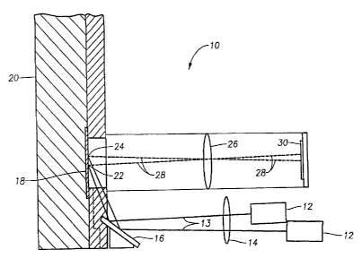
FIGURE 1 is a simplified block diagram of a noninvasive optical pressure
sensor according to one embodiment of the present invention. The pressure
sensor
can generally include light sources 12, source lens 14, mirror 16, pressure
chamber
20, flexible membrane 18, imaging lens 26, and image sensor 30. Other
embodiments
can comprise a single light source 12. A single light source 12 or multiple
light
sources 12 can also be focused directly onto flexible membrane 18. As will be
further
apparent to those familiar with the art, other optical elements can be used to
form an
optical path between light source 12 and diaphragm 18 to focus the light from
light
source 12 onto diaphragm 18.
Pressure chamber 20 can contain the fluid having a pressure to be measured
and may be made of any suitable material, such as metal, glass, or plastic,
and may be
of any suitable size or shape and can contain a port(s) (not shown) through
which the
pressure within chamber 20 may be varied. Diaphragm 18 is a flexible member
that
can be made of any suitably compliant material having good dimensional
stability,
such as stainless steel. Diaphragm 18 can further be a flat diaphragm, a pre-
curved
diaphragm (concave or convex) or a corrugated diaphragm. Diaphragm 18 should
have a consistent texture (if present) across the portion of its surface that
may receive
incident light from a light source 12. Light sources 12 can be any of a
variety of
8
CA 02511029 2005-06-28
Docket 2715US
Express Mail EV 224562275 US
commercially available light sources, such as a laser, laser diode, or LEF,
but
preferably a laser diode as known in the art.
Light sources 12 provide beams of light 13, which are directed to pass through
focusing lens 14 and onto mirror 16. Mirror 16 reflects light beams 13 onto
diaphragm 18. At diaphragm 18, light beams 13 are focused as spots 22 and 24
on
diaphragm 18 and are reflected as light beams 28, corresponding to the
reflected
images of spots 24 and 22 from diaphragm 18. Light beams 28 pass through lens
26.
Lens 26 focuses light beams 28 on image sensor 30. Image sensor 30 captures
the
reflected images of spots 24 and 22 and, in particular, the spatial separation
between
spots 24 and 22. Image sensor 30 can be any of a variety of commercially
available
devices such as a CCD (charge-coupled device) or a CMOS (complementary-metal-
oxide semiconductor) image sensor, or even a PD (photo sensitive diode)
capable of
capturing and differentiating between the reflected images of spots 24 and 22.
As shown in FIGURE 1, light beams 13 are focused by source lens 14 onto
mirror 16, which redirects light beams 13 onto diaphragm 18, forming spots 24
and
22. Light beams 13 are directed onto diaphragm 18 at a set incidence angle by
mirror
16. Spots 24 and 22 are reflected off of diaphragm 18 as light beams 28 and
refocused at image sensor 30 via imaging lens 26. When the pressure within
chamber
20 is at or near a reference pressure (e.g., ambient pressure), then as shown
in
FIGURE 1, diaphragm 18 will be at a preset reference position (e.g., flat as
shown).
In FIGURE 1, the reference position of diaphragm 18 is shown as flat for
purposes of
illustration, but the diaphragm 18 reference position can be any arbitrarily
determined
reference position at which the pressure sensor is calibrated for a reference
pressure,
such as ambient pressure.
Based on the relative positions of lights 12 to one another (a known and fixed
relationship) and the angle of incidence for light beams 13 provided by mirror
16,
spots 24 and 22 formed by reflected light beams 13 will be separated by a
preset
amount (i.e., have a fixed initial separation at a reference diaphragm
position). The
separation between spots 24 and 22 will be reproduced and detected at image
sensor
30 (in the embodiment shown, this is done via imaging lens 26, which focuses
the
9
CA 02511029 2005-06-28
Docket 2715US
Express Mail EV 224562275 US
reflection of spots 22 and 24 on the image sensor 30). When the pressure
within
chamber 20 is below the set reference pressure (e.g., ambient pressure), as
shown in
FIGURE 2, diaphragm 18 will be deflected inward (become concave), causing the
position of spots 22 and 24, both on the diaphragm and relative to one
another, to
change. The change in diaphragm position and in relative spatial separation
between
spots 22 and 24 is reproduced and detected at image sensor 30, as previously
described. One skilled in the art will recognize that in a similar manner,
pressure
changes in chamber 20 above the reference pressure will cause diaphragm 18 to
become convex (not shown), causing a shift in the position of spots 22 and 24
in a
io direction opposite to that of when pressure drops below the reference
pressure, with a
corresponding change in the separation between spots 22 and 24. The change in
position of spots 22 and 24 will similarly be reproduced and detected at image
sensor
30.
Thus, following a change in pressure, the change in relative separation and in
diaphragm position of spots 22 and 24 is detected and captured by image sensor
30,
and can then be analyzed using software well known in the art to calculate the
displacement of diaphragm 18. The displacement of diaphragm 18 can then be
correlated to a corresponding change in pressure. The displacement of
diaphragm 18
as indicated by the position changes of spots 22 and 24, directly relates to
the pressure
and pressure changes within chamber 20.
An alternative embodiment of the present invention can use a single light
source 12 to shine a single spot onto diaphragm 18. As the pressure in chamber
20
changes, diaphragm 18 will deflect as previously described and the change in
the
relative position of the single spot on diaphragm 18 can be correlated to the
change in
pressure in chamber 20. Alternative embodiments can also include directing the
light
source or sources 12 directly onto diaphragm 18 without the optical components
(path) formed by lens 14 and mirror 16. For example, a focusing assembly, such
as a
lens or a simple pinhole, can be incorporated into each light source 12. Other
focusing means for directing light from light sources 12 onto diaphragm 18
(and/or
from diaphragm 18 onto image sensor 30) will be known to those familiar with
the art
and are contemplated as being within the scope of this invention. A more
detailed
CA 02511029 2005-06-28
Docket 2715US
Express Mail EV 224562275 US
explanation for determining the angle of incidence of light beams 13 onto
diaphragm
18 and for calibrating a pressure sensor of the present invention (i.e.,
determining
reference positions and values) are provided later below.
FIGUREs 3A and 3B illustrate how the embodiments of this invention can
compensate for a change in the orientation of diaphragm 18 or chamber 20 from
a
reference position. Such a change in orientation might be caused, for example,
by a
variation in fit of a replaceable fluidics module in an ophthalmic surgical
system.
Such systems can use a replaceable fluidics cassette which can comprise a
pressure
io chamber having a diaphragm, corresponding to chamber 20 and diaphragm 18
of
FIGURE 1. As shown in FIGURE 3A, when diaphragm 18 (chamber 20) is at a
reference position (here shown as a flat diaphragm 18 and a chamber 20
oriented to a
reference position), spots 22 and 24 on diaphragm 18 are separated by a first
separation indicated by Line 40. In the event the initial position of a
chamber 20 and
diaphragm 18 changes from the reference position prior to a pressure
measurement
being made (e.g., due to a tilt from the reference position when a new
replaceable
chamber is inserted), then as shown in FIGURE 3B, the new position of spots 22
and
24 on diaphragm 18 can be measured and the tilt in the chamber 20/diaphragm 18
from the reference position can be compensated for prior to pressure
measurements
being made. This can be done, for example, as part of a calibration routine.
As when detecting changes in pressure during normal operation, image sensor
can be used to detect the change in the linear separation between spots 22 and
24
due to a tilt as described above and provide this information to a processing
system to
25 compensate for the tilt. Such a processing system can comprise a
processor, a
memory and computer executable software instructions stored within the memory
and
capable of being executed by the processor. A processing system in accordance
with
the teachings of this invention is described more fully later below. Software
for
correlating the change in separation of spots 24 and 22 to the change in
pressure
30 within a chamber 20, or to a change in reference position due to
variations between
replaceable chambers, will be known to those familiar with the art. Any such
software can be used with the embodiments of the present invention.
11
CA 02511029 2005-06-28
Docket 2715US
Express Mail EV 224562275 US
FIGURE 4 is a simplified diagram illustrating a method for calibrating the
incidence angle of light beams 13 onto diaphragm 18 (which form spots 22 and
24).
It is necessary to know the angle of incidence of light beams 13 onto
diaphragm 18
because the spacing of spots 22 and 24 on diaphragm 18 depends on the angle of
incidence of the light beams 13. To determine the angle of incidence, the
location of
a spot, such as spot 22 of FIGURE 1, is measured on a reference diaphragm,
such as
previously described above. The diaphragm 18 is then deflected from the
reference
position to a test position and the change in spot location is measured. Using
well-
known mathematical formulas, the angle of incidence is computed and the
process is
io repeated for each light source 12 as needed. In this way, the angle of
incidence of the
light beams 13 onto diaphragm 18 is known.
As discussed above, one embodiment of this invention can be implemented to
measure pressure in a surgical cassette of a surgical system. Such a cassette
can
include a chamber 20 bounded by a diaphragm 18 that is connected to the
aspiration
line of the fluidics portion of the corresponding surgical system. One side of
the
diaphragm 18 can be exposed to the ambient air pressure. The diaphragm 18 will
deform as described above in response to pressure differences between the
aspiration
line and ambient pressure.
In each embodiment of this invention, the relationship between diaphragm 18
deformation and pressure difference is monotonic (or very nearly so). Thus,
measurement of the diaphragm 18 deformation can be used to infer the chamber
20
pressure based on a calibration relationship or table. The diaphragm 18
deformation
can be uniquely quantified as the deflection of its center. This can be
determined by
projecting the narrow beams of light 13, which preferably are generated by a
laser
light source, onto the surface of the diaphragm 18 at an oblique angle and
imaging the
resulting scattered light spots 22 and 24 on an image sensor 30 (e.g. a CCD or
CMOS
image sensor chip).
The location of the image of the spot can be quantitatively determined from
the image data or a sub-set of the image, such as one or a few lines of pixel
data,
using one of a number of techniques. Options for quantifying the location of
the
12
CA 02511029 2005-06-28
Docket 2715US
Express Mail EV 224562275 US
peaks include determining the center of mass of the spot, correlating the
image with a
reference shape and finding the peak of the result, or fitting a curve to the
data and
determining the shift required to minimize the error of the fit. The
correlation
technique is preferred because it can be made to work with different beam
shapes
(including asymmetric beams), is effective at suppressing or averaging out
noise and
can be implemented efficiently with a digital signal processor.
Initially the laser spots' 22 and 24 location is measured for a reference
pressure (such as ambient, or no net pressure difference). Pressure
measurements are
lo made by comparing the location of the laser spots 22 and 24 on the image
sensor 30
for that pressure condition to their position for the reference pressure (or
alternatively,
directly through the use of the absolute location of the spots 22 and 24
images on the
image sensor 30, compared to known landmarks on the image sensor and/or the
diaphragm 18). For time critical applications, the relative motion of the
spots 22 and
24 can be directly converted to pressure using a pre-computed look-up table as
described below.
A number of elements must be considered to make the pressure sensor of this
invention accurate and robust. First, it is important to know the angle (or
average
angle) at which a light beam 13 is incident on the diaphragm 18. For some
applications it is desirable to know the incidence angle to an accuracy of
approximately 10. At the same time, it is difficult to insure that the
orientation of the
diaphragm 18 with respect to the chamber 20 and the position of the chamber 20
with
respect to a fluidics mechanism in which it may be implemented will be
reproducible
enough to insure that this condition will hold, in particular in the case
where the
chamber 20 and diaphragm 18 are implemented as a replaceable unit. Therefore,
it is
necessary to precisely measure the angle of incidence of the light beams 13
with
respect to a reference diaphragm 18 when a system implementing an embodiment
of
this invention is manufactured and then measure changes in these angles each
time a
new chamber 20/diaphragm 18 unit is inserted in the system for a surgical
procedure
or session.
13
CA 02511029 2005-06-28
Docket 2715US
Express Mail EV 224562275 US
=
At time of assembly, the angle of incidence of a light beam 13 with respect to
the normal of a flat (or other reference position) diaphragm 18 can be
measured in
several different ways. In the first method, a diaphragm 18 or test target is
moved
towards or away from the image sensor 30 in precisely measured increments that
approximately span the range of positions that the diaphragm 18 may occupy
during
actual pressure measurements. As shown in FIGURE 5, the height (y) of a laser
spot
such as spot 22 or 24 as a function of position of a target location (z) can
then be used
to precisely calculate the angle (0) at which the laser/light beam is incident
upon the
diaphragm. This can be done by regressing the height of the spot in diaphragm
coordinates against the location of the optical spot on the target using a
well know
least squares approach. The arc tangent of the slope of the line relating beam
height
to target spacing gives the angle of incidence.
Alternatively, a series of known pressures can be applied to the diaphragm 18
is and the resulting position of the spot on the diaphragm 18 can be
recorded. The
response of the diaphragm 18 (deflection as a function of pressure) must be
known in
advance. The angle of incidence can then be determined by comparing the spot
location on diaphragm 18 measured for each pressure to the predictions of a
model
(described below) that relates the location of the spot on the diaphragm 18 to
the
20 angle of incidence of the beam 13. The angle of incidence is determined
by
numerically solving for the angle that best aligns the model to the data.
One or both of the above procedures can be used during the manufacture of an
embodiment of the pressure sensor of this invention to establish a reference
angle of
25 incidence for any light source 12 within the system. Accurate pressure
measurements
(e.g. accuracy of the greater of 10 mm Hg or 10%), however, require that the
incidence angle be updated each pressure measuring session. This can be done
by
using two or more light sources. By measuring the locations of the two spots
22 and
24 at some reference pressure (e.g. no applied pressure) at manufacture time,
30 instrument reference positions can be determined for each spot. When a
system
implementing a pressure sensor in accordance with this invention is used in
the field,
the positions of each of the laser spots can again be determined for a known
pressure,
such as P=0 mm Hg or ambient. These session reference locations are compared
to
14
CA 02511029 2005-06-28
Docket 2715US
Express Mail EV 224562275 US
the original reference locations. The two (or more) spots provide additional
information that can be used to determine changes in both the angle of
incidence of
the light from the light sources 12 (more precisely, the angle of incidence
with respect
to the plane that contains the two light beams 13) and the distance between
the
diaphragm 18 and the instrument itself.
To calculate the change in the angle of incidence, it is helpful to define a
coordinate system. As shown in FIGURE 6, this can be done by setting the y
axis
parallel to the direction of beam motion with changes in pressure/diaphragm 18
io position and the x axis perpendicular to the y axis, but in the plane of
the diaphragm
18. For the case of two light sources 12, the y-component of the instrument
reference
spot locations can be designated as y10 and y20. The corresponding components
of
the spot positions for the session references can be designated as y11 and
y21. If the
angle between the two laser beams is assumed to not change (i.e., any change
that
occurs is small compared to their common change in angle of incidence with
respect
to the diaphragm) due to manufacturing tolerances and variability of the
positioning
of the chamber 20 in the instrument, then the change in the angle of incidence
80 for
the session configuration relative to the instrument reference is given by
SO = (y21 - y20 )C0402 - ¨ y,o )cot(Oi )
tan EQUATION I
Y20 - Y10
where 01 and 02 are the angles of incidence of beams 1 and 2 (from two light
sources
12) respectively.
Once the angle of incidence has been measured for a particular unit containing
a chamber 20 (installed for a particular session), a look-up table can be
produced to
directly relate pressure to the relative position of a laser spot. The direct
relationship
between spot location and pressure is difficult to calculate. However, it is
possible to
readily determine the spot location and pressure associated with a particular
deflection
of the vertex of the diaphragm 18. Therefore, a convenient method for
developing the
look-up table is to start with a somewhat arbitrary, but dense array of z-
plane
deflection values (Az) for the diaphragm 18 that range from the lowest to
highest
CA 02511029 2005-06-28
Docket 2715US
Express Mail EV 224562275 US
deflections that a system implementing an embodiment of the pressure sensor of
this
invention is intended to support. For example, if under the conditions of
interest the
diaphragm 18 vertex may move from ¨0.3 mm to 0.7 mm, it is desirable to have
on
the order of 100 or more points, so that the Az array can correspond to 10pm
steps in
position and include 101 points ¨ 0.3 mm, ¨ 0.29 mm, ¨ 0.28 mm, ..., 0.69 mm,
0.7 mm.
The pressure associated with different vertex deflections can be measured by
applying a series of calibrated pressures to the diaphragm 18 and measuring
the
deflection of the center of the diaphragm 18 using either a mechanical probe
or an
optical technique, such as the processes described herein (laser spot position
can be
readily converted to equivalent diaphragm 18 deflection if the angle of
incidence is
known). The pressures corresponding to the array of Az values described above
can
be computed by interpolating between the data points of the measured diaphragm
18
pressure response curve.
At the same time, the location where a light beam 13 would hit the diaphragm
for each of the diaphragm 18 deflections Az can be calculated by using the
well
known theory of exact ray tracing, as described, for example, on p. 309 of
Modem
Optical System Design, 3rd Edition, by Warren Smith. To use this ray analysis,
the
diaphragm 18 is assumed to assume an approximately spherical shape in response
to
an applied pressure. The curvature, c, of the diaphragm 18 can be approximated
as
¨3.333*Az
c= _______________________________________________ EQUATION 2
Az2 a2
where "Az" is the deflection of the vertex of the diaphragm 18 and "a" is the
radius of
the diaphragm 18. The factor of 3.333 in the numerator is used in this case
instead of
2 to account for the fact that the effective radius of the diaphragm 18 is
smaller than
its physical size because the fixed edge of the diaphragm 18 only allows its
center
portion to move. The ray trace procedure indicates where a light beam 13 will
hit the
diaphragm 18. This position can either be used directly or converted to a
relative
16
CA 02511029 2005-06-28
Docket 2715US
Express Mail EV 224562275 US
position (Ay) by comparing it to the position associated with the reference
position
where, for example, pressure chamber 20 is at zero pressure.
Using the procedure described above, it is possible to calculate both the
laser
spot 22 and 24 locations (Ay) and the pressures associated with each of the
initial
values of the Az array, shown in FIGURE 7. Once the Ay values and the
pressures
have been calculated, they form a look-up table that relates relative laser
spot 22 and
24 position to pressure. For ease in looking up values, interpolation can be
used to
place the Ay values on a regular spacing. Pressure measurements can then be
made
io by determining the location of the laser spot(s) 22 and 24 and using the
look up table
to convert this position or relative position to a pressure. This process can
be
performed by a processor operably connected to the image sensor 30.
Embodiments of a pressure measurement system in accordance with this
15 invention generate the light beams 13, image the location of the light
beams 13
scattering from the surface of the diaphragm 18, determine the location of the
imaged
spots 22 and 24, process the information as described above, and store
calibration
information. FIGURE 8 shows another embodiment of a pressure sensing assembly
of the present invention, illustrating in block form the various elements for
20 performing the above-described functions.
As shown in FIGURE 8, Processor 100 is a central processing unit for
coordinating the various functions of a system in accordance with this
invention.
Processor 100 can provide an input to light source drivers 110, as well as
receive as
25 an input information from light source drivers 110, for example, during
a calibration
procedure. Processor 100 can process the light spot 22 and 24 positional
information
received from image sensor 150 (corresponding to image sensor 30 of FIGURE 1)
and determine the pressure within chamber 20 in a manner as previously
described.
Processor 100 can provide the derived pressure information to a fluidics
system of, for
30 example, an ophthalmic surgical system, via a fluidics interface 170.
Such
information can be used by the surgical system to control various flows within
the
fluidic system, such as aspiration flow. Calibration interface 160 can be used
for
calibrating the pressure sensor, as previously described.
17
CA 02511029 2005-06-28
Docket 2715US
Express Mail EV 224562275 US
A memory 105 is operably coupled to processor 100 and is operable to store
computer executable software instructions for performing the various steps of
the
embodiments of the methods of this invention. Imaging optics 140 can comprise
any
optics as required for a given implementation (e.g., imaging lens 26) as can
light
source optics 130 (e.g., source lens 14). Light sources 120 correspond to
light sources
12 of FIGURE 1 and can comprise any such light source as described herein.
Processor 100 may be a single processing device or a plurality of processing
devices. Such a processing device may be a microprocessor, micro-controller,
digital
signal processor, microcomputer, central processing unit, field programmable
gate
array, programmable logic device, state machine, logic circuitry, analog
circuitry,
digital circuitry, and/or any device that manipulates signals (analog and/or
digital)
based on operational instructions. The memory 105 may be a single memory
device
or a plurality of memory devices. Such a memory device may be a read-only
memory, random access memory, volatile memory, non-volatile memory, static
memory, dynamic memory, flash memory, and/or any device that stores digital
information. Note that when the processor 100 implements one or more of its
functions via a state machine, analog circuitry, digital circuitry, and/or
logic circuitry,
the memory storing the corresponding operational instructions is embedded with
the
circuitry comprising the state machine, analog circuitry, digital circuitry,
and/or logic
circuitry. The memory 105 stores, and the processor 100 executes, operational
instructions corresponding to at least some of the steps and/or functions
illustrated in
Figures 1-9.
In a particular embodiment, the memory 105 is operably coupled processor
100 and includes operational instructions that cause the processor 100 to
direct (e.g.,
via the light source drivers 110) a plurality of coherent light beams, at a
known
incidence angle, onto a flexible diaphragm, wherein the flexible diaphragm
forms a
portion of a pressure chamber and is configured to flex in response to a
change in
pressure in the pressure chamber and wherein the plurality of coherent light
beams
form a pattern of light spots on the diaphragm; capture at an image sensor an
image of
the light spot pattern reflected from the diaphragm, wherein the light spot
pattern is
18
CA 02511029 2005-06-28
Docket 2715US
Express Mail EV 224562275 US
indicative of the pressure within the pressure chamber; and determine the
pressure
within the pressure chamber from the captured light spot pattern image. Memory
105
can further include operational instructions that cause the processor 100 to
calibrate
itself at a reference pressure by associating a reference light spot pattern
to a reference
diaphragm position corresponding to the reference pressure.
Returning to FIGURE 1, the opto-mechanical portion of an embodiment of the
pressure sensor of this invention is shown. A diaphragm 18 can be imaged in
sharp
focus for a range of diaphragm 18 positions. As the diaphragm 18 deflects due
to
changing pressures within chamber 20, the image of the spots 22 and 24 on the
image
sensor 30 can blur, making it harder to determine the precise location of the
spots 22
and 24. This can be compensated somewhat by tilting the image sensor 30 such
that a
hypothetical extended object occupying the same space as the incident light
beams 13
would be in focus at the image sensor 30. This is illustrated schematically in
FIGURE 9. The tilt of image sensor 30 can be a predetermined amount, or can be
adjustable based on the particular application of the pressure sensor.
FIGURE 10 is a graph illustrating one calibration curve for pressure (mm Hs)
to the spot shift on diaphragm 18 in millimeters. The spot shift in this case
is a
change in the linear separation between spots 24 and 22. Curves such as shown
in
FIGURE 10 can be generated so that appropriate software can be used to
correlate the
change in distance between spots 24 and 22 to a corresponding change in
pressure
within chamber 20. These generated curves can be different for different
pressure
sensors implemented in accordance with this invention.
The embodiments of the pressure sensor of the present invention can measure
pressure in a range from about minus 700 mm Hg to about 150 mm Hg. The
embodiments of this invention can provide this range of measurement with
accuracy
approximately that of the greater of +1- 10% of the range or 10 mmHg. Further,
peak
detection capabilities of the embodiments of this invention encompass, for low
pressure, up to 50 mm, and for high pressure, 70 mm. This can be achieved by
straightforward location of a pixel at image sensor 30 where the signal
(received light
intensity) is the highest.
19
CA 02511029 2011-09-29
Pressure sensing assembly 10 of the present invention thus allows for the
noninvasive measurement of pressure within a chamber 20 by optical means. One
skilled in the art will also recognize that by varying the thickness of
diaphragm 18, the
optical properties of lenses 14, 26, or mirror 16 and/or the relative position
of these
components, the pressure range that can be detected by noninvasive pressure
sensor
can be adjusted for a particular implementation
Although the present invention has been described in detail herein with
10 reference to the illustrated embodiments, it should be understood that
the description
is by way of example only and is not to be construed in a limiting sense. It
is to be
further understood, therefore, that numerous changes in the details of the
embodiments of this invention and additional embodiments of this invention
will be
apparent to, and may be made by, persons ordinarily skilled in the art having
reference to this description. Thus, while the present invention has been
described in
particular reference to the general area of fluidic surgical systems, the
teachings
contained herein apply equally wherever it is desirous to provide noninvasive
pressure
sensing to avoid, for example contact with a contaminated fluid.