Note: Descriptions are shown in the official language in which they were submitted.
CA 02511484 2005-06-21
WO 2004/060424 PCT/US2003/041494
SILK STENT GRAFTS
FIELD OF THE INVENTION
The present invention relates generally to pharmaceutical compositions,
methods and devices, more specifically to stmt grafts, and particularly to
stmt grafts
that contain silk and methods for making and using such stmt grafts.
BACKGROUND OF THE INVENTION
Stent grafts are utilized not only to hold open a passageway, but also to
bridge across diseased vasculature from healthy vessel to healthy vessel. A
common
application of stmt grafts is to bypass an abdominal aortic aneurysm (AAA).
Briefly, a
stmt graft is inserted over a guide wire, from the femoral or iliac artery,
and deployed
within the aneurysm, resulting in maintenance of blood flow from an aorta of
acceptable (usually normal) caliber above the aneurysm to a portion of aorta
or iliac
artery(s) of acceptable (usually normal) caliber below the aneurysm. Blood
flow is
thereby excluded from entering the aneurysm sac. Blood within this excluded
sac
thromboses and the aneurysm thus has no flow within it, presumably reducing
the
pressure and thus its tendency to burst.
While generally useful, presently available stmt grafts have a number of
shortcomings. For example, current stmt grafts are prone to persistent leakage
around
the area of the stmt graft. Hence, pressure within the aneurysm sac stays at
or near
arterial pressure, and there remains a risk that the sac will rupture. There
are three
common types of perigraft leakage. The first type is direct leakage around the
stmt
graft. This can be persistent from the time of insertion because of poor
sealing between
the stmt graft and vessel wall, or can develop later because the seal is lost.
In addition,.
this problem can develop due to changes in the position or orientation of the
stmt graft
in relation to the aneurysm as the aneurysm grows, shrinks, elongates or
shortens with
time after treatment. The second type of perigraft leak can occur because
there are side
arteries extending out from the treated segment of blood vessel. Once the
device
excludes the aneurysm, flow can reverse within these blood vessels and
continue to fill
the aneurysm sac around the stmt graft. The third type of perigraft leak can
occur
because of disarticulation of the device (in the case of modular devices) or
because of
the development of holes within the graft material. The continuous pulsation
of the
CA 02511484 2005-06-21
WO 2004/060424 PCT/US2003/041494
vessel can cause the graft material to rub against a metallic stmt tyne,
leading to hole
formation and eventually causing graft failure. In addition, disarticulation
of the device
can develop due to changes in shape of the aneurysm as it grows, shrinks,
elongates or
shortens with time after treatment.
Stent grafts are also limited in their application to only selected patients
with aneurysms. For example, endovascular stems are an advance in the
treatment of
AAA as they offer the avoidance of standard therapy, which is a major
operation with a
significant morbidity, mortality, long hospital stays, and prolonged recovery
time.
However, endovascular technology is only applicable to certain patients with
AAA
because of (a) lack of a suitable route of access via the blood vessels to the
intended site
of deployment which prevents insertion of the device and (b) the patient's
anatomy.
In order to effectively exclude an aneurysm, the graft material needs to
be of a certain strength and durability, or else it will tear. Typically, in
order to achieve
these properties, a polyester (e.g., polyester sold, e.g., under the trade
name DACRON
(E. I. DuPont De Nemours and Company, Wilmington, DE) or
poly(tetrafluoroethylene) (PTFE)) graft material of conventional "surgical"
thickness
may be used. This level of thickness is needed in order to convey adequate
strength to
the material. The thickness of the material results in the need for delivery
devices
typically of 24 to 27 French (8 to 9 millimeter diameter) and occasionally up
to 32
French. This requires surgical exposure of the insertion site, usually a
common femoral
artery, and limits the application of the technology, as a larger delivery
device is more
difficult to manipulate through the iliac artery to the intended site of
delivery. Even
"low profile" devices, which use thinner graft material, are of a sufficient
size that a
surgical exposure of the blood vessel through which the device is inserted is
required.
If the iliac arteries or aorta are very tortuous, (as is frequently the case
in AAA), or
heavily calcified and diseased (another frequent association with AAA), this
may be a
contraindication to treatment, or cause of failure of attempted treatment,
because of
inability to advance a device to the site of deployment or potential for iliac
artery
rupture.
A stmt graft is typically used to bridge a diseased artery (usually an
aneurysm), extending from a portion of artery of acceptable caliber above the
diseased
region to an artery of acceptable caliber below the diseased region. To
achieve a long
lasting seal, the artery of acceptable caliber above the diseased region
("proximal
2
CA 02511484 2005-06-21
WO 2004/060424 PCT/US2003/041494
neck") should be at least 1.5 cm long without a major branch vessel arising
from it.
The artery of acceptable caliber below the diseased region ("distal neck")
should be at
least 1.0 cm long without a major branch vessel arising within that 1 cm
length of
vessel. Shorter "necks" at either end of the diseased segment, necks which are
sloping
rather than cylindrical, or necks which are smaller than the aneurysm but
still dilated in
comparison to the normal diameter for a vessel in this location predispose to
failure of
sealing around the stmt graft or delayed perigraft leaks. One further
difficulty with
present stmt grafts is that over time certain devices have a tendency to
migrate distally
within the abdominal aorta. Such migration results in device failure,
perigraft leak and
vessel occlusion.
The present invention provides a stmt graft that overcomes problems
associated with existing stmt grafts.
BRIEF SUMMARY OF THE INVENTION
Briefly stated, the present invention provides silk-containing stmt grafts,
compositions for modifying or coating stmt grafts with silk, and methods for
making,
and using these grafts.
Within one aspect of the invention, a stent graft is provided that includes
an endoluminal stmt and a graft, wherein the stmt graft includes silk. The
silk induces
a response in a host who receives the stent graft, where the response can lead
to
enhanced adhesion between the silk stmt graft and the host's tissue that is
adjacent to
the silk of the silk stmt graft. In various aspects, the silk comprises
fibroin and/or
sericin. The silk may be natural, unmodified silk, or it may be chemically
modified
silk, e.g., acylated silk. However, the silk should not be modified to such an
extent that
it eliminates the ability of the silk to induce the host to generate a
biological response
that can increase adhesion between the stmt graft and the tissue in the host
that is
adjacent to the silk of the silk stmt graft. The silk may be from any of
various sources,
e.g., from a silkworm or from a spider, or from recombinant sources. The silk
may be
attached to the graft by any of various means, e.g., by interweaving the silk
into the
graft or by adhering the silk to the graft (e.g., by means of an adhesive or
by means of
suture). The silk may be in the form of a thread, a braid, a sheet, powder,
etc. As for
the location of the silk on the stmt graft, in one aspect, the silk may be
attached only the
exterior of the stmt, and/or in another aspect the silk may be attached to
distal regions
CA 02511484 2005-06-21
WO 2004/060424 PCT/US2003/041494
of the stmt graft, in order to assist in securing those distal regions to
neighboring tissue
in the host. In one aspect, a plurality of separated silk braids is attached
to the stmt
graft. The silk may be attached to the stmt portion of the stmt graft and/or
to the graft
portion of the stmt graft.
A wide variety of stmt grafts may be utilized within the context of the
present invention, depending on the site and nature of treatment desired.
Stent grafts
may be, fbr example, bifurcated or tube grafts, cylindrical or tapered, self
expandable or
balloon-expandable, unibody or, modular, etc.
In addition to silk, the stmt graft of the present invention may contain a
coating on some or all of the silk, where the coating degrades upon insertion
of the stmt
graft into a host, the coating thereby delaying contact between the silk and
the host.
Suitable coatings include, without limitation, gelatin, degradable polyesters
(e.g.,
PLGA, PLA, MePEG-PLGA, PLGA-PEG-PLGA, and copolymers and blends thereof),
cellulose and cellulose derivatives (e.g., hydroxypropyl cellulose),
polysaccharides
(e.g., hyaluronic acid, dextran, dextran sulfate, chitosan), lipids, fatty
acids, sugar
esters, nucleic acid esters, polyanhydrides, polyorthoesters and
polyvinylalcohol (PVA).
The silk-containing stmt grafts of the present invention may, in one
aspect, contain a biologically active agent, where the agent is released from
the stmt
graft and then induces an enhanced cellular response (e.g., cellular or
extracellular
matrix deposition) and/or fibrotic response in a host into which the stmt
graft has been
inserted. Exemplary agents include, without limitation, bleomycin or an
analogue or
derivative thereof, talcum powder, talc, ethanol, metallic beryllium and
oxides thereof,
silver nitrate, copper, silk, silica, crystalline silicates, quartz dust, and
vinyl chloride.
Exemplary polymeric agents include polyethylene-co-vinylacetate),
polyurethane,
polymers and copolymers of acrylic acid, and polymers of vinyl chloride. The
agent
may be an adhesive, such as, cyanoacrylate, crosslinked polyethylene glycol) -
methylated collagen, and derivatives thereof; a protein, carbohydrate or
peptide that
contains cellular adhesion sequences; an inflammatory cytokine (e.g., TGF(3,
PDGF,
VEGF, aFGF , bFGF, TNFoc, NGF, GM-CSF, IGh'-a, IL-1, IL-8, IL-6, growth
hormone,
EDGF, CTGF, and peptide and non-peptide agonists, analogues and derivatives
thereof); a component of extracellular matrix (e.g., vitronectin, fibronectin,
chondroitin
sulphate, laminin, hyaluronic acid, elastin, fibrin, fibrinogen, bitronectin,
proteins found
in basement membrane, fibrosin, or collagen); polylysine, chitosan, or N-
4
CA 02511484 2005-06-21
WO 2004/060424 PCT/US2003/041494
carboxybutylchitosan; a factor produced by immune cells (e.g., Interleukin-2
(IL-2),
Interleukin-4 (IL-4), Interleukin-1 (IL-1), Interleukin-8 (IL-8), Interleukin-
6 (IL-6) and
peptide and non-peptide agonists, analogues and derivatives thereof,
Granulocyte-
Monocyte Colony-Stimulating-Factor (GM-CSM), monocyte chemotactic protein,
histamine, and cell adhesion molecules; naturally occurring and synthetic
peptides
containing the RGD residue sequence; bone morphogenic protein (BMP) (e.g., BMP-
2,
BMP-3, BMP-4, BMP-5, BMP-6, or BMP-7); an inorganic and organic small anionic
molecule stimulant; and DNA and RNA sequences which are capable of promoting
synthesis of proteins that stimulate cell growth.
In one aspect, the stmt graft of invention further comprises a
proliferative agent that stimulates cellular proliferation. Representative
examples of
proliferative agents include dexamethasone, isotretinoin, 17-(3-estradiol,
diethylstibesterol, cyclosporin A, all-trans retinoic acid (ATRA), and
analogues and
derivatives thereof.
In another aspect, the stmt graft of the invention further comprises a
biologically active agent that inhibits or prevents expansion of an aneurysm,
such as a
caspase inhibitor (e.g., VX-799); an MMP inhibitor (e.g., BATIMASTAT or
MARIMISTAT); a tissue inhibitor of matrix metalloproteinases (TIMP); a
cytokine
inhibitor (e.g., chlorpromazine, mycophenolic acid, rapamycin, or loc-hydroxy
vitamin
D3); a MCP-1 antagonist (e.g., nitronaproxen, Bindarit, or 1-alpha-25
dihydroxy
vitamin D3); a TNFa antagonist or a TALE inhibitor (e.g., E-5531, AZD-4717,
glycophosphopeptical, UR-12715, cilomilast, infliximab, lentinan, or
etanercept); an
IL-1, ICE, and IRAK antagonist (e.g., E-5090, CH-172, CH-490, AMG-719,
iguratimod, AV94-88, pralnacasan, ESONARIMOD, or tranexamic acid); a chemokine
receptor antagonist (e.g., ONO-4128, L-38-1, CT-112, AS-900004, SCH-C, ZK-
811752,
PD-172084, UK-427857, SB-380732, vMIP II, SB-265610, DPC-168, TAK-779, TAK-
220, or KRH-1120); or an anti-inflammatory agent (e.g., dexamethasone,
cortisone,
fludrocortisone, prednisone, prednisolone, 6a-methylprednisolone,
triamcinolone,
betamethasone, and analogues and derivatives thereof).
In addition, the present invention provides methods for forming a silk-
containing stmt graft. In various aspects, which are exemplary only, the silk
may be
attached to the stmt graft by interweaving the silk into the graft, or the
silk may be
attached to the stmt graft by means of an adhesive, or the silk may be
attached to the
CA 02511484 2005-06-21
WO 2004/060424 PCT/US2003/041494
stmt graft by means of suture. In one aspect the silk is attached only to the
outside of
the stmt graft, andlor the silk may be attached to distal regions of the stmt
graft. In one
aspect, the silk is added to the stmt graft in an amount effective to induce a
biological
response in a host into which the stmt graft has been inserted, where the
biological
response is a cellular matrix deposition between the stent graft and tissue
adjacent to the
stmt graft. In a related aspect, the silk is added to the stmt graft in an
amount effective
to induce a biological response in a host into which the stent graft has been
inserted,
where the biological response is a cellular or extracellular matrix deposition
between
the stmt graft and tissue adjacent to the stmt graft. Optionally, the presence
of the silk
induces an enhanced biological response, i. e., a greater biological response
than would
have occurred in the absence of the silk on the stmt graft.
Also provided by the present invention are methods for treating patients
having aneurysms (e.g., abdominal, thoracic, or iliac aortic aneurysms), for
bypassing a
diseased portion of a vessel, or for creating communication or a passageway
between
one vessel and another (e.g., artery to vein or vice versa, or artery to
artery or vein to
vein), such that risk of rupture of the aneurysm is reduced. In one
embodiment, the
stmt graft is delivered into a patient (e.g., by balloon catheter) in a
constrained form,
and self expands into place after release of a constraining device. The
methods utilize
the silk-containing stmt grafts of the present invention. As utilized herein,
it should be
understood that "reduction in the risk of rupture" or "prevention of the risk
of rupture"
refers to a statistically significant reduction in the, number, timing, or,
rate of rupture,
and not to a permanent prohibition of any rupture. Likewise, a "reduction in
the risk of
perigraft leakage refers to statistically significant enhancement in the
effectiveness
and/or effective lifetime of a stmt graft, and not to a permanent or complete
cessation
of perigraft leakage.
The present invention addresses shortcomings in current stmt graft
technology by providing novel compositions, methods for preparing, and devices
related to silk-containing stmt grafts. The invention further provides other
related
advantages as disclosed below.
These and other aspects of the present invention will become evident
upon reference to the following detailed description and attached drawings. In
addition,
various references are set forth herein which describe in more detail certain
procedures
6
CA 02511484 2005-06-21
WO 2004/060424 PCT/US2003/041494
and/or compositions (e.g., polymers), and these references are incorporated by
reference
in their entirety.
BRIEF DESCRIPTION OF THE DRAWINGS
Figure 1 is a schematic illustration of a representative stmt graft.
Dashed lines indicate coating of the graft with a desired agent at each end of
the graft.
Figure 2 is a cross-sectional view of the stmt graft illustrated in Figure 1.
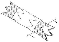
Figure 3 is a schematic illustration of a silk stmt graft of the present
invention having silk sutures that are secured to the stmt graft in a
horizontal, diagonal
or vertical manner.
Figure 4 is a schematic illustration of a silk stmt graft of the present
invention having silk sutures that are attached at either one end or both ends
of the silk
threads, where the silk extends some distance from the stmt graft.
Figure 5 is a graph showing the % activation of proliferation in smooth
1 S muscle cells as a function of cyclosporin A concentration.
Figure 6 is a bar graph showing the average number of cells migrating
for untreated and paclitaxel treated primary smooth muscle cells in response
to
rhPDDF-BB.
Figure 7 is a bar graph showing the area of granulation tissue in carotid
arteries exposed to silk coated perivascular PU films relative to arteries
exposed to
uncoated PU films.
Figure 8 is a bar graph showing the area of granulation tissue in carotid
arteries exposed to silk suture coated perivascular PU films relative to
arteries exposed
to uncoated PU films.
Figure 9 is a bar graph showing the area of granulation tissue in carotid
arteries exposed to natural and purified silk powder and wrapped with
perivascular PU
film relative to a control group in which arteries are wrapped with
perivascular PU film
only.
Figure 10 is a bar graph showing the area of granulation tissue (at 1
month and 3 months) in carotid arteries sprinkled with talcum powder and
wrapped
with perivascular PU film relative to a control group in which arteries are
wrapped with
perivascular PU film only.
7
CA 02511484 2005-06-21
WO 2004/060424 PCT/US2003/041494
Figure 11 is a photograph (100x) showing the cross section of a carotid
artery one month after insertion of a stmt graft (control) .
Figure 12 is a photograph (100x) showing the cross section of a carotid
artery one month after insertion of a silk covered stmt graft.
DETAILED DESCRIPTION OF THE INVENTION
DEFINITIONS
Prior to setting forth the invention, it may be helpful to an understanding
thereof to first set forth definitions of certain terms that are used
hereinafter.
"Stmt graft" refers to devices comprising a graft or wrap (composed of a
textile, polymer, or other suitable material such as biological tissue) which
maintains
the flow of fluids (e.g., blood) from one portion of a vessel to another, and
an
endovascular scaffolding or stmt (including expandable and inflatable stmt
structures)
that holds open a body passageway and/or supports the graft or wrap. The graft
or wrap
may be woven within a stent, contained within the lumen of a stmt, and/or be
located
exterior to a stmt.
"Fibrosis" or "Scarring" refers to the formation of fibrous tissue in
response to injury or medical intervention. Therapeutic agents which promote
fibrosis
or scarring (also referred to herein as fibrosing or fibrosis inducing agents)
can do so
through one or more mechanisms including: inducing or promoting angiogenesis,
stimulating migration or proliferation of comiective tissue cells (such as
fibroblasts,
and/or smooth muscle cells), inducing ECM (extracellular matrix) production,
and/or
promoting tissue remodeling. In addition, numerous therapeutic agents
described in
this invention will have the additional benefit of also promoting tissue
regeneration (the
replacement of injured cells by cells of the same type).
Silk refers to a fibrous protein, and may be obtained from a number of
sources, typically spiders and silkworms. Typical silks contain about 75% of
actual
fiber, referred to as fibroin, and about 25% sericin which is a gummy protein
that holds
the filaments together. Silk filaments are generally very fine and long - as
much as
300-900 meters long. There are several species of domesticated silkworm that
are used
in commercial silk production, however, Bonzbyx rnori is the most common, and
most
silk comes from this source. Other suitable silkworms include Philosamia
Cynthia
ricini, Antheraea yamamai, Antheraea perwyi, and Anthe~~aea mylitta. The silk
can be
CA 02511484 2005-06-21
WO 2004/060424 PCT/US2003/041494
processed to produce the raw silk or floss silk. Some of these processes
involve
degumming the silk. The steps to produce the different types of silk can
include steps
that can remove some or all of the sericin. Spider silk is relatively more
difficult to
obtain, however, recombinant techniques hold promise as a means to obtain
spider silk
at econonucal prices (see, e.g., U.S. Patent Nos. 6,268,169; 5,994,099;
5,989,894; and
5,728,810, which are exemplary only). Biotechnology has allowed researchers to
develop other sources for silk production, including animals (e.g., goats) and
vegetables
(e.g., potatoes). Silk from any of these sources may be used in the present
invention,
however, in one aspect of the invention the silk is not exclusively spider-
derived silk or
a genetically engineered spider silk as disclosed in, e.g., U.S. Patent
application No.
US2001/0053931 A1. In one aspect of the present invention, the silk is not
exclusively
biological or genetically-engineered spider silk or a derivative thereof, such
as spider
silk derived from Nephila clavipes, or a genetically engineered copy or
variant thereof.
In another aspect of the invention, the stmt graft does not include any spider
silk. In
another aspect, less than 50% of the silk present in a stmt graft of the
present invention
is biologically or genetically-engineered spider silk or a derivative thereof.
Raw silk is typically twisted into a strand sufficiently strong for weaving
or knitting. Four different types of silk thread may be produced by this
procedure:
organzine, crepe, tram and thrown singles. Organzine is a thread made by
giving the
raw silk a preliminary twist in one direction and then twisting two of these
threads
together in the opposite direction. Crepe is similar to organzine but is
twisted to a much
greater extent. Twisting in only one direction two or more raw silk threads
makes tram.
Thrown singles are individual raw silk threads that are twisted in only one
direction.
Any of these types of silk threads may be used in the present invention.
The sills can be used in the form of threads, monofilament yarn,
multifilament yarn, braids, powders as well as oligomers of the silk protein.
In addition to raw silk, commercially available silk sutures that are used
for surgical closure applications also can be used in the present invention.
Examples of
such commercially available silk sutures include, but are not limited to,
those sold by
Ethicon Ins. (Somerville, NJ), USSC/David&Geck/Tyco (Norwalk, CT) and Suru
International (India).
In addition to silk threads, fibers and yarns, silk in other forms can be
used. A commercially available silk protein is available from Croda, Ins., of
9
CA 02511484 2005-06-21
WO 2004/060424 PCT/US2003/041494
Paxsippany, NJ., and is sold under the trade names CROSILK LIQUID (silk amino
acids), CROSILK 10,000 (hydrolyzed silk), CROSILK POWDER (powdered silk), and
CROSILKQUAT (cocodiammonium hydroxypropyl silk amino acid). Another
example of a commercially available silk protein is SERICIN, available from
Pentapharm, LTD, a division of Kordia, BV, of the Netherlands. Further details
of such
silk protein mixtures can be found in U.S. Patent. No. 4,906,460, to Kim, et
al.,
assigned to Sorenco. Silk useful in the present invention includes natural
(raw) silk,
hydrolyzed silk, and modified silk, i.e., silk that has undergone a chemical,
mechanical,
or vapor treatment, e.g., acid treatment or acylation (see, e.g., U.S. Patent
5,747,015).
However, as mentioned above, in one aspect of the invention the silk is not
spider-
derived silk or genetically engineered spider silk. In a further optional
aspect, the stmt
graft of the present invention contains silk that induces a greater tissue
inflammatory
response than does spider silk. In yet another optional embodiment, the silk
present in
the stmt graft of the present invention promotes a tissue inflammatory
response.
The silk used in the present invention may be in any suitable form that
allows the silk to be joined (e.g., physically, mechanically, chemically or
via coating)
with the stmt graft, e.g., the silk may be in thread or powder-based forms.
Generally,
the silk is not released from the stmt graft after insertion into the patient,
however, in
certain applications, it may be desirable that the silk be released from the
stmt graft.
Furthermore, the silk may have any molecular weight. This molecular
weight can range from what is naturally found to molecular weights that can
typically
be obtained by the hydrolysis of natural silk, where the extent and harshness
of the
hydrolysis conditions determines the product molecular weight. For example,
silk
powders can have a molecular weight of about 100,000 to 300,000 Da while a
soluble
silk may have an average (number or weight) molecular weight of 200 to 5,000.
See,
e.g., JP-B-59-29199 (examined Japanese patent publication) for a description
of
conditions that may be used to hydrolyze silk.
A discussion of silk may be found in the following documents, which are
exemplary only: Hinman, M.B., et al. "Synthetic spider silk: a modular fibre"
Trends i~t
Biotechv~ology, 2000,18(9) 374-379; Vollrath, F. and Knight, D.P. "Liquid
crystalline
spinning of spider silk" Nature, 2001, 410(6828) 541-548; and Hayashi, C.Y.,
et al.
"Hypotheses that correlate the sequence, structure, and mechanical properties
of spider
CA 02511484 2005-06-21
WO 2004/060424 PCT/US2003/041494
silk proteins" I~t. J. Biol. Macromolecules, 1999, 24(2-3), 265-270; and U.S.
Patent No.
6,427,933.
The silk utilized in the present invention is intended to cause or induce a
biological reaction by the host who has received the stmt graft. In one
aspect, the silk
is utilized in order to induce a fibrotic reaction so that scarring occurs in
the vicinity of
the stmt graft. To this extent then, the silk is non-biocompatible.
As discussed above, the present invention provides compositions,
methods and devices relating to silk-containing stmt grafts, where the
presence of silk
greatly increases the success and application of the stmt graft. Described in
more detail
below are methods for constructing silk-containing stmt grafts, compositions
and
methods for generating silk-containing stmt grafts that adhere to a vessel
wall, and
methods for utilizing such stmt grafts.
STENT GRAFTS
As noted above, stmt grafts are devices that include a graft or wrap
which maintains the flow of fluids (e.g., blood) from one portion of a vessel
to another,
or from one blood vessel to another, and an endovascular scaffolding or stmt
which
holds open a body passageway and/or supports the graft or wrap. One
representative
stmt graft is illustrated in Figures 1 and 2.
The graft portion of the stmt may be composed of a textile, polymer, or
other suitable material such as biological tissue. Representative examples of
suitable
graft materials include textiles (including, e.g., woven and non-woven
materials) made
from polymeric fibers. Polymeric fibers for use in textiles may be formed from
a
variety of polymers, including, for example, nylon, acrylonitrile polymers and
copolymers (available, e.g., under the trade name ORLON (E. I. DuPont De
Nemours
and Company, Wilmington, DE)), polyesters (available, e.g., under the trade
name
DACRON (E. I. DuPont De Nemours and Company)), and poly(tetrafluoroethylene)
(available, e.g., under the trade name TEFLON (E. I. DuPont De Nemours and
Company)). Other representative examples of graft materials include non-
textiles, such
as expanded polytetrafluroethylene (ePTFE). The graft or wrap may be woven
within a
stmt, contained within the lumen of a scent andlor be located exterior to a
stmt.
Representative examples of stmt grafts, and methods for making and
utilizing such grafts are described in more detail in U.S. Patent No.
5,810,870 entitled
11
CA 02511484 2005-06-21
WO 2004/060424 PCT/US2003/041494
"Intraluminal Stent Graft"; U.S. Patent No. 5,776,180 entitled "Bifurcated
Endoluminal
Prosthesis"; U.S. Patent No. 5,755,774 entitled "Bistable Luminal Graft
Endoprosthesis"; U.S. Patent Nos. 5,735,892 and 5,700,285 entitled
"Intraluminal Stent
Graft"; U.S. Patent No. 5,723,004 entitled "Expandable Supportive Endoluminal
Grafts"; U.S. Patent No. 5,718,973 entitled "Tubular Intraluminal Graft"; U.S.
Patent
No. 5,716,365 entitled "Bifurcated Endoluminal Prosthesis"; U.S. Patent No.
5;713,917
entitled "Apparatus and Method for Engrafting a Blood Vessel"; U.S. Patent No.
5,693,087 entitled "Method for Repairing an Abdominal Aortic Aneurysm"; U.S.
Patent
No. 5,683,452 entitled "Method for Repairing an Abdominal Aortic Aneurysm";
U.S.
Patent No. 5,683,448 entitled "Intraluminal Stent and Graft"; U.S. Patent No.
5,653,747
entitled "Luminal Graft Endoprosthesis and Manufacture Thereof'; U.S. Patent
No.
5,643,208 entitled "Balloon Device of Use in Repairing an Abdominal Aortic
Aneurysm"; U.S. Patent No. 5,639,278 entitled "Expandable Supportive
Bifurcated
Endoluminal Grafts"; U.S. Patent No. 5,632,772 entitled "Expandable Supportive
Branched Endoluminal Grafts"; U.S. Patent No. 5,628,788 entitled "Self
Expanding
Endoluminal Stent-Graft"; U.S. Patent No. 5,591,229 entitled "Aortic Graft for
Repairing an Abdominal Aortic Aneurysm"; U.S. Patent No. 5,591,195 entitled
"Apparatus and Methods for Engrafting a Blood Vessel"; U.S. Patent No.
5,578,072
entitled "Aortic Graft and Apparatus for Repairing an Abdominal Aortic
Aneurysm";
U.S. Patent No. 5,578,071 entitled "Aortic Graft"; U.S. Patent No. 5,571,173
entitled
"Graft to Repair a Body Passageway"; U.S. Patent No. 5,571,171 entitled
"Method for
Repairing an Artery in a Body"; U.S. Patent No. 5,522,880 entitled "Method for
Repairing an Abdominal Aortic Aneurysm"; U.S. Patent No. 5,405,377 entitled
"Intraluminal Stent"; U.S. Patent No. 5,360,443 entitled "Aortic Graft for
Repairing an
Abdominal Aortic Aneurysm"; U.S. Patent No. 6,488,701 entitled "Stmt-graft
assembly with thin-walled graft component and method of manufacture"; U.S.
Patent
No. 6,482,227 entitled "Stmt graft having improved attachment within a body
vessel";
U.S. Patent No. 6,458,152 entitled "Coiled sheet graft for single and
bifurcated lumens
and methods of making and use"; U.S. Patent No. 6,451,050 entitled "Stmt graft
and
method"; U.S. Patent No. 6,395,018 entitled "Endovascular graft and process
for
bridging a defect in a main vessel near one of more branch vessels"; U.S.
Patent No.
6,390,098 entitled "Percutaneous bypass with branching vessel"; U.S. Patent
No.
6,361,637 entitled "Method of making a kink resistant stmt-graft"; U.S. Patent
No.
12
CA 02511484 2005-06-21
WO 2004/060424 PCT/US2003/041494
6,348,066 entitled "Modular endoluminal stent-grafts and methods for their
use"; U.S.
Patent No. 6,344,054 entitled "Endoluminal prosthesis comprising stmt and
overlying
graft cover, and system and method for deployment thereof'; U.S. Patent No.
6,325,820
entitled "Coiled-sheet stmt-graft with exo-skeleton"; U.S. Patent No.
6,322,585 entitled
"Coiled-sheet stmt-graft with slidable exo-skeleton"; U.S. Patent No.
6,319,278 entitled
"Low proftle device for the treatment of vasculax abnormalities"; U.S. Patent
No.
6,296,661 entitled "Self expanding stmt-graft"; U.S. Patent No. 6;245,100
entitled
"Method for making a self expanding stmt-graft"; U.S. Patent No. 6;238,432
entitled
"Stmt graft device for treating abdominal aortic aneurysms"; U.S. Patent No.
6,214,039
entitled "Covered endoluminal stmt and method of assembly"; U.S. Patent No.
6,168,610 entitled "Method for endoluminally excluding an aortic aneurysm";
U.S.
Patent No. 6,165,213 entitled "System and method for assembling an endoluminal
prosthesis"; U.S. Patent No. 6,165,210 entitled "Self expandable helical
intravascular
stmt and stmt-graft"; U.S. Patent No. 6,143,022 entitled "Stent-graft assembly
with
dual configuration graft component and method of manufacture"; U.S. Patent No.
6,123,722 entitled "Stitched scent grafts and methods for their fabrication";
U.S. Patent
No. 6,117,167 entitled "Endoluminal prosthesis and system for joining"; U.S.
Patent
No. 6,099,559 entitled "Endoluminal support assembly with capped ends"; U.S.
Patent
No. 6,042,605 entitled "Kink resistant stmt-graft"; U.S. Patent No. 6,015,431
entitled
"Endolumenal stent-graft with leak-resistant seal"; U.S. Patent No. 5,957,974
entitled
"Stmt graft with braided polymeric sleeve"; U.S. Patent No. 5,916,264 entitled
"Stent
graft"; U.S. Patent No. 5,906,641 entitled "Bifurcated stmt graft"; U.S.
Patent No.
5,891,191 entitled "Cobalt-chromium-molybdenum alloy stmt and stmt-graft";
U.S.
Patent No. 5,824,037 entitled "Modular intraluminal prostheses construction
and
methods"; U.S. Patent No. 5,824,036 entitled "Stent for intraluminal grafts
and device
and methods for delivering and assembling same"; U.S. Publication Nos.
2003/0120331; 2003/120338; and 2003/0125797; U.S. Patent No. 6,334,867, and
PCT
Publication No. WO 99/37242.
3O SILK STENT GRAFTS
In one aspect, the present invention provides a stmt graft to which silk
has been secured. The basic stmt graft may be any of the stmt grafts described
previously, or any other similar stmt graft. The silk that is present on the
stem graft
13
CA 02511484 2005-06-21
WO 2004/060424 PCT/US2003/041494
induces an enhanced fibrotic response between the stmt graft and the tissue
adjacent to
the ifz vivo stmt graft. Thus, in one aspect, the silk has the feature that it
will induce an
inflammatory response when contacted with a mammal. In another aspect, the
silk has'!
the feature that it will induce a cellular and/or extracellular matrix
deposition response
in an animal that is contacted with the silk. That is, absent the silk, the
stmt graft
would generate a "normal" adhesion between the adjacent tissue and the stmt
graft,
while in the presence of the silk the same stent/graft is capable of
generating an
enhanced adhesion via, e.g., an enhanced matrix deposition response to the
presence of
the silk. In one aspect of the invention, the silk excludes silks that do not
induce an
enhanced fibrotic response.
While the silk may be in any form or shape, e.g., sheet, powder, thread,
braid, filament, fiber, film, foam, and the like. In certain embodiments, the
silk is in the
form of a thread or powder. While the following discussion is primarily in
terms of
threads, the same principles and teachings apply to other forms and shapes of
the silk.
1 S The silk-containing threads will typically range in size from 1 nm to 3
mm in diameter although other sizes may be used and will also be effective.
The
threads can be individual thread (a monofilament), a multitude of threads
(multifilament
yarn), a braid, a knitted thread or a woven thread. The threads can be used
"as is", or
they can be further processed into a knitted or woven material that is then
attached to
the stmt graft. The threads can be made such that there are fibers) that
protrude from
the thread. These protruding fibers will further increase the exposed surface
area,
thereby enhancing the biological response when the stmt graft is inserted into
a host.
The fibers that protrude from the thread can be of the same composition as the
thread
material or they can comprise a different composition than the thread
material.
As discussed_in further detail below, the silk may be secured to the..stent
graft by any of a number of methods. Suitable methods include, without
limitation,
interweaving the silk into the graft, interweaving the silk into the stmt
structure;
attaching the silk to the stmt via knotting or suturing it around the stmt
structure;
attaching the silk to the stmt graft by means of an adhesive; and using one or
more
sutures to "sew" the silk onto the stmt graft. In one aspect, a plurality of
separated silk
braids or threads is attached to the stmt graft.
The silk itself may be natural sills, as obtained from, e.g., silkworms or
spiders. Alternatively, the silk may be a recombinant silk, or a chemically
modified silk
14
CA 02511484 2005-06-21
WO 2004/060424 PCT/US2003/041494
(e.g., acylated silk). In another aspect, the silk can be commercially
available silk
sutures. In one aspect, the silk includes fibroin, which is a component of
natural silk.
In another aspect, the silk includes sericin, which is also a component of
natural silk.
In one embodiment, the silk is secured only to the outside of the stmt
graft. In another embodiment, the silk is secured to distal regions of the
stent graft.
The silk may be attached to the stmt portion of the stmt graft, or it may be
attached to
the graft portion of the stmt graft, or it may be attached to both the stmt
and graft
portions of the stmt graft.
The silk threads can be located on the stmt-graft in various
configurations that may result in either partial or complete coverage of the
exterior of
the stmt-graft. The threads could be attached around the ends of the stmt-
graft, as
shown in Figure 3. The silk threads can be attached in bands along the stmt
graft. The
attaclunent could be in a vertical, horizontal or diagonal manner. Depending
on the
specific design of the stmt graft, the polymeric threads) can be attached to
either the
stmt component or the graft component of the stmt graft device. Alternatively,
or in
addition, the silk thread may be allowed to extend some distance from the
stent graft.
For example, as shown in Figure 4, only one end of the silk threads may be
secured to
the stmt graft, thereby allowing the other end of the thread to extend away
from the
graft. Alternatively, both ends of the thread may be secured to a stmt graft,
however,
the mid-portion of the thread is not secured to the stmt graft, and the ends
of the thread
are secured at a sufficiently short distance from one another that the mid-
portion is free
to extend away from the stmt graft.
In another embodiment, the ends of the silk threads can be attached to
the stmt graft, and/or one or more points along the silk thread can be
attached to the
stmt graft. In yet another embodiment, the ends of the silk thread are not
attached to
the stmt graft. Rather, one or more points along the silk thread are attached
to the stmt
graft. In yet another embodiment, the silk threads) can be made into a
preformed
structure (e.g., mesh, looped bundle, and the like) that is then attached to
the. stmt graft.
In one aspect, the invention provides a silk-containing stmt graft in
which the silk is present on the stmt graft in an amount effective to induce a
biological
response in a host into which the stmt graft has been inserted. The biological
response
may be manifested as a reduction in the risk of rupture of an aneurysm into
which the
stmt graft has been placed. In another aspect, the biological response is
manifested as a
CA 02511484 2005-06-21
WO 2004/060424 PCT/US2003/041494
reduction in perigraft leakage. The enhanced effectiveness of a silk-
containing stmt
graft may result from the silk inducing a cellular deposition between the
scent graft and
tissue adjacent to the stmt graft. The cellular proliferation and/or
extracellular matrix
secretion progresses over time to form a cellular or non-cellular matrix, more
commonly known as fibrotic tissue (i.e., tissue composed of fibroblasts,
smooth muscle
cells and extracellular matrix components such as collagen), which can hold
the stent-
graft in place within the vessel andlor act to fill part or all of the
aneurysm.
The stmt graft may, in addition to the silk, include a coating on some or
all of the silk. The coating can degrade or dissolve over a period of time
following
insertion of the stmt graft into a host. The presence of the coating functions
to delay
contact between the silk and the host. Suitable coatings for this purpose
include,
without limitation, gelatin, degradable polyesters (e.g., PLGA, PLA, MePEG-
PLGA,
PLGA-PEG-PLGA, copolymers and blends thereof), cellulose and cellulose
derivatives
(e.g., hydroxypropyl cellulose), polysaccharides (e.g., hyaluronic acid,
dextran, dextran
sulfate, chitosan), lipids, fatty acids, sugar esters, nucleic acid esters,
polyanhydrides
polyorthoesters and polyvinylalcohol (PVA). For example, in one embodiment of
the
invention, the silk is coated with a physical barrier. Such barriers can
include
biodegradable materials, such as gelatin, PLGA/MePEG film, PLA, polyethylene
glycol, and the like. In the case of PLGAI MePEG, once the PLGA/ MePEG becomes
exposed to blood, the MePEG will dissolve out of the PLGA, leaving channels
through
the PLGA to the underlying layer of silk. The exposed silk layer then is
available to
initiate its biological activity.
In another embodiment, the stmt graft can include a polymeric or non-
polymeric coating that further comprises silk. The silk can be in the form of
threads,
short fibers, particles, or a. combination thereof.
In another embodiment the stmt graft can include polymeric fibers,
yams or threads that are attached to the stmt graft. These fibers may be
composed of
polymers other than silk. Polymers that can be used include but are not
limited to
polyesters, such as DACRON, PTFE, nylon, poly(ethylene), polypropylene) or
degradable polyesters (e.g., PLGA, PCL, and poly(dioxanone)). These fibers can
have
one or more silk threads included in the polymeric fiber or yarn. In another
embodiment, these threads, fibers or yam can be coated with a polymeric or non-
polymeric carrier that further contains silk fibers, threads or particles. The
polymeric
16
CA 02511484 2005-06-21
WO 2004/060424 PCT/US2003/041494
carriers can be degradable or non degradable. Examples of polymer carriers and
non-
polymeric carriers that can be used are described below.
In addition, or instead of containing a coating as described above, the
silk-containing stmt graft of the present invention may further include a
biologically
active agent that is capable of inducing a fibrotic response in a host into
which the stmt
graft has been inserted. For example, the biologically active agent may induce
an
enhanced cellular deposition response andlor enhanced cellular matrix
deposition.
Exemplary agents include bleomycin and analogues and derivatives. Further
representative examples include talcum powder, talc, ethanol, metallic
beryllium and
oxides thereof, copper, silk, silver nitrate, quartz dust, crystalline
silicates and silica.
Other agents which may be used include components of extracellular matrix,
vitronectin, fibronectin, chondroitin sulphate, laminin, hyaluronic acid,
elastin, fibrin,
fibrinogen, bitronectin, proteins found in basement membrane, fibrosin,
collagen,
polylysine, vinyl chloride, polyvinyl chloride, polyethylene-co-vinylacetate),
polyurethane, polyester (e.g., DACRON), and inflammatory cytokines such as
TGF(3,
PDGF, VEGF (including VEGF-2, VEGF-3, VEGF-A,VEGF-B and VEGFC), aFGF,
bFGF, TNFa, NGF, GM-CSF, IGF-a, IL-1, IL-8, IL-6, growth hormone, EDGF
(epidermal growth factor), and CTGF (connective tissue growth factor), and
analogues
and derivatives thereof, and adhesives, such as cyanoacrylate or a crosslinked
polyethylene glycol) - methylated collagen composition, such as CT3 (Cohesion
Technologies, Palo Alto, CA). Additional agents include naturally occurring or
synthetic peptides containing the RGD (arginine-glycine-aspartic acid) residue
sequence, and factors produced by immune cells such as Interleukin-2 (IL-2),
Interleukin-4 (IL-4), Interleukin-1 (IL-1), Interleukin-8 (IL-8), Interleukin-
6 (IL-6),
Granulocyte-Monocyte Colony-Stimulating-Factor (GM-CSM), monocyte chemotactic
protein, histamine and cell adhesion molecules including integrins, and bone
morphogenic molecules including BMP-2, BMP-3, BMP-4, BMP-5, BMP-6 (Vgr-1),
BMP-7 (OP-1), BMP-8, BMP-9, BMP-10, BMP-1 l, BMP-12, BMP-13, BMP-14,
BMP-15 and BMP-16. Of these BMP's, BMP-2, BMP-3, BMP-4, BMP-5, BMP-6 and
BMP-7 are of particular utility. Other examples include peptide and non-
peptide
agonists of the above factors, and analogues and derivatives thereof,
proteins,
carbohydrates and peptides that contain cellular adhesion sequences, inorganic
or
17
CA 02511484 2005-06-21
WO 2004/060424 PCT/US2003/041494
organic small anionic molecule stimulants, and DNA or RNA sequences which
promote
the synthesis of proteins that stimulate cell growth.
In addition to, or instead of, containing a coating, as described above, the
silk-containing stmt graft of the present invention may further include a
biologically
active agent, wherein the agent induces an enhanced cellular proliferation
response in a
host into which the stmt graft has been inserted. Representative examples of
agents
that stimulate cellular proliferation include, without limitation,
dexamethasone,
isotretinoin, 17-(3-estradiol, diethylstibesterol, cyclosporin A and all-traps
retinoic acid
(ATRA) and analogues and derivatives thereof.
In another aspect of the invention, the biologically active agent may act
to inhibit processes which result in breakdown of the tissue within the
aneurysm which
can delay or prevent expansion of the aneurysm. Examples of such therapeutic
agents
include, without limitation, caspase inhibitors (e.g., VX-799), MMP inhibitors
(e.g.,
BATIMASTAT, also known as BB-94 and MARIMISTAT (both from British Biotech,
UK) and TIMP's (tissue inhibitors of matrix metalloproteinases)), cytokine
inhibitors
(e.g., chlorpromazine, mycophenolic acid, rapamycin, la-hydroxy vitamin D3),
MCP-1
antagonists (e.g., nitronaproxen, Bindarit, 1-alpha-25 dihydroxy vitamin D3 ),
TNFa
antagonists/TACE inhibitors (e.g., E-5531, AZD-4717, glycophosphopeptical, UR-
12715, cilomilast, infliximab, lentinan, and etanercept ), IL-1, ICE and IRAK
antagonists (e.g., E-5090, CH-172, CH-490, AMG-719, iguratimod, AV94-88,
pralnacasan, esonarimod, and tranexamic acid), chemokine receptor antagonists
(e.g.,
ONO-4128, L-381, CT-112, AS-900004, SCH-C, ZK-811752, PD-172084, UK-
427857, SB-380732, vMIP II, SB-265610, DPC-168, TAK-779, TAK-220, and KRH-
1120) and anti-inflammatory agents (e.g., dexamethasone, cortisone,
fludrocortisone,
prednisone, prednisolone, 6a-methylprednisolone, triamcinolone, and
betamethasone)
or analogues and derivatives thereof. It should be clear to one skilled in the
art that
these biologically active agents may be used individually or in combination or
may be
placed singly or in combination at various points within the stmt-graft and
that other
agents which act as therapeutic agents to prevent expansion of the aneurysm
can be
applied.
Within further aspects of the present invention, the silk-containing stent
grafts may include a polymeric carrier that is adapted to contain and release
a
18
CA 02511484 2005-06-21
WO 2004/060424 PCT/US2003/041494
therapeutic agent. Suitable polymeric carriers and therapeutic agents are
described
below.
In certain embodiments, the polymeric carrier may include regions,
pockets, or granules that contain one or more hydrophobic compounds (e.g.,
therapeutic
agents). For example, within one embodiment of the invention, hydrophobic
compounds may be incorporated within a matrix, followed by incorporation of
the
matrix within the polymeric carrier. A variety of matrices can be utilized in
this regard,
including for example, carbohydrates and polysaccharides, such as starch,
cellulose,
dextran, methylcellulose, chitosan and hyaluronic acid, and proteins or
polypeptides,
such as albumin, collagen and gelatin. Within alternative embodiments,
hydrophobic
compounds may be contained within a hydrophobic core, and this core contained
within
a hydrophilic shell. These and other carriers and therapeutic agents are
discussed in the
next section.
As mentioned above, the stmt graft may be of any type or configuration ,
that is suitable for the medical purpose intended. In various exemplary
aspects of the
invention, the stmt graft is bifurcated, the stmt graft is a tube graft, the
stmt graft is
cylindrical, the stmt graft is self expandable, and/or the stmt graft is
balloon-
expandable.
In one aspect, the stmt graft of the present invention is sterile. Many
pharmaceuticals are manufactured to be sterile and this criterion is defined
by the USP
XXII <1211>. Sterilization in this embodiment maybe accomplished by a number
of
means accepted in the industry and listed in the USP XXII <1211>, including
gas
sterilization or ionizing radiation. Sterilization may be maintained by what
is termed
aseptic processing, defined also in USP XXII <1211>. Acceptable gases used for
gas
sterilization include ethylene oxide. Acceptable radiation types used for
ionizing _
radiation methods include gamma, for instance from a cobalt 60 source and
electron
beam. A typical dose of gamma radiation is 2.5 MRad.
METHODS FOR MAKING SILK STENT GRAFTS
Silk may be attached to a stmt graft in any manner that creates a secure
bond between the stmt graft and the silk. This "bond" may be a chemical bond,
but it
may also be a mechanical bond, as described in further detail below. While the
19
CA 02511484 2005-06-21
WO 2004/060424 PCT/US2003/041494
following description is in terms of threads, silk of other configuration may
be applied
by the same techniques.
The polymeric silk threads can be attached to the stmt-graft in various
configurations that may result in either partial or complete coverage of the
exterior of
the stmt-graft. The threads could be attached around the ends of the stmt-
graft, as
shown in Figure 3. The attachment could be in a vertical, horizontal or
diagonal
manner. Depending on the specific design of the stmt graft, the polymeric
threads)
can be attached to either the stem component or the graft component of the
sfent graft
device.
In one embodiment, when the graft material is on the outer side of the
stmt, a preferred method of attachment is for the silk threads) to be attached
to the
graft material. In another embodiment, when the stmt is exterior to the graft
material, a
preferred method of attachment is for the silk threads) to be attached to
stmt. The silk
threads can be attached at a single point to the stmt graft or they can be
attached to the
stmt graft at multiple points. In addition, threads may be attached to the
central portion
of the stmt graft which will ultimately be located in the aneurysm. It is also
possible to
use a combination of all the above-described attachment methods.
The threads can be attached to the graft and/or the stmt material by use
of any one or a combination of the following exemplary methods: use of an
adhesive,
thermal welding, stitching, wrapping, weaving, knotting and looping. In one
aspect, an
adhesive is used to secure the silk to the stmt graft. In another aspect,
thermal welding
is used to secure the silk to the stmt graft. In another aspect, stitching is
used to secure
the silk to the stmt graft. In another aspect, wrapping is used to secure the
silk to the
stmt graft. In another aspect, weaving is used to secure the silk to the stmt
graft. In
another aspect, knotting is used to secure the silk to the stmt graft. 1n
another aspect,.
looping is used to secure the silk to the stmt graft.
In another aspect, the silk can be woven or knitted into a sheet or tubular
structure that is then attached to the exterior of the stmt graft structure.
This covering
can cover the entire exterior portion of the stmt graft or it can cover one or
more
specific portions of the stmt graft. In one embodiment, the covering is fixed
to the stmt
graft. The covering can be attached by knotting it or sewing it to the stmt
graft
structure, by using an adhesive to fix it to the stmt graft structure, or a
combination of
CA 02511484 2005-06-21
WO 2004/060424 PCT/US2003/041494
the above methods. In another embodiment, the covering is not fixed on the
stmt graft
and is simply placed as an outer covering on the stmt graft structure.
In one aspect, the stmt graft may be coated with a silk-containing
suspension, solution or emulsion. Examples of suitable emulsions or
suspensions
include aqueous formulations of commercially available silk powders (e.g.,
silk powder
available from Silk Biochemivcal Co., Ltd. (China), Nantong Dongchang Chemical
Industrial Co, Ltd. (China) and Wuxi Smiss Technology Co, Ltd. (China)), which
have
been formed into either a solution or an emulsion. Preferably, emulsions
contain
between about 5 to 50 wt. % solids.
In one embodiment, the silk threads can be coated with a material that
delays the time it takes for the silk to come into contact with the
surrounding tissue and
blood. This will allow placement of the stmt graft without concern of
thrombotic
events as a result of the silk threads. In one aspect, the. coating material
degrades or
dissolves during the deployment of the stmt, while in another aspect the
coating
material degrades or dissolves after the stmt graft has been implanted. These
coating
materials can be either polymeric or non-polymeric. Examples of coating
materials
include, without limitation, gelatin, degradable polyesters (e.g., PLGA, PLA,
MePEG-
PLGA, PLGA-PEG-PLGA, copolymers and blends thereof), cellulose and cellulose
derivatives (e.g., hydroxypropyl cellulose), polysaccharides (e.g., hyaluronic
acid,
dextran, dextran sulfate, chitosan), lipids, fatty acids, sugar esters,
nucleic acid esters,
polyanhydrides, polyorthoesters, and PVA.
The silk threads can be coated prior to attachment to the stmt graft or
they can be coated onto the silk threads once they have been attached to the
stmt graft.
This can be accomplished by using a spray-coating or dip-coating process.
In another embodiment, silk particle can. be incorporated into a
polymeric or a non-polymeric carrier which is in turn coated onto the stmt
graft. The
polymeric carriers can be either degradable or non-degradable. Examples of
polymer
carriers and non-polymeric carriers that can be used are described below.
In one embodiment, silk particles or silk fibers are added to a solution of
the polymeric or non polymeric carrier. The carrier solution forms a
suspension upon
addition of the sills particles or silk fibers. This suspension can be applied
to all or a
portion of the stmt graft by dipping, painting, or spraying.
21
CA 02511484 2005-06-21
WO 2004/060424 PCT/US2003/041494
In another embodiment the stmt graft can include polymeric fibers,
yarns or threads that are attached to the stmt graft. These fibers may be
composed of
polymers other than silk, such as, e.g., DACRON, PTFE, nylon, poly(ethylene),
polypropylene) or degradable polyesters (e.g., PLGA, PCL, and
poly(dioxanone)).
These fibers can have one or more silk threads included in the polymeric fiber
or yarn.
Iri another embodiment, threads, fibers or yarn can be coated with a polymeric
or non-
polymeric carrier that further contains silk fibers, threads or particles. The
polymeric
carriers can be either degradable or non degradable. The polymeric or non-
polymeric
carrier can be dissolved in a solvent that will not substantially dissolve the
polymeric
fiber during the exposure of the polymeric fiber to the solvent. Pieces of
silk fibers or
threads and/or silk particles can be added to the carrier solution. If
required, an
emulsifying agent or a surfactant can be added to the solution to aid in the
suspension of
the fibers, threads or particles. The polymeric threads, fibers, or yarn can
be coated
with the silk-containing carrier composition by dipping the polymeric threads,
fibers or
yarns into the silk/carrier suspension or spraying the silk/carrier suspension
onto the
polymeric threads, fibers or yarns. These coated systems can then be air dried
and if
required can be vacuum dried. The coated polymeric threads, fibers or yarn
then can be
attached to the stmt graft by methods disclosed herein.
In another embodiment, the polymeric thread, yarn, fiber, andlor the
stmt graft can be coated with a solution that contains a polymer or a non-
polymeric
carrier. The coating can be partially dried such that the coating is still
soft and tacky.
Silk thread, pieces of silk thread or silk powder then can be embedded into
the soft
coating. This can be accomplished by spraying the silk onto the soft coating,
by rolling
the coated form in the silk, by stamping the silk onto the coated form or by a
combination of these processes. The silk coated form can be further dried
to_remove
the residual solvent.
In one aspect of the invention, the graft (also referred to as a wrap or
sheath) may be prepared entirely from silk, where in one aspect the silk is
not a
biological or genetically engineered spider silk. For example, the entire
graft may be
formed from a biological or genetically engineered silkworm silk. However, in
a
different aspect, the stmt graft of the present invention contains a graft
that is not made
entirely of silk, however, silk is affixed to the stmt graft. This is a
preferred aspect
because, e.g., the amount of silk affixed to the stmt graft can be tailored to
achieve the
22
CA 02511484 2005-06-21
WO 2004/060424 PCT/US2003/041494
desired amount of biological response which is induced by the silk. Thus, in
one
aspect, the present invention provides a stmt graft wherein the graft is not
made entirely
from silk (or is not made from silk at all), however silk is affixed to the
stmt graft in a
manner as exemplified above. For example, the stmt graft may contain a graft
made
from non-silk material, e.g., polyester, polyamide, hydrocarbon polymer (e.g.,
polyethylene and polypropylene), polyurethane or fluoropolymer (or other
suitable
material) and silk is affixed to either the stmt or graft portion of the stmt
graft. In one
aspect, the stmt graft has a single graft, which in various separate
embodiments may be
woven within the stent, contained withitn the lumen of the stem, or be located
exterior to
the stem, where silk is affixed to this stmt graft. In another aspect, the
stmt graft has
two grafts, which in various embodiments may be woven within the stmt,
contained
within the lumen of the stmt, and/or be located exterior to the stmt, where
silk is
affixed to this stmt graft. When the stmt graft contains two grafts, the silk
is preferably
affixed to the graft in a manner that will allow the silk to contact the
vessel wall, e.g., it
may be affixed to the sheath which is located exterior to the stmt. As
mentioned
previously, in a preferred embodiment the silk is silkworm silk. For example,
fibers of
silkworm silk and fibers of a different material (polyester, polyamide, spider
silk, etc.)
may be combined together to form a sheath that is used to construct a stmt
graft of the
present invention.
In one embodiment, the silk or the silk/carrier compositions may further
contain a biologically active agent that reduces the probability of an
immediate
thrombotic event, where exemplary agents of this type include, without
limitation,
heparin and hydrophobic quaternary amine heparin (e.g., heparin-benzalkoiuum
chloride, heparin-tridodecylmethylammonium chloride) complexes. The heparin or
heparin complexes can be applied by dip coating or spray coating.
In another embodiment, the silk-containing thread, fiber, or yarn can
further contain a biologically active agent that enhances a cellular response
and/or a
fibrotic response. The agents that can be used in the present invention are
described
below. These agents can be incorporated by dip coating or spray coating the
silk-
containing threads, fibers or yarn with a solution that contains the
biologically active
agent. This solution can be a true solution, a suspension, a dispersion or an
emulsion.
The biologically active agents) can also be incorporated into a secondary
carrier. A
solution, suspension, dispersion or emulsion or the biologically active
agent/carrier can
23
CA 02511484 2005-06-21
WO 2004/060424 PCT/US2003/041494
be applied by a dip coating or spray coating process. These agents can be
applied to the
entire external surface of the stmt graft or to one or more specific locations
on the stmt
graft.
In another embodiment, the biologically active agent or biologically
active agent/secondary carrier (e.g., solution) can further comprise a
polymer. This
solution can be applied to the silk-containing thread, fiber or yarn.
In another embodiment, the biologically active agent and/or biologically
active agent/secondary carrier can be incorporated into a polymeric or non-
polymeric
carrier solution that contains silk. The solvent for the carrier may or may
not be a
solvent for the added biologically active agent. In the case where the solvent
is not a
solvent for the biologically active agent, the biologically active agent will
be in the
form of a suspension. In the case where the solvent fox the carrier is a
solvent for the
biologically active agent, a solution of the biologically active agent will be
formed. In
another embodiment, the solvent is a solvent for the biologically active
agent, but the
amount of the biologically active agent added to the solution is greater that
the
solubility limit of the biologically active agent. In this case, a saturated
suspension of
the biologically active agent will be formed. The silk- and biologically
active agent-
containing solution can be applied to the stent graft or the polymeric thread,
fiber or
yarn by a process of dip-coating or spray coating. The solution can be applied
to all of
the exterior of the stmt graft or to one or more regions of the stmt graft or
polymeric
thread, fiber or yarn.
In another embodiment, the coating includes a "biocompatible" polymer
that is coated with a polymer or other biologically active agent that results
in an
enhanced cellular response.
In one embodiment, the silk-containing stmt graft is coated with.a.
composition or a compound which promotes fibrosis and/or restenosis.
In another embodiment, the silk-containing stmt graft is coated with an
agent that is not released from the stmt graft but yet still results in an
enhanced cellular
and extracellular matrix deposition response. These agents can be coated
directly onto
the stmt graft or they can be incorporated into a non-degradable polymeric
carrier.
In one aspect, the silk-containing stmt grafts of the present invention are
coated with, or otherwise adapted to release an agent that induces adhesion to
vessel
walls. Stent grafts may be adapted to release such an agent by (a) directly
affixing to
24
CA 02511484 2005-06-21
WO 2004/060424 PCT/US2003/041494
the stmt graft a desired agent or composition (e.g., by either spraying the
stmt graft
with a polymer/agent film, or by dipping the stmt graft into a polymer/agent
solution,
or by other covalent or noncovalent means); (b) by coating the stmt graft with
a
substance such as a hydrogel which will in turn absorb the desired agent or
composition; (c) by interweaving an agent- or composition-coated thread into
the stmt
graft (e.g., a polymer which releases the agent formed into a thread); (d) by
inserting a
sleeve or mesh which is comprised of or coated with the desired agent or
composition;
(e) constructing the stmt graft itself with the desired agent or composition;
or (f)
otherwise impregnating the stmt graft with the desired agent or composition.
Suitable
fibrosis inducing agents may be readily determined based upon the animal
models
provided in Example 9 (Screening Protocol for Assessment of Perigraft
Reaction),
Example 14 (Ih vivo Evaluation of Perivascular PU Films Coated with Different
Silk
Suture Material), and Example 15 (.In vivo Evaluation of Perivascular Silk
Powder).
Exemplary agents which can result in an enhanced cellular response
and/or enhanced matrix deposition response, or more generally a scarring
response,
include bleomycin and analogues and derivatives. Further representative
examples
include talcum powder, talc, ethanol, metallic beryllium, copper, silk, silver
nitrate,
quartz dust, crystalline silicates and silica. Other agents which may be used
include
components of extracellular matrix, vitronectin, fibronectin, chondroitin
sulphate,
laminin, hyaluronic acid, elastin, fibrin, fibrinogen, bitronectin, proteins
found in
basement membrane, fibrosin, collagen, polylysine, vinyl chloride, polyvinyl
chloride,
polyethylene-co-vinylacetate), polyurethane, polyester (e.g., DACRON), and
inflammatory cytokines such as TGF(3, PDGF, VEGF (including VEGF-2, VEGF-3,
VEGF-A,VEGF-B and VEGFC), aFGF, bFGF, TNFa, NGF, GM-CSF, IGF-a, IL-1,
IL-8, IL-6, growth hormone; EDGF (epidermal growth factor), and CTGF
(connective
tissue growth factor), and analogues and derivatives thereof and adhesives,
such as
cyanoacrylate or a crosslinked polyethylene glycol) - methylated collagen
composition, such as CT3. Additional agents include naturally occurring or
synthetic
peptides containing the RGD (arginine-glycine-aspartic acid) residue sequence,
and
factors produced by immune cells such as Interleukin-2 (IL-2), Interleukin-4
(IL-4),
Interleukin-1 (IL-1), Interleukin-8 (IL-8), Interleukin-6 (IL-6), Granulocyte-
Monocyte
Colony-Stimulating-Factor (GM-CSM), monocyte chemotactic protein, histamine
and
cell adhesion molecules including integrins, and bone morphogenic molecules
including
CA 02511484 2005-06-21
WO 2004/060424 PCT/US2003/041494
BMP-2, BMP-3, BMP-4, BMP-S, BMP-6 (Vgr-1), BMP-7 (OP-1), BMP-8, BMP-9,
BMP-10, BMP-1 l, BMP-12, BMP-13, BMP-14, BMP-15 and BMP-16. Of
theseBMP's, BMP-2, BMP-3, BMP-4, BMP-5, BMP-6 and BMP-7 are of particular
utility. Furthermore, included are peptide and non-peptide agonists of the
above
factors, and analogues and derivatives thereof, proteins, carbohydrates or
peptides that
contain cellular adhesion sequences, cytokines, inorganic or organic small
anionic
molecule stimulants, and DNA or RNA sequences which promote the synthesis of
proteins that stimulate cell growth.
In another embodiment, the silk-containing stmt graft is coated with a
composition or a compound which stimulates cellular proliferation on the
exterior
surface of the graft. Representative examples of agents that stimulate
cellular
proliferation include, without limitation, dexamethasone, isotretinoin, 17-(3-
estradiol,
diethylstibesterol, cyclosporin A, all-trans retinoic acid (ATRA), and
analogues and
derivatives thereof.
In another embodiment, the silk-containing stmt graft is coated with a
composition or a compound which acts to inhibit processes which result in
pathological
change of the tissue within the aneurysm. The composition or compound thus can
prevent expansion of the aneurysm. Agents which inhibit such processes, but
not by
way of limitation, include caspase inhibitors, MMP inhibitors, MCP-1
antagonists,
TNFa antagonistsJTACE inhibitors, apoptosis inhibitors, IL-1, ICE and IRAK
antagonists, chemokine receptor antagonists and anti-inflammatory agents. The
following are examples of such agents: Caspase inhibitors (e.g., VX-799); MMP
inhibitors (e.g., D-9120, doxycycline (2-Naphthacenecarboxamide, 4-
(dimethylamino)-
1,4,4a,5,5a,6,11,12a-octahydro-3,5,10,12,12a-pentahydroxy-6-methyl-1,11-dioxo-
[45-
(4Alpha,4aAlpha,SAlpha,SaAlpha,6Alpha,l2aAlpha)]- [CAS]), BB-2827, BB-1101
(2 S-allyl-N 1-hydroxy-3 R-isobutyl-N4-( 1 S-methylcarbamoyl-2-phenylethyl)-
succinamide), BB-2983, solimastat (N'-[2,2-Dimethyl-1 (S)-[N-(2-
pyridyl)carbamoyl]propyl] N4-hydroxy-2(R)-isobutyl-3(S)-methoxysuccinamide),
BATIMASTAT (Butanediamide, N4-hydroxy-N1-[2-(methylamino)-2-oxo-1-
(phenylmethyl)ethyl]-2-(2-methylpropyl)-3-[(2-thienylthio)methyl]-, [2R-
[1(S*),2R*,3S*]]-[CAS]), CH-138, CH-5902, D-1927, D-5410, EF-13 (Gamma-
linolenic acid lithium salt), CMT-3 (2-Naphthacenecarboxamide,
1,4,4a,5,5a,6,11,12a-
octahydro-3,10,12,12a-tetrahydroxy-1,11-dioxo-, (4aS,5aR,12aS)- [CAS]),
26
CA 02511484 2005-06-21
WO 2004/060424 PCT/US2003/041494
MARIMASTAT (N-[2,2-Dimethyl-1(S)-(N-methylcarbamoyl)propyl]-N,3(S)-
dihydroxy-2(R)-isobutylsuccinamide), TIMP's (tissue inhibitors of matrix
metalloproteinases), ONO-4817, rebimastat (L-Valinamide, N-((2S)-2-mercapto-1-
oxo-
4-(3,4,4-trimethyl-2,5-dioxo-1-imidazolidinyl)butyl)-L-leucyl-N,3-dimethyl-
[CAS]),
PS-508, CH-715, nimesulide (Methanesulfonamide, N-(4-nitro-2-phenoxyphenyl)-
[CAS]), hexahydro-2-[2(R)-[1(RS)-(hydroxycarbamoyl)-4-phenylbutyl]nonanoyl]-N-
(2,2,6,6-etramethyl-4-piperidinyl)-3(S)-pyridazine carboxamide, Rs-113-080, Ro-
1130830, Cipemastat (1-Piperidinebutanamide,13-(cyclopentylmethyl)-N-hydroxy-
Gamma-oxo-Alpha-[(3,4,4-trimethyl-2,5-dioxo-1-imidazolidinyl)methyl]-
,(AlphaR,l3R)- [CAS]), 5-(4'-biphenyl)-5-[N-(4-
nitrophenyl)piperazinyl]barbituric acid,
6-methoxy-1,2,3,4-tetrahydro-norharman-1-carboxylic acid, Ro-31-4724 (L-
Alanine,
N-[2-[2-(hydroxyamino)-2-oxoethyl]-4-methyl-1-oxopentyl]-L-leucyl-, ethyl
ester[CAS]), prinomastat (3-Thiomorpholinecarboxamide, N-hydroxy-2,2-dimethyl-
4-
((4-(4-pyridinyloxy) phenyl)sulfonyl)-, (3R)- [CAS]), AG-3433 (1H-Pyrrole-3-
propanic
acid, 1-(4'-cyano[1,1'-biphenyl]-4-yl)-b-[[[(3S)-tetrahydro-4,4-dimethyl-2-oxo-
3-
furanyl]amino]carbonyl]-, phenylmethyl ester, (bS)- [CAS]), PNU-142769 (2H-
Isoindole-2-butanamide, 1,3-dihydro-N-hydroxy-Alpha-[(3S)-3-(2-methylpropyl)-2-
oxo-1-(2-phenylethyl)-3-pyrrolidinyl]-1,3-dioxo-, (AlphaR)- [CAS]), (S)-1-[2-
[[[(4;5-
Dihydro-5-thioxo-1,3,4-thiadiazol-2-yl)amino]-carbonyl] amino]-1-oxo-3-
(pentafluorophenyl)propyl]-4-(2-pyridinyl)piperazine, SU-5402 (1H-Pyrrole-3-
propanoic acid, 2-[(1,2-dihydro-2-oxo-3H-indol-3-ylidene)methyl]-4-methyl-
[CAS]),
SC-77964, PNU-171829, CGS-27023A, N-hydroxy-2(R)-[(4-methoxybenzene-
sulfonyl)(4-picolyl)amino]-2-(2-tetrahydrofuranyl)-acetamide, L-758354 ((1,1'-
Biphenyl)-4-hexanoic acid, Alpha-butyl-Gamma-(((2,2-dimethyl-1-
((methylamino)carbonyl)propyl)amino)carbonyl)-4'-fluoro-, (AlphaS-
(AlphaR*,GammaS*(R*)))- [CAS]), GI-155704A, CPA-926 or an analogue or
derivative thereof. Additional representative examples are included in U.S.
Patent Nos.
5,665,777; 5,985,911; 6,288,261; 5,952,320; 6,441,189; 6,235,786; 6,294,573;
6,294,539; 6,563,002; 6,071,903; 6,358,980; 5,852,213; 6,124,502; 6,160,132;
6,197,791; 6,172,057; 6,288,086; 6,342,508; 6,228,869; 5,977,408; 5,929,097;
6,498,167; 6,534,491; 6,548,524; 5,962,481; 6,197,795; 6,162,814; 6,441,023;
6,444,704; 6,462,073; 6,162,821; 6,444,639; 6,262,080; 6,486,193; 6,329,550;
6,544,980; 6,352,976; 5,968,795; 5,789,434; 5,932,763; 6,500,847; 5,925,637;
27
CA 02511484 2005-06-21
WO 2004/060424 PCT/US2003/041494
6,225,314; 5,804,581; 5,863,915; 5,859,047; 5,861,428; 5,886,043; 6,288,063;
5,939,583; 6,166,082; 5,874,473; 5,886,022; 5,932,577; 5,854,277; 5,886,024;
6,495,565; 6,642,255; 6,495,548; 6,479,502; 5,696,082; 5,700,838; 6,444,639;
6,262,080; 6,486,193; 6,329,550; 6,544,980; 6,352,976; 5,968,795; 5,789,434;
5,932,763; 6,500,847; 5,925,637; 6,225,314; 5,804,581; 5,863,915; 5,859,047;
5,861,428; 5,886,043; 6,288,063; 5,939,583; 6,166,082; 5,874,473; 5,886,022;
5,932,577; 5,854,277; 5,886,024; 6,495,565; 6,642,255; 6,495,548; 6,479,502;
5,696,082; 5,700,838; 5,861,436; 5,691,382; 5,763,621; 5,866,717; 5,902,791;
5,962,529; 6,017,889; 6,022,873; 6,022,898; 6,103,739; 6,127,427; 6,258,851;
6,310,084; 6,358,987; 5,872,152; 5,917,090; 6,1.24,329; 6,329,373; 6,344,457;
5,698,706; 5,872,146; 5,853,623; 6,624,144; 6,462,042; 5,981,491; 5,955,435;
6,090,840; 6,114,372; 6,566,384; 5,994,293; 6,063,786; 6,469,020; 6,118,001;
6,187,924; 6,310,088; 5,994,312; 6,180,611; 6,110,896; 6,380,253; 5,455,262;
5,470,834; 6,147,114; 6,333,324; 6,489,324; 6,362,183; 6,372,758; 6,448,250;
~ 6,492,367; 6,380,258; 6,583,299; 5,239,078; 5,892,112; 5,773,438; 5,696,147;
6,066,662; 6,600,057; 5,990,158; 5,731,293; 6,277,876; 6,521,606; 6,168,807;
6,506,414; 6,620,813; 5,684,152; 6,451,791; 6,476,027; 6,013,649; 6,503,892;
6,420,427; 6,300,514; 6,403,644; 6,177,466; 6,569,899; 5,594,006; 6,417,229;
5,861,510; 6,156,798; 6,387,931; 6,350,907; 6,090,852; 6,458,822; 6,509,337;
6,147,061; 6,114,568; 6,118,016; 5,804,593; 5,847,153; 5,859,061; 6,194,451;
6,482,827; 6,638,952; 5,677,282; 6,365,630; 6,130,254; 6,455,569; 6,057,369;
6,576,628; 6,110,924; 6,472,396; 6,548,667; 5,618,844; 6,495,578; 6,627,411;
5,514,716; 5,256,657; 5,773,428; 6,037,472; 6,579,890; 5,932,595; 6,013,792;
6,420,415; 5,532,265; 5,691,381; 5,639,746; 5,672,598; 5,830,915; 6,630,516;
5,324,634; 6,277,061; 6,140,099; 6,455,570; 5,595,885; 6,093,398; 6,379,667;
5,641,636; 5,698,404; 6,448,058; 6,008,220; 6,265,432; 6,169,103; 6,133,304;
6,541,521; 6,624,196; 6,307,089; 6,239,288; 5,756,545; 6,020,366; 6,117,869;
6,294,674; 6,037,361; 6,399,612; 6,495,568; 6,624,177; 5,948,780; 6,620,835;
6,284,513; 5,977,141; 6,153,612; 6,297,247; 6,559,142; 6,555,535; 6,350,885;
5,627,206; 5,665,764; 5,958,972; 6,420,408; 6,492,422; 6,340,709; 6,022,948;
6,274,703; 6,294,694; 6,531,499; 6,465,508; 6,437,177; 6,376,665; 5,268,384;
5,183,900; 5,189,178; 6,511,993; 6,617,354; 6,331,563; 5,962,466; 5,861,427;
5,830,869; 6,087,359; Cytokine inhibitors (e.g., chlorpromazine, mycophenolic
acid,
28
CA 02511484 2005-06-21
WO 2004/060424 PCT/US2003/041494
rapamycin, 1 a-hydroxy vitamin D3); MCP-1 antagonists (e.g., nitronaproxen,
Bindarit);
TNFa antagonists / TACE inhibitors (e.g., E-5531 (2-Deoxy-6-0-[2-deoxy-3-0-
[3(R)-
[5(Z)-dodecenoyloxy]-decyl]-6-0-methyl-2-(3-oxotetradecanamido)-4-O-phosphono-
13-
D-glucopyranosyl]-3-0-[3 (R)-hydroxydecyl]-2-(3-oxotetradecanamido)-Alpha-D-
glucopyranose-1-O-phosphate), AZD-4717, glycophosphopeptical, UR-12715
(Benzoic
acid, 2-hydroxy-5-[[4-[3-[4-(2-methyl-1H-imidazol[4,5-c]pyridin-1-yl]methyl]-1-
piperidinyl]-3-oxo-1-phenyl-1-propenyl]phenyl]azo] (Z) [CAS]), PMS-601, AM-87,
xyloadenosine (9H-Purin-6-amine, 9-13=D-xylofuranosyl- [CAS]), RDP-58, RDP-59,
BB2275, benzydamine, E-3330 (Undecanoic acid, 2-[(4,5-dimethoxy-2-methyl-3,6-
dioxo-1,4-cyclohexadien-1-yl)methylene]-, (E)- [CAS]), N-[D,L-2-
(hydroxyaminocarbonyl) methyl-4-methylpentanoyl]-L-3-(2'-naphthyl)alanyl-L-
alanine, 2-aminoethyl amide, CP-564959, MLN-608, SPC-839, ENMD-0997, Sch-
23863 ((2-[10,11-Dihydro-5-ethoxy-SH-dibenzo [a,d] cyclohepten-S-yl]-N, N-
dimethyl-ethanamine), SH-636, PKF-241-466, PKF-242-484, TNF-484A, cilomilast
(Cis-4-cyano-4-[3-(cyclopentyloxy)-4-methoxyphenyl] cyclohexane-1-carboxylic
acid),
GW-3333, GW-4459, BMS-561392, AM-87, cloricromene (Acetic acid, [[8-chloro-3-
[2-(diethylamino)ethyl]-4-methyl-2-oxo-2H-1-benzopyran-7-yl]oxy]-, ethyl ester
[CAS]), thalidomide (1H-Isoindole-1,3(2H)-dione, 2-(2,6-dioxo-3-piperidinyl)-
[CAS]),
vesnarinone (Piperazine, 1-(3,4-dimethoxybenzoyl)-4-(1,2,3,4-tetrahydro-2-oxo-
6-
quinolinyl)- [CAS]), infliximab, lentinan, etanercept (1-235-Tumor necrosis
factor
receptor (human) fusion protein with 236-467-immunoglobulin G1 (human gammal-
chain Fc fragment) [CAS]), diacerein (2-Anthracenecarboxylic acid, 4,5-
bis(acetyloxy)-
9,10-dihydro-9,10-dioxo- [CAS]) or an analogue or derivative thereof; IL-1,
ICE &
IR.AK antagonists (e.g., E-5090 (2-Propenoic acid, 3-(5-ethyl-4-hydroxy-3-
methoxy-1-
naphthalenyl)-2-methyl-, (Z)- [CAS]), CH-164, CH-172, CH-490, AMG-71-9,
iguratimod (N-[3-(Formylamino)-4-oxo-6-phenoxy-4H-chromen-7-yl]
methanesulfonamide), AV94-88, pralnacasan (6H-Pyridazino(1,2-a)(1,2)diazepine-
1-
carboxamide, N-((2R,3S)-2-ethoxytetrahydro-5-oxo-3-furanyl)octahydro-9-((1-
isoquinolinylcarbonyl)amino)-6,10-dioxo-, (1S,9S)- [CAS]), (2S-cis)-5-
[Benzyloxycarbonylamino-1,2,4,5,6,7-hexahydro-4-(oxoazepino[3,2,1-lu]indole-2-
carbonyl)-amino]-4-oxobutanoic acid, AVE-9488, ESONARIMOD (Benzenebutanoic
acid, Alpha-[(acetylthio)methyl]-4-methyl-Gamma-oxo- [CAS], from Taisho Co.,
Japan), pralnacasan (6H-Pyridazino(1,2-a)(1,2)diazepine-1-carboxamide, N-
((2R,3S)-2-
29
CA 02511484 2005-06-21
WO 2004/060424 PCT/US2003/041494
ethoxytetrahydro-5-oxo-3-furanyl)octahydro-9-(( 1-isoquinolinylcarbonyl)amino)-
6,10-
dioxo-, (1S,9S)- [CAS]), tranexamic acid (Cyclohexanecarboxylic acid, 4-
(aminomethyl)-, traps- [CAS]), Win-72052, Romazarit (Ro-31-3948) (Propanoic
acid,
2-[[2-(4-chlorophenyl)-4-methyl-5-oxazolyl]methoxy]-2-methyl-[CAS]), PD-
163594,
SDZ-224-015 (L-Alaninamide N-((phenylmethoxy)carbonyl)-L-valyl-N-((1S)-3-((2,6-
dichlorobenzoyl)oxy)-1-(2-ethoxy-2-oxoethyl)-2-oxopropyl)- [CAS]), L-709049 (L-
Alaninamide, N-acetyl-L-tyrosyl-L-valyl-N-(2-carboxy-1-formylethyl)-, (S)-
[CAS]),
TA-383 (1H-Imidazole, 2-(4-chlorophenyl)-4,5-dihydro-4,5-diphenyl-,
monohydrochloride, cis- [CAS]), EI-1507-1 (6a,12a-Epoxybenz[a]anthracen-
1,12(2H,7H)-dione, 3,4-dihydro-3,7-dihydroxy-8-methoxy-3-methyl- [CAS]), Ethyl
4-
(3,4-dimethoxyphenyl)-6,7-dimethoxy-2-(1,2,4-triazol-1-yl methyl)quinoline-3-
carboxylate, EI-1941-1, TJ-114, anakinra (Interleukin 1 receptor antagonist
(human
isoform x reduced), N2-L-methionyl- [CAS]) ) or an analogue or derivative
thereof;
Chemokine receptor antagonists (e.g., ONO-4128 (1,4,9-Triazaspiro(5.5)undecane-
2,5-
dione, 1-butyl-3-(cyclohexylmethyl)-9-((2,3-dihydro-1,4-benzodioxin-6-
yl)methyl-
[CAS]), L-381, CT-112 (L-Arginine, L-threonyl-L-threonyl-L-seryl-L-glutaminyl-
L-
valyl-L-arginyl-L-prolyl- [CAS]), AS-900004, SCH-C, ZK-811752, PD-172084, UK-
427857, SB-380732, vMIP II, SB-265610, DPC-168, TAK-779 (N, N-Dimethyl-N-[4-
[2-(4-methylphenyl)-6,7-dihydro-SH-benzocyclohepten-8-
ylcarboxamido]benyl]tetrahydro-2H-pyran-4-aminium chloride), TAK-220, KRH-
1120) or an analogue or derivative, and anti-inflammatory agents (e.g.,
dexamethasone,
cortisone, fludrocortisone, prednisone, prednisolone, 6a-methylprednisolone,
triamcinolone, betamethasone), or analogues and derivatives thereof.
It should be clear to one skilled in the axt that these biologically active
agents may be used individually or in combination or rnay be placed singly or
in ..
combination at various points within the stmt-graft and that other agents
which act as a
therapeutic agent to prevent expansion of the aneurysm can be applied.
Drubs and dosage: Therapeutic agents that may be used include but are
not limited to: (A) Stimulators of cell proliferation (e.g., dexamethasone,
isotretinoin,
17-(3-estradiol, diethylstibesterol, cyclosporine A and all-traps retinoic
acid (ATRA);
(B) Caspase inhibitors (e.g. VX-799); (C) MMP Inhibitors (e.g., doxycycline,
BATIMASTAT), (D) Cytokine inhibitors (e.g., chlorpromazine, mycophenolic acid,
rapamycin, la-hydroxy vitamin D3); (E) MCP-1 Antagonists (e.g., nitronaproxen,
CA 02511484 2005-06-21
WO 2004/060424 PCT/US2003/041494
Bindarit); (F) TNFa Antagonists/TACE inhibitors (e.g., E-5531, AZD-4717,
glycophosphopeptical, UR-12715, cilomilast, infliximab, lentinan, and
etanercept); (G)
ILl-ICE and IRAK antagonists (e.g., E-5090, CH-172, CH-490, AMG-719,
iguratimod,
AV94-88, pralnacasan, ESONARIMOD, tranexamic acid); (H) Chemokine receptor
antagonists (e.g., ONO-4128, L-381, CT-112, AS-900004, SCH-C, ZK-811752, PD-
172084, UK-427857, SB-380732, vMIP II, SB-265610, DPC-168, TAK-779, TAK-
220, and KRH-1120); and (I) Anti-inflammatory agents (e.g., dexamethasone,
cortisone, fludrocortisone, prednisone, prednisolone, 6a-methylprednisolone,
triamcinolone, betamethasone).
Drugs are to be used at concentrations that range from several times
more than to 10%, 5%, or even less than 1 % of the concentration typically
used in a
single therapeutic systemic dose application. Preferably, the drug is released
in
effective concentrations for a period ranging from 1- 90 days. (A) Stimulators
of cell
proliferation (e.g., dexamethasone, isotretinoin, 17-(3-estradiol,
diethylstibesterol,
cyclosporin A, all-trans retinoic acid (ATRA) and analogues and derivatives
thereof):
total dose not to exceed 50 mg (range of 0.1 ~g to 50 mg); preferred 1 ~,g to
10 mg.
The dose per unit area of 0.01 ~,g - 200 ~.g per mm2; preferred dose of 0.1
~g/mm2 - 20
~glmm2. Minimum concentration of 10-9 - 10~ M of agent is to be maintained on
the
device surface. (B) Caspase inhibitors (e.g., VX-799 and analogues and
derivatives
thereof): total dose not to exceed 100 mg (range of 0.1 ~g to 100 mg);
preferred 1 ~.g to
mg. The dose per unit area of 0.01 ~.g - 500 ~,g per mm~; preferred dose of
0.1
~g/mm2 - 50 ~,g/mm2. Minimum concentration of 10-9 - 10'4 M of agent is to be
maintained on the device surface. (C) MMP Inhibitors (e.g., doxycycline,
BATIMASTAT, and analogues and derivatives thereof): total dose not to exceed
100
25 mg (range of 0.1 ~g to 100 mg); preferred 1 ~,g to 25 mg. The dose per unit
area of
0.01 ~g - 500 ~,g per mm2; preferred dose of 0.1 ~g/mma - 50 ~,glmma. Minimum
concentration of 10-9 - 10-4 M of agent is to be maintained on the device
surface. (D)
Cytokine inhibitors (e.g., chlorpromazine, mycophenolic acid, rapamycin, la-
hydroxy
vitamin D3, and analogues and derivatives thereof): total dose not to exceed
100 mg
(range of 0.1 ~g to 100 mg); preferred 1 ~,g to 25 mg. The dose per unit area
of 0.01 ~g
- 500 ~.g per mm2; preferred dose of 0.1 ~.glmma - 50 ~,glmm2. Minimum
concentration of 10-9 - 104 M of agent is to be maintained on the device
surface. (E)
MCP-1 Antagonists (e.g., nitronaproxen, Bindarit and analogues and derivatives
31
CA 02511484 2005-06-21
WO 2004/060424 PCT/US2003/041494
thereof): total dose not to exceed 200 mg (range of 1.0 ~,g to 200 mg);
preferred 1 ~g to
50 mg. The dose per unit area of the device of 1.0 ~g - 100 ~g per mm2;
preferred dose
of 2.5 ~g/mm2 - 50 ~,g/mm~. Minimum concentration of 10-$ - 10-4 M of agent is
to be
maintained on the device surface. (F) TNFa Antagonists/TACE inhibitors (e.g.,
E-
5531, AZD-4717, glycophosphopeptical, UR-12715, cilomilast, infliximab,
lentinan,
etanercept, and analogues and derivatives thereof): total dose not to exceed
200 mg
(range of 1.0 ~,g to 200 mg); preferred 1 ~g to 50 mg. The dose per unit area
of the
device of 1.0 ~,g - 100 ~,g per mm2; preferred dose of 2.5 ~,glmm2 - 50
~g/mm2.
Minimum concentration of 10-8 - 10-4 M of agent is to be maintained on the
device
surface. (G) IL1-ICE and IRAK antagonists (e.g., E-5090, CH-172, CH-490, AMG-
719, iguratimod, AV94-88, pralnacasan, ESONARIMOD, tranexamic acid, and
analogues and derivatives thereof): total dose not to exceed 200 mg (range of
1.0 ~,g to
200 mg); preferred 1 ~,g to 50 mg. The dose per unit area of the device of 1.0
~,g - 100
~.g per mm2; preferred dose of 2.5 ~.~mm2 - 50 ~ynm2. Minimum concentration of
10-g - 10~ M of agent is to be maintained on the device surface. (H) Chemokine
receptor
antagonists (e.g., ONO-4128, L-381, CT-112, AS-900004, SCH-C, ZK-811752, PD-
172084, UK-427857, SB-380732, vMIP II, SB-265610, DPC-168, TAK-779, TAK-
220, KRH-1120 or an analogue or derivative thereof): total dose not to exceed
200 mg
(range of 1.0 ~g to 200 mg); preferred 1 ~,g to 50 mg. The dose per unit area
of the
device of 1.0 ~,g - 100 ~.g per mm2; preferred dose of 2.5 ~.glmm2 - 50
~,glmm2.
Minimum concentration of 10-8 - 10-4 M of agent is to be maintained on the
device
surface. (I) Anti-inflammatory agents (e.g., dexamethasone, cortisone,
fludrocortisone,
prednisone, prednisolone, 6a-methylprednisolone, triamcinolone,
betamethasorle, and
analogues and derivatives thereofj: total dose not to exceed 200 mg (range of
1.0 ~,g to
200 mg); preferred 1 ~,g to 50 mg. The dose per unit area of the device of 1.0
~,g - 100
~,g per mm2; preferred dose of 2.5 ~g/mm' - 50 ~glmma. Minimum concentration
of
10-8 - 10-4 M of agent is to be maintained on the device surface.
Optionally, within one embodiment of the invention, the silk-containing
stmt graft of the invention may include a polymer, which may be either
biodegradable
or non-biodegradable. Representative examples of biodegradable compositions
include
albumin, collagen, gelatin, hyaluronic acid, starch, cellulose and cellulose
derivatives
(e.g., methylcellulose, hydroxypropylcellulose, hydroxypropylmethylcellulose,
carboxymethylcellulose, cellulose acetate phthalate, cellulose acetate
succinate,
32
CA 02511484 2005-06-21
WO 2004/060424 PCT/US2003/041494
hydroxypropylmethylcellulose phthalate), casein, dextrans, polysaccharides,
fibrinogen,
poly(ether ester) multiblock copolymers, based on polyethylene glycol) and
poly(butylene terephthalate), tyrosine-derived polycarbonates (e.g., U.S.
Patent No.
6,120,491 ), poly(hydroxyl acids), poly(D,L-lactide), poly(D,L-lactide-co-
glycolide),
poly(glycolide), poly(hydroxybutyrate), polydioxanone, poly(alkylcarbonate)
and
poly(orthoesters), polyesters, poly(hydroxyvaleric acid), polydioxanone,
polyethylene
terephthalate), poly(malic acid), poly(tartronic acid), poly(acrylamides),
polyanhydrides, polyphosphazenes, poly(amino acids), poly(alkylene oxide)-
polyester)
block copolymers (e.g., X-Y, X-Y-X or Y-X-Y, where X is a polyalkylene oxide
and Y
is a polyester (e.g., PLGA, PLA, PCL, polydioxanone and copolymers thereof)
and
their copolymers as well as blends thereof. [see generally, Illum, L., Davids,
S.S. (eds.)
"Polymers in Controlled Drug Delivery" Wright, Bristol, 1987; Arshady, J.
Controlled
Release 17:1-22, 1991; Pitt, 132t. J. Phar. 59:173-196, 1990; Holland et al.,
J. Controlled
Release 4:155-0180, 1986]. Representative examples of non-degradable polymers
suitable for the delivery of fibrosing agents include polyethylene-co-vinyl
acetate)
("EVA") copolymers, silicone rubber, acrylic polymers [polyacrylic acid,
polymethylacrylic acid, polyrnethylmethacrylate, poly(butyl methacrylate)],
poly(alkylcynoacrylate) [e.g., poly(ethylcyanoacrylate),
poly(butylcyanoacrylate)
poly(hexylcyanoacrylate) poly(octylcyanoacrylate)], polyethylene,
polypropylene,
polyamides (nylon 6,6), polyurethane, polyester urethanes), poly(ether
urethanes),
polyester-urea), polyethers [poly(ethylene oxide), polypropylene oxide),
polyalkylene
oxides (e.g., PLURONIC compounds from BASF Corporation, Mount Olive, NJ), and
poly(tetramethylene glycol)], , styrene-based polymers [polystyrene,
polystyrene
sulfonic acid), poly(styrene)-block-poly(isobutylene)-block-poly(styrene),
poly(styrene)-poly(isoprene) block copolymers], and vinyl polymers
(polyvinylpyrrolidone, polyvinyl alcohol), polyvinyl acetate phthalate) as
well as
copolymers and blends thereof. Polymers may be anionic (e.g., alginate,
carrageenan,
carboxymethyl cellulose, poly(acrylamido-2-methyl propane sulfonic acid) and
copolymers thereof, poly(methacrylic acid and copolymers thereof and
poly(acrylic
acid) and copolymers and blends thereof), or cationic (e.g., chitosan, poly-L-
lysine,
polyethylenimine, and poly(a11y1 amine)) and copolymers and blends thereof
(see
generally, Dunn et al., J. Applied Polymef- Sci. 50:353-365, 1993; Cascone et
al., J.
Materials Sci.: Materials in Medicine 5:770-774, 1994; Shiraishi et al., Biol.
Phar~c.
33
CA 02511484 2005-06-21
WO 2004/060424 PCT/US2003/041494
Bull. 16(11):1164-1168, 1993; Thacharodi and Rao, Int'l J. Pharnz. 120:115-
118, 1995;
Miyazaki et al., Int'l J. Phas-rn. 118:257-263, 1995). Particularly preferred
polymeric
carriers include polyethylene-co-vinyl acetate), polyurethanes, poly (D,L-
lactic acid)
oligomers and polymers, poly (L-lactic acid) oligomers and polymers, poly
(glycolic
acid), copolymers of lactic acid and glycolic acid, poly (caprolactone), poly
(valerolactone), polyanhydrides, copolymers of poly (caprolactone) or poly
(lactic acid)
with a polyethylene glycol (e.g., MePEG), silicone rubbers, poly(styrene)block-
poly(isobutylene)-block-poly(styrene), poly(acrylate) polymers and blends,
admixtures,
or co-polymers of any of the above. Other preferred polymers include collagen,
poly(alkylene oxide)-based polymers, polysaccharides such as hyaluronic acid,
chitosan
and fucans, and copolymers of polysaccharides with degradable polymers.
Other representative polymers capable of sustained localized delivery of
fibrosis-inducing agents include carboxylic polymers, polyacetates,
polyacrylamides,
polycarbonates, polyethers, polyesters, polyethylenes, polyvinylbutyrals,
polysilanes,
polyureas, polyurethanes, polyoxides, polystyrenes, polysulfides,
polysulfones,
polysulfonides, polyvinylhalides, pyrrolidones, rubbers, thermal-setting
polymers,
cross-linkable acrylic and methacrylic polymers, ethylene acrylic acid
copolymers,
styrene acrylic copolymers, vinyl acetate polymers and copolymers, vinyl
acetal
polymers and copolymers, epoxy, melamine, other amino resins, phenolic
polymers,
and copolymers thereof, water-insoluble cellulose ester polymers (including
cellulose
acetate propionate, cellulose acetate, cellulose acetate butyrate, cellulose
nitrate,
cellulose acetate phthalate, and mixtures thereof), polyvinylpyrrolidone,
polyethylene
glycols, polyethylene oxide, polyvinyl alcohol, polyethers, polysaccharides,
hydrophilic
polyurethane, polyhydroxyacrylate, dextran, xanthan, hydroxypropyl cellulose,
methyl
cellulose, and homopolymers and copolymers of N-vinylpyrrolidone, N-
vinyllactam, N-
vinyl butyrolactam, N-vinyl caprolactam, other vinyl compounds having polar
pendant
groups, acrylate and rnethacrylate compounds having hydrophilic esterifying
groups,
hydroxyacrylate, and acrylic acid, and combinations thereof. Other examples
include
cellulose esters and ethers, ethyl cellulose, hydroxyethyl cellulose,
cellulose nitrate,
cellulose acetate, cellulose acetate butyrate, cellulose acetate propionate,
polyurethane,
polyacrylate, natural and synthetic elastomers, rubber, acetal, nylon,
polyester, styrene
polybutadiene, acrylic resin, polyvinylidene chloride, polycarbonate,
homopolymers
and copolymers of vinyl compounds, polyvinylchloride, and polyvinylchloride
acetate.
34
CA 02511484 2005-06-21
WO 2004/060424 PCT/US2003/041494
Representative examples of patents relating to drug-delivery polymers
and their preparation include PCT Publication Nos. WO 98/19713, WO 01/17575,
WO
01/41821, WO 01/41822, and WO 01/15526 (as well as their corresponding U.S.
applications), U.S. Patent Nos. 4,500,676, 4,582,865, 4,629,623, 4,636,524,
4,713,448,
4,795,741, 4,913,743, 5,069,899, 5,099,013, 5,128,326, 5,143,724, 5,153,174,
5,246,698, 5,266,563, 5,399,351, 5,525,348, 5,800,412, 5,837,226, 5,942,555,
5,997,517, 6,007,833, 6,071,447, 6,090,995, 6,106,473, 6,110,483, 6,121,027,
6,156,345, 6,214;901, 6,368,611 6,630,155, 6,528,080, RE37,950, 6,46,1631,
6,143,314, 5,990,194, 5,792,469, 5,780,044, 5,759,563, 5,744,153, 5,739,176,
5,733,950, 5,681,873, 5,599,552, 5,340,849, 5,278,202, 5,278,201, 6,589,549,
6,287,588, 6,201,072, 6,117,949, 6,004,573, 5,702,717, 6,413,539, and
5,714,159,
5,612,052, and U.S. Publication Nos. 200310068377, 2002/0192286, 2002/0076441,
and 2002/0090398.
It should be obvious to one of skill in the art that the polymers as
described herein can also be blended or copolymerized in various compositions
as
required to deliver therapeutic doses of fibrosis-inhibiting agents.
Polymeric carriers for fibrosis-inhibiting agents can be fashioned in a
variety of forms, with desired release characteristics and/or with specific
properties
depending upon the stmt graft or composition being utilized. For example,
polymeric
carriers may be fashioned to release a fibrosing or other therapeutic agent
upon
exposure to a specific triggering event such as pH (see, e.g., Heller et al.,
"Chemically
Self Regulated Drug Delivery Systems," in Polymers in Medicine III, Elsevier
Science
Publishers B.V., Amsterdam, 1988, pp. 175-188; Kang et al., J. Applied Polymer
Sci.
48:343-354, 1993; Dong et al., J. Controlled Release 19:171-178, 1992; Dong
and
Hoffman, J. Controlled Release.15:141-152, 1991; Kim et al., J. Controlled
Release
28:143-152, 1994; Cornejo-Bravo et al., J. Cout~~olled Release 33:223-229,
1995; Wu
and Lee, Pharm. Res. 10(10):1544-1547, 1993; Serres et al., Pharm. Res.
13(2):196-
201, 1996; Peppas, "Fundamentals of pH- and Temperature-Sensitive Delivery
Systems," in Gurny et al. (eds.), Pulsatile Drug Delivery, Wissenschaftliche
Verlagsgesellschaft mbH, Stuttgart, 1993, pp. 41-55; Doelker, "Cellulose
Derivatives,"
1993, in Peppas and Langer (eds.), Biopolyrraers I, Springer-Verlag, Berlin).
Representative examples of pH-sensitive polymers include poly(acrylic acid)
and its
derivatives (including for example, homopolymers such as poly(aminocarboxylic
acid);
CA 02511484 2005-06-21
WO 2004/060424 PCT/US2003/041494
poly(acrylic acid); poly(methyl acrylic acid), copolymers of such
homopolymers, and
copolymers of poly(acrylic acid) and acrylmonomers such as those discussed
above.
Other pH sensitive polymers include polysaccharides such as cellulose acetate
phthalate; hydroxypropylmethylcellulose phthalate;
hydroxypropylmethylcellulose
acetate succinate; cellulose acetate trimellilate; and chitosan. Yet other pH
sensitive
polymers include any mixture of a pH sensitive polymer and a water-soluble
polymer.
Likewise, fibrosis-inducing and other therapeutic agents can be delivered
via polymeric carriers which are temperature sensitive (see, e.g., Chen et
al., "Novel
Hydrogels of a Temperature-Sensitive PLURONIC Grafted to a Bioadhesive
Polyacrylic Acid Backbone for Vaginal Drug Delivery," in Proceed. Inter. 8ymp.
Conty~ol. Rel. Bioact. Mater'. 22:167-168, Controlled Release Society, Inc.,
1995;
Okano, "Molecular Design of Stimuli-Responsive Hydrogels for Temporal
Controlled
Drug Delivery," in Proceed. Intern. Symp. Cont~~ol. Rel. Bioact. Mate. 22:111-
112,
Controlled Release Society, Inc., 1995; Johnston et al., Pha~m. Res. 9(3):425-
433,
1992; Tung, Int'l J. Phar~m. 107:85-90, 1994; Harsh and Gehrke, J. Cont~~olled
Release
17:175-186, 1991; Bae et al., Pharm. Res. 8(4):531-537, 1991; Dinarvand and
D'Emanuele, J. CohtfAolled Release 36:221-227, 1995; Yu and Grainger, "Novel
Thermo-sensitive Amphiphilic Gels: Poly N-isopropylacrylamide-co-sodium
acrylate-
co-N-alkylacrylamide Network Synthesis and Physicochemical Characterization,"
Dept.
of Chemical & Biological Sci., Oregon Graduate Institute of Science &
Technology,
Beaverton, OR, pp. 820-821; Zhou and Smid, "Physical Hydrogels of Associative
Star
Polymers," Polymer Research Institute, Dept. of Chemistry, College of
Environmental
Science and Forestry, State Univ. of New York, Syracuse, NY, pp. 822-823;
Hoffinan
et al., "Characterizing Pore Sizes and Water 'Structure' in Stimuli-Responsive
Hydrogels," Center for Bioengineering, Univ. of Washington, Seattle, WA, p.
828; Yu
and Grainger, "Thermo-sensitive Swelling Behavior in Crosslinked N-
isopropylacrylamide Networks: Cationic, Anionic and Ampholytic Hydrogels,"
Dept.
of Chemical & Biological Sci., Oregon Graduate Institute of Science &
Technology,
Beaverton, OR, pp. 829-830; Kim et al., Pharm. Res. 9(3):283-290, 1992; Bae et
al.,
Phaf~na. Res. 8(5):624-628, 1991; Kono et al., J. Cofit~~olled Release 30:69-
75, 1994;
Yoshida et al., J. Controlled Release 32:97-102, 1994; Okano et al., J.
Controlled
Release 36:125-133, 1995; Chun and Kim, J. Cohtr~olled Release 38:39-47, 1996;
D'Emanuele and Dinarvand, Int'l J. Phar~m. 118:237-242, 1995; Katono et al.,
,I.
36
CA 02511484 2005-06-21
WO 2004/060424 PCT/US2003/041494
Controlled Release 16:215-228, 1991; Hoffman, "Thermally Reversible Hydrogels
Containing Biologically Active Species," in Migliaresi et al. (eds.), Polymers
in
Medicine III, Elsevier Science Publishers B.V., Amsterdam, 1988, pp. 161-167;
Hoffman, "Applications of Thermally Reversible Polymers and Hydrogels in
Therapeutics and Diagnostics," in Third International Symposium on Recent
Advances
in Drug Delivery Systems, Salt Lake City, UT, Feb. 24-27, 1987, pp. 297-305;
Gutowska et al., J. Controlled Release 22:95-104, 1992; Palasis and Gehrke, J.
Controlled Release 1~:1-12, 1992; Paavola et al., Pharm. Res. 12(12):1997-
2002,
1995).
Representative examples of thermogelling polymers, and their gelatin
temperature [LCST (°C)] include homopolymers such as poly(N-methyl-N-
propylacrylamide), 19.8; poly(N- propylacrylamide), 21.5;
poly(N-methyl-N-isopropylacrylamide), 22.3; poly(N- propylmethacrylamide),
28.0;
poly(N-isopropylacrylamide), 30.9; poly(N, n-diethylacrylamide), 32.0;
poly(N-isopropylmethacrylamide), 44.0; poly(N-cyclopropylacrylamide), 45.5;
poly(N-ethylmethyacrylamide), 50.0; poly(N-methyl-N-ethylacrylamide), 56.0;
poly(N-cyclopropylmethacrylamide), 59.0; poly(N-ethylacrylamide), 72Ø
Moreover
thermogelling polymers may be made by preparing copolymers between (among)
monomers of the above, or by combining such homopolymers with other water-
soluble
polymers such as acrylmonomers (e.g., acrylic acid and derivatives thereof
such as
methylacrylic acid, acrylate and derivatives thereof such as butyl
methacrylate,
acrylamide, and N- butyl acrylamide).
Other representative examples of thermogelling polymers include
cellulose ether derivatives such as hydroxypropyl cellulose, 41 °C;
methyl cellulose,
55°C; hydroxypropylmethyl cellulose, 66°C; and ethylhydroxyethyl
cellulose,... .
polyalkylene oxide-polyester block copolymers of the structure X-Y, Y-X-Y and
X-Y-
X, where X is a polyalkylene oxide and Y is a biodegradable polyester (e.g.,
PLG-PEG-
PLG), and polyalkylene oxides, such as PLURONIC F-127, 10 - 15°C; L-
122, 19°C;
L-92, 26°C; L-81, 20°C; and L-61, 24°C (BASF
Corporation, Mount Olive, NJ).
Representative examples of patents relating to thermally gelling
polymers and their preparation include U.S. Patent Nos. 6,451,346; 6,201,072;
6,117,949; 6,004,573; 5,702,717; and 5,484,610; and PCT Publication Nos. WO
37
CA 02511484 2005-06-21
WO 2004/060424 PCT/US2003/041494
99/07343; WO 99/18142; WO 03/17972; WO 01/82970; WO 00/18821; WO 97/15287;
WO 01/41735; WO 00/00222 and WO 00/38651.
Fibrosis-inducing agents may be linked by occlusion in the matrices of
the polymer, bound by covalent linkages, or encapsulated in microcapsules.
Within
certain embodiments of the invention, therapeutic compositions are provided in
non-
capsular formulations such as microspheres (ranging from nanometers to
micrometers
in size), pastes, threads of various size, films and sprays.
Within certain aspects of the present invention, the therapeutic
composition is biocompatible and releases one or more fibrosis-inducing agents
over a
period of several hours, days, or, months. Further, therapeutic compositions
of the
present invention should preferably be stable for several months and capable
of being
produced and maintained under sterile conditions.
Within certain aspects of the present invention, therapeutic compositions
may be fashioned in any size ranging from 50 nm to 500 Vim, depending upon the
particular use. These compositions can be in the form of microspheres,
microparticles
and/or nanoparticles. These compositions can be formed by spray-drying
methods,
milling methods, coacervation methods, W/O (water/oil) emulsion methods, W/O/W
(water/oilfwater) emulsion methods, and solvent evaporation methods. In
another
embodiment, these compositions can include microemulsions, emulsions,
liposomes
and micelles. Alternatively, such compositions may also be readily applied as
a
"spray", which solidifies into a film or coating for use as a device surface
coating or to
line the tissues of the implantation site. Such sprays may be prepared from
microspheres of a wide array of sizes, including for example, from 0.1 pm to 3
~,m,
from 10 ~.m to 30 Vim, and from 30 ~m to 100 Vim.
Therapeutic compositions of the present invention may-also~be prepared
in a variety of "paste" or gel forms. For example, within one embodiment of
the
invention, therapeutic compositions are provided which are liquid at one
temperature
(e.g., temperature greater than 37°C, such as 40°C, 45°C,
50°C, 55°C or 60°C), and
solid or semi-solid at another temperature (e.g., ambient body temperature, or
any
temperature lower than 37°C). Such "thermopastes" may be readily made
utilizing a
variety of techniques (see, e.g., PCT Publication WO 98/24427). Other pastes
may be
applied as a liquid, which solidify in vivo due to dissolution of a water-
soluble
38
CA 02511484 2005-06-21
WO 2004/060424 PCT/US2003/041494
component of the paste and precipitation of encapsulated drug into the aqueous
body
environment. These "pastes" and "gels" containing fibrosis-inducing agents are
particularly useful for application to the surface of tissues that will be in
contact with
the implant or device.
Within yet other aspects of the invention, the therapeutic compositions
of the present invention may be formed as a film or tube. These films or tubes
can be
porous or non-porous. Preferably, such films or tubes are generally less than
5, 4, 3, 2,
or 1 mm thick, more preferably less than 0.75 mm, 0.5 mm, 0.25 mm, or, 0.10 mm
thick. Films or tubes can also be generated of thicknesses less than 50 Vim,
25 ~.m or 10
Vim. Such films are preferably flexible with a good tensile strength (e.g.,
greater than
50, preferably greater than 100, and more preferably greater than 150 or 200
N/cm2),
good adhesive properties (i.e., adheres to moist or wet surfaces), and have
controlled
permeability. Fibrosis-inducing agents contained in polymeric films are
particularly
useful for application to the surface of a stmt graft as well as to the
surface of tissue,
cavity or an organ.
Within certain embodiments of the invention, the therapeutic
compositions may also include additional ingredients such as surfactants
(e.g.,
PLIJRONICs F-127, L-122, L-101, L-92, L-81, and L-61), anti-inflammatory
agents,
antithrombotic agents, preservatives, antioxideants, andl or anti-platelet
agents.
Within certain embodiments, the composition may include radio-opaque
or echogenic materials and magnetic resonance imaging (MRI) responsive
materials
(i. e., MRT contrast agents) to aid in visualization of the silk-containing
stmt graft under
ultrasound, fluoroscopy and/or MRI. For example, a stmt graft may be made with
or
coated with a composition which is echogenic or radiopaque (e.g., made with
echogenic
or radiopaque with materials such as powdered tantalum, tungsten, barium
carbonate,
bismuth oxide, barium sulfate, Metrazimide, Iopamidol, Iohexol, Iopromide ,
Iobitridol
Iomeprol , Iopentol, Ioversol, Ioxilan, Iodixanol,Iotrolan, Acetrizoic Acid
derivatives,
Diatrizoic Acid derivatives, Iothalamic Acid derivatives , Ioxithalamic Acid
derivatives,
Metrizoic Acid derivatives, Iodamide, lypophylic agents, Iodipamide and
Ioglycamic
Acid or, by the addition of microspheres or bubbles which present an acoustic
interface). For visualization under MRI, contrast agents (e.g., Gadolinium
(III) chelates
or iron oxide compounds) may be incorporated into the stmt graft, such as, for
39
CA 02511484 2005-06-21
WO 2004/060424 PCT/US2003/041494
example, as a component in a coating or within the void volume of the device
(e.g.,
within a lumen, reservoir, or within the structural material used to form the
device).
Within further aspects of the present invention, polymeric carriers are
provided which are adapted to contain and release a hydrophobic fibrosis-
inducing
compound, and/or the carrier containing the hydrophobic compound in
combination
with a carbohydrate, protein or polypeptide. Within certain embodiments, the
polymeric carrier includes regions, pockets, or granules of one or more
hydrophobic
compounds. For example, within one embodiment of the invention, hydrophobic
compounds may be incorporated within a matrix, followed by incorporation of
the
matrix within the polymeric carrier. A variety of matrices can be utilized in
this regard,
including for example, carbohydrates and polysaccharides such as starch,
cellulose,
dextran, methylcellulose, sodium alginate, heparin, chitosan and hyaluronic
acid,
proteins or polypeptides such as albumin, collagen and gelatin. Within
alternative
embodiments, hydrophobic compounds may be contained within a hydrophobic core,
and this core contained within a hydrophilic shell.
Other carriers that may likewise be utilized to contain and deliver
fibrosis-inducing agents described herein include: hydroxypropyl cyclodextrin
(Cserhati
and Hollo, Int. J. Pha~na. 108:69-75, 1994), liposomes (see, e.g., Sharma et
al., Cancer
Res. 53:5877-5881, 1993; Sharma and Straubinger, Pha~m. Res. ll (60):889-896,
1994;
WO 93/18751; U.S. Patent No. 5,242,073), liposome/gel (WO 94/26254),
nanocapsules
(Bartoli et al., J. Micf°oencapsulation 7(2):191-197, 1990), micelles
(Alkan-Onyuksel et
al., Pharm. Res. ll (2):206-212, 1994), implants (Jampel et al., Invest.
Ophthalm. Tlis.
Science 34(11):3076-3083, 1993; Walter et al., Cancer Res. 54:22017-2212,
1994, and
U.S. Patent No. 4,882,168), nanoparticles (Violante and Lanzafame PAACR),
nanoparticles - modified (U.S. Patent No. 5,.145,684), nanoparticles
(surface.modified)
(U.S. Patent No. 5,399,363), micelle (surfactant) (U.S. Patent No. 5,403,858),
synthetic
phospholipid compounds (U.S. Patent No. 4,534,899), gas borne dispersion (U.S.
Patent
No. 5,301,664), liquid emulsions, foam, spray, gel, lotion, cream, ointment,
dispersed
vesicles, particles or droplets, solid- or liquid- aerosols, microemulsions
(U.S. Patent
No. 5,330,756), polymeric shell (nano- and micro- capsule) (U.S. Patent No.
5,439,686), emulsions (Tart et al., Pharrn Res. 4: 62-165, 1987), and
nanospheres
(Hagan et al., Proc. Intern. Symp. Control Rel. Bioact. Mater. 22, 1995; Kwon
et al.,
Phaf~rn Res. 12(2):192-195; Kwon et al., Pha~°m Res. 10(7):970-974;
Yokoyama et al.,
CA 02511484 2005-06-21
WO 2004/060424 PCT/US2003/041494
J. Contr~. Rel. 32:269-277, 1994; Gref et al., Science 263:1600-1603, 1994;
Bazile et al.,
J. PharnZ. Sci. 84:493-498, 1994).
Within another aspect of the present invention, polymeric carriers may
be materials that are formed in-situ. In one embodiment, the precursors can be
monomers or macromers that contain unsaturated groups that can be polymerized.
The
monomers or macromers can then, for example, be injected into the treatment
area or
onto the surface of the treatment area and polymerized in-situ using a
radiation source
(e.g., visible light or UV light) or a free radical system (e.g., potassium
persulfate and
ascorbic acid or iron and hydrogen peroxide). The polymerization step can be
performed immediately prior to, simultaneously with, or after injection of the
reagents
into the treatment site. Representative examples of compositions that undergo
free
radical polymerization reactions are described in PCT Publication Nos. WO
01/44307,
WO 01/68720, WO 02/072166, WO 03/043552, WO 93/17669, and WO 00/64977,
U.S. PatentNos. 5,900,245; 6,051,248; 6,083,524, 6,177,095; 6,201,065;
6,217,894;
6,166,130; 6,323,278; 6,639,014; 6,352,710; 6,410,645; 6,531,147; 5,567,435;
5,986,043; and 6,602,975, and U.S. Publication Nos. 2002/012796, 2002/0127266,
2002/0151650, 2003/0104032, 2002/0091229, and 2003/0059906.
In another embodiment, the reagents can undergo an electrophilic-
nucleophilic reaction to produce a crosslinked matrix. For example, a 4-aimed
thiol
derivatized polyethylene glycol can be reacted with a 4 armed NHS-derivatized
polyethylene glycol under basic conditions (pH > about 8 ). Representative
examples
of compositions that undergo electrophilic-nucleophilic crosslinking reactions
are
described in U.S. Patent. Nos. 5,752,974; 5,807,581; 5,874,500; 5,936,035;
6,051,648;
6,165,489; 6,312,725; 6,458,889; 6,495,127; 6,534,591; 6,624,245; 6,566,406;
6,610,033; 6,632,457; U.S. Publication No. 2003/0077272; and co-pending.
patent _.
applications entitiled "Tissue Reactive Compounds and Compositions 'and Uses
Thereof' (U.S. Serial No. 60/437,384, filed December 30, 2002, and U.S. Serial
No.
60/44,924, filed January 17, 2003) and "Drug Delivery from Rapid Gelling
Polymer
Composition" (U.S. Serial No. 60/437,471, filed December 30, 2002, and U.S.
Serial
No. 60/440,875, filed January 17, 2003). Other examples of in-situ forming
materials
that can be used include those based on the crosslinking of proteins
(described, e.g., in
U.S. PatentNos. RE38158; 4,839,345;.5,514,379, 5,583,114; 6,458,147;
6,371,975,
41
CA 02511484 2005-06-21
WO 2004/060424 PCT/US2003/041494
U.S. Publication Nos 2002/0161399 and 2001/0018598, and PCT Publication Nos.
WO
03/090683; WO 01/45761; WO 99/66964, and WO 96/03159).
In another embodiment, the fibrosing agent can be coated onto all of the
stmt graft or a portion of the stmt graft. This can be accomplished by
dipping,
spraying, painting or by vacuum deposition.
As described above, the fibrosing agent can be coated onto the stmt graft
using the polymeric coatings described above. In addition to the coating
compositions
and methods described above, there are various other coating compositions and
methods that are known in the art. Representative examples of these coating
compositions and methods are described in U.S. Patent. Nos. 6,610,016;
6,358,557;
6,306,176; 6,110,483; 6,106,473; 5,997,517; 5,800,412; 5,525,348; 5,331,027;
5,001,009; 6,562,136; 6,406,754; 6,344,035; 6,254,921; 6,214,901; 6,077,698;
6,603,040; 6,278,018; 6,238,799; 6,096,726; 5,766,158; 5,599,576; 4,119,094;
4,100,309; 6,599,558; 6,369,168; 6,521,283; 6,497,916; 6,251,964; 6,225,431;
6,087,462; 6,083,257; 5,739,237; 5,739,236; 5,705,583; 5,648,442; 5,645,883;
5,556,710; 5,496,581; 4,689,386; 6,214,115; 6,090,901; 6,599,448; 6,054,504;
4,987,182; 4,847,324; and 4,642,267; U.S. Publication Nos. 2003/0129130,
2001/0026834; 2003/0190420; 2001/0000785; 2003/0059631; 2003/0190405;
2002/0146581; 2003/020399; 2003/0129130, 2001/0026834; 2003/0190420;
200110000785; 2003/0059631; 2003/0190405; 2002/0146581; 2003/020399, and PCT
PublicationNos. WO 02/055121; WO 01/57048; WO 01/52915; and WO 01/01957.
Within another aspect of the invention, the biologically active agent can
be delivered with non-polymeric agents. These non-polymeric agents can include
sucrose derivatives (e.g., sucrose acetate isobutyrate, sucrose oleate);
sterols such as
cholesterol, stigmasterol, (3-sitosterol, and estradiol;.cholesteryl esters
such as
cholesteryl stearate; C12 -Ca4 fatty acids such as lauric acid, myristic acid,
palmitic acid,
stearic acid, arachidic acid, behenic acid, and lignoceric acid; Cl$ -C36 mono-
, di- and
triacylglycerides such as glyceryl monooleate, glyceryl monolinoleate,
glyceryl
monolaurate, glyceryl monodocosanoate, glyceryl monomyristate, glyceryl
monodicenoate, glyceryl dipalmitate, glyceryl didocosanoate, glyceryl
dimyristate,
glyceryl didecenoate, glyceryl tridocosanoate, glyceryl trimyristate, glyceryl
tridecenoate, glycerol tristearate and mixtures thereof; sucrose fatty acid
esters such as
sucrose distearate and sucrose palmitate; sorbitan fatty acid esters such as
sorbitan
42
CA 02511484 2005-06-21
WO 2004/060424 PCT/US2003/041494
monostearate, sorbitan monopalmitate and sorbitan tristearate; C16-C1$ fatty
alcohols,
such as cetyl alcohol, myristyl alcohol, stearyl alcohol, and cetostearyl
alcohol; esters of
fatty alcohols and fatty acids such as cetyl palmitate and cetearyl palmitate;
anhydrides
of fatty acids such as stearic anhydride; phospholipids including
phosphatidylcholine
(lecithin), phosphatidylserine, phosphatidylethanolamine,
phosphatidylinositol, and
lysoderivatives thereof; sphingosine and derivatives thereof; spingomyelins
such as
stearyl, palmitoyl, and tricosanyl spingomyelins; ceramides such as stearyl
and
palmitoyl ceramides; glycosphingolipids; lanolin and lanolin alcohols, calcium
phosphate, sintered and unscintered hydoxyapatite, zeolites, paraffin wax; and
combinations and mixtures thereof.
Representative examples of patents relating to non-polymeric delivery
systems and their preparation include U.S. Patent Nos. 5,736,152; 5,888,533;
6,120,789; 5,968,542; and 5,747,058.
The fibrosis-inducing agent may be delivered as a solution and may be
incorporated directly into the solution to provide a homogeneous solution or
dispersion.
In certain embodiments, the solution is an aqueous solution. The aqueous
solution may
further include buffer salts, as well as viscosity modifying agents (e.g.,
hyaluronic acid,
alginates, carboxymethyl cellulose (CMC), and the like). In another aspect of
the
invention, the solution can include a biocompatible solvent, such as ethanol,
DMSO,
glycerol, PEG-200, PEG-300 or NMP.
Within another aspect of the invention, the fibrosis-inhibiting agent can
further include a secondary carrier. The secondary carrier can be in the form
of
microspheres (e.g., PLGA, PLLA, PDLLA, PCL, gelatin, polydioxanone,
poly(alkylcyanoacrylate)), nanospheres (PLGA, PLLA, PDLLA, PCL, gelatin,
polydioxanone, poly(alkylcyanoacrylate)), liposomes, emulsions,
microemulsions,
micelles (SDS, block copolymers of the form X-Y, X-Y-X or Y-X-Y where X is a
poly(alkylene oxide) or alkyl ether thereof and Y is a polyester (e.g., PLGA,
PLLA,
PDLLA, PCL, and polydioxanone), zeolites or cyclodextrins.
The composition may further include preservatives, stabilizers, and dyes.
In one aspect, the compositions of the present invention include one ar more
preservatives or bacteriostatic agents present in an effective amount to
preserve a
composition and/or inhibit bacterial growth in a composition, for example,
bismuth
tribromophenate, methyl hydroxybenzoate, bacitracin, ethyl hydroxybenzoate,
propyl
43
CA 02511484 2005-06-21
WO 2004/060424 PCT/US2003/041494
hydroxybenzoate, erythromycin, chlorocresol, benzalkonium chlorides, and the
like.
Examples of preservatives include paraoxybenzoic acid esters, chlorobutanol,
benzylalcohol, phenethyl alcohol, dehydroacetic acid, sorbic acid, and the
like. In one
aspect, the compositions of the present invention include one or more
bactericidal (also
known as bacteriacidal) agents.
A variety of excipients may be added to impart specific properties to the
formulation including, e.g., colorants, antioxidants, preservatives, binders
to form
granules, pore formers, density, tonicity, pH or osmotic pressure adjusting
materials, or
degradation accelerants such as acids or bases. In certain embodiments, the
compositions of the invention may further include water andlor have have a pH
of about
3-9.
METHODS FOR UTILIZING STENT GRAFTS
Silk stmt grafts of the present invention may be utilized to induce a
perigraft reaction or to otherwise create a tight adhesive bond between an
endovascular
prosthesis and the vascular wall in a host. Such grafts are capable of
providing a
solution to the following common problems associated with endovascular stmt
graft
technology.
1. Persistent Perigraft Leaks - a formation of fibrotic response or
adhesion or tight adhesive bond between the proximal and distal interfaces
between the
stmt portion of the stmt graft and the vessel wall results in a more
efficacious sealing
around the device, and prevents late perigraft leaks arising at either end of
the device
even with a change in aneurysm morphology. Moreover, formation of a fibrous
response or tight adhesion between the body of the graft and the aneurysm
itself may
result in occlusion of, or prevention of a perigraft leak due to retrograde
flow (i~e.,
persistence of, or late reopening of the inferior mesenteric artery or lumbar
arteries
extending into the aneurysm).
2. Size of the Delivery Device - one difficulty with present delivery
devices is that they axe quite large due to the required thickness of the stmt
graft. By
inducing a reaction in the wall, which in itself conveys strength to the graft
portion of
the stmt graft prosthesis, a thinner graft material may be utilized in stmt
grafts of the
present invention compared to standard stmt grafts. Thus, in the various
aspects of the
44
CA 02511484 2005-06-21
WO 2004/060424 PCT/US2003/041494
invention, the silk stmt graft has a thickness of less than 24 French, or less
than 23
French, or less than 22 French, or less than 21 French, or less than 20
French.
3. Anatomic Factors which limit Patients with Aneurismal Disease
who are Candidates for Treatment with Endovasculaf° Stent Grafts - by
inducing a
fibrotic reaction or creating a tight durable adhesive bond between the
prosthesis and
the vascular wall at the proximal and distal margins of the grafted portion of
the
prosthesis, the length of the neck, particularly the proximal neck, can be
shorter than the
presently suggested 1.5 centimeters. This benefit is realized because the
fibrotic
reaction or tight adhesion between graft and vessel wall will enhance sealing
of the
graft even when there is a short length of contact between the graft and
vessel wall. In
an aneurysm, the walls are dilated and thus extend away from the graft. When
there is a
long neck, apposition between graft material and vessel wall is only between
the portion
of vessel wall of "normal" diameter. In some cases, the portion of the vessel
to which
the device is to be anchored is dilated, e.g., a dilated iliac artery distal
to an abdominal
aortic aneurysm. If this segment of the vessel is too dilated, it tends to
continue
expansion after graft insertion, resulting in late perigraft leaks. Patients
with dilated
iliac arteries or aortic neck might be denied therapy with uncoated devices
but can
advantageously receive a silk-containing stmt graft of the present invention.
Creation
of a firm bond between the graft and the vessel wall will prevent the neck
from
expanding ftu-ther.
4. Stent Graft Migration - as the silk stmt graft of the present
invention becomes firmly fixed against the vessel wall by more than just hooks
or force
of expansion between the stmt graft and the vessel wall, migration of the stmt
graft or
portions of the scent graft is prevented or reduced.
5. Expansion of Applications of Stent Grafts - Present applications
of stmt grafts for practical purposes are limited to situations where the stmt
graft is
wholly deployed within a blood vessel. By strengthening the seal between the
blood
vessel wall and the device, this expands the possibility that the device can
be used as an
extravascular or even extra-anatomic conduit such as, but not limited to,
between
arteries, between an artery and a vein, or between veins, or between a vein
and the
peritoneal cavity. The expansion of stent grafts for these purposes is limited
at least
partially by the risk of leak of bodily fluid such as blood because of poor
sealing at the
site where the stmt graft enters of leaves a body tube such as a blood vessel)
or cavity.
CA 02511484 2005-06-21
WO 2004/060424 PCT/US2003/041494
Thus, stmt grafts, which are adapted by the inclusion of silk to adhere to
vessel walls, can be utilized in a wide variety of therapeutic applications.
For example,
a silk stmt graft can be utilized to connect one artery to another, either
intra-
anatomically, e.g., to bypass aneurysms (e.g., carotid artery, thoracic aorta,
abdominal
aorta, subclavian artery, iliac artery, coronary artery, venous); to treat
dissections (e.g.,
carotid artery, coronary artery, iliac artery, subclavian artery); to bypass
long segment
disease (e.g., carotid artery, coronary artery, aorta, iliac artery, femoral
artery, popliteal
artery), or to treat local rupture (e.g., carotid artery, aorta, iliac artery,
renal artery,
femoral artery). Silk stmt grafts may also be utilized extra-anatomically, for
example,
for arterial-to-arterial dialysis fistula; or for percutaneous bypass grafts.
Stent grafts of the present invention may also be utilized to connect an
artery to a vein (e.g., a dialysis fistula), or one vein to another (e.g., a
portacaval shunt
or venous bypass).
A. Abdominal Aortic Aneurysms
In one representative example, silk stmt grafts may be inserted into an
Abdominal Aorta Aneurysm (AAA), in order to treat or prevent rupture of the
abdominal aorta. Briefly, using sterile conditions, under appropriate
anesthesia and
analgesia, the common femoral artery is surgically exposed and an arteriotomy
is
performed after clamping of the artery. A guide wire is manipulated through
the iliac
arterial system and over this a catheter is inserted into the proximal
abdominal aorta and
an angiogram or intravascular ultrasound is performed. Subsequently the
diagnostic
catheter is exchanged over a guide wire for a delivery system, usually a
sheath,
containing the aortic portion of the stmt graft system. If the device is an
articulated
bifurcated system, the most common iteration, than the ipsilateral iliac
portion of the
prosthesis is connected to the aortic portion. The device is deployed by
releasing it
from its constrained configuration, in the case of a stmt graft composed of
self
expanding stems. If the stmt graft skeleton is composed of balloon expandable
stems,
it is released by withdrawal of the sheath and inflating a balloon to expand
the stmt
graft in place. After release of the aortic and ipsilateral iliac portion of
the prosthesis,
surgical exposure and cut down of the opposite iliac artery is performed and a
guide
wire is manipulated so that it passes through the deployed portion of the
prosthesis. A
similax delivery device containing the contralateral iliac limb of the
prosthesis is then
manipulated into the deployed aortic portion of the prosthesis and under
fluoroscopic
46
CA 02511484 2005-06-21
WO 2004/060424 PCT/US2003/041494
guidance is released in an appropriate position. The position is chosen so
that the entire
grafted portion of the stmt graft sits below the renal arteries and preferably
is deployed
above the internal iliac arteries although one or both may be occluded.
Depending on
the patient's anatomy, further limb extensions may be inserted on either side.
If the
device is a tube graft, or a one piece bifurcated device, insertion via only
one femoral
artery may be required. A final angiogram is normally obtained by an
angiographic
catheter position with its distal portion in the upper abdominal aorta.
B. Thoracic Aortic Aneurysm or Dissection
In another representative example, a stent graft may be utilized to treat
or prevent a thoracic aortic aneurysm. Briefly, under appropriate anesthesia
and
analgesia, using sterile technique, a catheter is inserted via the right
brachial artery into
the ascending thoracic aorta and an angiogram performed. Once the proximal and
distal
boundaries of the diseased segment of the aorta to be treated are defined, an
operative
exposure of one of the common femoral arteries, usually the right, and an
operative
arteriotomy is performed. A guide wire is manipulated through the diseased
segment of
the aorta and over this, the delivery device, usually a sheath, is advanced so
that the
device is positioned across the diseased segment with the grafted portion of
the stmt
immediately below the origin of the left subclavian artery. After contrast is
injected to
define the precise position of the stmt graft, the device is deployed usually
by
withdrawing an outer sheath in the case of self expanding stems so that the
device is
positioned immediately distal to the left subclavian artery and with its
distal portion
extending beyond the diseased portion of the thoracic aorta but above the
celiac axis. A
final angiogram is performed via the catheter inserted by the right brachial
artery. The
vascular access wounds are then closed.
C. Delay of Onset of Activity of the Stent Coating rt..
The time it takes to insert the device can be very long. For instance, it
theoretically could be hours between the time that the first part of a device
(usually the
aortic segment) is deployed and the second part of the device is deployed. It
is not until
all the parts of the device are inserted that an adequate exclusion of the
aneurysm is
achieved. In other words, the coating on the device may cause blood clots to
form on or
around the device. Because blood is rushing around as well as through the
device until
it is fully deployed, thereby excluding the aneurysm, such blood clots could
be
dislodged and washed downstream, or, might propagate distally. This could
result in
47
CA 02511484 2005-06-21
WO 2004/060424 PCT/US2003/041494
the inadvertent and undesirable occlusion or partial occlusion of blood
vessels
downstream from the intended site of insertion of the device, which the
operator had
intended to keep open. Several strategies may be employed to address such
difficulties.
For example, as discussed in more detail above, stmt grafts may be
- constructed which are designed to delay the onset of activity of the
fibrosis inducing,
and/or fibrosis forming response to the silk (e.g., by coating the stmt graft
with a
material such as heparin or PLGA which delays adhesion or fibrosis).
The following examples are offered by way of illustration, and not by
way of limitation.
EXAMPLES
EXAMPLE 1
ATTACHMENT OF SILK BRAID TO A STENT GRAFT - HOT MELT GLUE
Silk braid (Ethicon Inc., 4-0, 638) was cut into lengths of approx 10 cm
lengths. The end of a length of the silk braid was secured to the graft
material of a stmt
graft (WALLGRAFT Endoprosthesis, Ref: 50019, Boston Scientific, Natick, MA)
using a hot melt glue. The stmt graft was then elongated and the silk braid
was secured
to the graft portion of the stmt graft at approx. 2 cm spacings using the hot
melt glue.
The excess silk at the end was removed using a pair of scissors. The
attachment of the
silk was continued until 8 strands of silk were attached to the stmt graft.
Upon release
of the stent graft from the elongated conformation, the contraction of the
stmt graft
resulted in the silk braid forming protruding loops from the surface of the
graft.
EXAMPLE 2
ATTACHMENT OF SILK BRAID TO A STENT GRAFT - SUTURES
Silk braid (Ethicon Inc., 4-0, 638) was cut into approx 10 cm lengths.
The end of a length of the silk braid was secured to the graft material of a
stent graft
(WALLGRAFT Endoprosthesis, Ref: 50019, Boston Scientific) using a PROLENE 7-0
suture (Ethicon Inc.). The silk braid was secured to the graft portion of the
stmt graft at
48
CA 02511484 2005-06-21
WO 2004/060424 PCT/US2003/041494
approx. 2 cm spacings using additional PROLENE 7-0 sutures in such a manner
that the
silk braid formed loops that protruded from the stmt graft's exterior surface.
The
excess silk at the end was removed using a pair of scissors. The attachment of
the silk
was continued until 8 strands of silk were attached to the stmt graft.
EXAMPLE 3
COATING OF THE SILK BRAID WITH A BIOLOGICALLY AGENT - DIRECT DIPPING
Silk braid (Ethicon Inc., 4-0, 638) was cut into approx 10 cm lengths.
The silk braid was dipped into a methanol solution of bleomycin. The
concentration of
the bleomycin in the methanol solution was altered from 0.1 % to a saturated
solution.
The silk braid was immersed in the bleomycin solution for 5 minutes. The silk
braid
was then removed and air-dried. The bleomycin-loaded silk braid was then
further
dried under vacuum. The silk braid was then attached to the graft portion of
the stmt
graft using PROLENE 7-0 sutures as described in Example 2.
EXAMPLE 4
1 S COATING OF THE SILK BRAID WITH A POLYMER/BIOLOGICALLY AGENT - DIRECT
DIPPING
Silk braid (Ethicon Inc., 4-0, 638) is cut into approx 10 cm lengths. The
silk braid is dipped into an ethyl acetate solution of poly(lactide-co-
glycolide) [PLGA]
(9K, 50:50, Birmingham Polymers) and bleomycin. The concentration of the PLGA
is
altered from 0.1 % to 20% (w/v) and concentration of the bleomycin in the
solution is
altered from 0.1 % to a saturated solution. The silk braid is immersed in the
PLGA/bleomycin solution for 5 minutes. The silk braid is then removed and air-
dried.
The bleomycin loaded silk braid is then further dried under vacuum. The silk
braid is
then attached to the graft portion of the stmt graft using PROLENE 7-0 sutures
as
described in Example 2.
49
CA 02511484 2005-06-21
WO 2004/060424 PCT/US2003/041494
EXAMPLE 5
COATING OF THE STENT GRAFT WITH A BIOLOGICALLY ACTIVE AGENT AND
ATTACHMENT OF POLYMERIC THREADS
A stmt graft (WALLGRAFT Endoprosthesis, Ref: 50019, Boston
Scientific) is pushed onto a 1 mL plastic pipette tip. The open end of the
pipette tip is
attached to a stainless steel rod that is attached to a Fisher overhead
stirrer that is
orientated horizontally. The stirrer is set to rotate at 30 rpm. A 2% PLGA
(9K, 50:50,
Birmingham Polymers) solution (ethyl acetate) that contains bleomycin is
sprayed onto
the rotating stmt graft using an airbrush spray device. The concentration of
the
bleomycin in the PLGA solution is altered from 0.1 % to a saturated solution.
After the
spraying process, the stmt graft is allowed to air dry for 30 minutes while
still rotating.
The stmt graft is then removed from the pipette tip and is further dried under
vacuum
for 24 h. Silk braid is then attached to the coated stmt graft as described in
Example 2.
EXAMPLE 6
1 S PREPARATION OF SILK POWDER
Several pieces of silk braid (Ethicon, 4-0, 638) are cut into lengths of
approx 0.4 cm. These cut pieces are placed in a 100 mL round bottom flask that
contains 50 mL 2M NaOH. The sample is stirred using a magnetic stirrer at room
temperature for 24 h. The sample is neutralized using concentrated HCI. The
neutralized contents are then dialyzed against deionized water using cellulose-
based
dialysis tubing (WMCO approx 3000) (NBS Biologicals-Spectrum Laboratories).
The
sample is dialyzed for 48 hours with 5 water changes. The dialyzed sample is
then
poured into a 100 mL round bottom flask. The sample is frozen and freeze-dried
to
yield a fluffy powdered material.
CA 02511484 2005-06-21
WO 2004/060424 PCT/US2003/041494
EXAMPLE 7
COATING OF THE STENT GRAFT WITH A POWDERED SILK/PLGA COATING
A stmt graft (WALLGRAFT Endoprosthesis, Ref: 50019, Boston
Scientific) is pushed onto a 1 mL plastic pipette tip. The open end of the
pipette tip is
attached to a stainless steel rod that is attached to a Fisher overhead
stirrer that is
orientated horizontally. The stirrer is set to rotate at 30 rpm. A 2% PLGA
(9K, 50:50,
Birmingham Polymers, Birmingham, AL) solution (ethyl acetate) that contains
the
powdered silk is sprayed onto the rotating stmt graft using an airbrush spray
device.
The concentration of the powdered silk in the PLGA solution is altered from
0.1 % to
50%. After the spraying process, the stmt graft is allowed to air dry for 30
minutes
while still rotating. The stmt graft is then removed from the pipette tip and
is further
dried under vacuum for 24 h.
EXAMPLE 8
COATING A POLYMERIC THREAD WITH A SILK POWDER/CARRIER
A 2.5% (w/v) ChonoFlex AL 85A (CardioTech International Inc.,
Woburn, MA) solution in THF was prepared. Various amounts of silk powder (5-
60%
w/w compared to the ChronoFlex) were added to the polymer solution. A nylon
suture
(4-0 Black Monofilament Nylon (Ethicon Inc.) was pulled through the polymer
silk
solution. The coated suture was allowed to air-dry, after which it was further
dried
under vacuum. The coated suture was then attached to the graft portion of the
stmt
graft using Prolene 7-0 sutures as described in Example 2.
EXAMPLE 9
SCREENING PROCEDURE FOR ASSESSMENT OF PERIGRAFT REACTION
Large domestic rabbits are placed under general anesthetic. Using
aseptic precautions, the infrarenal abdominal aorta is exposed and clamped at
its
superior and inferior aspects. A longitudinal arterial wall arteriotomy is
performed and
a 2 millimeter diameter, 1 centimeter long segment of PTFE graft is inserted
within the
51
CA 02511484 2005-06-21
WO 2004/060424 PCT/US2003/041494
aorta and the proximal and distal aspect of the graft is sewn so that the
entire aortic
blood flow is through the graft wluch is contained in the abdominal aorta in
the manner
of open surgical abdominal aortic repair in humans (except that no aneurysm is
present
in this model). The aortotomy is then surgically closed and the abdominal
wound
closed and the animal recovered.
The animals are randomized to receive standard PTFE grafts, silk stmt
grafts, or silk stmt grafts coated with other agents as described above.
The animals are sacrificed between 1 and 6 weeks post surgery, the aorta
is removed en bloc and the area in relation to the graft is grossly examined
for adhesive
reaction. Any difference in morphology or histology of the vessel wall from
portions of
the artery that contain no graft, portion which contain graft without coating,
and portion
which contained graft with coating is noted.
EXAMPLE 10
SCREENING ASSAY FOR ASSESSING THE EFFECT OF CYCLOSPORIN A ON CELL
PROLIFERATION
Smooth muscle cells at 70-90% confluency are trypsinized, replated at
600 cells/well in media in 96-well plates and allowed to attachment overnight.
Cyclosporin A is prepared in DMSO at a concentration of 10-' M and diluted 10-
fold to
give a range of stock concentrations (10-8 M to 10-2 M). Drug dilutions are
diluted
1/1000 in media and added to cells to give a total volume of 200 ~.L/well.
Each drug
concentration is tested in triplicate wells. Plates containing smooth muscle
cells and
cyclosporin A are incubated at 37°C for 72 hours. To terminate the
assay, the media is
removed by gentle aspiration. A 1/400 dilution of CYQUANT 400X GR dye
indicator
(Molecular Probes; Eugene, OR) is added to 1X Cell Lysis buffer, and 200 ~,L
of the
mixture is added to the wells of the plate. Plates are incubated at room
temperature,
protected from light for 3-5 minutes. Fluorescence is read in a fluorescence
microplate
reader at 480 nm excitation wavelength and 520 nm emission maxima. Activation
of
proliferation is determined by taking the average of triplicate wells and
comparing
average relative fluorescence units to the DMSO control. The results of the
assay are
shown in Figure 5. References: In vitro toxicol. (1990) 3: 219; Biotech.
Histoclzem.
(1993) 68: 29; Anal. Biochern. (1993) 213: 426.
52
CA 02511484 2005-06-21
WO 2004/060424 PCT/US2003/041494
EXAMPLE 11
SCREENING ASSAY FOR ASSESSING THE EFFECT OF PDGF ON SMOOTH MUSCLE CELL
MIGRATION
Primary human smooth muscle cells are starved of serum in smooth
muscle cell basal media containing insulin and human basic fibroblast growth
factor
(bFGF) for 16 hours prior to the assay. For the migration assay, cells are
trypsinized to
remove cells from flasks, washed with migration media and diluted to a
concentration
of 2-2.5 X 1 OS cells/mL in migration media. Migration media consists of
phenol red
free Dulbecco's Modified Eagle Medium (DMEM) containing 0.35% human serum
albumin. A 100 ~,L volume of smooth muscle cells (approximately 20,000-25,000
cells) is added to the top of a Boyden chamber assembly (QCM Chemotaxis 96-
well
migration plate; Chemicon International Inc., Temecula, CA). To the bottom
wells, the
chemotactic agent, recombinant human platelet derived growth factor (rhPDGF-
BB) is
added at a concentration of 10 ng/mL in a total volume of 150 ~,L. Paclitaxel
is
prepared in DMSO at a concentration of 10-2 M and serially diluted 10-fold to
give a
range of stock concentrations (108 M to 10-2 M). Paclitaxel is added to cells
by directly
adding paclitaxel DMSO stock solutions, prepared earlier, at a 111000
dilution, to the
cells in the top chamber. Plates are incubated for 4 hours to allow cell
migration.
At the end of the 4 hour period, cells in the top chamber are discarded
and the smooth muscle cells attached to the underside of the filter are
detached for 30
minutes at 37°C in Cell Detachment Solution (Chemicon). Dislodged cells
are lysed in
lysis buffer containing the DNA binding CYQUANT GR dye and incubated at room
temperature for 15 minutes. Fluorescence is read in a fluorescence microplate
reader at
480 nm excitation wavelength and 520 nm emission maxima. Relative fluorescence
units from triplicate wells are averaged after subtracting background
fluorescence
(control chamber without chemoattractant) and average number of cells
migrating is
obtained from a standard curve of smooth muscle cells serially diluted from
25,000
cells/well down to 98 cells/well. Inhibitory concentration of 50% (ICSO) is
determined
by comparing the average number of cells migrating in the presence of
paclitaxel to the
positive control (smooth muscle cell chemotaxis in reponse to rhPDGF-BB). The
53
CA 02511484 2005-06-21
WO 2004/060424 PCT/US2003/041494
results of the assay are shown in Figure 6. References: Biotechniques (2000)
29: 81;
J. Imnaunol Methods (2001) 254: 8S
EXAMPLE 12
ANIMAL ABDOMINAL AORTIC ANEURYSM MODEL
S Pigs or sheep are placed under general anesthetic. Using aseptic
precautions the abdominal aorta is exposed. The animal is heparinized and the
aorta is
cross-clamped below the renal arteries and above the bifurcation. Collaterals
are
temporarily controlled with vessel loops or clips that are removed upon
completion of
the procedure. A longitudinal aortotomy is created in the arterial aspect of
the aorta,
and an elliptical shaped patch of rectus sheath from the same animal is
sutured into the
aortotomy to create an aneurysm. The aortic clamps from the lumbar arteries
and
collaterals are removed and the abdomen closed. After 30 days, the animal is
reanesthesized and the abdominal wall again opened. A cutdown is performed on
the
iliac artery and through this, a stmt graft is positioned across the
infrarenal abdominal
1 S aorta aneurysm extending from normal infrarenal abdominal aorta above to
normal
infrarenal abdominal aorta below the surgically created aneurysm and the
device is
released in a conventional way.
Animals are randomized into groups of S receiving uncoated stmt grafts,
and S animals that receive a silk-containing stmt graft. After closure of the
arteriotomy
and of the abdominal wound, the animal is allowed to recover. At 6 weeks and 3
months post stem graft insertion, the animal is sacrificed and the aorta
removed en bloc.
The infrarenal abdominal aorta is examined for evidence of histological
reaction and
perigraft leaking.
EXAMPLE 13
2S IN-VIVO EVALUATION OF SILK COATED PERIVASCULAR PU FILMS
Wistar rats weifhing 300g to 400g are anesthetized with halothane. The
skin over the neck region is shaved and the skin is sterilized. A vertical
incision is
made over the trachea and the left carotid artery is exposed. A polyurethane
film
S4
CA 02511484 2005-06-21
WO 2004/060424 PCT/US2003/041494
covered with silk strands or a control uncoated PU film is wrapped around a
distal
segment of the common carotid artery. The wound is closed and the animal is
recovered. After 2~ days, the rats are sacrificed with carbon dioxide and
pressure-
perfused at 100 mmHg with 10% buffered formaldehyde. Both carotid arteries are
harvested and processed for histology. Serial cross-sections will be cut every
2 mm in
the treated left carotid artery and at corresponding levels in the untreated
right carotid
artery. Sections are stained with H8~E and Movat's stains to evaluate tissue
growth
around the carotid artery. Area of perivascular granulation tissue is
quantified by
computer-assisted morphometric analysis. Area of the granulation tissue is
significantly higher in the silk coated group than in the control uncoated
group. See
Figure 7.
EXAMPLE 14
IN-VIVO EVALUATION OF PERIVASCULAR PU FILMS COATED WITH DIFFERENT SILK
SUTURE MATERIAL
Wistar rats weighing 300g to 400g are anesthetized with halothane. The
skin over the neck region is shaved and the skin is sterilized. A vertical
incision is
made over the trachea and the left carotid artery is exposed. A polyurethane
film
covered with silk sutures from one of three different manufacturers (3-0 Silk -
Black
Braided sutures from Davis & Geck, 3-0 silk sutures from US Surgicall Davis &
Geck,
sold under the tradename SOFSILK, and 3-0 Silk -Black Braided sutures from
Ethicon
Inc., sold under the tradename LIGAPAK) are wrapped around a distal segment of
the
common carotid artery. (The polyurethane film can also be coated with other
agents
that can induce fibrosis.) The wound is closed and the animal is recovered.
After 28 days, the rats are sacrificed with carbon dioxide and pressure-
perfused at 100 mmHg with 10% buffered formaldehyde. Both carotid arteries are
harvested and processed for histology. Serial cross-sections will be cut every
2 mm in
the treated left carotid artery and at corresponding levels in the untreated
right carotid
artery. Sections are stained with H&E and Movat's stains to evaluate tissue
growth
around the carotid artery. Area of perivascular granulation tissue is
quantified by
computer-assisted morphometric analysis. Thickness of the granulation tissue
is
CA 02511484 2005-06-21
WO 2004/060424 PCT/US2003/041494
approximately the same in the three groups showing that tissue proliferation
around silk
suture is independent of manufacturing processes. See Figure 8.
EXAMPLE 15
IN-VIVO EVALUATION OF PERIVASCULAR SILK POWDER
Wistar rats weighing 300g to 400g are anesthetized with halothane. The
skin over the neck region is shaved and the skin is sterilized. A vertioal
incision is
made over the trachea and the left carotid artery is exposed. Silk powder is
sprinkled
on the exposed artery that is then wrapped with a PU film. Natural silk powder
or
purified silk powder (without contaminant proteins) is used in different
groups of
animals. Carotids wrapped with PU films only are used as a control group. The
wound
is closed and the animal is recovered. After 28 days, the rats are sacrificed
with carbon
dioxide and pressure-perfused at 100 mm Hg with 10% buffered formaldehyde.
Both
carotid arteries are harvested and processed for histology. Serial cross-
sections will be
cut every 2 mm in the treated left carotid artery and at corresponding levels
in the
untreated right carotid artery. Sections are stained with H&E and Movat's
stains to
evaluate tissue growth around the carotid artery. Area of tunica intima,
tunica media
and perivascular granulation tissue is quantified by computer-assisted
morphometric
analysis.
The natural silk caused a severe cellular inflammation consisting mainly
of a neutrophil and lymphocyte infiltrate in a fibrin network without any
extracellular
matrix or blood vessels. In addition, the treated arteries were seriously
damaged with
hypocellular media, fragmented elastic laminae and thick intimal hyperplasia.
Intimal
hyperplasia contained many inflammatory cells and was occlusive in 2/6 cases:-
This
severe immune response was likely triggered by antigenic proteins coating the
silk
protein in this formulation. On the other end, the regenerated silk powder
triggered
only a mild foreign body response surrounding the treated artery. This tissue
response
was characterized by inflammatory cells in extracellular matrix, giant cells
and blood
vessels. The treated artery was intact. These results show that removing the
coating
proteins from natural silk prevents the immune response and promotes benign
tissue
growth. Degradation of the regenerated silk powder was underway in some
histology
56
CA 02511484 2005-06-21
WO 2004/060424 PCT/US2003/041494
sections indicating that the tissue response will likely mature and heal over
time. See
Figure 9.
EXAMPLE 16
IN-VIVO EVALUATION OF PERIVASCULAR TALCUM POWDER
Wistar rats weighing 300g to 400g are anesthetized with halothane. The
skin over the neck region is shaved and the skin is sterilized. A vertical
incision is
made oventhe trachea and the left carotid artery is exposed. Talcum powder is
sprinkled on the exposed artery that is then wrapped with a ~PU film. Carotids
wrapped
with PU films only are used as a control group. The wound is closed and the
animal is
recovered. After 1 or 3 months, the rats are sacrificed with carbon dioxide
and
pressure-perfused at 100 mmHg with 10% buffered formaldehyde. Both carotid
arteries
are harvested and processed for histology. Serial cross-sections will be cut
every 2 mm
in the treated left carotid artery and at corresponding levels in the
untreated right carotid
artery. Sections are stained with H&E and Movat's stains to evaluate tissue
growth
around the carotid artery. Thickness of tunica intima, tunica media and
perivascular
granulation tissue is quantified by computer-assisted morphometric analysis.
Histopathology results and morphometric analysis showed the same local
response to
talcum powder at 1 month and 3 months. A large tissue reaction trapped the
talcum
powder at the site of application around the blood vessel. This tissue was
characterized
by a large number of macrophages within a dense extracellular matrix with few
neutrophiles, lymphocytes and blood vessels. The treated blood vessel appeared
intact
and unaffected by the treatment. Qverall, this result showed that talcum
powder
induced a mild long-lasting fibrotic reaction that was subclinical in nature
and did not
harm any adjacent tissue. See Figure 10.
EXAMPLE 17
IN-VIVO EVALUATION OF SILK COATED STENT-GRAFTS
Sheep are anesthetized with an IV injection of Penthota and maintained
with halothane. The skin over the neck is prepared for sterile surgery. A
vertical skin
57
CA 02511484 2005-06-21
WO 2004/060424 PCT/US2003/041494
incision is made over the sternocleidomastoid muscle on one side of the neck.
The
common carotid artery and the external jugular will be exposed. A 2 cm long
arteriotomy will be performed after clamping the artery. A segment of the vein
will be
excised. One end of the vein graft is sutured to the arteriotomy with an end-
to-side
anastomosis. The other end is closed with suture thus creating a saccular
aneurysm.
After release of the clamps, the wound is closed in layers and the animal will
then be
recovered.
Two weeks later; the animal is anesthetized as previously described.
Using sterile surgical technique, the right femoral artery is exposed and a
vascular
sheath inserted. A catheter is advanced through the sheath and guided by
fluoroscopy
into the carotid artery. A first angiogram of the aneurysm is performed. A
DACRON
stmt-graft coated with silk strands or a control DACRON stmt-graft without
silk is
inserted across the aneurysm thereby excluding it. A second angiogram is
performed to
check graft position. Catheter and sheath are removed. The femoral artery is
repaired,
the wound is closed and the animal is recovered.
One month after stmt-implantation, the animals are anesthetized as
previously described. The left femoral artery is exposed and a vascular sheath
inserted.
A final angiogram is performed. The animal is then euthanized and pressure-
perfused
with formalin. The grafts and aneurysms are harvested, sectioned and stained
with
H&E and Movat's stains. Histopathology assessment of the stented arteries
reveals that
the space 10 between silk strands 20, stmt graft 30 (where circular region 35
remains
after removal of the stmt tynes of stmt graft 30) and vessel wall 40 is filled
with tissue
growth 50 (i.e., granulation tissue) which fills voids that are present after
graft
deployment and provides a tight seal (see, Figure 12). In comparison, control
grafts 60
without silk strands (shown in Figure 11, where circular regions 70 remain
after
removal of the stmt tynes of stmt graft 60) exhibit no tissue growth between
the graft
60 and the vessel wall 40.
All of the above U.S. patents, U.S. patent application publications, U.S.
patent applications, foreign patents, foreign patent applications and non-
patent
publications referred to in this specification and/or listed in the
Application Data Sheet
are incorporated herein by reference, in their entirety.
From the foregoing it will be appreciated that, although specific
embodiments of the invention have been described herein for purposes of
illustration,
58
CA 02511484 2005-06-21
WO 2004/060424 PCT/US2003/041494
various modifications may be made without deviating from the spirit and scope
of the
invention. Accordingly, the invention is not limited except as by the appended
claims.
59