Note: Descriptions are shown in the official language in which they were submitted.
CA 02511602 2005-06-23
-1
DESCRIPTION
AQUEOUS HUMOR DRAINAGE IMPLANT FOR
GLAUCOMA TREATMENT
TECHNICAL FIELD
The present invention relates to a treating device for
effectively draining aqueous humor from the interior of the eye
to the exterior of the conjunctiva, used to reduce intraocular
pressure in glaucoma or other diseases associated with elevated
intraocular pressure.
BACKGROUND ART
In a normal eye, the aqueous humor produced by the
eiliary body circulates through the anterior and posterior
chambers before it is drained through the Schlemm's canal and
trabecular meshwork providing certain outflow resistance
against the drained aqueous humor. Intraocular pressures no
greater than 21 mmHg is considered to be in the normal range.
Glaucoma is believed to be a consequence of interrupted outflow
of aqueous humor through the Schlemm's canal and trabecular
meshwork, which occurs either essentially, or secondarily due
to inflammations or the like, leading to excess level of aqueous
CA 02511602 2005-06-23
_2_
humor in the eye and elevated intraocular pressure. Glaucoma
is a disease characterized by damage to the optic nerve caused
by elevated intraocular pressure which, if not checked, may
lead to a narrowing of the field of vision or visual loss, and
eventually to blindness.
Currently, the only way to treat glaucoma is to adjust
intraocular pressure. The treatment intends to stop the
progress of optic nerve atrophy by lowering intraocular pressure.
This is achieved either by suppressing production of aqueous
humor or by facilitating outflow of aqueous humor. The
treatment is classified into a conservative method and invasive
method. The conservative method intends to lower intraocular
pressure with use of an eye drops or oral medicine. The invasive
method is used when the conservative treatment alone is not
sufficient to reduce intraocular pressure. The invasive
treatment is given to facilitate outflow of the aqueous humor.
A representative example of the invasive treatment is
trabeculectomy. In trabeculectomy, an art~cial opening to the
anterior chamber is formed through the sclerocornea to provide
a drainage for the aqueous humor, and a filtering bleb is formed
under the conjunctiva in order to drain the aqueous humor
from the anterior chamber to the tissues under the conjunctiva
and have these tissues absorb the aqueous humor. However,
this method may cause many complications. For example, in
the early stage of operation, the trabecuiectomy may cause
problems associated with excess drainage of aqueous humor,
such as hypoplasia of the anterior chamber, choroidal
detachment, low intraocular pressure maculopathy, and
malignant glaucoma. In the late stage of operation, the
trabeculectomy may cause problems associated with wound
healing, such as clogging of the aqueous humor drainage, high
intraocular pressure due to malabsorption of the aqueous
CA 02511602 2005-06-23
-3-
humor caused by fusion of the conjunctiva to the sclera,
leakage of the aqueous humor from the filtering bleb, and
endophthalmiti s.
In the light of such problems of the trabeculectomy, there
have been developed numerous aqueous humor drainage
devices (aqueous humor drainage implants) implantable to the
human body. As does the trabeculectomy, the aqueous humor
drainage implant currently in use drains the aqueous humor to
the region under the conjunctiva and the aqueous humor is
absorbed by the tissues under the conjunctiva. For this purpose,
the aqueous humor drainage implant includes a tube that
communicates between the interior of the eye and the space
under the conjunctiva, and a plate provided in the space under
the conjunctiva. That is, the aqueous humor drainage implant
is a device for providing a space for absorbing aqueous humor.
This is achieved by the tube that prevents the aqueous humor
drainage from being clogged, and the plate that prevents fusion
between the conjunctiva and sclera.
However, after extended time periods since the operation,
there are cases where the underlying tissue of the conjunctiva
around the plate fuses together and leaves a scar as a result of
a xonobiotic reaction against the aqueous humor drainage
implant, or wound healing. In this case, the aqueous humor
cannot be absorbed easily, and effective drainage of the aqueous
humor cannot be achieved.
In order to overcome such problems of the aqueous humor
drainage implant, there have been a number of proposals to
drain the aqueous humor from the interior of the eye to the
exterior of the conjunctiva in particular.
For example, there has been proposed a method in which
aqueous humor is passed to the nasolacrimal duct through a
tube (see Patent Document 1, for example). However, owning to
CA 02511602 2005-06-23
-4-
the fact that the drainage tube is inserted either directly into
the nasolacrimal duct by forming a temporary path in the
lacrimal sac, or from the lacrimal canaliculus through the eyelid
tissue, the method involves complex procedures and the
surgical operation is highly invasive.
The filter used to prevent reflux infection is a flat plate
and a Millipore f lter is used therefor {the product of Millipore
Corporation). The filter is positioned at a stump proximal to the
nasolacrimal duct. However, the method involves complex
procedures and the surgical operation is highly invasive.
Further, depending on the size of the filter, the filter may
damage the nasolacrimal duct after installation, yet no specific
countermeasure is disclosed as to the problem of filter size.
As to the function of the Millipore filter, a pore diameter
range of from 0.1 um to 10 um is simply described as being
preferable. However, with such a filter function, it would be
impossible to prevent reflux infection due to viruses, such as
the parvovirus, having a diameter of about 0.02 um.
There has also been proposed a method in which a tube
equipped with a filter is positioned to extend into the exterior of
the conjunctiva from the eye, leaving the tube hung in the
conjunctiva) sac (see Patent Document 2, for example). as
disclosed in this publication, the filter used to prevent reflex
infection is rectangular in shape, and has a polycarbonate filler.
Further, as described in the publication, the filter is positioned
in the conjunctiva) sac. However, with the rectangular shape, it
is highly likely that the filter will damage the conjunctiva or
cornea after the installation. As to the function of the filter, the
publication simply describes using a filter with a pore diameter
of approximately 0.22 Hm for the purpose of preventing entry of
bacteria. However, with a pore diameter of approximately 0.22
um, it would be impossible to prevent reflex infection due to
CA 02511602 2005-06-23
-5-
viruses, such as the parvovirus, having a diameter of about
0.02 urn. Further, the publication does not disclose anything
about a method of placing the aqueous humor drainage device
in the lacrimal canaliculus, lacrimal sac, or nasolacriznal duct.
There has also been proposed a method in which a
drainage tube using a hollow fiber membrane is implanted
through the scleroeornea so as to drain aqueous humor through
the tube placed in the conjunetival sac (see Patent Document 3,
for example). However, since the drainage tube (silicon tube) of
this aqueous humor drainage device is passed through the
sclcrocornca, it poses the great danger of causing problems
associated with scarnng, such as aqueous humor leakage,
infection, or damage to the endothelium camerae anterioris.
Further, the drainage tube of the aqueous humor drainage
implant has a. onc-piece structure instead of being provided as
multiple tubes, and lacks flexibility. Thus, when placed in the
conjunedval sac, the aqueous humor drainage implant may
cause conjunetival hermorrhage, allergic reaction, or
foreign-body sensation when blinking. As to the filter, the
publication discloses using a hollow fiber membrane employing
a porous membrane with a pore diameter of approximately
0.005 lxm to 0.3 urxr.
A drawback of the aqueous humor drainage device
disclosed in Patent Document 3, however, is that when the filter
function of the hollow fiber membrane is exploited for extended
periods of time, clogging occurs in the hollow fiber membrane
due to proteins or other substances contained in the aqueous
humor, deteriorating permeability of the filter. The reduced filer
permeability may lead to increased outflow resistance of the
aqueous humor and elevation of intraocular pressure. The
publication dues not disclose anything about such a possibility
or countermeasures for the problem.
CA 02511602 2005-06-23
-6-
Further, because the aqueous humor drainage device
disclosed in Patent Document 3 is placed through the cornea,
withdrawing and replacing the aqueous humor drainage device
is highly invasive. Indeed, it is highly likely that it leads to
various complications such as endophthalrx~itis, corneal
astigmatism, and suture failure in the cornea.
[Patent Document 1] U.S. Patent No. 4,886,488 {published
on December 12, 1989)
jPatent Document 2] U.S. Patent No. 5,346,464 (published
on September 13, 1994)
[Patent Document 3] Japanese Laid-Open Patent
Publication No. 117267/ 1996 {Tokukaihei 8-11726'7, published
on May I4, 1996)
[Problems to be solved by the invention]
As described above, none of the aqueous humor drainage
devices disclosed in the foregoing Patent Documents 1 through
3 can drain aqueous humor to the exterior of the conjunctiva
while preventing reflex infection at the viral level, and sustain
the intraocular pressure reducing effect for extended time
periods over the entire lifespan of the patient. Further, owning
to the fact that the aqueous humor drainage devices require a
complex procedure for positioning and highly invasive surgical
procedures, the devices pose the danger of damaging the eye or
nasolacrimal duct after the installation.
The present invention was made in view of the foregoing
problems, and an object of the invention is to provide an
aqueous humor drainage implant for glaucoma treatment,
which can be used with reduced surgical invasiveness and
reduced risk of damaging the eye or nasolacrimal duct after the
installation, and which can drain aqueous humor to tile exterior
of the conjunctiva while preventing reflex infection, and sustain
the intraoeular pressure reducing effect for extended time
CA 02511602 2005-06-23
_7_
periods over the lifespan of the patient.
DISCLOSURE OF INVENTION
The inventors of the present invention diligently worked to
solve the foregoing problems and accomplished the invention
providing an aqueous humor drainage implant for glaucoma
treatment. ~ The aqueous humor drainage implant of the
invention can be positioned with reduced invasiveness while
preventing damage to the eye or nasolacrimal duct after the
installation and at the same time preventing reflux infection.
This was achieved by providing an eye-side guiding tube part
and an outside-conjunctiva guiding tube part in a guiding tube
part used to guide aqueous humor to a filter part positioned
externally to .the eye, and by connecting the eye-side guiding
tube part to the filter part via the outside-conjunctiva guiding
tube part.
In order to solve the foregoing problems, the present
invention provides an aqueous humor drainage implant fox
draining aqueous humor in an eye to exterior of the conjunctiva
for glaucoma treatment, the aqueous humor drainage implant
including: a guiding tube part for guiding the aqueous humor to
exterior of the eye; and a filter part, connected to one end of the
guiding tube part, for preventing reflux infection from the
exterior to interior of the eye, wherein the guiding tube part
includes an eye-side guiding part and an outside-conjunctiva
guiding tube part, and wherein the outside-conjunctiva guiding
tube part at least includes an outside-conjunctiva eye-side
guiding tube part, and an outside-conjunctiva filter-side guiding
tube part having different properties from the
outside-conjunctiva eye-side guiding tube part.
According to the invention, in installing the aqueous
CA 02511602 2005-06-23
- 7/ 1 -
humor drainage implant for glaucoma treatment (simply
"aqueous humor drainage implant" hereinafter) in the patient,
the guiding tube part can easily be positioned with reduced
invasiveness based on the anatomical structure of the eye and
nearby organs. That is, in the aqueous humor drainage implant
CA 02511602 2005-06-23
_ 8 -
of the present invention, because the guiding tube part for
guiding the aqueous humor in the eye to the filter part
externally positioned to the eye has the eye-side guiding tube
part and the outside-conjunctiva guiding tube part, the shape
and characteristics of each tube part can be independently
determined depending on the intended position where the tube
is placed.
Specifically, moderate Flexibility and good biocompatibility
are required for the eye-side guiding tube part positioned in the
living tissues such as in the anterior chamber or sclera, or
under the conjunctiva, because the eye-side guiding tube part is
little affected by the eye movement. On the other hand, the
outside-conjunctiva guiding tube part requires a highly flexible
and highly biocompatible material because it is more directly
affected by the eye movement and needs to accommodate the
complex anatomical structures outside the eye.
The guiding tube part of the present invention includes
the eye-side guiding tube part and the outside-conjunctiva
guiding tube part. Thus, the shape and characteristics of each
tube part can easily be determined as desired. Further, with the
junction of the eye-side guiding tube part and the
outside-conjunctiva guiding tube part positioned in the vicinity
of a surface of the conjunctiva, the guiding tube part can be
positioned with reduced invasiveness. l~irther, by tailoring the
shape and structure of the outside-conjunctiva guiding tube
part for individual patients, damage to the eye, nasolacrimal
duct, or other organs can be avoided after the aqueous humor
drainage implant is installed.
Further, with the filter part connected to one end
{outside-conjunctiva stamp) of the guiding tube part of the
aqueous humor drainage implant, reflux infection from the
exterior to interior of the eye can be prevented. That is, the
CA 02511602 2005-06-23
-9-
aqueous humor can be safely drained out of the eye into the
exterior of the conjunctiva.
In order to solve the foregoing problems, the aqueous
humor drainage implant of the present invention may be
adapted so that the outside-conjunctiva guiding tube part has
an outer diameter smaller than an inner diameter of the
nasolacrimal duct.
According to the invention, with the guiding tube
positioned in the lacrimal passage including the lacrimal
canaliculus, lacrimal sac, and nasolacrimal duct, the aqueous
humor can drain into the nasal cavity through the lacrimal
passage. That is, the guiding tube can be positioned with
reduced invasiveness. Specifically, by setting the outer diameter
of the guiding tube smaller than the inner diameter of the
lacrimal canaliculus where the inner diameter is the smallest in
the lacrimal passage, the guiding tube can be positioned
anywhere in the lacrimal canaliculus, lacrimal sac, or
nasolacrimal duct without undergoing the surgical operation of,
for example, making an incision to position the
outside-conjunctiva guiding tube. As a result, the aqueous
humor can easily drain into the nasal cavity.
The inner diameter of the lacrimal canaliculus generally
ranges from about 1 mm to 1.5 mm, though there are individual
differences. Thus, the outer diameter of the outside-conjunctiva
guiding tube part can be made smaller than the inner diameter
of the lacrimal canaliculus by confining it within the range of
0.5 mm to 1.5 mm. The filter part is shaped according to the
position where it is placed, but is preferably cylindrical with an
outer diameter smaller than the lacrimal canaliculus as with
the outside-conjunctiva guiding tube part.
In order to solve the foregoing problems, the aqueous
humor drainage implant of the present invention may be
CA 02511602 2005-06-23
- 1~ -
adapted so that the outside-conjunctiva guiding tube part and
the filter part are shaped to have a curved outer surface and
sized to have substantially the same outer diameter.
According to the invention, the outside-conjunctiva
guiding tube part and the filter part can be easily positioned
along the eye wall. Further, damage to the conjunctiva or the
sense of foreign object can be relieved. That is, with the curved
outer surface structure, the outside-conjunctiva guiding tube
part and the filter part can be positioned on the conjunctiva
with reduced invasiveness.
An example of such a curved outer surface structure of
the outside-conjunctiva guiding tube part and the filter part is a
cylinder with substantially the same outer diameter, i.e., a
single tube structure connecting the outside-conjunctiva
guiding tube part and the filter part.
Further, with the outside-conjunctiva guiding tube part of
the invention having a smaller outer diameter than the inner
diameter of the lacrimal canaliculus, the aqueous humor
drainage implant and the drainage passage of aqueous humor
can be suitably positioned according to patient conditions.
The outside-conjunctiva guiding tube and filter part of the
aqueous humor drainage implant of the present invention can
be realized by directly fitting two tubes made of soft polymer
material, or by . joining the two via a joint. As used herein,
"substantially the same outer diameter" refers to an outer
diameter that enables the tubes to be smoothly fitted, or joined
via a joint.
As such, "substantially the same outer diameter" includes
variations due to the flexural modulus, outer diameter-to-inner
diameter ratio, or other factors associated with the soft polymer
materials of the outside-conjunctiva guiding tube and the filter
part. Specifically, by "substantially the same outer diameter," it
CA 02511602 2005-06-23
-I1-
means that the outer diameter of one of the outside-conjunctiva
guiding tube and the filter part is no greater than two times, or
more preferably no greater than 1.5 times the outer diameter of
the other.
In order to solve the foregoing problems, the aqueous
humor drainage implant of the present invention may be
adapted so that the filter part includes a hollow fiber membrane
made of at least one kind of polymer material selected from the
group consisting of a polyolefin polymer, a polyvinyl alcohol
polymer, an ethylene-vinyl alcohol copolymer, a polysulfone
polymer, a polyacrylonitrile polymer, a cellulose polymer,
cellulose acetate polymer, a polymethyl methacrylate polymer,
and a polyamide polymer.
According to the invention, the filter part can easily
prevent reflux infection at the viral level. That is, with the
extremely small pore diameter, the hollow fiber membrane can
prevent reflux infection at the viral level.
Further, it is preferable that the hollow fiber membrane
have an average pare diameter of no greater than 0.3 um, or
more preferably no greater than 0.02 um. In this way, the
hollow fiber membrane can reliably capture viral particles. As
used herein, the "average pore diameter" of the hollow fiber
membrane is a converted value obtained by a method commonly
used for hollow fiber membranes for artificial kidneys, as
described in Takeshi SATO et al., Functions and Adaptations of
Various Blood Purification Methods-Performance Evaluation
and Functional Classification of Blood Purifier, The Journal of
Japanese Society for Dialysis Therapy, 29(8), 1231-1245, 1996,
Japanese Society for Dialysis Therapy.
In order to solve the foregoing problems, the aqueous
humor drainage implant of the present invention may be
adapted so that the filter part includes a chemically bound
CA 02511602 2005-06-23
-12-
anionic group or cationic group.
According to the invention, the reflux infection at the viral
level can be prevented more reliably, and a sufficient amount of
aqueous humor can be drained to reduce intraocular pressure.
With the ability to electrically block viruses, the f lter part can
block viruses more effectively as compared with a filter part
without such ability. That is, given the same pore diameter, the
hollow fiber membrane can block viruses more effectively when
it has a chemically bound anionic group or cationic group given
by the electrical treatment. This enables the pore diameter of
the hollow fiber membrane to be increased while maintaining
desirable virus blocking ability, thereby readily draining
aqueous humor in a sufficient amount to reduce intraocular
pressure.
In order to solve the foregoing problems, the aqueous
humor drainage implant of the present invention may be
adapted so that the guiding tube part and the filter part are
rendered hydrophilic.
According to the invention, the guiding tube part and the
filter part can have improved biocompatibility, and the filter
part can drain a sufficient amount of aqueous humor necessary
to reduce intraocular pressure.
In order to solve the foregoing problems, the aqueous
humor drainage implant of the present invention may be
adapted to further include a joint part for detachably
connecting the eye-side guiding tube part and the
outside-conjunctiva guiding tube part.
According to the invention, the outside-conjunctiva
guiding tube part can be detached at the joint part to replace
the filter part as required. That is, the filter part can be
replaced when it is deteriorated or damaged over the course of
using the aqueous humor drainage implant. In this way, the
CA 02511602 2005-06-23
-13-
intraocular pressure relieving effect of the aqueous humor
drainage implant can be sustained for extended periods of time
in a simpler, less expensive, and more patient friendly method,
as compared with the case where the entire aqueous humor
drainage implant is reinstalled.
Further, the invention is advantageous in that the
aqueous humor drainage implant can shaped and positioned to
accommodate individual differences of eyes and surrounding
tissues of the patients to a certain extent. For example, with the
eye-side guiding tube parts and outside-conjunctiva guiding
tube parts prepared according to individual differences of eyes
and tissues of the patients, suitable combinations of eye-side
guiding tube part and outside-conjunctiva guiding tube part
can be made according to the individual differences among the
patients. That is, the shape and position of the eye-side guiding
tube part and outside-conjunctiva guiding tube part can be
readily adjusted to a certain extent as compared with the
construction in which the guiding tube is provided in one piece.
In order to solve the foregoing problems, the aqueous
humor drainage implant of the present invention may be
adapted so that the outside-conjunctiva guiding tube part has a
flexural modulus of no greater than 2000 Mpa at ordinary
temperature.
In this way, the invention can effectively prevent problems
caused by eye movement, such as invasiveness to the ocular
tissue, patient's pain, and shifting of the aqueous humor
drainage implant. That is, with the outside-conjunctiva having a
flexural modulus of no greater than 2000 Mpa at ordinary
temperature, the aqueous humor drainage implant can easily
deform according to eye movement, and can have flexibility
enough to relieve the invasiveness to the ocular tissue. By thus
absorbing the influence of eye , movement by the
CA 02511602 2005-06-23
-14_
outside-conjunctiva guiding tube part in particular, the
invention can more effectively prevent problems associated with
the outside-conjunctiva guiding tube part, such as invasiveness
to the ocular tissue, patient's pain, and shifting of the aqueous
humor drainage implant.
In order to solve the foregoing problems, an aqueous
humor drainage implant of the present invention may be
adapted so that the outside-conjunctiva guiding tube part
includes an outside-conjunctiva eye-side guiding tube part and
an outside-conjunctiva filter-side guiding tube part, and that
the outside-conjunctiva eye-side guiding tube part and the
outside-conjunctiva filter-side guiding tube part are connected
to each. other, and wherein the outside-conjunctiva eye-side
guiding tube part has a smaller flexural modules than. the
outside-conjunctiva filter-side guiding tube part at ordinary
temperature.
According to the invention, the influence of eye movement
is more reliably absorbed by the outside-conjunctiva guiding
tube part, which is particularly susceptible to the influence of
eye movement. Thus, the invention can effectively prevent
problems such as invasiveness to the ocular tissue, patient's
pain, and shifting of the aqueous humor drainage implant. That
is, by taking advantage of the fact that flexibility improves as
the flexural rnodulus is decreased, the flexural modules of the
outside-conjunctiva eye-side guiding tube part at ordinary
temperature is made smaller than that of the
outside-conjunctiva filter-side guiding tube part at ordinary
temperature. In this way, the influence of eye movement is
absorbed by the outside-conjunctiva eye-side guiding tube part,
the outside-conjunctiva filter-side guiding tube part and the
filter part can be reliably protected from the influence of eye
movement.
CA 02511602 2005-06-23
- I5-
As used herein, the "flexural modulus" of a tube refers to
a value measured and calculated at ordinary temperature by a
common method (ASTM D790) .
For a fuller understanding of the nature and advantages
of the invention, reference should be made to the ensuing
detailed description taken in conjunction with the
accompanying drawings.
BRIEF DESCRIPTION OF DRAWINGS
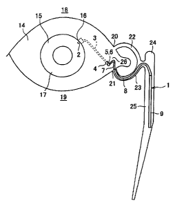
Fig. 1 is a schematic diagram illustrating how an aqueous
humor drainage implant for the treatment of glaucoma
according to one embodiment of the present invention is
positioned in the eye when it is inserted into the nasolacrimal
duct.
Fig. 2 is a diagram illustrating the overall structure of the
aqueous humor drainage implant for the treatment of glaucoma
shown in Fig. I, separately as an anterior part and a posterior
part.
Fig. 3 is a cross sectional view illustrating an example in
which a plurality of pores are provided at the front end of an
outer sheath part, schematizing a structure of a filter part of
the aqueous humor drainage implant for the treatment of
glaucoma shown in Fig. I, cut along the direction of extension
of the filter part.
Fig. 4 is a cross sectional view of the filter part taken
along the line A-A', schematizing a structure of the filter part of
the aqueous humor drainage implant for the treatment of
glaucoma shown in Fig. 1.
Fig. 5 is a schematic diagram illustrating how the aqueous
humor drainage implant for the treatment of glaucoma shown in
Fig. 1 is positioned on the conjunctiva of the eye.
CA 02511602 2005-06-23
- 16-
BEST MODE FOR CARRYING OUT THE INVENTION
Referring to Fig. 1 through Fig. 5, the following will
describe an exemplary structure of an aqueous humor drainage
implant for glaucoma treatment (hereinafter simply referred to
as "aqueous humor drainage implant'°). Fig. 1 schematizes how
an aqueous humor drainage implant according to one
embodiment of the present invention is positioned in an eye by
being inserted into the nasolacrimal duct. Fig. 2 illustrates the
overall structure of the aqueous humor drainage implant shown
in Fig. 1, separately as an anterior part and a posterior part.
As shown in Fig. i and Fig. 2, the aqueous humor
drainage implant of the present embodiment has three major
parts: a first tube (guiding tube part, eye-side guiding tube part)
3; frst and second joints (joint parts) 5 and 6; and a posterior
part 10. Note that, in the following, the first joint 5 and the
second joint 6 will be collectively referred to simply as joints 5
and 6, unless otherwsrise noted.
Specifically, the first tube 3 constitutes the anterior part
of the aqueous humor drainage implant 1, connecting the
anterior chamber of the eye to the exterior of a conjunctiva 14,
and positioned along the sclera wall under the conjunctiva I4.
The posterior part 10 of the aqueous humor drainage implant 1
is positioned such that it extends from an angulus oculi
medialis 26, through an upper lacrimal punctum 20 (or lower
lacrimal punctum 21}, into an upper lacrimal canaliculus 22 (or
lower lacrimal canaliculus 23), a lacrimal sac 24, or a
nasolacrimal duct 25. The joints 5 and 6 of the aqueous humor
drainage implant 1 connect the first tube 3 (anterior part) to the
posterior part 1D. Nate that, in the exaxrxple shown in Fig. 1, the
posterior part 1D is positioned in the lacrimal sac 24 or the
nasolacrimal duct 25. Alternatively, the posterior part 10 may
be positioned on the conjunctiva 14, as will be described later.
CA 02511602 2005-06-23
- 17-
As used herein, the term "conjunctiva'° 14 includes the bulbar
conjunctiva, conjunctiva cul-de-sac, and palpebral conjunctiva.
The posterior part 10 includes a second tube (guiding
tube part, outside-conjunctiva guiding tube part,
outside-conjunctiva eye-side guiding tube part) 7, a third tube
(guiding tube part, outside-conjunctiva guiding tube part,
outside-conjunctiva filter-side guiding tube part) 8, and a filter
part 9. The first tube 3, the second tube 7, and the third tube 8
correspond to a guiding tube part of the present invention, and
the filter part 9 corresponds to a filter part of the present
invention. Further, the joints 5 and 6 correspond to a joint part
of the present invention, and the second tube 7 and the third
tube 8 correspond to an outside-conjunctiva guiding tube part
of the present invention. Farther, the second tube 7 and the
third tube 8 correspond to an outside-conjunctiva eye-side
guiding tube part and an outside-conjunctiva filter-side guiding
tube part of the present invention, respectively.
With this arrangement, the aqueous humor in the anterior
chamber of the eye is guided into the joints S and fi through the
first tube 3. Through the joints 5 and 6, the aqueous humor is
ejected out of the conjunctiva 14 and, via the posterior part 10,
drains into the upper lacrimal canaliculus 22 (or lower lacrimal
canaliculus 23), the Iacrimal sac 24, or the nasolacrimal duct
25. The aqueous humor drained out of the aqueous humor
drainage implant 1 flows through the upper lacrimal
canaliculus 22 (or Iower lacrimal canaliculus 23) and the
lacrimal sac 24, and is absorbed in the nasolacrimal duct 25
and the nasal cavity (not shown) connecting to the nasolacrimal
duct 25.
Although Fig. 1 illustratcs an Gxample in which the
aqueous humor flown through the posterior part 10 is absorbed
in the nasal cavity, the aqueous humor may alternatively be
CA 02511602 2005-06-23
-18_
drained onto the conjunctiva 14 through the posterior part 10,
as will be described later. Tn this case, the aqueous humor is
absorbed by the tissues of the conjunctiva 14.
To describe the first tube 3 of the aqueous humor
drainage implant 1 of the present embodiment more specifically,
the first tubc 3 constituting the anterior part is a single silicon
tube with an inner diameter of 0.5 mm, an outer diameter of 1.0
mm, and a length of 10 mm, and is connected to the joint 5 at a
conjunctiva-side stump 4. The first tube 3 is surgically
positioned along the sclera wall under the conjunctiva 14. Here,
an anterior chamber-side stump 2 of the first tube 3 is inserted
into the anterior chamber of the eye, and the conjunctiva-side
stump 4 and the joint 5 are placed external of the conjunctiva
14 at the angulus oculi medialis 26.
Specifically, a flap of conjunctiva 14 and the underlying
tissue is opened with an incision to expose a sclera 16. Here,
any breeding should be contro3led. The conjunctiva-side stump
4 of the first tube 3 of the aqueous humor drainage implant 1 is
secured with a suture to the sclera wall at the angulus oculi
medialis 26. 'Then, the anterior chamber-side stump 2 of the
first tube 3 of the aqueous humor drainage implant 1 is
inserted into the sclera 16 by a known method, and positioned
in the anterior chamber by inserting it between an iris 17 and a
cornea Z 5.
The first tube 3 is properly secured to the sclera wall with
a suture, so as to position it as shown in Fig. 1. The conjunctiva
14 is then restored and the incision is closed with a suture.
Here, the incision of conjunctiva Z4 around the conjunctiva-side
stump 4 of the first tube 3 is closed using a known method
employing, for example a purse-string suture, a biologically
acceptable adhesive, and the like, so that the first joint 5
connected to the conjunctiva-side stump 4 of the first tube 3 is
CA 02511602 2005-06-23
_19_
exposed external of the conjunctiva 14.
With the first tube 3 and the joint 5 positioned in the
described manner, the aqueous humor in the anterior chamber
can be guided into the exterior of the conjunctiva 14 through
the first tube 3 and the joint 5.
Considering that the first tube 3 is positioned in the living
tissues such as the anterior chamber, the conjunctiva 14, and
the sclera 16, any material can be used for the first tube 3 as
long as it is sufficiently flexible and has good biocompatibility.
Specific examples of the first tube 3 are various polymers,
including: silicone resins; polyolefin resins such as polyethylene,
polypropylene, polyisobutylene, ethylene-vinyl acetate
copolymer, and polynorbornene; polyurethane resins; synthetic
rubbers such as polybutadiene, polyisoprene, SBR (Styrene
Butadiene Rubber}, and SIR; and natural rubbers. In light of
the proved performance and reliability, silicone resins and
polyurethane resins are preferably used.
It is preferable that the first tube 3 have substantially the
same outer diameter as the posterior part 10 which the first
tube 3 is connected to. Generally, the outer diameter of the first
tube 3 ranges from about 0.5 mm to about 1.5 mm, ignoring
individual differences. Namely, a suitable outer diameter of the
first tube 3 generally ranges from about 0.5 mm to about 1.5
mm, though it varies from patient to patient requiring the
aqueous humor drainage implant 1. ~rther, the first tube 3 is
generally about 5 mrn to 20 mm in length, though it depends on
where the anterior chamber-side stump 2 is inserted.
As described above, the first tube 3 is secured along the
sclera wall. To this end, the first tube 3 may have any stnacture
using known techniques, so long as it assists the procedure of
securing the first tube 3. For example, using a known technique,
the first tube 3 may have a projection-Like structure along its
CA 02511602 2005-06-23
-20-
outer surface. With such a structure, the first tube 3 can be
secured along the sclera wall more easily.
The conjunctiva-side stump 4 of the first tube 3 is
connected to the first joint 5. For this purpose, the
conjunctiva-side stump 4 and the first joint 5 are desirably
fastened together in advancc. In this way, surgical procedures
can be performed more easily, and infection from the junction
can be prevented more reliably.
As illustrated in Fig. 1 and Fig. 2, the posterior part 10 of
the aqueous humor drainage implant 1 of the present
embodiment has three parts, including: the second tube 7
connected to the second joint 6; the filter part 9; and the third
tube 8 bridging the second tube 7 and the filter part 9. With the
shape and structure described below, the posterior part 10 can
be positioned with reduced invasiveness in any of the upper
lacrimal canaliculus 22, -the lower lacrimal canaliculus 23, the
lacrimal sac 24, the nasolacrimal duct 25, or on the conjunctiva
14, an effect which has not been realized with conventional
aqueous humor drainage implants.
First, description is made below as to the posterior part
positioned in the upper lacrimal canaliculus 22, the lower
lacrimal canaliculus 23, the lacrimal sac 24, or the
nasolacrimal duct 25, as shown in Fig. 1. In the human body,
there is a canaliculus, called the lacrimal duct, that passes the
lacrimal fluid from the angulus oculi medialis 25 to the nasal
cavity (not shown}, as illustrated in Fig. 1 and Fig. 5. The
lacrimal duct is a single duct with a diameter of about 1 mm to
1.5 mm, and a length of 10 mm to 30 mm. The lacrimal duct
includes the upper Iacrimal punctum 20, the lower lacrimal
punctum 21, the upper lacrimal canaliculus 22, the lower
lacrimal canaliculus 23, the lacrimal sac 24, and the
nasolacrimal duct 25, and connects the angulus oculi medialis
CA 02511602 2005-06-23
-21-
26 to the nasal cavity. {The lacrimal duct communicates
between the angulus oculi medialis 25 and the nasal cavity.)
The present invention enables the posterior part 10 to be
inserted into the lacrirnal duct by taking advantage of the
anatomical feature of the lacrimal duct draining the lacrimal
fluid into the nasal cavity. That is, the aqueous humor drainage
implant 1 of the present embodiment enables the aqueous
humor in the anterior chamber to be drained into the nasal
cavity with the structure of the lacrimal duct intact, thereby
realizing installation with reduced invasiveness, unattained by
conventional techniques. Namely, with the posterior part 1.0
shaped into a single tube to be inserted into the lacrimal duct
as described below, the aqueous humor drainage implant 1 of
the present embodiment can be inserted into the lacrimal duct
with reduced invasiveness.
Next, the following will describe the case where the
posterior part 10 is positioned on the conjunctiva 14. Fig. S
schematizes how the aqueous humor drainage implant 1 of the
present embodiment is positioned on the conjunctiva of the eye.
It should be noted here that members and portions having the
same functions are those described with reference to Fig. 1 are
given the same reference numerals and explanations thereof are
omitted.
In positioning the posterior part 10 on the conjunctiva 14
as shown in Fig. 5, care must be taken not to damage the
cornea or conjunctiva with the posterior part 10 after it is
placed in position, or not to cause any discomfort to the patient,
in addition to avoiding any invasiveness due to the installation.
To this end, the posterior part 10 needs to be positioned on the
conjunctiva 14 along the curved surface of the eye wall, as
shown in Fig. S, so as to minimize the influence of eye
movement, instead of simply hanging the aqueous humor
CA 02511602 2005-06-23
-22-
drainage implant in the conjunctival sac as in the conventional
technique.
By being positioned on the conjunctiva 14 as shown in Fig.
5, the posterior part 10 becomes part of the eye through the
conjunctiva 14, and is therefore able to follow the eye movement
in any direction. That is, with the single tube construction, the
posterior part 10 can easily be positioned along the eye wall.
Farther, the single tube construction allows the posterior part
10 to be positioned on the conjunctiva 14 with reduced
invasiveness, without causing much damage or discomfort to
the conjunctiva 14 by the posterior part 10 positioned thereon.
Note that, in the example shown in Fig. 5, the posterior part 10
is positioned on the conjunctival cul-de-sac 27 of the
conjunctiva I4.
It should be noted here that the posterior part 10 is
connected to the second joint 6 regardless of whether the
posterior part 10 is positioned in the lacrimal duct as shown in
Fig. 1, or on the conjunctiva 14 along the eye wall as shown in
Fig. 5. For this reason, the posterior part 10 is directly
influenced by the eye movement. However, the influence of eye
movement is minimized by the segmented structure of the
posterior part 10 divided into the second tube 7, the third tube
8, and the filter part 9.
That is, by setting required levels of flexibility and
biocornpatibility for each of these different parts, the influence
of eye movement on the posterior part 10 can be minimized. In
the present embodiment, the posterior part 10 is segmented
into three parts; however, the number of segments is not just
limited to three.
As described above, the posterior part 10 has a single
tube structure, and is segmented into plural parts, specifically,
the second tube 7, the third tube 8, and the filter part 9. This
CA 02511602 2005-06-23
-23-
enables the posterior part 10 of the aqueous humor drainage
implant 1 to be positioned with reduced invasiveness in any of
the upper lacrimal canaliculus 22, the lower lacrimal
canaliculus 23, the lacrimal sac 24, and the nasolacrimal duct
25, or on the conjunctiva 14. Details of the single tube
structure of the posterior part 10 will be described later.
As described above, the posterior part 10 desirably has
substantially the same outer diameter as the first tube 3,
preferably and generally in a range of 0.5 mm to 1.5 mrn.
More specifically, in the aqueous humor drainage implant
1 of the present embodiment, the second tube 7 and the third
tube 3 are both silicon tubes with an inner diameter of 0.5 mm
and an outer diameter of 1.0 mm. That is, the posterior part 10
includes two silicon tubes. The second tube 7 is S mm in length,
and the third tube 8 is 10 mm_ to 30 mm in length. The filter
part 9 is constructed from, for example, a polyethylene tube
sheath with an inner diameter of 0.8 mm, an outer diameter of
1.0 mm, and a length of 10 mm, and an 8 mm-long hollow fiber
membrane provided therein with an outer diameter of 0.7 mm.
Using the hollow fiber membrane as a filter enables the
filter part 9 to be constructed as a single tube like the second
tube 7 and the third tube 8. The second tube 7, the third tube 8,
and the filter part 9 are shaped and sized based on the anatomy
of the lacrimal duct, so as to enable the posterior part 10 to be
positioned with reduced invasiveness in any of the upper
lacrimal canaliculus 22, the lower Iacrimal canaliculus 23, the
Iacrimal sac 24, and the nasolacrimal duct 25. Further, the
anatomy of the eyeball is also taken into account in designing
the shape and size of the second tube ?, the third tube 8, and
the filter part 9, so that the posterior part 10 can be positioned
on the conjunctiva 14 with reduced invasiveness.
As described above, depending on patient conditions, the
CA 02511602 2005-06-23
-24-
posterior part 10 is surgically positioned in any of the upper
lacrimal eanaliculus 22, the lower lacrirnal canaliculus 23, the
lacrimal sac 24, and the nasolacrimal duct 25, or on the
conjunctiva 14. In the case where the posterior part 10 is
positioned in the upper lacrimal canaliculus 22, the lower
lacrimal canalieulus 23, the lacrinnal sac 24, or the
nasolacrimal duct 25 as shown in Fig. 1, the posterior part 10
is inserted into the upper lacrimal punctum 20 {or lower
lacrimal punctum 21} by a known nasolacrimal duct bougienage
method, and is positioned in the upper lacrimal canaliculus 22,
(lower lacrimal canaliculus 23), the lacrimal sac 24, or the
nasolacriznal duct 25.
Here, the second tube 7 is positioned outside the upper
lacrimal punetum 20 {or lower lacrimal punctum 21) so as to
allow the posterior part 10 to follow the eye movement. The
stump of the second tube 7 is connected to the first joint 5 via
the second joint 5. In the case where the posterior part 10 is
positioned on the conjunctiva 14 as shown in Fig. S, the stump
of the second tube 7 is connected to the first joint 5 via the
second joint 5, and the posterior part 10 is positioned on the
conjunctiva 14. Note that, in Fig. 1 and Fig. 5, the upper eyelid
and lower eyelid are shown as 18 and 19, respectively.
As used herein, "patient conditions'° refers to situations
where the nasolacrimal duct is clogged, the drained aqueous
humor affects vision, or the patient feels discomfort by the
presence of the posterior part 10. For example, for patients
suffering from a clogged nasolacrimal duct, the posterior part
is desirably positioned on the conjunctiva 14. If, for example,
the posterior part I0 positioned on the conjunctiva 14 Leads to
affected vision by the drained aqueous humor, or discomfort
{unpleasant sensation) due to the posterior part 10 during eye
movement, the posterior part 10 is desirably positioned in the
CA 02511602 2005-06-23
-25-
upper lacrimal canalieulus 22, the lower lacrimal canaliculus
23, the lacrimal sac 24, or the nasolacrimal duct 25.
In any case, the lengths of the second tube 7 and third
tube 8 can be adjusted as required to accommodate different
patient conditions. Further, the second joint fi and the posterior
part 10 are desirably fastened together in advance. In this way,
surgical procedures can be performed more easily, and infection
from the junction of the second joint 5 and the posterior part 10
can be prevented more reliably.
Here, because the first tube 3 and the joints 5 and 5 are
secured to the sclera wall, the second tube 7 extending
therefrom is under the direct mechanical force of eye movement.
Here, eye movement is restricted if the second tube 7 is not
elastic enough to follow the eye movement within the movable
range of the eye. This may lead to ambiopia or displacement of
the aqueous humor drainage implant 1.
In order to prevent arnbiopia or displacement of the
aqueous humor drainage implant 1, the second tube 7
particularly requires good elasticity, flexibility, and ease of
deformation sufficient to accommodate the eye movement. That
is, it is required that the second tube 7 be made of a material
that provides good elasticity, flexibility, and case of deformation.
Depending on the movable range of the eye, there are cases
where the second tube 7 is brought info contact with the cornea
or other ocular tissues for a brief moment. Thus, in order to
ensure that the second tube 7 does not damage the ocular
tissues, it is important that the second tube 7 be made of a
material that offers good elasticity, flexibility, and ease of
deformation. That is, the second tube 7 requires a highly
flexible and biocornpatible material that can easily deform to
follow eye movement and that can relieve invasiveness to the
ocular tissues. With the posterior part 10 including the second
CA 02511602 2005-06-23
-26-
tube 7 satisfying such conditions, problems associated with the
eye movement, such as invasiveness to the ocular tissues, pain,
and displacement of the aqueous humor drainage implant 1 can
be effectively prevented.
The material of the second tube 7 is not particularly
limited as long as it offers good elasticity, flexibility, ease of
deformation, and biocompatibility. Some of the representative
examples are various types of polymer materials, including:
silicone resins; polyolefin resins such as polyethylene,
polypropylene, polyisobutylene, ethylene-vinyl acetate
copolymer, and polynorbornene; polyurethane resins; natural
rubbers; and synthetic rubbers. Among these materials, silicone
resins and polyurethane resins are particularly preferable. The
second tube 7 desirably has substantially the same outer
diameter as the first tube 3 and the third tube 8. Further,
taking into account the expansion and contraction due to the
eye movement, the second tube 7 is generally about 5 mm to 20
mm in length, though it depends on where the joints 5 and 6,
and the posterior part 10 are positioned, Farther, the second
joint 6 and the second tube 7 are desirably fastened together in
advance. In this way, surgical procedures can be performed
more easily, and infection from the junction can be prevented
more reliably.
Considering that the third tube 8 is positioned on the
conjunctiva Z4, or in other living tissues such as the upper
lacrimal punctum 20, the lower lacrimal punctum 21, the upper
lacrimal canaliculus 22, the lower lacrimal canalicuIus 23, and
the Iacrimal sac 24, any maternal can be used for the third tube
S as long as it is sufficiently flexible and has good
biocompatibility. Some of the representative examples of the
third tube 8 are various polymers, including: silicone resins;
polyolefm resins such as polyethylene, polypropylene,
CA 02511602 2005-06-23
-27-
polyisobutylene, and ethylene-vinyl acetate copolymer;
polyurethane resins; synthetic rubbers; and natural rubbers.
Among these materials, silicone resins and polyurethane resins
are particularly preferable.
Considering that the third tube 8 is positioned in the
upper lacrimal canaliculus 22, the lower lacrimal canalieulus
23, the laerimal sac 24, and the nasolacrimal duct 25, it is
required that the third tube 8 have a narrower outer diameter
than the inner diameter of any of the upper lacrimal punctum
20, the lower lacrimal punctum 21, the upper lacrimal
canaliculus 22, the lower lacrimal canaliculus 23, and the
lacrimal sac 24. Generally, the outer diameter of the third tube
8 desirably ranges from about 0.5 mm to about 1.5 mm,
ignoring individual differences. Further, the third tube 8 is
gexlerally about 5 mm to 20 mm in length, though it depends on
where the posterior part 10 is positioned and ignoring
individual differences among patients.
The second tube 7 and the third tube 8 are highly flexible
with a flexural modules of no greater than 2000 Mpa at
ordinary temperature, thereby preventing problems associated
with eye movement, such as invasiveness to the eye, pain, and
displacement of the aqueous humor drainage implant 1. In the
present embodiment, the second tube 7 and the third tube 8
have the same flexural modules at ordinary temperature, i.e.,
the same flexibility. However, the second tube 7 may have a
smaller flexural modules than the third tube 8 at ordinary
temperature. This enables the second tube 7 to absorb the
influence of eye movement more reliably.
Depending on the elasticity of the second tube 7 or
movement of the filter part 9 in the nasolacrimal duct 5, there
are cases where the position of the third tube 8 may be affected.
For example, with the posterior part 10 positioned in the upper
CA 02511602 2005-06-23
-28-
lacrimal canaliculus 22, the lower lacrimal canaliculus 23, the
lacrimal sac 24, or the nasolacrimal duct 25, the third tube 8
may slip out of the upper lacrimal punctum 20 or the lower
lacrimal punctum 21, or drawn into the nasolacrimal duct 25.
Such situations can be avoided by securing the posterior
part 10 to a suitable position. The method of securing the
posterior part 10 is not particularly limited, and conventional
methods can be used. For example, the following methods can
he used when the posterior part 10 is positioned in the upper
lacrimal canaliculus 22, the lower lacrimal canalicuIus 23, the
lacrimal sac 24, or the nasolacrimal duct 25. In the first method,
the upper lacrimal punctum 20 or the lower lacrimal punctum
21 is tightened by ligation. In the second method, the second
tubc 7 or the third tube 8 is temporarily secured to the skin
around the upper lacrimal punctum 20 or the lower lacrimal
punctum 21 by ligation. In the third method, a wing-like
projection serving as a stopper is provided at the boundary of
the second tube 7 and the third tube 8.
In the case where the posterior part 10 is positioned on
the conjunctiva 14, the third tube 8 may be omitted as required
to directly join the second tube 7 and the filter part 9. However,
regardless of whether the third tube 8 is omitted or not, there is
always a possibility, whenever the posterior part 10 is
positioned on the conjunctiva 14, that the posterior part 10
r~aoves out of position and becomes unstable due to the eye
movement. This can be avoided by securing the posterior part
to a suitable position on the conjunctiva 14. The method of
securing the posterior part 10 on the conjunctiva 14 is not
particularly limited, and conventional methods can be used. As
one example, the posterior part 10 may be secured to the
conjunctiva 14 by a suture.
Fig. 3 is a cross sectional view schematizing a structure of
CA 02511602 2005-06-23
-29-
the filter part cut along the direction of extension of the filter
part 9 of the aqueous humor drainage implant 1 of the present
embodiment. As shown in Fig. 3, the filter part 9 includes a
hollow fiber membrane part 11 and an outer sheath part 12. It
should be noted here that the outer sheath part 12 is optionally
provided according to the hardness of the hollow fiber
membrane part 11. As such, the filter part 9 may only include
the hollow fiber membrane part 11.
Fig. 4 is a cross sectional view of the filter part taken
along the line A-A', schematizing a structure of the filter part of
the aqueous humor drainage implant shown in Fig. 1. As
illustrated in Fig. 4, the filter part 9 of the present embodiment
includes the hollow fiber membrane part 11 inside the outer
sheath section 12.
Considering that the f lter part 9 is positioned in the
upper Iacrimal canaliculus 22, the lower Iacrimal canaliculus
23, the lacrimal sac 24, and the nasolacrirnal duct 25, it is
required that the filter part 9 have a narrower outer diameter
than the , inner diameter of any of the upper lacrimal punctum
20, the Iower lacrimal punctum 21, the upper lacrimal
canaliculus 22, the lower lacrimal canaliculus 23, and the
lacriznal sac 24. Generally, the outer diameter of the filter part 9
desirably ranges from about 0.5 mm to about 1.5 mm, ignoring
individual differences among patients. As such, the outer
diameter of the filter part 9 is desirably about 0.5 mm to 1.5
mm. Further, the filter part 9 is generally about 5 rnm to 20 mm
in length, though it depends on individual differences among
patients. Note that, if the filter part 9 includes only the hollow
fiber membrane part 11, the outer diameter of the filter part 9
coincides with the outer diameter of the hollow fiber membrane
part II. On the other hand, if the filter part 9 includes the
outer sheath part I2, the outer diameter of the filter part 9
CA 02511602 2005-06-23
-30-
coincides with the outer diameter of the outer sheath part 12.
As shown in Fig. 3, the hollow fiber membrane part 11
opens into the third tube 3 at an end connecting thereto, and is
closed at a stump 13 to provide a dead end. That is, the
aqueous humor that flows into the aqueous humor drainage
implant 1 from the anterior chamber of the eye all passes
through the hollow fiber membrane part 11 and drains out of
the filter part 9 through pores formed through a side wall of the
hollow fiber membrane part 11.
As to a method of closing the stump 13 of the hollow fiber
membrane part 11, any conventional method can be used as
long as it can close the stump 13. For example, a method using
a polyurethane adhesive, or a method employing heat fusion is
available.
The hollow fiber membrane part 11 of the filter part 9 is
provided to reduce intraocular pressure by draining the
aqueous humor, and to prevent viruses, bacteria, fungi, or other
microorganisms that exist outside the conjunctiva 14 from
entering the first tube 3 and the posterior part 10. In this way,
intraocular pressure is reduced, and at the same time, reflux
infection from the conjunctiva 14 is prevented.
For the purpose of draining aqueous humor, the hollow
fiber membrane 11 must accommodate an aqueous humor
production rate in a range of 2.0 ltl/min to 3.0 lxl/min, and
attain a target intraocular pressure of about 10.0 mmHg to 20.0
mmHg. As such, the hollow fiber membrane part 11 must
provide an aqueous humor flow rate of no less than 2.0 Ltl/min
to 3.0 ul/min .under a hydraulic pressure of about I0.0 mmHg
to 20.0 mmHg.
In order to examine whether a hollow fiber membrane
used for the hollow fiber membrane part 11 of the aqueous
humor drainage implant 1 of the present embodiment satisfy
CA 02511602 2005-06-23
-31-
these conditions, following series of experiments were
conducted.
[Experimental method for the evaluation of hollow fiber
membrane]
With a hollow fiber membrane used for the hollow fiber
membrane part 11 of the aqueous humor drainage implant 1 of
the present embodiment, the amount of aqueous humor flown
under a certain hydraulic pressure was measured. Specifically,
in a vertically placed pipe, the pseudo aqueous humor aBSS
plus° (the product of SANTEN PHERMACEUTICAL CO., LTD.)
was charged to about 13 cm from the bottom cnd of the pipe.
Then, with the hollow fiber membrane part 11, 10 mm long,
fitted to the bottom end of the pipe, the outflow weight of BSS
plus per unit time was measured. From the measured Weight of
BSS plus and its specific gravity, an outflow volume of BSS plus
was calculated. As the hollow fiber membrane used as the
hollow fiber membrane part 11, two kinds of prototype EVAL
membranes with different average pore diameters and different
outer diameters were used. Note that, the foregoing procedure
was carried out with the BSS plus maintained at 37°C.
[Preparation method of prototype. EVAL membranes]
For the preparation of the prototype EVAL membranes, a
starting solution was prepared first by heating, stirring, and
dissolving at 90°C 15 parts by weight of ethylene-vinyl alcohol
copolymer with the ethylene content of 32 mol% and
saponificated to 99 mol% (the KURARAY Co., LTD. product
EVAL EC-F100A), 73 parts by weight of dimethylsulfoxide, 10
parts by weight of water, and 2 parts by weight of lithium
chloride.
The starting solution so prepared had a LST (Lower
Solution Temperature) of 28°C. The starting solution was a
transparent homogeneous solution at high temperatures, but
CA 02511602 2005-06-23
- 32 -
underwent phase separation and became clouded with
decreasing temperature. When allowed to stand for extended
periods of time, the solution separated into two layers. In the
present embodiment, a temperature at which such phase
separation occurs will be referred to as the LST.
Using a double annular nozzle, the starting solution
maintained at 40°C was extruded with water injected through
the center of the nozzle. Then, the solution was allowed to pass
through an air layer and was solidified in a water bath. After
water washing, wet heating, drying, and heat treatment
according to ordinary method, a dry hollow fiber membrane was
obtained as a hollow fiber membrane El, i.e., the prototype
EVAL membrane.
In addition, another kind of prototype EVAL membrane,
hollow fiber membrane E2, was prepared under the same
conditions as for the hollow fiber membrane E1, except that the
ethylene-vinyl alcohol copolymer saponificated to 99 mol% was
used in 17 parts by weight, and that the dimethylsulfoxide was
used in 71 parts by weight. The starting solution used for the
preparation of the hollow fiber membrane E2 had a LST of 30°C.
The preparation method of the prototype EVAL membrane,
i.e., the ethylene vinyl alcohol copolymer is described in more
detail in Japanese Laid-Open Patent Publication No.
286744/2001 (Tokukaihei 13-286'740).
Table 1 below shows results of evaluation experiment for
the average pore diameter and outer diameter of the hollow fiber
membranes E1 and E2. As shown in Table 1, the hollow fiber
membranes E1 and E2 both had a flow rate that satisfied the
required conditions for the production rate of aqueous humor
noted above. The results therefore showed the effectiveness of
the aqueous humor drainage implant 1 of the present invention
in draining the aqueous humor and thereby keeping the
CA 02511602 2005-06-23
-33-
intraocular pressure within a normal range.
[Table 1]
Outer The nunckberFlow rate
of
Hollow 'overage diameter of hollow Pseudo
fiber of
pore hollow aqueous
membrane fiber f ber
she (um) membrane humor
membranes
m min
E1 0.004 780 1 5.68
E2 0.005 _ 4 2.2
300
It should be noted here that, depending on the
performance of the hollow fiber membrane 11, there are cases
where the aqueous humor drainage implant 1 of the present
invention may drain the aqueous humor in excess. Such excess
draining of the aqueous humor may lead to low intraocular
pressure after the surgical operation. In order to prevent such a
situation, a pressure-controlled check valve or regulator valve
may be suitably provided in the first tube 3, the first joint 5, the
second joint 6, or the posterior part 10 according to the
performance of the hollow fiber membrane part 11 used.
The pressure-controlled check valve opens and closes to
maintain the intraocular pressure within the normal intraocular
pressure range of about 7 mmHg to 20 mmHg. Any type of
conventional pressure-controlled check valve may be used as
long as it has a structure meeting this purpose. For example, a
slit check valve used for Krupin-Denver eye shunt (USP
5,454,796) and a check valve used for the Ahznedglaucoma
implant (USP 5,071,408, USP 6,261,256) may be used. The
pressure-contr~tled check valve, with its check valve structure,
prevents backflow of the aqueous humor in situations where
there is abrupt pressure increase inside the nasolacrimal duct
as in nose blowing or sneezing.
From the standpoint of preventing reflux infection due to
CA 02511602 2005-06-23
-34-
viruses or other microorganisms, the hollow fiber membrane of
the hollow fiber membrane part 11 needs to~ have an average
pore diameter of no greater than 0.3 um, preferably 0.0001 um
to 0.02 urn, or more preferably 0.0001 lZm to 0.01 um, taking
into account the diameter of viral particles ranging from about
0.02 lxm to 0.3 um. With ari average pore diameter of hollow
fiber membrane exceeding these ranges, it may become
increasingly difficult tv block viral particles.
However, the foregoing condition required for the average
pore diameter of the hollow fiber membrane is adjustable within
a range that can achieve the object of the hollow fiber
membrane 11, i.e., to prevent refIux infection at the viral level.
To describe more specifically, in the case where the hollow fiber
membrane 11 has the additional function of electrically
blocking viruses as will be described Iater, the viruses are also
captured electrically, in addition to being captured by the small
average pore diameter of the hollow fiber membrane. That is,
the hollow fiber membrane used for the hollow fiber membrane
part 11 may have an average pore diameter greater than the
foregoing ranges as long as it serves to prevent reflux infection
at the viral level.
The material of the hollow fiber membrane used for the
hollow fiber membrane part I1 is not particularly limited as
long as it is moderately water permeable and serves to prevent
reflux infection at the viral level. For example, various polymers
such as a polyolefin polymer, a polyvinyl alcohol polymer, an
ethylene-vinyl alcohol copolymer, a polysulfone poiyrner, a
polyacrylonitrile polymer, a cellulose polymer, cellulose acetate
polymer, a polymethyl methacrylate polymer, and a polyamide
polymer are available.
Applicable areas of hollow fiber membrane extend to
various fields. In medical applications, the hollow fiber
CA 02511602 2005-06-23
-35-
membrane has been used primarily in artificial kidneys.
Generally, the hollow fiber membrane used for this purpose has
an average.pore diameter of about 0.005 um to 0.008 ltrn, and
this satisfies the foregoing condition required for the hollow
fiber membrane of the hollow fiber membrane part 11 of the
aqueous humor drainage implant 1 of the present invention.
Thus, the hollow fiber membrane for the present invention can
be suitably selected from industrially available hollow fiber
membranes for artificial kidneys.
Specific examples of such a hollow fiber membrane for
artificial kidneys include those used for the dialyzer of devices
such as the APS-150, AM-FP-130, AM-GP-13, AM-UF-13
(products of Asahi Kasei Medical Co., Ltd.}, Meltrax 140,
Meltrax 160 (products of MERA}, FB-130U (product of NIPRO
CORPORATION}, BS-1.6 (Toray Industries, Inc.}, and PS-1.6N
(KAWASUMI LABORATORIES, INC.}. (Seisuke TAKASHIMA,
Essential Properties of Membrane Materials, Clinical
Engineering, 1997, Vol. 8, No. 6, pp. 479-492).
For the purpose of preventing reflux infection at the viral
level rx~ore reliably while maintaining sufficient flow rate for the
aqueous humor, the hollow fiber membrane part II may have
the function of electrically blocking viruses, in addition to
capturing viruses by the pore diameter of the hollow Fber
membrane.
It is knowr~ that viral particles as a whole are negatively
charged under normal neutral pH range conditions as are many
microorganisms. By taking advantage of this fact, passage of
viral particles through the hollow fiber mennbrane can be
prevented by negatively charging the hollow fiber membrane
part 11 with chemically bound (introduced} anionic groups and
thereby causing the viral particles to repel the negative ions
that exist in the hollow fiber membrane. Alternatively, the
CA 02511602 2005-06-23
-36-
hollow fiber membrane 11 may be positively charged by
chemically binding cationic groups thereto. In this case, the
viral particles are drawn to the hollow fiber membrane part 11
by being attracted to the positive ions that exist in the hollow
fiber membrane, with the result that passage of the viral
particles is prevented.
Meanwhile, the protein, which is one of the constituents
of the virus, is an ampholyte, including cationic groups
(primarily amino groups) and anionic groups (primarily carboxyl
groups). It is envisaged that, by the same mechanism as the ion
exchange membrane, the anionic group or cationic group
chemically bound to the hollow fiber membrane part 11
captures the amino group or carboxyl group of the protein by
forming an ion pair.
That is, by "electrically blocking viruses," it means that
passage of viral particles through the hollow fiber membrane is
preventcd ~by the electric force. Further, with the ability to
electrically block viruses, the hollow fiber membrane part 11
can block passage of viruses with a larger pore diameter
(average pore diameter) as compared with a non-charged
membranc with no electrical capabilities.
The method of introducing ionic groups into the hollow
fiber membrane part 11 is not particularly limited as long as it
can introduce ionic groups into the hollow fiber membrane of
the hollow fibcr mcmbrane part 11. For example, methods
employing known acid treatment, alkali treatment, oxidation
process, photo irradiation, addition reaction, or graft reaction
may be used. In the case of a polymer material having hydroxyl
groups in its molecules for example, a sulfuric acid group,
carboxyl group, amino group, or other ionic groups can be
easily introduced by, for example, esterification, etherification,
or Michael addition. (See Seisu.ke TAKASHIMA et al., Research on
CA 02511602 2005-06-23
-37-
removal of I-1B antigen by absorbent, The Journal of Japanese
Medical Instruments, 1986, vol. 56, No. 11, pp. 499-505, Japanese
Patent Nos. 1695758, 1695760.)
Depending on hardness of the hollow fiber membrane part 11,
the hollow fiber membrane part 11 may be optionally provided with
the outer sheath part 12, in order to assist installation of the hollow
fiber membrane part 11 outside the conjunctiva 14 and improve
durability of the filter part 9. As illustrated in Fig. 3, the outer
sheath part 12 on its front end (stump) has a plurality of pores,
providing passageways for the aqueous humor drained through the
sidewall of the hollow fiber membrane part 11. Note that, in the
present embodiment, the outer sheath part 12 has a plurality of
pores at its front end to provide passageways for the aqueous
humor. However, the pores provided through the outer sheath part
12 are not limited to this arrangement as long as they can pass the
aqueous humor. For example, the outer sheath part 12 may have
one or more openings (pores) through the sidewali, or one or more
openings (pores) through the sidewall and front end.
The material of the outer sheath part 12 is not particularly
limited as long as it can pmvide adequate hardness and good
biocompatibility. Some of the examples include various polymer
materials, including silicone resin, polyethylene resin,
polypropylene resin, polyvinyl alcohol resin, ethylene-vinyl alcohol
copolymer, polyurethane resin, synthetic rubber, Natural rubber,
trane-polyisoprene resin, and polycarbonate resin. Among these
materials, silicone resin, polyurethane resin, and
trans-polyisoprene resin are particularly preferable.
For the purpose sustaining a flow rate of the aqueous humor
in the hollow fiber membrane part lI and improving
biocompatibility of the posterior part 10, the posterior part 10 may
be rendered hydrophilic. For the hydrophilic treatment, any
conventional method may ,.be used. For example, methods
CA 02511602 2005-06-23
~$
employing surface grafting, oxidation process, acid treatment, alkali
treatment, and Ivrichael addition are available.
Joining the first tube 3 and the posterior part 10 with the
joints 5 and fi allows the posterior part IO and the subsequent
parts to be replaced as required. Fox example, there are cases
where the filter funEtion-~f the -hollow fiber membrane part 11 used
in the filter part 9 of the posterior part 10 may deteriorate over time
as the protein or other substances contained in the aqueous humor
clogs the hollow fiber membrane. In this case, the posterior part I0,
including the filter part 9, can be replaced with a new replacement
part by detaching the posterior part 10 at the joints 5 and 6. In this
way, the intraocular pressure reducing effect can be sustained for
extended periods of time.
Further, because only the posterior part 10 is replaced, the
cost of replacement is much cheaper than the case where the
aqueous humor drainage implant 1 needs to be re-installed entirely.
In addition, the physical pain the patient must endure is greatly
relieved. Further, because the joints 5 and 6 are positioned external
to but in contact with living tissues such as the conjunctiva I4, the
upper eyelid 18, and the lower eyelid 19, it is preferable that the
joints 5 and 5 be made of material with good biocompatibility and
good durability. The type of material is not particularly limited as
long as it has such characteristics. For example, polymer materials
such as polyacetal resin, silicone resin, polyethylene resin,
polypropylene resin, ethylene-vinyl alcohol copolymer, polyurethane
resin, ABS (Acrylonitrile-Butadiene-Stylene) resin, and
polyearbonate resin are available. In addition, ceramics such as
alumina and titania, or metals such as stainless steel can also be
used.
The joints 5 and 6 may have any conventional structure as
long as it serves to prevent entry of foreign substances and join the
first tube 3 to the second tube 7 of the posterior part 10. Examples
CA 02511602 2005-06-23
-39-
of such structures include a tapered connector, a threaded
connector, a ball joint, a coupler (the product of NITTO KOHKI CO.,
LTD.), and a tube fitter (the product of NITTO KOHKI CO., LTD.).
Among these different structures, those employed, for example, by
the coupler and tube fitter (both the products of NITTO KOHKI CO.,
LTD.) are particularly preferable because such structures are easily
detachable and allow an operator to check whether the joints are in
place by the sound or feel of clicking.
The joints 5 and 6 may be sized and shaped in any manner
as long as invasiveness of the conjunctiva 14, the upper eyelid 18,
and the lower eyelid 19 following eye movement is controlled. For
example, the joints 5 and 6 may be sized to 1 mm3 to S mm3 each,
and may have a curved surface as shown in Fig. 2. With the joints 5
and 6 sized and shaped this way, invasiveness to the body can be
minimized.
While a representative structure and embodiment of the
aqueous humor drainage implant of the present invention is
described above with reference to Fig. 1 through Fig. 5, the
invention is not limited in any way by the foregoing examples. It
should be understood that the foregoing examples are not intended
to limit the invention to the particular forms disclosed, but on the
contrary, the invention is to cover all modifications, equivalents,
and alternatives falling within the scope of the invention as defined
in the appended claims.
An aqueous humor drainage implant of the present invention
may be implemented as follows.
Specifically, an aqueous humor drainage implant may be
implemented as a first aqueous humor drainage implant for
draining aqueous humor from the interior of the eye to the exterior
of the conjunctiva, the first aqueous humor drainage implant
including a guiding tube part for guiding aqueous humor to exterior
of the eye, and a filter part far preventing reflex infection from the ._
CA 02511602 2005-06-23
- 40 -
exterior to interior of the eye, and being structured and shaped to
be positioned in the eye and outside the conjunctiva with reduced
invasiveness.
According to the invention, the aqueous humor drainage
implant can easily be positioned with reduced invasiveness, and the
aqueous humor can be drained to the exterior of the aqueous
humor both safely and reliably.
The first aqueous humor drainage implant may be shaped
and structured to be positioned in the lacrimal canaliculus,
lacrimal sac, or nasolacrimal duct. F~u-ther, the first aqueous
humor drainage implant may be shaped and structured to be
positioned on the conjunctiva.
According to the invention, the aqueous humor drainage
implant can be positioned on the conjunctiva, or in the lacrimaI
canaliculus, lacrimal sac, or nasolacrirnal duct, depending on
patient conditions. F~xrther, regardless of the position, the aqueous
humor drainage implant can be installed with reduced
invasiveness.
The first aqueous humor drainage implant may be adapted so
that the filter part uses a hollow fiber membrane for preventing
reflex infection at the viral level.
According to the invention, reflex infection caused by viruses
or any other pathogens can be prevented.
The first aqueous humor drainage implant may be adapted so
that the filter part uses a hollow fiber membrane that has been
treated to electrically block viruses or other microorganisms.
According to the invention, reflex infection at the viral level
can be prevented more reliably, while draining a sufficient amount
of aqueous humor to reduce intraocular pressure.
The first aqueous humor drainage implant may be adapted so
that its outer surface is rendered hydrophilic.
According to the invention, the guiding tube part and the
CA 02511602 2005-06-23
-41-
$lter part can have improved biocompatibility, and the hollow fiber
membrane of the filter part can drain a sufficient amount of
aqueous humor to reduce intraocular pressure.
The first aqueous humor drainage implant may be adapted to
optionally include a joint for enabling the filter part to be replaced.
According to the invention, the filter part can be replaced
when it is deteriorated or damaged. That is, the intraocular
pressure reducing effect of the aqueous humor drainage implant
can be sustained for extended periods of time by a simpler, less
expensive, and more patient friendly method, as compared with
the case where the entire aqueous humor drainage implant is
reinstalled.
The first aqueous humor drainage implant may be
adapted so that a portion of the guiding tube part to be
positioned outside the conjunctiva includes a tube that can
easily deform according to eye movement and that is flexible
enough to relieve invasiveness to the ocular tissue.
In this way, the invention can prevent problems caused by
eye movement, such as invasiveness to the ocular tissue,
patient's pain, and displacement of the aqueous humor
drainage implant.
As described above, in the aqueous humor drainage
implant for glaucoma treatment, the guiding tube part includes
an eye-side guiding tube part and an outside-conjunctiva
g~ziding tube part.
In this way, in installing the aqueous humor drainage
implant for glaucoma treatment in the patient, the guiding tube
part can easily be positioned with reduced invasiveness based
on the anatomical structure of the eye and nearby organs.
Further, with the filter part connected to one end of the
guiding tube part, reflux infection from the exterior to interior of
the cyc can be prevented. That is, the aqueous humor can be
CA 02511602 2005-06-23
-42-
safely drained out of the eye to the exterior of the conjunctiva.
Further, the outside-conjunctiva guiding tube part may
have an outer diameter smaller than an inner diameter of the
nasolacrincxal duct. In this way, with the guiding tube positioned
in the lacrimal passage including the lacrimal canaliculus,
lacrimal sac, and nasolacrimal duct, the aqueous humor can
drain into the nasal cavity through the lacrimal passage. That is,
the guiding tube can be positioned with reduced invasiveness.
Further, the outside-conjunctiva guiding tube part and
the filter part may be shaped to have a curved outer surface and
may be sized to have substantially the same outer diameter. In
this way, the outside-conjunctiva guiding tube part and the
filter part can be easily positioned along the eye wall. Further,
damage to the conjunctiva or the sense of foreign object can be
relieved.
Further, the filter part may include a hollow fiber
membrane made of at least one kind of polymer material
selected from the group consisting of a polyolcfin polymer, a
polyvinyl alcohol polymer, an ethylene-vinyl alcohol copolymer,
a polysulfone polymer, a polyacrylonitrile polymer, a cellulose
polymer, cellulose acetate polymer, a polymethyl methacrylate
polymer, and a polyamide polymer. Further, it is preferable that
the hollow fiber membrane have an average pore diameter of no
greater than 0.3 um, or more preferably no greater than 0.02
~.tm. In this way, the hollow fiber membrane can prevent reflux
infection at the viral level.
Further, the filter part may include a chemically bound
anionic group or cationic group. In this way, the reflux infection
at the viral level can be prevented more reliably, and a sufficient
amount of aqueous humor can be drained to reduce intraocular
pressure.
Further, the guiding tube part and the filter part may be
CA 02511602 2005-06-23
-43-
rendered hydrophilic. In this way, the guiding tube part and the
filter part can have improved biocompatibility, and the filter
part can stably drain a sufficient amount of aqueous humor
necessary to reduce intraocular pressure.
The aqueous humor drainage implant for glaucoma
treatment may be adapted to further include a joint part for
detachably connecting the eye-side guiding tube part and the
outside-conjunctiva guiding tube part. In this way, the
intraocular pressure relieving effect of the aqueous humor
drainage implant can be sustained for extended periods of time
by a simpler, less expensive, and more patient friendly method.
Further, the outside-conjunctiva guiding tube part may
have a flexural moduIus of no greater than 2000 Mpa at
ordinary temperature. Further, the outside-conjunctiva guiding
tube part may include an outside-conjunctiva eye-side guiding
tube part and an outside-conjunctiva filter-side guiding tube
part, and the outside-conjunctiva eye-side guiding tube part
and the outside-conjunctiva filter-side guiding tube part may be
connected to each other, wherein the outside-conjunctiva
eye-side guiding tube part has a smaller flexural modulus than
the outside-conjunctiva filter-side guiding tube part at ordinary
temperature.
In this way, the invention prevents problems caused by
eye movement, such as invasiveness to the ocular tissue,
patient's pain, and displacement of the aqueous humor
drainage implant.
The invention being thus described, it will be obvious that
the same may be varied in many ways. Such variations are not
to be regarded as a departure from the spirit and scope of the
invention, and all such modifications as would be obvious to
one skilled in the art are intended to be included within the
scope of the following claims.
CA 02511602 2005-06-23
INDUSTRIAL APPLICABILITY
-44-.
As dcscribed above, the aqueous humor drainage implant
for glaucoma treatment can be positioned in the eye and outside
the conjunctiva with reduced invasiveness, and is therefore
useful as a device for the treatment of glaucoma.