Note: Descriptions are shown in the official language in which they were submitted.
CA 02512142 2005-07-26
WO 2004/069026 PCT/US2004/000211
SPINAL DISC ANNULUS RECONSTRUCTION METHOD
AND SPINAL DISC ANNULUS STENT
Description
Cross-Reference to a Related Application
[001] This application is a continuation-in-part of U.S. Patent
Application No. 10/133,339, filed April 29, 2002, which is a continuation-in-
part of U.S. Patent Application No. 09/947,078, filed September 5, 2001,
which is a continuation of U.S. Patent Application No. 09/484,706, filed
January 18, 2000, which claims the benefit of U.S. Provisional Application No.
60/160,710, filed October 20, 1999. This application also claims the benefit
of U.S. Provisional Application No. 60/309,105, filed July 31, 2001. This
application is also a continuation in part of U.S. Patent Application
10/075,615, filed on February 15, 2002. The entire contents of each of the
above are incorporated herein by reference.
Field of the Invention
[002] The invention generally relates to methods and implantable
medical devices for the closure, sealing, and/or repair of an aperture in the
intervertebral disc annulus. The term "aperture" refers to a hole in the
annulus that is a result of a surgical incision into the intervertebral disc
annulus, or the consequence of a naturally occurring tear (rent). The
invention generally relates to surgical devices and methods for intervertebral
disc wall repair or reconstruction. The invention further relates to an
annular
repair device, or stent, for annular disc repair. These stents can be of
natural
or synthetic materials. The effects of said reconstruction are restoration of
CA 02512142 2005-07-26
WO 2004/069026 PCT/US2004/000211
disc wall integrity and reduction of the failure rate (3-21 %) of a common
surgical procedure (disc fragment removal or discectomy). This surgical
procedure is performed about 390,000 times annually in the United States.
Background of the Invention
[003] The spinal column is formed from a number of bony vertebrae,
which in their normal state are separated from each other by intervertebral
discs. These discs are comprised of the annulus fibrosus, and the nucleus
pulposus, both of which are soft tissue. The intervertebral disc acts in the
spine as a crucial stabilizer, and as a mechanism for force distribution
between adjacent vertebral bodies. Without the disc, collapse of the
intervertebral space occurs in conjunction with abnormal joint mechanics and
premature development of arthritic changes.
[004] The normal intervertebral disc has an outer ligamentous ring
called the annulus surrounding the nucleus pulposus. The annulus binds the
adjacent vertebrae together and is constituted of collagen fibers that are
attached to the vertebrae and cross each other so that half of the individual
fibers will tighten as the vertebrae are rotated in either direction, thus
resisting
twisting or torsional motion. The nucleus pulposus is constituted of loose
tissue, having about 85% water content, which moves about during bending
from front to back and from side to side.
[005] The aging process contributes to gradual changes in the
intervertebral discs. The annulus loses much of its flexibility and
resilience,
becoming more dense and solid in composition. The aging annulus may also
be marked by the appearance or propagation of cracks or fissures in the
2
CA 02512142 2005-07-26
WO 2004/069026 PCT/US2004/000211
annular wall. Similarly, the nucleus desiccates, increasing viscosity and thus
losing its fluidity. In combination, these features of the aged intervertebral
discs result in less dynamic stress distribution because of the more viscous
nucleus pulposus, and less ability to withstand localized stresses by the
annulus fibrosus due to its desiccation, loss of flexibility, and the presence
of
fissures. Fissures can also occur due to disease or other pathological
conditions. Occasionally fissures may form rents through the annular wall. In
these instances, the nucleus pulposus is urged outwardly from the subannular
space through a rent, often into the spinal column. Extruded nucleus
pulposus can, and often does, mechanically press on the spinal cord or spinal
nerve rootlet. This painful condition is clinically referred to as a ruptured
or
herniated disc.
[006] In the event of annulus rupture, the subannular nucleus
pulposus migrates along the path of least resistance forcing the fissure to
open further, allowing migration of the nucleus pulposus through the wall of
the disc, with resultant nerve compression and leakage of chemicals of
inflammation into the space around the adjacent nerve roots supplying the
extremities, bladder, bowel, and genitalia. The usual effect of nerve
compression and inflammation is intolerable back or neck pain, radiating into
the extremities, with accompanying numbness, weakness, and in late stages,
paralysis and muscle atrophy, and/or bladder and bowel incontinence.
Additionally, injury, disease, or other degenerative disorders may cause one
or more of the intervertebral discs to shrink, collapse, deteriorate, or
become
displaced, herniated, or otherwise damaged and compromised.
3
CA 02512142 2005-07-26
WO 2004/069026 PCT/US2004/000211
[007] The surgical standard of care for treatment of herniated,
displaced, or ruptured intervertebral discs is fragment removal and nerve
decompression without a requirement to reconstruct the annular wall. While
results are currently acceptable, they are not optimal. Various authors report
3.1- 21 % recurrent disc herniation, representing a failure of the primary
procedure and requiring re-operation for the same condition. An estimated
10% recurrence rate results in 39,000 re-operations in the United States each
year.
[003] An additional method of relieving the symptoms is thermal
annuloplasty, involving the heating of sub-annular zones in the non-herniated
painful disc, seeking pain relief, but making no claim of reconstruction of
the
ruptured, discontinuous annulus wall.
[009] Some have also suggested that the repair of a damaged
intervertebral disc might include the augmentation of the nucleus pulposus,
and various efforts at nucleus pulposus replacement have been reported.
The present invention is directed at the repair of the annulus, whether or not
a
nuclear augmentation is also warranted.
[010] In addition, there has been experimentation in animals to
assess various surgical incisions with and without the direct surgical repair
of
the annulus. These studies were performed on otherwise healthy animals
and involved no removal or augmentation of nucleus pulposus. The authors
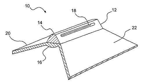
of these experiments conclude that direct repair of the annulus does not
influence the healing of the disc.
4
CA 02512142 2005-07-26
WO 2004/069026 PCT/US2004/000211
[011] There is currently no known method of annulus reconstruction,
either primarily or augmented with an annulus stent.
Brief Summary of the Invention
[012] The present invention provides methods and related materials
for reconstruction of the disc wall in cases of displaced, herniated,
ruptured,
or otherwise damaged intervertebral discs. In accordance with the invention,
a method is disclosed for intervertebral disc reconstruction for treating a
disc
having an aperture in the wall of the annulus fibrosis, wherein the aperture
provides a path for the migration of nucleus pulposus from the subannular
space, the method including the steps of providing an expandable patch
having a first configuration dimensioned to pass through the aperture and a
second expanded configuration having at least one dimension at least as
large as the aperture and having at least one dimension larger than a
corresponding dimension in said first configuration; inserting the patch
through the aperture into the subannular space when the device is in the first
collapsed configuration; and causing or allowing the patch to expand in the
subannular space into the second expanded configuration to bridge the
aperture, thereby occluding the aperture and preventing the migration of
nucleus pulposus therethrough.
[013] The objects and various advantages of the invention will be
apparent in consideration of the description which follows. In general, the
implantable medical device is placed, positioned, and affixed to the annulus
to reduce re-extrusion of the nucleus through the aperture by: acting as a
mechanical barrier; restoring the natural integrity of the wall of the
annulus;
CA 02512142 2005-07-26
WO 2004/069026 PCT/US2004/000211
and promoting the healing of the annulus through the reapproximation of disc
wall tissue. Increased integrity and faster and/or more thorough healing of
the aperture is intended to reduce future recurrence of herniation of the disc
nucleus from the intervertebral disc, and the recurrence of resulting back
pain. In addition, it is believed that the repair of the aperture could
promote
enhanced biomechanics and reduce the possibility of intervertebral disc
height collapse and segmental instability, thus resulting in a decrease in the
recurrence of back pain after a surgical procedure.
[014] Moreover, the repair of the aperture with the reduction of the re-
extrusion of the nucleus may also advantageously reduce adhesion formation
surrounding the nerve roots. The nuclear material of the disc is toxic to the
nerves and is believed to cause increased inflammation surrounding the
nerves, which in turn can cause increased scar formation (adhesions or
epidural fibrosis) upon healing. Adhesions created around the nerve roots
can cause continued back pain. Any reduction in adhesion formation is
believed to reduce future recurrence of pain.
[015] One of the objects of the present inventions is to act as a
mechanical barrier to the extrusion of the nucleus from the disc space, add
mechanical integrity to the annulus and the tissue surrounding the aperture,
and to promote faster and a more complete healing of the aperture.
[016] Although much of the discussion is directed toward the repair of
the intervertebral disc after a surgical procedure, such as discectomy (a
surgical procedure performed to remove herniated fragments of the disc
nucleus), it is contemplated that the device could be used in other procedures
6
CA 02512142 2005-07-26
WO 2004/069026 PCT/US2004/000211
that involve incisions into the annulus of the intervertebral disc. An example
of another procedure that could require a repair technique involves the
replacement of the nucleus - nucleus replacement - with an implantable
nucleus to replace the functioning of the natural nucleus when it is
degenerated. The object of the invention in this case would be similar in that
the repair would maintain the replacement nucleus within the disc space.
[017] According to the invention, a sub-annular patch/stent can be
employed to repair an intervertebral disc annulus. In its simplest form, the
repair of the annulus involves the placement and fixation of a fascial
autograft
patch to the sub-annular space which can additionally employ two or more
sutures, while re-approximating the tissues surrounding the aperture. The
invention, through involvement of the sub-annular space and wall for the
repair of the aperture, has several advantages over the prior art; for
example,
sealing the aperture only on the outer surface, or sealing the aperture only
within the aperture. The first advantage of a repair that involves the sub-
annular surface derives itself from the physical nature of a circular (or an
elliptical) compressed chamber with a radius, like an intervertebral disc.
Sealing the inside wall has the inherent advantage of being at a smaller
radius of curvature versus the outer wall and thus, according to LaPlace's
Law, the patch would be subjected to lower stresses at any given pressure, all
else held equal.
[018] Another advantage of utilizing the inner surface to accomplish
sealing is that the natural pressure within the disc can enhance the sealing
of
the device against the inner wall of the disc space. Conversely, if the repair
is
7
CA 02512142 2005-07-26
WO 2004/069026 PCT/US2004/000211
performed on the outer surface of the annulus there is an inherent risk of
leakage around the periphery of the device, with the constant exposure to the
pressure of the disc.
[019] Another advantage of the present invention over the prior art in
utilizing the inner surface of the annulus is the reduction of the risk of
having a
portion of the device protruding from the exterior surface of the annulus.
Device materials protruding from the exterior of the annulus pose a risk of
damaging the nerve root and/or spinal canal which are in close proximity.
Damage to these structures can result in continued pain, incontinence, bowel
dysfunction, and paralysis.
[020] The present invention also incorporates the concept of pulling
the tissues together that surround the aperture, the inner surface, and the
outer surface of the annulus to help increase the integrity of the repair.
[021] An example of the technique and placement of the device
according to the invention is as follows:
[022] 1. An aperture is created measuring approximately, for
example, 6 mm x 2 mm in the wall of the annulus after performing a
discectomy procedure in which a portion of the nucleus is also removed from
the disc space, as shown in FIGs. 32a, 32b, 33a and 33b.
[023] 2. Two or more sutures are passed through the upper and
lower surfaces of the aperture and they are pushed within the intervertebral
disc space to create a "sling" to receive the fascial autograft as shown for
example in FIG. 34.
8
CA 02512142 2005-07-26
WO 2004/069026 PCT/US2004/000211
[024] 3. A piece of para-spinal fascial tissue is removed from the
patient measuring approximately, for example, 10 mm X 5 mm.
[025] 4. The autograft is folded and compressed to pass through
the aperture in the annulus, as shown for example in FIG. 35.
[026] 5. The autograft takes a second shape, within the annulus
that is uncompressed and oriented to be in proximity of the subannular wall of
the annulus, within the sling, as shown for example in FIG. 36. The autograft
may be inserted entirely into the subannular space, or a portion may extend
into the rent as depicted in FIG. 36.
[027] 6. The sutures are tightened, as shown for example in FIG.
37, thus tightening the sling surrounding the autograft, to bring the
autograft in
close proximity with the subannular wall, while providing tension to bring the
patch at the subannular surface together with the outer surface of the annular
wall, thus creating increased integrity of the annulus surrounding the
aperture,
as well as causing the autograft to take a second shape that is larger than
the
aperture. Furthermore, the tightening, and eventual tying of the sutures also
promotes the re-approximation of the tissue at the outer surface of the
annulus and within the aperture.
[028] 7. The sutures are tied and the ends of the sutures are cut.
[029] 8. A piece of autograft fat tissue may be placed over the
discectomy site for the prevention of adhesion formation, a typical surgical
technique.
[030] 9. Standard surgical techniques are utilized to close the
access site of the surgical procedures.
9
CA 02512142 2005-07-26
WO 2004/069026 PCT/US2004/000211
[031 ] Several devices according to the present invention can be used
to practice the above illustrative inventive steps to accomplish the sealing
and/or repair of the intervertebral disc. In each of the representative
devices
described herein, there is: an expandable patchlstent (note: patch, stent and
device are used interchangeably) that has, in use, at least a portion of the
device in proximity to the sub-annular space of the intervertebral disc
annulus; a means to affix the patch to stay in proximity with the annulus; a
means to draw the patch and the annular tissue together and fasten in
tension; and a means to help reduce the relative motion of the surfaces of the
aperture after fixation, and thus promote healing. According to one feature
and object of the present invention, close approximation of tissue, while
reducing the motion of the surfaces, provides the optimal environment for
healing.
[032] The concepts disclosed hereinbelow accomplish these
objectives, as well as advantageously additionally incorporating design
elements to reduce the number of steps (and time), andlor simplify the
surgical technique, and/or reduce the risk of causing complications during the
repair of the intervertebral disc annulus. In addition, it is an objective of
the
following devices to become incorporated by the surrounding tissues, or to act
as a scaffold in the short-term (3 - 6 months) for tissue incorporation.
[033] In an exemplary embodiment, one or more mild biodegradable
surgical sutures can be placed at about equal distances along the sides of a
pathologic aperture in the ruptured disc wall (annulus) or along the sides of
a
surgical incision in the annular wall, which may be weakened or thinned.
CA 02512142 2005-07-26
WO 2004/069026 PCT/US2004/000211
[034J Sutures are then tied in such fashion as to draw together the
sides of the aperture, effecting reapproximation or closure of the opening, to
enhance natural healing and subsequent reconstruction by natural tissue
(fibroblasts) crossing the now surgically narrowed gap in the disc annulus.
[035] A 25-30% reduction in the rate of recurrence of disc nucleus
herniation through this aperture has been achieved using this method.
[036] In another exemplary embodiment, the method can be
augmented by creating a subannular barrier in and across the aperture by
placement of a patch of human muscle fascia (muscle connective tissue) or
any other autograft, allograft, or xenograft acting as a bridge or a scaffold,
providing a platform for traverse of fibroblasts or other normal cells of
repair
existing in and around the various layers of the disc annulus, prior to
closure
of the aperture.
[037] A 30-50% reduction in the rate of recurrence of disc herniation
has been achieved using the aforementioned fascial augmentation with this
embodiment.
[038] In still another embodiment, a braided patch can be formed
having a first collapsed configuration having a major longitudinal dimension
with first and second ends. When these ends are moved toward each other
along the longitudinal axis, a portion' of the device between the ends can
deploy outwardly to form an expanded configuration.
[039] Having demonstrated that human muscle fascia is adaptable for
annular reconstruction, other biocompatible membranes can be employed as
a bridge, stent, patch or barrier to subsequent migration of the disc nucleus
11
CA 02512142 2005-07-26
WO 2004/069026 PCT/US2004/000211
through the aperture. Such biocompatible materials may be, for example,
medical grade biocompatible fabrics, biodegradable polymeric sheets, or form
fitting or non-form fitting fillers for the cavity created by removal of a
portion of
the disc nucleus pulposus in the course of the disc fragment removal or
discectomy. The prosthetic material can be placed in and around the
intervertebral space, created by removal of the degenerated disc fragments.
[040] Additional objects and advantages of the invention will be set
forth in part in the description which follows, and in part will be obvious
from
the description, or may be learned by practice of the invention. The objects
and advantages of the invention will be realized and attained by means of the
elements and combinations particularly pointed out in the appended claims.
[041] It is to be understood that both the foregoing general description
and the following detailed description are exemplary and explanatory only and
are not restrictive of the invention, as claimed.
Brief Description of the Drawings
[042] The accompanying drawings, which are incorporated in and
constitute a part of this specification, illustrate illustrative embodiments
of the
invention and, together with the description, serve to explain the principles
of
the invention.
[043] FIG. 1 shows a perspective view of an illustrative embodiment of
an annulus stent.
[044] FIG. 2 shows a front view of the annulus stent of FIG. 1.
[045] FIG. 3 shows a side view of the annulus stent of FIG. 1.
12
CA 02512142 2005-07-26
WO 2004/069026 PCT/US2004/000211
[046] FIGs. 4A-4C show a front view of alternative illustrative
embodiments of an annulus stent.
[047] FIGs. 5A-5B show the alternative embodiment of a further
illustrative embodiment of an annulus stent.
[048] FIGs. 6A-6B show the alternative embodiment of a further
illustrative embodiment of an annulus stent.
[049] FIG. 7 shows a primary closure of an opening in the disc
annulus.
[050] FIGs. 8A-8B show a primary closure with a stent.
[051] FIG. 9 shows a method of suturing an annulus scent into the
disc annulus utilizing fixation points on vertebral bodies.
[052] FIGs. 10A-10B show a further illustrative embodiment of an
annulus stent with flexible bladder being expanded into the disc annulus.
[053] FIGs. 11A-11D show an annulus stent being inserted into and
expanded within the disc annulus.
[054] FIGs. 12A- 12B show an annulus stent with a flexible bladder
being expanded.
[055] FIG. 13 shows a perspective view of a further illustrative
embodiment of an annulus stent.
[056] FIG. 14 shows a first collapsed view of the annulus stent of FIG.
13.
[057] FIG. 15 shows a second collapsed view of the annulus stent of
FIG. 13.
13
CA 02512142 2005-07-26
WO 2004/069026 PCT/US2004/000211
[058] FIGs. 16A-16C show the annulus stent of FIG. 13 being
inserted into the disc annulus.
[059] FIGs. 17A-17C show a method of inserting the annulus stent of
F1G. 13 into the disc annulus.
[060] FIGs. 18A-18B show a further illustrative embodiment of an
annulus stent with a flexible bladder.
[061 ] FIGs. 19A-19B show another illustrative embodiment of an
annulus stent with a flexible bladder.
[062] FIG. 20 shows an expanded annulus stent with barbs on the
radial extension.
[063] FIG. 21 shows a still further illustrative embodiment of an
annulus stent with a compressible core.
[064] FIG. 22 shows a still further illustrative embodiment of an
introduction device for an annulus stent.
[065] FIG. 23 shows a modification of the device depicted in FIG. 22.
[066] FIG. 24 shows an exemplary introduction tool for use with the
devices of FIGs. 22 and 23 with a stent deflected proximally.
[067] FIG. 25 shows an exemplary introduction tool for use with the
devices of FIGs. 22 and 23 with a stent deflected distally.
[068] FIG. 26 shows an exemplary introduction tool for use with the
devices of FIGs. 22 and 23 with a stent deflected partially distally and
partially
proximally.
[069] FIG. 27 shows a still further illustrative embodiment of a stent
device having a grasping feature and fixation devices in the form of barbs.
14
CA 02512142 2005-07-26
WO 2004/069026 PCT/US2004/000211
[070] F1G. 28 shows the illustrative embodiment in FIG. 27 deployed
subannularly.
[071] FIG. 29 shows a still further illustrative embodiment of an
annulus stent employing a secondary barbed fixation device.
[072] FIG. 30 shows a still further illustrative embodiment of an
annulus stent employing another example of a secondary barbed fixation
device.
[073] FIG. 31 shows the frame of a still further illustrative embodiment
of an annulus stent having a metal substrate being machined from flat stock.
[074] FIG. 32a shows a herniated disc in perspective view, and FIG.
32b shows the same disc after discectomy.
[075] FIG. 33a shows a top view of the disc post-discectomy, and
FIG. 33b shows a posteriolateral view of the disk showing an incision.
[076] FIG. 34 shows schematically the creation of a subannular sling
using sutures.
[077] FIG. 35 schematically shows the introduction of a compressed
autograft stentfpatch into the subannular space.
[078] FIG. 36 schematically shows the autograft of FIG. 35 in an
expanded shape within the annulus.
[079] FIG. 37 schematically shows the tightening of the sutures to
reapproximate the annulus aperture and draw the stent/patch of FIG. 35
toward the annular wall.
[080] FIG. 38 shows an exemplary collar for use in repairing a disc
annulus.
CA 02512142 2005-07-26
WO 2004/069026 PCT/US2004/000211
[081] FIG. 39 schematically depicts the collar of FIG. 38 in use for
-- disc annulus repair.
[082] FIG. 40 shows a still further exemplary embodiment of the
present invention using a bag to contain the patch/stent.
[083] FIG. 41a-a show still further illustrative embodiments of the
present invention having frames.
[084] FIG. 42 shows an illustrative method for placing a barbed
expandable patch in the subannular disc space.
[085] FIG. 43 shows the patch of FIG. 42 being fixed to the inside wall
of the annulus fibrosus.
[086] FIGs. 44a-g show a still further illustrative embodiment of an
introduced and expanded annulus stent/patch being fixated and the aperture
reapproximated.
[087] FIGs. 45a-c schematically depict a still further embodiment of
the present invention where an expandable stent/patch is tethered in situ
using a cinch line.
[088] FIGs. 46a-c schematically depict the cinch line of FIG. 45 being
fixated through use of a surgical staple device.
[089] FIGs. 47a-b show an illustrative embodiment of a suturing
arrangement for securing a patch/stent in the annulus.
[090] FIG. 48a-b depict a still further illustrative embodiment where
fixation sutures are placed into the vertebral body or the Sharpey fibers.
16
CA 02512142 2005-07-26
WO 2004/069026 PCT/US2004/000211
[091 ] FIGs. 49a-c schematically depict a still further embodiment of
the present invention where an expandable stent/patch is tethered in situ
using a cinch line.
[092] FIGs. 50a-c schematically depict the cinch line of FIG. 49 being
fixated through use of a barbed surgical staple device that penetrates the
patch/stent.
[093] FIG. 51 depicts an exemplary use of filler tissue within the
aperture during placement of a patch/stent tethered by a cinch line.
[094] FIGs. 52a-a shows exemplary embodiments of various
additional patch/stent fixation techniques.
[095] FIG. 53 shows a still further illustrative embodiment of a
stentlpatch having a frame.
[096] FIG. 54a-f shows a still further illustrative embodiment of an
annular stent/patch having a self contained fixation tightening feature.
[097] FIG. 55 shows a still further exemplary embodiment of the
present invention having external fixation anchors.
[098] FIG. 56a-c shows a still further exemplary embodiment of the
present invention having external fixation anchors.
[099] FIG. 57a-c shows a still further exemplary embodiment of the
present invention having external fixation anchors.
[0100] FIG. 58 shows a still further exemplary embodiment of the
present invention having external fixation anchors.
[0101] FIG. 59 shows a still further exemplary embodiment of the
present invention having a springing arrangement.
17
CA 02512142 2005-07-26
WO 2004/069026 PCT/US2004/000211
[0102] FIG. 60 shows a lateral view of a still further exemplary
embodiment of the present invention having a braided arrangement in a
collapsed configuration.
[0103] FIG. 61 shows an axial view of the exemplary embodiment of
FIG. 60 in an expanded configuration.
[0104] FIG. 62 shows a lateral view of the exemplary embodiment of
FIG. 60 in a collapsed configuration mounted on an illustrative delivery
device.
[0105] FIG. 63 shows a lateral cutaway view of the exemplary
embodiment of FIG. 60 in a collapsed configuration.
[0106] FIG. 64 shows a lateral cutaway view of the exemplary
embodiment of FIG. 60 in an expanded configuration.
[0107] FIG. 65 shows a lateral view of an illustrative delivery member
as shown in the exemplary embodiment of FIGs. 63 and 64.
[0108] FIG. 66 shows a lateral view of an exemplary embodiment of the
invention in an expanded configuration subannularly.
Detailed Description of the Invention
[0109] Reference will now be made in detail to an illustrative
embodiment of the invention, which appears in the accompanying drawings.
Wherever possible, the same reference numbers will be used throughout the
drawings to refer to the same or like parts.
[0110] In one embodiment of the present invention, as shown in FIG. 7,
a damaged annulus 42 is repaired by use of surgical sutures 40. One or
more surgical sutures 40 are placed at about equal distances along the sides
18
CA 02512142 2005-07-26
WO 2004/069026 PCT/US2004/000211
of a pathologic aperture 44 in the annulus 42. Reapproximation or closure of
the aperture 44 is accomplished by tying the sutures 40 so that the sides of
the aperture 44 are drawn together. The reapproximation or closure of the
aperture 44 enhances the natural healing and subsequent reconstruction by
the natural tissue (e.g., fibroblasts) crossing the now surgically narrowed
gap
in the annulus 42. Preferably, the surgical sutures 40 are biodegradable, but
permanent non- biodegradable may be utilized.
[0111] Additionally, to repair a weakened or thinned wall of a disc
annulus 42, a surgical incision can be made along the weakened or thinned
region of the annulus 42 and one or more surgical sutures 40 can be placed
at about equal distances laterally from the incision. Reapproximation or
closure of the incision is accomplished by tying the sutures 40 so that the
sides of the incision are drawn together. The reapproximation or closure of
the incision enhances the natural healing and subsequent reconstruction by
the natural tissue crossing the now surgically narrowed gap in the annulus 42.
Preferably, the surgical sutures 40 are biodegradable, but permanent non-
biodegradable materials may be utilized.
[0112] In an alternative embodiment, the method can be augmented by
the placement of a patch of human muscle fascia or any other autograft,
allograft or xenograft in and across the aperture 44. The patch acts as a
bridge in and across the aperture 44, providing a platform for traverse of
fibroblasts or other normal cells of repair existing in and around the various
layers of the disc annulus 42, prior to closure of the aperture 44.
19
CA 02512142 2005-07-26
WO 2004/069026 PCT/US2004/000211
[0113] In a further embodiment, as shown in FIGs. 8A-B a
biocompatible membrane can be employed as an annulus stent 10, being
placed in and across the aperture 44. The annulus stent 10 acts as a bridge
in and across the aperture 44, providing a platform for a traverse of
fibroblasts
or other normal cells of repair existing in and around the various layers of
the
disc annulus 42, prior to closure of the aperture 44. In some embodiments
the. device, stent or patch can act as a scaffold to assist in tissue growth
that
healingly scars the annulus.
[0114] In an illustrative embodiment, as shown in FIGs. 1-3, the
annulus stent 10 comprises a centralized vertical extension 12, with an upper
section 14 and a lower section 16. The centralized vertical extension 12 can
be trapezoid in shape through the width and may be from about 8mm - l2mm
in length.
[0115] Additionally, the upper section 14 of the centralized vertical
extension 12 may be any number of different shapes, as shown in FIGs. 4A
through 4C, with the sides of the upper section 14 being curved or with the
upper section 14 being circular in shape. Furthermore, the annulus stent 10
may contain a recess between the upper section 14 and the lower section 16,
enabling the annulus stent 10 to form a compatible fit with the edges of the
aperture 44.
[0116] The upper section 14 of the centralized vertical extension 12
can comprise a slot 18, where the slot 18 forms an orifice through the upper
section 14. The slot 18 is positioned within the upper section 14 such that it
traverses the upper section's 14 longitudinal axis. The slot 18 is of such a
CA 02512142 2005-07-26
WO 2004/069026 PCT/US2004/000211
size and shape that sutures, tension bands, staples or any other type of
fixation device known in the art may be passed through, to affix the annulus
stent 10 to the disc annulus 42.
[0117] In an alternative embodiment, the upper section 14 of the
centralized vertical extension 12 may be perforated. The perforated upper
section 14 contains a plurality of holes that traverse the longitudinal axis
of
upper section 14. The perforations are of such a size and shape that sutures,
tension bands, staples or any other type of fixation device known in the art
may be passed through, to affix the annulus stent 10 to the disc annulus 42.
[0118] The lower section 16 of the centralized vertical extension 12 can
comprise a pair of lateral extensions, a left lateral extension 20 and a right
lateral extension 22. The lateral extensions 20 and 22 comprise an inside
edge 24, an outside edge 26, an upper surface 28, and a lower surface 30.
The lateral extensions 20 and 22 can have an essentially constant thickness
throughout. The inside edge 24 is attached to and is about the same length
as the lower section 16. The outside edge 26 can be about 8mm-16mm in
length. The inside edge 24 and the lower section 16 meet to form a
horizontal plane, essentially perpendicular to the centralized vertical
extension
12. The upper surface 28 of the lateral extensions 20 and 22 can form an
angle from about 0°-60° below the horizontal plane. The width of
the annulus
stent 10 may be from about 3mm-8mm.
21
CA 02512142 2005-07-26
WO 2004/069026 PCT/US2004/000211
[0119] Additionally, the upper surface 28 of the lateral extensions 20
and 22 may be barbed for fixation to the inside surface of the disc annulus 42
and to resist expulsion through the aperture 44.
[0120] In an alternative embodiment, as shown in FIG. 4B, the lateral
extensions 20 and 22 have a greater thickness at the inside edge 24 than at
the outside edge 26.
[0121] In an illustrative embodiment, the annulus stent 10 is a solid
unit, formed from one or more of the flexible resilient biocompatible or
bioresorbable materials well know in the art. The selection of appropriate
stent materials may be partially predicated on specific stent construction and
the relative properties of the material such that, after fixed placement of
the
stent, the repair may act to enhance the healing process at the aperture by
relatively stabilizing the tissue and reducing movement of the tissue
surrounding the aperture.
[0122] For example, the annulus stent 10 may be made from:
[0123] A porous matrix or mesh of biocompatible and bioresorbable
fibers acting as a scaffold to regenerate disc tissue and replace annulus
fibrosus as disclosed in, for example, U, S. Patent Nos. 5,108,438 (Stone)
and 5,258,043 (Stone), a strong network of inert fibers intermingled with a
bioresorbable (or bioabsorbable) material which attracts tissue ingrowth as
disclosed in, for example, U.S. Patent No, 4,904,260 (Ray et al.).
[0124] a biodegradable substrate as disclosed in, for example, U.S.
Patent No. 5,964,807 (Gan at al.); or
22
CA 02512142 2005-07-26
WO 2004/069026 PCT/US2004/000211
[0125] an expandable polytetrafluoroethylene (ePTFE), as used for
conventional vascular grafts, such as those sold by W.L. Gore and
Associates, Inc. under the trademarks GORE-TEX and PRECLUDE, or by
Impra, Inc. under the trademark IMPRA.
[0126] Furthermore, the annulus, stent 10, may contain hygroscopic
material for a controlled limited expansion of the annulus stent 10 to fill
the
evacuated disc space cavity.
[0127] Additionally, the annulus stent 10 may comprise materials to
facilitate regeneration of disc tissue, such as bioactive silica-based
materials
that assist in regeneration of disc tissue as disclosed in U.S. Patent No.
5,849,331 (Ducheyne, et al.), or other tissue growth factors well known in the
art.
[0128] Many of the materials disclosed and described above represent
embodiments where the device actively promotes the healing process. It is
also possible that the selection of alternative materials or treatments may
modulate the role in the healing process, and thus promote or prevent healing
as may be required. It is also contemplated that these modulating factors
could be applied to material substrates of the device as a coating, or similar
covering, to evoke a different tissue response than the substrate without the
coating.
[0129] In further embodiments, as shown in FIGs. 5AB-6AB, the left
and right lateral extensions 20 and 22 join to form a solid pyramid or cone.
Additionally, the left and right lateral extensions 20 and 22 may form a solid
trapezoid, wedge, or bullet shape. The solid formation may be a solid
23
CA 02512142 2005-07-26
WO 2004/069026 PCT/US2004/000211
biocompatible or bioresorbable flexible material, allowing the lateral
extensions 20 and 22 to be compressed for insertion into aperture 44, then to
expand conforming to the shape of the annulus' 42 inner wall.
[0130] Alternatively, a compressible core may be attached to the lower
surface 30 of the lateral extensions 20 and 22, forming a pyramid, cone,
trapezoid, wedge, or bullet shape. The compressible core may be made from
one of the biocompatible or bioresorbable resilient foams well known in the
art. The core can also comprise a fluid-expandable membrane, e.g., a
balloon. The compressible core allows the lateral extensions 20 and 22 to be
compressed for insertion into aperture 44, then to expand conforming to the
shape of the annulus' 42 inner wall and to the cavity created by pathologic
extrusion or surgical removal of the disc fragment.
[0131] In an illustrative method of use, as shown in FIGs. 11A-D, the
lateral extensions 20 and 22 are compressed together for insertion into the
aperture 44 of the disc annulus 42. The annulus scent 10 is then inserted into
the aperture 44, where the lateral extensions 20, 22 expand. In an expanded
configuration, the upper surface 28 can substantially conform to the contour
of the inside surface of the disc annulus 42. The upper section 14 is
positioned within the aperture 44 so that the annulus stent 10 maybe secured
to the disc annulus 42, using means well known in the art.
[0132] In an alternative method, where the length of the aperture 44 is
less than the length of the outside edge 26 of the annulus stent 10, the
annulus stent 10 can be inserted laterally into the aperture 44. The lateral
extensions 20 and 22 are compressed, and the annulus stent 10 can then be
24
CA 02512142 2005-07-26
WO 2004/069026 PCT/US2004/000211
laterally inserted into the aperture 44. The annulus stent 10 can then be
rotated inside the disc annulus 42, such that the upper section 14 can be held
back through the aperture 44. The lateral extensions 20 and 22 are then
allowed to expand, with the upper surface 28 contouring to the inside surface
of the disc annulus 42. The upper section 14 can be positioned within, or
proximate to, the aperture 44 in the subannular space such that the annulus
stent 10 may be secured to the disc annulus, using means well known in the
art.
[0133] In an alternative method of securing the annulus stent 10 in the
aperture 44, as shown in FIG. 9, a first surgical screw 50 and second surgical
screw 52, with eyeholes 53 located at the top of the screws 50 and 52, are
inserted into the vertebral bodies, illustratively depicted as adjacent
vertebrae
54 and 56. After insertion of the annulus stent 10 into the aperture 44, a
suture 40 is passed down though the disc annulus 42, adjacent to the
aperture 44, through the eye hole 53 on the first screw 50 then back up
through the disc annulus 42 and through the orifice 18 on the annulus stent
10. This is repeated for the second screw 52, after which the suture 40 is
secured. One or more surgical sutures 40 are placed at about equal
distances along the sides of the aperture 44 in the disc annulus 42.
Reapproximation or closure of the aperture 44 is accomplished by tying the
sutures 40 in such a fashion that the sides of the aperture 44 are drawn
together. The reapproximation or closure of the aperture 44 enhances the
natural healing and subsequent reconstruction by the natural tissue crossing
CA 02512142 2005-07-26
WO 2004/069026 PCT/US2004/000211
the now surgically narrowed gap in the annulus 42. Preferably, the surgical
sutures 40 are biodegradable but permanent non-biodegradable forms may
be utilized. This method should decrease the strain on the disc annulus 42
adjacent to the aperture 44, precluding the tearing of the sutures through the
disc annulus 42.
[0134] It is anticipated that fibroblasts will engage the fibers of the
polymer or fabric of the intervertebral disc stent 10, forming a strong wall
duplicating the currently existing condition of healing seen in the normal
reparative process.
[0135] In an additional embodiment, as shown in FIGs. 10A-B, a
flexible bladder 60 is attached to the lower surface 30 of the annulus stent
10.
The flexible bladder 60 comprises an internal cavity 62 surrounded by a
membrane 64, where the membrane 64 is made from a thin flexible
biocompatible material. The flexible bladder 60 is attached to the lower
surface 30 of the annulus stent 10 in an unexpanded condition. The flexible
bladder 60 is expanded by injecting a biocompatible fluid or expansive foam,
as known in the art, into the internal cavity 62. The exact size of the
flexible
bladder 60 can be varied for different individuals. The typical size of an
adult
nucleus is about 2 cm in the semi-minor axis, 4 cm in the semi-major axis,
and 1.2 cm in thickness.
[0136] In an alternative embodiment, the membrane 64 is made of a
semi-permeable biocompatible material. The mechanical properties of the
injectate material may influence the performance of the repair and it is
contemplated that materials which are "softer" or more compliant as well as
26
CA 02512142 2005-07-26
WO 2004/069026 PCT/US2004/000211
materials that are less soft and less compliant than healthy nucleus are
contemplated within the scope of certain embodiments of the invention. It
must be understood that in certain embodiments the volume added to the
subannular space may be less than equal to or larger than the nucleus
volume removed. The volume of the implant may vary over time as well in
certain embodiments.
[0137] In an illustrative embodiment, a hydrogel is injected into the
internal cavity 62 of the flexible bladder 60. A hydrogel is a substance
formed
when an organic polymer (natural or synthetic) is cross-linked via, covalent,
ionic, or hydrogen bonds to create a three-dimensional open-lattice structure,
which entraps water molecules to form a gel. The hydrogel may be used in
either the hydrated or dehydrated form.
[0138] In a method of use, where the annulus stent 10 has been
inserted into the aperture 44, as has been previously described and shown in
FIGs. 12 A-B, an injection instrument, as known in the art, such as a syringe,
is used to inject the biocompatible fluid or expansive foam into the internal
cavity 62 of the flexible bladder 60. The biocompatible fluid or expansive
foam is injected through the annulus stent 10 into the internal cavity 62 of
the
flexible bladder 60. Sufficient material is injected into the internal cavity
62 to
expand the flexible bladder 60 to fill the void in the intervertebral disc
cavity.
The use of the flexible bladder 60 is particularly useful when it is required
to
remove all or part of the intervertebral disc nucleus.
[0139] The surgical repair of an intervertebral disc may require the
removal of the entire disc nucleus, being replaced with an implant, or the
27
CA 02512142 2005-07-26
WO 2004/069026 PCT/US2004/000211
removal of a portion of the disc nucleus thereby leaving a ~~oid in the
intervertebral disc cavity. The flexible bladder 60 allows for the removal of
only the damaged section of the disc nucleus, with the expanded flexible
bladder 60 filling the resultant void in the intervertebral disc cavity. A
major
advantage of the annulus scent 10 with the flexible bladder 60 is that the
incision area in the annulus 42 can be reduced in size, as there is no need
for
the insertion of an implant into the intervertebral disc cavity.
[0140] In an alternative method of use, a dehydrated hydrogel is
injected into the internal cavity 62 of the flexible bladder 60. Fluid, from
the
disc nucleus, passes through the semipermeable membrane 64 hydrating the
dehydrated hydrogel. As the hydrogel absorbs the fluid the flexible bladder
60 expands, filling the void in the intervertebral disc cavity.
[0141] In an alternative embodiment, as shown in FIG. 13, the annulus
stent 10 is substantially umbrella shaped, having a central hub 66 with
radially
extending struts 67. Each of the struts 67 is joined to the adjacent struts 67
by a webbing material 65, forming a radial extension 76 about the central hub
66. The radial extension 76 has an upper surface 68 and a lower surface 70,
where the upper surface 68 contours to the shape of the disc annulus' 42
inner wall when inserted as shown in FIG. 17A-C, and where the lower
surface 70 contours to the shape of the disc annulus' 42 inner wall when
inserted as shown in FIG. 16A-C. The radial extension 76 may be
substantially circular, elliptical, or rectangular in plan shape.
Additionally, as
shown in FIG. 20, the upper surface 68 of the radial extension 76 may be
28
CA 02512142 2005-07-26
WO 2004/069026 PCT/US2004/000211
barbed 82 for fixation to the disc annulus' 42 inner wall and to resist
expulsion
through the aperture 42.
[0142] As shown in FIGs. 14 and 15, the struts 67 are formed from
flexible material, allowing the radial extension 76 to be collapsed for
insertion
into aperture 44, then the expand conforming to the shape of the inner wall of
disc annulus 42. In the collapsed position, the annulus stent 10 is
substantially frustoconical or shuttlecock shaped, and having a first end 72,
comprising the central hub 66, and a second end 74.
[0143] In an alternative embodiment, the radial extension 76 has a
greater thickness at the central hub 66 edge than at the outside edge.
[0144] In an embodiment, the annulus stent 10 is a solid unit, formed
from one or more of the flexible resilient biocompatible or bioresorbable
materials well known in the art.
[0145] Additionally, the annulus stent 10 may comprise materials to
facilitate regeneration of disc tissue, such as bioactive silica based
materials
that assist in regeneration of disc tissue as disclosed in U.S. Patent No.
5,849,331 (Ducheyne, et al.), or other tissue growth factors well known in the
art.
[0146] Alternatively, as shown in FIG. 21, a compressible core 84 may
be attached to the lower surface 70 of the radial extension 76. The
compressible core 84 may be made from one of the biocompatible or
bioresorbable resilient foams well known in the art. The compressible core 84
allows the radial extension 76 to be compressed for insertion into aperture 44
then to expand conforming to the shape of the disc annulus' 42 inner wall and
29
CA 02512142 2005-07-26
WO 2004/069026 PCT/US2004/000211
to the cavity created by pathologic extrusion or surgical removal of the disc
fragment.
[0147] In an additional embodiment, as shown in FIG. 18A and 18B, a
flexible bladder 80 is attached to the lower surface 70 of the annulus stent
10.
The flexible bladder 80 comprises an internal cavity 86 surrounded by a
membrane 88, where the membrane 88 is made from a thin flexible
biocompatible material. The flexible bladder 86 is attached to the lower
surface 70 of the annulus stent 10 in an unexpanded condition. The flexible
bladder 80 is expanded by injecting a biocompatible fluid or expansive foam,
as known in the art, into the internal cavity 86. The exact size of the
flexible
bladder 80 can be varied for different individuals. The typical size of an
adult
nucleus is 2 cm in the semi-minor axis, 4 cm in the semi-major axis and 1.2
cm in thickness.
[0148] In an alternative embodiment, the membrane 88 is made of a
semi-permeable biocompatible material.
[0149] In a method of use, as shown in FIGs. 16A-16C, the radial
extension 76 is collapsed together, for insertion into the aperture 44 of the
disc annulus 42. The radial extension 76 is folded such the upper surface 68
forms the outer surface of the cylinder. The annulus stent 10 is then inserted
into the aperture 44, inserting the leading end 72 though the aperture 44
until
the entire annulus stent 10 is within the disc annulus 42. The radial
extension
76 is released, expanding within the disc 44. The lower surface 70 of the
annulus stent 10 contours to the inner wall of disc annulus 42. The central
CA 02512142 2005-07-26
WO 2004/069026 PCT/US2004/000211
hub 66 is positioned within the aperture 44 so that the annulus stent 10 may
be secured to the disc annulus 42 using means well known in the art.
[0150] It is anticipated that fibroblasts will engage the fibers of the
polymer of fabric of the annulus stent 10, forming a strong wall duplicating
the
currently existing condition of healing seen in the normal reparative process.
[0151] In an alternative method of use, as shown in FIGs. 17A-17C, the
radial extension 76 is collapsed together for insertion into the aperture 44
of
the disc annulus 42. The radial extension 76 is folded such that the upper
surface 68 forms the outer surface of the stent, for example in a
frustoconical
configuration as illustrated. The annulus stent 10 is then inserted into the
aperture 44, inserting the tail end 74 through the aperture 44 until the
entire
annulus stent 10 is in the disc. The radial extension 76 is released,
expanding within the disc. The upper surface 68 of the annulus stent 10
contours to the disc annulus' 42 inner wall. The central hub 66 is positioned
within the aperture 44 so that the annulus stent 10 may be secured to the disc
annulus 42, using means well known in the art.
[0152] In one illustrative embodiment, the barbs 82 on the upper
surface 68 of one or more strut 67 or other feature of the radial extension
76,
engage the disc annulus' 42 inner wall, holding the annulus stent 10 in
position.
[0153] In a method of use, as shown in FIGs. 12A-12B, where the
annulus stent 10 has been inserted into the aperture 44, as has been
previously described. Similarly, for the stent shown in FIGs. 18 through 21,
an injection instrument, as known in the art, such as a syringe, can be used
to
31
CA 02512142 2005-07-26
WO 2004/069026 PCT/US2004/000211
inject the biocompatible fluid or expansive foam into the internal cavity 86
of
the flexible bladder 80. The biocompatible fluid or expansive foam is injected
through the annulus stent 10 into the internal cavity 86 of the flexible
bladder
80. Sufficient material is injected into the internal cavity 86 to expand the
flexible bladder 80 to fill the void in the intervertebral disc cavity. The
material
can be curable (i.e., glue). The use of the flexible bladder 80 is
particularly
useful when it is required to remove all or part of the intervertebral disc
nucleus.
[0154] It should be noted that in any of the "bag" embodiments
described herein one wall or barrier can be made stiffer and less resilient
than
others. This relatively stiff wall member can then be placed proximate the
annulus wall and can advantageously promote, in addition to its reparative
properties, bag containment within the annulus.
[0155] FIG. 22 shows a further aspect of the present invention.
According to a further illustrative embodiment, a simplified schematic cross
section of a vertebral pair is depicted including an upper vertebral body 110,
a
lower vertebral body 112 and an intervertebral disc 114. An aperture or rent
116 in the annulus fibrosus (AF) is approached by a tube 118, which is used
to deliver a device 120 according to a further aspect of the present
invention.
The device 120 may be captured by a delivery tool 122 through the use of a
ring or other fixation feature 124 mounted on the repair device 120.
[0156] FIG. 23 shows a delivery method similar to that depicted in FIG.
22, with the exception that the tube 118A has a reduced diameter so that it
32
CA 02512142 2005-07-26
WO 2004/069026 PCT/US2004/000211
may enter into the sub-annular space of the disc 114 through the aperture or
rent,
[0157] Turning to FIG. 25, according to a further aspect of the present
invention, the delivery of the device 120 through the delivery tube 118 or
118A may be facilitated by folding the arms or lateral extensions 128, 130 of
the device to fit within the lumen of the tube 118 or 118A so that the scent
or
device 120 is introduced in a collapsed configuration. The device 120 is
moved through the lumen of the tubes 118 or 118A through the use of
delivery tool 122. FIG. 25 shows the arms deflected in a distal, or forward
direction for insertion into the delivery tube 118 or 118A while FIG. 24 shows
the arms 128, 130 deflected into a proximal position. FIG. 26 shows the
device 120 curled so that one arm 128 is projecting distally, or in a forward
direction, and the other arm 130 is projecting proximally, or in a rearward
direction. Because the lateral extent of the device is relatively flexible,
whether the device is of natural or synthetic material, other collapsible
configurations consistent with the intent of this invention are also possible,
including twisting, balling, crushing, etc.
[0158] FIG. 27 shows the device 120 having a series of peripheral barb
structures typified by barb 132 located at the edges. In operation, these
barbs may be forced into the annulus fibrosus as seen in connection with FIG.
28. Barb placement can be anywhere on the device 120 provided that at
least some number of barbs are likely to find annulus fibrosus tissue to
anchor in during placement. For a simple aperture or rent, placement on the
periphery of the device body is a reasonable choice, but for complex tears, it
33
CA 02512142 2005-07-26
WO 2004/069026 PCT/US2004/000211
may be desirable to place a plurality of barbs on the device not knowing in
advance which barbs will find tissue to anchor in during placement.
[0159] FIG. 29 shows an alternative fixation strategy where a pair of
barbs 134 and 136 are plunged into the annulus fibrosus from the exterior of
the annulus while the device 120 is retained in the sub-annular space by
means of a tether 142. Although there are a wide variety of fixation devices
in
this particular example, a tether 142 may be knotted 145 with the band 144
holding the barbs 134 and 136 together to fix the device in the sub-annular
space. The knot is shown in an uncinched position to clarify the relationship
between the tether 142 and the bands 144. Using this approach, the device
can be maintained in a subannular position by the barbed bands while the
tether knot is cinched, advantageously simultaneously reapproximating the
annulus to close the aperture while drawing the device into sealing, bridging
engagement with the subannular wall of the annulus fibrosus.
[0160] FIG. 30 shows an alternative fixation strategy where the barbs
148 and 150 are sufficiently long that they can pierce the body of the device
120 and extend all the way through the annulus fibrosus into the device 120.
In this configuration, the band 144 connecting the barbs 148 and 150 may be
tightened to gently restrain and position the device 120 in the sub-annular
space, or tightened with greater force to reapproximate the aperture or rent.
[0161] FIG. 31 shows a still further illustrative embodiment according to
another aspect of the present invention. In this embodiment, a metal
substrate 160 is incorporated into the device 120. This piece can be
machined from flat stock and includes the loop 162 as well as barbs typified
34
CA 02512142 2005-07-26
WO 2004/069026 PCT/US2004/000211
by barb 164. When formed in to the device 120 the structure shown in FIG.
31 is used in a manner analogous to FIG. 27 and FIG. 28.
[0162] Stents can expand to be planar, for example as shown
hereinabove in FIGs 4, 8, 9, 11 and 12', or they can expand to be three-
dimensional as shown hereinabove in FIGs. 5 and 10. FIGs 34-36 depict a
further three dimensional patch/stent using an autograft formed of fascial
tissue. FIG 34 shows the superior vertebral body 202 and the inferior
vertebral body 204 surrounding a disc having an annulus fibrosus 206 and
nucleus pulposus 203 in the subannular space. According to this illustrative
embodiment of the invention, a suture 210 is passed from outside the annulus
through the wall of the annulus on one side of an aperture 208 and into the
subannular space as shown. The suture is then passed back out through the
annular wall on an opposing side of the aperture 208 leaving a loop or sling
212 of suture in the subannular space. As shown in the posterior view on the
right side of FIG. 34, more than one suture can be applied. Turning to FIG.
35, a fascial autograft 214 is then inserted through the aperture 208 into the
subannular space using, for example, forceps 216. FIG. 36 shows the fascial
stent/patch 214 fully inserted into the subannular space within the suture
sling
212. The closure of the aperture is accomplished simultaneously with pulling
the autograft 214 toward the annular wall as shown in FIG. 37. The suture
210 can be cinched 218 or tied to maintain the closure and the fixation of the
patch/stent.
[0163] Patches can be folded and expanded in a single plane or in
three dimensions. As shown in FIGs. 24-25 and 41 for example, collapsing
CA 02512142 2005-07-26
WO 2004/069026 PCT/US2004/000211
the patch can be accomplished laterally, whether the device is a single
material or composite. Other embodiments, such as that shown in FIG. 1 can
collapse vertically , and still others such as that shown in FIG. 26,
longitudinally. Others can collapse in three dimensions, such as those shown
in FIGs. 13-15 and 36. Devices which expand in three dimensions can be
packaged in a restraining jacket, such as a gelatine shell or "gelcap" for
example, or a mesh of biosorbable or dissolvable material, that would allow
for facile placement and subsequent expansion.
[0164] Patches can also be constructed of a single component, as
shown for example in Fig. 36, made of autograft or a synthetic material such
as Dacron, or for example where the stent is a gelcap. They can be made of
multiple components. An exemplary stent (not shown) can be made from a
polymeric material, for example silicone rubber, which can be formed to have
a natural unstressed shape, for example that of a "Bulb". A stylet or push-rod
can, for example, be inserted on the inside of the bulb to stretch the bulb
into
a second shape which is thinner and elongated. The second shape is
sufficient to place within the aperture in the annulus. Upon placement of the
device within the sub-annular space, the push-rod is removed and the bulb
assumes it natural, unstressed state, assuming a larger dimension within the
sub-annular space. Although silicone is used in this example, other metallic
constructs could also be envisioned such as a Nitinol braided device that has
a natural unstressed shape and assumes a second shape under tension for
the delivery of the device. It is also contemplated that the opposite scenario
can also accomplish the similar objective. In this alternative embodiment, the
36
CA 02512142 2005-07-26
WO 2004/069026 PCT/US2004/000211
device can have a first configuration that is unstressed and elongated and
assumes a second, larger configuration (bulb) under stress. In this
embodiment, a portion of the stylet or rod that is used to mechanically
activate the device would be left behind to hold the expansion element in its
stressed configuration.
[0165] Multiple components could include a frame to help with
expansion of the device and a covering to obtain biocompatibility and tissue
ingrowth. Examples of different frame configurations might include an
expandable "Butterfly" or "Figure-8" configuration that could be constructed
of
wire material, such as Nitinol or multiple wires. Exemplary embodiments
showing frame members 502 are depicted in FIG. 41A-E. Of course, other
configurations such as diamonds or other rounded or polygonal shapes can
be used. The diamond frame is a construct that takes a first form that is
smaller and expands to a larger frame. The diamond elements could be
constructed from a single wire or from multiple wires. Alternatively, the
members could be constructed of elements that are moveable. fixed at each
of the ends to allow expansion. A tether or attachment device 504 is also
depicted, which may be a suture, a wire, a screw, or other attachment means
known in the art.
[0166] The frame could be cut from a single material, such as flat
stock Nitinol to accomplish the same objective, as shown for example in FIG.
31. Such shapes can be cut from flat stock using known methods, for
example, laser cutting. A heat forming step could also be employed, as
37
CA 02512142 2005-07-26
WO 2004/069026 PCT/US2004/000211
known in the art, to form barbs 132 in a shape that passes out of the flat
plane of the stock material, as shown in FIG. 27 for example.
[0167] Another frame configuration, also not shown, is that of a spiral
or coif. The "Coil" design can be, for example, a spring steel or other
biocompatible material that is wrapped to a first "wound" smaller
configuration
and expands to a larger unwrapped, unwound configuration.
[0168] Depending on the size of the openings in the frames described
above, each of these concepts may or may not have a covering over them in
order to assure that the nucleus does not re-extrude from the intervertebral
disc space after placement of the device, as well as to serve as substrate for
the surrounding tissue to naturally incorporate the device. Coverings might
include ePTFE, polyester, silicone, or other biocompatible materials.
Coverings could also include natural materials such as collagen, cellulose,
autograft, xenograft, allograft or similar materials. The covering could also
be
biodegradable in nature, such as polyvinyl lactic acid.
[0169] Frames that are not covered may be permeable, such as a
patch that is porous and allow for normal movement of fluids and nutrients
through the patch into and out of the annular ring while maintaining nucleus
fragments larger than the porosity of the stent/patch within the s.ubannular
space. Depending on the material that the frame is constructed, a surface
finish may be added to promote tissue ingrowth into the patch. For example,
a titanium sputtering of the device may allow it to be more easily
incorporated
within the disc space. Alternatively, a NiTi or tantalum foam could be added
to the outer surface of the patch to promote tissue ingrowth.
38
CA 02512142 2005-07-26
WO 2004/069026 PCT/US2004/000211
[0170] It is understood that there can be a variety of device designs of
patches to accomplish the expansion of a device from a first configuration, to
a second configuration to occupy the sub-annular space and reduce re-
extrusion of the nucleus. The following device concepts are further discussed
for additional embodiments of a device and/or system for the repair of an
intervertebral disc annulus.
[0171 ] As mentioned hereinabove, the stent/patch according to the
present invention may comprise a mass of fascial autograft, and that
autograft may be contained in a covering of material to form what will be
referred to herein as a "bag". Of course, this term is used not necessarily to
connote a five-sided closed container so much as to denote the notion of
flexibly surrounding the volume of a patch/stent material so that it can be
manipulated in space.
[0172] In the most simplistic form, a prefabricated device of sutures
could be used to form the "sling" to hold the fascial implant as discussed
above. The advantage of this design over simple placement of sutures to hold
the autograft is better containment and control of the autograft during and
after implantation. The "sling" or a "bag" surrounds the fascial autograft to
hold it in place. It is contemplated that other materials, such as a polyester
mesh, could be used instead of the fascial autograft.
[0173] Figure 38 shows an example of a pre-fabricated sling 300.
There are three sutures used in this example, 302, 304, and 306, although
there could be more or less sutures as would be understood by one of
ordinary skill in the art. A collar member 308 has apertures or other features
39
CA 02512142 2005-07-26
WO 2004/069026 PCT/US2004/000211
for attaching to the sutures. In this example, the third suture 306 passes
along or within the collar 308 to form a loop extending from the lateral
extent
of the collar 308. The first and second sutures 302, 304 form loops from the
superior and inferior extents of the collar 308. Intersections 310 can secure
the loops to each other with small Poops or knots in the sutures, small fabric
attachment pieces, or by small preformed devices resembling grommets
placed on the suture to aid in securement. Other knot tying techniques
known in the art can also be employed. Turning to FIG. 39, the collar is
depicted within the subannular space where the loops surround a fascial
autograft 314 which by pulling proximally the sutures 302, 304, 306 the graft
is collapsed into contact with the annular wall in a sealing manner. The
sutures can be made of known materials, e.g., biodegradable, bioabsorbable
or~bioresorbable Vicryl or biocompatible nylon. The collar can be made of a
fabric material, e.g., polyester. During placement, one end of some or each
suture can be passed through the inferior wall of the annulus and the other
end can be passed through the superior wall surrounding the aperture. After
the placement of the sling into the wall of the annulus, the fascial autograft
is
placed within the sling. The sutures are tightened to bring the tissues
together and also to help reapproximate the aperture, as the collar size will
be
selected based on the surgeon's judgment according to the degree of
reapproximation desired.
[0174] Other constructions can also be used to accomplish the same
objective, such as a "bag" 404 formed of expandable PTFE as shown in Fig.
40. The bag is placed through an aperture in the annulus 402. Additionally, a
CA 02512142 2005-07-26
WO 2004/069026 PCT/US2004/000211
one way seal 406 can be positioned behind the aperture 408. Suturing
techniques for introducing cardiac valves could be employed to place the
seal. It is understood that there could be multiple constructs to accomplish
the same objective and this is only given as an example.
[0175] The are a variety of ways to affix the device to the sub-annular
wall of the annulus in addition to those discussed hereinabove. The following
exemplary embodiments are introduced here to provide inventive illustrations
of the types of techniques that can be employed to reduce the time and skill
required to affix the patch to the annulus, versus suturing and tying a knot.
Discussed hereinabove is the use of sutures, staples and other fixation
devices, such as those passed through slot 18 to affix the patch to the
annulus as shown.~in FIG. 1. FIG. 20 also depicts the use of "barbs" on the
surface of the stent to facilitate fixation to the annulus. In a simple
example,
as shown in FIG. 20, a patch/stent could be compressed, passed through a
guide tube such as tubes 18, 18A shown in FIGs. 22 and 23, and expanded
within the sub-annular space. As shown in FIG. 42, the expanded patch 602
is shown having barbs 604, along with detachable delivery tool 608 and guide
tube 606. Once expanded, barbs 604 on the outer surface of patch 602 can
be used to fix the patch into the inner wall 610 of the annulus 612 by pulling
the patch back proximally, into the sub-annular wall 610, and pushing forward
distally on the guide tube 606, thus driving the barbs 604 into the annulus
and
drawing the inner and outer tissues of the annulus together and
reapproximating the disc on either side of the aperture, as shown in FIG. 43.
41
CA 02512142 2005-07-26
WO 2004/069026 PCT/US2004/000211
After the placement of the patch, the delivery tool and guide tube are
removed.
[0176] The advantage of this design described above is that it requires
very little time and skill to place and secure the patch to the annulus while
also drawing the tissues together.
[0177] Materials of the patch could be similar to materials discussed
hereinabove. Anchoring barbs could be made of a biocompatible material, for
example a metallic material (e.g., NiTi alloy, Stainless steel, Titanium), or
a
polymeric material (e.g., polypropylene, polyethylene, polyurethane).
Anchoring barbs could also be a biodegradable/bioabsorbable material, such
as a polyglycolic acid (PGA), a polylevolactic acid (PPLA), a polydioxanone
(PDA) or for example a racemic polylactic acid (PDLLA). If the barbs included
a biodegradable/bioabsorbable material, it is anticipated that the barbs might
have sufficient holding strength for a sufficient period of time to allow the
patch to be incorporated into the annulus during the healing process. The
advantage of having the anchoring barb of FIGs. 42 and 43 being
biodegradable/bioabsorbable is that after the incorporation of the patch into
the annulus there may be no need for the barbs to provide fixation. However,
barbs pointing toward the outer surface of the annulus could pose a long term
risk of penetration out of the annulus due to migration, and potentially
impinging on the nerve root and spinal canal. Biodegradablelbioabsorbable
barbs address and advantageously reduce any long-term risk in this regard.
[0178] It is also possible that the barbs could be made of both a
biocompatible component and a biodegradable/bioabsorbable component.
42
CA 02512142 2005-07-26
WO 2004/069026 PCT/US2004/000211
For example, the very tip of the barb could be made of a biodegradable
material. The barb could penetrate the annulus wall with a rather sharp point,
but after degradation the point of the barb would become dull. In this
embodiment, the point would no longer induce continued scar formation after
the patch has been incorporated, nor pose a risk of penetrating out of the
annulus onto the nerve root.
[0179] Another fixation means includes the passing of "anchoring
bands" into the wall of the annulus, vertebral bodies (superior, inferior, or
both), or the Sharpey's Fibers (collagenous fibers between the junction of the
annular fibers and vertebral bodies). In the following example of anchors, the
barbs or bands are affixed to the annulus/vertebral bodies/Sharpey's fibers.
Another element, for example a suture, cinch line, or a staple is utilized to
attach the anchor bands to the patch, and thus hold the patch in proximity to
the inner wall of the annulus. In addition, these bands may re-approximate
the tissues at the aperture.
[0180] Revisiting one example of using barbs to anchor the device is
shown in FIG. 9, described hereinabove. Barbs or bone anchor screws 50
ands 52 are passed into the superior and inferior vertebral bodies 54 and 56,
respectively. Superiorly, suture 40 is passed through the outer wall of the
annulus, to the sub-annular space. The suture is then passed through the
eyelet 53 of bone anchor 52 and then passed through the wall of the annulus
from the sub-annular space to the outer wall of the annulus. The inferior end
of the suture is similarly passed through the annulus, eyelet of the bone
anchor, and back through the wall of the annulus. Both ends of suture 40 are
43
CA 02512142 2005-07-26
WO 2004/069026 PCT/US2004/000211
tightened and tied. The advantage of this concept is that it allows for
fixation
of the device to a surface that is known to be present in all discectomy
procedures - the vertebral bodies. Whereas, it is possible, depending on the
location and size of a natural rent that there may not be sufficient annulus
accessible to fixate the device directly to the annulus. In addition to
providing
a location for fixation, anchoring into the vertebral bodies may provide a
more
stable anchor surface.
[0181 ] Another example of fixating the device to inner wall of the
annulus is shown in FIG. 29, and is further illustrated by FIGs 44-47. As
discussed hereinabove, with reference to FIGs. 22-30, a patch 120 is placed
with a delivery tool 122, through the inner lumen of a guide tube 118, into
the
sub-annular space and then expanded. This step can also be seen in FIGs.
45 and 46, where a patch 702 is folded and passed through a guide tube 706
and is held by a delivery tool 704. Also shown is a anchor band or staple 709
and an anchor band delivery device 708. Within the guide tube, or within the
delivery tool, there is a suture line or cinch line 710 that is attached to
the
center of the patch 702. This can be seen in FIG. 44a with the guide tube
706 removed. As seen in FIGs. 45C and 46A, the guide tube 706 is retracted
after the patch 702 has been expanded and deployed. Next, an anchor band
delivery tool 708 is used to deliver one or more "bands" 709 onto the outer
surface of the annulus. These are intended to be anchored into the wall of
the annulus with barb shapes that do not allow for the barbs to be pulled back
through the annulus. The anchor bands resemble a construction of a "staple".
The bands could actually be constructed by connecting two barbed elements
44
CA 02512142 2005-07-26
WO 2004/069026 PCT/US2004/000211
with, for example, a suture between the two barbed elements. The barbs and
the connection band between the barbs could be constructed of the same
material or of different materials. For example, the barbed part of the anchor
band could be a biodegradable/bioabsorbable material (such as polyglycolic
acid) or could be constructed of a metallic or polymeric biocompatible
material
(e.g., titanium, NiTi alloy, stainless steel, polyurethane, polypropylene). In
addition, the band that connects these barbs can be constructed of materials
that are similar to the barbs, or different materials. For example, the
connection band could be a biodegradable/bioabsorbable suture, such as
Vicryl, or a biocompatible material such as polypropylene. In addition, it is
possible that these elements are constructed from multiple materials to
accomplish the objective of anchoring into the annulus and providing for a
fixation site to draw the patch within proximity of the sub-annular wall.
[0182] FIGs. 44B and 44C show the placement of the anchor bands
709 into the annulus 712 with the anchor band delivery tool 708. FIGs. 46A
and 46B schematically show the placement of the anchor bands 709 into the
wall of the annulus 712 and the retraction of the anchor band delivery device
708, with the patch delivery tool 704 still in place. FIG. 44D depicts a
representative anchor band 709, having a pair of stainless steel barbs
709"connected by a suture 709'. FIG. 44E shows the patch 702, anchor
bands 709, and cinch line or suture 710 with the delivery tools removed, prior
to drawing the patch and the tissues of the annulus together. In this
embodiment there is a pre-fabricated knot 714 on the cinch line, which is
described further in Fig. 47B, although other knots are possible. FIG 47a also
CA 02512142 2005-07-26
WO 2004/069026 PCT/US2004/000211
shows a posterior view of the patching of the annulus with this device with
knot 714. In this stent/patch 702 a pair of loops of 7mm suture 709 are
shown, which engage the cinch line and slip knot. These suture loops
connect to the barbs directly, as in FIG. 44, or loop to surgical staples, or
are
placed directly into the annulus. The presence of a pre-fabricated knot on the
cinch line makes the process of repairing quicker since there is no need to
tie
a knot. It also facilitates drawing the tissues together. The use of the cinch
line and a pre-fabricated knot can be placed by, for example, an external tube
such as a knot pusher. Fig. 44E is similar to the FIG. 29 described
hereinabove prior to "tying" the knot 145. Fig. 44F shows the drawing of the
patch and the annular tissues together by pulling on the suture in the
direction
"A" indicated by the arrow. In this case, the Knot Pusher has been removed
from the cinch line 710. The suture 710 is drawn proximally to draw the patch
702 into engagement with the inner wall of the annulus to seal the aperture
from within, as well as draw the walls of the annulus together to
reapproximate the annular aperture. FIG. 46C and FIG. 44G show the cinch
line suture 710 tied and drawing the annular tissues together, after the
excess
suture line has been cut. It is also apparent from this device, fixation and
delivery system that the outer surfaces of the aperture are also drawn
together for re-approximation.
[0183] The cinching of the bands and the patch also allows for taking-
up the slack that allows for the accommodation of varying sizes. For
example, the thickness of the annular wall surrounding the aperture can vary
from 1 mm up to 10 mm. Therefore, if the anchor bands have a set length,
46
CA 02512142 2005-07-26
WO 2004/069026 PCT/US2004/000211
this design with an cinch line accommodates different dimensions of the
thickness of the wall of the annulus by drawing the "slack" of the bands
together within the aperture.
[0184] Although it has been described here as patch placement that
involves two lateral anchor bands with a suture to draw the patch, bands and
tissues together, one or more bands could be used and two bands is only an
example. Furthermore, the anchor bands were placed with the barbs in a
superior-inferior fashion. One skilled in the art would recognize that these
could be placed at different locations surrounding the aperture. Moreover,
although it was described that the bands are placed into the annulus, these
anchor bands could also be placed in the vertebral bodies as shown in FIG.
48A generally at 800, or the Sharpey's Fibers 802, as shown in FIG. 48B
generally at 804.
[0185] Although the patch depicted in the example above does not
have barbs attached to the patch, it is also possible to have the barbs as
described hereinabove to further promote the fixation of the patch to the
inner
wall of the annulus.
[0186] Finally, although the drawings depict an aperture that lends itself
to re-approximating the tissues, it is conceivable that some apertures,
whether natural or surgically made, may be relatively large and therefore
might require the placement of additional material within the aperture to act
as
a scaffold for tissue in growth, between the patch on the inner wall of the
annulus and the anchor bands located on the outer wall. An example of
material to fill the aperture might include autograft para-spinal fascial
tissue,
47
CA 02512142 2005-07-26
WO 2004/069026 PCT/US2004/000211
xenograft, allograft, or other natural collagenous materials. The filler
material
could also be of a biocompatible material such as a Dacron material. FIG. 51
shows the illustrative filling of an aperture with implant material 716 prior
to
cinching the suture 710.
[0187] As an alternative embodiment of the present invention, the
anchor bands 709 as described previously (anchor bands into annulus) could
be sufficiently long enough to pass through the annulus and then through the
patch. The barbs in this embodiment have an engaging involvement with the
patch. This concept was previously discussed hereinabove in connection with
FIG 30. Further illustration of such a system is schematically shown in FIGs.
49 and 50. Passing the barbs through the patch, in this embodiment,
provides additional security and safety of reducing the possibility that the
barbs may migrate after implantation. In this application of the invention,
the
suture cinch line may (FIG. 50) or may not (Fig. 30) be used in addition to
the
anchor bands to draw the tissues together and reduce tissue movement
surrounding the aperture.
[0188] In addition, although the bands shown in Fig. 49 and 50 take the
form of a "barb", they could as easily take a form of a simple T-barb 720, as
shown in FIG. 52E, or a C- type element wherein the object is to have
irrevocable engagement with the patch device 702 after the penetration
through the patch. A T-type attachment, when aligned longitudinally with the
suture, passes through the patch. The T section then rotates to prevent the
suture anchor from being pulled back through the patch. In another
embodiment a "C' retainer made of a superelastic material may be attached
48
CA 02512142 2005-07-26
WO 2004/069026 PCT/US2004/000211
to the end of the suture band. The C retainer is loaded into a needle wherein
it is held straight. The needle is used to pass the C retainer and suture
through the patch and deploy the retainer in a second configuration in the
shape of a "C".
[0189] It is also foreseen within the scope of the invention that there
may be patch designs which will accommodate the placement and
securement of the anchor to the fabric that covers the frame of the patch. For
example, a frame for a patch that is made out of metal such as Nitinol can
provide for "windows". The device, covered with a mesh fabric, for example
silicone or Dacron, would therefore allow the anchoring barbs to be passed
through the "windows" in the frame of the patch. In this case, the barb can be
secured to the patch in the fabric covering the frame.
[0190] Alternatively, the patch can be secured by passing barbs that
engage the lattice of the patch frame. These embodiments of the invention
illustrate designs in which the barbs engage with the vertical, horizontal or
criss-crossed structures/members of the frame. In this case, the barbs would
pass through the mesh or lattice of the frame and they would be unable to
pass back out of the structure.
[0191]Although this discussion refers to "anchor bands" that are shown
to be two anchors connected by a suture, it is also contemplated that single
barbs with sutures are placed and the sutures' ends, at the outer surface of
the annulus, are tied after placement through the patch.
[0192] One objective in the designs discussed hereinabove is to
provide a way to "pull up the slack" in a system to adjust the length of
sutures
49
CA 02512142 2005-07-26
WO 2004/069026 PCT/US2004/000211
and for anchor bands. According to the present invention, a technique
referred to as the "Lasso Cinch Knot" was developed as a means to draw the
anchor bands together with a suture cinch line that is incorporated into the
patch design. Fig. 53 gives further description of the use of the Lasso
embodiment. In essence, patch and frame constructs are used that
incorporate the "barbs through the patch" design. Once the barbs have
passed through the patch, an internal lasso 722 is drawn tight around the
sutures of the anchor bands and thus draws the extra suture material within
the patch. The internal lasso gathers the sutures of the bands, and as the
lasso is tightened, it cinches together the sutures of the bands and therefore
tightens them and eliminates slack, bringing the patch/stent into closer or
tighter engagement with the annulus wall. The patch in FIG. 53 additionally
provides for a diamond shape grid pattern, which advantageously provides a
grid which will while allowing a probe or similar instrument to pass through
with little resistance, provides resistance to a barb or other restraining
feature
on the instrument. The frame shown can be made from nitinol, and the
locking and holding windows shown at the center of the figure would allow for
rotation about the z-axis during placement. A slipknot technique using, for
example a knot pusher, would aid in the loop pulling process by the lasso.
The internal loop (lasso) can be tacked to the outside corners of the
patch/stent, in order to hold the loop at the outer edges of the patch frame.
When cinching the lasso knot, the loop can be pulled free from some or all of
its tacked attachment points to the frame, to prevent deformation of the
planar
shape of the frame when cinching the lasso. As above, the frame can be a
CA 02512142 2005-07-26
WO 2004/069026 PCT/US2004/000211
composite structure or sandwich formed with some type of mesh fabric. The
proximal mesh fabric can be bonded fully to the patch frame, for example
through the use of an adhesive, for instance a silicone. Adhesive,
advantageously, can fill the interstices of the grid pattern while allowing
for
easy probe penetration and protection of the suture lines. Protection of the
suture lines is advantageous when the lasso is used to pull and bunch a
group of band sutures together.
[0193] It is also contemplated within the scope of the present invention
that sutures 710' can be preattached directly to a stent/patch. As shown in
FIG. 52A several separate barbs 709"' into the annulus 712 can be directly
attached to the patch 702. Each "barb" of FIG. 52A can be independently
placed into the annulus after the patch is deployed. This can be seen to be
similar to the embodiment including barbs 709"" of FIG. 55.
[0194] An alternative embodiment for securing a patch 902 and
reapproximating a rent is providing each of the separate barbs with sutures
having variable lengths as shown in FIG. 56. Each independent suture barb
904 is placed into the annulus 906 or into the patch 902 with the barb
delivery
tool 908. After the placement, all of the suture lines 910 are drawn taught,
by
drawing on the free ends that exit the patch delivery tool 912. A locking
element 914 that uses a gasket 916 and threading mechanism is attached to
the patch 902 and is used to tighten the gasket 916 around the distal ends of
the sutures 910. The patch delivery tool 912 is removed and the extra suture
length is cut. It is also possible that the gasket mechanism could be a press-
fit to accommodate the tightening of the sutures to the patch.
51
CA 02512142 2005-07-26
WO 2004/069026 PCT/US2004/000211
[0195] Alternatively, the locking mechanism can be as shown in FIG.
57, although in this case the engagement of the locking element 914' takes
part on the patch. Pulling the suture 910 in the direction of arrow B will
tighten and lockingly hold in tension to aid in securement and
reapproximation. The adjustable length suture band between the two
anchors allows slack to be taken up between the anchors 916. Two T-type
anchors are illustratively shown in this example, but multiple anchors of
differing configurations could be used. The locking features can be included
on the feature band, as depicted here, and allow for substantially one-way
locking engagement with the anchor members. This adjustability
advantageously promotes for the accommodation of varying thickness of the
annulus from patient to patient. The suture slack in this embodiment may be
taken up to close the defect in the annulus and/or to shorten the band
between anchors for a secondary cinching of multiple tensioned suture bands
as described hereinabove.
[0196] The cinch line and the Lasso concepts in essence try to
facilitate the re-approximation and drawing of tissues together in a fast and
simple way. Other contemplated embodiments for "tension" elements
include using an elastic coupler as a part of the anchor band used to fixate
the device. The elastic coupler can be expanded for placement, and upon
release, can draw tension to pull the tissues together. The coupler could be
made of a biocompatible metal or polymer, or could be constructed of a
biodegradable/bioabsorbable material.
52
CA 02512142 2005-07-26
WO 2004/069026 PCT/US2004/000211
[0197] Similarly, an alternative embodiment to cause tension within the
device and draw the tissues together after placement of the anchor bands
might include an elastic band or band with a spring which one end can be
attached to the anchor bands and the other end attached to the patch.
Alternatively, the anchor bands might, in and of themselves may be made of
an elastic band between the barbs, or may contain a spring element between
the barbs. Such an embodiment can be made to resemble a so-called
"Bobber Spring." Again, it is contemplated that the elastic or resilient
element could be made from a wide variety of metals, polymeric, or
biodegradablelbioabsorbable material.
[0198] FIG 59 describes an embodiment where the patch element
1002 takes the form of a mesh seal. The securement is effected by a hook
having a barb element 1004 that penetrates the inner surface of the annulus
1006, while the inner connection of the hook (barb) 1004 is attached to the
patch in such a fashion as to add tension between the outer surface of the
annulus and the inner surface in proximity to the patch, thus drawing the
annular tissues together. The patch/stent 1002 contains a spring ribbon
element 1008 which can be formed from nitinol or other spring material.
Hooks 1010 are then deployed to "grab" the annulus, either through
penetration or through grasping into the aperture 1012 as shown.
[0199] Figs. 54a-f shows another embodiment of a means to draw the
suture lines together to cause tension between the inner and outer tissues of
the annulus. Anchor bands, for example T-barbs 720' are placed through the
annulus and the patch, and they are secured to the patch 702. "Slack" in the
53
CA 02512142 2005-07-26
WO 2004/069026 PCT/US2004/000211
suture of the anchor band is "rotated" around a detachable portion of the
delivery tool 704' and a locking element, for example a screw configuration
724 as shown in the drawing, is used to lock the extra suture line in place
affixed to threads 726 with the patch 702. The delivery tool 704' is then
removed.
[0200] Fig. 58 shows alternative embodiments for tightening "anchoring
barbs" with different configurations of sutures and cinch lines. For example
in
Fig. 58B each independent barb has a looped suture attached to it. Through
each of these loops is passed a cinch line, which contains a knot. After
placement of the barbs within the annulus, and possibly through the patch,
the cinch line draws the loops of the barbs together. The advantage of this
embodiment is that it allows for the independent placement of multiple barbs
and the ability to draw all of them together.
[0201 ] Although cinch lines have been described as using a knot to
"lock" the length of the suture, other mechanisms could also lock the length,
as shown in Figure 57. The locking of the suture length is accomplished
through a mechanical element located on the barb which engages with three
dimensional elements attached to the suture line which mechanically press fit
through the engagement element on the barb, thus locking the length of the
suture line into place.
[0202] Although the embodiments of Fig. 57 and Fig. 58 depict the use
of a single locking mechanism (e.g., knot on cinch line), it is conceivable
that
various designs could use more than one locking element to achieve the re-
approximation and drawing together the tissue surrounding an aperture.
54
CA 02512142 2005-07-26
WO 2004/069026 PCT/US2004/000211
[0203] A further exemplary embodiment of the invention further
describes a concept illustrated by Figure 41e, for example. A braided patch
1100 such as depicted in FIGs. 60 through 66, is a further illustrative
embodiment of the present invention that can be deployed into the
subannular space to act as a barrier to the extrusion of the nucleus pulposus.
[0204] The "patch" 1100 is constructed from a braided tube of
filaments 1102. The ends 1104 of the braided tube are heat-sealed to keep
the braid from unraveling and the seals also provide structural integrity to
the
patch when deployed. The braided patch 1100 is woven on a braiding
machine with multiple filaments 1102 to create the structure. For example,
the patch can be woven with 72 polyester filaments in order to create the
construct that readily deploys into the annular defect, promotes cell ingrowth
into the device, and retains an anchor after it has been placed through the
wall of the annulus and through the patch. Changing the number of filaments
1102 in the patch, the material of the filaments, the dimension of the
filaments
(e.g., diameter), as well as the configuration of the filaments (e.g., cross-
sectional area), or changing the braid pattern, can create differences in the
characteristics of the patch. The braided patch can be made on a standard
Steeger braider, or similar type braiding machine, that can handle braiding
from anywhere from 16 filaments at a time, to up to 196 filaments. Preferably
the patch is braided with between 32 to 144 filaments.
[0205] The filaments 1102 of the patch can be made of different
materials or all of the filaments in a patch can be of like material and
dimensions. The filaments can be metallic, such as a stainless steel, a nickel
CA 02512142 2005-07-26
WO 2004/069026 PCT/US2004/000211
titanium alloy, or other metallic materials. The patch 1100 can also be made
from biocompatible polymeric material such as polyethyleneteraphthalate,
polyester, polyethylene, or polypropylene, for example. It is also conceivable
that the patch can be braided from biodegradable materials, such as
polyglycolic acid (PGA), polylactic acid (PLA), or other material that may
degrade and be re-absorbed by the body over time.
[0206] It is also possible to braid the patch 1100 with multiple materials
and/or multiple dimensions of the filaments. For example, the patch can be
braided with 32 filaments of a polymeric PET material and 32 filaments of
polyester yarn material to create a patch that is optimal for sealing an
annulus. The combination of different filament materials, sizes, cross-
sectional configuration, number of filaments, and braiding pattern can be
used to construct a braided patch that can be delivered into the sub-annular
space, while acting as a scaffold to induce healing of the aperture.
[0207] The braided patch has advantages in that it can be placed
through an aperture in the wall of the annulus that is relatively small, but
then
expand to a dimension that is substantially greater than the aperture. For
example, it is possible to construct the braided tube to be less than 5 mm in
diameter, whereas in its fully deployed state it could be greater than, for
example, 20 mm.
[0208] Referring to FIG. 62, the non-deployed braided patch 1100 is
affixed on the distal end of the patch delivery tool 1200. It is situated in a
fashion that is co-axial with the delivery tool's delivery members. Further
detail of the deployment mechanism can be seen in FIG. 63. The braided
56
CA 02512142 2005-07-26
WO 2004/069026 PCT/US2004/000211
patch 1100 is placed on the distal end of the inner delivery member 1202.
The heat-set distal cuff 1104 of the patch is situated within a depressed
region on the distal region of the inner delivery member 1216. The distal
portion of the delivery member 1216 is slotted as shown in FIG. 65, and, in
the non-deployed state, contains a co-axial retention member 1208 that acts
to press the slotted potions of the inner delivery member apart, and thus
securing the distal cuff of the patch 1104 on the distal region of the inner
delivery member 1202. The proximal portion of the patch abuts and is in
contact with an outer pusher member 1204. In the non-deployed state, the
delivery device is passed into the aperture of the annulus. Once inside the
annular aperture, the outer pusher member 1204 of the delivery device 1200
is pushed toward the distal end of the device, while the inner delivery member
1202 is pulled proximally. This action of moving these members in such a
fashion results in the braided patch expanding perpendicular to tube's axis,
as
shown in FIGs. 61 and 64.
[0209] Once the patch 1100 has been expanded to its fully expanded
state, a cinch line 1212 that is connected to the distal and proximal ends of
the patch can be tightened and a knot, such as a Roeder knot, can be used to
hold the braided patch in its expanded configuration. Although, the device is
shown with a cinch knot 1214, it is possible that a locking element may not be
needed, depending on the means used to fixate the patch into the annulus. It
is possible that no locking means is necessary . It is also possible that
alternative locking means can be contemplated to keep the braided patch in
57
CA 02512142 2005-07-26
WO 2004/069026 PCT/US2004/000211
its expanded form. A knot pusher 1210 can also be employed to manipulate
the knot locking device 1214.
[0210] Once the device patch has been expanded into its final
configuration in the aperture and subannular space, the retention member
can be removed from the distal portion of the inner member by slidably pulling
the proximal end of the retention member in a proximal direction. Removing
the retention member relieves the stress holding the distal cuff of the patch
in
place and allows the patch to be slideably removed from the distal end of the
delivery device, and thus deployed into the subannular space.
[0211 ] As depicted in FIG. 66, the patch 1100 can be affixed to the
inner surface either before or after the deployment of the patch from the
delivery device. It is also contemplated that this patch can be affixed to the
inner surface of the annulus by the various fixation means described in other
parts of this application. For example, anchor bands as shown in Figure 29
could be used to penetrate the annulus and the patch to anchor the patch into
the sub-annular space. It is also conceivable that single T-anchors 1310 with
a band 1314 (e.g., suture) could be delivered through the annulus 1306 and
patch 1100 with the portion of the suture on the outer surface of the annulus
locked to the outer surface with a knot, pledget, or other locking device
1316.
It is also conceivable that the patch could be affixed to the inner surface of
the annulus through the use of adhesives, such as cyanoacrylate, fibrin glue,
polymer protein, polyurethane or other material used to cure the patch in the
subannular space in situ. Path 1312 illustrated another possible suture path
58
CA 02512142 2005-07-26
WO 2004/069026 PCT/US2004/000211
through the bone of the vertebra to penetrate and hold a T-anchor member
1310 in the patch.
[0212] A device suitable for affixing a stent or patch to a disc annulus
is disclosed in copending US Patent Application No. 10/327,106, filed on
December, 24, 2002, and commonly assigned herein, the contents of which
are incorporated herein by reference.
[0213] The advantages of this design, given the right selection of
filament dimension, configuration, material, braid pattern, and number of
filaments is that it can be easily delivered to the annular repair site, have
the
flexibility to take the shape of the annular defect while maintaining the
mechanical integrity needed to remain within the disc space upon loading.
Another advantage, again with the appropriate selection of material, filament
configuration, braiding, dimensional considerations, and multiple filament
weaves, is that one can construct a patch that is conducive, in its deployed
state, for incorporation of fibrosis and the fibrotic healing of the annular
defect. Finally, the patch can be designed so that when it is in its delivered
state, it can easily receive one or more anchor bands through the braided
filaments while retaining the T-anchor or other similar type fixation device,
after passing the fixation device through the patch.
[0214] All patents referred to or cited herein are incorporated by
reference in their entirety to the extent they are not inconsistent with the
explicit teachings of this specification, including; U.S. Patent No. 5,108,438
(Stone), U.S. Patent No. 5,258,043 (Stone), U.S. Patent No. 4,904,260 (Ray
59
CA 02512142 2005-07-26
WO 2004/069026 PCT/US2004/000211
et al.), U.S. Patent No. 5,964,807 (Gan et al.), U.S. Patent No. 5,849,331
(Ducheyne et al.), U.S. Patent No. 5,122,154 (Rhodes), U.S. Patent No.
5,204,106 (Schepers at al.), U.S. Patent No. 5,888,220 (Felt et al.) and U.S.
Patent No. 5,376,120 ~(Sarver et al.).
[0215] Various materials know to those skilled in the art can be
employed in practicing the present invention. By means of example only, the
body portions of the stent could be made of NiTi alloy, plastics including
polypropylene and polyethylene, stainless steel and other biocompatible
metals, chromium cobalt alloy, or collagen. Webbing materials can include
silicone, collagen, ePTFE, DACRON, polyester, polypropylene, polyethylene,
and other biocompatible materials and can be woven or non-woven.
Membranes might be fashioned of silicone, propylene, polyester, SURLYN,
PEBAX, polyethylene, polyurethane or other biocompatible materials.
Inflation fluids for membranes can include gases, liquids, foams, emulsions,
and can be or contain bioactive materials and can also be for mechanical,
biochemical and medicinal purposes. The stent body, webbing and/or
membrane can be drug eluting or bioabsorbable, as known in the medical
implant arts.
[0216] Other embodiments of the invention will be apparent to those
skilled in the art from consideration of the specification and practice of the
invention disclosed herein. It is intended that the specification and examples
be considered as exemplary only, with a true scope and spirit of the invention
being indicated by the following claims.