Note: Descriptions are shown in the official language in which they were submitted.
CA 02512172 2005-06-29
WO 2004/060460 PCT/US2003/041852
MEDICAL DEVICES
TECHNICAL FIELD
[0001] The invention relates to medical devices, such as medical balloons,
catheters having
balloons, and stems.
BACKGROUND
[0002] The body includes various passageways such as arteries, other blood
vessels, and
other body lumens. These passageways sometimes become occluded by a tumor or
restricted
by plaque. To widen an occluded body vessel, balloon catheters can be used,
for example, in
angioplasty.
[0003] A balloon catheter can include an inflatable and deflatable balloon
carried by a long
and narrow catheter body. The balloon is initially folded around the catheter
body to reduce
the radial profile of the balloon catheter for easy insertion into the body.
[0004] During use, the folded balloon can be delivered to a target location in
the vessel, e.g.,
a portion occluded by plaque, by threading the balloon catheter over a guide
wire emplaced
in the vessel. The balloon is then inflated, e.g., by introducing a fluid into
the interior of the
balloon. Inflating the balloon can radially expand the vessel so that the
vessel can permit an
increased rate of blood flow. In some cases, it is desirable to incise at
least a portion of the
plaque, which can further widen the vessel and increase the rate of blood
flow. After use, the
balloon is deflated and withdrawn from the body.
[0005] In another technique, the balloon catheter can also be used to position
a medical
device, such as a stmt or a stmt-graft, to open and/or to reinforce a blocked
passageway. For
example, the stmt can be delivered inside the body by a balloon catheter that
supports the
stmt in a compacted or reduced-size form as the stmt is transported to the
target site. Upon
reaching the site, the balloon can be inflated to deform and to fix the
expanded stmt at a
predetermined position in contact with the lumen wall. The balloon can then be
deflated, and
the catheter withdrawn.
-1-
CA 02512172 2005-06-29
WO 2004/060460 PCT/US2003/041852
SUMMARY
[0006] The invention relates to medical devices, such as medical balloons,
catheters having
balloons, stems, and stmt-grafts. In one aspect, the invention features a
medical device
including one or more cutting elements, or atherotomes.
[0007] In another aspect, the invention features a medical device including an
inflatable
balloon, a cutting element carried by the balloon, and a deformable member
different than the
balloon. The member is disposed over a portion of the cutting element.
[0008] Embodiments can include one or more of the following features. The
cutting element
includes diamond. The cutting element includes a material having a hardness of
greater than
about 5,700 kg/mm2. The cutting element has a cutting edge, and the deformable
member,
e.g., a polymer, extends over the cutting edge. The polymer can be a silicone
rubber or a
urethane. The device further includes a second member between the cutting
element and the
balloon, the second member having a hardness greater than the hardness of the
deformable
member. The second member can be a polyimide or a polyamide. The medical
device does
not include the deformable member.
[0009] The device can include a plurality of cutting elements carned by the
balloon. The
plurality of cutting elements can be arranged collinearly and carried by the
balloon. The
device can include two adjacent cutting elements arranged overlapping relative
to a
longitudinal direction of the balloon.
[0010] The cutting element can have a cutting edge with a radius of curvature
less than about
50 nanometers, e.g., less than about 20 nanometers, or less than about 10
nanometers.
[0011] The balloon can be smaller than a 10 French balloon, e.g., smaller than
a 7 French, S
French, or 3 French balloon.
[0012] In another aspect, the invention features a medical device including an
inflatable
balloon, and a cutting element carned by the balloon. The cutting element has
a hardness
greater than about 5,700 kg/mm2. The cutting element can include diamond. The
balloon
can be smaller than a 10 French balloon, e.g., a 7 French, 5 French, or 3
French balloon.
[0013] In another aspect, the invention features a medical device including an
inflatable
balloon smaller than a 10 French balloon, and a cutting element carried by the
balloon. The
-2-
CA 02512172 2005-06-29
WO 2004/060460 PCT/US2003/041852
cutting element can include diamond. The balloon can be smaller than a 10
French balloon,
e.g., a 7 French, 5 French, or 3 French balloon.
[0014] In another aspect, the invention features a method of using a medical
device. The
method includes providing the device having an inflatable balloon, a cutting
element carried
by the balloon, and a deformable member different than the balloon, the member
being
disposed over the cutting element, and expanding the balloon, the cutting
element piercing
the deformable member.
[0015] The cutting element can have a cutting edge, and the method further
includes radially
reducing the balloon, the deformable member covering the cutting edge. The
cutting element
can pierce through the deformable member as the deformable member is contacted
against a
vessel wall.
[0016] In another aspect, the invention features an endoprosthesis including
an expandable
tubular member, and a cutting element disposed on the tubular member.
[0017] Embodiments can include one or more of the following features. The
cutting element
includes diamond. The cutting element includes a material having a hardness
greater than
about 5,700 kglmm2. The cutting element has a cutting edge with a radius of
curvature of
less than about 50 nanometers. The cutting element has a cutting edge, and the
stmt further
comprises a deformable member extending over the cutting edge. The
endoprosthesis further
includes a polymeric layer over a portion of the tubular member. The
endoprosthesis further
includes a drug-releasing layer over a portion of the tubular member. The
endoprosthesis
includes a plurality of cutting elements disposed on the tubular member. The
tubular member
is balloon-expandable or self expandable.
[0018] Embodiments may include one or more of the following advantages. The
cutting
element can be formed relatively sharp, and as a result, can provide well-
defined, regular cuts
with relatively low forces. Reducing random, uncontrolled cracking can reduce
inflammation andlor restenosis of a body vessel. The cutting element can be
formed
relatively thin and flexible, e.g., without compromising strength. As a
result, the cutting
element can be supported by relatively small medical devices, such as
catheters and stems,
that are capable of being delivered through tortuous and narrow body lumens.
The cutting
element is biocompatible. The cutting element is compatible with magnetic
resonance
imaging (MRI). Endoprostheses having cutting elements can provide the cutting
action of a
-3-
CA 02512172 2005-06-29
WO 2004/060460 PCT/US2003/041852
cutting balloon and the expansion of an endoprosthesis in one step, which can
be convenient
and save time.
[0019] Other features and advantages of the invention will be apparent from
the description
of the preferred embodiments thereof and from the claims.
DESCRIPTION OF DRAWINGS
[0020] Fig. 1 is an illustration of an embodiment of a medical device.
[0021] Fig. 2A is a cross sectional view of the medical device of Fig. 1,
taken along line 2-2;
and Fig. 2B is a detailed view of a portion of Fig. 2A.
[0022] Figs. 3A, 3B, and 3C illustrate an embodiment of a method of using the
medical
device of Fig. 1.
[0023] Figs. 4A, 4B, and 4C are cross sectional views of the medical device
shown in Figs.
3A, 3B, and 3C, respectively, taken along lines 3A-3A, 3B-3B, and 3C-3C,
respectively.
[0024] Fig. 5 is a schematic plan view of an embodiment
of a medical device.
[0025] Fig. 6 is a schematic plan view of an embodiment
of a medical device.
[0026] Fig. 7 is a cross sectional view of an embodiment
of a medical device.
[0027] Fig. 8 is a cross sectional view of an embodiment
of a medical device.
[0028] Fig. 9 is a cross sectional view of an embodiment
of a medical device.
[0029] Fig. 10 is a perspective view of an embodiment of
a medical device.
DETAILED DESCRIPTION
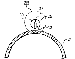
[0030] Refernng to Fig. 1, a balloon catheter 20 includes a catheter body 22,
an inflatable
balloon 24 attached to the catheter body, and one or more cutting elements 26,
or
atherotomes, (here, two) carried by the balloon. Referring to Figs. 2A and 2B,
cutting
element 26 is supported by a base 32 (e.g., by an adhesive) that is secured to
balloon 24, for
example, by an adhesive such as a urethane. Cutting element 26 includes a
cutting edge 28,
which is covered by a resilient material 30. Resilient material 30 can protect
balloon 24 from
the cutting edge and/or protect a vessel wall from the cutting edge, e.g.,
during insertion and
withdrawal of catheter 20. During use, described in detail below, cutting edge
28 can pierce
CA 02512172 2005-06-29
WO 2004/060460 PCT/US2003/041852
through resilient material 30, thereby allowing cutting element 26 to cut, for
example, plaque
or a calcified material that is occluding a body vessel.
[0032] Cutting elements 26 are elongated members (e.g., blades) preferably
formed of
diamond. Because of its physical properties, diamond enhances cutting element
26 by
allowing cutting edge 28 to be formed with relatively high sharpness. A sharp
cutting edge
typically lowers the forces needed for cutting. Lower cutting forces can
provide a relatively
controlled, precise cut with enhanced regularity and less distortion, thereby
reducing the
occurrence of damage to the vessel wall. Sharpness can be measured as a radius
of curvature
of cutting edge 28, e.g., using scanning electron microscopy (SEM). In
embodiments,
cutting edge 28 has a radius of curvature less than about 50 nanometers. The
radius of
curvature can be equal to or less than about 50, 40, 30, 20, 10, 5, or 3
nanometers; and/or
equal to or greater than about 3, 5, 10, 20, 30, or 40 nanometers. In
embodiments of catheter
20 having multiple cutting elements 26, two or more of the cutting elements
can have the
same or different sharpness. Different sharpness can provide different degrees
of cutting.
[0033] A cutting element formed of diamond can also be relatively hard, and
have a small
friction coefficient and a small thermal expansion coefficient. In
embodiments, cutting
element 26 has a hardness of greater than or equal to about 4,000 kg/mm2,
e.g., between
about 5,700-19,400 kg/mmz (Knoops hardness at 298K), and/or an elongation
modulus of
greater than or equal to about 1,140 GPa. The enhanced hardness allows cutting
element 26
to be formed relatively thin, e.g., without compromising strength. As a
result, cutting
element 26 can be relatively flexible. Enhanced flexibility allows cutting
element 26 to
travel well through tortuous paths of a body vessel or passageway. In
embodiments, cutting
element 26 has a width (W, Fig. 2B) less than or equal to about 0.006 inch,
e.g., less than or
equal to about 0.005 inch, 0.004 inch, 0.003 inch, 0.002 inch, or 0.001 inch.
The length of
cutting element 26 can be about 1-30~mm, e.g., about S-20 mm, or about 10-15
mm. The
height (H, Fig. 2B) of cutting element 26 can be less than or equal to about
0.013 inch, e.g.,
less than or equal to about 0.011 inch, 0.009 inch, 0.007 inch, or 0.005 inch.
The dimensions
of cutting element 26 typically are a function of the size of balloon catheter
20 that carries
the cutting element(s). In embodiments of catheter 20 having multiple cutting
elements 26,
two or more of the cutting elements can have the same or different dimensions.
Different
dimensions can provide different degrees of cutting and/or flexibility.
-5-
CA 02512172 2005-06-29
WO 2004/060460 PCT/US2003/041852
[0034] In some embodiments, only a portion of cutting element 26 is formed of
diamond.
For example, a diamond member defining a cutting edge can be attached (e.g.,
glued) to a
support, such as a metal (e.g., steel or Nitinol), ceramic, or polymer
support, earned by the
balloon. A support, such as a Nitinol or polymer support, can be flexible,
which can enhance
the flexibility of the balloon. The diamond member can be made relatively
small, e.g.,
smaller relative to the dimensions described above. One or more of the total
dimensions of
the diamond member and the support can be generally as described above.
[0035] Furthermore, since cutting element 26 can be formed relatively thin,
while still
providing effective cutting, a relatively small balloon catheter can be used
to carry the cutting
element. In embodiments, balloon catheter 20 is a 10 French device or smaller.
For
example, balloon catheter 20 can be equal to smaller than a 9 French, 8
French, 7 French, 6
French (2.00 mm O.D.), 5 French, 4 French, 3 French, 2 French, or 1 French
(1.00 mm O.D.)
device. As a result, catheter 20 can be delivered to relatively narrow target
sites, such as
coronary arteries (e.g., those having a diameter less than about 2 mm),
cranial arteries, and
peripheral arteries (e.g., those in the lower extremities).
[0036] Diamond cutting elements 26 are commercially available from GFD
Gesellschaft fiir
Diamantprodukte mbH (Ulm, Germany), which uses a plasma polishing process.
Methods of
making diamond cutting tools are described in U.S. Patent No. 4,989,578, and
methods of
polishing diamond is described in U.S. Patent No. 6,284,315.
[0037] Resilient material 30 can be made of any material that can deform upon
compression.
It is also desirable that the material can elastically recover to an
undeformed state when the
compression is removed. Resilient material 30 can be made of a polymer, such
as a rubber
(e.g., a silicone rubber) or a soft polyurethane (e.g., Tecothane SSD).
Resilient material 30
can be covered with a coating, such as a hydrophilic coating, to reduce
friction. Examples of
suitable materials include a hydrogel layer having a hydrophilic polymeric
material such as
(alkoxy) polyalkylene glycol, a copolymer of methylvinyl ether and malefic
acid,
poly(vinylpyrrolidone) (PVP), poly(N-alkylacrylamide), poly(acrylic acid),
polyvinyl
alcohol), poly(ethyleneimine), polyamide, (carboxy)methyl cellulose, polyvinyl
sulfonic
acid, heparin, dextran, modified dextran and chondroitin sulfate, polyethylene
oxide,
polyvinyl pyrrolidone), or a PVP/vinyl acetate copolymer. Resilient material
30 can be
attached base 32 using an adhesive or by heat bonding.
-G-
CA 02512172 2005-06-29
WO 2004/060460 PCT/US2003/041852
[0038] Base 32 can be any material that is compatible, e.g., can be bonded
with, balloon 24,
cutting element 26, the adhesive, and/or resilient member 30. The material for
base 32 can
be relatively hard to provide a rigid support for cutting element 26,
particularly during
cutting, and to spread the cutting force from the cutting element to a larger
area on balloon
24. In some cases, base 32 can be formed of a metal (such as stainless steel,
tantalum,
tungsten, Nitinol, titanium, or niobium) anchored or molded to balloon 24, or
a polymer
(such as a polyimide or a polyamide, e.g., Nylon 12). Certain materials for
base 32 are MRI
compatible, such as Nitinol, titanium, or niobium. Base 32 can be generally
triangular, as
shown, or non-triangular, e.g., rectangular, rhomboid, or trapezoid.
[0039] Examples of balloon catheter 20 are described in, for example, Wang
U.S. 5,195,969,
and Hamlin U.S. 5,270,086, both hereby incorporated by reference; and are
exemplified by
the Express~ or Maverick~ systems available from Boston Scientific Scimed,
Maple Grove,
MN. In some cases, balloon 24 is a non-elastic balloon, e.g., a nondistendable
balloon made
of, e.g., PET. Balloon 24 can include one or more biaxially-oriented layers.
[0040] Refernng to Figs. 3A, 3B, and 3C, a method of using catheter 20 is
shown. Catheter
20 is delivered to a target site 51, e.g., one having a calcified region 50,
using conventional
methods, such as by threading catheter body 22 over an emplaced guide wire
(not shown)
(Fig. 3A). Balloon 24 is unexpanded, and cutting edge 28 is covered by
resilient material 30
to prevent the cutting edge from contacting vessel wall 52 (Fig. 4A). After
catheter 20 is
properly positioned, balloon 24 is radially expanded (arrows A), e.g., by
introducing a fluid
into the interior of the balloon via an inflation lumen (not shown) extending
along catheter
body 22. As balloon 24 is expanded, resilient material 30 and cutting element
26 are
advanced radially toward calcified region 50. As balloon 24 if expanded
further, resilient
material 30 contacts calcified region 50 and compresses against the calcified
region. With
further expansion and compression, resilient material 30 deforms (e.g.,
compresses and
bulges), and cutting edge 28 pierces through the resilient member, thereby
cutting calcified
region 50 (Fig. 4B). Catheter 20 can be moved (e.g., translated and/or
rotated) to provide a
desired cutting action. Subsequently, balloon 24 is radially reduced, thereby
withdrawing
cutting elements 26 away from calcified region 50. As a result, the
compressive forces
against resilient material 30 are removed. The resilient material elastically
recovers to its
undeformed state in which the material covers cutting edge 28. Catheter 20 can
be removed
according to conventional methods.
CA 02512172 2005-06-29
WO 2004/060460 PCT/US2003/041852
[0041 ] Other Embodiments
[0042] In some embodiments, a drug or therapeutic agent, e.g., heparin, is
placed or
encapsulated between resilient material 30 and cutting element 26. The drug or
agent is
released into a cutting area as edge 28 penetrates through resilient material
30 during use.
Examples of drugs and therapeutic agents are disclosed in U.S. Patent No.
5,674,242;
U.S.S.N. 09/895,415, filed July 2, 2001; and U.S.S.N. 10/232,265, filed August
30, 2002.
The therapeutic agents or pharmaceutically active compounds can include, for
example, anti-
thrombogenic agents, antioxidants, anti-inflammatory agents, anesthetic
agents, anti-
coagulants, and antibiotics.
[0043] In other embodiments, a sponge coating can be placed on balloon 24 or
an
endoprosthesis (described below). The sponge coating, e.g., a non-hydrogel
polymer having
voids, can be loaded with a drug, e.g., heparin, to release the drug during
expansion of the
balloon or endoprosthesis. Examples of sponge coatings, including methods of
making them,
and suitable drugs, are described in U.S. Patent No., 6,364,856.
[0044] While catheter 20 is shown having two cutting elements 26, in other
embodiments,
the catheter can have one, three, four, five, six, seven, eight, or more
cutting elements.
Cutting elements 26 can be equally and/or unequally spaced around the
circumference of
balloon 24. For example, looking at a radial cross section (e.g., Fig. 4A) of
a balloon having
six cutting elements 26, the cutting elements can be formed at 2 o'clock, 3
o'clock, 4 o'clock,
8 o'clock, 9 o'clock, and 10 o'clock. Cutting element 26 at 3 o'clock is
equally spaced from
the cutting elements at 3 o'clock and 4 o'clock; but, for example, the cutting
element at 4
o'clock is unequally spaced from the cutting elements at 3 o'clock and 8
o'clock. Cutting
elements 26 can be symmetrically or asymmetrically positioned around the
circumference of
balloon 24.
[0045] Multiple cutting elements 26, e.g., two, three, four, five, six, or
more, can be arranged
collinearly (e.g., spaced and end-to-end) along balloon 24 (Fig. S), which can
enhance the
flexibility of the balloon. Multiple cutting elements 26 can be arranged side-
by-side, e.g.,
adjacent to each other. Multiple cutting elements 26 can be arranged adjacent
to each other
and overlapping along the longitudinal direction of balloon 24 (Fig. 6). A
balloon can have
one or more sets of cutting elements arranged as described above.
_g_
CA 02512172 2005-06-29
WO 2004/060460 PCT/US2003/041852
"." ,: ~~ ".~;.. ,~"," ".n~, ~~"...
[0046] Other methods of attaching cutting elements 26 to balloon 24 are
possible. Cutting
elements 26 can be thermally and/or mechanically bonded. For example, cutting
elements 26
can include projections, e.g., hooks, at their base that embed into the wall
of balloon 24. The
projections can be embedded manually. The cutting elements can be
appropriately
positioned in the balloon-forming mold with the projections extending into the
cavity of the
mold. The projections are embedded into the wall of the balloon as a parison
or a tube is
radially expanded (e.g., blow molded) to form the balloon. Cutting element 26
may include
one or more openings through which the material of base 32 can extend, thereby
further
securing the cutting element to the base. Cutting elements) 26 can be attached
directly to
balloon 24, e.g., without base 32. For example, cutting element can be
elongated blades
having a triangular cross section in which the base is attached to the balloon
and the cutting
edge is formed at the apex of the triangular section (Fig. 5).
[004'7] Alternatively or in addition to resilient material 30, balloon 24 can
be folded (Fig. 7)
using the methods described in Vigil U.S. 5,209,799 and 5,336,234, both hereby
incorporated
by reference, to protect cutting elements 26. In some cases, referring to Fig.
8, relatively
compliant areas of balloon 24, e.g., flaps 60, can be folded over cutting
elements 26 to
further protect the body lumen from cutting edges 28. Folding can be performed
by
engaging, e.g., grasping, flaps 60 with a chuck, and rotating the chuck.
Folding can be
performed during heat treatment of balloon 24, as described in Vigil U.S.
5,209,799. Other
methods of folding balloon 24 are described in U.S.S.N. 10/087,303.
[0048] In other embodiments, balloon 24 and/or catheter body 22 can have a
wall having a
plurality of layers formed of polymers. Multilayer devices are described in
Hamlin U.S.
5,270,086; Wang U.S. 5,195,969; Hamilton U.S. 5,797,877; and U.S.S.N.
09/798,749,
entitled "Multilayer Medical Device" and filed on March 2, 2001, all hereby
incorporated by
reference in their entirety. The layers can be selected to provide catheter
body 22 and/or
balloon 24 with desired properties. Different combinations of layering, e.g.,
materials,
sequence, and/or thickness, can be used, as described in U.S.S.N. 09/798,749.
[0049] Referring to Fig. 9, in embodiments, balloon 24 can be co-extruded to
include a
matrix material 62 and discrete (e.g., individually distinct) striped portions
64 (here, four)
surrounded by the matrix material. Cutting elements 26 are attached to balloon
24 over
striped portions 64. In embodiments, striped portions 64 are formed of a
materials) having a
lower compliancy than materials) that are not in the striped portions, such as
those of matrix
-9-
CA 02512172 2005-06-29
WO 2004/060460 PCT/US2003/041852
material 62. Alternatively or in addition, striped portions 64 are formed of a
materials)
having a lower distensibility than materials) that are not in the striped
portions. Compliancy
and distensibility may apply to the radial direction and/or the longitudinal
direction of
balloon 24. Alternatively or in addition, striped portions 64 are stiffer,
harder, and/or
stronger than non-striped portions of balloon 24.
[0050] Attaching cutting elements 26 over striped portions 64 enhances the
attachment
between the cutting elements and balloon 24. For example, as balloon 24 is
inflated (e.g., up
to 10 atm or higher) and deflated during use, striped portions 64 are less
likely to change,
e.g., grow or distend, longitudinally and/or radially, relative to non-striped
portions of the
balloon, such as compliant portions made of the matrix material. The interface
between
cutting elements 26 and striped portions 64 can remain relatively constant
during use. As a
result, mechanical stress between cutting elements 26 and balloon 24 reduced,
and
attachment therebetween is enhanced.
[0051] Striped portions 64 can also enhance folding and refolding of balloon
24. A striped
portion 64 and areas adjacent to the striped portions can behave like a hinge.
For example, a
(relatively non-compliant) striped portion 62 can act as a stationary member
of a hinge and
the (relatively compliant) adjacent areas can act as moveable members of the
hinge that pivot
about the interfacial region between the striped portion and the adjacent
areas. When balloon
24 is deflated, it can fold along the interfacial region so that compliant
areas form flaps, and
striped portions 64 are positioned in furrows. As a result, balloon 24 can be
formed and used
with a relatively low profile and a relatively predictable folding
configuration, thereby
providing desirable insertion and withdrawal of catheter 20 from a subject.
Embodiments of
balloon 24 and stripes portions 64 are described in U.S.S.N. 10/083,926,
entitled "Medical
Device" and filed on February 27, 2002, hereby incorporated by reference.
Cutting
elements) 26 can be attached directly to the balloon or indirectly, e.g.,
through base 32.
[0052] Refernng to Fig. 10, in other embodiments, one or more cutting elements
26 can be
carried by an endoprosthesis 70, such as a stmt or a stmt-graft, here shown on
a support such
as a balloon catheter or a catheter shaft 72. As shown, cutting elements 26
are mounted on
the struts of endoprosthesis 70. During expansion, cutting elements 26 can cut
a calcified
region, which can reduce the amount of force used to expand endoprosthesis.
Cutting
elements 26 can be mounted on endoprosthesis 70 using a biocompatible adhesive
or cement,
such as DiamondLinkTM (available from Biodent, Quebec, Canada).
-10-
CA 02512172 2005-06-29
WO 2004/060460 PCT/US2003/041852
[0053] Embodiments of the cutting elements on endoprosthesis 70 can be the
same as those
described above, and can be arranged on the endoprosthesis the same as those
on balloon 24.
Alternatively or in addition to resilient material 30, the cutting elements on
endoprosthesis 70
can be sprayed with a polymer, such as styrene-isobutylene-styrene (SIBS, a
tri-blocked
polymer) to protect the cutting edges, e.g., during delivery and/or crimping.
In other
embodiments, a drug or therapeutic agent (e.g., placitaxel or those described
above) can be
placed between cutting elements 26 and resilient member 30 or the polymer. The
drug can
be released to a cutting site as endoprosthesis 70 is expanded and cutting
elements 26
penetrate the site.
[0054] To crimp endoprosthesis 70, a soft polymer tube, e.g., one thicker than
the cutting
elements, can be placed inside the cavity of a crimper or around the
endoprosthesis. The tube
can distribute the crimping force over a broad area of endoprosthesis 70,
thereby reducing
damage to the cutting elements. Alternatively or in addition, the polymer,
e.g., SIBS, can be
applied to the cutting elements after crimping. The polymer can be applied to
cutting
elements 26 on balloon 24. Methods of crimping endoprostheses and devices for
crimping
are described, for example, in Austin, U.S. Patent No. 6,360,577, hereby
incorporated by
reference. Other suitable systems are described in U.S.S.N. 10/087,303.
Crimping devices
are also commercially available, e.g., from Machine Solutions Inc. (Flagstaff,
AZ).
[0055] In general, the endoprostheses can be of any desired shape and size
(e.g., coronary
stems, aortic stems, peripheral stems, gastrointestinal stems, urology stems
and neurology
stems). In certain embodiments, a coronary stmt can have an expanded diameter
of from
about 2 millimeters to about 6 millimeters. In some embodiments, a peripheral
stmt can
have an expanded diameter of from about 5 millimeters to about 24 millimeters.
In certain
embodiments, a gastrointestinal and/or urology stmt can have an expanded
diameter of from
about 6 millimeters to about 30 millimeters. In some embodiments, a neurology
stmt can
have an expanded diameter of from about 1 millimeter to about 12 millimeters.
The
endoprostheses can be balloon-expandable, self expandable, or a combination of
both (e.g.,
U.S. Patent No. 5,366,504).
[0056] In other embodiments, the endoprosthesis can include and/or be attached
to a
biocompatible, non-porous or semi-porous polymer matrix made of
polytetrafluoroethylene
(PTFE), expanded PTFE, polyethylene, urethane, or polypropylene. The
endoprosthesis can
include a releasable therapeutic agent or a pharmaceutically active compound,
such as
-11-
CA 02512172 2005-06-29
WO 2004/060460 PCT/US2003/041852
described in U.S. Patent No. 5,674,242, and commonly assigned U.S.S.N.
09/895,415, filed
July 2, 2001. The therapeutic agents or pharmaceutically active compounds can
include, for
example, anti-thrombogenic agents, antioxidants, anti-inflammatory agents,
anesthetic
agents, anti-coagulants, and antibiotics.
[005'7] The endoprosthesis can be used, e.g., delivered and expanded,
according to
conventional methods. Suitable catheter systems are described in, for example,
Wang U.S.
5,195,969, and Hamlin U.S. 5,270,086. Suitable stems and stmt delivery are
also
exemplified by the NIR on RangerC~ system, available from Boston Scientific
Scimed, Maple
Grove, MN. Other methods of carrying and delivering an endoprosthesis is
described in
U.S.S.N. 10/283,815, filed October 30, 2002, and entitled "Medical Devices
With Magnetic
Powered Actuation".
[0058] The medical devices described above can include radiopaque markers
and/or markers
that are visible by magnetic resonance imaging (MRI)) portions or markers to
help the user
position the devices. For example, a portion of cutting element 26 can be
coated with a
radiopaque and/or MRI visible material; balloon 24 may include one or more
marker bands;
and the wire of the stmt or stmt-graft can be radiopaque and/or MRI visible.
Suitable
radiopaque materials include, for example, gold, platinum, tungsten, tantalum,
and metal
alloys containing a sufficient percentage of heavy elements. Suitable MRI
visible materials
include, for example, non-ferrous metal-alloys containing paramagnetic
elements (e.g.,
dysprosium or gadolinium) such as terbium-dysprosium, dysprosium, terbium, and
gadolinium; non-ferrous metallic bands coated with an oxide or a carbide layer
of
dysprosium or gadolinium (e.g., Dy203 or Gd203); non-ferrous metals (e.g.,
copper, silver,
platinum, or gold) coated with a layer of superparamagnetic material, such as
nanocrystalline
Fe304, CoFe204, MnFe204, or MgFez04; and nanocrystalline particles of the
transition metal
oxides (e.g., oxides of Fe, Co, Ni).
[0059] All publications, references, applications, and patents referenced in
this application
are herein incorporated by reference in their entirety.
[0060] Other embodiments are within the claims.
-12-