Note: Descriptions are shown in the official language in which they were submitted.
CA 02512453 2010-09-09
TISSUE CLOSURE TREATMENT SYSTEM AND METHOD WITH EXTERNALLY-APPLIED
PATIENT INTERFACE
BACKGROUND OF THE INVENTION
1. Field of the Invention
The present invention relates generally to medical devices and methods for
treating closed wounds and incisions, and in particular to a system and method
for draining and/or
irrigating tissue separations, such as surgical incisions, and for compressing
and stabilizing a
dissected or traumatized field with ambient air pressure created by an
external patient interface
component and a vacuum source.
2. Description of the Related Art
Tissue separations can result from surgical procedures and other causes, such
as
traumatic and chronic wounds. Various medical procedures are employed to close
tissue separations.
An important consideration relates to securing separate tissue portions
together in order to promote
closure and healing. Incisions and wounds can be closed with sutures, staples
and other medical
closure devices. The "first intention" (primary intention healing) in surgery
is to "close" the incision.
For load-bearing tissues, such as bone, fascia, and muscle, this requires
substantial material, be it
suture material, staples, or plates and screws. For the wound to be "closed,"
the epithelial layer
must seal. To accomplish this, the "load bearing" areas of the cutaneous and
subcutaneous layers
(i.e., the deep dermal elastic layer and the superficial fascia or fibrous
layers of the adipose tissue,
CA 02512453 2005-06-30
WO 2004/060148 PCT/US2003/041667
-2-
respectively) must also at least be held in approximation long enough for
collagen deposition to take
place to unite the separated parts.
Other important considerations include controlling bleeding, reducing
scarring,
eliminating the potential of hematoma, seroma, and "dead-space" formation and
managing pain.
Dead space problems are more apt to occur in the subcutaneous closure.
Relatively shallow
incisions can normally be closed with surface-applied closure techniques, such
as sutures, staples,
glues and adhesive tape strips. However, deeper incisions may well require not
only skin surface
closure, but also time-consuming placement of multiple layers of sutures in
the load-bearing planes.
Infection prevention is another important consideration. Localized treatments
include various antibiotics and dressings, which control or prevent bacteria
at the incision or wound
site. Infections can also be treated and controlled systemically with suitable
antibiotics and other
pharmacologics.
Other tissue-separation treatment objectives include minimizing the traumatic
and
scarring effects of surgery and minimizing edema. Accordingly, various closure
techniques,
postoperative procedures and pharmacologics are used to reduce postoperative
swelling, bleeding,
seroma, infection and other undesirable, postoperative side effects. Because
separated tissue
considerations are so prevalent in the medical field, including most
surgeries, effective, expedient,
infection-free and aesthetic tissue closure is highly desirable from the
standpoint of both patients
and health-care practitioners. The system, interface and method of the present
invention can thus be
widely practiced and potentially provide widespread benefits to many patients.
Fluid control considerations are typically involved in treating tissue
separations.
For example, subcutaneous bleeding occurs at the fascia and muscle layers in
surgical incisions.
Accordingly, deep drain tubes are commonly installed for the purpose of
draining such incisions.
CA 02512453 2005-06-30
WO 2004/060148 PCT/US2003/041667
-3-
Autotransfusion has experienced increasing popularity in recent years as
equipment and techniques
for reinfusing patients' whole blood have advanced considerably. Such
procedures have the
advantage of reducing dependence on blood donations and their inherent risks.
Serous fluids are
also typically exuded from incision and wound sites and require drainage and
disposal. Fresh
incisions and wounds typically exude blood and other fluids at the patient's
skin surface for several
days during initial healing, particularly along the stitch and staple lines
along which the separated
tissue portions are closed.
Another area of fluid control relates to irrigation. Various irrigants are
supplied
to separated tissue areas for countering infection, anesthetizing, introducing
growth factors and
otherwise promoting healing. An effective fluid control system preferably
accommodates both
draining and irrigating functions sequentially or simultaneously.
Common orthopedic surgical procedures include total joint replacements (TJRs)
of the hip, knee, elbow, shoulder, foot and other joints. The resulting tissue
separations are often
subjected to flexure and movement associated with the articulation of the
replacement joints.
Although the joints can be immobilized as a treatment option, atrophy and
stiffness tend to set in
and prolong the rehabilitation period. A better option is to restore joint
functions as soon as possible.
Thus, an important objective of orthopedic surgery relates to promptly
restoring to patients the
maximum use of their limbs with maximum ranges of movement.
Similar considerations arise in connection with various other medical
procedures.
For example, arthrotomy, reconstructive and cosmetic procedures, including
flaps and scar
revisions, also require tissue closures and are often subjected to movement
and stretching. Other
examples include incisions and wounds in areas of thick or unstable
subcutaneous tissue, where
splinting of skin and subcutaneous tissue might reduce dehiscence of deep
sutures. The demands of
CA 02512453 2005-06-30
WO 2004/060148 PCT/US2003/041667
-4-
mobilizing the extremity and the entire patient conflict with the restrictions
of currently available
methods of external compression and tissue stabilization. For example, various
types of bandage
wraps and compressive hosiery are commonly used for these purposes, but none
provides the
advantages and benefits of the present invention
The aforementioned procedures, as well as a number of other applications
discussed below, can benefit from a tissue-closure treatment system and method
with a surface-
applied patient interface for fluid control and external compression.
Postoperative fluid drainage can be accomplished with various combinations of
tubes, sponges, and porous materials adapted for gathering and draining bodily
fluids. The prior art
includes technologies and methodologies for assisting drainage. For example,
the Zamierowski U.S.
Patents No. 4,969,880; No. 5,100,396; No. 5,261,893; No. 5,527,293; and No.
6,071,267 disclose
the use of pressure gradients, i.e., vacuum and positive pressure, to assist
with fluid drainage from
wounds, including surgical incision sites. Such pressure gradients can be
established by applying
porous sponge material either internally or externally to a wound, covering
same with a permeable,
semi-permeable, or impervious membrane, and connecting a suction vacuum source
thereto. Fluid
drawn from the patient is collected for disposal. Such fluid control
methodologies have been shown
to achieve significant improvements in patient healing. Another aspect of
fluid management,
postoperative and otherwise, relates to the application of fluids to wound
sites for purposes of
irrigation, infection control, pain control, growth factor application, etc.
Wound drainage devices
are also used to achieve fixation and immobility of the tissues, thus aiding
healing and closure. This
can be accomplished by both internal closed wound drainage and external, open-
wound vacuum
devices applied to the wound surface. Fixation of tissues in apposition can
also be achieved by
bolus tie-over dressings (Stent dressings), taping, strapping and (contact)
casting.
CA 02512453 2005-06-30
WO 2004/060148 PCT/US2003/041667
-5-
Heretofore there has not been available a tissue closure system, patient
interface
and method with the advantages and features of the present invention.
SUMMARY OF THE INVENTION
In the practice of the present invention, a system and method are provided for
enhancing closure of separated tissue portions using a surface-applied patient
interface. Subsurface
drainage, irrigation and autotransfusion components can optionally be used in
conjunction with the
surface-applied, external interface. The external interface can be
advantageously placed over a
stitch or staple line and includes a primary transfer component comprising a
strip of porous material,
such as rayon, applied directly to the patient for wicking or transferring
fluid to a secondary transfer
component comprising a sponge or foam material. An underdrape is placed
between the transfer
elements for passing fluid therebetween through an underdrape opening, such as
a slot. An
overdrape is placed over the secondary transfer component and the surrounding
skin surface. The
patient interface is connected to a negative pressure source, such as a vacuum
assisted closure
device, wall suction or a mechanical suction pump. A manual control embodiment
utilizes a finite
capacity fluid reservoir with a shut-off valve for discontinuing drainage when
a predetermined
amount of fluid is collected. An automatic control embodiment utilizes a
microprocessor, which is
adapted for programming to respond to various inputs in controlling the
operation of the negative
pressure source. A closed wound or incision treatment method of the present
invention involves
three phases of fluid control activity, which correspond to different stages
of the healing process. In
a first phase active drainage is handled. In a second phase components can be
independently or
sequentially disengaged. In a third phase the secondary transfer component can
optionally be left in
CA 02512453 2012-03-12
-6-
place for protection and to aid in evacuating any residual fluid from the
suture/staple line
through the primary transfer component.
In other embodiments of the invention, components of the dressing system can
be premanufactured for efficient application. A foam piece can be provided
with a full or
partial rayon cover and a close-fitting overdrape. An access panel with a
reclosable seal strip
can be installed on the overdrape for access to the foam pieces and the wound
area. A
premanufactured external dressing can be provided with a sheath receiving a
foam piece,
which is accessible through a reclosable seal strip for replacement or
reorientation.
Treatment area access is also provided through the seal strip.
Thus, one embodiment of the invention includes a dressing assembly for a
wound or incision, which comprises an external patient interface and a
pressure source. The
external patient interface includes an external fluid transfer component which
is adapted for
transferring fluid from the closed wound or incision. The external fluid
transfer component
includes a porous foam core and a wicking material liner fluidically
communicating with the
foam core. The external patient interface includes a sheath comprising an
overdrape having
an interior compartment receiving the foam core placed over the external fluid
transfer
component in contact with a surrounding skin surface. The pressure source is
connected to
the external fluid transfer component. The fluid discharge port is connected
to the pressure
source. The wicking material liner encloses the foam core, forming an
intermediate layer
between the overdrape and the foam core. The wicking material liner also forms
an
intermediate layer between the external fluid transfer component and the skin
surface at the
wound or incision at a lower portion of the dressing assembly. The wicking
material liner is
adapted for initiating a wicking action for enhancing a transfer of fluid from
the wound or
incision to the external fluid transfer component and from the external fluid
transfer
component to the fluid discharge port.
Another embodiment of the invention includes a system for applying fluid
pressure to tissue. The system includes a dressing, a fluid port, and a
pressure source. The
dressing includes a dressing cover with an interior opening. The dressing
cover is adapted for
mounting on, a patient's skin. The dressing includes a force fluid transfer
component including
a compressible, reticulated core positioned in the dressing cover and adapted
for interacting
with a patient through the opening. The fluid port is in the dressing cover,
and is adapted for
fluidic communication with the transfer component. The pressure source is
connected to the
fluid port.
CA 02512453 2012-03-12
-6a-
BRIEF DESCRIPTION OF THE DRAWINGS
The drawings constitute a part of this specification and include exemplary
embodiments of the present invention and illustrate various objects and
features thereof.
Fig. 1 is a schematic, block diagram of a tissue closure treatment and
system embodying the present invention.
Fig. 2 is a perspective view of an incision tissue separation with a deep
drain tube installed.
Fig. 3 is a perspective view thereof, showing the separated tissue sutured
together at the skin.
Fig. 4 is a perspective view thereof, showing the separated tissue sutured
together at the deep dermal layer below the skin surface.
CA 02512453 2005-06-30
WO 2004/060148 PCT/US2003/041667
-7-
Fig. 5 is a perspective view thereof, showing a rayon strip primary fluid
transfer
component (FTC.1) and an underdrape being placed on the stitch line.
Fig. 6 is a perspective view thereof, showing FTC. 1 and the underdrape in
place
on the stitch line.
Fig. 7 is a perspective view thereof, showing a secondary fluid transfer
component (FTC.2) in place.
Fig. 8 is a perspective view thereof, showing an overdrape in place.
Fig. 9 is a perspective view thereof, showing a connecting fluid transfer
component (FTC.3) in place for connecting the system to a negative pressure
source.
Fig. 10 is a cross-sectional view thereof, taken generally along line 10-10 in
Fig.
9 and particularly showing FTC.3.
Fig. 11 a is a perspective view thereof, showing FTC.3 removed and the
overdrape
scored for ventilation.
Fig. 1 lb is a perspective view thereof, showing the patient interface removed
along a perforated tear line in the underdrape and a slit line in the
overdrape.
Fig. 11 c is a perspective view of a patient interface adapted for
prepackaging,
application to a patient and connection to a negative pressure source.
Figs. 12a-d show alternative embodiment elbow connecting devices FTC.3a-d
respectively.
CA 02512453 2005-06-30
WO 2004/060148 PCT/US2003/041667
-8-
Figs. 12e,f show a modified FTC.2a with removable wedges to facilitate
articulation, such as flexure of a patient joint.
Figs. 12g,h show alternative embodiment external patient interface assemblies.
Figs. 13a-c comprise a flowchart showing a tissue closure treatment method
embodying the present invention.
Fig. 14 is a schematic, block diagram of an automated tissue closure treatment
system comprising an alternative embodiment of the present invention.
Fig. 15 is a cross-sectional view of the alternative embodiment automated
tissue
closure treatment system.
Fig. 16 is a partial flowchart of an alternative embodiment automated tissue
closure treatment method embodying the present invention.
Fig. 17 is a fragmentary, perspective view of a tissue closure treatment
system
comprising an alternative embodiment of the present invention, with a
reclosable access panel.
Fig. 18 is a perspective view of the reclosable access panel.
Fig 19 is a cross-sectional view of the tissue closure treatment system, taken
generally along line 19-19 in Fig 18.
Fig 20 is an enlarged, cross-sectional view of the tissue closure system,
particularly showing a reclosable seal strip thereof.
CA 02512453 2005-06-30
WO 2004/060148 PCT/US2003/041667
-9-
Fig. 21 is a perspective view of the tissue closure system, showing the seal
strip
open.
Fig 22 is a perspective view of the tissue closure system, showing the seal
strip
open and a foam piece removed.
Fig 23 is a cross-sectional view of an external dressing assembly, which
comprises an alternative embodiment of the present invention.
Fig. 24 is a cross-sectional view of an alternative embodiment tissue closure
system with internal and external foam pieces.
5
Fig. 25 is a cross-sectional view of the system shown in Fig 24, showing the
progressive healing of tissue in the wound.
Fig. 26 is a cross-sectional view of the system shown in Fig 24, showing the
reepithelialization of the wound.
Fig 27 is a cross-sectional view of a foam piece partially enclosed in rayon.
Fig. 28 is a cross-sectional view of an alternative embodiment tissue closure
system, with an external foam piece and an internal foam piece assembly.
Fig. 29 is a cross-sectional view thereof, shown partially collapsed under
ambient
atmospheric pressure.
Fig. 30 is a perspective view of an alternative construction dressing with a
reclosable seal strip and fluid access ports.
CA 02512453 2005-06-30
WO 2004/060148 PCT/US2003/041667
-10-
Fig. 31 is a perspective view of the underside of the dressing, showing a
middle
backing strip being removed.
Fig. 32 is a perspective view of the dressing, showing side backing strips
being
removed.
Fig. 33 is a perspective view of the dressing, shown with a squeeze bulb
evacuator attached to a fluid port thereof.
Fig 34 is a perspective view of the dressing, shown partially-collapsed under
atmospheric pressure.
Fig. 35 is a perspective view of the dressing, shown with the seal strip open.
Fig. 36 is a perspective view of the dressing, shown with the foam piece
removed.
Fig. 37 is a cross-sectional view of a foam piece fully-enclosed in rayon.
Fig 38 is a perspective view of an alternative embodiment dressing with a
separate liner and foam piece.
Fig. 39 is a perspective view of the dressing, shown with the foam piece for
moved.
Fig. 40 is a perspective view of the dressing, shown with the liner removed.
Fig. 41 is a cross-sectional view of an alternative embodiment dressing with a
sheath bottom panel comprising a wicking material.
CA 02512453 2005-06-30
WO 2004/060148 PCT/US2003/041667
-11-
Fig. 42 is a cross-sectional view of an alternative embodiment dressing system
with a covered foam-core transfer element.
Fig. 43 is a cross-sectional view thereof, showing the dressing compressed
under
pressure.
Fig. 44 is a top plan view thereof.
Fig. 45 is a cross-sectional view thereof, showing the dressing configuration
prior
to application to a patient and taken generally along line 45-45 in Fig. 44.
Fig. 46 is a top plan view of an application involving multiple dressings
covering
an elongated tissue separation, such as a surgical incision.
DETAILED DESCRIPTION OF THE PREFERRED EMBODIMENT
1. Introduction and Environment
As required, detailed embodiments of the present invention are disclosed
herein;
however, it is to be understood that the disclosed embodiments are merely
exemplary of the
invention, which may be embodied in various forms. Therefore, specific
structural and functional
details disclosed herein are not to be interpreted as limiting, but merely as
a basis for the claims and
as a representative basis for teaching one skilled in the art to variously
employ the present invention
in virtually any appropriately detailed structure.
II. Tissue Closure System 2
CA 02512453 2010-09-09
-12-
Referring to the drawings in more detail, the reference numeral 2 generally
designates a tissue closure treatment system embodying the present invention.
As shown in Fig. 1,
the system 2 is adapted for use on a patient 4 with an incision or wound 6,
which can be closed by a
stitch line 8 consisting of sutures 10, staples or other suitable medical
fasteners.
A patient interface 12 consists of an optional deep drain 14 connected to a
deep
drain negative pressure source 15 associated with a deep drainage reservoir 17
and an external
patient interface 16 including a primary fluid transfer component FTC.1
comprising a strip of rayon
or other suitable porous material, an underdrape 20 generally covering FTC.1
and including a slot
20a, a secondary fluid transfer component FTC.2 comprising a hydrophobic
sponge and an
overdrape 24.
A fluid handling subsystem 26 includes the deep drain negative pressure source
and a surface drain negative pressure source 28, which can be combined for
applications where a
common negative pressure source and a collection receptacle are preferred. The
negative pressure
sources 15, 28 can operate either manually or under power. Examples of both
types are well-known
15 in the medical art. For example, a manually operable portable vacuum source
(MOPVS) is shown in
U.S. Patent No. 3,115,138. The MOPVS is available
from Zimmer, Inc. of Dover, Ohio under the trademark HEMOVAC . Bulb-type
actuators, such as
that shown in U.S. Patent No.4,828,546 and available from
Surgidyne, Inc. of Eden Prairie, Minnesota, can be used on smaller wounds, for
shorter durations or
in multiples. Moreover, power-actuated vacuum can be provided by vacuum
assisted closure
equipment available under the trademark THE VAC from Kinetic Concepts, Inc.
of San Antonio,
CA 02512453 2010-09-09
-13-
Texas. Still further, many health-care facilities, particularly hospitals and
clinics, are equipped with
suction systems with sources of suction available at wall-mounted outlets.
A finite capacity reservoir 30 is fluidically connected to the negative
pressure
source 28 and is adapted to discharge to a waste receptacle 32. A shut-off
valve 34 is associated
with the reservoir 30 and is adapted to automatically discontinue drainage
when the reservoir 30 is
filled to a predetermined volume.
An optional autotransfusion subsystem 36 can be connected to the deep drain 14
and is adapted for reinfusing the patient 4 with his or her own blood. U.S.
Patent No. 5,785,700
discloses such an autotransfusion system with a portable detachable vacuum
source, which is
available from Zimmer, Inc.
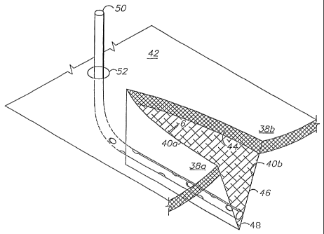
Fig. 2 shows an incision 6 forming first and second separated tissue portions
38a,b with incision edges 40a,b. The incision 6 extends from and is open at
the skin 42, through the
deep dermal layer 44 and the subcutaneous layer 46, to approximately the
fascia 48. A deep drain
tube 50 is placed in a lower part of the incision 6 and penetrates the skin 42
at an opening 52.
Fig. 3 shows the incision edges 40a,b secured together by sutures 54 forming a
stitch line 56 at the skin surface 42. As an alternative to sutures 54,
various other medical fasteners,
such as staples, can be used. Fig. 4 shows sutures 55 placed in the deep
dermal layer 44 below the
skin surface 42.
Fig. 5 shows application of FTC.1 on top of the stitch line 8. FTC.1
preferably
comprises a suitable porous wicking material, such as rayon, which is well-
suited for wicking the
CA 02512453 2005-06-30
WO 2004/060148 PCT/US2003/041667
-14-
fluid that exudes along the stitch line 8. Rayon also tends to dry relatively
quickly, and thus
efficiently transfers fluid therethrough. The underdrape 20 is placed over
FTC. 1 and the adjacent
skin surface 42. Its slot 20a is generally centered along the centerline of
FTC. 1 and directly above
the stitch line 8. FTC. 1 and the underdrape 20 can be preassembled in a roll
or some other suitable
configuration adapted to facilitate placement on the stitch line 8 in any
desired length. Fig. 6 shows
FTC. 1 and the underdrape 20 in place.
The secondary fluid transfer component FTC.2 is shown installed in Fig. 7. It
preferably comprises a suitable hydrophobic foam material, such as
polyurethane ether (PUE),
which comprises a reticulated, lattice-like (foam) material capable of being
collapsed by vacuum
force (negative pressure) in order to exert positive "shrink-wrap" type
compression on skin surface
and still maintain channels that allow passage of fluid. As shown, its
footprint is slightly smaller
than that of the underdrape 20, thus providing an underdrape margin 20b. The
wicking layer of
FTC.1 can, as an alternative, be sized equal to or almost equal to the
footprint of FTC.2. This
configuration lends itself to prefabrication as an individual, pre-assembled
pad that can be employed
by simply removing a releasing layer backing from an adhesive lined
underdrape. This
configuration also lends itself to easy total removal and replacement of the
central part of the
assembly without removing drape already adhered to skin if removal and
replacement is the desired
clinical option rather then staged removal or prolonged single application.
Fig. 8 shows the overdrape 24 applied over FTC.2 and the underdrape 20, with a
margin 24a extending beyond the underdrape margin 22b and contacting the
patient's skin surface
(dermis) 42. Figs. 9 and 10 show a patch connector 58 mounted on FTC.2 and
comprising a
hydrophobic foam (PUE) material core 58a sandwiched between drape layers 58b.
A vacuum drain
CA 02512453 2005-06-30
WO 2004/060148 PCT/US2003/041667
-15-
tube 60 includes an inlet end 60a embedded in the foam core 58a and extends
between the drape
layers 58b to an outlet end 60b connected to the surface drainage negative
pressure source 28.
Fig. 11 a shows FTC.3 removed, e.g. by cutting away portions of the overdrape
24
to provide an overdrape opening 54. In addition, the overdrape 24 can be slit
at 55 to further
ventilate FTC.2. Draining FTC.2 under negative pressure, and further drying it
with air circulation
(Fig. I l a) can provide significant healing advantages by reducing the growth
of various microbes
requiring moist environments in FTC.2. Such microbes and various toxins
produced thereby can
thus be evaporated, neutralized and otherwise prevented from reentering the
patient. Microbe
control can also be accomplished by introducing antiseptics in and irrigating
various components of
the patient interface 12, including the drapes 20, 24; FTC.1; FTC.2; and
FTC.3.
Fig. 1 lb shows the patient interface 12 removed along underdrape perforated
tear
lines 56 and slit lines 59 in overdrape 24. It will be appreciated that
substantially the entire patient
interface 12, except for underdrape and overdrape margins 20b, 24a can thus be
removed to provide
access to the stitch line 8 and the dermis 42 for visual inspection,
evaluation, cleaning, stitch
removal, dressing change (e.g., with prepackaged patient interface 12a as
shown in Fig. 11c),
consideration of further treatment options, etc. For example, the overdrape 24
can be slit to around
the perimeter or footprint of FTC.2 to permit removing the same. Preferably
FTC.2 is easily
releasable from the underdrape 20 and FTC.1 whereby FTC.2 can be grasped and
lifted upwardly to
facilitate running a scalpel through the overdrape 24 and into a separation
between the underside of
FTC.2 and the underdrape 20. The FTC.1 can then optionally be removed by
tearing the underdrape
20 along its tear lines 56 and removing same as shown in Fig. 1 lb.
CA 02512453 2005-06-30
WO 2004/060148 PCT/US2003/041667
-16-
Fig. 11 c shows a prepackaged patient interface 12a adapted for initial or
"dressing change" application. Optionally, the rayon strip FTC.1 can have the
same configuration or
"footprint" as the foam sponge FTC.2, thus eliminating the underdrape 20. The
prepackaged patient
interface 12a can be sterilely packaged to facilitate placement directly on a
stitch line 8.
Alternatively, the patient interface components can be prepackaged
individually or in suitable
groups comprising subassemblies of the complete patient interface 12. For
example, the
underdrape/FTC. 1 and the overdrape/FTC.2 subassemblies respectively can be
prepackaged
individually. Various sizes and component configurations of the patient
interface can be
prepackaged for application as indicated by particular patient conditions.
Preferably, certain sizes
and configurations would tend to be relatively "universal" and thus applicable
to particular medical
procedures, such as TJRs, whereby patient interface inventory can be
simplified. Alternatively, the
individual components can be assembled in various sizes and configurations for
"custom"
applications.
Figs 12a-d show alternative connecting fluid transfer components FTC.3a-d for
connecting FTC.2 to the surface drainage negative pressure source 28. FTC.3a
(Fig. 12a) shows a
patch connector with a similar construction to FTC.3 and adapted for placement
at any location on
the overdrape 24. FTC.3a is provided with a Leur lock connector 62. FTC.3b
(Fig. 12b) comprises
a strip of hydrophobic (PUE) foam material partially covered by an overdrape
64, which can be
configured as a wrap around a patient's limb or extremity 66. FTC.3c (Fig.12c)
is an elbow-type
connector. FTC.3d (Fig. 12d) is a bellows-type elbow connector, which is
adapted to accommodate
deflection of the vacuum drain tube 60.
CA 02512453 2005-06-30
WO 2004/060148 PCT/US2003/041667
-17-
Figs.12e,f show an alternative construction of FTC.2a with multiple, removable
wedges 57 formed therein and adapted for accommodating articulation, such as
joint flexure. The
flexibility of FTC.2a can thus be considerably enhanced for purposes of
patient comfort, mobility
and flexibility. Such wedges can extend transversely and/or longitudinally
with respect to FTC.2a.
FTC.2a functions in a similar manner with and without the wedges 57 in place
or removed.
Fig. 12g shows a modified patient interface 312 with the underdrape 20 placed
below FTC. 1. This configuration permits removing FTC. 1 without disturbing
the underdrape 20.
Fig. 12h shows a further modified patient interface 412 with FTC.1 having the
same configuration
or footprint as FTC.2, whereby they can be fabricated and bonded together. In
this configuration the
underdrape 20 can be omitted.
III. Treatment Method
Figs.13a-c comprise a flowchart for a method embodying the present invention.
From start 70 the method proceeds to patient diagnosis and evaluation at 72
and treatment plan at
74. Deep drains 14 are installed at 76 as necessary, and the incision is
sutured at 78. Surface
interface components 12 are applied at 80 and connected to the external
components (i.e., negative
pressure sources 15, 28) at 82. The collection reservoir capacity is preset at
84 based on such factors
as nature of wound/incision, blood flow, etc.
Phase 1
Deep drainage occurs at 86 and active surface drainage occurs at 88, both
being
influenced by the negative pressure sources 15, 28. The negative pressure
source 28 causes the PUE
CA 02512453 2005-06-30
WO 2004/060148 PCT/US2003/041667
-18-
foam FTC.2 to partially collapse, which correspondingly draws down the
overdrape 24 and exerts a
positive, compressive force on the closed wound or incision 6. In the closed
environment of the
patient interface 12, such force is effectively limited to ambient atmosphere.
This limiting control
feature protects the patient from excessive force exerted by the patient
interface 12. The steady
force of up to one atmosphere applied across the closed wound or incision 6
functions similarly to a
splint or plaster cast in controlling edema and promoting healing.
A "Reservoir Full" condition is detected at 90 and branches to an interrupt of
the
surface drainage negative pressure at 92, after which the reservoir contents
are inspected and
disposed of at 94. If surface bleeding is detected by visual inspection at
decision box 96, the method
branches to a "Discontinue Active Surface Drainage" step at 98. If the suture
line is actively
draining at decision box 100, the method loops to the active surface drainage
step 88 and continues,
otherwise active surface drainage discontinues at 98, i.e. when the
wound/incision is neither
bleeding nor exuding fluids.
Phase 1 is generally characterized by deep drainage (interactive or passive)
and
active surface drainage under the influence of manual or powered suction. The
normal duration is
approximately two to three days, during which time post-operative or post-
trauma swelling normally
reaches its maximum and begins to recede.
Phase 2
Fig. 13b shows Phase 2 commencing with a "Staged Component Removal?"
decision box 102. An affirmative decision leads to independently deactivating
and removing
components at 103, including discontinuing active suction at 104, which
transforms the hydrophobic
CA 02512453 2005-06-30
WO 2004/060148 PCT/US2003/041667
-19-
PUE foam (FTC.2) internal pressure from negative to positive and allows the
collapsed FTC.2 to
reexpand at 106, potentially increasing surface composite pressure from
ambient to positive.
Preferably this transition occurs without applying undue pressure to the
surface from the
decompressed, expanding FTC.2. During Phase 1, negative pressure (i.e.,
suction/vacuum) tends to
compress FTC.2 and correspondingly contracts the overdrape 24, adding to the
compression exerted
by FTC.2. When the application of negative pressure discontinues, either
manually or
automatically, FTC.2 re-expands against the constraints of the overdrape 24,
and in an equal and
opposite reaction presses against the skin 42, particularly along the stitch
line 8. FTC.2 can thus
automatically transform from ambient to positive pressure simply by
discontinuing the application
of the vacuum source.
The positive pressure exerted on the skin 42 continues to compress and
stabilize
tissue along the suture line 8 (step 108) in order to reduce swelling and
cooperates with the
operation of FTC. 1 and FTC.2 to continue drainage by evaporation at the
suture line 8 at step 110.
A negative determination at decision box 102 leads to interface removal at 112
and, unless treatment
is to be terminated, stitch line inspection and treatment at 113 and interface
replacement at 114,
which can involve all or part of the patient interface 12. The method then
proceeds to Phase 3.
Phase 3
Fig. 13c shows Phase 3 of the treatment method wherein deep drainage is
discontinued and the tube(s) is removed at 118. The overdrape 24 and FTC.2 are
removed at 120,
122 respectively. The underdrape 20 and FTC.1 are preferably configured to
permit visual
inspection of the suture line 8 therethrough at 124. When the suture line 8
has closed sufficiently,
the underdrape 20 and FTC.l are removed at 126 and the treatment ends at 128.
Alternatively and if
CA 02512453 2005-06-30
WO 2004/060148 PCT/US2003/041667
-20-
indicated by the patient's condition, all or part of the interface 12 can be
replaced in Phase 3 and
treatment continued.
IV. Alternative Embodiment Tissue Closure System 202
Fig. 14 schematically shows a tissue closure system 202 comprising an
alternative embodiment of the present intention, which includes a
microprocessor or controller 204,
which can be connected to one or more sensors 206 coupled to the patient
interface 12 for sensing
various conditions associated with the patient 4. The microprocessor 204 can
be programmed to
operate a solenoid 208 coupled to a valve 210 associated with the reservoir 30
and controlling fluid
flow induced by a negative pressure source 228 through its connection to the
patient interface 12.
Fig. 15 shows the tissue closure system 202 with the microprocessor 204
connected to multiple sensors 206a,b,c each of which is associated with a flow
control component,
such as a valve, 210a,b,c respectively. Each flow control component 210a,b,c
is associated with a
respective negative pressure source 228a,b,c, which in turn controls fluid
discharge into canisters or
reservoirs 212a,b,c respectively. For example, the patient interface 12 can
comprise an external
patient interface 16 as described above and a pair of deep drainage tubes
50a,b. The patient
interface 12 includes an optional supply component 214, which can comprise one
or more fluid
reservoirs, pumps (manual or powered) and associated controls, which can
connect to the
microprocessor 204 for system control. The supply component 214 optionally
takes to one or more
of the tubes 50, 60 for delivering fluid to the patient through the deep
drainage tubes 50 or through
the external patient interface 16. Such fluids can comprise, for example,
antibiotics, and aesthetics,
CA 02512453 2005-06-30
WO 2004/060148 PCT/US2003/041667
-21-
irrigating agents, growth factor, and any other fluid beneficial in promoting
healing, countering
infection and improving patient comfort.
The methodology of the treatment with the alternative embodiment tissue
closure
system 202 is shown in Fig. 16 and generally involves modified pretreatment
230 and Phase 1
procedures. From "Start" the method proceeds to a diagnosis/evaluation step
234, a treatment plan
step 236, deep drain installation 238, suturing at 240, external interface
component application 242,
microprocessor programming 244 and connection of the application components at
246, such as
connection of the tubing. Phase 1 commences with deep drainage at 248, active
suction interface at
250 and a "Suture Line Actively Draining?" decision box 252. If the suture
line is actively
draining, the method loops back to the active suction interface step 250,
otherwise (negative
determination at 252) it proceeds to Phase 2.
V. Applications
Without limitation on the generality of useful applications of the tissue
closure
systems 2 and 202 of the present invention, the following partial list
represents potential patient
conditions and procedures, which might indicate application of the present
invention.
= Over closed tissue separations, such as surgical incisions.
= Over joints where the incision is subject to movement and stretching, such
as arthrotomy,
reconstructive proceedures, cosmetic procedures, flaps, scar revisions, Total
Joint
Replacement (TJR) procedures, i.e., hip, knee, elbow, shoulder and foot.
= Any wound in an area of thick or unstable. subcutaneous tissue, where
splinting of skin and
subcutaneous tissue might reduce dehiscence of deep sutures.
CA 02512453 2005-06-30
WO 2004/060148 PCT/US2003/041667
-22-
= Wounds over reconstructive procedures in which irregular cavities are
created. These include
resection of tumors, implants, bone, and other tissues. Changes in length and
geometry of
limbs, and changes in size, position, and contour of bones and other deep
structures.
= Wounds in which elimination and prevention of dead space is important.
= Treatment of hematomas and seromas.
= Amputation stumps.
= Abdominal, thoracic, flank, and other wounds in which splinting of the wound
might assist
closing and mobilizing the patient during the postoperative interval.
= Wounds in areas of fragile or sensitive skin, where repeated removal and
replacement of tape
or other adhesives might produce pain, irritation, or blistering of skin in
the vicinity of the
wound. Also where dressing changes might produce shear or displacement of
tissue so as to
compromise primary wound healing.
= Wounds in cases where the patient wishes to bathe before the skin has healed
sufficiently to
allow protection from contamination with bath or shower water.
= Wounds subject to contamination with feces, urine, and other body fluids.
= Pediatric, geriatric, psychiatric, and neurologic patients, and other
patients likely to disturb
dressings and wounds.
= Patients with multiple consultants and care givers, where repeated
inspection of the wound
might compromise healing.
= Deep closure and surface sutures and staples.
= Any clean surgical or traumatic incision, open, or fully or partially closed
by sutures, or
where the skin edges can be apposed to a gap no wider than the width of the
negative
CA 02512453 2005-06-30
WO 2004/060148 PCT/US2003/041667
-23-
pressure zone of the dressing, i.e. where the maximum separation is less than
or equal to the
width of FTC.1 (rayon strip).
= In cosmetic and reconstructive surgery, the systems and methods of the
present invention can
control and conceal the effects of early bleeding, exudation, ecchymosis, and
edema of the
wound.
= In surgery on the limbs, where compression and drainage by this method might
eliminate or
reduce the need for circumferential compressive wrapping.
= Tissue separations that are prone to protracted drainage, such as hip and
knee incisions, and
tissue separations in patients with health conditions, such as diabetes, that
tend to inhibit
healing. Shortened hospital stays might result from swelling reduction and
control of
drainage.
VI. Case Studies
= General concept: sequential surface application of foam material (FTC.2) to
surgical site and
other wounds. Air-drying at the suture line is facilitated by the rayon strip
(FTC. 1).
= Phase 1: deep drainage (drain tube(s)), active or passive; active suction
applied to surface
PUE foam (placed on top of surgical incision, drains bleeding and exudate from
suture line);
active suction compresses PUE foam, thus applying positive compression to the
entire
dissection field; adhesive-lined film underdrape with an MVTR of 3-800 on skin
underlying
PUE foam; rayon (or other suitable porous wicking material) strip on suture
line; similar
type of adhesive film overdrape (MVTR of 3-800) overlying PUE foam material.
= Duration: approximately 2-3 days, i.e. effective time for active drainage
from incision/stitch
line to cease and for suture line to dry and heal.
CA 02512453 2005-06-30
WO 2004/060148 PCT/US2003/041667
-24-
Phase 2: Remove active suction by cutting off (elbow) connector and leave
FTC.2 in place.
Released from suction, FTC.2 expands against the overdrape and exerts positive
pressure
differential on the operation site. May maintain continued mild compression
throughout
Phase 2; residual drainage function through rayon strip and into FTC.2
provides continued
drying of suture line. Deep drain tubes remain in place during Phase 2 for
active deep
drainage.
= Duration: approximately three days, i.e. days 3-6 after operation.
= Phase 3: remove overdrape and FTC.2; leave underdrape and rayon strip in
place; visually
observe wound healing progress; transparency desirable.
= Duration: several (e.g., up to three) weeks.
= Clinical trial confirmation: Closure of surgical site in upper chest area in
patient with severe
healing problems showed excellent results and rapid wound healing.
= Subcuticular (subepidermal) sutures avoid conflict with rayon strip and need
for early suture
removal, or pressure on skin sutures beneath compressive black sponge.
= Option: use pressure transducer for interface pressure mapping of wound site
and automate
control and monitor pressures, flow, etc.
VII. Alternative Embodiment Tissue Closure System 302.
A tissue closure system 302 comprising an alternative embodiment of the
present
invention is shown in Figs. 17-22. The system 302 is adapted for closing a
wound 304 with an
undermined area 306 just above the fascia and an upper tissue separation 308
located primarily in
the dermis and in the subcutaneous layer. A wedge-shaped internal fluid
transfer component (foam
CA 02512453 2005-06-30
WO 2004/060148 PCT/US2003/041667
-25-
piece) 310 is located in the tissue separation area 308 and is installed
between side drapes 312
located on either side of the wound 304. An external fluid transfer component
(foam piece) 314 is
placed on top of the internal component 310 and the side drapes 312, and is
covered by an outer
drape 316. An optional innermost foam piece 330 can be located in and sized to
fit the undermined
area 306 and can transfer fluid and gradient forces to and from the internal
foam piece 310.
A reclosable access panel 318 is placed over an opening formed in the outer
drape 316 and includes an adhesive-coated perimeter 320 surrounding an
adhesive-free center area
322 with a reclosable seal strip 324 extending longitudinally down the
centerline thereof. The seal
strip 324 includes a rib or bead 326, which is releasably captured in a
channel 328 (Fig. 20).
In operation, the reclosable access panel 318 is adhesively secured around its
perimeter 322 to the outer drape 316 and provides access to the foam pieces
310, 314 of the dressing
system 302. For example, the foam pieces 310, 314 can be changed (Figs. 21 and
22), treatments
can be applied and wound healing progress can be visually monitored.
VIII. Alternative External Dressing 402.
Figs. 23-27 show an external dressing 402, which can be premanufactured or
preassembled and used for various wound treatment and closure applications.
The dressing 402
includes a foam piece 404 partially enclosed in a rayon covering 406, which
includes an open top
408 secured to an upper perimeter 410 of the foam piece 404, for example, by
sutures, staples,
adhesive or some other suitable mechanical fastener as shown at 412. The
dressing 402 is
preferably preassembled with an outer drape 414 including a foam-covering
central portion 416 and
a perimeter, patient-contact skirt portion 418. A tucked margin 420 is formed
at the intersection of
CA 02512453 2005-06-30
WO 2004/060148 PCT/US2003/041667
-26-
the drape portions 416, 418 and partially underlies the foam piece 404 in
order to protect the skin
and prevent the formation of low-pressure, vacuum voids around the edge of the
foam piece 404
whereat blistering could otherwise occur. In operation, the dressing 402 can
be easily changed by
cutting around the margin 420, removing the foam piece 404 and the drape outer
portion 416. The
wound can thus be inspected, cleaned, debrided, treated, etc. and a new
dressing 402 put in place.
The patient-contact skirt portion 418 of the original dressing can remain in
place.
Fig. 23 shows a fluid flow (discharge) directional arrow 421 from an elbow
coupling 417 and a discharge tube 419. Alternatively, fluid could be injected
into the dressing 402
through the tube 419 and the coupling 417. Hydraulic/pneumatic compressive
force arrows 423 are
shown in Fig. 23 and represent the downward (i.e. into patient) forces, which
can be established by
compressing the foam piece 4.04 under suction and then releasing the negative
pressure differential,
thus transitioning the dressing to a positive pressure differential. In a
positive pressure differential
mode of operation, the dressing 402 controls edema by pressing the foam piece
404 against the
tissue adjacent to the wound. There are many potential medical benefits from
controlling edema in
this manner. For example, healing is promoted, scar tissue is minimized and
patient discomfort can
be reduced.
Fig. 24 shows the external dressing 402 used in conjunction with an internal
foam
piece 422, which is located below the dermis at the top of the subcutaneous
layer. The internal foam
piece 422 is adapted for applying a pressure differential within the
subcutaneous layer whereby
tissue growth and closure are promoted. The inside/outside configuration of
the dressing system
shown in Fig. 24 can rehabilitate and make pliable a wound edge 424 that has
contracted and
become hard, immobile and edematous by applying pressure differentials across
the external and
CA 02512453 2005-06-30
WO 2004/060148 PCT/US2003/041667
-27-
internal foam pieces 404, 422, such as compression (positive pressure
differential) for edema
control.
Fig. 25 shows the wound confined to the dermis 426 with another internal foam
piece 428 in place. The subcutaneous layer is substantially healed. Fig. 26
shows the external foam
piece 404 in place alone for drawing the wound edges 430 together at the
epidermis. Fig. 27 shows
the external. foam piece 404 covered on the sides and bottom by the rayon
covering 406, leaving an
open top 408.
IX. Alternative Embodiment Dressing System 502
Fig. 28 shows yet another alternative embodiment internal/external dressing
system configuration 502 with an external foam piece 504 similar to the foam
piece 404 described
above and an internal foam assembly 506 located in the dermis and in the
subcutaneous layer. The
assembly 506 consists of a proximate internal foam piece 508, which can be
located at the bottom of
the subcutaneous layer on top of the fascia in an undermined cavity 510 formed
by the wound , and
a distal internal foam piece 412 located primarily in the dermis and the
subcutaneous layer portions
of the wound between the external foam piece 504 and the proximate internal
foam piece 508.
The dressing system configuration 502 can be configured and reconfigured as
necessary to accommodate various wound configurations in various stages of
healing. For example,
the proximate internal foam piece 508 can be removed when the undermined
cavity 510 closes.
Likewise, the distal internal foam piece 512 can be removed when the
subcutaneous layer and the
dermis have healed. Moreover, the foam pieces 504, 508 and 512 can be replaced
with different
sizes of foam pieces as necessary in connection with dressing changes and as
the wound
CA 02512453 2005-06-30
WO 2004/060148 PCT/US2003/041667
-28-
configuration changes. Such sizes and configurations can be chosen to optimize
the beneficial
effects of pressure gradients (both positive and negative), fluid control,
edema control, antibacterial
measures, irrigation and other treatment protocols. Still further, the access
panel 318 described
above can be used in conjunction with the dressing system 502 in order to
provide access to the
foam pieces thereof and to the wound itself.
Fig. 29 shows the internal/external dressing system 502 compressed under the
vacuum effects of an external vacuum source with the drape 316 drawn tightly
down on the
compressed outer foam piece 504. Thus compressed, the system 502 is adapted to
transfer positive
pressure differential, compressive forces to the area of the wound.
X. Alternative Embodiment Dressing Assembly 602
Figs. 30-37 show a reclosable, preassembled external dressing assembly 602
comprising an alternative embodiment of the present invention. The dressing
assembly 602 includes
a foam piece 604, which can be completely covered in rayon 606 or some other
suitable material
with the desired absorbent and/or wicking capabilities. The foam piece 604
also includes a core 605
comprising a suitable material, such as polyurethane, hydrophobic foam.
Alternatively, other foam
materials with hydrophobic or hydrophilic properties can be utilized. Various
sizes and shapes of
the foam piece 604 can also be employed, including cutting and trimming it to
size during the course
of a medical procedure.
The foam piece 604 is removably placed in a reclosable sheath 608 including a
bottom panel 610 selectively covered by removable, adhesive backing strips
612, 614 and 616
forming a central opening 618. As shown in Fig. 31, a central opening 618 in
the bottom panel 610
CA 02512453 2005-06-30
WO 2004/060148 PCT/US2003/041667
-29-
is initially covered by the center backing strip 614. Removing the center
backing strip 614 exposes
the foam piece 604 through the opening 618. The reclosable sheath 608 also
includes a top panel
620 with a reclosable seal strip 622 extending from end-to-end and generally
longitudinally
centered. The seal strip 622 can be similar in construction to the reclosable
seal strip 324 described
above. The top panel 620 also includes fluid ports 324, 326, which can
comprise, for example, Leur
lock connectors or some other suitable fluid connection device.
The sheath 608 can comprise polyethylene or some other suitable material
chosen
on the basis of performance criteria such as permeability, flexibility,
biocompatibility and
antibacterial properties. Various permeable and semi-permeable materials are
commonly used as
skin drapes in medical applications where healing can be promoted by exposure
to air circulation.
The sheath 608 can be formed from such materials for applications where
continuous vacuum
suction is available and the dressing 602 is not required to be airtight.
According to an embodiment of the method of the present invention, a dressing
assembly 602 can be premanufactured, or custom-assembled from suitable
components for
particular applications. In a premanufactured version, the dressing 602 is
preferably presterilized
and packaged in sterile packaging.
A common application of the dressing 602 is on a recently-closed surgical
incision for controlling bleeding and other fluid exudate. For example, the
dressing 602 can be
placed on the patient with its bottom panel opening 618 located over a stitch
line 636 (Fig. 36). The
center backing strip 614 is peeled from the bottom panel 610 to expose the
opening 618 and the
adhesive 628 on the bottom panel 610 (Fig. 33). The opening 618 provides a
fluid transfer, which
CA 02512453 2005-06-30
WO 2004/060148 PCT/US2003/041667
-30-
can also be provided by constructing the sheath bottom panel 610 from a
permeable material, or by
providing other passage configurations therethrough. The dressing 602 can then
be placed on the
patient, with the bottom panel adhesive providing temporary fixation. The side
backing strips 612,
616 can then be removed, as shown in Fig. 32, and the bottom panel 610
completely secured to the
patient.
The fluid ports 624, 626 are adapted for either extraction or infusion of
fluids, or
both, depending on the particular treatment methodology. For extraction
purposes a vacuum source
can be attached to one or both of the ports 624, 626, and can comprise a
mechanical, powered
pressure differential source, such as wall suction. Alternatively, hand-
operated mechanical suction
can be provided, such as a suction bulb 630 (Fig. 33) or a Hemovac device
available from Zimmer
Corp. of Warsaw, Indiana. Such hand-operated suction devices can accommodate
patient mobility
and tend to be relatively simple to operate. Powered suction and fluid pump
devices can be
preprogrammed to provide intermittent and alternating suction and infusion,
and to automatically
respond to patient condition feedback signals. As shown in Fig. 33, the
application of a negative
pressure differential (suction) collapses the sheath 608 onto the foam piece
604. The various
dynamic fluid forces and fluid movement effects described above can thus be
brought into operation
and controlled.
Fig. 34 shows the sheath 608 further collapsing on the foam piece 604 as a
result
of evacuation from both of the fluid ports 24, as indicated by the fluid flow
arrows 632. The
ambient air pressure force arrows 634 show the application of this force,
which tends to collapse the
sheath 608 onto the foam piece 604.
CA 02512453 2005-06-30
WO 2004/060148 PCT/US2003/041667
-31-
Fig. 35 shows opening the seal strip 622 for access to the interior of the
dressing
602. The foam piece 604 can then be removed, as shown in Fig. 36, whereby the
stitch line 636 can
be visually inspected and/or treated. The foam piece 604 can be flipped over
or replaced, as
necessary. Fig. 37 shows a cross-section of the foam piece 604, which can be
completely covered in
rayon or some other suitable wicking material 606 in order to accommodate
placement of either side
against the stitch line 636.
XI. Alternative Embodiment Dressing Assembly 702
Figs. 38-40 show a dressing assembly 702 comprising an alternative embodiment
of the present invention and including a foam piece 704 comprising any
suitable hydrophobic or
hydrophilic foam material. The foam piece 704 is selectively and removably
located in a sheath
708, which can be similar to the sheath 608 described above. A liner 706 can
comprise a piece of
rayon or some other suitable material adapted to wick fluid from the stitch
line 636 into the foam
piece 704, and further adapted to isolate the patient from direct contact with
the foam piece 704.
The liner 706 can be sized to lay flat against the bottom panel of the sheath
708.
In operation, the dressing assembly 702 is adapted to utilize readily
available
components, such as the foam piece 704 and the liner 706, in a dressing
adapted for wound
inspection, wound treatment and component change procedures, all without
having to remove the
sheath or disturb its adhesive attachment to the patient. Fig. 39 shows
removing the foam piece 704,
which can be flipped over for reuse or replaced. Fig. 40 shows removing the
liner 706, which can
also be easily replaced. With the liner 706 removed, the stitch line 636 is
exposed for stitch
removal, inspection, treatment, irrigation and other procedures. The sheath
708 can then be reclosed
and vacuum-assisted and/or other treatment can resume.
CA 02512453 2005-06-30
WO 2004/060148 PCT/US2003/041667
-32-
XII. Alternative Embodiment Dressing Assembly 802
A dressing assembly 802 comprising an alternative embodiment of the present
invention is shown in Fig. 41 and includes a foam piece 804 in a sheath 806
adapted for opening and
closing through a reclosable seal strip 808. The sheath 806 includes an upper
drape portion 810,
which can comprise a suitable semi-permeable or impervious drape material. The
sheath 806
includes a perimeter 812, which can be provided with an optional adhesive
perimeter seal 813
adapted for providing a relatively fluid-tight seal around the sheath 806. The
perimeter seal 813 can
be relatively narrow in order to minimize patient discomfort, skin maceration,
etc. A bottom panel
814 comprises a suitable wicking material, such as rayon, and extends to the
sheath perimeter 812.
The materials comprising the dressing 802 can be chosen for permeability or
occlusiveness,
biocompatibility, hydrophobic or hydrophilic reaction to liquids,
bacteriastatic and antimicrobial
properties, and other performance-related properties and criteria.
In operation, the dressing 802 is placed on the patient over a wound or stitch
line.
The perimeter adhesive 813 can provide temporary fixation and sealing. A strip
of tape 816 can be
placed over the sheath perimeter 812 for securing the sheath 806 in place.
Fluid is transferred
through the wicking material layer 814 to the foam piece 804 for evacuation
through suitable fluid
connectors, as described above, which can be attached to a vacuum source.
Moreover, the dressing
802 is adapted for providing a positive pressure gradient, also as described
above. The seal strip 808
permits access to the foam piece 804 for flipping over or changing, as
indicated.
The foam piece 804, the drape upper portion 810 and the wicking material layer
814 can be assembled for independent movement whereby the only attachment
among these
CA 02512453 2005-06-30
WO 2004/060148 PCT/US2003/041667
-33-
components occurs around the perimeter 812 where the drape upper portion 810
is connected to the
wicking material layer 814. Such independent freedom of movement permits the
dressing assembly
802 to reconfigure itself and conform to the patient and various applied
forces, such as pressure
gradients. The individual components can thus expand and contract
independently of each other
without distorting the other components or interfering with the performance
and comfort of the
dressing assembly 802.
XIII. Alternative Embodiment Dressing System 902
A dressing system 902 comprising another alternative aspect or embodiment of
the present invention is shown in Figs. 42-46 and includes a dressing 904
adapted for controlling the
application of positive, compressive forces and/or negative, suction forces to
a patient with an
incision-type tissue separation 906. Without limitation of the generality of
useful applications of the
system 902, the incision 906 can comprise a surgical incision, which can
optionally be closed with
stitches 908 or other suitable wound-closure procedures, including staples,
adhesives, tapes, etc.
The incision 906 can include a closed suction drainage tube 910 in the base of
the incision, which
can be brought to the skin surface through a stab incision, using well-known
surgical procedures.
The dressing 904 includes a dressing cover 909 with an optional perimeter base
ring 912, which comprises a semi-permeable material with a layer of skin-
compatible adhesive 914
applied to a lower face thereof. Prior to application of the dressing 904, the
base ring adhesive 914
mounts a release paper backing 916 (Fig. 45) with a release tab 917 (Fig. 44).
The base ring 912
defines a central, proximal opening 918, through which the dressing 904 is
downwardly open. A
cover superstructure 920 includes a distal panel 922, a perimeter 924
generally defining a folding,
collapsible edge, and a proximal return ring 926 secured to the base ring 912
around the central
CA 02512453 2005-06-30
WO 2004/060148 PCT/US2003/041667
-34-
opening 918 at another folding, collapsible edge. The base and return rings
912, 926 thus form an
invaginated, double-thickness base structure 928 adapted to expand and
collapse. A distal cover
opening 930 is formed in the distal panel 922 and communicates with a
flexible, bellows-shaped
collapsible sheath, which in turn mounts a length of rigid tubing 934
terminating distally in a
connector 936 comprising, for example, a needle-free, leur lock hub or other
suitable tubing
connection/closure device, such as an air valve. The tubing 934 includes a
proximal end 935
communicating with the interior of the dressing cover 909
An optional transfer assembly or element 938 is positioned within the cover
909
and is exposed through the central opening 918 thereof. The transfer assembly
938 optionally
includes a compressible, reticulated core 940, which can comprise, for
example, polyurethane ether
foam material chosen for its hydrophobic, resilient and memory performance
characteristics. The
transfer assembly 938 also includes a porous, flexible liner 942 comprising a
material such as
Owens rayon surgical dressing with liquid-wicking properties and
biocompatibility for direct
contact with patients' skin.
Without limitation on the generality of useful applications of the dressing
system
902, post-operative incision dressing applications are particularly well-
suited for same. The
dressing 904 can be preassembled and sterile-packaged for opening under
sterile conditions, such as
those typically maintained in operating rooms. The central opening 918 can be
sized to
accommodate the tissue separation 906 with sufficient overlap whereby the
perimeter base ring
adhesive 914 adheres to healthy skin around the area of the tissue separation
906 and beyond the
area of underlying internal operative dissection. Multiple dressings 904 can
be placed end-to-end
(Fig. 46) or side-by-side in order to effectively cover relatively long
incisions 950. In such multiple
CA 02512453 2005-06-30
WO 2004/060148 PCT/US2003/041667
-35-
dressing applications, the stitch line 952 can be covered with an intervening
barrier layer strip 948 at
locations where the adhesive-coated base ring crosses same for purposes of
patient comfort. The
barrier layer strips 948 can comprise, for example: Xeroform gauze available
from Integrity
Medical Devices, Inc. of Elwood, New Jersey; Vaseline gauze; or straps of
Owens rayon,.
The base ring adhesive 914 preferably forms a relatively fluid-tight
engagement
around the treatment area. Optionally, the base ring 912 can comprise a
suitable semi-permeable
membrane material, with suitable breathability characteristics for enhancing
patient comfort and
avoiding maceration in the contact areas. A suitable differential pressure
source 944 is coupled to
the tubing connector 936. Without limitation, the pressure source 944 can
comprise automated and
manual pressure sources. For example, automated wall suction is commonly
available in operating
rooms and elsewhere in health-care facilities.
For post-operative incision dressings, operating room wall suction can be
attached to the connector 936, the dressing 904 evacuated, and the wall
suction disconnected
whereby the connector 936 seals the system. It will be appreciated that a
"steady-state" condition of
equilibrium can be achieved with positive, ambient air pressure acting
externally on the dressing
cover 909 and the transfer assembly 938 compressed internally, and thus
exerting compressive
forces on the incision 906 and the surrounding area via compressive force
arrows 939 (Fig. 43).
For example, Fig. 43 shows the dressing 904 collapsed with the rayon dressing
liner 942 extending beyond the polyurethane ether foam core 940 and forming a
double-thickness
liner perimeter 946 located within the double-folded cover perimeter 924. In
this configuration any
liquid exudate from the incision 906 is effectively transferred by wicking
action of the rayon liner
CA 02512453 2005-06-30
WO 2004/060148 PCT/US2003/041667
-36-
942 away from the incision 906 via fluid transfer arrows 941. Serosanguineous
fluid emissions can
be expected from an incision line for a short period, commonly a day or two,
after an operation. The
wicking action of the rayon liner 942, coupled with the slight ambient air
circulation admitted
through the semi-permeable base ring 912, cooperate to maintain the incision
906 and the healthy
skin around it relatively dry in order to avoid maceration. The pressure
differential provided by
components of the dressing 904 can also contribute to extraction and removal
of wound exudates, in
cooperation with the wicking action described above. With the dressing 904 in
its compressed
configuration (Fig. 43), the tubing proximal end 935 can engage and be pushed
into the transfer
element 938 for direct fluid transfer therebetween.
The evacuated dressing 904 provides a number of medical incision-closure and
healing benefits. The stabilizing and fixating effects on the incision and the
surrounding tissue
resulting from the forces applied by the dressing 904 tend to promote contact
healing, as opposed to
gap healing or healing wherein opposing edges are sliding and moving one on
the other. Moreover,
edema and ecchymosis control are accomplished by exerting positive pressure,
compressive force
via the compressive force arrows 939 in the compressed core 940, which tends
to resume its pre-
compression shape and volume as pressure is released within the dressing 904.
Thus, the effects of
restricted or controlled leakage, for example around the base ring 912, tend
to be offset by the
controlled expansion of the core 940. The limited air movement through the
dressing 904 can be
beneficial for controlling internal moisture, reducing maceration, etc.
The system 902 is adapted for adjustment and replacement as necessary in the
course of closing and healing an incision. Additional air displacement can be
applied via the
connector 936 from automated or manual sources. Wall suction, mechanized pumps
and other
CA 02512453 2005-06-30
WO 2004/060148 PCT/US2003/041667
-37-
automated sources can be applied. Manual vacuum sources include: squeeze-type
bulbs (630 in Fig.
33); (Snyder) Hemovac evacuators available from Zimmer, Inc. of Warsaw,
Indiana; and vacuum
tubes. Inspection of the incision 906 can be accomplished by making an L-
shaped cut in the
dressing cover superstructure 920 and extracting or lifting the transfer
assembly 938, thereby
exposing the incision 906. The transfer assembly 938 can be flipped over or
replaced. The dressing
904 can then be resealed by applying a replacement portion of the cover 909,
whereafter the
dressing 904 can be evacuated as described above. After treatment is
completed, the cover
superstructure 920 can be cut away and the transfer assembly 938 can be
discarded. The base ring
912 can be peeled away from the skin, or simply left in place until the
adhesive 914 releases.
The stabilizing, fixating and closing forces associated with the dressing 904
tend
to facilitate healing by maintaining separated tissue portions in contact with
each other, and by
controlling and/or eliminating lateral movement of the tissue, which can
prevent healing. The
positive pressure, compressive force components associated with the forces in
the dressing 902 tend
to close the tissue separation 906 and retain the opposing tissue edges in
fixed contact with each
other whereby healing is promoted. Various other dynamic forces tending to
displace the wound
edges relative to each other can be effectively resisted.
It is to be understood that while certain embodiments and/or aspects of the
invention have been shown and described, the invention is not limited thereto
and encompasses
various other embodiments/aspects.