Note: Descriptions are shown in the official language in which they were submitted.
CA 02512673 2005-07-06
WO 2004/062592 PCT/US2004/000332
-1-
2-0 SULFATASE COMPOSITIONS AND RELATED METHODS
FIELD OF THE INVENTION
The invention relates to 2-0 sulfatase, related compositions, and methods of
use
thereof
BACKGROUND OF THE INVENTION
Sulfated glycosaminoglycans such as heparin and the related heparan sulfate
(HSGAGs) are complex, linear carbohydrates possessing considerable chemical
heterogeneity (Esko, J. D., and Lindahl, U. (2001) J Clin Invest 108(2), 169-
73, Shriver, Z.,
Liu, D., and Sasisekharan, R. (2002) Trends Cardiovasc Med 12(2), 71-72).
Their structural
diversity is largely a consequence of the variable number and position of
sulfates present
within a single HSGAG chain. Because of their highly anionic character, these
polysaccharides historically have been relegated to an exclusively structural
role, namely as a
sort of hydration gel and scaffold comprising the extracellular matrix (ECM).
Contrary to
this limited perception, however, HSGAGs actually play an important and
dynamic function
in many critical biological processes ranging from development (Perrimon, N.,
and Bernfield,
M. (2000) Nature 404(6779), 725-8) and tissue repair (Simeon, A., Wegrowski,
Y.,
Bontemps, Y., and Maquart, F. X. (2000) J Invest Dennatol 115(6), 962-8) to
apoptosis
(Ishikawa, Y., and Kitamura, M. (1999) Kidney Int 56(3), 954-63, Kapila, Y.
L., Wang, S.,
Dazin, P., Tafolla, E., and Mass, M. J. (2002) J Biol Chem 277(10), 8482-91).
These
polysaccharides are also central players in several pathological conditions
such as cancer
(Selva, E. M., and Perrimon, N. (2001) Adv Cancer Res 83, 67-80, Sasisekharan,
R., Shriver,
Z., Venkataraman, G., and Narayanasami, U. (2002) Nat Rev Cancer 2(7), 521-8),
angiogenesis (Folkman, J., and Shing, Y. (1992) Adv Exp Med Biol 313, 355-64,
Vlodavsky,
I., Elkin, M., Pappo, 0., Aingorn, H., Atzmon, R., Ishai-Michaeli, R., Aviv,
A., Pecker, I.,
and Friedmann, Y. (2000) Isr Med Assoc J2 Suppl, 37-45), certain
neurodegenerative
diseases such as Alzheimers (Cohlberg, J. A., Li, J., Uversky, V. N., and
Fink, A. L. (2002)
Biochemistry 41(5), 1502-11), athleroscelerosis (Sehayek, E., Olivecrona, T.,
Bengtsson-
Olivecrona, G., Vlodavsky, I., Levkovitz, H., Avner, R., and Eisenberg, S.
(1995)
Atherosclerosis 114(1), 1-8), and microbial infectivity (Liu, J., and Thorp,
S. C. (2002) Med
Res Rev 22(1), 1-25). HSGAGs do so as part of proteoglycans found at the cell
surface and
CA 02512673 2005-07-06
WO 2004/062592 PCT/US2004/000332
-2-
within the ECM where they mediate signaling pathways and cell-cell
communication by
modulating the bioavailability and temporal-spatial distribution of growth
factors, cytokines,
and morphogens (Tumova, S., Woods, A., and Couchman, J. R. (2000) Int J
Biochem Cell
Biol 32(3), 269-88) in addition to various receptors and extracellular
adhesion molecules
(Lyon, M., and Gallagher, J. T. (1998) Matrix Biol 17(7), 485-93). HSGAG
structure and
function are inextricably related.
A study of the HSGAG structure-function paradigm (Gallagher, J. T. (1997)
Biochenz
Soc Trans 25(4), 1206-9) requires the ability to determine both the overall
composition of
biologically relevant HSGAGs as well as ultimately ascertaining their actual
linear sequence
(fine structure). Therefore the availability of several chemical and enzymatic
reagents which
are able to cleave HSGAGs in a structure-specific fashion have proven to be
valuable. One
example of an important class of GAG degrading enzymes is the heparin lyases
(heparinases)
originally isolated from the gram negative soil bacterium F. heparinum (Ernst,
S., Langer, R.,
Cooney, C. L., and Sasisekharan, R. (1995) Grit Rev Biochem Mol Biol 30(5),
387-444).
Each of the three heparinases encoded by this microorganism cleave both
heparin and
heparan sulfate with a substrate specificity that is generally based on the
differential sulfation
pattern which exists within each GAG chain (Ernst, S., Langer, R., Cooney, C.
L., and
Sasisekharan, R. (1995) Grit Rev Biochem Mol Biol 30(5), 387-444, Rhomberg, A.
J., Ernst,
S., Sasisekharan, R., and Biemann, K. (1998) Proc Natl Acad Sci USA 95(8),
4176-81). In
fact, F. heparinum uses several additional enzymes in an apparently sequential
manner to
first depolymerize and then subsequently desulfate heparin/heparan sulfate. In
addition to
heparinase I (Sasisekharan, R., Bulmer, M., Moremen, K. W., Cooney, C. L., and
Langer, R.
(1993) Proc Nat! Acad Sci U S A 90(8), 3660-4), we have recently cloned one of
these
enzymes, the A 4,5 unsaturated glycuronidase (Myette, J. R., Shriver, Z.,
Kiziltepe, T.,
McLean, M. W., Venkataraman, G., and Sasisekharan, R. (2002) Biochemistry
41(23), 7424-
7434). This enzyme has been recombinantly expressed in E. coli as a highly
active enzyme.
Because of its rather unique substrate specificity (Wamick, C. T., and Linker,
A. (1972)
Biochemistry 11(4), 568-72), this enzyme has already proven to be a useful
addition to our
PEN-MALDI based carbohydrate sequencing methodology (Venkataraman, G.,
Shriver, Z.,
Raman, R., and Sasisekharan, R. (1999) Science 286(5439), 537-42).
CA 02512673 2012-08-21
64371-698
-3-
SUMMARY OF THE INVENTION
2-0 sulfatase has been cloned from the F. lzeparinum genome and its subsequent
recombinant expression in E. coli as a soluble, highly active enzyme has been
accomplished.
Thus in one aspect the invention provides for a recombinantly produced 2-0
sulfatase.
Recombinant expression may be accomplished in one embodiment with an
expression vector.
An expression vector may be a nucleic acid for SEQ ID NO:1, optionally
operably linked to a
promoter. In another embodiment the expression vector may be a nucleic acid
for SEQ ID
NO: 3 or a variant thereof also optionally linked to, a promoter. In one
embodiment the
recombinantly expressed 2-0 sulfatase is produced using a host cell comprising
the
to 'expression vector. In another embodiment the expression vector may
comprise any of the
isolated nucleic acid molecules provided herein. In some embodiments the
protein yields
using the recombinantly expressed 2-0 sulfatases provided herein exceed 100 mg
of sulfatase
enzyme per liter of induced bacterial cultures. In other embodiments the
Protein yield is 110,
115, 120, 125, 130, 150, 175, 200 mg or more per liter of induced bacterial
culture. In other
aspects methods of achieving such protein yields are provided comprising
recombinantly
expressing 2-0 sulfatase and using at least one chromatographic step.
In another aspect of the invention isolated nucleic acid molecules are
provided. The
nucleic acid molecules may be (a) nucleic acid molecules which hybridize under
stringent
conditions to a nucleic acid molecule having a nucleotide sequence set forth
as SEQ ID NO:
1 or SEQ ID NO: 3, and which code for a 2-0 sulfatase, (b) nucleic acid
molecules that differ
from the nucleic acid molecules of (a) in codon sequence due to degeneracy of
the genetic
code, or (c) complements of (a) or (b). In one embodiment the isolated nucleic
acid molecule
comprises the nucleotide sequence set forth as SEQ ID NO: 1. In another
embodiment the
isolated nucleic acid molecule comprises the nucleotide sequence set forth as
SEQ ID NO: 3.
In still other embodiments the isolated nucleic acid molecule codes for SEQ ID
NO: 2, and in
yet other embodiments the isolated nucleic acid molecule codes for SEQ ID NO:
4.
CA 02512673 2012-08-21
64371-698
- 4 -
In a particular embodiment, the invention relates to an isolated nucleic acid
molecule selected from the group of nucleic acid molecules consisting of: (a)
nucleic acid
molecules which hybridize under stringent conditions to a nucleic acid
molecule consisting of
a nucleotide sequence complementary to that selected from the group consisting
of nucleotide
sequences set forth as SEQ ID NOs: 1 and 3, and which code for a 2-0
sulfatase, (b) nucleic
acid molecules that differ from the nucleic acid sequences set forth as SEQ ID
NOs: 1 and 3
in codon sequence due to degeneracy of the genetic code, and (c) complements
of (a) or (b),
wherein the stringent conditions are (a) hybridization at 65 C in
hybridization buffer
containing 3.5XSSC, 0.02% Ficoll*, 0.02% polyvinylpyrrolidone, 0.02% Bovine
Serum
Albumin, 2.5 mM NaH2PO4 (pH7), 0.5% SDS, and 2 mM EDTA, and (b) washing in
2 x SSC at room temperature and then in 0.1-0.5 x SSC/0.1 x SDS at 68 C, and
wherein SSC
is 0.15M sodium chloride/0.015M sodium citrate, pH7; SDS is sodium dodecyl
sulphate; and
EDTA is ethylenediaminetetracetic acid.
The isolated nucleic acid molecules of the invention are also intended to
encompass homologs and alleles. In one aspect of the invention, the isolated
nucleic acid
molecules are at least about 90% identical to the nucleotide sequence set
forth as
SEQ ID NO: 1 or 3. In other embodiments, isolated nucleic acid molecules that
are at least
91%, 92%, 93%, 94%, 95%, 96%, 97%, 98%, or 99% identical to SEQ ID NO: 1 or 3
are
given. In still other embodiments the isolated nucleic acid molecules are at
least 99.5% or
99.9% identical to the nucleotide sequence set forth as SEQ ID NO: 1 or 3.
In another aspect the invention relates to an expression vector comprising the
isolated nucleic acid molecule as described herein operably linked to a
promoter, and a host
cell comprising this expression vector.
*Trade mark
CA 02512673 2012-08-21
64371-698
- 4a -
Therefore, in one aspect of the invention a 2-0 sulfatase molecule produced by
expressing the nucleic acid molecules provided herein is given. In some
embodiments, as
described above, the nucleic acid molecule is expressed recombinantly. In one
embodiment
the recombinant expression is carried out in E. coli.
In another aspect the 2-0 sulfatase of the invention is a polypeptide having
an amino
acid sequence. of SEQ ID NO: 2,.or a functional variant thereof. In yet
another aspect the
polypeptide has an amino acid sequence of SEQ ID NO: 4, or a functional
variant thereo
still another aspect Of the invention the 2-0 sulfatase is an isolated 2-0
sulfatase. In yet
another embodinient the isolated 2-0 sulfatase is synthetic. In yet another
aspect of the
invention an isolated pOlypeptide which comprises a 2-0 sulfatase is also
provided. The
isolated polypeptide in some embodiments comprises a 2-0 sulfatase having an
amino acid
sequence set forth as SEQ ID NO: 1 In other embodiments, the isolated
polypeptide
comprises a 2-0 sulfatase which has the amino acid sequence as set forth as
SEQ ID NO: 4.
In still other embodiments the isolated polypeptide comprises a 2-0 sulfatase
which has the
amino acid sequence as set forth as SEQ II) NO: 2 or 4 or functional variants
thereof.
In one aspect of the invention, therefore, 2-0 sulfatase functional variants
are
provided. In one embodiment the 2-0 sulfatase functional variants include 2-0
sulfatases
that contain at least one amino acid substitution. In another embodiment the 2-
0 sulfatase
functional variants contain 1, 2, 3,4, 5,6, 7, 8, 9, 10, 11, 12, 13, 14, 15,
16, 17, 18, 19, 20, 25,
30,40 or more amino acid substitutions. In some of these embodiments the 2-0
sulfatase
functional variants are 2-0 sulfatdses that function similarly to the native 2-
0 sulfatase. In
other embodiments the 2-0 sulfatase functional variants are 2-0 sulfatases
that function
differently than the native 2-0 sulfatase. The different function can be, for
instance, altered
enzymatic activity or different substrate affinity. For example, as described
herein, there are
specific active site amino acids that are positioned to interact with specific
constituents of
glycosaminoglycans (e.g., Lys 175, Lys 238 with the planar carboxyl group of
the uronic
acid; Lys 107 and possibly Thr 104 with the 6-0 sulfate of the glucosamine;
and Lys 134,
Lys 308 with the 2-0 sulfate). Therefore, 2-0 sulfatase functional variants
can maintain
these residues or contain amino acid substitutions at these residues to
maintain or alter,
respectively, the enzyme's function on a specific substrate. In yet other
embodiments the
CA 02512673 2005-07-06
WO 2004/062592
PCT/US2004/000332
-5-
amino acid substitutions occur outside of the active and binding sites as
described herein. In
still other embodiments the active and binding sites are targeted for
substitution. In some of
the foregoing embodiments the amino acid substitutions occur outside of the
catalytic domain
given in SEQ ID NO: 6. In other embodiments the amino acid substitutions occur
within this
catalytic domain. In still other embodiments the choice of amino acid
substitutions can be
based on the residues that are found to be conserved between the various
sulfatase enzymes
(e.g., see the sequence alignments provided in Figs. 3, 9 and 16) (e.g.,
highly conserved His
136, His 191, Asp 42, Asp 63, Asp 295). Amino acid substitutions can be
conservative or
non-conservative.
In one aspect of the invention the amino acid sequence of the isolated
polypeptide
contains (a) at least one residue selected from Arg 86, Asp 42, Asp 159, Asp
295, Cys 82,
FGly 82, Gln 43, Gln 237, Glu 106, Gln 309, His 136, His 296, Leu 390, Leu
391, Leu 392,
Lys 107, Lys 134, Lys 175, Lys 238, Lys 308 or Thr 104 and (b) at least one
amino acid
substitution. In one embodiment of the invention the amino acid sequence of
the isolated
polypeptide contains a Cys 82 residue and at least one amino acid
substitution. In another
embodiment the isolated polypeptide contains a Cys 82 residue which is
subsequently
modified to formyl glycine and at least one amino acid substitution. In still
other
embodiments the isolated polypeptide contains a FGly 82 residue and at least
one amino acid
substitution.
In another aspect of the invention functional variants include a 2-0 sulfatase
which
contains at least one amino acid residue that has been substituted with a
different amino acid
than in native 2-0 sulfatase and wherein the residue that has been substituted
is selected from
Arg 86, Asp 42, Asp 159, Asp 295, Gln 43, Gln 237, Glu 106, Gln 309, His 136,
His 296,
Leu 390, Leu 391, Leu 392, Lys 107, Lys 134, Lys 175, Lys 238, Lys 308 and Thr
104.
In another aspect, the invention is a composition comprising, an isolated 2-0
sulfatase
having a higher specific activity than native 2-0 sulfatase. In some
embodiments, the 2-0
sulfatase has a specific activity that is at least about 5- fold higher than
native 2-0 sulfatase.
The specific activity of the 2-0 sulfatase in other embodiments may be 6-, 7-,
8-, 9-, 10-, 11-,
12-, 13-, 14-, 15-, 16-, 17-, 18-, or 19-fold higher than the specific
activity of the native
enzyme. In other embodiments the specific activity may be about 20-, 25-, 30-,
40- or 50-
fold higher. In one embodiment the 2-0 sulfatase has a specific activity that
is about ten fold
higher than the specific activity of the native enzyme.
CA 02512673 2012-08-21
=
64371-698
- 6 -
In another aspect the invention also provides a method of degrading a
glycosaminoglycan. The method may be performed by contacting the
glycosaminoglycan
with a 2-0 sulfatase of the invention in an effective amount to degrade the
glycosaminoglycan. In other embodiments the method may be performed by
contacting the
glycosaminoglycan with at least one other glycosaminoglycan degrading enzyme.
In some
embodiments the at least one other glycosaminoglycan degrading enzyme is
heparinase or
glycuronidase. In other embodiments the glycosaminoglycan is contacted with
the at least
one other glycosaminoglycan degrading enzyme concomitantly with the 2-0
sulfatase. In still
other embodiments the glycosaminoglycan is contacted with the at least one
other
glycosaminoglycan degrading enzyme prior to or subsequent to contacting the
glycosaminoglycan with 2-0 sulfatase. In still another embodiment the
glycosaminoglycan is
contacted with a heparinase prior to contact with a 2-0 sulfatase.
In a particular embodiment, the invention relates to a method of degrading a
glycosaminoglycan in vitro or ex vivo, comprising: contacting a
glycosaminoglycan with the
2-0 sulfatase as described herein, or the polypeptide as described herein, in
an effective
amount to degrade the glycosaminoglycan; wherein the glycosaminoglycan is 2-0-
sulfated.
In some embodiments the glycosaminoglycan is a long chain saccharide. In
such embodiments the glycosaminoglycan is a tetrasaccharide or a
decasaccharide. In other
embodiments the glycosaminoglycan contains a 2-0 sulfated uronic acid at the
non-reducing
end. In still other embodiments the glycosaminoglycan contains a 131 --> 4
linkage. In yet
another embodiment the glycosaminoglycan is a chondroitin sulfate. In other
embodiments
the glycosaminoglycan is a highly sulfated glycosaminoglycan. In such
embodiments the
highly sulfated glycosaminoglycan contains a 6-0 sulfated glucosamine. In yet
other
embodiments the highly sulfated glycosaminoglycan contains a glucosamine
sulfated at the
N-position.
In some aspects of the invention degraded glycosaminoglycans prepared by the
methods described herein are provided. In still other aspects of the invention
a composition
which contains a degraded glycosaminoglycan is given. In still another aspect
of the
CA 02512673 2012-08-21
64371-698
- 6a -
invention the composition is a pharmaceutical preparation which also contains
a
pharmaceutically acceptable carrier.
The present invention also provides methods for the analysis of a
glycosaminoglycan or group of glycosaminoglycans. In one aspect the invention
is a method
of analyzing a glycosaminoglycan by contacting a glycosaminoglycan with the 2-
0 sulfatase
of the invention in an effective amount to analyze the glycosaminoglycan.
In a particular embodiment, the invention relates to a method of analyzing a
glycosaminoglycan in vitro or ex vivo, comprising: contacting a
glycosaminoglycan with an
effective amount of the 2-0 sulfatase as described herein, or the polypeptide
as described
herein, and analyzing the product profile of the glycosaminoglycan after
treatment with the
2-0 sulfatase; wherein the glycosaminoglycan is 2-0-sulfated.
CA 02512673 2005-07-06
WO 2004/062592 PCT/US2004/000332
-7-
The present invention also provides 2-0 sulfatase immobilized on a solid
support. In
another embodiment at least one other glycosaminoglycan degrading enzyme is
also
immobilized on the solid support.
In one aspect of the invention a method for identifying the presence of a
particular
glycosaminoglycan in a sample is provided. In another aspect of the invention
a method for
determining the identity of a glycosaminoglycan in a sample is provided. In
yet another
aspect of the invention a method for determining the purity of a
glycosaminoglycan in a
sample is also provided. In still a further aspect of the invention a method
for determining
the composition of a glycosaminoglycan in a sample is provided. Yet another
aspect of the
invention is a method for determining the sequence of saccharide units in a
glycosaminoglycan. In some embodiments, these methods can further comprise an
additional
analytical technique such as mass spectrometry, gel electrophoresis, capillary
electrophoresis
or HPLC.
In another aspect the invention is a method of inhibiting angiogenesis by
administering to a subject in need thereof an effective amount of any of the
pharmaceutical
preparations described herein for inhibiting angiogenesis.
In another aspect a method of treating cancer by administering to a subject in
need
thereof an effective amount of any of the pharmaceutical preparations
described herein for
treating cancer is also provided.
Yet another aspect of the invention is a method of inhibiting cellular
proliferation by
administering to a subject in need thereof an effective amount of any of the
pharmaceutical
preparations described herein for inhibiting cellular proliferation.
In yet another aspect of the invention a method of treating neurodegenerative
disease
by administering to a subject in need thereof an effective amount of any of
the
pharmaceutical preparations described herein for treating neurodegenerative
disease is
provided. In one embodiment the neurodegenerative disease is Alzheimer's
disease.
Another aspect of the invention is a method of treating atherosclerosis by
administering to a subject in need thereof an effective amount of any of the
pharmaceutical
preparations described herein for treating atherosclerosis.
In another aspect of the invention a method of treating or preventing
microbial
infection by administering to a subject in need thereof an effective amount of
any of the
CA 02512673 2011-05-18
64371-698
8
pharmaceutical preparations described herein for treating or
preventing microbial infection is given.
In yet another aspect of the invention a method of
controlling apoptosis by administering to a subject in need
thereof an effective amount of any of the pharmaceutical
preparations described herein for controlling apoptosis is
provided.
In another aspect, the invention provides a method of
hydrolyzing a chondroitin disaccharide, comprising: reacting
the chondroitin disaccharide with the 2-0 sulfatase as
described herein.
In other aspects of the invention methods of
repairing tissue or controlling development are also provided.
In some embodiments of the methods of the
invention the 2-0 sulfatase is used concurrently with, prior
to or following treatment with at least one other
glycosaminoglycan degrading enzyme. In some embodiments the
at least one other glycosaminoglycan degrading enzyme is
heparinase or glycuronidase. In some embodiments of the
compositions or pharmaceutical preparations of the invention
other enzymes such as heparinase and/or glycuronidase may be
included.
In other aspects of the invention, compositions,
pharmaceutical preparations and therapeutic methods are
provided with/using the 2-0 sulfatase or the degraded
glycosaminoglycans alone or in combination.
CA 02512673 2012-08-21
=
64371-698
- 8a -
Compositions of any of the 2-0 sulfatases, degraded glycosaminoglycans,
nucleic acids, polypeptides, host cells or vectors described herein are also
encompassed
in the invention. Pharmaceutical preparations of any composition provided
herein are
also provided in some embodiments. In these embodiments the pharmaceutical
preparations contain a pharmaceutically acceptable carrier.
In another aspect, the invention relates to a commercial package
comprising the 2-0 sulfatase as described herein, or the polypeptide as
described herein,
together with instructions for use of the 2-0 sulfatase or the polypeptide for
degrading
glycosaminoglycan that is 2-0-sulfated.
In still another aspect of the invention, a substantially pure, non-
recombinantly produced 2-0 sulfatase that has a purity that is about 3000-fold
greater
than crude bacterial lysate is provided. In some embodiments the purity of the
substantially pure, non-recombinantly produced 2-0 sulfatase is about 4000-,
5000-,
6000-, 7000-, 8000-, 9000- or 10,000-fold more pure than crude bacterial
lysate. In some
embodiments the substantially pure, non-recombinantly produced 2-0 sulfatase
is
obtained by a multi-step fractionation method. In one embodiment the method is
a five-
step fractionation method. In this aspect of the invention, the term
"substantially pure"
means that the proteins are essentially free of other substances to an extent
practical and
appropriate for their intended use.
In another aspect, the invention relates to an isolated polypeptide
comprising: a 2-0 sulfatase that is otherwise identical to native 2-0
sulfatase having the
amino acid sequence of SEQ ID N0:2 or SEQ ID N0:4 except that it contains at
least
one amino acid residue that has been substituted with a different amino acid
than in the
native 2-0 sulfatase and wherein the at least one residue that has been
substituted is
selected from the group consisting of Arg 86, Asp 42, Asp 159, Asp 295, Gin
43,
Gin 237, Glu 106, Gin 309, His 136, His 296, Leu 390, Leu 391, Leu 392, Lys
107,
Lys 134, Lys 175, Lys 238, Lys 308 and Thr 104.
CA 02512673 2012-08-21
64371-698
- 8b -
In another aspect, the invention provides use of the 2-0 sulfatase
described herein for inhibiting angiogenesis in a subject, and in the
preparation of a
medicament therefor. Also provided are commercial packages based on such use.
In another aspect, the invention provides use of the 2-0 sulfatase
In another aspect, the invention provides use of the 2-0 sulfatase
described herein for inhibiting cellular proliferation in a subject, and in
the preparation of
a medicament therefor. Also provided are commercial packages based on such
use.
In another aspect, the invention provides use of the 2-0 sulfatase
described herein for treating neurodegenerative disease in a subject, and in
the
preparation of a medicament therefor. Also provided are commercial packages
based on
such use.
In another aspect, the invention provides use of the 2-0 sulfatase
In another aspect, the invention provides use of the 2-0 sulfatase
described herein for treating or preventing microbial infection in a subject,
and in the
preparation of a medicament therefor. Also provided are commercial packages
based on
In another aspect, the invention provides use of the 2-0 sulfatase
described herein for degrading a glycosaminoglycan that is 2-0-sulfated. Also
provided
are commercial packages based on such use.
CA 02512673 2011-05-18
64371-698
-9-
Each of the limitations of the invention can encompass various embodiments of
the
invention. It is, therefore, anticipated that each of the limitations of the
invention involving
any one element or combinations of elements can be included in each aspect of
the invention.
These and other aspects of the invention, as well as various advantages and
utilities,
will be more apparent with reference to the detailed description of the
preferred
embodiments.
BRIEF DES( RIPTIONOF THE FIGURES
Fig. 1 provides the results of Flavobacterium 2-0 sulfatase purification and
to proteolysis. Panel (A) provides the final RP-HPLC chromatography of blue-
Sepharose CL-
6B purified sulfatase. Panel (B) illustrates the C4 RP-HPLC chromatographic
resolution of
sulfatase peptides generated by a limit trypsin digestion of the major peak
shown in
Panel (A).
Fig. 2 provides the F. heparinum 2-0 sulfatase coding sequence (open reading
frame
from genomic clone S4A. The nucleic acid and amino acid sequence (SEQ 1D NOs:
1 and 2,
respectively) of the full length gene for the 2-0 sulfatase begins with the
first methionine (the
nucleic acid and amino acid sequences including the sequence upstream of the
first
methionine are provided as SEQ ID NOs: 38 and 39, respectively). The nucleic
acid and
amino acid sequence of the truncated 2-0 sulfatase which lacks the first 24
amino acids
(herein referred to as 2-0 MI-24) of the full length gene are given as SEQ lD
NOs: 3 and 4,
respectively. Translation initiation and termination codons are shown in bold.
Pruners used
in original PCR screen are noted by horizontal arrows. Internal Nde 1 site is
double
underscored. Corresponding amino acid sequence of select sulfatase peptides
are boxed.
Sulfatase consensus sequence CXPXR)OCXXS/TG (SEQ ED NO: 5) is boxed and shaded
with active site cysteine at position 82 noted by an asterisk. Putative signal
sequence is
overscored with predicted peptidase cleavage site represented by a vertical
arrow.
Fig. 3 depicts a 2-0 sulfatase multiple sequence alignment. The flavobacterial
enzyme is a member of a large sulfatase family. Alignment shown excludes 2-0
sulfatase
carboxy tenninus (amino acids 374-468). The putative active site is boxed with
critically
modified cysteine noted by an asterisk. Invariant residues are shaded in dark
gray, partial
identity in light gay, conservative substitutions in charcoal. Multiple
sequence alignment
was generated by ClustalW using only select bacterial sequences identified
from a BLASTP
*Trade -mark
CA 02512673 2005-07-06
WO 2004/062592 PCT/US2004/000332
-10-
search of the protein database. Mammalian sulfatases are not included. Most
sequences
listed correspond to the open reading frame of genes to which only a putative
sulfatase
function has been ascribed. GenBank accession numbers are as follows: AA605721
(Pseuodmonas aeruginoasa.); AL355753 (Streptomyces coelicolor ); BAB79937 E.
co/i
0157:H7); AAF'72520 (Prevotella sp. MdsA gene); AAL:45441 (Agrobacterium
tumefaciens); AAL19003 (Salmonella typhimurium).
Fig. 4 provides the results from the purification of recombinant 2-0 sulfatase
from E.
coli lysates by Ni+2 chelation chromatography. Enzyme purity following each
fractionation
step was assessed by silver-staining of 12% SDS-polyacrylamide gels.
Approximately 200
ng of total protein was loaded in each well. Lane 1, bacterial lysate from
uninduced (minus
LPTG) control; lane 2, whole cell lysate; lane 3, 20,000 X g supernatant
(column pre-load);
lane 4, eluate from Ni+2 chelation chromatography; lane 5, 2-0 sulfatase
following thrombin
cleavage to remove NH2 6X histidine purification tag. Molecular weight markers
(Mr) and
their corresponding masses are also shown.
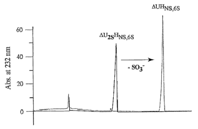
Fig. 5 illustrates the exclusive desulfation of the 2-0H position by the
recombinant
sulfatase. Panel (A) depicts the enzyme desulfating activity assayed by
capillary
electrophoresis using the 2-0 containing trisulfated heparin disaccharide
AU2sHNs,6s. Panel
(B) depicts the activity using its disulfated counterpart to AU2sHNs,6s
lacking a sulfate at the
2-0H position. Only in Panel (A) is a loss of sulfate observed. Minus enzyme
control is
shown as a dotted line.
Fig. 6 provides the in vitro biochemical reaction conditions for the
recombinant 2-0
sulfatase. Panel (A) illustrates the effect of pH. Sulfatase catalytic
efficiency (kcat/Kiii) was
measured as a function of varying pH from 5 to 8 using two overlapping
buffers: 50 mM
MES (solid circles) and 50 mM MOPS (open circles). Inset: Relative effect of
three
different assay buffers (each at pH 6.5) on optimal enzyme activity. 1. 50 mM
MES; 2. 50
mM imidazole; 3. 50 mM sodium phosphate. Panel (B) illustrates the effect of
ionic
strength. Shown here is % activity normalized to 50 mM NaCl. Panel (C)
illustrates the
effect of reaction temperature. Data is normalized to 30 C activity (100%).
The unsaturated
disaccharide AU2sHNs was used in all three experiments.
Fig. 7 illustrates the substrate-product relationship between the 2-0
sulfatase and the
A 4,5 glycuronidase. 2 mM of the unsaturated, 2-0 sulfated heparin
disaccharide AU2sHNs
CA 02512673 2005-07-06
WO 2004/062592 PCT/US2004/000332
-11-
was preincubated with either 250 nM A 4,5 glycuronidase or 25 nM 2-0 AN1-24
for two
minutes at 30 C in a 100 iLit, reaction. Following this preincubation, the
reciprocal enzyme
was added to the reaction for up to six extra minutes. A 4,5 glycuronidase
activity was
measured in real time as the rate of substrate disappearance monitored by the
loss of UV
absorption at 232 run. Zero time on the x-axis represents the time following
the
preincubation during which the second enzyme was added.
'
Fig. 8 illustrates the results of the tandem use of 2-0 sulfatase and A 4,5
glycuronidase in HSGAG compositional analyses. Panel (A) provides the results
of
exhaustively cleaving 200 pg heparin with heparinase I, II and III. These
heparinase-
generated saccharides were then subjected to hydrolysis by the A 4,5
glycuronidase. Panel
(B) provides the results of subsequent hydrolysis by 2-0 sulfatase after the
heparinase
treament. Panel (C) illustrates subsequent hydrolysis by 2-0 sulfatase and by
A 4,5
glycuronidase added simultaneously. Panel (D) depicts the 7 disaccharide peaks
(and one
tetrasaccharide peak) resolved by capillary electrophoresis (each numbered
separately). Their
compositional assignments are as follows: AU2sHNs,6s (1); AUHNAc,6SGHNS,3S,6S
MI
tetrasaccharide (2); AU2sHNs (3); A
¨ ¨NS,6S (4); AU2s1INAc,6s (5); AUHNs (6); AU2sHNAc (7);
and AUHNAL,6s (8).
Fig. 9 illustrates the multiple sequence alignment of sulfatases using
ClustalW. The
sequence of F. heparinum 2-0 sulfatase (F20S) was aligned with human
arylsulfatase B
(ARSB), human arylsulfatase A (ARSA) and P. aeruginosa arylsulfatase (PARS).
The
amino and carboxyl termini are not shown. The sequence numbers for each
sulfatase are
listed on the right. The numbers listed above the alignment correspond
specifically to F2OS
sequence positions (see Figure 2 above). The critical active site cysteines
are highlighted in
black. Other highly conserved amino acids are highlighted in gray.
Fig. 10 provides the structural model of 2-0 sulfatase and topology of the
active site.
Panel (A) is the ribbon diagram of the proposed 2-0 sulfatase structure
constructed using
homology modeling of the crystal structure of human arylsulfatase B. The 13
strands are
shown as thicker areas of the ribbon diagram, and the a helices are shown as
cylindrically
shaped areas. The geminal diol form of the modified cysteine is also depicted
(rendered as
CPK; carbon and oxygen molecules are shown). The direction of substrate
diffusing into the
active site is indicated by an arrow. Panel (B) provides the CPK rendering of
the top view of
CA 02512673 2005-07-06
WO 2004/062592 PCT/US2004/000332
-12-
the structure shown in Panel (A). The modified cysteine, the surrounding basic
amino acids
(Arg, His and Lys), acidic amino acids (Asp, Glu), and Gin and Asn are all
shown. Note that
the active site geminal diol is located in the bottom of a deep cleft.
Fig. 11 depicts the active site amino acids and their interaction with
AU25HN5,6s.
Panel (A) is the stereo view of the 2-0 sulfatase active site highlighting
important amino
acids (shown here by a stick representation). Acidic amino acids (Asp), Gin,
Thr, Leu, and
FGly 82 are depicted. The docked disaccharide is also shown using a stick
representation.
The sulfur atom of the 2-0 sulfate group (next to the lowest positioned
oxygen) and oxygen
atoms (circled) of the 2-0 sulfate group and the planar carboxyl group are
also depicted.
Panel (B) provides the schematic representation of the amino acids shown in
Panel (A).
Potential metal ion coordination is also shown with the divalent cation (Mg2+)
depicted as a
gray circle.
Fig. 12 illustrates the exolytic activity of the 2-0 sulfatase by analyzing
the ability of
the sulfatase to hydrolyze internally positioned 2-0 sulfates within the AT10
decasaccharide
and subsequent compositional analyses of the heparinase-treated product. Panel
(A) shows
the AT-10 decasaccharide sequence with PEN-MALDI nomenclature and outline of
experimental design. Panel (B) provides the capillary electrophoretogram for
both the
control and sulfatase pre-treated samples along with their saccharide
compositional
assignments. Heparinase cleavage products following sulfatase pre-treatment
are shown as a
dashed line (with gray fill). Minus sulfatase control is shown as a white line
(no fill). The
pentasulfated tetrasaccharide (4, -7) is also noted. Disappearance of the
trisulfated
disaccharide (D) by one-third and the corresponding appearance of the 2-0
desulfated
product (AUHNsfis) are depicted by vertical arrows. The minor tetrasaccharide
contaminant
is noted by an asterisk.
Fig. 13 illustrates the steady-state kinetics for various unsaturated
disaccharide
substrates. Panel (A) provides the initial rates determined using 25 nM enzyme
under
standard conditions. Substrate saturation data were fit to pseudo-first order
Michaelis-
Menten assumptions using a non-linear least squares analysis. AU2sHNa, (A);
AU2sHNac,65 (=);
AU25HN5 ( A); AU2sHN5,65 (0); AU25GaiNAA6s (+).
CA 02512673 2005-07-06
WO 2004/062592
PCT/US2004/000332
-13-
Fig. 14 provides the comparable CD spectroscopy of the wild-type 2-0 AN1-24
sulfatase and C82A site-directed mutant¨wild-type enzyme (=), C82A mutant (0).
Band
intensities are expressed as molar ellipticities with units indicated.
Fig. 15 illustrates the identification of 2-0 sulfatase active site
modification (FGly) by
chemical labeling and mass spectrometry. Wild-type sulfatase (2-0 AN1-24) and
C82A
mutant were reacted with Texas Red Hydrazide and subjected to trypsin
proteolysis as
described in Materials and Methods. The molecular masses of the resultant
peptides were
subsequently characterized by MALDI-MS. Panel (A) shows the unlabeled wild-
type
sulfatase control. Panel (B) shows the covalently labeled wild-type sulfatase.
Panel (C)
shows the C82A mutant refractory to chemical labeling. A unique molecular mass
signature
in Panel (B) is noted by an asterisk.
Fig. 16 shows a multiple sequence alignment of the sulfatases using ClustalW.
The
putative active site is boxed, with critically modified cysteine noted by an
asterisk. Invariant
residues are shaded in dark gray, those with partial identity in light gray,
and conservative
substitutions in charcoal. Multiple sequence alignment was generated by
ClustalW using
only select sequences identified from a BLASTP search of the protein data
base. Mammalian
sulfatases are included. Enzymes are abbreviated as follows. FH2S, F.
heparinum 2-0-
sulfatase; PARS, P. aeruginosa arylsulfatase; MDSA, Prevotella sp. MdsA gene;
HGa16S,
human N-acetylgalactosamine- 6-sulfate sulfatase (chondroitin 6-sulfatase);
HARSA, human
cerebroside-3-sulfate sulfatase (arylsulfatase A); HARSB, human N-
acetylgalactosamine-4
sulfate sulfatase (arylsulfatase B); HI2S, human iduronate-2-sulfate
sulfatase; cons,
consensus sequence. The GenBankTM protein accession numbers for sulfatases
listed are as
follows: CAA88421, P. aeruginosa arylsulfatase; AAF72520, Prevotella sp. MdsA
mucin
desulfating gene; AAC51350, Homo sapiens N-acetylgalactosamine-6-sulfate
sulfatase;
AAB03341, H. sapiens cerebroside-3-sulfate sulfatase (arylsulfatase A);
AAA51784, H.
sapiens N-acetylgalactosamine-4-sulfate sulfatase (arylsulfatase B); AAA63197,
H. sapiens
iduronate-2-sulfate sulfatase.
DETAILED DESCRIPTION OF THE INVENTION
Heparin and heparin sulfate glycosaminoglycans (HSGAGs) are structurally
complex
linear polysaccharides (Esko, J. D., and Lindahl, U. (2001) J Clin Invest
108(2), 169-73,
CA 02512673 2005-07-06
WO 2004/062592
PCT/US2004/000332
-14-
Lindahl, U., Kusche-Gullberg, M., and Kjellen, L. (1998) J Biol Chem 273(39),
24979-82)
comprised of repeating disaccharides of uronic acid (a-L-iduronic or(-D-
glucuronic) linked
1-> 4 to a-D-glucosamine. The extensive chemical heterogeneity of these
biopolymers derives
from both the variable number of their constituent disaccharides as well as
the combinatorial
potential for chemical modification at specific positions within each of these
building blocks.
Such modifications include acetylation or sulfation at the N-position of the
glucosamine,
epimerization of glucuronic acid to iduronic acid and additional sulfations at
the 2-0 position
of the uronic acid in addition to the 3-0, 6-0 position of the adjoining
glucosamine. It is a
highly variable sulfation pattern, in particular, that ascribes to each GAG
chain a unique
structural signature. In turn, this signature dictates specific GAG-protein
interactions
underlying critical biological processes related to cell and tissue function.
One of the more formidable challenges currently facing the glycobiology field
is the
design of effective analytical methods to study this structure-function
relationship at the
molecular level. Given this critical structure-function relationship of GAG
sulfation, enzymes
which can hydrolyze these sulfates in a structurally-specific manner become
important in
several ways. To begin with, the systematic desulfation of GAGs at discrete
positions is
central to GAG catabolism that occurs in divergent organisms ranging from
bacteria to
mammals. In addition, the in vivo desulfation of intact GAG chains both at
discrete chemical
positions and in a cell specific, temporally relevant context is also likely
to serve as an
important molecular switch for abrogating targeted GAG-protein interactions.
2-0 sulfatase is a desulfating enzyme that can be now added to the repertoire
of
enzymes used to analyze GAGs and degrade them in a specific manner. As used
herein, the
term "degraded glycosaminoglycan" or "GAG fragment" is intended to encompass a
glycosaminoglycan that has been altered from its original form by the activity
of a 2-0
sulfatase or other enzyme that can act thereon. The degraded glycosaminoglycan
includes
glycosaminoglycans that have been altered by the activity of a 2-0 sulfatase
in some
combination with other glycosaminoglycan degrading enzymes as described
herein. The
degraded glycosaminoglycan may be desulfated, cleaved or desulfated and
cleaved. Any of the
degraded products produced by the activity of the 2-0 sulfatase and/or other
enzymes on the
glycosaminoglycan are intended to be used in the compositions, pharmaceutical
preparations
and methods of the invention. In addition, this sulfatase can be used in
treatment methods
along with the GAG fragments they degrade. 2-0 sulfatase is a member of a
large enzyme
CA 02512673 2005-07-06
WO 2004/062592 PCT/US2004/000332
-15-
family that hydrolyze a wide array of sulfate esters (for a review, see
(Parenti, G., Meroni, G.,
and Ballabio, A. (1997) CUrr Opin Genet Dev 7(3), 386-91, von Figura, K.,
Schmidt, B.,
Selmer, T., and Dierks, T. (1998) Bioessays 20(6), 505-10)). This enzyme
exhibits 2-0
specific sulfatase activity as measured using the trisulfated, unsaturated
heparin disaccharide
AU2sHNso as a substrate (described below). The activity of the enzyme is not
limited to 2-0
desulfation alone, however, as 2-0 sulfatase was found to hydrolyze at the 6-0
and 2N
positions of glucosamine. 2-0 sulfatase can be used to hydrolyze heparin and
chondroitin
disaccharides and can also desulfate GAGs with longer chain lengths such as
tetra- and
decasaccharides. Furthermore, 2-0 sulfatase has been found to work with other
GAG
degrading enzymes such as heparinases and A 4,5 glycuronidase and can be used
in
conjunction with these other enzymes as described herein.
Like the A 4,5 glycuronidase, which we have recently cloned and expressed
(Myette,
J. R., Shriver, Z., Kiziltepe, T., McLean, M. W., Venkataraman, G., and
Sasisekharan, R.
(2002) Biochemistry 41(23), 7424-7434), we have successfully cloned from
Flavobacterium
heparinum and expressed the 2-0 sulfatase in E. coli, from which milligram
quantities of
highly active, soluble enzyme were readily purified. As was also the case for
the
glycuronidase, we found that the yield of soluble recombinant enzyme was
greatly improved
by the engineered removal of the hydrophobic N-terminal signal sequence
comprised of the
first 24 amino acids. This signal sequence was predicted by the von Heinje
method which
also identified the likely signal peptidase cleavage recognition sequence
AXAXA. By
engineering a 2-0 sulfatase N-terminal truncation lacking this sequence
(herein referred to as
2_0 AN1- ,)24, we achieved protein yields exceeding 100 mg of relatively pure
sulfatase per
liter of induced bacterial cultures using a single chromatographic step.
The invention, therefore, provides, in part, a recombinantly produced 2-0
sulfatase.
As used herein, a "recombinant 2-0 sulfatase" is a 2-0 sulfatase that has been
produced
through human manipulation of the nucleic acid that encodes the enzyme. The
human
manipulation usually involves joining the nucleic acid that encodes the 2-0
sulfatase to the
genetic material of a different organism and, generally, a different species.
"Recombinant" is
a term of art that is readily known to one of skill, and techniques for the
recombinant
expression of 2-0 sulfatase are readily available to those of skill in the art
and include those
described in Sambrook et al., Molecular Cloning--A Laboratory Manual, Cold
Spring Harbor
Laboratory, Cold Spring Harbor, N.Y., (1989) or Current Protocols in Molecular
Biology
CA 02512673 2005-07-06
WO 2004/062592 PCT/US2004/000332
-16-
Volumes 1-3, John Wiley & Sons, Inc. (1994-1998). Other techniques for
recombinant
expression including examples of expression systems are described further
below.
As provided herein, recombinant technology can be used to produce a 2-0
sulfatase
encoded by the nucleic acid sequence of SEQ ID NO: 1 or having the amino acid
sequence of
SEQ ID NO: 2. In other aspects of the invention a 2-0 sulfatase encoded by the
nucleic acid
sequence of SEQ ID NO: 3 or having the amino acid sequence of SEQ ID NO: 4 can
be
prepared. The 2-0 sulfatase as provided herein is, in general, produced
through the
manipulation of isolated nucleic acids.
The invention also provides the isolated nucleic acid molecules that code for
a 2-0
sulfatase as described herein. The term "isolated nucleic acid", as used
herein, means: (i)
amplified in vitro by, for example, polyrnerase chain reaction (PCR); (ii)
recombinantly
produced by cloning; (iii) purified, as by cleavage and gel separation; or
(iv) synthesized by,
for example, chemical synthesis. An isolated nucleic acid is one which is
readily
manipulable by recombinant DNA techniques well known in the art. Thus, a
nucleotide
sequence contained in a vector in which 5' and 3' restriction sites are known
or for which
polymerase chain reaction (PCR) primer sequences have been disclosed is
considered
isolated but a nucleic acid sequence existing in its native state in its
natural host is not. An
isolated nucleic acid may be substantially purified, but need not be. For
example, a nucleic
acid that is isolated within a cloning or expression vector is not pure in
that it may comprise
only a tiny percentage of the material in the cell in which it resides. Such a
nucleic acid is
isolated, however, as the term is used herein because it is readily
manipulable by standard
techniques known to those of ordinary skill in the art.
According to the invention, isolated nucleic acid molecules that code for a 2-
0
sulfatase include: (a) nucleic acid molecules which hybridize under stringent
conditions to a
molecule selected from a group consisting of the nucleotide sequences set
forth as SEQ ID
NO: 1 and 3 and which code for a 2-0 sulfatase or parts thereof, (b)
deletions, additions and
substitutions of (a) which code for a 2-0 sulfatase or parts thereof, (c)
nucleic acid molecules
that differ from the nucleic acid molecules of (a) or (b) in codon sequence
due to the
degeneracy of the genetic code, and (d) complements of (a), (b) or (c). The
isolated nucleic
acid molecules include isolated nucleic acid molecules that code for a 2-0
sulfatase which
has an amino acid sequence set forth as SEQ ID NOs: 2 and 4.
CA 02512673 2005-07-06
WO 2004/062592 PCT/US2004/000332
-17-
The invention also includes degenerate nucleic acids which include alternative
codons
to those present in the native materials. For example, serine residues are
encoded by the
codons TCA, AGT, TCC, TCG, TCT and AGC. Each of the six codons is equivalent
for the
purposes of encoding a serine residue. Thus, it will be apparent to one of
ordinary skill in the
art that any of the serine-encoding nucleotide triplets may be employed to
direct the protein
synthesis apparatus, in vitro or in vivo, to incorporate a serine residue into
an elongating 2-0
sulfatase. Similarly, nucleotide sequence triplets which encode other amino
acid residues
include, but are not limited to: CCA, CCC, CCG and CCT (proline codons); CGA,
CGC,
CGG, CGT, AGA and AGG (arginine codons); ACA, ACC, ACG and ACT (threonine
codons); AAC and AAT (asparagine codons); and ATA, ATC and ATT (isoleucine
codons).
Other amino acid residues may be encoded similarly by multiple nucleotide
sequences. Thus,
the invention embraces degenerate nucleic acids that differ from the
biologically isolated
nucleic acids in codon sequence due to the degeneracy of the genetic code.
The isolated nucleic acid molecules of the invention are also intended to
encompass
homologs and alleles which can be identified by conventional techniques.
Identification of
human and other organism homologs of 2-0 sulfatase polypeptides will be
familiar to those
of skill in the art. In general, nucleic acid hybridization is a suitable
method for identification
of homologous sequences of another species (e.g., human, cow, sheep), which
correspond to
a known sequence. Standard nucleic acid hybridization procedures can be used
to identify
related nucleic acid sequences of selected percent identity. For example, one
can construct a
library of cDNAs reverse transcribed from the mRNA of a selected tissue and
use the nucleic
acids that encode a 2-0 sulfatase identified herein to screen the library for
related nucleotide
sequences. The screening preferably is performed using high-stringency
conditions to
identify those sequences that are closely related by sequence identity.
Nucleic acids so
identified can be translated into polypeptides and the polypeptides can be
tested for activity.
The term "stringent conditions" as used herein refers to parameters with which
the art
is familiar. Such parameters include salt, temperature, length of the probe,
etc. The amount
of resulting base mismatch upon hybridization can range from near 0% ("high
stringency") to
about 30% ("low stringency"). Nucleic acid hybridization parameters may be
found in
references that compile such methods, e.g. Molecular Cloning: A Laboratory
Manual, J.
Sambrook, et al., eds., Second Edition, Cold Spring Harbor Laboratory Press,
Cold Spring
Harbor, New York, 1989, or Current Protocols in Molecular Biology, F.M.
Ausubel, et al.,
CA 02512673 2011-05-18
64371-698
-18-
eds., John Wiley & Sons, Inc., New York. One example of high-stringency
conditions is
hybridization at 65 C in hybridization buffer (3.5X SSC, 0.02% Ficoll, 0.02%
polyvinyl
pyrrolidone, 0.02% Bovine Serum Albumin, 2.5mM NaH2PO4(pH7), 0.5% SDS, 2mM
EDTA). SSC is 0.15M sodium chloride/0.15M sodium citrate, pH7; SDS is sodium
dodecyl
sulphate; and EDTA is ethylenediaminetetracetic acid. After hybridization, a
membrane
upon which the nucleic acid is transferred is washed, for example, in 2X SSC
at room
temperature and then at 0.1 - 0.5X SSC/0.1X SDS at temperatures up to 68 C.
The skilled artisan also is familiar with the methodology for screening cells
for
expression of such molecules, which then are routinely isolated, followed by
isolation of the
to pertinent nucleic acid. Thus, homologs and alleles of the 2-0 sulfatase
of the invention, as
well as nucleic acids encoding the same, may be obtained routinely, and the
invention is not
intended to be limited to the specific sequences disclosed. It will be
understood that the
skilled artisan will be able to manipulate the conditions in a manner to
permit the clear
identification of homologs and alleles of the 2-0 sulfatase nucleic acids of
the invention. The
skilled artisan also is familiar with the methodology for screening cells and
libraries for
expression of such molecules which then are routinely isolated, followed by
isolation of the
pertinent nucleic acid molecule and sequencing.
In general, homologs and alleles typically will share at least 90% nucleotide
identity
and/or at least 95% amino acid identity to the sequences of 2-0 sulfatase
nucleic acids and
polypeptides, respectively, in some instances will share at least 95%
nucleotide identity
and/or at least 97% amino acid identity, in other instances will share at
least 97% nucleotide
identity and/or at least 98% amino acid identity, in other instances will
share at least 99%
nucleotide identity and/or at least 99% amino acid identity, and in other
instances will share
at least 99.5% nucleotide identity and/or at least 99.5% amino acid identity.
The homology
can be calculated using various, publicly available software tools developed
by NCBI
(Bethesda, Maryland) that can be obtained through the internet. Exemplary
tools include the
BLAST system available from the website of the National Center for
Biotechnology
Information (NCBI) at the National Institutes of Health. Pairwise and ClustalW
alignments
(BLOSUM30 matrix setting) as well as Kyte-Doolittle hydropathic analysis can
be obtained
using the MacVector sequence analysis software (Oxford Molecular Group).
Watson-Crick
complements of the foregoing nucleic acids also are embraced by the invention.
*Trade -mark
CA 02512673 2005-07-06
WO 2004/062592 PCT/US2004/000332
-19-
In screening for 2-0 sulfatase related genes, such as homologs and alleles of
2-0
sulfatase, a Southern blot may be performed using the foregoing conditions,
together with a
radioactive probe. After washing the membrane to which the DNA is finally
transferred, the
membrane can be placed against X-ray film or a phosphoimager plate to detect
the
radioactive signal.
The recombinantly produced 2-0 sulfatase as provided herein exhibited robust,
2-0
specific sulfatase activity. The success with expressing a highly active 2-0
sulfatase clearly
validates our use of E. coli as a recombinant expression system for the large-
scale production
of active enzyme. Therefore, active isolated 2-0 sulfatase polypeptides
(including whole
proteins and partial proteins) are provided herein which include isolated 2-0
sulfatase
polypeptides that have the amino acid sequence of SEQ ID NO: 2 or SEQ ID NO:
4.
Polypeptides can be isolated from biological samples, and can also be
expressed
recombinantly in a variety of prokaryotic and eukaryotic expression systems,
such as those
described above, by constructing an expression vector appropriate to the
expression system,
introducing the expression vector into the expression system, and isolating
the recombinantly
expressed protein. Polypeptides can also be synthesized chemically using well-
established
methods of peptide synthesis.
As used herein, "isolated polypeptide" means the polypeptide is separated from
its
native environment and present in sufficient quantity to permit its
identification or use. This
means, for example: (i) selectively produced by expression cloning or (ii)
purified as by
chromatography or electrophoresis. Isolated proteins or polypeptides may be,
but need not
be, substantially pure. Because an isolated polypeptide may be admixed with a
pharmaceutically acceptable carrier in a pharmaceutical preparation, the
polypeptide may
comprise only a small percentage by weight of the preparation. The polypeptide
is
nonetheless isolated in that it has been separated from the substances with
which it may be
associated in living systems, i.e., isolated from other proteins.
As used herein, the term "substantially pure" means that the proteins are
essentially
free of other substances to an extent practical and appropriate for their
intended use. In
particular, the proteins are sufficiently pure and are sufficiently free from
other biological
constituents of their hosts cells so as to be useful in, for example, protein
sequencing, or
producing pharmaceutical preparations. As used herein, a "substantially pure 2-
0 sulfatase"
is a preparation of 2-0 sulfatase which has been isolated or synthesized and
which is greater
CA 02512673 2005-07-06
WO 2004/062592 PCT/US2004/000332
-20-
than about 90% free of contaminants. Preferably, the material is greater than
91%, 92%,
93%, 94%, 95%, 96%, 97%, 98%, or even greater than 99% free of contaminants.
The
degree of purity may be assessed by means known in the art. One method for
assessing the
purity of the material may be accomplished through the use of specific
activity assays.
The cloned, full-length gene of the 2-0 sulfatase encodes an open reading
frame
(ORF) of 468 amino acids (Fig. 2), with a predicted molecular mass of 51.9
kDa. This
theoretical molecular weight is approximately 10 kDa less than the value
reported in the
literature (McLean, M. W., Bruce, J. S., Long, W. F., and Williamson, F. B.
(1984) Eur J
Biochem 145(3), 607-15). Based on its amino acid composition, the encoded
protein is quite
basic (theoretical IA of 8.75). A further analysis of its primary amino acid
sequence
unequivocally places this ORF as a member of a larger sulfatase family. As
members of a
large enzyme family, the sulfatases hydrolyze a wide array of sulfate esters
(for a review, see
(Parenti, G., Meroni, G., and Ballabio, A. (1997) Curr Opin Genet Dev 7(3),
386-91, von
Figura, K., Schmidt, B., Selmer, T., and Dierks, T. (1998) Bioessays 20(6),
505-10)). Their
respective substrates include sulfated complex carbohydrates such as the
glycosaminoglycans
(GAGs), steroids, sphingolipids, xenobiotic compounds, and amino acids such as
tyrosine.
Additionally, many of these enzymes are able to hydrolyze in vitro smaller
synthetic
substrates (e.g., 4-nitrophenyl sulfate and catechol sulfate). It is for this
reason that these
enzymes are often generically described as "arylsulfatases" (even when their
preferred in vivo
substrate is ill-defined). Despite their disparate substrate specificities,
the members of this
enzyme family share both considerable structural homology and a common
catalytic
mechanism with one another (Waldow, A., Schmidt, B., Dierks, T., von Bulow,
R., and von
Figura, K. (1999) J Biol Chem 274(18), 12284-8).
The fiavobacterial 2-0 sulfatase possesses considerable sequence homology to
other
bacterial (and non-bacterial) sulfatases, especially within its amino terminus
in which resides a
highly conserved sulfatase domain. This signature catalytic domain is readily
identified by the
consensus sequence C/SXPXRXXXXS/TG (SEQ. ID NO: 6). The conserved cysteine (or
less
commonly serine) within this sulfatase motif is of particular functional
importance as it is
covalently modified to a L-Ca- formylglycine (L-2-amino-3-0xo-propionic acid).
The
ubiquitous importance of this chemical modification was first functionally
identified by its
relationship to the etiology of multiple sulfatase deficiency (MSD), a
genetically recessive
disorder in which there is a complete loss of sulfatase activity due to a lack
of this critical
CA 02512673 2005-07-06
WO 2004/062592 PCT/US2004/000332
-21-
aldehyde (FGly) within the active site of all expressed sulfatases (Koloclny,
E. H. a. F., A. L.
(1995) in The Metabolic and Molecular Bases of Inherited Disease (Scriver, C.
R., Beaudet,
A. L., Sly, W. S., and Valle, D., ed), pp. 2693-2741, McGraw-Hill, New York).
We have
identified the conserved sulfatase active site by sequence homology which we
have found
includes a cysteine and not a serine as the critical amino acid predicted to
be chemically
modified as a formylglycine in vivo. An empirical demonstration of this active-
site aldehyde at
this position is presented in Examples.
While the cloned flavobacterial sulfatase exhibits the highest sequence
similarity to the
bacterial arylsulfatases (especially the arylsulfatase from Pseudomonas
aeruginosa), we point
out that a limited homology of the 2-0 sulfatase does extend to the mammalian
glycosaminoglycan sulfatases functioning in the lysosomal degradation pathway.
As is the
case for the bacterial enzymes, this sequence homology is strongest in the NH2-
terminus where
the putative sulfatase domain resides. Among the human lysosomal enzymes, it
is the
galactosamine (N-acetyl)-6-sulfate sulfatase (chondroitin 6-0 sulfatase) which
exhibits the
closest similarity with the flavobacterial 2-0 sulfatase; the two enzymes
possess approximately
26% identity when comparing their entire protein sequences. There are also two
functionally
related lysosomal sulfatases which specifically hydrolyze the 2-0H position of
uronic acid.
These enzymes are the iduronate 2-sulfate sulfatase (IDS) (Bielicki, J.,
Freeman, C., Clements,
P. R., and Hopwood, J. J. (1990) Biochem J271(1), 75-86) and the glucuronic-2-
sulfate
sulfatase (Freeman, C., and Hopwood, J. J. (1989) Biochem J259(1), 209-16).
The IDS and
flavobacterial 2-0 sulfatase exhibit only a limited sequence homology (less
than 22% identity),
however.
Both of these enzymes desulfate heparan sulfate, the iduronate-2-sulfate
sulfatase (IDS)
also acts on dermatan sulfate. Both enzymes possess an acidic pH optima for
activity, a fact
consistent with their location within the lysosome. The two sulfatases
initially exist as
precursors which must be proteolytically processed for activity. The native
molecular weight
of the human IDS precursor has been reported in the range of 42 to 65 kDa
(Bielicki, J.,
Freeman, C., Clements, P. R., and Hopwood, J. J. (1990) Biochem J271(1), 75-
86), while its
theoretical mass based entirely on its amino acid composition is approximately
62 kDa. As
such, the mammalian lysosomal IDS is somewhat larger than its flavobacterial
counterpart,
while also requiring substantial posttranslational modification for maximal
enzyme activity.
The acidic pH optima for the lysosomal enzymes would also appear to limit
their in vitro use
CA 02512673 2011-05-18
64371-698
-22-
for the determination of HSGAG composition, at least when used in tandem with
other
flavobacterial HSGAG degrading enzymes such as the heparinases or the A 4,5
glycuronidase;
these latter enzymes all possess a pH optima much closer to neutrality.
A homology-based structural model of the 2-0 sulfatase active site was
constructed
using as a framework the available crystallographic data for three highly
related arylsulfatases.
In this model, we have identified important structural parameters within the
enzyme active site
relevant to enzyme function, especially as relates to its substrate
specificity (substrate binding
and catalysis). By docking various disaccharide substrates, we were also able
to make specific
predictions concerning structural determinants present within these potential
substrates that
would complement this unique active site architecture. These determinants
included the
position and number of sulfates present on the glucosamine, oligosaccharide
chain length, the
presence of a A 4,5 unsaturated double bond, and the exolytic vs. endolytic
potential of the
enzyme. These predictions were then tested against biochemical and kinetic
data which largely
validated our substrate specificity predictions. Our modeling approach was
further
complemented experimentally using aldehyde-specific chemical labeling, peptide
mapping in
tandem with mass spectrometry and site-directed mutagenesis to physically
demonstrate the
presence of a covalently modified cysteine (formyl glycine (FGly)) within the
active site. This
combinatorial approach of structure modeling and biochemical studies has
provided insight
into the molecular basis of enzyme function.
The crystal structures of two human lysosomal sulfatases, cerebroside-3-
sulfate 3-
sulfohydrolase (arylsulfatase A), (Lukatela, Gõ Krauss, N., Theis, K., Selmer,
T., Giesehnann,
V., von Figura, K., and Saenger, W. (1998) Biochemishy 37(11), 365464, von
Bulow, R.,
Schmidt, B., Dierks, T., von Figura, K., and Uson, I. (2001) J Mol Biol
305(2), 269-77)
N-acetylgalactosamine-4-sulfate 4-sulfohydrolase (arylsulfatase B) (Bond, C.
S., Clements, P.
R., Ashby, S. J., Collyer, C. A., Harrop, S. J., Hopwood, J. J., and Guss, J.
M. (1997) Structure
5(2), 277-89), and a bacterial arylsulfatase from Pseudomonas aeruginosa
(Boltes, L,
Czapinska, H., Kahnert, A., von Bulow, R., Dierks, T., Schmidt, B., von
Figura, K., Kertesz,
M. A., and Uson, I. (2001) Structure (Camb) 9(6), 483-91) have been solved.
These three
sulfatases share an identical alkaline-phosphatase like structural fold
(according to Structural
Classification of Proteins database) comprised of a series of mixed parallel
and
antiparallelfl strands flanked by long and short a helices on either side
(Lukatela, G., Krauss,
N., Theis, K., Selmer, T., Gieselmaim, V., von Figura, K., and Saenger, W.
(1998)
CA 02512673 2005-07-06
WO 2004/062592 PCT/US2004/000332
-23-
Biochemistiy 37(11), 3654-64, Bond, C. S., Clements, P. R., Ashby, S. J.,
Collyer, C. A.,
Harrop, S. J., Hopwood, J. J., and Guss, J. M. (1997) Structure 5(2), 277-89,
Bolles, I.,
Czapinska, H., Kahnert, A., von Bulow, R., Dierks, T., Schmidt, B., von
Figura, K., Kertesz,
M. A., and Uson, I. (2001) Structure (Camb) 9(6), 483-91, von Bulow, R.,
Schmidt, B., Dierks,
T., von Figura, K., and Uson, I. (2001) J Mol Biol 305(2), 269-77). In
addition to their
common structural fold, these sulfatase structures also possess a high degree
of homology
within their respective active sites, especially in the region localized
around the modified
cysteine (FGly). Taken together, these crystal structures present a clear and
consistent
description of conserved active site residues at least as it relates to a
likewise conserved
mechanism of sulfate ester hydrolysis. At the same time, this strong
structural homology is
somewhat surprising considering that at least two of these sulfatases act on
notably different
substrates, e.g., sulfated sphingolipid vs. sulfated glycosaminoglycan (GAG).
It was discovered that 2-0 sulfatase has a relatively high cysteine content.
Apart from
the catalytic cysteine at position 82, none of the remaining seven cysteines
appeared to be
highly conserved among other members of the sulfatase family. Enzyme activity
was not
inhibited with the addition of DTNB (Ellman's reagent) or DTT. This general
lack of
inhibition by these two cysteine-reactive agents suggests at least two
probabilities. First, the 2-
sulfatase does not require intramolecular disulfide linkages to critically
stabilize a
catalytically active conformation. Second, free sulfhydryls are not directly
participating in
catalysis. It is possible, however, that a few of these cysteines are buried
and therefore not
accessible to sulfhydryl exchange. At least five of the eight cysteines,
however, do react with
DTNB under nondenaturing conditions. This latter fact suggests an alternate
role for these
solvent-accessible cysteines (along with specific histidines) ie., metal-
coordinating thiolates.
Comparison between the 2-0 sulfatase and alkaline phosphatase reveals that
these enzymes are
esterases with similar catalytic mechanisms, including the presumptive
formation of a covalent
intermediate. The two hydrolytic enzymes also possess structurally related
domains, in
particular, a highly superimposible active site that includes a divalent metal
binding pocket. In
the case of alkaline phosphatase, it is zinc rather than calcium (or Mg+2)
that is coordinated
within this pocket.
The 2-0 sulfatase possesses 67 basic amino acids, including the catalytic
histidine at
position 136, a proximal lysine at position 134 and an invariant arginine at
position 86 found
within the defining sulfatase consensus sequence. Moreover, crystal structures
of the active
CA 02512673 2005-07-06
WO 2004/062592 PCT/US2004/000332
-24-
site of related sulfatases each clearly show at least four basic residues
participating in catalysis
which was also found in our homology model. A masking of these important
charges by
exogenous ions would interfere with their catalytic function.
Of the 8 histidines present in the flavobacterial 2-0 sulfatase, 11136 is
invariantly
conserved among the structurally related bacterial sulfatases examined. For
each of these
enzymes, this highly conserved histidine is found within a putative consensus
sequence
GKWHX (SEQ. ID NO: 7) (where X is a hydrophobic amino acid). Other conserved
histidines
include His 296 and His 303. Catalytically important histidines have been
observed within the
active site of several sulfatase crystal structures including human lysosomal
N-
acetylgalactosamine-4 sulfatase (arylsulfatase B) (Bond, C. S., Clements, P.
R., Ashby, S. J.,
Collyer, C. A., Harrop, S. J., Hopwood, J. J., and Guss, J. M. (1997)
Structure 5(2), 277-89)
and arylsulfatase A (Lukatela, G., Krauss, N., Theis, K., Selmer, T.,
Gieselmann, V., von
Figura, K., and Saenger, W. (1998) Biochemistry 37(11), 3654-64) as well as
the arysulfatase
from Pseudonzonas aeriginosa (Bolles, I., Czapinska, H., Kahnert, A., von
Bulow, R., Dierks,
T., Schmidt, B., von Figura, K., Kertesz, M. A., and Uson, I. (2001) Structure
(Camb) 9(6),
483-91) to which the flavobacterial 2-0 sulfatase appears to be most closely
related. In the
latter case, His 211 appears to hydrogen bond with the sulfate oxygen (04)
contributing
perhaps to proper sulfate coordination. Additionally, the N51 of His 115 of P.
aeruginosa (His
242 in the human 4-S sulfatase) is within hydrogen bonding distance to the 012
of the catalytic
formylglycine. The presence of His 136 in the active site and its
participation in catalysis is
strongly supported by our homology studies.
The flavobacterial 2-0 sulfatase possesses 52 acidic amino acids, several of
which are
highly conserved (e.g., Asp 42, Asp 269, Asp 286, Asp 295, and Asp 342).
Interestingly, four
acidic side chains are also found in a consensus active site also observed in
known crystal
structures. In this snapshot, these four carboxylates appear to coordinate a
divalent metal ion
(typically calcium). This divalent metal in turn coordinates with the
formylglycine hydroxylate
and possibly the 0-y1 group of the sulfate.
Based on the understanding of the important residues involved in the function
of 2-0
sulfatase, the invention also embraces functional variants. As used herein, a
"functional
variant" of a 2-0 sulfatase polypeptide is a polypeptide which contains one or
more
modifications to the primary amino acid sequence of a 2-0 sulfatase
polypeptide. The
polypeptide can contain 1, 2, 3, 4, 5, 6, 7, 8, 9, 10, 11, 12 ,13 ,14, 15, 16,
17, 18, 19, 20, 25,
CA 02512673 2005-07-06
WO 2004/062592 PCT/US2004/000332
-25-
30, 35, 40, 50 or more amino acid modifications. These modifications are
intended to
encompass modifications that result in a 2-0 sulfatase with altered activity
relative to the
native 2-0 sulfatase but also include modifications that do not result in
altered activity
relative to the native enzyme. The term "native" as used herein refers to the
2-0 sulfatase as
it would be found in nature. Modifications which create a 2-0 sulfatase
polypeptide
functional variant are typically made to the nucleic acid which encodes the 2-
0 sulfatase
polypeptide, and can include deletions, point mutations, truncations, amino
acid substitutions
and addition of amino acids or non-amino acid moieties to: 1) enhance a
property of a 2-0
sulfatase polypeptide, such as protein stability in an expression system or
the stability of
protein-protein binding; 2) provide a novel activity or property to a 2-0
sulfatase polypeptide,
such as addition of a detectable moiety; or 3) to provide equivalent or better
interaction with
other molecules (e.g., heparin). Alternatively, modifications can be made
directly to the
polypeptide, such as by cleavage, addition of a linker molecule, addition of a
detectable
moiety, such as biotin, addition of a fatty acid, and the like. Modifications
also embrace
fusion proteins comprising all or part of the 2-0 sulfatase amino acid
sequence. One of skill
in the art will be familiar with methods for predicting the effect on protein
conformation of a
change in protein sequence, and can thus "design" a functional variant 2-0
sulfatase
polypeptide according to known methods. One example of such a method is
described by
Dahiyat and Mayo in Science 278:82-87, 1997, whereby proteins can be designed
de novo.
The method can be applied to a known protein to vary only a portion of the
polypeptide
sequence. By applying the computational methods of Dahiyat and Mayo, specific
variants of
a polypeptide can be proposed and tested to determine whether the variant
retains a desired
conformation.
Functional variants can include 2-0 sulfatase polypeptides which are modified
specifically to alter a feature of the polypeptide unrelated to its
physiological activity. For
example, cysteine residues can be substituted or deleted to prevent unwanted
disulfide
linkages. Similarly, certain amino acids can be changed to enhance expression
of a 2-0
sulfatase polypeptide by eliminating proteolysis by proteases in an expression
system (e.g.,
dibasic amino acid residues in yeast expression systems in which KEX2 protease
activity is
present). Functional variants, therefore, can also include variant 2-0
sulfatase that maintain
the same enzymatic function as the native 2-0 sulfatase but include some
modification to the
amino acid sequence that does not alter native enzyme activity. These
modifications include
CA 02512673 2011-05-18
64371-698
-26-
conservative amino acid substitutions as well as non-conservative amino acid
substitutions
that are remote from the binding and catalytic sites of the enzyme.
Mutations of a nucleic acid which encodes a 2-0 sulfatase polypeptide
preferably
preserve the amino acid reading frame of the coding sequence, and preferably
do not create
regions in the nucleic acid which are likely to hybridize to form secondary
structures, such as
hairpins or loops, which can be deleterious to expression of the variant
polypeptide.
Mutations can be made by selecting an amino acid substitution, or by random
mutagenesis of a selected site in a nucleic acid which encodes the
polypeptide. Variant
polypeptides are then expressed and tested for one or more activities to
determine which
mutation provides a variant polypeptide with the desired properties. Further
mutations can be
made to variants (or to non-variant 2-0 sulfatase polypeptides) which are
silent as to the
amino acid sequence of the polypeptide, but which provide preferred codons for
translation in
a particular host. The preferred codons for translation of a nucleic acid in,
e.g., E. coli, are
well known to those of ordinary skill in the art. Still other mutations can be
made to the
noncoding sequences of a 2-0 sulfatase gene or cDNA clone to enhance
expression of the
polypeptide.
In the description that follows, reference will be made to the amino acid
residues and
residue positions of native 2-0 sulfatase disclosed in SEQ ID NO: 1. In
particular, residues
and residue positions will be referred to as "corresponding to" a particular
residue or residue
position of 2-0 sulfatase. As will be obvious to one of ordinary skill in the
art, these
positions are relative and, therefore, insertions or deletions of one or more
residues would
have the effect of altering the numbering of downstream residues. In
particular, N-terminal
insertions or deletions would alter the numbering of all subsequent residues.
Therefor; as
used herein, a residue in a recombinant modified heparinase will be referred
to as
"corresponding to" a residue of the full 2-0 sulfatase if, using standard
sequence comparison
programs, they would be aligned. Many Such sequence alignment programs are now
'available to one of ordinary skill in the art and their use in sequence
comparisons has become
standard (e.g., "LALIGN" ). As used herein, this convention of referring to
the positions
of residues of the recombinant modified heparinases by their corresponding 2-0
sulfatase
residues shall extend not only to embodiments including N-terminal insertions
or deletions
but also to internal insertions or deletions (e.g, insertions or deletions in
"loop" regions).
CA 02512673 2005-07-06
WO 2004/062592 PCT/US2004/000332
-27-
One type of amino acid substitution is referred to as a "conservative
substitution." As
used herein, a "conservative amino acid substitution" or "conservative
substitution" refers to
an amino acid substitution in which the substituted amino acid residue is of
similar charge as
the replaced residue and is of similar or smaller size than the replaced
residue. Conservative
substitutions of amino acids include substitutions made amongst amino acids
within the
following groups: (a) the small non-polar amino acids, A, M, I, L, and V; (b)
the small polar
amino acids, G, S, T and C; (c) the amido amino acids, Q and N; (d) the
aromatic amino
acids, F, Y and W; (e) the basic amino acids, K, R and H; and (f) the acidic
amino acids, E
and D. Substitutions which are charge neutral and which replace a residue with
a smaller
residue may also be considered "conservative substitutions" even if the
residues are in
different groups (e.g., replacement of phenylalanine with the smaller
isoleucine). The term
"conservative amino acid substitution" also refers to the use of amino acid
analogs.
Methods for making amino acid substitutions, additions or deletions are well
known
in the art. The terms "conservative substitution", "non-conservative
substitutions", "non-
polar amino acids", "polar amino acids", and "acidic amino acids" are all used
consistently
with the prior art terminology. Each of these temis is well-known in the art
and has been
extensively described in numerous publications, including standard
biochemistry text books,
such as "Biochemistry" by Geoffrey Zubay, Addison-Wesley Publishing Co., 1986
edition,
which describes conservative and non-conservative substitutions, and
properties of amino
acids which lead to their definition as polar, non-polar or acidic.
Even when it is difficult to predict the exact effect of a substitution in
advance of
doing so, one skilled in the art will appreciate that the effect can be
evaluated by routine
screening assays, preferably the biological assays described herein.
Modifications of peptide
properties including thermal stability, enzymatic activity, hydrophobicity,
susceptibility to
proteolytic degradation or the tendency to aggregate with carriers or into
multimers are
assayed by methods well known to the ordinarily skilled artisan. For
additional detailed
description of protein chemistry and structure, see Schulz, G. E. et al.,
Principles of Protein
Structure, Springer-Verlag, New York, 1979, and Creighton, T. E., Proteins:
Structure and
Molecular Principles, W. H. Freeman & Co., San Francisco, 1984.
Additionally, some of the amino acid substitutions are non-conservative
substitutions.
In certain embodiments where the substitution is remote from the active or
binding sites, the
non-conservative substitutions are easily tolerated provided that they
preserve a tertiary
CA 02512673 2005-07-06
WO 2004/062592 PCT/US2004/000332
-28-
structure characteristic of, or similar to, native 2-0 sulfatase, thereby
preserving the active
and binding sites. Non-conservative substitutions, such as between, rather
than within, the
above groups (or two other amino acid groups not shown above), which will
differ more
significantly in their effect on maintaining (a) the structure of the peptide
backbone in the
area of the substitution (b) the charge or hydrophobicity of the molecule at
the target site, or
(c) the bulk of the side chain.
Nearly every active, recombinantly expressed sulfatase reported in the
literature
possesses a cysteine (and not a serine) within the active site sequence
C/SXPXRXXXXS/TG
(SEQ. ID NO: 6) (Lukatela, G., Krauss, N., Theis, K., Selmer, T., Gieselmann,
V., von Figura,
K., and Saenger, W. (1998) Biochemistry 37(11), 3654-64). It seemed likely,
therefore, that a
cysteine-specific modifying machinery functionally exists in E. coli. This
idea was supported
by our initial attempts to produce a recombinant cysteine--> serine 2-0
sulfatase variant which
led to the production of insoluble protein when expressed in E. coli. We note
that the E. coli
genome encodes for at least three different putative sulfatase genes in
addition to the atsB gene
which, by homology, has been proposed to encode for this cysteine-specific
modifying activity.
All of these genes are located as a cluster within the bacterial chromosome
(Kertesz, M. A.
(2000) FEMS Microbiol Rev 24(2), 135-75). It would appear, however, that the
E. coli
sulfatase genes are normally cryptic. At the very least, E. coli lacks the
specific enzymes for
desulfating heparin/heparan sulfate glycosaminoglycans, but the bacterium
fortuitously
provides the necessary enzymology to effectively modify select heterologous
sulfatases such as
the 2-0 sulfatase. Therefore, the 2-0 sulfatases as described herein can be
produced
recombinantly in E. coli. However, the recombinant production of the 2-0
sulfatases provided
are not limited to their expression in E. coli. The 2-0 sulfatases can also be
recombinantly
produced in other expression systems described below.
The 2-0 sulfatases, can be recombinantly produced using a vector including a
coding
sequence operably joined to one or more regulatory sequences. As used herein,
a coding
sequence and regulatory sequences are said to be "operably joined" when they
are covalently
linked in such a way as to place the expression or transcription of the coding
sequence under
the influence or control of the regulatory sequences. If it is desired that
the coding sequences
be translated into a functional protein the coding sequences are operably
joined to regulatory
sequences. Two DNA sequences are said to be operably joined if induction of a
promoter in
the 5' regulatory sequences results in the transcription of the coding
sequence and if the nature
CA 02512673 2005-07-06
WO 2004/062592 PCT/US2004/000332
-29-
of the linkage between the two DNA sequences does not (1) result in the
introduction of a
frame-shift mutation, (2) interfere with the ability of the promoter region to
direct the
transcription of the coding sequences, or (3) interfere with the ability of
the corresponding
RNA transcript to be translated into a protein. Thus, a promoter region would
be operably
joined to a coding sequence if the promoter region were capable of effecting
transcription of
that DNA sequence such that the resulting transcript might be translated into
the desired
protein or polypeptide.
The precise nature of the regulatory sequences needed for gene expression may
vary
between species or cell types, but shall in general include, as necessary, 5'
non-transcribing
and 5' non-translating sequences involved with initiation of transcription and
translation
respectively, such as a TATA box, capping sequence, CAAT sequence, and the
like.
Especially, such 5' non-transcribing regulatory sequences will include a
promoter region
which includes a promoter sequence for transcriptional control of the operably
joined gene.
Promoters may be constitutive or inducible. Regulatory sequences may also
include
enhancer sequences or upstream activator sequences, as desired.
As used herein, a "vector" may be any of a number of nucleic acids into which
a
desired sequence may be inserted by restriction and ligation for transport
between different
genetic environments or for expression in a host cell. Vectors are typically
composed of
DNA although RNA vectors are also available. Vectors include, but are not
limited to,
plasmids and phagemids. A cloning vector is one which is able to replicate in
a host cell, and
which is further characterized by one or more endonuclease restriction sites
at which the
vector may be cut in a determinable fashion and into which a desired DNA
sequence may be
ligated such that the new recombinant vector retains its ability to replicate
in the host cell. In
the case of plasmids, replication of the desired sequence may occur many times
as the
plasmid increases in copy number within the host bacterium, or just a single
time per host as
the host reproduces by mitosis. In the case of phage, replication may occur
actively during a
lytic phase or passively during a lysogenic phase. An expression vector is one
into which a
desired DNA sequence may be inserted by restriction and ligation such that it
is operably
joined to regulatory sequences and may be expressed as an RNA transcript.
Vectors may
further contain one or more marker sequences suitable for use in the
identification of cells
which have or have not been transformed or transfected with the vector.
Markers include, for
example, genes encoding proteins which increase or decrease either resistance
or sensitivity
CA 02512673 2005-07-06
WO 2004/062592
PCT/US2004/000332
-30-
to antibiotics or other compounds, genes which encode enzymes whose activities
are
detectable by standard assays known in the art (e.g., 13-galactosidase or
alkaline phosphatase),
and genes which visibly affect the phenotype of transformed or transfected
cells, hosts,
colonies or plaques. Preferred vectors are those capable of autonomous
replication and
' 5 expression of the structural gene products present in the DNA
segments to which they are
operably joined.
For prokaryotic systems, plasmid vectors that contain replication sites and
control
sequences derived from a species compatible with the host may be used.
Examples of
suitable plasmid vectors include pBR322, pUC18, pUC19 and the like; suitable
phage or
bacteriophage vectors include kgt10, kgt11 and the like; and suitable virus
vectors include
pMAM-neo, pKRC and the like. Preferably, the selected vector of the present
invention has
the capacity to autonomously replicate in the selected host cell. Useful
prokaryotic hosts
include bacteria, in addition to E. coli, Flavobacterium heparinum, Bacillus,
Streptomyces,
Pseudomonas, Salmonella, Serratia, and the like.
To express the 2-0 sulfatase of the invention in a prokaryotic cell, it is
desirable to
operably join the nucleic acid sequence of a 2-0 sulfatase of the invention to
a functional
prokaryotic promoter. Such promoter may be either constitutive or, more
preferably,
regulatable (i.e., inducible or derepressible). Examples of constitutive
promoters include the
int promoter of bacteriophage 7, the bla promoter of the 13-lactamase gene
sequence of
pBR322, and the CAT promoter of the chloramphenicol acetyl transferase gene
sequence of
pPR325, and the like. Examples of inducible prokaryotic promoters include the
major right
and left promoters of bacteriophage X (PL and PR), the trp, recA, lacZ, lad,
and gal promoters
of E. coli, the a-amylase (Ulmanen et al., J. BacterioL 162:176-182 (1985))
and the -28-
specific promoters of B. subtilis (Gilman et al., Gene sequence 32:11-20
(1984)), the
promoters of the bacteriophages of Bacillus (Gryczan, In: The Molecular
Biology of the
Bacilli, Academic Press, Inc., NY (1982)), and Streptomyces promoters (Ward et
al., MoL
Gen. Genet. 203:468-478 (1986)).
Prokaryotic promoters are reviewed by Glick (J. Ind. MicrobioL 1:277-282
(1987));
Cenatiempo (Biochimie 68:505-516 (1986)); and Gottesman (Ann. Rev. Genet.
18:415-442
(1984)).
CA 02512673 2005-07-06
WO 2004/062592 PCT/US2004/000332
-31-
Proper expression in a prokaryotic cell also requires the presence of a
ribosome
binding site upstream of the encoding sequence. Such ribosome binding sites
are disclosed,
for example, by Gold et al. (Ann. Rev. Microbiol. 35:365-404 (1981)).
Because prokaryotic cells may not produce the 2-0 sulfatase of the invention
with
normal eukaryotic glycosylation, expression of the 2-0 sulfatase of the
invention of the
eukaryotic hosts is useful when glycosylation is desired. Preferred eukaryotic
hosts include,
for example, yeast, fungi, insect cells, and mammalian cells, either in vivo
or in tissue culture.
Mammalian cells which may be useful as hosts include HeLa cells, cells of
fibroblast origin
such as VERO or CHO-K1, or cells of lymphoid origin, such as the hybridoma
SP2/0-AG14
or the myeloma P3x635g8, and their derivatives. Preferred mammalian host cells
include
SP2/0 and J558L, as well as neuroblastoma cell lines such as IMR 332 that may
provide
better capacities for correct post-translational processing. Embryonic cells
and mature cells
of a transplantable organ also are useful according to some aspects of the
invention.
In addition, plant cells are also available as hosts, and control sequences
compatible
with plant cells are available, such as the nopaline synthase promoter and
polyadenylation
signal sequences.
Another preferred host is an insect cell, for example in Drosophila larvae.
Using
insect cells as hosts, the Drosophila alcohol dehydrogenase promoter can be
used (Rubin,
Science 240:1453-1459 (1988)). Alternatively, baculovirus vectors can be
engineered to
express large amounts of the 2-0 sulfatase of the invention in insect cells
(Jasny, Science
238:1653 (1987); Miller et al., In: Genetic Engineering (1986), Setlow, J.K.,
et al., eds.,
Plenum, Vol. 8, pp. 277-297).
Any of a series of yeast gene sequence expression systems which incorporate
promoter and termination elements from the genes coding for glycolytic enzymes
and which
are produced in large quantities when the yeast are grown in media rich in
glucose may also
be utilized. Known glycolytic gene sequences can also provide very efficient
transcriptional
control signals. Yeast provide substantial advantages in that they can also
carry out post-
translational peptide modifications. A number of recombinant DNA strategies
exist which
utilize strong promoter sequences and high copy number plasmids which can be
utilized for
production of the desired proteins in yeast. Yeast recognize leader sequences
on cloned
mammalian gene sequence products and secrete peptides bearing leader sequences
(i.e., pre-
peptides).
CA 02512673 2005-07-06
WO 2004/062592 PCT/US2004/000332
-32-
A wide variety of transcriptional and translational regulatory sequences may
be
employed, depending upon the nature of the host. The transcriptional and
translational
regulatory signals may be derived from viral sources, such as adenovirus,
bovine papilloma
virus, simian virus, or the like, where the regulatory signals are associated
with a particular
gene sequence which has a high level of expression. Alternatively, promoters
from
mammalian expression products, such as actin, collagen, myosin, and the like,
may be
employed. Transcriptional initiation regulatory signals may be selected which
allow for
repression or activation, so that expression of the gene sequences can be
modulated. Of
interest are regulatory signals that are temperature-sensitive so that by
varying the
temperature, expression can be repressed or initiated, or which are subject to
chemical (such
as metabolite) regulation.
As discussed above, expression of the 2-0 sulfatase of the invention in
eukaryotic
hosts requires the use of eukaryotic regulatory regions. Such regions will, in
general, include
a promoter region sufficient to direct the initiation of RNA synthesis.
Preferred eukaryotic
promoters include, for example, the promoter of the mouse metallothionein I
gene sequence
(Hamer et al., J. Mol. AppL Gen. 1:273-288 (1982)); the TK promoter of Herpes
virus
(McKnight, Cell 31:355-365 (1982)); the SV40 early promoter (Benoist et al.,
Nature
(London) 290:304-310 (1981)); the yeast gal4 gene sequence promoter (Johnston
et al., Proc.
NatL Acad. Sci. (USA) 79:6971-6975 (1982); Silver et al., Proc. NatL Acad.
Sci. (USA)
81:5951-5955 (1984)).
As is widely known, translation of eukaryotic mRNA is initiated at the codon
which
encodes the first methionine. For this reason, it is preferable to ensure that
the linkage
between a eukaryotic promoter and a DNA sequence which encodes the 2-0
sulfatase of the
invention does not contain any intervening codons which are capable of
encoding a
methionine (i.e., AUG). The presence of such codons results either in the
formation of a
fusion protein (if the AUG codon is in the same reading frame as the 2-0
sulfatase of the
invention coding sequence) or a frame-shift mutation (if the AUG codon is not
in the same
reading frame as the 2-0 sulfatase of the invention coding sequence).
In one embodiment, a vector is employed which is capable of integrating the
desired
gene sequences into the host cell chromosome. Cells which have stably
integrated the
introduced DNA into their chromosomes can be selected by also introducing one
or more
markers which allow for selection of host cells which contain the expression
vector. The
CA 02512673 2005-07-06
WO 2004/062592 PCT/US2004/000332
-33-
marker may, for example, provide for prototrophy to an auxotrophic host or may
confer
biocide resistance to, e.g., antibiotics, heavy metals, or the like. The
selectable marker gene
sequence can either be directly linked to the DNA gene sequences to be
expressed, or
introduced into the same cell by co-transfection. Additional elements may also
be needed for
optimal synthesis of the 2-0 sulfatase mRNA. These elements may include splice
signals, as
well as transcription promoters, enhancers, and termination signals. cDNA
expression
vectors incorporating such elements include those described by Okayama, Molec.
Cell. Biol.
3:280 (1983).
In another embodiment, the introduced sequence will be incorporated into a
plasmid
or viral vector capable of autonomous replication in the recipient host. Any
of a wide variety
of vectors may be employed for this purpose. Factors of importance in
selecting a particular
plasmid or viral vector include: the ease with which recipient cells that
contain the vector
may be recognized and selected from those recipient cells which do not contain
the vector;
the number of copies of the vector which are desired in a particular host; and
whether it is
desirable to be able to "shuttle" the vector between host cells of different
species. Preferred
prokaryotic vectors include plasmids such as those capable of replication in
E. coli (such as,
for example, pBR322, ColE1, pSC101, pACYC 184, and 7rVX). Such plasmids are,
for
example, disclosed by Sambrook, et al. (Molecular Cloning: A Laboratory
Manual, second
edition, edited by Sambrook, Fritsch, & Maniatis, Cold Spring Harbor
Laboratory, 1989)).
Bacillus plasmids include pC194, pC221, pT127, and the like. Such plasmids are
disclosed
by Gryczan (In: The Molecular Biology of the Bacilli, Academic Press, NY
(1982), pp. 307-
329). Suitable Streptomyces plasmids include pIJ101 (Kendall et al., J.
BacterioL 169:4177-
4183 (1987)), and streptomyces bacteriophages such as qc=C31 (Chater et al.,
In: Sixth
International Symposium on Actinomycetales Biology, Akademiai Kaido, Budapest,
Hungary
(1986), pp. 45-54). Pseudomonas plasmids are reviewed by John et al. (Rev.
Infect. Dis.
8:693-704 (1986)), and Izaki (Jpn. J. BacterioL 33:729-742 (1978)).
Preferred eukaryotic plasmids include, for example, BPV, EBV, SV40, 2-micron
circle, and the like, or their derivatives. Such plasmids are well known in
the art (Botstein et
al., Miami Wiltr. Synzp. 19:265-274 (1982); Broach, In: The Molecular Biology
of the Yeast
Saccharomyces: Life Cycle and Inheritance, Cold Spring Harbor Laboratory, Cold
Spring
Harbor, NY, p. 445-470 (1981); Broach, Cell 28:203-204 (1982); Bollon et al.,
J. Clin.
CA 02512673 2005-07-06
WO 2004/062592
PCT/US2004/000332
-34-
Hematol. Oncol. 10:39-48 (1980); Maniatis, In: Cell Biology: A Comprehensive
Treatise,
Vol. 3, Gene Sequence Expression, Academic Press, NY, pp. 563-608 (1980)).
Other
preferred eukaryotic vectors are viral vectors. For example, and not by way of
limitation, the
pox virus, herpes virus, adenovirus and various retroviruses may be employed.
The viral
vectors may include either DNA or RNA viruses to cause expression of the
insert DNA or
insert RNA.
Once the vector or DNA sequence containing the construct(s) has been prepared
for
expression, the DNA construct(s) may be introduced into an appropriate host
cell by any of a
variety of suitable means, i.e., transformation, transfection, conjugation,
protoplast fusion,
electroporation, calcium phosphate-precipitation, direct microinjection, and
the like.
Additionally, DNA or RNA encoding the 2-0 sulfatase of the invention may be
directly
injected into cells or may be impelled through cell membranes after being
adhered to
microparticles. After the introduction of the vector, recipient cells are
grown in a selective
medium, which selects for the growth of vector-containing cells. Expression of
the cloned
gene sequence(s) results in the production of the 2-0 sulfatase of the
invention. This can take
place in the transformed cells as such, or following the induction of these
cells to differentiate
(for example, by administration of bromodeoxyuracil to neuroblastoma cells or
the like).
One of skill in the art may also substitute appropriate codons to produce the
desired
amino acid substitutions in SEQ ID NOs: 2 or 4 by standard site-directed
mutagenesis
techniques. One may also use any sequence which differs from the nucleic acid
equivalents of
SEQ ID NO: 2 or 4 only due to the degeneracy of the genetic code as the
starting point for site
directed mutagenesis. The mutated nucleic acid sequence may then be ligated
into an
appropriate expression vector and expressed in a host such as E. coli.
Our initial assessment of 2-0 sulfatase activity was based upon the use of a
few select
unsaturated heparin disaccharide substrates. Desulfation was unequivocally
specific for the
2-0 position (Fig. 5). This substrate discrimination was based on the extent
of sulfation and
largely manifested as a Km effect. In particular., the presence of a 6-0
sulfate on the adjoining
glucosamine conferred a significantly lower Km relative to its counterpart
lacking such a
sulfate ester. In terms of catalytic efficiency, the trisulfated disaccharide
(AU2sHNs,6s) was
the more efficient substrate whereas the mono-sulfated disaccharide (AU25HNAc)
was less
efficient.
CA 02512673 2005-07-06
WO 2004/062592 PCT/US2004/000332
-35-
The 2-0 sulfated chondroitin. disaccharide AU2sGalNAL,6s, however, was also,
albeit
neglibly, hydrolyzed under the same kinetic conditions. The enzyme did
desulfate this
disaccharide to an appreciable extent, however, under reaction conditions
involving a 4X
higher enzyme concentration and a longer incubation time. Under these
conditions,
approximately 40% of the substrate was desulfated over a 20 minute period. In
contrast, less
than 10% of chondroitin disaccharide AU2sGaINALAs was hydrolyzed during the
same time
period. Under exhaustive conditions, both chondroitin disaccharides were
greater than 95%
desulfated at the 2-0 position. The apparent kinetic discrimination points to
an underlying
structural determinant, namely a preference for glucosamine sulfated at the 6-
0H and 2N
positions.
In addition, examination of the biochemical conditions for optimal enzymatic
activity
yielded several observations. First, 2-0 sulfatase activity exhibited a pH
profile with a
narrower pH range (6.0-7.0) in which the enzyme was most active. The enzyme
exhibited
maximal catalytic efficiency at pH 6.5 with essentially no activity observed
at the outlying pH
values of 5 and 8 (Fig. 6, Panel (A)). A sharply defined pH optima of 6.5
implicates a catalytic
role of one or more histidines. Second, the observed NaC1 titration profile
(Fig. 6, Panel (B))
demonstrates a clearly inhibitory effect of ionic strength on sulfatase
activity, even at relatively
low NaC1 concentrations. That is, while 50% inhibition occurred in the
presence of
approximately 200 mM NaC1, even 100 mM NaCl was slightly inhibitory to 2-0
sulfatase
activity. This is a rather sharp activity transition for both the A 4,5
glycuronidase and other
recombinantly expressed F. heparinuin GAG degrading enzymes. The correlation
between
activity and the ionic buffer composition is reasonable, given the anionic
character of the
saccharide substrates conferred by both the presence of sulfates and the
uronic acid
carboxylates within each disaccharide unit. For the 2-0 sulfatase in
particular, charge
interactions between basic side chains and the sulfate oxygen anion may be
involved in
substrate orientation.
The results described herein suggest that the 2-0 sulfatase activity is
upstream from the
hydrolysis of the unsaturated uronic acid by the A 4,5 glycuronidase. This
scenario would also
make the 2-0 sulfatase a so-called "early" enzyme in the HSGAG degradation
pathway that
occurs in vivo. The substrate-product correlation between the 2-0 sulfatase
and the A 4,5
glycuronidase has been demonstrated with the two experiments summarized in
Figs. 7 and 8.
Fig. 8 in particular demonstrates how these two enzymes (along with the
heparinases) can be
CA 02512673 2005-07-06
WO 2004/062592 PCT/US2004/000332
-36-
used in tandem as analytical tools for HSGAG compositional analyses. The
results have
demonstrated the utility of the sulfatase as a tool for probing HSGAG
composition, especially
when the enzyme is used in tandem with the 4,5 glycuronidase.
The present invention provides for the use of 2-0 sulfatase as an enzymatic
tool due
to its substrate specificity and specific activity. As described herein, it
was found that the
activity of the cloned enzyme is not compromised by its recombinant expression
in E. coli.
The "native 2-0 sulfatase specific activity" is the measure of enzymatic
activity of the native
2-0 sulfatase obtained from cell lysates of F. heparinunz also described in
the Examples
below. Therefore, based on the disclosure provided herein, those of ordinary
skill in the art
will be able to identify other 2-0 sulfatases having altered enzymatic
activity with respect to
the native 2-0 sulfatase such as functional variants.
The term "specific activity" as -Used herein refers to the enzymatic activity
of a
preparation of 2-0 sulfatase. In general, it is preferred that the
substantially pure and/or
isolated 2-0 sulfatase preparations of the invention have a specific activity
of at least about 7
nanomoles of substrate (DiS) hydrolized per minute per microgram of enzyme. It
also
generally more preferred that the substantially pure and/or isolated 2-0
sulfatase preparations
of the invention have a specific activity of at least about 40 nanomoles of
substrate (DiS)
hydrolized per minute per microgram of enzyme. As provided herein, the
recombinant 2-0
sulfatase purified by (nickel chromatography with the histidine tag) was found
to have an
about six-fold higher specific activity than native 2-0 sulfatase. The
recombinant 2-0
sulfatase without the histidine tag was found to have an about ten-fold higher
specific activity
than the native 2-0 sulfatase. Therefore, in one aspect of the invention
preparations of 2-0
sulfatase with about a 5-, 6-, 7-, 8-, 9-, 10-, 11-, 12-, 13-, 14-, 15-, 20-,
25-, and 30- fold
specific activity are provided.
The invention, therefore, provides for the degradation of glycosaminoglycans
using
the 2-0 sulfatase described herein. The 2-0 sulfatase of the invention may be
used to
specifically degrade an HSGAG by contacting the HSGAG substrate with the 2-0
sulfatase
of the invention. The invention is useful in a variety of in vitro, in vivo
and ex vivo methods
in which it is useful to degrade HSGAGs.
As used herein the terms "HSGAG" and "glycosaminoglycan" and "GAG" are used
interchangeably to refer to a family of molecules having heparin-like/heparan
sulfate-like
structures and properties. These molecules include but are not limited to low
molecular
CA 02512673 2005-07-06
WO 2004/062592 PCT/US2004/000332
-37-
weight heparin (LMWH), heparin, biotechnologically prepared heparin,
chemically modified
heparin, synthetic heparin, and heparan sulfate. The term "biotechnological
heparin"
encompasses heparin that is prepared from natural sources of polysaccharides
which have
been chemically modified and is described for example in Razi et al., Bioche.
J. 1995 Jul
15;309 (Pt 2): 465-72. Chemically modified heparin is described in Yates et
al.,
Carbohydrate Res (1996) Nov 20;294:15-27, and is known to those of skill in
the art.
Synthetic heparin is well known to those of skill in the art and is described
in Petitou, M. et
al., Bioorg Med Chem Lett. (1999) Apr 19;9(8):1161-6.
Analysis of a sample of glycosaminoglycans is also possible with 2-0 sulfatase
alone
or in conjunction with other enzymes. Other HSGAG degrading enzymes include
but are not
limited to heparinase-I, heparinase- II, heparinase-III, A 4, 5 glycuronidase,
other sulfatases,
modified versions of the enzymes, variants and functionally active fragments
thereof. In
particular, 2-0 sulfatase can be used subsequent to or concomitantly with a
heparinase to
degrade a glycosaminoglycan. In addition 2-0 sulfatase may be used prior to
and also
concomitantly with A 4, 5 glycuronidase.
The methods that may be used to test the specific activity of 2-0 sulfatase of
the
present invention are known in the art, e.g., those described in the Examples.
These methods
may also be used to assess the function of variants and functionally active
fragments of 2-0
sulfatase. The kcat value may be determined using any enzymatic activity assay
to assess the
activity of a 2-0 sulfatase enzyme. Several such assays are well-known in the
art. For
instance, an assay for measuring keat is described in Ernst, S. E.,
Venkataraman, G., Winkler,
S., Godavarti, R., Langer, R., Cooney, C. and Sasisekharan. R. (1996) Biochem.
J. 315, 589-
597. Therefore, based on the disclosure provided herein, those of ordinary
skill in the art will
be able to identify other 2-0 sulfatase molecules having enzymatic activity
that is similar to
or altered in comparison with the native 2-0 sulfatase molecule such as 2-0
sulfatase
functional variants.
Due to the activity of 2-0 sulfatase on glycosaminoglycans, the product
profile
produced by a 2-0 sulfatase may be determined by any method known in the art
for
examining the type or quantity of degradation products produced by 2-0
sulfatase alone or in
combination with other enzymes. One of skill in the art will also recognize
that the 2-0
sulfatase may also be used to assess the purity of glycosaminoglycans in a
sample. One
= CA 02512673 2011-05-18
64371-698
-38-
preferred method for determining the type and quantity of product is described
in Rhomberg,
A.J. et at., PNAS, v. 95, p. 4176-4181, (April 1998). The method disclosed in
the Rhomberg
reference utilizes a combination of mass spectrometry and capillary
electrophoretic techniques to
identify the enzymatic products produced by heparinase. The Rhomberg study
utilizes heparinase to
degrade HSGAGs to produce HSGAG oligosaccharides. MALDI (Matrix-Assisted Laser
Desorption Ionization) mass spectrometry can be used for the identification
and
serniquantitative measurement of substrates, enzymes, and end products in the
enzymatic
reaction. The capillary electrophoresis technique separates the products to
resolve even small
differences amongst the products and is applied in combination with mass
spectrometry to
quantitate the products produced. Capillary electrophoresis may even resolve
the difference
between a disaccharide and its semicarbazone derivative. Detailed methods for
sequencing
polysaccharides and other polymers are disclosed in U.S. Patent Serial Nos.
7,412,332 and
6,597,996, both filed on April 24,2000 and having common inventorship.
Briefly, the method is performed by enzymatic digestion, followed by mass
spectrometry and capillary electrophoresis. The enzymatic assays can be
performed in a
variety of manners, as long as the assays are performed identically on the
HSGAG, so that the
results may be compared. In the example described in the Rhomberg reference,
enzymatic
reactions are performed by adding 1 mL of enzyme solution to 5 mL of substrate
solution.
The digestion is then carried out at room temperature (22 C), and the reaction
is stopped at
various time points by removing 0.5 mL of the reaction mixture and adding it
to 4.5 mL of a
MALDI matrix solution, such as caffeic acid (approximately 12 mg/mL) and 70%
=acetonitrile/water. The reaction mixture is then subjected to MALDI mass
spectrometry.
The MALDI surface is prepared by the method of Xiang and Beavis (Xiang and
Beavis
(1994) Rapid. Conunun. Mass. Spectrom. 8, 199-204). A two-fold lower access of
basic
peptide (Arg/Gly)15 is premixed with matrix before being added to the
oligosaccharide
solution. A 1 mL aliquot of sample,/matrix mixture containing 1-3 picomoles of
oligosaccharide is deposited on the surface. After crystallization occurs
(typically within 60
seconds), excess liquid is rinsed off with water. MALDI mass spectrometry
spectra is then
acquired in the linear mode by using a PerSeptive Biosystems (Framingham, MA)
Voyager
CA 02512673 2011-05-18
64371-698
-39-
Elite reflectron time-of-flight instrument fitted with a 337 nanometer
nitrogen laser. Delayed
extraction is used to increase resolution (22 kV, grid at 93%, guidewire at
0.15%, pulse delay
150 ns, low mass gate at 1,000, 128 shots averaged). Mass spectra may be
calibrated
externally by using the signals for proteinated (Arg/Gly)15 and its complex
with the
oligosaccharide.
Capillary electrophoresis may then be performed on a Hewlett-Packard3D CE unit
by
using uncoated fused silica capillaries (internal diameter 75 micrometers,
outer diameter 363
micrometers, lad 72.1 cm, and 'tot 85 cm). Analytes are monitored by using UV
detection at
230 mai and an extended light path cell (Hewlett-Packard). The electrolyte is
a solution of 10
to mL dextran sulfate and 50 naillimolar Tris/phosphoric acid (pH2.5).
Dextran sulfate is used
to suppress nonspecific interactions of the heparin oligosaccharides with a
silica wall.
Separations are carried out at 30 kV with the anode at the detector side
(reversed polarity). A
mixture of a 1/5-naphtalenedisulfonic acid and 2-naphtalenesulfonic acid (10
micromolar
each) is used as an internal standard.
Other methods for assessing the product profile may also be utilized. For
instance,
other methods include methods which rely on parameters such as viscosity
(Jandik, K.A., Gu,
K. and Linhardt, R.J., (1994), Glycobiology, 4:284-296) or total UV absorbance
(Ernst, S. et
al., (1996), Biochem. 1, 315:589-597) or mass spectrometry or capillary
electrophoresis
alone.
The 2-0 sulfatase molecules of the invention are also useful as tools for
sequencing
HSGAGs. Detailed methods for sequencing polysaccharides and other polymers are
disclosed in U.S. Patent Serial Nos. 7,412,332 and 6,597,996, both filed on
April 24,2000
and having common inventorship. These methods utilize tools such as
heparinases in the
sequencing process. The 2-0 sulfatase of the invention is useful as such a
tool.
2-0 sulfatase as well as the combinations of 2-0 sulfatase with other enzymes
can,
therefore, be used in any method of analyzing HSGAGs. In addition, these
enzymes as
described can be used to determine the presence of a particular
glycosaminoglycan in a
sample or the composition of a glycosaminoglycans in a sample. A "sample", as
used herein,
refers to any sample that may contain a GAG.
One of ordinary skill in the art, in light of the present disclosure, is
enabled to produce
substantially pure preparations of HSGAG and/or GAG fragment compositions
utilizing the
CA 02512673 2005-07-06
WO 2004/062592 PCT/US2004/000332
-40-
2-0 sulfatase molecules alone or in conjunction with other enzymes. The GAG
fragment
preparations are prepared from HSGAG sources. A "HSGAG source" as used herein
refers
to heparin-like/heparan sulfate-like glycosaminoglycan composition which can
be
manipulated to produce GAG fragments using standard technology, including
enzymatic
degradation etc. As described above, HSGAGs include but are not limited to
isolated
heparin, chemically modified heparin, biotechnology prepared heparin,
synthetic heparin,
heparan sulfate, and LMWH. Thus HSGAGs can be isolated from natural sources,
prepared
by direct synthesis, mutagenesis, etc.
The 2-0 sulfatase is, in some embodiments, immobilized on a support. The 2-0
sulfatase may be immobilized to any type of support but if the support is to
be used in vivo or
ex vivo it is desired that the support is sterile and biocompatible. A
biocompatible support is
one which would not cause an immune or other type of damaging reaction when
used in a
subject. The 2-0 sulfatase may be immobilized by any method known in the art.
Many
methods are known for immobilizing proteins to supports. A "solid support" as
used herein
refers to any solid material to which a polypeptide can be immobilized.
Solid supports, for example, include but are not limited to membranes, e.g.,
natural
and modified celluloses such as nitrocellulose or nylon, Sepharose, Agarose,
glass,
polystyrene, polypropylene, polyethylene, dextran, amylases, polyacrylamides,
polyvinylidene difluoride, other agaroses, and magnetite, including magnetic
beads. The
carrier can be totally insoluble or partially soluble and may have any
possible structural
configuration. Thus, the support may be spherical, as in a bead, or
cylindrical, as in the
inside surface of a test tube or microplate well, or the external surface of a
rod. Alternatively,
the surface may be flat such as a sheet, test strip, bottom surface of a
microplate well, etc.
The 2-0 sulfatase of the invention may also be used to remove active GAGs from
a
GAG containing fluid. A GAG containing fluid is contacted with the 2-0
sulfatase of the
invention to degrade the GAG. The method is particularly useful for the ex
vivo removal of
GAGs from blood. In one embodiment of the invention the 2-0 sulfatase is
immobilized on a
solid support as is conventional in the art. The solid support containing the
immobilized 2-0
sulfatase may be used in extracorporeal medical devices (e.g. hemodialyzer,
pump-oxygenator) for systemic heparinization to prevent the blood in the
device from
clotting. The support membrane containing immobilized 2-0 sulfatase is
positioned at the
end of the device to neutralize the GAG before the blood is returned to the
body.
CA 02512673 2005-07-06
WO 2004/062592
PCT/US2004/000332
-41-
2-0 sulfatase and the resulting GAG fragments also have many therapeutic
utilities.
A "therapeutic GAG fragment" as used herein refers to a molecule or molecules
which are
degraded GAGs or pieces or fragments thereof that have been degraded through
the use of
the 2-0 sulfatase possibly along with other GAG ¨ degrading enzymes, (e.g.
native and/or
modified heparinases). Such compounds may be generated using 2-0 sulfatase to
produce
therapeutic fragments or they may be synthesized de novo. Putative GAG
fragments can be
tested for therapeutic activity using any of the assays described herein or
known in the art.
Thus the therapeutic GAG fragment may be a synthetic GAG fragment generated
based on
the sequence of the GAG fragment identified when the tumor is contacted with 2-
0 sulfatase,
or having minor variations which do not interfere with the activity of the
compound.
Alternatively the therapeutic GAG fragment may be an isolated GAG fragment
produced
when the tumor is contacted with 2-0 sulfatase.
The 2-0 sulfatase and/or GAG fragments can be used for the treatment of any
type of
condition in which GAG fragment therapy has been identified as a useful
therapy, such as
preventing coagulation, inhibiting angiogenesis, preventing
neovascularization, inhibiting
proliferation, regulating apoptosis, etc. The methods of the invention also
enable one of skill
in the art to prepare or identify an appropriate composition of GAG fragments,
depending on
the subject and the disorder being treated. These compositions of GAG
fragments may be
used alone or in combination with the 2-0 sulfatase and/or other enzymes.
Likewise 2-0
sulfatase and/or other enzymes may also be used to produce GAG fragments in
vivo.
The invention is useful for treating and/or preventing any disease/condition
in a
subject whereby glycosaminoglycans have been found to be important in the
development
and/or progress of the disease. The terms "treat" and "treating" as used
herein refers to
reversing or blocking the progression of the disease in the subject. Treating
a disease also
includes exacting a desired improvement in the disease or symptoms of the
disease. For
example to treat a subject with tumor cell proliferation refers to inhibiting
completely or
partially the proliferation or metastasis of a cancer or tumor cell, as well
as inhibiting or
preventing any increase in the proliferation or metastasis of a cancer or
tumor cell.
A "subject having a disease" is a subject that can be diagnosed as having the
disease,
e.g., a person having cancer is identified by the presence of cancerous cells.
A "subject at
risk of having a disease" as used herein is a subject who has a high
probability of developing
the disease. These subjects include, for instance, subjects having a genetic
abnormality, the
CA 02512673 2011-05-18
64371-698
-42-
presence of which has been demonstrated to have a correlative relation to a
higher likelihood
of developing the disease. For diseases brought about by exposure to disease
causing agents,
subjects at risk are those who are exposed to the disease causing agents such
as tobacco,
asbestos, chemical toxins, viruses, parasites, etc. A subject at risk also
includes those who
have previously been treated for the disease and have the possibility of
having a recurrence of
the disease. When a subject at risk of developing a disease is treated with a
2-0 sulfatase, a
cocktail of 2-0 sulfatase along with other GAG ¨ degrading enzymes (e.g.
heparinase and
A4, 5 glycuronidase) or degradation products thereof the subject is able to
prevent the
occurrence of the disease or reduce the possibility of developing the disease.
The compositions of the invention, therefore, can be used for the treatment of
any
type of condition in which GAG fragment therapy has been identified as a
useful therapy.
Thus, the invention is useful in a variety of in vitro, in vivo and ex vivo
methods in which
therapies are useful. For instance, GAG fragments can also be useful for
treating or
preventing cancer, atherosclerosis, neurodegenerative disease (eg.
Alzheimer's), microbial
infection, psoriasis, etc. GAG fragments can also be useful in tissue repair.
The GAG
fragment compositions may also be used in in vitro assays, such as a quality
control sample.
Each of these disorders mentioned herein is well-known in the art and is
described,
for instance, in Harrison's Principles of Internal Medicine (McGraw Hill,
Inc., New York).
In one embodiment the preparations of the invention are used for inhibiting
angiogenesis. An effective amount for inhibiting angiogenesis of the GAG
fragment
preparation is administered to a subject in need of treatment thereof.
Angiogenesis as used
herein is the inappropriate formation of new blood vessels. "Angiogenesis"
often occurs in
tumors when endothelial cells secrete a group of growth factors that are
mitogenic for
endothelium causing the elongation and proliferation of endothelial cells
which results in a
generation of new blood vessels. Several of the angiogenic mitogens are
heparin binding
peptides which are related to endothelial cell growth factors. The inhibition
of angiogenesis
can cause tumor regression in animal models, suggesting a use as a therapeutic
anticancer
agent. An effective amount for inhibiting angiogenesis is an amount of GAG
fragment
preparation which is sufficient to diminish the number of blood vessels
growing into a tumor.
CA 02512673 2005-07-06
WO 2004/062592 PCT/US2004/000332
-43-
This amount can be assessed in an animal model of tumors and angiogenesis,
many of which
are known in the art.
Thus, the 2-0 sulfatase molecules are useful for treating or preventing
disorders
associated with coagulation. A "disease associated with coagulation" as used
herein refers to
a condition characterized by an interruption in the blood supply to a tissue
due to a blockage
of the blood vessel responsible for supplying blood to the tissue such as is
seen for
myocardial or cerebral infarction. A cerebral ischemic attack or cerebral
ischemia is a form
of ischemic condition in which the blood supply to the brain is blocked. This
interruption in
the blood supply to the brain may result from a variety of causes, including
an intrinsic
blockage or occlusion of the blood vessel itself, a remotely originated source
of occlusion,
decreased perfusion pressure or increased blood viscosity resulting in
inadequate cerebral
blood flow, or a ruptured blood vessel in the subarachnoid space or
intracerebral tissue.
A "disease associated with coagulation" as used herein also is intended to
encompass
atherosclerosis. Atherosclerosisis a disease of the arteries whereby blood
flow can be
reduced due to the development of atheromatous plaques along the interior
walls of the
arteries. These plaques begin by the initial deposition of cholesterol
crystals which grow
larger with time. In addition to the cholesterol deposition, plaques also grow
due to the
proliferation of the surrounding cells. In time, the artery may become
completely occluded
due to this plaque growth.
The 2-0 sulfatase or the GAG fragments generated therewith may be used alone
or in
combination with a therapeutic agent for treating a disease associated with
coagulation.
Examples of therapeutics useful in the treatment of diseases associated with
coagulation
include anticoagulation agents, antiplatelet agents, and thrombolytic agents.
Anticoagulation agents prevent the coagulation of blood components and thus
prevent
clot formation. Anticoagulants include, but are not limited to, heparin,
warfarin, coumadin,
dicumarol, phenprocoumon, acenocoumarol, ethyl biscoumacetate, and indandione
derivatives.
Antiplatelet agents inhibit platelet aggregation and are often used to prevent
thromboembolic stroke in patients who have experienced a transient ischemic
attack or
stroke. Antiplatelet agents include, but are not limited to, aspirin,
thienopyridine derivatives
such as ticlopodine and clopidogrel, dipyridamole and sulfmpyrazone, as well
as RGD
mimetics and also antithrombin agents such as, but not limited to, hirudin.
CA 02512673 2011-05-18
6437 1 - 6 9 8
-44-
Thrombolytic agents lyse clots which cause the thromboembolic stroke.
Thrombolytic agents have been used in the treatment of acute venous
thromboembolism and
pulmonary emboli and are well known in the art (e.g. see Hennekens et at, JAnz
Coll Cardiol;
v. 25 (7 supp), p. 18S-22S (1995); Holmes, et al, J Am Coll Cardiol; v.25 (7
suppl), p. 10S-
17S(1995)). Thrombolytic agents include, but are not limited to, plasminogen,
a2-
antiplasmin, streptokinase, antistreplase, tissue plasminogen activator (tPA),
and urokinase.
"tPA" as used herein includes native tPA and recombinant tPA, as well as
modified forms of
tPA that retain the enzymatic or fibrinolytic activities of native tPA. The
enzymatic activity
of tPA can be measured by assessing the ability of the molecule to convert
plasminogen to
to plasmin. The fibrinolytic activity of tPA may be determined by any in
vitro clot lysis activity
known in the art, such as the purified clot lysis assay described by Carlson,
et. at., Anal.
Biochetn. 168, 428-435 (1988) and its modified form described by Bennett, W.
F. et al., 1991,
J. Biol. Chem. 266(8):5191-5201.
The compositions as described herein can also be used to prevent or treat
"neurodegenerative disease" is defined herein as a disease in which
progressive loss of
neurons occurs either in the peripheral nervous system or in the central
nervous system.
Examples of neurodegenerative disorders include familial and sporadic
amyotrophic lateral
sclerosis (FALS and ALS, respectively), familial and sporadic Parkinson's
disease,
Huntington's disease, familial and sporadic Alzheimer's disease, multiple
sclerosis,
olivopontocerebellar atrophy, multiple system atrophy, progressive
supranuclear palsy,
diffuse Lewy body disease, corticodentatonigral degeneration, progressive
familial myoclonic
epilepsy, strionigral degeneration, torsion dystonia, familial tremor, Down's
Syndrome,
Gilles de la Tourette syndrome, Hallervorden-Spatz disease, diabetic
peripheral neuropathy,
dementia pugilistica, AIDS dementia, age related dementia, age associated
memory
impairment, amyloidosis-related neurodegenerative diseases such as those
caused by the
prion protein (PrP).which is associated with transmissible spongifonn
encephalopathy
(Creutzfeldt-Jakob disease, Gerstmann-Straussler-Scheinker syndrome, scrapie,
bovine
spongiform encephalopathy and kuru), and those caused by excess cystatin C
accumulation
(hereditary cystatin C angiopathy), traumatic brain injury (e.g., surgery-
related brain injury),
cerebral edema, peripheral nerve damage, spinal cord injury, Wernicke-
Korsakoff's related
dementia (alcohol induced dementia), and presenile dementia_ The foregoing
examples are
CA 02512673 2005-07-06
WO 2004/062592 PCT/US2004/000332
-45-
not meant to be comprehensive but serve merely as an illustration of the term
"neurodegenerative disease".
The invention also provides treatment or prevention of a neurodegenerative
disease by
the administration of the 2-0 sulfatase and/or GAG fragment compositions
described herein
possibly in conjunction with other therapeutic agents for the particular
condition being
treated. The administration the other therapeutics may be performed
concomitantly,
sequentially or at different time points.
For example, when treating Alzheimer's Disease, the therapeutic agents which
can be
combined with the compositions of the invention include, but are not limited
to, estrogen,
vitamin E (alpha-tocopherol), Tacrine (tetrahydroacridinamine), selegiline
(deprenyl), and
Aracept (donepezil). One of ordinary skill in the art will be familiar with
additional
therapeutic agents useful for the treatment of neurodegenerative diseases.
Critically, HSGAGs (along with collagen) are key components of the cell
surface-
extracellular matrix (ECM) interface. While collagen-like proteins provide the
necessary
extracellular scaffold for cells to attach and form tissues, the complex
polysaccharides fill the
space created by the scaffold and act as a molecular sponge by specifically
binding and
regulating the biological activities of numerous signaling molecules like
growth factors,
cytokines etc. Therefore, the compositions provided herein can also be used in
methods of
repairing tissues.
In addition, as it had been found that viruses and parasites utilize
glycosaminoglycans
such as heparan sulfate as receptors to infect target cells (Liu, J., and
Thorp, S. C. (2002) Med
Res Rev 22(1), 1-25), the compositions of the invention may also be used to
treat or prevent
microbial infections. The compositions of the invention can also be
administered in
combination with other antiviral agents or antiparasitic agents.
Antiviral agents are compounds which prevent infection of cells by viruses or
replication of the virus within the cell. There are several stages within the
process of viral
infection which can be blocked or inhibited by antiviral agents. These stages
include,
attachment of the virus to the host cell (immunoglobulin or binding peptides),
uncoating of
the virus (e.g., amantadine), synthesis or translation of viral mRNA (e.g.,
interferon),
replication of viral RNA or DNA (e.g., nucleoside analogues), maturation of
new virus
proteins (e.g., protease inhibitors), and budding and release of the virus.
CA 02512673 2005-07-06
WO 2004/062592 PCT/US2004/000332
-46-
Examples of antiviral agents known in the art are nucleotide analogues which
include,
but are not limited to, acyclovir (used for the treatment of herpes simplex
virus and varicella-
zoster virus), gancyclovir (useful for the treatment of cytomegalovirus),
idoxuridine, ribavirin
(useful for the treatment of respiratory syncitial virus), dideoxyinosine,
dideoxycytidine, and
zidovudine (azidothyrnidine).
It has also been recently been recognized that cells synthesize distinct HSGAG
sequences and decorate themselves with these sequences, using the
extraordinary information
content present in the sequences to bind specifically to many signaling
molecules and thereby
regulate various biological processes. The processes include apoptosis
(Ishikawa, Y., and
Kitamura, M. (1999) Kidney Int 56(3), 954-63, Kapila, Y. L., Wang, S., Dazin,
P., Tafolla,
E., and Mass, M. J. (2002) J Biol Chem 277(10), 8482-91). Regulation of
apoptosis with the
compositions of the invention can prove important to a variety of diseases
whereby an
increase or decrease in cell death is warranted. Apoptosis is known to play a
role in
numerous physiologic and pathologic events such as embryogenesis and
metamorphosis,
hormone-dependent involution in the adult, cell death in tumors, atrophy of
some organs and
tissues, etc.
As the compositions of the invention are useful for the same purposes as
heparinases
and the degradation products of heparinases (HSGAG fragments), they are also
useful for
treating and preventing cancer cell proliferation and metastasis. Thus,
according to another
aspect of the invention, there is provided methods for treating subjects
having or at risk of
having cancer. The cancer may be a malignant or non-malignant cancer. Cancers
or tumors
include but are not limited to biliary tract cancer; brain cancer; breast
cancer; cervical cancer;
choriocarcinoma; colon cancer; endometrial cancer; esophageal cancer; gastric
cancer;
intraepithelial neoplasms; lymphomas; liver cancer; lung cancer (e.g. small
cell and
non-small cell); melanoma; neuroblastomas; oral cancer; ovarian cancer;
pancreas cancer;
prostate cancer; rectal cancer; sarcomas; skin cancer; testicular cancer;
thyroid cancer; and
renal cancer, as well as other carcinomas and sarcomas.
The invention also encompasses screening assays for identifying therapeutic
GAG
fragments for the treatment of a tumor and for preventing metastasis. The
assays are
accomplished by treating a tumor or isolated tumor cells with 2-0 sulfatase
and/or other
native or modified heparinases and isolating the resultant GAG fragments.
Surprisingly,
these GAG fragments have therapeutic activity in the prevention of tumor cell
proliferation
= CA 02512673 2011-05-18
64371-698
-47-
and metastasis. Thus the invention encompasses individualized therapies, in
which a tumor
or portion of a tumor is isolated from a subject and used to prepare the
therapeutic GAG
fragments. These therapeutic fragments can be re-administered to the subject
to protect the
subject from further tumor cell proliferation or metastasis or from the
initiation of metastasis
if the tumor is not yet metastatic. Alternatively the fragments can be used in
a different
subject having the same type or tumor or a different type of tumor.
The invasion and metastasis of cancer is a complex process which involves
changes in
cell adhesion properties which allow a transformed cell to invade and migrate
through the
extracellular matrix (ECM) and acquire anchorage-independent growth properties
(Liotta, L.
A., et at., Cell 64:327-336, 1991). Some of these changes occur at focal
adhesions, which are
cell/ECM contact points containing membrane-associated, cytoskeletal, and
intracellular
signaling molecules. Metastatic disease occurs when the disseminated foci of
tumor cells
seed a tissue which supports their growth and propagation, and this secondary
spread of
tumor cells is responsible for the morbidity and mortality associated with the
majority of
cancers. Thus the term "metastasis" as used herein refers to the invasion and
migration of
tumor cells away from the primary tumor site.
The barrier for the tumor cells may be an artificial barrier in vitro or a
natural barrier
in vivo. In vitro bathers include but are not limited to extracellular matrix
coated
membranes, such as Matrigel. Thus the 2-0 sulfatase compositions or
degradation products
thereof can be tested for their ability to inhibit tumor cell invasion in a
Matrigel invasion
assay system as described in detail by Parish, C.R., et al., "A Basement-
Membrane
Permeability Assay which Correlates with the Metastatic Potential of Tumour
Cells," Int. J.
Cancer, 1992, 52:378-383. Matrigel is a reconstituted basement membrane
containing type
IV collagen, laminin, heparan sulfate proteoglycans such as perlecan, which
bind to and
localize bFGF, vitronectin as well as transforming growth factor- p (TGF-13),
urokinase-type
plasminogen activator (uPA), tissue plasminogen activator (tpA), and the
serpin known as
plasminogen activator inhibitor type 1 (PAI-1). Other in vitro and in vivo
assays for
metastasis have been described in the prior art, see, e.g., U.S. Patent No.
5,935,850, issued
on August 10, 1999. An in vivo barrier refers to a cellular barrier present in
the body of a
subject.
*Trade -mark
CA 02512673 2005-07-06
WO 2004/062592
PCT/US2004/000332
-48-
Effective amounts of the 2-0 sulfatase, functional variants thereof or
therapeutic
GAGs of the invention are administered to subjects in need of such treatment.
Effective
amounts are those amounts which will result in a desired improvement in the
condition or
symptoms of the condition, e.g., for cancer this is a reduction in cellular
proliferation or
metastasis, without causing other medically unacceptable side effects. Such
amounts can be
determined with no more than routine experimentation. It is believed that
doses ranging from
1 nanogram/kilogram to 100 milligrams/kilogram, depending upon the mode of
administration, will be effective. The absolute amount will depend upon a
variety of factors
(including whether the administration is in conjunction with other methods of
treatment, the
number of doses and individual patient parameters including age, physical
condition, size and
weight) and can be determined with routine experimentation. It is preferred
generally that a
maximum dose be used, that is, the highest safe dose according to sound
medical judgment.
The mode of administration may be any medically acceptable mode including
oral,
subcutaneous, intravenous, etc.
In general, when administered for therapeutic purposes, the formulations of
the
invention are applied in pharmaceutically acceptable solutions. Such
preparations may
routinely contain pharmaceutically acceptable concentrations of salt,
buffering agents,
preservatives, compatible carriers, adjuvants, and optionally other
therapeutic ingredients.
The compositions of the invention may be administered per se (neat) or in the
form of
a pharmaceutically acceptable salt. When used in medicine the salts should be
pharmaceutically acceptable, but non-pharmaceutically acceptable salts may
conveniently be
used to prepare pharmaceutically acceptable salts thereof and are not excluded
from the scope
of the invention. Such pharmacologically and pharmaceutically acceptable salts
include, but
are not limited to, those prepared from the following acids: hydrochloric,
hydrobromic,
sulphuric, nitric, phosphoric, maleic, acetic, salicylic, p-toluene sulphonic,
tartaric, citric,
methane sulphonic, formic, malonic, succinic, naphthalene-2-sulphonic, and
benzene
sulphonic. Also, pharmaceutically acceptable salts can be prepared as alkaline
metal or
alkaline earth salts, such as sodium, potassium or calcium salts of the
carboxylic acid group.
Suitable buffering agents include: acetic acid and a salt (1-2% WN); citric
acid and a
salt (1-3% WN); boric acid and a salt (0.5-2.5% WN); and phosphoric acid and a
salt
(0.8-2% WN). Suitable preservatives include benzalkonium chloride (0.003-0.03%
WN);
CA 02512673 2005-07-06
WO 2004/062592 PCT/US2004/000332
-49-
chlorobutanol (0.3-0.9% WN); paraben.s (0.01-0.25% WN) and thimerosal (0.004-
0.02%
WN).
The present invention provides pharmaceutical compositions, for medical use,
which
comprise 2-0 sulfatase, functional variants thereof or therapeutic GAG
fragments together
with one or more pharmaceutically acceptable carriers and optionally other
therapeutic
ingredients. The term "pharmaceutically-acceptable carrier" as used herein,
and described
more fully below, means one or more compatible solid or liquid filler,
dilutants or
encapsulating substances which are suitable for administration to a human or
other animal.
In the present invention, the term "carrier" denotes an organic or inorganic
ingredient, natural
or synthetic, with which the active ingredient is combined to facilitate the
application. The
components of the pharmaceutical compositions also are capable of being
commingled with
the 2-0 sulfatase of the present invention or other compositions, and with
each other, in a
manner such that there is no interaction which would substantially impair the
desired
pharmaceutical efficiency.
A variety of administration routes are available. The particular mode selected
will
depend, of course, upon the particular active agent selected, the particular
condition being
treated and the dosage required for therapeutic efficacy. The methods of this
invention,
generally speaking, may be practiced using any mode of administration that is
medically
acceptable, meaning any mode that produces effective levels of an immune
response without
causing clinically unacceptable adverse effects. A preferred mode of
administration is a
parenteral route. The term "parenteral" includes subcutaneous injections,
intravenous,
intramuscular, intraperitoneal, intra sternal injection or infusion
techniques. Other modes of
administration include oral, mucosal, rectal, vaginal, sublingual, intranasal,
intratracheal,
inhalation, ocular, transdermal, etc.
For oral administration, the compounds can be formulated readily by combining
the
active compound(s) with pharmaceutically acceptable carriers well known in the
art. Such
carriers enable the compounds of the invention to be formulated as tablets,
pills, dragees,
capsules, liquids, gels, syrups, slurries, suspensions and the like, for oral
ingestion by a
subject to be treated. Pharmaceutical preparations for oral use can be
obtained as solid
excipient, optionally grinding a resulting mixture, and processing the mixture
of granules,
after adding suitable auxiliaries, if desired, to obtain tablets or dragee
cores. Suitable
excipients are, in particular, fillers such as sugars, including lactose,
sucrose, mannitol, or
CA 02512673 2011-05-18
64371-698
-50-
sorbitol; cellulose preparations such as, for example, maize starch, wheat
starch, rice starch,
potato starch, gelatin, gum tragacanth, methyl cellulose, hydroxypropylmethyl-
cellulose,
sodium carboxymethylcellulose, and/or polyvinylpyrrolidone (PVP). If desired,
disintegrating agents may be added, such as the cross-linked polyvinyl
pyrrolidone, agar, or
alginic acid or a salt thereof such as sodium alginate. Optionally the oral
formulations may
also be formulated in saline or buffers for neutralizing internal acid
conditions or may be
administered without any carriers.
Dragee cores are provided with suitable coatings. For this purpose,
concentrated
sugar solutions may be used, which may optionally contain gum arabic, talc,
polyvinyl
to pyrrolidone, carbopol gel, polyethylene glycol, and/or titanium dioxide,
lacquer solutions,
and suitable organic solvents or solvent mixtures. Dyestuffs or pigments may
be added to the
tablets or dragee coatings for identification or to characterize different
combinations of active
compound doses.
Pharmaceutical preparations which can be used orally include push-fit capsules
made
of gelatin, as well as soft, sealed capsules made of gelatin and a
plasticizer, such as glycerol
or sorbitol. The push-fit capsules can contain the active ingredients in
admixture with filler
such as lactose, binders such as starches, and/or lubricants such as talc or
magnesium stearate
and, optionally, stabilizers. In soft capsules, the active compounds may be
dissolved or
suspended in suitable liquids, such as fatty oils, liquid paraffin, or liquid
polyethylene
glycols. In addition, stabilizers may be added. Microspheres formulated for
oral
administration may also be used. Such microspheres have been well defined in
the art. All
formulations for oral administration should be in dosages suitable for such
administration.
For buccal administration, the compositions may take the form of tablets or
lozenges
formulated in conventional manner.
For administration by inhalation, the compounds for use according to the
present
invention may be conveniently delivered in the form of an aerosol spray
presentation from
pressurized packs or a nebulizer, with the use of a suitable propellant,
e.g.,.
dichlorodifluorometha.n.e, trichlorofluoromethane, dichlomtetrafluoroethane,
carbon dioxide
or other suitable gas. In the case of a pressurized aerosol the dosage unit
may be determined
by providing a valve to deliver a metered amount. Capsules and cartridges of
e.g. gelatin for
use in an inhaler or insufflator may be formulated containing a powder mix of
the compound
and a suitable powder base such as lactose or starch.
*Trade -mark
CA 02512673 2005-07-06
WO 2004/062592 PCT/US2004/000332
-51-
The compounds, when it is desirable to deliver them systemically, may be
formulated
for parenteral administration by injection, e.g., by bolus injection or
continuous infusion.
Formulations for injection may be presented in unit dosage form, e.g., in
ampoules or in
multi-dose containers, with an added preservative. The compositions may take
such forms as
suspensions, solutions or emulsions in oily or aqueous vehicles, and may
contain formulatory
agents such as suspending, stabilizing and/or dispersing agents.
Pharmaceutical formulations for parenteral administration include aqueous
solutions
of the active compounds in water-soluble form. Additionally, suspensions of
the active
compounds may be prepared as appropriate oily injection suspensions. Suitable
lipophilic
solvents or vehicles include fatty oils such as sesame oil, or synthetic fatty
acid esters, such as
ethyl oleate or triglycerides, or liposomes. Aqueous injection suspensions may
contain
substances which increase the viscosity of the suspension, such as sodium
carboxymethyl
cellulose, sorbitol, or dextran. Optionally, the suspension may also contain
suitable
stabilizers or agents which increase the solubility of the compounds to allow
for the
preparation of highly concentrated solutions.
Alternatively, the active compounds may be in powder form for constitution
with a
suitable vehicle, e.g., sterile pyrogen-free water, before use.
The compounds may also be formulated in rectal or vaginal compositions such as
suppositories or retention enemas, e.g., containing conventional suppository
bases such as
cocoa butter or other glycerides.
In addition to the formulations described previously, the compounds may also
be
formulated as a depot preparation. Such long acting formulations may be
formulated with
suitable polymeric or hydrophobic materials (for example as an emulsion in an
acceptable
oil) or ion exchange resins, or as sparingly soluble derivatives, for example,
as a sparingly
soluble salt.
The pharmaceutical compositions also may comprise suitable solid or gel phase
carriers or excipients. Examples of such carriers or excipients include but
are not limited to
calcium carbonate, calcium phosphate, various sugars, starches, cellulose
derivatives, gelatin,
and polymers such as polyethylene glycols.
Suitable liquid or solid pharmaceutical preparation forms are, for example,
aqueous or
saline solutions for inhalation, microencapsulated, encochleated, coated onto
microscopic
gold particles, contained in liposomes, nebulized, aerosols, pellets for
implantation into the
CA 02512673 2011-05-18
6 4 3 7 1 - 6 9 8
-52-
skin, or dried onto a sharp object to be scratched into the skin. The
pharmaceutical
compositions also include granules, powders, tablets, coated tablets,
(micro)capsules,
suppositories, syrups, emulsions, suspensions, creams, drops or preparations
with protracted
release of active compounds, in whose preparation excipients and additives
and/or auxiliaries
such as disintegrants, binders, coating agents, swelling agents, lubricants,
flavorings,
sweeteners or solubilizers are customarily used as described above. The
pharmaceutical
compositions are suitable for use in a variety of drug delivery systems. For a
brief review of
methods for drug delivery, see Langer, Science 249:1527-1533, 1990.
The compositions may conveniently be presented in unit dosage form and may be
prepared by any of the methods well known in the art of pharmacy. All methods
include the
step of bringing the active 2-0 sulfatase into association with a carrier
which constitutes one
or more accessory ingredients. In general, the compositions are prepared by
uniformly and
intimately bringing the polymer into association with a liquid carrier, a
finely divided solid
carrier, or both, and then, if necessary, shaping the product. The
compositions may be stored
lyophilized.
Other delivery systems can include time-release, delayed release or sustained
release
delivery systems. Such systems can avoid repeated administrations of the
heparinases of the
invention, increasing convenience to the subject and the physician. Many types
of release
delivery systems are available and known to those of ordinary skill in the
art. They include
polymer based systems such as polylactic and polyglycolic acid, polyanhydrides
and
polycaprolactone; nonpolymer systems that are lipids including sterols such as
cholesterol,
cholesterol esters and fatty acids or neutral fats such as mono-, di and
triglycerides; hydrogel
release systems; silastic systems; peptide based systems; wax coatings,
compressed tablets
using conventional binders and excipients, partially fused implants and the
like. Specific
examples include, but are not limited to: (a) erosional systems in which th6
polysaccharide is
contained in a form within a matrix, found in U.S. Patent Nos. 4,452,775
(Kent); 4,667,014
(Nestor et al.); and 4,748,034 and 5,239,660 (Leonard) and (b) diffusional
systems in which
an active component permeates at a controlled rate through a polymer, found in
U.S. Patent
Nos. 3,832,253 (Higuchi et al.) and 3,854,480 (Zaffaroni). In addition, a pump-
based
hardware delivery system can be used, some of which are adapted for
implantation.
A subject is any human or non-human vertebrate, e.g., dog, cat, horse, cow,
pig.
CA 02512673 2005-07-06
WO 2004/062592 PCT/US2004/000332
-53-
When administered to a patient undergoing cancer treatment, the 2-0 sulfatase
or
therapeutic GAG compounds may be administered in cocktails containing other
anti-cancer
agents. The compounds may also be administered in cocktails containing agents
that treat the
side-effects of radiation therapy, such as anti-emetics, radiation
protectants, etc.
Anti-cancer drugs that can be co-administered with the compounds of the
invention
include, but are not limited to Acivicin; Aclarubicin; Acodazole
Hydrochloride; Acronine;
Adriamycin; Adozelesin; Aldesleukin; Altretamine; Ambomycin; Ametantrone
Acetate;
Aminoglutethimide; Amsacrine; Anastrozole; Anthramycin; Asparaginase;
Asperlin;
Azacitidine; Azetepa; Azotomycin; Batimastat; Benzodepa; Bicalutamide;
Bisantrene
Hydrochloride; Bisnafide Dimesylate; Bizelesin; Bleomycin Sulfate; Brequinar
Sodium;
Bropirimine; Busulfan; Cactinomycin; Calusterone; Caracemide; Carbetimer;
Carboplatin;
Carmustine; Carubicin Hydrochloride; Carzelesin; Cedefingol; Chlorambucil;
Cirolemycin;
Cisplatin; Cladribine; Crisnatol Mesylate; Cyclophosphamide; Cytarabine;
Dacarbazine;
Dactinomycin; Daunorubicin Hydrochloride; Decitabine; Dexonnaplatin;
Dezaguanine;
Dezaguanine Mesylate; Diaziquone; Docetaxel; Doxorubicin; Doxorubicin
Hydrochloride;
Droloxifene; Droloxifene Citrate; Dromostanolone Propionate; Duazomycin;
Edatrexate;
Eflornithine Hydrochloride; Elsamitrucin; Enloplatin; Enpromate; Epipropidine;
Epirubicin
Hydrochloride; Erbulozole; Esorubicin Hydrochloride; Estramustine;
Estramustine Phosphate
Sodium; Etanidazole; Etoposide; Etoposide Phosphate; Etoprine; Fadrozole
Hydrochloride;
Fazarabine; Fenretinide; Floxuridine; Fludarabine Phosphate; Fluorouracil;
Flurocitabine;
Fosquidone; Fostriecin Sodium; Gemcitabine; Gemcitabine Hydrochloride;
Hydroxyurea;
Idarubicin Hydrochloride; Ifosfamide; Ilmofosine; Interferon Alfa-2a;
Interferon Alfa-2b;
Interferon Alfa-nl; Interferon Alfa-n3; Interferon Beta- I a; Interferon Gamma-
I b;
Iproplatin; Irinotecan Hydrochloride; Lanreotide Acetate; Letrozole;
Leuprolide Acetate;
Liarozole Hydrochloride; Lometrexol Sodium; Lomustine; Losoxantrone
Hydrochloride;
Masoprocol; Maytansine; Mechlorethamine Hydrochloride; Megestrol Acetate;
Melengestrol
Acetate; Melphalan; Menogaril; Mercaptopurine; Methotrexate; Methotrexate
Sodium;
Metoprine; Meturedepa; Mitindomide; Mitocarcin; Mitocromin; Mitogillin;
Mitomalcin;
Mitomycin; Mitosper; Mitotane; Mitoxantrone Hydrochloride; Mycophenolic Acid;
Nocodazole; Nogalamycin; Ormaplatin; Oxisuran; Paclitaxel; Pegaspargase;
Peliomycin;
Pentamustine; Peplomycin Sulfate; Perfosfamide; Pipobroman; Piposulfan;
Piroxantrone
Hydrochloride; Plicamycin; Plomestane; Porfimer Sodium; Porfiromycin;
Prednimustine;
CA 02512673 2005-07-06
WO 2004/062592
PCT/US2004/000332
-54-
Procarbazine Hydrochloride; Puromycin; Puromycin Hydrochloride; Pyrazofurin;
Riboprine;
Rogletimide; Safingol; Safingol Hydrochloride; Semustine; Simtrazene;
Sparfosate Sodium;
Sparsomycin; Spirogermanium Hydrochloride; Spiromustine; Spiroplatin;
Streptonigrin;
Streptozocin; Sulofenur; Talisomycin; Tecogalan Sodium; Tegafur; Teloxantrone
Hydrochloride; Temoporfin; Teniposide; Teroxirone; Testolactone; Thiamiprine;
Thiogu.anine; Thiotepa; Tiazofurin; Tirapazamine; Topotecan Hydrochloride;
Toremifene
Citrate; Trestolone Acetate; Tiiciribine Phosphate; Trimetrexate; Trimetrexate
Glucuronate;
Triptorelin; Tubulozole Hydrochloride; Uracil Mustard; Uredepa; Vapreotide;
Verteporfin;
Vinblastine Sulfate; Vincristine Sulfate; Vindesine; Vindesine Sulfate;
Vinepidine Sulfate;
Vinglycinate Sulfate; Vinleurosine Sulfate; Vinorelbine Tartrate; Vinrosidine
Sulfate;
Vinzolidine Sulfate; Vorozole; Zeniplatin; Zinostatin; Zorubicin
Hydrochloride.
The 2-0 sulfatase or therapeutic GAG compounds may also be linked to a
targeting
molecule. A targeting molecule is any molecule or compound which is specific
for a
particular cell or tissue and which can be used to direct the 2-0 sulfatase or
therapeutic GAG
to the cell or tissue. Preferably the targeting molecule is a molecule which
specifically
interacts with a cancer cell or a tumor. For instance, the targeting molecule
may be a protein
or other type of molecule that recognizes and specifically interacts with a
tumor antigen.
Tumor-antigens include Melan-AJMART-1, Dipeptidyl peptidase IV (DPPIV),
adenosine deaminase-binding protein (ADAbp), cyclophilin b, Colorectal
associated antigen
(CRC)--0017-1A1GA733, Carcinoembryonic Antigen (CEA) and its immunogenic
epitopes
CAP-1 and CAP-2, etv6, amll, Prostate Specific Antigen (PSA) and its
immunogenic
epitopes PSA-1, PSA-2, and PSA-3, prostate-specific membrane antigen (PSMA), T-
cell
receptor/CD3-zeta chain, MAGE-family of tumor antigens (e.g., MAGE-Al, MAGE-
A2,
MAGE-A3, MAGE-A4, MAGE-A5, MAGE-A6, MAGE-A7, MAGE-A8, MAGE-A9,
MAGE-Al 0, MAGE-All, MAGE-Al2, MAGE-Xp2 (MAGE-B2), MAGE-Xp3 (MAGE-
B3), MAGE-Xp4 (MAGE-B4), MAGE-C1, MAGE-C2, MAGE-C3, MAGE-C4, MAGE-
C5), GAGE-family of tumor antigens (e.g., GAGE-1, GAGE-2, GAGE-3, GAGE-4, GAGE-
S, GAGE-6, GAGE-7, GAGE-8, GAGE-9), BAGE, RAGE, LAGE-1, NAG, GnT-V, MUM-
1, CDK4, tyrosinase, p53, MUC family, HER2/neu, p2lras, RCAS1, a-fetoprotein,
E-
cadherin, a-catenin, p-catenin and 7-catenin, pl2Octn, gp100Pm6117, PRAME, NY-
ESO-1,
brain glycogen phosphorylase, SSX-1, SSX-2 (HOM-MEL-40), SSX-1, SSX-4, SSX-5,
SCP-
CA 02512673 2011-05-18
6437 1 - 6 9 8
=
-55-
1, CT-7, cdc27, adenomatous polyposis coli protein (APC), fodrin, PIA,
Connexin 37, Ig-
idiotype, p15, gp75, GM2 and GD2 gangliosides, viral products such as human
papilloma
virus proteins, Smad family of tumor antigens, Imp-1, EBV-encoded nuclear
antigen
(EBNA)-1, and c-erbB-2.
The present invention is further illustrated by the following Examples, which
in no way
should be construed as further limiting.
EXAMPLES
Materials And Methods
Reagents¨Heparin and chondroitin disaccaharides were purchased from Calbiochem
(La
Jolla, CA). Unfi-actionated heparin was obtained from Celsus Laboratories
(Cincinatti, OH).
The unsaturated heparin tetrasaccharide AU2sHNs,6s12s1INs,6s (Ti) and
decasaccharide
AU2sHNsfisI2sHNs,6sI2sHris,6silINAc,6sGHNs,3s,6s (AT-10) were generated by a
partial
heparinase digestion and purified as described (Toida, T., Hileman, R. E.,
Smith, A. E.,
Vlahova, P. 1., and Linhardt, R. J. (1996)J Biol Chem 271(50), 32040-7).
Materials for
)ZAP II genomic library construction, screening and phagemid excision
including
bacteriophage host strain XL1B1ue MRF and the helper-resistant strain SOLR
were obtained
from Stratagene (La Jolla, CA) and used according to the Manufacturer's
instructions.
Restriction endonucleases and molecular cloning and PCR enzymes were purchased
from
New England Biolabs (Beverly, MA). DNA oligonucleotide primers were
synthesized by
Invitrogen/Life Technologies custom primer service (Carlisbad, CA). TOP10
chemically
competent cells for PCR cloning and subcloning were also obtained from
1nvitrogen.
[321]dCTP radionuclides were purchased from NEN (Boston, MA). Additional
molecular
cloning reagents were obtained from the manufacturers listed. Modified trypsin
(sequencing
grade) was purchased from Roche Molecular Biochemicals (Indianapolis, IN).
Texas Red
hydrazine was purchased from Molecular Probes (Eugene, OR). All other reagents
were
from Sigma-Aldrich (St. Louis, MO) unless otherwise noted.
CA 02512673 2011-05-18
6437 1 - 6 9 8
-56-
Purification of the Flavobacteriunz heparinunz 2-0 sulfatase and subsequent
proteolysis¨
The 2-0 sulfatase was purified from 20 liter fermentation cultures. Briefly,
the large-scale
cultures were grown at 25 C for 48 hours. Cell lysates were obtained by a
repeated passage
of a resuspended cell pellet through an Aminco French-pressure cell. The
homogenate was
clarified by centrifugation (37000 X g). The 2-0 sulfatase was purified from
this cell-free
supernatant by employing five chromatographic steps carried out in the
following sequence:
cation-exchange (CM-Sepaharose CL-6B) --> hydroxyapatite (Bio-Gel HTP) -> gel
filtration
(Sephadex*G-50) -> taurine-Sepharose CL-4B blue-Sepharose CL-6B. 2-0 sulfatase
activity was measured at each chromatography step as described (McLean, M. W.,
Bruce, J.
S., Long, W. F., and Williamson, F. B. (1984) Eur Biochenz 145(3), 607-15).
Fractions
from 6 initial CM-sepharose chromatography were also assayed for heparinase,
chondroitinase (AC and B) and A 4,5 glycuronidase activities as well as any co-
eluting 6-0 or
N sulfatase activities. The highly purified 2-0 sulfatase pool from the final
blue-Sepharose
chromatography step was free from any contaminating glycosaminoglycan
degrading
activity.
Generation of 2-0 sulfatase peptides and protein sequencing¨In preparation for
proteolysis,
the purified flavobacterial sulfatase was first desalted by reverse phase
chromatography (RP-
HPLC) on a 150 mrn X 4.6 mm C4 column (Phenomenex, Torrance, CA). Protein was
elated
by applying a linear gradient from 0-80% acetonitrile in 0.1% TFA. During this
elution, both
a major and minor protein peak was detected by UV absorbance at 210 nm and 277
nrn
(Fig. 1 Panel (A)). The two separate fractions were lyophilized to dryness and
resuspended
in 50 of denaturation buffer (8M Urea, 0.4 M ammonium bicarbonate, pH
7.5). Both
protein fractions were digested with modified trypsin for approximately 18
hours at 37 C.
Trypsin was added at a 1:40 ratio (w/w) relative to each sulfatase fraction.
Prior to
proteolysis, cysteimes were first subjected to reductive carboxymethylation by
the addition of
5 m1\4 dithiothreitol for 1 hour at 50 C, followed by the addition of 20 mM
iodoacetic acid
for 30 minutes (room temperature). The alkylation reaction was quenched by the
addition of
50 L denaturation buffer. The resulting peptides were resolved by RP-HPLC on a
250 mm
X 2 mm C4 column using a linear gradient of 2-80% acetonitrile in 0.1 %
trifluoracetic acid
carried out over a 120 minute timecourse. Select peptides corresponding to
chromatography
peaks 2, 3, 4, 5, and 8 (Fig. 1 Panel (B)) were sequenced using an on-line
Model 120
*Trademark
CA 02512673 2005-07-06
WO 2004/062592
PCT/US2004/000332
-57-
phenylthiohydantoin-derivative analyzer (Biopolymers Laboratory, Massachusetts
Institute of
Technology).
Molecular cloning of the flavobacterial 2-0 sulfatase¨The 2-0 sulfatase was
cloned from a
)..ZAP II flavobacterial genomic library constructed and screened essentially
as described for
the 4,5 glycuronidase (Myette, J. R., Shriver, Z., Kiziltepe, T., McLean,
M. W.,
Venkataraman, G., and Sasisekharan, R. (2002) Biochemistry 41(23), 7424-7434).
A 600
base pair DNA plaque hybridization probe was generated by PCR using degenerate
primers
5' ATHGAYATHATHCCNACNATH 3' (forward, SEQ ID NO: 8) and 5'
DATNGTYTCATTNCCRTGYTG 3' (reverse, SEQ ID NO: 9). PCR was carried out for 35
cycles using a 52 C annealing temperature and 2 minute extensions at 72 C. The
specificity
of this probe was established by DNA sequence analysis, which indicated a
direct
correspondence of its translated sequence to peak 1 tryptic peptides. Based on
this
information, the non-degenerate primers 5' CATACACGTATGGGCGATTAT 3' (forward,
SEQ ID NO: 10) and 5' GATGTGGGGATGATGTCGAT 3' (reverse, SEQ ID NO: 11) were
subsequently used in place of the original degenerate primers. PCR amplified
DNA probe
was gel purified and subsequently 32P radiolabeled using the Prime-it II
random priming kit
(Stratagene). Plaques were lifted on to nylon membranes (Nytran Supercharge,
Schleicher
and Schuell, Keene, NH) and DNA was crosslinked to each filter by UV-
irradiation. Plaque
hybridizations were completed overnight at 42 C according to standard methods
and
solutions (Current Protocols in Molecular Biology (1987) (Ausubel, F. M.,
Brent, R.,
Kingston, R.E., Moore, D.D., Seidman, J.G., Smith, J.A., and Struhl, K., Ed.)
1-3 vols., John
Wiley and Sons, New York). Positive clones were visualized by phosphor imaging
(Molecular Dynamics, Piscataway, NJ) and/or 32P autoradiography. Clones were
further
purified by secondary and tertiary screens and the recombinant phage was
excised as a
double-stranded phagemid (pBluescript) as described by the manufacturer
(Stratagene).
Recombinants were confirmed by DNA sequencing using both T7 and T3 primers.
Insert
size was determined by restriction mapping of pBluescript inserts using Not 1,
Xba 1, and
Xho 1.
The full-length sulfatase gene (phagemid clone S4A) was subcloned into the T7-
based
expression plasmid pET28a in three steps. In the first PCR step, Nde 1 and Xho
lrestriction
sites were introduced at the 5' and 3' termini of the 2-0 sulfatase coding
sequence by using
CA 02512673 2005-07-06
WO 2004/062592 PCT/US2004/000332
-58-
primers 5' TGTTCTAGACATATGAAGATGTACAAATCGAAAGG 3' (SEQ ID NO: 12)
and 5' GTCTCGAGGAT CCTTATTTTTTTAATGCATAAAACGAATCC 3' (SEQ ID NO:
13), respectively. At the same time, the Nde 1 restriction site already
present within the
sulfatase gene starting at position 1049 (Fig. 2) was abolished by silent
mutagenesis
(CATATG ¨> CATCTG) using the mutagenic primers 5'
GATATTATCCCCACCATCTGTGGCTTTGCCGGAA 3' (SEQ ID NO: 14) and 5'
TTCCGGCAAAGCCACAGA TGGTGGGGATAATATC 3' (SEQ ID NO: 15), with the A
to C transversion noted in bold. In the second step, the final PCR product was
gel purified
and ligated into the TOPO/TA PCR cloning vector pCR 2.1 (Invitrogen) following
the
addition of 3' dA overhangs with 0.5 units of Taq polymerase and 300 M dATP
(10
minutes, 72 C). Ligated DNA was transfolined into One-shot TOP10 chemically
competent
cells. Positive clones were identified by blue/white colony selection and
confirmed by PCR
colony screening. In the third step, the 1.5 kb sulfatase gene was excised
from pCR 2.1
TOPO and pasted into pET28a (Novagen, Madison, WI) as an Nde 1-Xho 1 cassette.
Final
expression clones were confirmed by plasmid DNA sequencing.
A 2-0 sulfatase amino terminal truncation lacking the first 24 amino acids (2-
0N1
24)24x was
) PCR cloned as above except the forward primer 5'
TCTAGACATATGCAAACCTCAAAA GTAGCAGCT 3' (SEQ ID NO: 16) was used in
place of original outside 5' primer listed. In this DNA construct, the 2-0
sulfatase-specific
sequence begins with Q25 (Fig. 2) and reads MQTSKVAASRPN (SEQ ID NO: 17).
Recombinant Expression and protein purification of a 6X histidine-tagged 2-0
sulfatase (and
2_0 "I-
24)¨Both the full-length enzyme and the truncated enzyme (2-0 AN/-24) were
recombinantly expressed in the E. coli strain BL21 (DE3) (Novagen) initially
as NH2-
terminal 6X histidine fusion proteins to facilitate purification. The protocol
for their
expression and subsequent one-step purification by nickel chelation
chromatography was as
previously described for the A 4,5 glycuronidase (Myefte, J. R., Shriver, Z.,
Kiziltepe, T.,
McLean, M. W., Venkataraman, G., and Sasisekharan, R. (2002) Biochemistry
41(23), 7424-
7434). Greater than 90% of the enzyme was eluted from a 5 ml column in a
single 12.5 ml
fraction following the addition of high imidazole elution buffer (50 mM Tris-
HCL, pH 7.9,
0.5 M NaCl, and 250 mM imidazole). The enzyme was immediately diluted with 2
volumes
of cold enzyme dilution buffer (50 mM Tris, pH 7.5, 100 mM NaC1). Cleavage of
the 6X
CA 02512673 2011-05-18
64371-698
-59-
histidine tag by thrombin was achieved by the step-wise addition of 10 units
of biotinylated
thrombin (total 50 units) to 30 mL of diluted enzyme over the course of
several hours while
gently mixing by inversion at 4 C. Substantial precipitation of the sulfatase
routinely
occurred during the cleavage reaction. Thrombin was recovered by the addition
of
streptavidin agarose using the thrombin cleavage capture kit (Novagen).
Capture was carried
out a 4 C for 2 hours with gentle mixing. Bound thrombin was collected by
centrifugation
for 5 minutes at 500 X g. Supernatant containing soluble 2-0 sulfatase was
then dialyzed at
4 C against 12 liters of enzyme dilution buffer using 20.4 mm diameter
Spectra/Por dialysis
tubing (Spectrum Laboratories, Rancho Dominguez, CA) with a 10,000 MWCO.
Following
to dialysis, the purified sulfatase was concentrated using a Centriplus
YM10 ultrafiltration
device (Millipore, Watertown, MA). The enzyme was stable for at least two
weeks at 4 C.
Long-term storage was carried out at -85 C in the presence of 10% glycerol
without any
subsequent loss of activity due to freezing and thawing.
Protein concentrations were determined by the Bio-Rad protein assay and
confirmed
by UV spectroscopy using a theoretical molar extinction coefficient (280) of
77,380 K1 for
2-0 ANI-24 with the histidine tag removed. Protein purity was assessed by
silver-staining of
SDS-polyacrylamide gels.
Computational inethods¨Sulfatase multiple sequence alignments were made from
select
BLASTP database sequences (with scores exceeding 100 bits and less than 6%
gaps) using
the CLUSTALW program (version 1.81) preset to an open gap penalty of 10.0, a
gap
extension penalty of 0.20, and both hydrophilic and residue-specific gap
penalties turned on.
Signal sequence predictions were made by SignalP V1.1 using the von Heijne
computational
method (Nielsen, H., Engelbrecht, J., Brunak, S., and von Heijne, G. (1997)
Protein Eng
10(1), 1-6).
Molecular mass determinations by MALDI-MS---The molecular weight of the 2-0
sulfatase
NH2 truncated enzyme (2-0 AN") was determined by matrix-assisted laser
desorption
ionization mass spectrometry (MALDI-MS) essentially as described (Rhomberg, A.
J., Ernst,
S., Sasisekharan, R., and Biemann, K. (1998) Proc Nail Acad Sci USA 95(8),
4176-81). The
NH2-terminal histidine tag of the recombinant protein was cleaved by thrombin
prior to mass
analysis. 1 AL of .a 2-0 sulfatase solution (diluted in water to 0.5 mg/mL)
was added to 1111
*Trade -mark
CA 02512673 2005-07-06
WO 2004/062592
PCT/US2004/000332
-60-
of a saturated sinapinic acid matrix solution previously deposited onto the
plate. The
observed mass of the recombinant enzyme was corrected according to an external
calibration
using mass standards.
2-0 sulfatase assay and determination of biochemical reaction conditions-2-0
sulfatase
activity was measured using the unsaturated heparin trisulfated disaccharide
AU2s1INs,6s or
the disulfated disaccharide AU2sHNs as well as the disulfated disaccharide
,SUHNs,6s lacking a
sulfate at the 2-0H position. Standard reactions included 50 mM imidazole, pH
6.5, 50 mM
NaC1, 500 04 disaccharide, and 25 nIVI of enzyme (2-0 AN1-24) in a 20 ti,L
reaction volume.
The reaction was carried out for 30 seconds at 30 C. Prior to its addition,
the enzyme was
serially diluted to 250 nM in ice cold 1X imidazole buffer. The assay was
initiated by the
addition of 2 L of this 10X enzyme stock to 18 jtL of reaction mixture. The
enzyme was
inactivated by heating at 95 C 11 for five minutes in pre-heated 0.5 mL
eppendorf tubes.
Desulfation at the 2-0H position of the disaccharide was measured by capillary
electrophoresis. Resolution of substrate and product were achieved under
standard conditions
described for HSGAG compositional analyses (Rhomberg, A. J., Ernst, S.,
Sasisekharan, R.,
and Biemann, K. (1998) Proc Natl Acad Sci US A 95(8), 4176-81). Activity was
generally
measured as moles of desulfated product formed and was calculated from the
measured area
of the product peak based on molar conversion factors empirically determined
from standard
curves. For the detection of mono- and di-sulfated disaccharide products,
total
electrophoresis time was 20 minutes. Each unsaturated disaccharide peak was
detected by
UV absorption at 232 nm.
For pilot experiments measuring the relative effect of ionic strength on 2-0
sulfatase
activity, the NaC1 concentration was varied from 0.05 to 1 M in 50 mM MES
buffer (pH 6.5)
that included 500 uM of the disulfated disaccharide AU2sHNs,6s and 50 nM
enzyme. The
effect of pH on sulfatase activity was assessed as a function of catalytic
efficiency by
measuring kinetic parameters in the following two overlapping pH buffer
systems ranging
from 5.0 to 8.0: 50 mM MES at pH 5.0, 5.5, 6.5, and 7.0; 50 mM MOPS at pH 6.5,
7.0, 7.5
and 8Ø Assays included 25 nM enzyme, 50 mM NaC1 and varying concentrations
of the
disulfated disaccharide substrate AU2sHNs. Km and lccat values were
extrapolated from Vo vs.
[S] curves fit to the Michaelis- Menten equation by a non-linear least squares
regression and
the relative Iccat/Km ratios plotted as a function of buffer pH. Based on this
profile, relative
CA 02512673 2005-07-06
WO 2004/062592
PCT/US2004/000332
-61-
enzyme activity was also measured in four different buffers (MES, imidazole,
ADA, and
sodium phosphate) each present as a 50 mM concentration at pH 6.5. Relative
activities were
measured at a single saturating substrate concentration (4mM) using AU2sHNS.
Tandem use of 2-0 sulfatase and A 4,5 glycuronidase in HSGAG compositional
analyses-
200 lig of heparin was first digested with all three heparinases in an
overnight digestion in
glycuronidase reaction buffer which included 50 mM PIPES, pH 6.5, 50 mM NaC1
and a 100
p,L reaction volume. The heparinase digestion mix was split into 4 X 20 1AL
reactions which
were individually treated as follows: Tube 1, no addition (heparinase only
control); Tube 2, 5
jig of A 4,5 glycuronidase, 30 C 1 hour; Tube 3, 5p,g 2-0 sulfatase (2-0 AN1-
24) 37 C, 1 hour;
Tube 4, 2-0 sulfatase and A 4,5 glycuronidase added simultaneously, 30 C, 1
hour. A 4,5
glycuronidase activity was ascertained by a disappearance of unsaturated
disaccharide peaks
due to the loss of UV absorption at 232 urn.
The substrate-product relationship between the two enzymes was examined by
directly measuring A 4,5 glycuronidase activity either before or following the
addition of
recombinant 2-0 sulfatase. Reactions were carried out at 30 C and included 50
mM MES,
pH 6.5, 100 mM NaC1, and 2 mM AU2sHNs in a 100 p,L reaction volume. In these
experiments, 250 nM A 4,5 glycuronidase and 25 nM 2-0 AN1-24 were sequentially
added as
follows: A 4,5 alone, A 4,5 followed by 2-0 sulfatase, or 2-0 sulfatase
followed by A 4,5. In
each case, the first enzyme was added to the reaction in a 2 minute
preincubation step. A 4,5
glycuronidase activity was measured immediately following the addition of the
second
enzyme by determining the rate of substrate disappearance as monitored by the
loss of UV
absorption at 232 nm (Myette, J. R., Shriver, Z., Kiziltepe, T., McLean, M.
W.,
Venkataraman, G., and Sasisekharan, R. (2002) Biochemistry 41(23), 7424-7434).
A 4,5
activity for the corresponding 2-0 desulfated disaccharide AUHNs was also
measured under
identical conditions.
Homology modeling of 2-0 sulfatase¨The crystal structure of human
arylsulfatase A, human
arylsulfatase B, and the P. aeruginosa arylsulfatase (von Bulow, R., Schmidt,
B., Dierks, T.,
von Figura, K., and Uson, I. (2001) J Mol Biol 305(2), 269-77) were used to
obtain a
structural model for the 2-0 sulfatase enzyme. A multiple sequence alignment
was
performed using CLUSTALW algorithm (Higgins, D. G., Thompson, J. D., and
Gibson, T. J.
CA 02512673 2005-07-06
WO 2004/062592 PCT/US2004/000332
-62-
(1996) Methods Enzymol 266, 383-402) on the 2-0 sulfatase and the sulfatase
sequences
whose crystal structures have been solved (human arylsulfatase A, B and P.
aeruginosa
arylsulfatase) (Figs. 9 and 16). Based on this multiple sequence alignment,
three model
structures of 2-0 sulfatase were obtained corresponding to its alignment with
the other three
Molecular docking of disaccharide substrates into the active site of the
modeled 2-0
sulfatase¨Heparin derived disaccharides with a AU at the non-reducing end were
modeled
as follows. The coordinates of the trisulfated AU containing disaccharide
(AU2sHNs,6s) were
obtained from the co-crystal structure of a heparinase derived hexasacchmide
with fibroblast
The orientation of the cleavable sulfate group relative to 071 of the geminal
diol in
the active site of human arylsulfatase A and the bacterial arylsulfatase was
identical as
CA 02512673 2005-07-06
WO 2004/062592 PCT/US2004/000332
-63-
observed in their respective crystal structures. This orientation was such
that one of the faces
of the tetrahedral formed by the 3 oxygen atoms of S03- was oriented towards
071
facilitating the nucleophilic attack of the sulfur atom and the transfer of
the S03- group to
071 (Waldow, A., Schmidt, B., Dierks, T., von Bulow, R., and von Figura, K.
(1999) J Biol
Chem 274(18), 12284-8). This highly specific orientation of the sulfate group
helped in
positioning the disaccharide substrates relative to the active site of the 2-0
sulfatase. After
fixing the orientation of the 2-0 sulfate group, the glycosidic torsion angles
and exocyclic
torsion angles were adjusted manually to remove unfavorable steric contacts
with the amino
acids in the active site. The enzyme substrate complexes were minimized using
200 steps of
steepest descent followed by 400 steps of Newton-Raphson minimization
including charges.
Most of the enzyme was kept rigid and only the loop regions constituting the
active site were
allowed to move freely. To model the disaccharide structure, a forcing
constant of 7000
kcal/mole was applied to the ring torsion angles during the energy
minimization calculations
while simultaneously fixing the ring conformation of the individual
monosaccharide units.
The manual positioning of the substrates was done using the Viewer module,
building of the
disaccharide structures from the reference structures was done using the
Builder module and
the energy minimization was done using the Discover module of Insight II.
Heparin compositional analyses by capillary electrophoresis and MALDI-MS-
Approximately 10 lag of the AT-10 oligosaccharide were incubated with 100
picomoles of 2-
0 AN-1-24 in a 40 !IL reaction volume at 30 C. 15 !IL aliquots were removed at
4 hours and 17
hours and heat inactivated at 95 C. The oligosaccharide reaction products
(along with 15 tiL
of a minus sulfatase control) were subjected to an exhaustive heparinase I and
III digestion
prior to CE-based compositional analysis. Desulfation of the decasaccharide
was assayed in
parallel by MALDI-MS using established methods (Rhomberg, A. J., Ernst, S.,
Sasisekharan,
R., and Biemann, K. (1998) Proc Natl Acad Sci USA 95(8), 4176-81.).
Substrate specificity and kinetics experiments using different disaccharide
substrates¨For
substrate specificity experiments, the following heparin disaccharide
substrates were used:
AU2sHNAc, AU2sHNAc,6S, AU2sHNs, and AU2sHNsfis= In addition, the chondroitin
disaccharides
AU2sGalNAcAs and AU2sGalivm,6s were also studied. Disaccharide concentrations
for each
respective substrate were varied from 0.1 mM to 4 mM. Initial rates (V.) were
extrapolated
CA 02512673 2012-08-21
64371-698
- 64 -
from linear activities representing <20% substrate turnover and fit to pseudo
fast-order
kinetics. Standard reactions included 50 mM imidazole, pH 6.5, 50 mM NaC1, 500
AM
disaccharide, and 25 nM of enzyme (2-0 AN1-24) in a 20 p.L reaction volume.
The reaction
was carried out for 30 seconds at 30 C. Prior to its addition, the enzyme was
serially diluted
to 250 nM in ice cold IX imidazole buffer. The assay was initiated by the
addition of 2pL of
this 10X enzyme stock to 18 pl of reaction mixture. Sulfatase activity was
inactivated for
five minutes at 95 C in pre-heated 0.5 mL eppendorf tubes. DesuLfation at the
2-0H position
of the disaccharide was measured by capillary electrophoresis. Resolution of
substrate and
product were achieved under standard conditions described for HSGAG
compositional
analyses (Venkataraman, G., Shriver, Z., Raman, R., and Sasisekharan, R.
(1999) Science
286(5439), 537-42). Activity was measured as moles of desulfated product
formed and was
calculated from the measured area of the product peak based on molar
conversion factors
empirically determined from standard curves. For the detection of mono- and di-
sulfated
disaccharide products, total electrophoresis time was 25 minutes. Each
unsaturated
disaccharide peak was detected by UV absorption at 232 urn. All the substrate
saturation
kinetics were measured under Michaelis-Menten conditions.
2-0 sulfatase active site labeling and peptide mapping¨Approximately 500 jig
of 6X
histidine-tagged 2-0 ANI-24 (wild-type enzyme and C82A site-directed mutant)
were
lyophilized by Speed-Vac centrifugation and vigorously resuspended in 90 pL
denaturation
buffer containing 6M guandinium hydrochloride, 0.1 M Tris-HCL, pH 7.5. Active
site
aldehydes were fluorescently labeled by adding 25 pL of Texas Red hydrazine
made up as a
10 mM stock in dimethyl formamide (DMF). Labeling was carried out for three
hours at
room temperature with gentle mixing on a rotating platform. The hydrazone
linkage was
stabilized by the addition of 10 pL of a fresh 5M sodium cyanoborohydride
stock made up in
1N NaOH. Reduction was carried out for 1 hour at room temperature. Unreacted
fluorophore was removed by repeated acetone precipitation (added 5:1 v:v).
Acetone was
prechilled at -20 C. Samples were chilled at -85 C for 20 minutes prior to
spinning in a
microfuge for 10 minutes, maximum speed, at 4 C. Pellets were briefly dried by
Speed-Vac
centrifugation.
The labeled sulfatase (and unlabeled control) were proteolyzed with sequence
grade-
modified trypsin for 20 hours at 37 C in digestion buffer that contained 0.1 M
Tris-HCL, pH
*Trade -mark
CA 02512673 2012-08-21
64371-698
- 65 -
8.5, 1 mM EDTA, 1 mM DTT and 10% acetonitrile (v/v) in a 30 pl reaction
volume.
Trypsin was first reconstituted as a 2.5 mg/mL stock in 1% acetic acid and
added at a 1:5
ratio (w/w) relative to the target protein. Following trypsin digestion,
peptide cysteines were
reduced by the addition of 50mM DTT (50 C under argon, 1 hour). Reduced
cysteines were
subsequently alkylated for 30 minutes at 37 C (in the dark) by the addition of
150 inM
iodoacetamide, added from a 2M stock made up in 0.1M Tris-HCL, pH 8.5. This
reduction-
alkylation cycle was repeated one more time.
Molecular masses of select peptides were determined by MALDI-MS as described
(Myette, J.R., Shriver, Z., Liu, J., Venkataraman, G., Rosenberg, R., and
Sasisekharan, R.
(2002) Biochem Biophys Res COMMU11290(4), 1206-13) using 1 pi, of a-cyano-4-
hydroxycinnamic acid (CHCA) in 50% acetonitrile, 0.3% TFA as a matrix.
Site-directed inutagenesis of the C82A active site mutant¨The site-directed
mutant C82A
was cloned by recombinant PCR using outside primers 5' TCT AGA CAT ATG CAA ACC
TCA AAA GTA GCA GCT 3' (forward, SEQ ID NO: 18) and (5' GT CTC GAG GAT CCT
TAT TTT Til AAT GCA TAA AAC GAA TCC 3' (reverse, SEQ ID NO: 19) in addition to
the following mutagenic primer pair: 5' C CAG CCG CTC GCT ACA CCT TCA CG 3'
(forward, SEQ ID NO: 20) and 5' CG TGA AGG TGT AGC GAG CGG CTG G 3' (reverse,
SEQ ID NO: 21). The engineered codon change for each DNA strand is underlined.
Subcloning into pET28a, recombinant expression in the E. coli 'strain BL21
(DE3), and
subsequent purification by nickel chelation chromatography using the N-
terminal 6X
histidine purification tag are as described above for 2-0 AN1-24.
Circular dichroism¨Reeombinantly expressed 2-0 sulfatase and the inactive C82A
mutant
were concentrated and buffer-exchanged into 50 mM sodium phosphate, pH 7.0,
using a
Centricon 10 ultrafiltration device (Millipore). CD spectra were collected on
an Aviv 62DS
spectropolarimeter equipped with a thermostatic temperature control and
interfaced to an
IBM microcomputer. Measurements were performed in a quartz cell with a 1 mm
path
length. Spectra were recorded at 25 C in an average of 5 scans between 205 and
270 urn
with a 1.0 urn bandwidth and a scan rate of 12 mm/mm. CD band intensities are
expressed as
molar ellipticities, OM, in degrees=cm2.dmori.
*Trade-mark
CA 02512673 2005-07-06
WO 2004/062592 PCT/US2004/000332
-66-
Results
Molecular cloning and recombinant expression of the F. heparinum 2-0
sulfatase¨As a first
step towards the cloning the 2-0 sulfatase gene, we purified the enzyme
directly from the
native bacterium followed by a partial determination of its amino acid
sequence. After a five-
step chromatographic fractionation of flavobacterial lysates, we achieved a
greater than 3000-
fold purification of sulfatase activity. Further fractionation of this
activity by reverse phase
HPLC chromatography yielded two separate polypeptides (Fig. 1, Panel (A)).
Both proteins
were subjected to a limit trypsin digestion and the resultant peptides
likewise purified by
reverse phase HPLC (Fig. 1, Panel (B)). From select peak 1 peptide sequences,
degenerate
primers were synthesized. We initially screened primer pairs corresponding
exclusively to
peak 1 protein sequence (Table 1), given the fact that this sulfatase fraction
represented the
major protein species present in the final purification step. PCR
amplification of genomic
DNA using degenerate primers corresponding to peptide peaks 3 and 5 yielded a
discrete 600
bp DNA product. Sequence analysis of this amplified DNA indicated a translated
amino acid
sequence to which three of the isolated peak 1 peptides mapped. We used this
DNA,
therefore, as a hybridization probe to screen a XZAP flavobacterial genomic
library and
isolate a full-length clone. Several positive clones were isolated; most of
them contained an
average insert size between 4-5 kb. One genomic clone of approximately 7 kb
(clone S4A)
was subjected to direct DNA sequencing. This clone contained at least one open
reading
frame (ORF) in particular that encodes a putative protein of 468 amino acids
in length (464
amino acids from first methionine) and whose primary sequence includes all of
the sulfatase
peptides for which we had obtained sequence information (Fig. 2). Based on its
amino acid
composition, the encoded protein is quite basic (theoretical pI of 8.75), with
67 basic side
chains comprising 14 approximately 14% on a molar basis. The putative
sulfatase also
possesses 8 cysteines in addition to 46 aromatic amino acids.
CA 02512673 2005-07-06
WO 2004/062592 PCT/US2004/000332
-67-
Table 1: 2-0-sulfatase peptides and corresponding degenerate primers
Peak Peptide Sequence Degenerate Primers
No.
5' NCCY FIRTCRTANACDATRTA 3' (SEQ ID NO:28)
3 TYPSVGWNESQWR (SEQ. ID. NO:23) 5' CARCAYGGNTTYGARACNAT 3' (SEQ ID
NO:29)
5' DATNGTYTCATTNCCRTGYTG 3' (SEQ ID NO:30)
4 KMPHETGFTGNTPEKDGQWPDSVLMMGK 5' TAYATHGTNTAYGAYAARGG 3' (SEQ ID
NO:31)
(SEQ ID NO:24) 5' NCCY ITRTANACDATRTA 3' (SEQ ID
NO:32)
VAQHGFETIENTGMGDYTDAVTPSQCANFNK 5' ATHGAYATHATHCCNACNAT 3' (SEQ ID NO:33)
(SEQ ID NO:25) 5' DATNGINGGDATDATRTCDAT 3' (SEQ ID
NO:34)
8 TDDQLVCNGIDIIPTICGFAGIAK 5' GAYATHATHCCNACNATHTGYTT 3' (SEQ
ID NO:35)
(SEQ ID NO:26) 5' AARCADATNGTNGGDATDA'TRTC 3' (SEQ
ID NO:36)
Select RP-HPLC purified tryptic peptides (see also Fig. 1, Panel (B)) were
subjected to amino acid sequencing.
5 Also shown are the corresponding degenerate primers.
Upon a closer examination of its primary sequence, we also identified a
conserved
sulfatase domain. This signature domain included the consensus sequence
C/SXPXRXXXXS/TG (SEQ ID NO: 6) presumably comprising (at least in part) the
sulfatase
active site and possessing the cysteine (denoted in bold) that is most likely
modified as a
formylglycine. in vivo. The putative 2-0 sulfatase that we cloned from F.
heparinum exhibits
substantial homology to many members of a highly conserved sulfatase family
(Fig. 3)
(Bond, C. S., Clements, P. R., Ashby, S. J., Collyer, C. A., Harrop, S. J.,
Hopwood, J. J., and
Guss, J. M. (1997) Structure 5(2), 277-89, Parenti, G., Meroni, G., and
Ballabio, A. (1997)
Curr Opin Genet Dev 7(3), 386-91). A structurally-oriented description of this
homology
and its correlation to enzyme function is found below.
From this sequence information, we were confident that we had indeed cloned a
sulfatase from the flavobacterial genome. To ultimately establish its
functionality, we next
set out to recombinantly express this protein in E. coli: The full-length gene
(beginning at the
first methionine noted in Fig. 2) was subcloned into the T7-based expression
vector, pET28a
for expression as an NH2-terminal 6X histidine-tagged protein to facilitate
purification.
Induction with IPTG led to a limited soluble expression of a polypeptide whose
apparent
molecular weight roughly corresponded to the theoretical mass of the fusion
protein
(approximately 54 kDa). Using Ni+2 chelation chromatography, we were able to
partially
purify this polypeptide from the bacterial lysate and unequivocally measure 2-
0 specific
sulfatase activity using the trisulfated, unsaturated heparin disaccharide
AU2sHNs,6s as a
substrate.
CA 02512673 2005-07-06
WO 2004/062592 PCT/US2004/000332
-68-
We identified a putative signal sequence for the flavobacterial 2-0 sulfatase
comprised of the first 24 amino acids (see Fig. 2). By engineering a 2-0
sulfatase N-terminal
truncation lacking this sequence (herein referred to as 2-0 AN1-24), we
achieved high
expression levels of soluble, highly active enzyme. Protein yields exceeding
100 mg of
relatively pure sulfatase per liter of induced bacterial cultures were
routinely achieved using a
single chromatographic step (Fig. 4). The specific activity of the recombinant
sulfatase was
considerably enhanced following the removal of the N-terminal 6X-histidine tag
by thrombin
cleavage. Removal of this purification tag resulted in a greater than 10-fold
purification of
sulfatase activity relative to the crude bacterial lysate (Table 2). For this
reason, we used the
cleaved protein in all subsequent experiments. The molecular weight of this
recombinantly
expressed sulfatase as determined by MALDI-MS is 50,120.8 Daltons. This
empirical value
closely agrees with its theoretical mass of 49,796 Daltons that is based
entirely on its amino
acid composition.
Table 2: Purification of recombinant 2-0-sulfatase
Fraction Total Protein Specific Activity Fold-purification
(mg) (nartornoles of DiSknining protein)
lysate 329 4.14
Ni72 (with His Tag) 122 743 1,8
His Tap removal 15 42.3 10.2
200 ng of total protein from each purification step was assayed for 2-0-
sulfatase activity as described in
Materials and Methods using the unsaturated heparin disaccharide (DiS) U25HNs
as a substrate.
Fold purification is relative to crude bacterial lysate.
*Soluble enzyme remaining after substantial loss due to protein precipitation.
To establish the recombinant enzyme's exclusivity for the uronic acid 2-0
sulfate, we
initially compared two related unsaturated heparin disachharides: AU2sHNs,6s
versus
za.IHNs,6s. The recombinant sulfatase only hydrolyzed a single sulfate,
namely, the one found
at the 2-0H position (Fig. 5).
Biochemical conditions for optimal in vitro activity Having successfully
achieved the
recombinant expression and purification of the flavobacterial sulfatase as a
soluble enzyme as
well as demonstration of its unequivocal specificity for the uronic acid 2-0
sulfate, we next
set out to define the reaction conditions required for optimal enzyme activity
in vitro. These
CA 02512673 2005-07-06
WO 2004/062592
PCT/US2004/000332
-69-
parameters included pH, temperature, ionic strength, and possible divalent
metal ion
dependency. In brief, the enzyme exhibited a pH activity range between 6.0 and
7.0, with
optimum activity occurring at pH 6.5 (Fig. 6, Panel (A)). The enzyme was
essentially
inactive at the outlying pH values of 5.0 and 8Ø In terms of different
buffer systems (all at
pH 6.5), an imidazole-based buffer demonstrated the highest relative activity
as compared
with buffers containing 50 mM MES, ADA, or phosphate. As expected, phosphate
buffer
was clearly inhibitory (Fig. 6, Panel (A) inset).
We also examined 2-0 sulfatase activity relative to ionic composition. The
recombinant enzyme was optimally active at approximately 50 mM NaCl. Activity
was
sharply inhibited by [NaCl] exceeding 100 mM, with 50 % inhibition occurring
at less than
250 mM NaC1 (Fig. 6, Panel (B)). Maximal enzyme activity was largely
unaffected by the
addition of EDTA up to a 1 mM concentration. Addition of exogenous CaCl2,
MgC12, or
MnC12 (up to 10 mM) also had no substantive effect, indicating that these
particular divalent
metal ions are not required. A preincubation of the enzyme with 5 mM EDTA did
result in
an approximately 10 % inhibition of activity using the trisulfated
disaccharide as a substrate.
37 C was the default temperature at which all of the preliminary biochemical
experiments were conducted. We measured both relative enzyme activity and
stability as a
function of varying reaction temperature (Fig. 6, Paenl (C)). The 2-0
sulfatase was active
over a fairly broad temperature range (25 C to 37 C), with optimal activity
occurring at 30 C.
Enzyme activity was compromised at 42 C. Enzyme stability at this temperature
was
likewise affected as assessed in pre-incubation experiments conducted at
varying
temperatures (30 C - 42 C) prior to measuring 2-0 sulfatase activity at 30 C.
The substrate-product relationship between the 2-0 sulfatase and A 4,5
glycuronidase¨As
we have already noted, the flavobacterial A 4,5 glycuronidase is unable to
hydrolyze
unsaturated saccharides possessing a uronic acid 2-0 sulfate at the non-
reducing end (Myette,
J. R., Shriver, Z., Kiziltepe, T., McLean, M. W., Venkataraman, G., and
Sasisekharan, R.
(2002) Biochemistry 41(23), 7424-7434). We hypothesized that, an obligatory
substrate- '
product relationship between the 2-0 sulfatase and the A 4,5 glycuronidase may
exist. We
examined a possible kinetic relationship between these two enzymes by looking
at their
sequential action (Fig. 7). In this experiment, A 4,5 glycuronidase activity
was measured
directly either during or following the addition of the recombinant 2-0
sulfatase using the
CA 02512673 2005-07-06
WO 2004/062592 PCT/US2004/000332
-70-
disaccharide substrate AU2sHNs. When this disaccharide was incubated with the
A 4,5
enzyme alone, it was completely refractory to glycuronidase-mediated
hydrolysis as
measured by a loss of absorbance at 232 rim. A 2 minute preincubation of the
substrate with
the 2-0 sulfatase, however resulted in robust linear glycuronidase activity.
This rate was
comparable to the rate of hydrolysis measured for the control substrate ALTHNs
using the A
4,5 enzyme alone. In the reciprocal experiment (i.e., whereby the 2-0
sulfatase was added
second), we observed an initial lag in A 4,5 activity. This lag was followed
by a linear A 4,5
activity, albeit at a slower rate than in the case where the 2-0 sulfatase was
added first. The
observed delay in activity was presumably due to the prerequisite 2-0
desulfation of the
substrate which must occur prior to being acted on by the glycuronidase. This
experiment
clearly demonstrates at least a functional linkage between these two HSGAG
degrading
enzymes.
With the results just described, we considered the parallel use of these two
enzymes
(along with the heparinases) as complementary tools for HSGAG compositional
analyses.
The utility of this combinatorial approach is shown in Fig. 8. 200 jig of
heparin were first
subjected to an exhaustive heparinase treatment. Subsequent treatment of the
cleavage
products with the A 4,5 glycuronidase resulted in the disappearance of select
saccharide
peaks, namely those that did not possess a 2-0 sulfated uronic acid at the non-
reducing end
(Fig. 8, Panel (B)). Conversely, subsequent treatment of the heparinase-
derived saccharides
with the 2-0 sulfatase results in both the disappearance of 2-0 sulfated
disaccharides as well
as a concomitant appearance of their desulfated products (Fig. 8, Panel (C)).
When both the
A 4,5 glycuronidase and the 2-0 sulfatase were added simultaneously to the
heparinase
cleavage products, essentially all of the saccharides were hydrolyzed by the A
4,5
glycuronidase as evident by a lack of any UV absorbable electrophoresis
products (Fig. 8
Panel (D).
Structure-based homology modeling of the 2-0 sulfatase active site¨The crystal
structures
of three sulfatases have been solved. These sulfatases are human arylsulfatase
A (Lukatela,
G., Krauss, N., Theis, K., Selmer, T., Gieselmann, V., von Figura, K., and
Saenger, W.
(1998) Biochemistry 37(11), 3654-64, von Bulow, R., Schmidt, B., Dierks, T.,
von Figura,
K., and Uson, I. (2001) JMolBiol 305(2), 269-77), arylsulfatase B (N-
acetylgalactosamine-
4-sulfatase) (Bond, C. S., Clements, P. R., Ashby, S. J., Collyer, C. A.,
Harrop, S. J.,
CA 02512673 2005-07-06
WO 2004/062592 PCT/US2004/000332
-71-
Hopwood, J. J., and Guss, J. M. (1997) Structure 5(2), 277-89), and a
bacterial arylsulfatase
from Pseudomonas aeruginosa (Bolles, I., Czapinska, H., Kahnert, A., von
Bulow, R.,
Dierks, T., Schmidt, B., von Figura, K., Kertesz, M. A., and Uson, I. (2001)
Structure (Camb)
9(6), 483-91). In comparing their structures, we observed a structural
homology between
each of them, especially as it pertained to a conservation of critical active
site residues and
their spatial arrangement. By extension, most of these amino acids were
likewise conserved
in the flavobacterial 2-0 sulfatase as evident by a direct alignment of their
primary sequences
(Figs. 9 and 16). We used this close structural relationship to construct
three homology-
based models for the flavobacterial 2-0 sulfatase, each one based on one of
the three crystal
structures examined. We ultimately chose as our representative 2-0 sulfatase
structure the
homology model constructed using the N-acetylgalactosamine-4-sulfatase
(arylsulfatase B)
(Fig. 10). This decision was largely based on it also being a GAG desulfating
enzyme. In
this model, we replaced cysteine 82 with a formylglycine (FGly 82). We chose
to represent
FGly 82 in the hydrated state as a geminal diol {-C(OH)2, consistent with the
proposed
resting state (before catalysis) of the enzyme (Lukatela, G., Krauss, N.,
Theis, K., Selmer, T.,
Gieselmann, V., von Figura, K., and Saenger, W. (1998) Biochemistry 37(11),
3654-64,
Waldow, A., Schmidt, B., Dierks, T., von Bulow, R., and von Figura, K. (1999)
J Biol Chem
274(18), 12284-8).
Upon inspection of the 2-0 sulfatase structure, several amino acids that
potentially
constitute the active site were identified (Table 3). There are several
structurally conserved
basic amino acids in the proximity of FGly 82 including Arg 86, Lys 134, His
136 and Lys
308. The topology of the active site as observed in our structural model
indicated that the
critical FGly 82 and the basic amino acid cluster are located at the bottom of
a deep pocket
(Fig. 10, Panel (B)). Such restrictive access to the active site would appear
to impose a clear
structural constraint on the substrate as it relates to the position of the 2-
0 sulfate group
within the oligosaccharide chain (i.e., externally vs. internally positioned)
upon which the
enzyme acts. We predicted from this topology that a sulfate group present at
the non-
reducing end of the oligosaccharide will be favorably positioned for
catalysis; the
juxtaposition of an internal sulfate into the active site would require a
substantial bending of
the oligosaccharide chain. Such chain distortion would be sterically
unfavorable. Based on
these constraints, therefore, we predicted the sulfatase to hydrolyze 2-0
sulfates in an
exclusively exolytic fashion. This exclusivity for the non-reducing end does
not necessarily
CA 02512673 2005-07-06
WO 2004/062592 PCT/US2004/000332
-72-
preclude, however, the enzyme acting on longer chain oligosaccharides (i.e.,
those exceeding
a disaccharide in length) provided that they in fact possess sulfates at the
terminal 2-0H
position. The model does suggest a likely kinetic preference for disaccharide
substrates as
they would most readily diffuse into and out of this narrow active site (see
enzyme-substrate
structural modeling below).
Table 3: Structure-based comparison of sulfatase active site residues
2-0 sulratase Arylsulfatase A Arylsulfatzse B Ar fisnifata.se
Rheparinum Human Manful P.aerugtoosa
Cy-82 qe-s-69 Cy-91 Cys-51
Are-86 Are-73 Art-95 Art-55
Lys-134 Lys-123 Lys-145 Lys-113
His-136 H1s-125 His-147 His-115
Lys-308 Lys-302 Lys-318 Lys-375
Gin-237 His-229 His-242 1-115-211
Asp-42 Asp-29 Asp-53 Asp-13
Gln-43 Asp-30 Asp-54 Asp44
Asp-295 Asp-281 Asp-300 Asp-317
His-296 Asa-282 Asn-301 Asn318
Lys,23r 1),T-230 Trp-212
Lys-175= Art) Pro461
Asp-159 ITIN-151 Ser472. Ala-139
Thr-104' Va1-91
Glu-106 Val-93 Trp-115
Lys-107 Pro416
Gln-309" Gly-303 Ti-3l9 Ala-376
Highly conserved amino acids are listed in black. Non-conserved amino acids
are listed in gray. Amino acids
in the 2-0 sulfatase that could be potentially involved in substrate binding
are noted by an asterisk. Structural
alignment of the modeled 2-0 sulfatase structure with the other sulfatases was
obtained based on superposition
of their Ca traces using the combinatorial extension algorithm (McLean, M. W.,
Bruce, J. S., Long, W. F., and
Williamson, F. B. (1984) Eur J Biochem 145(3), 607-15). Regions of deletion in
the structural alignment are
noted with a minus sign.
The surface of the active site pocket is comprised of many amino acids that
can
potentially interact with a disaccharide substrate. These include Lys 107, Lys
175, Lys 238,
Gin 237 and Gin 309, Thr 104, Glu 106 and Asp 159. Lysines and glutamines are
commonly
occurring amino acids in heparin binding sites that interact with the sulfate
and carboxylate
group. Unlike the amino acids proximal to the FGly 82, these residues are not
conserved in
the other sulfatases that we examined (Table 3, denoted in gray), suggesting a
potentially
CA 02512673 2005-07-06
WO 2004/062592 PCT/US2004/000332
-73-
unique role of these amino acids in dictating oligosaccharide substrate
specificity. This
disparity is particularly true when directly comparing the 2-0 sulfatase and
arylsulfatase A;
many of the non-conserved amino acids in the 2-0 sulfatase are charged while
those in
arylsulfatase A are predominantly hydrophobic. This observation is consistent
with the
structural distinction of their respective substrates, i.e., the highly
sulfated HSGAG substrates
of the 2-0 sulfatase vs. the long hydrophobic alkyl chains of cerebroside-3-
sulfate substrate
of arylsulfatase A.
Enzyme-substrate structural complex: Interaction between 2-0 sulfatase and
disaccharides-
Since the active site can readily accommodate disaccharide substrates, we
modeled several
unsaturated glycosaminoglycan disaccharides. Our choice of z 4,5 unsaturated
substrates
was logical for two reasons: 1) 0-eliminative cleavage of a HS polysaccharide
by the
flavobacterial lyases that naturally occurs in vivo results in the formation
of disaccharides
and other small oligosaccharides all possessing a A4-5 unsaturated bond at the
non-reducing
end uronic acid and; 2) the obligatory substrate-product relationship between
the 2-0
sulfatase and the 4,5 glycuronidase that exists both in vitro and in vivo. A
representative
structural complex involving the trisulfated disaccharide AU2sHNs,6s (Fig. 11)
was used to
describe the molecular interactions between the enzyme and the substrate. This
choice was
ultimately validated by the substrate kinetics. A description of these
interactions and their
proposed functional roles is shown in Table 4. The functional roles of the
conserved active
site amino acids (listed in bold) were proposed based on their interactions
with the 2-0
sulfate group and/or the geminal diol of the formylglycine at position 82.
Identical roles have
been proposed for the corresponding amino acids in the three known sulfatase
crystal
structures (Table 3).
CA 02512673 2005-07-06
WO 2004/062592
PCT/US2004/000332
-74-
Table 4. Functional assignment of 2-0-sulfatase active site amino acids¨
Active site Proposed functional role
Amino adds
Gys-82 Modified into hydrated form of the FGly ¨Oil
positioned for nucleophilic attack on
sulfate group,
Arg-86, His-136 Stabilizing the hydrated FGly by interaction with
cryL His-136 is also positioned
favorably for abstraction of proton from 0y2 after catalysis to eliminate the
sulfate
group and regenerate geminal diaL
Lys-134, Lys-30S, Gin- Coordinate with the oxygen atoms of 2-0 sulfate group
to enhance electron density
237 withdrawal from sulfate group thereby increasing the
electmphilicity of sulfur
center., Lys308 is also positioned to protonate the oxygen atom on the leaving
substrate,
Asp-295 Enhances nucleophilicity of Oyl by proton donation
Lys-23S, Lys-175 Interaction with planar carboxyl group of ALI may he
critical for substrate
recoanition and positioning the 2-0 sulfate group.
Thr-104, Lys-107 Interaction with 6-0 sulfate on glucosaraine may be
critical for positioning of 2-0
sulfate group
Leu-390, Len-391, Lett- Better positioned to make favorable hydrophobic
contacts with the N-acetyl group,
392
The amino acids listed in the first column were identified by inspection of
the structural model presented in
Fig. 3. The critical active site Cys-82 is indicated in boldface.
A closer inspection of the modeled enzyme-substrate complex revealed some
interesting possibilities pertaining to the role of the non-conserved amino
acids in substrate
recognition and binding. The planar carboxylate group attached to the C5 atom
of the A4-5
uronic acid is oriented in such a manner as to potentially interact with Lys
175, Lys 238.
These amino acids could play an important role, therefore, in favorably
orienting the 2-0
sulfate within the active site. We were further interested in this arrangement
given the
additional constraint imposed upon the planar carboxyl group of the uronic
acid by the
presence of the C4-05 double bond. This constraint may further influence
substrate
orientation within the active site. Given this possibility, we predicted a
substrate
discrimination exhibited by the 2-0 sulfatase which is based on the presence
of the A 4,5
double bond at the oligosaccharide non-reducing terminus. In the absence of
this double
bond, the favorable orientation of the 2-0 sulfate and the C5 carboxylate
afforded by charge
interactions with Lys 178 and Lys 238, respectively, would not occur.
To better understand this likely structural constraint, we superimposed onto
our
trisulfated model substrate disaccharides containing a non-reducing end
iduronic acid in
either the 1C4 or 2S0 conformation. The superimposition was such that the S-0-
C2-C1 atoms
of all the uronic acids coincided, thereby fixing the orientation of the 2-0
sulfate group. In
this model, the carboxylate groups of the iduronic acid containing
disaccharide substrates
CA 02512673 2005-07-06
WO 2004/062592 PCT/US2004/000332
-75-
were, in fact, pointing away from the active site pocket and were not
positioned to interact as
favorably with the active site amino acids (i.e., Lys 175, Lys 238) as
compared with the
original disaccharide substrate possessing a planar C5 carboxylate.
Our structural model of a sulfatase-frisulfated disaccharide complex also
points out
key interactions involving additional sulfates (other than the uronic acid 2-
0H position)
present on the adjoining glucosamine. In particular, the 6-0 sulfate group
interacts with the
basic side chain of Lys 107 within the enzyme active site (Fig. 11). This
putative charge
interaction would likely play an important role in stabilizing the orientation
of the substrate in
the active site. In contrast, the N-sulfate group of the disaccharide
glucosamine is proximal
to a contiguous stretch of leucines (390-392). In such an arrangement, it is
the methyl group
of an N-acetylated glucosamine rather than a sulfate at this position which is
more likely to
make favorable hydrophobic contacts with these residues. This prediction was
borne out in
one of our models docking the AU2sHNAc,6s substrate in the active site.
We also modeled enzyme-substrate complexes containing two unsaturated
chondroitin sulfate disaccharides (AU25GalNAcAs and AU2sGalNAc,6s). In
comparison to our
original model using the heparin disaccharide substrate, we found interactions
with the 2-0
sulfate and carboxyl group of the AU monosaccharide that were identical to
that of
AU2sHNs,6s. There were few interactions involving the 4-sulfate and 6-sulfate
groups,
however. This particular model, therefore, does not exclude the ability of the
so-called
"heparin/heparan sulfate" 2-0 sulfatase to hydrolyze 2-0 sulfated chondroitin
disaccharides.
Given a lack of additional favorable contacts between the enzyme and substrate
(e.g., with
either the 4-0 or 6-0 sulfates), we would anticipate a lower catalytic
efficiency for the
chondroitin disaccharides relative to the structurally corresponding heparin
disaccharides.
In discussing this model, we must briefly consider the potential role of
divalent metal
ions. We decided not to include any such metal ions in our model of the 2-0
sulfatase as we
could find no divalent metal requirement for enzymatic activity. A divalent
metal ion is
present, however, in all three sulfatase crystal structures that we examined.
In each case, the
metal ion coordinates with the oxygen atoms of the sulfate group of the
respective substrate.
Additionally, a cluster of four highly conserved acidic amino acids has been
observed to
coordinate with this divalent metal ion. In the case of human arylsulfatase B,
for example,
the oxygen atoms of Asp 53, Asp 54, Asp 300 and Asn 301 are coordinated with a
Ca2+ ion.
Three of the four corresponding amino acids in the flavobacterial sulfatase
model that we
CA 02512673 2005-07-06
WO 2004/062592 PCT/US2004/000332
-76-
have identified as potentially coordinating with a metal ion are Asp 42, Gin
43 and Asp 295
(Table 3). The fourth amino acid in the 2-0 sulfatase corresponding spatially
to Asn 301 of
arylsulfatase B is His 296. The positive charge of this position, however,
does not favor the
proximal location of a divalent metal cation. It is perhaps this unfavorable
charge interaction
which interferes with proper metal ion coordination.
Enzyme-substrate model: Mechanism for catalysis¨Nearly identical mechanisms
for the
hydrolysis of the sulfate ester bond involving the conserved active site amino
acids have been
proposed for human arylsulfatases A and B and the bacterial sulfatase from
Pseudomonas
aeruginosa. The resting state of the active sulfatase in each of the crystal
structures is
proposed to contain the geminal diol which is stabilized by interactions with
basic residues.
His 136 and Arg 86 of the flavobacterial enzyme are positioned appropriately
in the active
site to do so (Fig. 10, Panel (B)). A critical step in catalysis involves the
correct positioning
the 2-0 sulfate group such that the sulfur atom is accessible to the 0-y1 of
the geminal diol.
We have already described how interactions of specific active site amino acids
with the
planar carboxyl group of the uronic acid (Lys 175, Lys 238), with the 6-0
sulfate of the
glucosamine (e.g., Lys 107 and possibly Thr 104) and with the 2-0 sulfate
itself (Lys 134,
Lys 308) are likely to serve in this capacity (Table 4). At the same time,
interaction of the 2-
0 sulfate group with charged amino acids would also enhance any electron
density
withdrawal from the oxygen atoms, thereby increasing the electrophilicity of
the sulfur
center. It has also been suggested that the nucleophilicity of the 0-y1 atom
is enhanced by a
possible proton donation to a neighboring aspartic acid residue. In our
structural model of the
2-0 sulfatase, this residue would correspond to Asp 295.
An SN2 mechanism may follow the above steps and eventually lead to the
cleavage of
the sulfate ester bond. In this mechanism, the exocyclic oxygen atom on the
leaving substrate
may be protonated by water or potentially by neighboring amino acids. In the 2-
0 sulfatase
active site model, Lys 308 is juxtaposed to protonate the leaving group (Fig.
11). The
resulting sulfate group on the geminal diol is subsequently eliminated by
abstraction of a
proton from 0-Y2 regenerating the formylglycine. His 136 is positioned to
abstract this
proton.
As we have already pointed out, our homology-based model of the 2-0 sulfatase
has
several structure-function implications relating to substrate specificity.
Many of these points
CA 02512673 2005-07-06
WO 2004/062592 PCT/US2004/000332
-77-
are summarized in Table 4. When examined from the perspective of
oligosaccharide
structure, our model addresses the issue of substrate specificity principally
as it relates to the
following parameters: 1) the exolytic action of the enzyme; 2) the influence
of
oligosaccharide chain length; 3) the presumed requirement for an unsaturated
double bond at
the non-reducing end; 4) the number and position of additional sulfates
present within the
glucosamine adjoining the 2-0 sulfated uronic acid and; 5) the nature of the
glycosidic
linkage position between these two monosaccharides. In the example which
follows, each of
these predictions is empirically examined through biochemical and kinetic
studies defining
substrate preference.
Exolytic Action of the 2-0 sulfatase¨We addressed this important question
using as a
substrate the purified heparin-derived AT-10 decasaccharide
AU2sHNs,6sI2sHNs,6sI2sHNs,6s1HNAA6sGHNs,3s,6s. This oligosaccharide possesses
a A 4,5
unsaturated uronic acid at the non-reducing end and both externally and
internally positioned
2-0 sulfates. The substrate was first exhaustively treated with the 2-0
sulfatase. The 2-0
desulfated decasaccharide was then subjected to an exhaustive heparinase
treatment. CE-
based compositional analyses indicated the disappearance of the disaccharide
AU2sHNs,6s by
only one-third; two-thirds of this trisulfated disaccharide remained after
sequential treatment
with the 2-0 sulfatase and heparin lyases (Fig. 12). Loss of a single sulfate
was
independently determined by mass spectrometry. The loss of the single sulfate
to the
terminal 2-0H position is suggested given the fact that the internally
positioned iduronic acid
2-0 sulfates are structurally identical and should therefore possess the same
potential for
desulfation. Based on this assumption, the 2-0 sulfatase would appear to act
in an exolytic
fashion. Our model clearly predicts a strong preference for sulfates
positioned at the non-
reducing end where these sulfates would not be constrained by the narrow
topology of the
enzyme active site.
The requirement for an unsaturated A 4,5 non-reducing terminus¨In a related
experiment,
we assessed the ability of the 2-0 sulfatase to hydrolyze size-fractionated
hexasaccharides
derived from the nitrous acid treatment of heparin. Unlike enzymatic cleavage,
these
chemically-derived heparin saccharides do not possess a A 4,5 unsaturated bond
at their
respective non-reducing ends. A majority of the resultant tetrasaccharides,
however, do
CA 02512673 2005-07-06
WO 2004/062592 PCT/US2004/000332
-78-
contain an I2s at this end. Using MALDI-MS, we were unable to detect any
enzyme-
dependent desulfation of treated hexasaccharides. This result strongly
suggests a structural
requirement for the A 4,5 bond. The rationale for this is described above in
relation to our
molecular modeling. In particular, the physical connection between this bond
and the planar
C5 carboxylate of the uronic acid carboxylate and how such a constraint
permits critical
enzyme-substrate interactions for the proper orientation of the 2-0 sulfate
within the enzyme
active site was described.
Determination of disaccharide substrate kinetics and specificity¨We were
interested in
ascertaining any kinetic discrimination the enzyme may possess for its
disaccharide
substrates based on the following structural considerations. 1) the number and
position of
sulfates on the adjoining hexosamine; 2) the glycosidic linkage position
(i.e., 01-> 4 versus
al- > 3); and 3) glucosamine vs. galactosamine as the adjoining hexosamine. We
examined
substrate saturation kinetics measured under Michaelis-Menten conditions. For
these
experiments, several heparin disaccharide substrates were used, each with a
uronic acid
possessing a 2-0 sulfate and a A 4,5 unsaturated bond at the non-reducing end,
but differing
in the degree of sulfation within the glucosamine. In addition the two
unsaturated
chondroitin disaccharides AU2sGalNAA4s and AU2sGalNAc,6s were also examined as
possible
substrates. These latter two disaccharides differ from those derived from
heparin/heparan
sulfate in possessing a 01-> 3 glycosidic linkage and a galactosamine in place
of a
glucosamine. The results are summarized in Fig. 13 and Table 5. All of the
heparin
disaccharides examined were hydrolyzed at substantial rates that included kcat
values which
varied from approximately 600 to 1700 sec-1. At the same time, the 2-0
sulfatase did exhibit
a substrate discrimination apparently based on the extent of sulfation and
largely manifested
as a Km effect. In particular, the presence of a 6-0 sulfate on the adjoining
glucosamine
conferred a significantly lower Km relative to its counterpart lacking such a
sulfate ester. In
terms of catalytic efficiency, the trisulfated disaccharide (AU2sHNs,6s) was
clearly the
preferred substrate whereas the mono-sulfated disaccharide (AU2sHN,Ae) was
least preferred.
CA 02512673 2005-07-06
WO 2004/062592 PCT/US2004/000332
-79-
Table 5. 2-0-sulfatase disaccharide substrate specificity
Disaccharide k. (see) C (mM) kent1Km
aths1-11.4k.rs 1672 0.515 1247
AU2s1-1NsAs 814 0.087 9356
AU2sHNs 911 L06 859
AthsHmic 673 4.66 144 =
Al_32sG alme.Ge <100 > 10 N. D.
Kinetic parameters were derived from a non-linear regressional analyses of
substrate saturation data depicted in
Fig. 13. *Kinetic values for the unsaturated chondroitin disaccharide were
approximated from double reciprocal
plots. N.D. not determined.
The 2-0 sulfated chondroitin disaccharide AU2sGalNAL,65 was only neglibly
hydrolyzed under the same kinetic conditions. The enzyme did desulfate this
disaccharide to
an appreciable extent, however, under reaction conditions involving a 4X
higher enzyme
concentration and a longer incubation time. Under these conditions,
approximately 40% of
the substrate was desulfated over a 20 minute period. In contrast, less than
10% of
chondroitin disaccharide AU25GalNA,,4s was hydrolyzed during the same time
period. To
determine whether either or both of these 2-0 sulfated chondroitin
disaccharides could be
quantitatively desulfated under exhaustive conditions, we carried out an 18
hour incubation at
30 C that included 5 mM of substrate and 5 ttM enzyme. Under these conditions,
both
chondroitin disaccharides were greater than 95% desulfated at the 2-0
position. This result
indicates that while linkage position and/or hexosamine isomerization are
discriminating
kinetic factors, these physical parameters are not absolute determinants for 2-
0 sulfatase
substrate recognition. It is interesting to consider this latter observation
in the context of the
lysosomal pathway for glycosaminoglycan degradation in mammals where one
enzyme
desulfates both chondroitin and HS oligosaccharides at this position.
The apparent kinetic discrimination described above points to an underlying
structural
determinant, namely a preference for glucosamine sulfated at the 6-0H and 2N
positions.
Our model does predict a favorable interaction with the 6-0 sulfate in correct
optimal
orientation. At the same time, we would predict a bias in favor of acetylation
of the N-
position rather than sulfation due to potential hydrophobic interactions.
2-0 sulfatase peptide mapping and chemical modification of active site
formylglycine¨
Finally, in describing the structure-function relationship of the 2-0
sulfatase active site, we
CA 02512673 2011-05-18
6 4 3 7 1 - 6 9 8
-80-
come to the central catalytic player itself¨the formylglycine at position 82.
The
recombinant expression of catalytically active 2-0 sulfatase in E. coli
functionally argues for
this covalent modification of the active site in vivo. ,We established the
catalytic function of
Cys 82 by site-directed mutagenesis. The mutant (C82A) was recombinantly
expressed and
purified as a histidine-tagged protein in the same manner employed for the
wild-type enzyme.
Comparable expression levels of soluble protein were achieved. The C82A
mutant, however,
was completely inactive. Both the wild-type and mutant possessed the same
secondary
structure as exhibited by their virtually superimposible CD spectra (Fig. 14),
arguing against
any adverse global conformational changes induced by the molecular replacement
of the
cysteine by alanine.
We also set out to demonstrate the physical presence of the FGly at position
82 by the
tandem use of protein chemistry and mass spectrometry. 10 nanomoles of wild-
type sulfatase
(2-0 ANI -24) and the C82A mutant were reacted with Texas red hydrazide
(620.74 Da) as
described in Materials and Methods. The two sulfatase fractions were
subsequently
trypsinized under mildly denaturing conditions followed by reductive
methylation of the
unmodified, cysteines. The molecular masses of the resultant peptides were
determined by
MALDI-MS (Fig. 15). In this experiment, we identified a single ionized species
uniquely
present in the labeled sulfatase experiment (Fig. 15, Panel (B)), but absent
in the active site
mutant (Fig. 15, Panel (C)) or in the unlabeled control (Fig. 15, Panel (A)).
The empirical
mass of this species corresponded most closely to the peptide sequence
FTRAYCAQPLCTPSR (SEQ ID NO: 37) resultant from a partial trypsin cleavage.
This
peptide contains the sulfatase consensus sequence CKPXR which includes the
critical active
site cysteine (denoted in bold) at position 82. The mass of this peptide is
consistent with first
the conversion of this cysteine to a formylglycine (FGly 82) followed by the
covalent
hydrazone linkage of the aldehyde-reactive fluorophore at this position. It
also takes into
account the carbamidomethyla.tion of the second (unmodified) cysteine present
in this
peptide. These data, taken together with the loss of function observed for the
C82A mutant,
establish the important structure-function relationship for this active site
modification.
Having described the presently preferred embodiments, and in accordance with
the
present invention, it is believed that other modifications, variations and
changes will be
suggested to those skilled in the art in
CA 02512673 2005-07-06
WO 2004/062592
PCT/US2004/000332
-81-
view of the teachings set forth herein. It is, therefore, to be understood
that all such
variations, modifications, and changes are believed to fall within the scope
of the present
invention as defined by the appended claims.
We claim:
CA 02512673 2005-07-06
VIM) 2004A62592
PCT/US2004/000332
SEQUENCE LISTING
<110> Massachusetts Institute of Technology
<120> 2-0 Sulfatase Compositions and Related Methods
<130> M0656.70096W000
<140> Not yet assigned
<141> 2004-01-07
<150> JP 2003-271653
<151> 2003-07-07
<150> US 60/438,810
<151> 2003-01-08
<160> 39
<170> Patentln version 3.2
<210> 1
<211> 1395
<212> DNA
<213> Flavobacterium heparinum
<400> 1
atgaagatgt acaaatcgaa aggctggttg atagccatgc ttatacttgc aggttttgga 60
gatgcagggg cgcaaacctc aaaagtagca gcttccaggc ctaacatcat tatcatcatg 120
acagatcagc aaacagctga tgccatgagc aatgctggta ataaggacct gcatacacct 180
gcaatggatg ttttggctgc aaacggtacc cgttttacac gtgcctattg tgcccagccg 240
ctctgtacac cttcacgctc cgcgatattt agcggaaaaa tgccacatga aaccggcttt 300
acggggaata caccggaaaa ggacggacag tggcccgatt ctgtgctgat gatgggcaaa 360
atatttaagg caggaggcta taaaaccggc tacgtcggaa aatggcacct gcctgttcct 420
gttactaaag tagcacaaca tggatttgag actattgaga atacaggtat gggcgattat 480
accgatgcag ttaccccatc gcaatgcgcc aacttcaata aaaagaataa agacaaccca 540
tttttactgg tagcatcctt tttgaaccca cacgatattt gtgaatgggc aaggggtgat 600
aatttgaaaa tggatgttct ggatgcagcg ccggatacag cattttgtcc gaaattacct 660
gccaactggc caattccggc ttttgagcct gccattgtaa gggaacagca aaaggtgaac 720
ccgcgtactt atccttcggt aggctggaac gaaagccagt ggcgcaaata ccgctgggcc 780
tataaccgdc tggtagagaa ggtagacaat tatatggcca tggtattggg ttcgttaaaa 840
aaatatggta tagaagacaa taccatcatc atctttacca gcgatcatgg tgatggttat 900
gcggcacatg agtggaacca gaagcagatt ttgtatgagg aggctgccag gatacctttt 960
atcatctcga agatcggaca atggaaagcc agaaccgatg atcagctggt ttgcaatggc 1020
atcgatatta tccccaccat atgtggcttt gccggaattg ctaaacctgt tggtttaaaa 1080
1/18
CA 02512673 2005-07-06
WO 2004/062592
PCT/US2004/000332
ggcctggatt taagtaaacg tattgccaac ccttcggtta aactacggga tactttagtg 1140
atagaaaccg attttgctga taacgaactg ttgctgggta ttaagggcag ggcagtgatt 1200
accaaagatt ttaaatacat tgtttatgac aagggggaga tccgggaaca attgtttgac 1260
ctggaaaaag acgcaggaga aatggataac ctggctgtta aacccgccta taaaaagaaa 1320
ttgaatgaaa tgcgcgctta cctgaaacta tggtgtaaac agcaccagga ttcgttttat 1380
gcattaaaaa aataa 1395
<210> 2
<211> 464
<212> PRT
<213> Flavobacterium heparinum
<400> 2
Met Lys Met Tyr Lys Ser Lys Gly Trp Leu Ile Ala Met Leu Ile Leu
1 5 10 15
Ala Gly Phe Gly Asp Ala Gly Ala Gin Thr Ser Lys Val Ala Ala Ser
20 25 30
Arg Pro Asn Ile Ile Ile Ile Met Thr Asp Gin Gin Thr Ala Asp Ala
35 40 45
Met Ser Asn Ala Gly Asn Lys Asp Leu His Thr Pro Ala Met Asp Val
50 55 60
Leu Ala Ala Asn Gly Thr Arg Phe Thr Arg Ala Tyr Cys Ala Gin Pro
65 70 75 80
Leu Cys Thr Pro Ser Arg Ser Ala Ile Phe Ser Gly Lys Met Pro His
85 90 95
Glu Thr Gly Phe Thr Gly Asn Thr Pro Glu Lys Asp Gly Gin Trp Pro
100 10,5 110
Asp Ser Val Leu Met Met Gly Lys Ile Phe Lys Ala Gly Gly Tyr Lys
115 120 125
Thr Gly Tyr Val Gly Lys Trp His Leu Pro Val Pro Val Thr Lys Val
130 135 140
Ala Gin His Gly Phe Glu Thr Ile Glu Asn Thr Gly Met Gly Asp Tyr
145 150 155 160
Thr Asp Ala Val Thr Pro Ser Gin Cys Ala Asn Phe Asn Lys Lys Asn
165 170 175
Lys Asp Asn Pro Phe Leu Leu Val Ala Ser Phe Leu Asn Pro His Asp
180 185 190
Ile Cys Glu Trp Ala Arg Gly Asp Asn Leu Lys Met Asp Val Leu Asp
195 200 205
Ala Ala Pro Asp Thr Ala Phe Cys Pro Lys Leu Pro Ala Asn Trp Pro
210 215 220
2/18
CA 02512673 2005-07-06
WO 2004/062592
PCT/US2004/000332
Ile Pro Ala Phe Glu Pro Ala Ile Val Arg Glu Gin Gin Lys Val Asn
225 230 235 240
Pro Arg Thr Tyr Pro Ser Val Gly Trp Asn Glu Ser Gin Trp Arg Lys
245 250 255
Tyr Arg Trp Ala Tyr Asn Arg Leu Val Glu Lys Val Asp Asn Tyr Met
260 265 270
Ala Met Val Leu Gly Ser Leu Lys Lys Tyr Gly Ile Glu Asp Asn Thr
275 280 285
Ile Ile Ile Phe Thr Ser Asp His Gly Asp Gly Tyr Ala Ala His Glu
290 295 300
Trp Asn Gln Lys Gin Ile Leu Tyr Glu Glu Ala Ala Arg Ile Pro Phe
305 310 315 320
Ile Ile Ser Lys Ile Gly Gin Trp Lys Ala Arg Thr Asp Asp Gin Leu
325 330 335
Val Cys Asn Gly Ile Asp Ile Ile Pro Thr Ile Cys Gly Phe Ala Gly
340 345 350
Ile Ala Lys Pro Val Gly Leu Lys Gly Leu Asp Leu Ser Lys Arg Ile
355 360 365
Ala Asn Pro Ser Val Lys Leu Arg Asp Thr Leu Val Ile Glu Thr Asp
370 375 380
Phe Ala Asp Asn Glu Leu Leu Leu Gly Ile Lys Gly Arg Ala Val Ile
385 390 395 400
Thr Lys Asp Phe Lys Tyr Ile Val Tyr Asp Lys Gly Glu Ile Arg Glu
405 410 415
Gin Leu Phe Asp Leu Glu Lys Asp Ala Gly Glu Met Asp Asn Leu Ala
420 425 430
Val Lys Pro Ala Tyr Lys Lys Lys Leu Asn Glu Met Arg Ala Tyr Leu
435 440 445
Lys Leu Trp Cys Lys Gin His Gin Asp Ser Phe Tyr Ala Leu Lys Lys
450 455 460
<210> 3
<211> 1323
<212> DNA
<213> Flavobacterium heparinum
<400> 3
caaacctcaa aagtagcagc ttccaggcct aacatcatta tcatcatgac agatcagcaa 60
acagctgatg ccatgagcaa tgctggtaat aaggacctgc atacacctgc aatggatgtt 120
ttggctgcaa acggtacccg ttttacacgt gcctattgtg cccagccgct ctgtacacct 180
tcacgctccg cgatatttag cggaaaaatg ccacatgaaa ccggctttac ggggaataca 240
ccggaaaagg acggacagtg gcccgattct gtgctgatga tgggcaaaat atttaaggca 300
3/18
CA 02512673 2005-07-06
WO 2004/062592 PCT/US2004/000332
ggaggctata aaaccggcta cgtcggaaaa tggcacctgc ctgttcctgt tactaaagta 360
gcacaacatg gatttgagac tattgagaat acaggtatgg gcgattatac cgatgcagtt 420
accccatcgc aatgcgccaa cttcaataaa aagaataaag acaacccatt tttactggta 480
gcatcctttt tgaacccaca cgatatttgt gaatgggcaa ggggtgataa tttgaaaatg 540
gatgttctgg atgcagcgcc ggatacagca ttttgtccga aattacctgc caactggcca 600
attccggctt ttgagcctgc cattgtaagg gaacagcaaa aggtgaaccc gcgtacttat 660
ccttcggtag gctggaacga aagccagtgg cgcaaatacc gctgggccta taaccgcctg 720
gtagagaagg tagacaatta tatggccatg gtattgggtt cgttaaaaaa atatggtata 780
gaagacaata ccatcatcat ctttaccagc gatcatggtg atggttatgc ggcacatgag 840
tggaaccaga agcagatttt gtatgaggag gctgccagga taccttttat catctcgaag 900
atcggacaat ggaaagccag aaccgatgat cagctggttt gcaatggcat cgatattatc 960
cccaccatat gtggctttgc cggaattgct aaacctgttg gtttaaaagg cctggattta 1020
agtaaacgta ttgccaaccc ttcggttaaa ctacgggata ctttagtgat agaaaccgat 1080
tttgctgata acgaactgtt gctgggtatt aagggcaggg cagtgattac caaagatttt 1140
aaatacattg tttatgacaa gggggagatc cgggaacaat tgtttgacct ggaaaaagac 1200
gcaggagaaa tggataacct ggctgttaaa cccgcctata aaaagaaatt gaatgaaatg 1260
cgcgcttacc tgaaactatg gtgtaaacag caccaggatt cgttttatgc attaaaaaaa 1320
taa 1323
<210> 4
<211> 440
<212> PRT
<213> Flavobacterium heparinum
<400> 4
Gin Thr Ser Lys Val Ala Ala Ser Arg Pro Asn Ile Ile Ile Ile Met
1 5 10 15
Thr Asp Gin Gin Thr Ala Asp Ala Met Ser Asn Ala Gly Asn Lys Asp
20 25 30
Leu His Thr Pro Ala Met Asp Val Leu Ala Ala Asn Gly Thr Arg Phe
35 40 45
Thr Arg Ala Tyr Cys Ala Gin Pro Leu Cys Thr Pro Ser Arg Ser Ala
50 55 60
Ile Phe Ser Gly Lys Met Pro His Glu Thr Gly Phe Thr Gly Asn Thr
65 70 75 80
Pro Glu Lys Asp Gly Gin Trp Pro Asp Ser Val Leu Met Met Gly Lys
85 90 95
Ile Phe Lys Ala Gly Gly Tyr Lys Thr Gly Tyr Val Gly Lys Trp His
4/18
CA 02512673 2005-07-06
WO 2004/062592 PCT/US2004/000332
105 110
Leu Pro Val Pro Val Thr Lys Val Ala Gin His Gly Phe Glu Thr Ile
115 120 125
Glu Asn Thr Gly Met Gly Asp Tyr Thr Asp Ala Val Thr Pro Ser Gin
130 135 140
Cys Ala Asn Phe Asn Lys Lys Asn Lys Asp Asn Pro Phe Leu Leu Val
145 150 155 160
Ala Ser Phe Leu Asn Pro His Asp Ile Cys Glu Trp Ala Arg Gly Asp
165 170 175
Asn Leu Lys)Met Asp Val Leu Asp Ala Ala Pro Asp Thr Ala Phe Cys
180 185 190
Pro Lys Leu Pro Ala Asn Trp Pro Ile Pro Ala Phe Glu Pro Ala Ile
195 200 205
Val Arg Glu Gin Gin Lys Val Asn Pro Arg Thr Tyr Pro Ser Val Gly
210 215 220
Trp Asn Glu Ser Gin Trp Arg Lys Tyr Arg Trp Ala Tyr Asn Arg Leu
225 230 235 240
Val Glu Lys Val Asp Asn Tyr Met Ala Met Val Leu Gly Ser Leu Lys
245 250 255
Lys Tyr Gly Ile Glu Asp Asn Thr Ile Ile Ile Phe Thr Ser Asp His
260 265 270
Gly Asp Gly Tyr Ala Ala His Glu Trp Asn Gin Lys Gin Ile Leu Tyr
275 280 285
Glu Glu Ala Ala Arg Ile Pro Phe Ile Ile Ser Lys Ile Gly Gin Trp
290 295 300
Lys Ala Arg Thr Asp Asp Gin Leu Val Cys Asn Gly Ile Asp Ile Ile
305 310 315 320
Pro Thr Ile Cys Gly Phe Ala Gly Ile Ala Lys Pro Val Gly Leu Lys
325 330 335
Gly Leu Asp Leu Ser Lys Arg Ile Ala Asn Pro Ser Val Lys Leu Arg
340 345 350
Asp Thr Leu Val Ile Glu Thr Asp Phe Ala Asp Asn Glu Leu Leu Leu
355 360 365
Gly Ile Lys Gly Arg Ala Val Ile Thr Lys Asp Phe Lys Tyr Ile Val
370 375 380
Tyr Asp Lys Gly Glu Ile Arg Glu Gin Leu Phe Asp Leu Glu Lys Asp
385 390 395 400
Ala Gly Glu Met Asp Asn Leu Ala Val Lys Pro Ala Tyr Lys Lys Lys
405 410 415
Leu Asn Glu Met Arg Ala Tyr Leu Lys Leu Trp Cys Lys Gin His Gin
420 425 430
5/18
CA 02512673 2005-07-06
WO 2004/062592
PCT/US2004/000332
Asp Ser Phe Tyr Ala Leu Lys Lys
435 440
<210> 5
<211> 11
<212> PRT
<213> Artificial Sequence
<220>
<223> Sulfatase Consensus Sequence
<220>
<221> MISC_FEATURE
<222> (2)..(2)
<223> Xaa=any amino acid
<220>
<221> MISC_FEATURE
<222> (4)..(4)
<223> Xaa=any amino acid
<220>
<221> MISC_FEATURE
<222> (6)..(9)
<223> Xaa=any amino acid
<220>
<221> MISC_FEATURE
<222> (10)..(10)
<223> Xaa=serine or threonine
<400> 5
Cys Xaa Pro Xaa Arg Xaa Xaa Xaa Xaa Xaa Gly
1 5 10
<210> 6
<211> 11
<212> PRT
<213> Artificial Sequence
<220>
<223> Sulfatase Consensus Sequence
<220>
<221> MISC_FEATURE
<222> (1)..(1)
<223> Xaa=cysteine or serine
<220>
<221> MISC_FEATURE
<222> (2)..(2)
<223> Xaa=any amino acid
<220>
<221> MISC_FEATURE
<222> (4)..(4)
<223> Xaa=any amino acid
6/18
CA 02512673 2005-07-06
VIM) 2004A62592
PCT/US2004/000332
<220>
<221> MISC_FEATURE
<222> (6)..(9)
<223> Xaa=any amino acid
<220>
<221> MISC_FEATURE
<222> (10)..(10)
<223> Xaa=serine or threonine
<400> 6
Xaa Xaa Pro Xaa Arg Xaa Xaa Xaa Xaa Xaa Gly
1 5 10
<210> 7
<211> 5
<212> PRT
<213> Artificial Sequence
<220>
<223> Putative Consensus Sequence
<220>
<221> MISC_FEATURE
<222> (5)..(5)
<223> Xaa=any hydrophobic amino acid
<400> 7
Gly Lys Trp His Xaa
1 5
<210> 8
<211> 21
<212> DNA
<213> Artificial Sequence
<220>
<223> Synthetic Oligonucleotide
<220>
<221> misc_feature
<222> (15)..(15)
<223> n is a, c, g, or t
<220>
<221> misc_feature
<222> (18)..(18)
<223> n is a, c, g, or t
<400> 8
athgayatha thccnacnat h 21
<210> 9
7/18
CA 02512673 2005-07-06
WO 2004/062592
PCT/US2004/000332
<212> DNA
<213> Artificial Sequence
<220>
<223> Synthetic Oligonucleotide
<220>
<221> misc_feature
<222> (4)..(4)
<223,> n is a, c, g, or t
<220>
<221> misc_feature
<222> (13)..(13)
<223> n is a, c, g, or t
<400> 9
datngtytca ttnccrtgyt g 21
<210> 10
<211> 21
<212> DNA
<213> Artificial Sequence
<220>
<223> Synthetic Oligonucleotide
<400> 10
catacacgta tgggcgatta t 21
<210> 11
<211> 20
<212> DNA
<213> Artificial Sequence
<220>
<223> Synthetic Oligonucleotide
<400> 11
gatgtgggga tgatgtcgat 20
<210> 12
<211> 35
<212> DNA
<213> Artificial Sequence
<220>
<223> Synthetic Oligonucleotide
<400> 12
tgttctagac atatgaagat gtacaaatcg aaagg 35
<210> 13
<211> 41
8/18
CA 02512673 2005-07-06
VIM) 2004M62592
PCT/US2004/000332
<213> Artificial Sequence
<220>
<223> Synthetic Oligonucleotide
<400> 13
gtctcgagga tccttatttt tttaatgcat aaaacgaatc c 41
<210> 14
<211> 34
<212> DNA
<213> Artificial Sequence
<220>
<223> Synthetic Oligonucleotide
<400> 14
gatattatcc ccaccatctg tggctttgcc ggaa 34
<210> 15
<211> 34
<212> DNA
<213> Artificial Sequence
<220>
<223> Synthetic Oligonucleotide
<400> 15
ttccggcaaa gccacagatg gtggggataa tatc 34
<210> 16
<211> 33
<212> DNA
<213> Artificial Sequence
<220>
<223> Synthetic Oligonucleotide
<400> 16
tctagacata tgcaaacctc aaaagtagca gct 33
<210> 17
<211> 12
<212> PRT
<213> Flavobacterium heparinum
<400> 17
Met Gln Thr Ser Lys Val Ala Ala Ser Arg Pro Asn
1 5 10
<210> 18
9/18
CA 02512673 2005-07-06
VIM) 2004/062592
PCT/US2004/000332
<211> 33
<212> DNA
<213> Artificial Sequence
<220>
<223> Synthetic Oligonucleotide
<400> 18
tctagacata tgcaaacctc aaaagtagca gct 33
<210> 19
<211> 41
<212> DNA
<213> Artificial Sequence
<220>
<223> Synthetic Oligonucleotide
<400> 19
gtctcgagga tccttatttt tttaatgcat aaaacgaatc c 41
<210> 20
<211> 24
<212> DNA
<213> Artificial Sequence
<220>
<223> Synthetic Oligonucleotide
<400> 20
ccagccgctc gctacacctt cacg 24
<210> 21
<211> 24
<212> DNA
<213> Artificial Sequence
<220>
<223> Synthetic Oligonucleotide
<400> 21
cgtgaaggtg tagcgagcgg ctgg 24
<210> 22
<211> 10
<212> PRT
<213> Flavobacterium heparinum
<400> 22
Tyr Ile Val Tyr Asp Lys Gly Glu Ile Arg
1 5 10
<210> 23
10/18
CA 02512673 2005-07-06
WO 2004/062592
PCT/US2004/000332
<211> 13
<212> PRT
<213> Flavobacterium heparinum
<400> 23
Thr Tyr Pro Ser Val Gly Trp Asn Glu Ser Gin Trp Arg
1 5 10
<210> 24
<211> 28
<212> PRT
<213> Flavobacterium heparinum
<400> 24
Lys Met Pro His Glu Thr Gly Phe Thr Gly Asn Thr Pro Glu Lys Asp
1 5 10 15
Gly Gin Trp Pro Asp Ser Val Leu Met Met Gly Lys
20 25
<210> 25
<211> 31
<212> PRT
<213> Flavobacterium heparinum
<400> 25
Val Ala Gin His Gly Phe Glu Thr Ile Glu Asn Thr Gly Met Gly Asp
1 5 10 15
Tyr Thr Asp Ala Val Thr Pro Ser Gin Cys Ala Asn Phe Asn Lys
20 25 30
<210> 26
<211> 24
<212> PRT
<213> Flavobacterium heparinum
<400> 26
Thr Asp Asp Gin Leu Val Cys Asn Gly Ile Asp Ile Ile Pro Thr Ile
1 5 10 15
Cys Gly Phe Ala Gly Ile Ala Lys
<210> 27
<211> 20
<212> DNA
<213> Artificial Sequence
<220>
<223> Synthetic Oligonucleotide
<220>
11/18
CA 02512673 2005-07-06
WO 2004/062592
PCT/US2004/000332
<221> misc_feature
<222 (9)..(9)
<223> n is a, c, g, or t
<400> 27
tayathgtnt aygayaargg 20
<210> 28
<211> 21
<212> DNA
<213> Artificial Sequence
<220>
<223> Synthetic Oligonucleotide
<220>
<221> misc_feature
<222> (1)..(1)
<223> n is a, c, g, or t
i
<220>
<221> misc_feature
<222> (13)..(13)
<223> n is a, c, g, or t
<400> 28
nccyttrtcr tanacdatrt a 21
<210> 29
<211> 20
<212> DNA
<213> Artificial Sequence
<220>
<223> Synthetic Oligonucleotide
<220>
<221> misc_feature
<222> (9)..(9)
<223> n is a, c, g, or t
<220>
<221> misc_feature
<222> (18)..(18)
<223> n is a, c, g, or t
<400> 29
carcayggnt tygaracnat 20
<210> 30
<211> 21
<212> DNA
<213> Artificial Sequence
<220>
1/(18
CA 02512673 2005-07-06
WO 2004/062592
PCT/US2004/000332
<223> Synthetic Oligonucleotide
<220>
<221> misc_feature
<222> (4)..(4)
<223> n is a, c, g, or t
=
<220>
<221> misc_feature
<222> (13)..(13)
<223> n is a, c, g, or t
<400> 30
datngtytca ttnccrtgyt g 21
<210> 31
<211> 20
<212> DNA
<213> Artificial Sequence
<220>
<223> Synthetic Oligonucleotide
<220>
<221> misc_feature
<222> (9)..(9)
<223> n is a, c, g, or t
<400> 31
tayathgtnt aygayaargg 20
<210> 32
<211> 18
<212> DNA
<213> Artificial Sequence
<220>
<223> Synthetic Oligonucleotide
<220>
<221> misc_feature
<222> (1)..(1)
<223> n is a, c, g, or t
<220>
<221> misc_feature
<222> (10)..(10)
<223> n is a, c, g, or t
<400> 32
nccyttrtan acdatrta 18
<210> 33
<211> 20
<212> DNA
<213> Artificial Sequence
11(18
CA 02512673 2005-07-06
WO 2004/062592
PCT/US2004/000332
<220>
<223> Synthetic Oligonucleotide
<220>
<221> misc_feature
<222> (15)..(15)
<223> n is a, c, g, or t
<220>
<221> misc_feature
<222> (18)..(18)
<223> n is a, c, g, or t
<400> 33
athgayatha thccnacnat 20
<210> 34
<211> 21
<212> DNA
<213> Artificial Sequence
<220>
<223> Synthetic Oligonucleotide
<220>
<221> misc_feature
<222> (4)..(4)
<223> n is a, c, g, or t
<220>
<221> misc_feature
<222> (7)..(7)
<223> n is a, c, g, or t
<400> 34
datngtnggd atdatrtcda t 21
<210> 35
<211> 23
<212> DNA
<213> Artificial Sequence
<220>
<223> Synthetic Oligonucleotide
<220>
<221> misc_feature
<222> (12)..(12)
<223> n is a, c, g, or t
<220>
<221> misc_feature
<222> (15)..(15)
<223> n is a, c, g, or t
<400> 35
14/18
CA 02512673 2005-07-06
WO 2004/062592
PCT/US2004/000332
gayathathc cnacnathtg ytt 23
<210> 36
<211> 23
<212> DNA
<213> Artificial Sequence
<220>
<223> Synthetic Oligonucleotide
<220>
<221> misc_feature
<222> (9)..(9)
<223> n is a, c, g, or t
<220>
<221> misc_feature
<222> (12)..(12)
<223> n is a, c, g, or t
<400> 36
aarcadatng tnggdatdat rtc 23
<210> 37
<211> 15
<212> PRT
<213> Flavobacterium heparinum
<400> 37
Phe Thr Arg Ala Tyr Cys Ala Gln Pro Leu Cys Thr Pro Ser Arg
1 5 10 15
<210> 38
<211> 1407
<212> DNA
<213> Flavobacterium heparinum
<400> 38
agtaaacata acatgaagat gtacaaatcg aaaggctggt tgatagccat gcttatactt 60
gcaggttttg gagatgcagg ggcgcaaacc tcaaaagtag cagcttccag gcctaacatc 120
attatcatca tgacagatca gcaaacagct gatgccatga gcaatgctgg taataaggac 180
ctgcatacac ctgcaatgga tgttttggct gcaaacggta cccgttttac acgtgcctat 240
tgtgcccagc cgctctgtac accttcacgc tccgcgatat ttagcggaaa aatgccacat 300
gaaaccggct ttacggggaa tacaccggaa aaggacggac agtggcccga ttctgtgctg 360
atgatgggca aaatatttaa ggcaggaggc tataaaaccg gctacgtcgg aaaatggcac 420
ctgcctgttc ctgttactaa agtagcacaa catggatttg agactattga gaatacaggt 480
atgggcgatt ataccgatgc agttacccca tcgcaatgcg ccaacttcaa taaaaagaat 540
aaagacaacc catttttact ggtagcatcc tttttgaacc cacacgatat ttgtgaatgg 600
15/18
CA 02512673 2005-07-06
WO 2004/062592
PCT/US2004/000332
gcaaggggtg ataatttgaa aatggatgtt ctggatgcag cgccggatac agcattttgt 660
ccgaaattac ctgccaactg gccaattccg gcttttgagc ctgccattgt aagggaacag 720
caaaaggtga acccgcgtac ttatccttcg gtaggctgga acgaaagcca gtggcgcaaa 780
taccgctggg cctataaccg cctggtagag aaggtagaca attatatggc catggtattg 840
ggttcgttaa aaaaatatgg tatagaagac aataccatca tcatctttac cagcgatcat 900
ggtgatggtt atgcggcaca tgagtggaac cagaagcaga ttttgtatga ggaggctgcc 960
aggatacctt ttatcatctc gaagatcgga caatggaaag ccagaaccga tgatcagctg 1020
gtttgcaatg gcatcgatat tatccccacc atatgtggct ttgccggaat tgctaaacct 1080
gttggtttaa aaggcctgga tttaagtaaa cgtattgcca acccttcggt taaactacgg 1140
gatactttag tgatagaaac cgattttgct gataacgaac tgttgctggg tattaagggc 1200
agggcagtga ttaccaaaga ttttaaatac attgtttatg acaaggggga gatccgggaa 1260
caattgtttg acctggaaaa agacgcagga gaaatggata acctggctgt taaacccgcc 1320
tataaaaaga aattgaatga aatgcgcgct tacctgaaac tatggtgtaa acagcaccag 1380
gattcgtttt atgcattaaa aaaataa 1407
<210> 39
<211> 468
<212> PRT
<213> Flavobacterium heparinum
<400> 39
Ser Lys His Asn Met Lys Met Tyr Lys Ser Lys Gly Trp Leu Ile Ala
1 5 10 15
Met Leu Ile Leu Ala Gly Phe Gly Asp Ala Gly Ala Gin Thr Ser Lys
20 25 30
Val Ala Ala Ser Arg Pro Asn Ile Ile Ile Ile Met Thr Asp Gln Gin
35 40 45
Thr Ala Asp Ala Met Ser Asn Ala Gly Asn Lys Asp Leu His Thr Pro
50 55 60
Ala Met Asp Val Leu Ala Ala Asn Gly Thr Arg Phe Thr Arg Ala Tyr
65 70 75 80
Cys Ala Gin Pro Leu Cys Thr Pro Ser Arg Ser Ala Ile Phe Ser Gly
85 90 95
Lys Met Pro His Glu Thr Gly Phe Thr Gly Asn Thr Pro Glu Lys Asp
100 105 110
Gly Gin Trp Pro Asp Ser Val Leu Met Met Gly Lys Ile Phe Lys Ala
115 120 125
Gly Gly Tyr Lys Thr Gly Tyr Val Gly Lys Trp His Leu Pro Val Pro
16/18
CA 02512673 2005-07-06
WO 2004/062592
PCT/US2004/000332
130 135 140
Val Thr Lys Val Ala Gin His Gly Phe Glu Thr Ile Glu Asn Thr Gly
145 150 155 160
Met Gly Asp Tyr Thr Asp Ala Val Thr Pro Ser Gin Cys Ala Asn Phe
165 170 175
Asn Lys Lys Asn Lys Asp Asn Pro Phe Leu Leu Val Ala Ser Phe Leu
180 185 190
Asn Pro His Asp Ile Cys Glu Trp Ala Arg Gly Asp Asn Leu Lys Met
195 200 205
Asp Val Leu Asp Ala Ala Pro Asp Thr Ala Phe Cys Pro Lys Leu Pro
210 215 220
Ala Asn Trp Pro Ile Pro Ala Phe Glu Pro Ala Ile Val Arg Glu Gin
225 230 235 240
Gin Lys Val Asn Pro Arg Thr Tyr Pro Ser Val Gly Trp Asn Glu Ser
245 250 255
Gin Trp Arg Lys Tyr Arg Trp Ala Tyr Asn Arg Leu Val Glu Lys Val
260 265 270
Asp Asn Tyr Met Ala Met Val Leu Gly Ser Leu Lys Lys Tyr Gly Ile
275 280 285
Glu Asp Asn Thr Ile Ile lie Phe Thr Ser Asp His Gly Asp Gly Tyr
290 295 300
Ala Ala His Glu Trp Asn Gin Lys Gin Ile Leu Tyr Glu Glu Ala Ala
305 310 315 320
Arg Ile Pro Phe Ile Ile Ser Lys Ile Gly Gin Trp Lys Ala Arg Thr
325 330 335
Asp Asp Gin Leu Val Cys Asn Gay Ile Asp Ile Ile Pro Thr Ile Cys
340 345 350
Gly Phe Ala Gly Ile Ala Lys Pro Val Gly Leu Lys Gly Leu Asp Leu
355 360 365
Ser Lys Arg Ile Ala Asn Pro Ser Val Lys Leu Arg Asp Thr Leu Val
370 375 380
Ile Glu Thr Asp Phe Ala Asp Asn Glu Leu Leu Leu Gly Ile Lys Gly
385 390 395 400
Arg Ala Val Ile Thr Lys Asp Phe Lys Tyr Ile Val Tyr Asp Lys Gly
405 410 415
Glu Ile Arg Glu Gin Leu Phe Asp Leu Glu Lys Asp Ala Gly Glu Met
420 425 430
Asp Asn Leu Ala Val Lys Pro Ala Tyr Lys Lys Lys Leu Asn Glu Met
435 440 445
Arg Ala Tyr Leu Lys Leu Trp Cys Lys Gin His Gln Asp Ser Phe Tyr
450 455 460
17/18
CA 02512673 2005-07-06
WO 2004/062592
PCT/US2004/000332
Ala Leu Lys Lys
465
18/18