Note: Descriptions are shown in the official language in which they were submitted.
CA 02513104 2005-07-11
WO 2004/067023 PCT/DK2004/000062
1
SURVIVIN-DERIVED PEPTIDES AND USE THEREOF
FIELD OF INVENTION
The present invention relates to novel survivin-derived peptides and their use
for diagnos-
tic and therapeutic purposes, specifically in cancer. In particular, the novel
peptides are
MHC Class I-restricted T-cell epitopes that are capable of eliciting cytotoxic
T-cell re-
sponses in cancer patients including in situ and ex vivo responses.
Specifically, such novel
peptides are derived from the apoptosis inhibitor protein survivin, a
recognized tumor as-
sociated antigen (TAA).
TECHNICAL BACKGROUND AND PRIOR ART
The process by which the mammalian immune system recognizes and reacts to
foreign or
alien materials is a complex one. An important facet of the system is the T-
cell response.
This response requires that T cells recognize and interact with complexes of
cell surface
molecules referred to as human leukocyte antigens (HLA) constituting the human
major
histocompatibility complex (MHC), and peptides. The peptides are derived from
larger
molecules, which are processed by the cells, which also present the HLA/MHC
molecule.
The interaction of T cells and complexes of HLA/peptide is restricted,
requiring a T cell that
is specific for a particular combination of an HLA molecule and a peptide. If
a specific T cell
is not present, there is no T-cell response even if its partner complex is
present. Similarly,
there is no response if the specific complex is absent, but the T cell is
present.
The mechanism by which T cells recognize cellular abnormalities has also been
implicated
in cancer. E.g. in W092/20356, a family of genes is disclosed which are
processed into
peptides which, in turn, are expressed on cells surfaces, and can lead to
lysis of the tu-
mour cells by specific CTLs. These genes are referred to as the MAGE family
and are said
to code for "tumour rejection antigen precursors" or "TRAP" molecules, and the
peptides
derived therefrom are referred to as "tumour rejection antigens" or "TRAs".
In WO 94/05304, nonapeptides are disclosed which bind to the HLA-A1 molecule.
The re-
ference discloses that given the known specificity of particular peptides for
particular HLA
molecules, one should expect a particular peptide to bind one HLA molecule,
but not
others. This is significant, because different individuals possess different
HLA phenotypes.
As a result, while identification of a particular peptide as being a partner
for a specific HLA
molecule has diagnostic and therapeutic ramifications, these are only relevant
for individu-
als with that particular HLA phenotype.
Several peptides presented by MHC molecules have been characterized and it has
been
found that some of these may carry posttranslational modifications possibly
having an
impact on the functionality of the HLA-peptide complex. Thus, a number of
studies have
CA 02513104 2005-07-11
WO 2004/067023 PCT/DK2004/000062
2
associated alterations in the pattern of phosphorylation with malignant
transformation.
Furthermore, it has been shown that phosphorylation could have a neutral,
negative or
even a positive effect on peptide binding to class I MHC and that
phosphopeptide-specific
CTL, which discriminated between the phosphorylated and the non-phosphorylated
versions of the peptide, could be generated, showing that such CTL most likely
are part of
the class I MHC-restricted CTL repertoire. Recently, it has been shown that
phosphorylated
peptides indeed are processed naturally and presented by MHC class I molecules
in vivo.
Additionally, the presence of phosphorylated peptides in extracts from
isolated class I
molecules from several different EBV-transformed B-cells has been established.
Thus, it is well established that peptide epitopes derived from tumor
associated antigens
(TAAs) can be recognized as antigens by cytotoxic T lymphocytes (CTLs) in the
context of
MHC molecules (1). However, although it is generally accepted that most if not
all, tu-
mours are antigenic, only a few are indeed immunogenic in the sense that
tumour pro-
gression is readily controlled by the immune system.
To overcome this limitation, several immunotherapeutic trials have been
initiated, e.g.
vaccinations with TAA-derived peptides. For melanoma, the tumour for which the
largest
number of CTL-defined TAAs has been characterized, powerful CTL responses
against anti-
gens have been induced by vaccination and some patients experienced a complete
remis-
sion of their disease (2,3). However, most of the peptide epitopes used in
these vaccina-
tion trials are melanocyte specific, and these peptides cannot be applied for
tumours of
non-melanocyte origin. Furthermore, expression of these TAAs is heterogeneous
among
tumours from different patients and can even vary among metastases obtained
from one
patient. However, during the last couple of years a number of tumour specific
peptide an-
tigens, which are expressed in a number of different cancers, have been
identified, i.e.
HER-2 (4), Muc-1 (5) and telomerase (6).
It has also been shown that by proper manipulation tumor antigens present in
tumors can
be exposed to the immune system. Studies have shown that the CD8+ CTL arm of
the
immune response, alone or in combination with CD4+ Th cells, constitutes the
primary
anti-tumor effector arm of the adaptive immune response. Up till now the focus
has mainly
been on the CTL arm of the immune response. However, it is becoming more and
more
clear that the CD4 T cell response plays an essential role in tumor rejection,
especially in
the induction phase or in the extension of a CTL response in vivo.
Consequently, the
incorporation of class II-restricted tumor antigens into effective tumor
vaccination
protocols might increase the effectiveness of the vaccines.
Apoptosis is a genetic program of cellular suicide, and inhibition of
apoptosis has been
suggested to be an important mechanism involved in cancer formation by
extending the
life span of cells favouring the accumulation of transforming mutations (7).
Survivin is a
recently identified member of the family of inhibitors of apoptosis proteins
(IAPs). In a
global gene expression analysis of about 4 million transcripts, survivin was
identified as
one of the top genes invariably up-regulated in many types of cancer but not
in normal
CA 02513104 2010-01-27
3
tissue (8). Solid malignancies overexpressing survivin include lung, colon,
breast, pan-
creas, and prostate cancer as well as hematopoletlc malignancies (9).
Additionally, series
of melanoma and non-melanoma skin cancers have been reported to be invariably
survivin
positive (10,11). The overexpression of survivin in most human cancers
suggests a general
role of apoptosis Inhibition in tumor progression, a notion substantiated by
the observation
that in the case of colorectal and bladder cancer, as well as neuroblastoma,
expression of
survivin was associated with an unfavourable prognosis. In contrast, survhrin
Is undetect-
able in normal adult tissues. These characteristics qualify survivin as a
suitable TAA for
both diagnostic and therapeutic purposes.
Thus, during the last decade a large number of TAAs have been identified which
are recog-
nized by CTLs In a major histocompatibility complex (MHC)-restrlcted fashion.
As survivin
Is overexpressed In most human cancers and inhibition of Its function results
in Increased
apoptosis, this protein may serve as a target for therapeutic CTL responses.
The survivin
protein and the potential diagnostic and therapeutic use hereof are disclosed
in (8) and US
6.245.523. Survivin Is a 16.5 kDa cytoplasmic
protein containing a single BIR and a highly charged carboxy-terminal coifed
region Instead
of a RING finger, which inhibits apoptosis induced by growth factor (IL-3)
withdrawal when
transferred in 8 cell precursors. The gene coding for survivin is nearly
identical to the
sequence of Effector Cell Protease Receptor-1(EPR-1), but oriented In the
opposite
direction, thus suggesting the existence of two separate genes duplicated In a
head-to-
head configuration. Accordingly, survivin can be described as an antisense EPR-
1 product.
Functionally, Inhibition of survivin expression by up-regulating its natural
antisense EPR-1
transcript results In massive apoptosis and decreased cell growth.
US 6.245.523 discloses the Isolation of purified survivin and it provides
nucleic acid mole-
cules that encode the survivin protein, and antibodies and other molecules
that bind to
survivin. US 6.245.523 also discloses anti-apoptotieaily active fragments of
the survivin
protein and variants hereof wherein an amino acid residue has been Inserted N-
or C-ter-
minal to, or within, the disclosed survivin sequence. It is specifically
disclosed that such
peptides should contain key functional residues required for apoptosis, l.e.
Trp at position
67, Pro at position 73 and Cys at position 84.
The present Invention is based on the discovery that MI-IC Class I restricted
peptides can
be derived from the survivin protein, which are capable of binding to MHC
Class I HIA
molecules and thereby eliciting both ex vivo and in situ CTL immune responses
in patients
suffering from a wide range of cancer diseases. These findings strongly
suggest that sur-
vivin acts as a TRAP molecule, which is processed by cells Into peptides
having TRA func-
tionality. Evidently, these findings open the way for novel therapeutic and
diagnostic ap-
proaches which, due to the fact that survivin appears to be expressed
universally by tu-
mour cells; are generally applicable in the control of cancer diseases.
CA 02513104 2005-07-11
WO 2004/067023 PCT/DK2004/000062
4
SUMMARY OF THE INVENTION
Accordingly, the invention pertains in a first aspect to a MHC Class I-
restricted epitope
peptide derived from survivin, said epitope having at least one of the
following characte-
ristics:
(i) capable of binding to the Class I HLA molecule to which it is restricted
at an affinity as
measured by the amount of the peptide that is capable of half maximal recovery
of the
Class I HLA molecule (C50 value) which is at the most 50 M as determined by
the assem-
bly binding assay as described herein,
(ii) capable of eliciting INF-y -producing cells in a PBL population of a
cancer patient at a
frequency of at least 1 per 104 PBLs as determined by an ELISPOT assay, and/or
(iii) capable of in situ detection in a tumour tissue of CTLs that are
reactive with the epi-
tope peptide.
Preferably, the peptide of the invention has at least two, most preferably all
of these three
features.
In further aspects the invention provides a pharmaceutical composition and a
composition
for ex vivo or in situ diagnosis of the presence in a cancer patient of
survivin reactive T-
cells among PBLs or in tumour tissue, which composition comprises a peptide as
defined
above.
In yet further as the invention relates to a diagnostic kit for ex vivo or in
situ diagno-
sis of the presence in a cancer patient of survivin reactive T-cells among
PBLs or in tumor
tissue, which kit comprises a peptide according of the invention, and a
complex of such a
peptide and a Class I HLA molecule or a fragment of such molecule.
In another aspect there is also provided a method of detecting in a cancer
patient the
presence of survivin reactive T-cells, the method comprising contacting a
tumour tissue or
a blood sample with a complex as defined above and detecting binding of the
complex to
the tissue or the blood cells.
In still further aspects the invention pertains to a molecule that is capable
of binding spe-
cifically to a peptide of the invention such as an antibody or a fragment
hereof, and to a
molecule that is capable of blocking the binding of such a molecule.
An important aspect of the invention relates to the use of the peptides of the
invention for
the preparation of a medicament for the treatment of cancer. A further aspect
relates to
the use of the composition or the molecule as mentioned above for the
preparation of a
medicament for the treatment of cancer.
CA 02513104 2005-07-11
WO 2004/067023 PCT/DK2004/000062
Still further aspects relate independently to a method for treating cancer in
a mammal,
such as a human, comprising the administration to a patient suffering from the
disease an
effective amount of the peptide, composition or a molecule of the invention.
5
DETAILED DISCLOSURE OF THE INVENTION
The novel MHC Class I-restricted peptide of the invention is characterised by
having at
least one of several features, one of which is the ability to bind to the
Class I HLA molecule
to which it is restricted at an affinity, which, when it is measured by the
amount of the
peptide that is capable of half maximal recovery of the Class I HLA molecule
(C50 value) in
an assembly assay as described herein, is at the most 50 M. This assembly
assay is car-
ried out as described previously (12,13), and it is based on stabilisation of
the HLA mole-
cule after loading of peptide to the peptide transporter deficient cell line
T2. Subsequently,
correctly folded stable HLA heavy chains are immunoprecipitated using
conformation de-
pendent antibodies and the peptide binding is quantitated.
This assay provides a simple means of screening candidate peptides for their
ability to bind
to a given HLA allele molecule at the above affinity. In preferred
embodiments, the peptide
of the invention in one having a C50 value, which is at the most 30 M, such
as a C50 value,
which is at the most 20 M including C50 values of at the most 10 M, at the
most 5 M
and at the most 2 M.
As mentioned above, the HLA system represents the human major
histocompatibility
(MHC) system. Generally, MHC systems control a range of characteristics:
transplantation
antigens, thymus dependent immune responses, certain complement factors and
predispo-
sition for certain diseases. More specifically, the MHC codes for three
different types of
molecules, i.e. Class I, II and III molecules, which determine the more
general characte-
ristics of the MHC. Of these molecules, the Class I molecules are so-called
HLA-A, HLA-B
and HLA-C molecules that are presented on the surface of most nucleated cells
and throm-
bocytes.
The peptides of the present invention are characterised by their ability to
bind to (being
restricted to) a particular MHC Class I HLA molecule. Thus, in one embodiment
the peptide
is one which is restricted to a MHC Class I HLA-A molecule including HLA-A1,
HLA-A2, HLA-
A3, HLA-A9, HLA-A10, HLA-A11, HLA-Aw19, HLA-A23(9), HLA-A24(9), HLA-A25(10),
HLA-
A26(10),, HLA-A28, HLA-A29(w19), HLA-A30(w19), HLA-A31(w19), HLA-A32(w19), HLA-
Aw33(w19), HLA-Aw34(10), HLA-Aw36, HLA-Aw43, HLA-Aw66(10), HLA-Aw68(28), HLA-
A69(28). More simple designations are also used throughout the literature,
where only the
primary numeric designation is used, e.g. HLA-A19 or HLA-A24 instead of HLA-
Aw19 and
HLA-A24(9), respectively. In specific embodiments, the peptide of the
invention is
restricted to a MHC Class I HLA species selected from the group consisting of
HLA-A1, HLA-
A2, HLA-A3, HLA-A11 and HLA-A24.
CA 02513104 2005-07-11
WO 2004/067023 PCT/DK2004/000062
6
The peptides of the invention are derived from the known sequence of survivin,
e.g. the
sequence disclosed in US 6.245.523. The selection of peptides potentially
having the ability
to bind to a particular HLA molecule can be made by the alignment of known
sequences
that bind to a given particular HLA molecule to thereby reveal the
predominance of a few
related amino acids at particular positions in the peptides. Such predominant
amino acid
residues are also referred to herein as "anchor residues" or "anchor residue
motifs". By
following such a relatively simple procedure based on known sequence data that
can be
found in accessible databases, peptides can be derived from the survivin
protein molecule
which are likely to bind to the particular HLA molecule. Representative
examples of such
analyses for a range of HLA molecules are given in the below table:
HLA Position Position Position Position Position Position C-
allele 1 2 3 5 6 7 terminal
HLA-Al T,S D,E L Y
HLA-A2 L, M V L,V
HLA-A3 L,V,M F,Y K, Y, F
HLA-A11 V,I,F,Y M,L,F,Y,I K, R
HLA-A23 I,Y W,I
HLA-A24 Y I,V F I,L,F
HLA-A25 M,A,T I W
HLA-A26 E,D V,T,I,L,F I,L,V Y,F
HLA-A28 E,D V,A,L A,R
HLA-A29 E Y,L
HLA-A30 Y, L, F,V Y
HLA-A31 L, M, F,Y R
HLA-A32 I, L W
HLA-A33 Y, I, L,V R
HLA-A34 V, L R
HLA-A66 E,D T,V R,K
HLA-A68 E,D T,V R,K
HLA-A69 V,T,A V, L
HLA-A74 T V, L
HLA-B5 A,P F,Y I,L
HLA-B7 P L,F
HLA-B8 K K,R L
HLA-B14 R,K L,V
HLA-B15 Q,L,K,P,H, F,Y,W
CA 02513104 2005-07-11
WO 2004/067023 PCT/DK2004/000062
7
(B62) V,I,M,S,T
HLA-B17 L,V
HLA-B27 R Y, K,F,L
HLA-B35 P I, L, M, Y
H LA-B37 D, E I, L, M
HLA-B38 H D,E F,L
HLA-B39 R,H L,F
HLA-B40 E F,I,V L,V,A,W,M,
(B60,61) T,R
HLA-B42 L,P Y,L
H LA-B44 E F,Y,W
H LA-B46 M, I, L, V Y,F
HLA-B48 Q,K L
H LA-B51 A, P, G F,Y, I,V
HLA-B52 Q F,Y I,V
HLA-B53 P W,F,L
HLA-B54 P
HLA-B55 P A,V
HLA-B56 P A,V
HLA-B57 A,T,S F,W,Y
HLA-B58 A,T,S F,W,Y
HLA-B67 P L
H LA-B73 R P
H LA-Cw l A, L L
HLA-Cw2 A, L F,Y
HLA-Cw3 A,L L,M
HLA-Cw4 Y, P, F L, M, F,Y
HLA-Cw6 Y L,Y,F,Y
HLA-Cw8 Y L,I,
HLA-Cw16 A,L L,V
Thus, as an example, nonapeptides potentially having the ability to bind to
HLA-A1 would
have one of the following sequences: Xaa-T-D-Xaa-Xaa-Xaa-L-Xaa-Y, Xaa-T-E-Xaa-
Xaa-
Xaa-L-Xaa-Y; Xaa-S-D-Xaa-Xaa-Xaa-L-Xaa-Y or Xaa-S-E-Xaa-Xaa-Xaa-L-Xaa-Y (Xaa
indi-
cating any amino acid residue). In a similar manner, sequences potentially
having the
ability to bind to any other HLA molecule can be designed.
CA 02513104 2005-07-11
WO 2004/067023 PCT/DK2004/000062
8
It will be appreciated that the person of ordinary skill in the art will be
able to identify
further "anchor residue motifs" for a given HLA molecule.
Thus, in useful embodiments, the peptides of the invention include peptides,
the se-
quences of which comprise, for each of the specific HLA alleles listed in the
table, any of
the amino acid residues as indicated in the table.
Thus, a simple approach to identifying peptides of the invention includes the
following
steps: selecting a particular HLA molecule, e.g. one occurring at a high rate
in a given
population, carrying out an alignment analysis as described above to identify
"anchor resi-
due motifs" in the survivin protein, isolating or constructing peptides of a
suitable size that
comprise one or more of the identified anchor residues and testing the
resulting peptides
for (i) capability to bind to the particular HLA molecule using the assembly
assay as de-
scribed herein, (ii) the capability of the peptides to elicit INF-y -producing
cells in a PBL
population of a cancer patient at a frequency of at least 1 per 104 PBLs as
determined by
an ELISPOT assay as described herein, and/or (iii) the capability of the
peptides to detect
in situ in a tumour tissue CTLs that are reactive with the epitope peptides
being tested.
In specific embodiments, the peptide of the invention is an HLA-A2 restricted
survivin-de-
rived peptide having a sequence selected from the following: FLKLDRERA
(surviviniol-1o9)
(SEQ ID NO:1), TLPPAWQPFL (survivin5-14) (SEQ ID NO:2), ELTLGEFLKL (survivin95-
104)
(SEQ ID NO:3), LLLGEFLKL (SEQ ID NO:4) and LMLGEFLKL (SEQ ID NO:5). (The
designa-
tions in brackets indicate the positions of the residues in the survivin
protein as disclosed
in US 6.245.523). LLLGEFLKL (SEQ ID NO:4) is a sequence derived from
survivin96-104 by
substituting "T" in position 2 of the peptide with an "L" and LMLGEFLKL (SEQ
ID NO:5) is
derived from survivin96-104 by substituting "T" in position 2 with "M".
In further useful embodiments, the peptide of the invention is a peptide,
which is restricted
by a MHC Class I HLA-B molecule including any of the following: HLA-B5, HLA-
B7, HLA-B8,
HLA-B12, HLA-B13, HLA-B14, HLA-B15, HLA-B16, HLA-817, HLA-B18, HLA-B21, HLA-
Bw22, HLA-B27, HLA-B35, HLA-B37, HLA-B38, HLA-B39, HLA-B40, HLA-Bw41, HLA-
Bw42,
HLA-844, HLA-B45, HLA-Bw46 and HLA-Bw47. In specific embodiments, the MHC
Class I
HLA-B species to which the peptide of the invention is capable of binding is
selected from
HLA-B7, HLA-B35, HLA-B44, HLA-B8, HLA-B15, HLA-B27 and HLA-851.
In specific embodiments, the peptide of the invention is an HLA-B35-restricted
survivin-
derived peptide having a sequence selected from the following: CPTENEPDL
(survivin46-54)
(SEQ ID NO:6), EPDLAQCFF (survivin51_59) (SEQ ID NO:7), CPTENEPDY (SEQ ID
NO:8) and
EPDLAQCFY (SEQ ID NO:9). (The designations in brackets indicate the positions
of the
residues in the survivin protein as disclosed in US 6.245.523). CPTENEPDY (SEQ
ID NO:8)
is a sequence derived from survivin46-54 by substituting "L" in the C-terminal
of the peptide
with a "Y" and EPDLAQCFY (SEQ ID NO:9) is derived from survivin51 59 by
substituting an
"F" residue in the C-terminal 2 with a "Y".
CA 02513104 2009-01-23
9
In further specific embodiments, the peptide of the invention is a HLA-A1
restricted
peptide having a sequence selected from the following: Survivin38_16 (Sur38Y9)
(a C
changed to a Y at P9, MAEAGFIHY)(SEQ ID NO:38), Survivin47_56 (Sur47Y10) (a Q
changed
to a Y at P10, PTENEPDLAY(SEQ ID NO:39)), Survivin92_101 (Sur92-101)
(QFEELTLGEF)
(SEQ ID NO:27), and Survivin93.101 (Sur93T2 (a E changed to at at P2,
FTELTLGEF (SEQ
ID NO:36)). The peptide of the invention may also be a HLA-A3 restricted
peptide such as
Survivin,e_24 (Sur18K10) (a F changed to a K at P10, RISTFKNWPK (SEQ ID NO:58)
and/or a
HLA-Al 1 restricted peptide such as Survivin53.62 (Sur53-62) (DLAQCFFCFK) (SEQ
ID NO:45)
and/or a HLA-A2 restricted peptide such as Survivin18_28 (Sur18-28)
(RISTFKNWPFL)
(SEQ ID NO:66).
In further useful embodiments, the peptide of the invention is a peptide,
which is restricted
to a MHC Class I HLA-C molecule including any of the following: HLA-Cwl, HLA-
Cw2, HLA-
Cw3, HLA-Cw4, HLA-CwS, HLA-Cw6, HLA-Cw7 and HLA-Cw16.
Preferably, the peptide of the Invention comprises less than 50 amino acid
residues, and
more preferably It comprises at the most 20 amino acid residues, such as at
the most 10
amino acid residues. In specific embodiments, the peptide is a heptapeptide,
an octopep-
tide, a nonapeptide, a decapeptide or an undecapeptide.
The peptide of the invention is, as mentioned above, derived from a survivin
protein or a
fragment hereof. The survivin protein from which the peptide can be derived is
survivin
protein from any animal species in which the protein is expressed. In
preferred embodi-
ments, the survivin starting protein is from a mammal species including a
rodent species,
rabbit and a primate species such as humans. Based on the sequence of the
selected sur-
vivin protein, the peptide of the invention is derived by any appropriate
chemical or enzy-
matic treatment of the survivin starting material that results In a peptide of
a suitable size
as Indicated above, or it can be synthesised by any conventional peptide
synthesis proce-
dures with which the person of ordinary skills in the art is familiar.
The peptide of the invention may have a sequence which is a native sequence of
the sur-
vivin protein from which is derived. However, peptides having a higher
affinity to any given
HLA molecule may be derived from such a native sequence by modifying the
sequence by
substituting, deleting or adding at least one amino acid residue, e.g. on the
basis of the
procedure described above whereby anchor residue motifs in respect of the
given HLA
molecule are Identified.
Accordingly, to increase the immuogenicity of survivin-derived peptides, amino
acid
substitutions can be introduced at anchor positions, but not at TCR contact
residues, to
increase peptide binding to the HLA class I molecule. This has resulted in
more
immunogenic epitopes, e.g., this has enhanced the capacity to induce cancer-
reactive CTL
and it has been demonstrated to be more suitable for the induction of
clinically meaningful
CTL responses. Importantly, however, the target cancer cells do only express
and present
the native survivin-derived peptide on the cell-surface. In that respect, it
is of crucial
CA 02513104 2005-07-11
WO 2004/067023 PCT/DK2004/000062
importance that therapy-induced CTL specific for the modified survivin-derived
peptides
cross-react with the native analogues.
The present invention also encompasses variants and functional equivalents of
the
5 survivin-derived peptides as disclosed herein. "Functional equivalents" as
used in the
present context is established by means of reference to the corresponding
functionality of
a predetermined fragment of the sequence in question. Functional equivalence
can be
established by e.g. similar binding affinities to HLA class I molecules, or
similar potency
demonstrated by the ELISPOT assay.
Functional equivalents or variants of a survivin-derived peptide as described
herein will be
understood to exhibit amino acid sequences gradually differing from the
preferred,
predetermined sequences, as the number and scope of insertions, deletions and
substitutions including conservative substitutions, increases. This difference
is measured as
a reduction in homology between a preferred, predetermined sequence and the
survivin-
derived variant or survivin-derived functional equivalent.
The homology between amino acid sequences may be calculated using algorithms
well
known in the art. Fragments sharing homology with fragments comprising or
consisting of
consecutive survivin-derived amino acid residues are to be considered as
falling within the
scope of the present invention when they are preferably at least about 90%
homologous,
such as at least 94% homologous, including 95%, 96%, 97%, 98% or 99%
homologous
with a predetermined survivin-derived peptide.
Furthermore, it may be advantageous to carry out post-translational
modifications of the
peptides of the invention. It has been shown that exposure of breast carcinoma
MCF-7 or
cervical carcinoma HeLa cells to anticancer agents including Adriamycin,
Taxol, or UVB
resulted in a 4-5-fold increased survivin expression. Changes in survivin
levels after
anticancer treatment did not involve modulation of survivin mRNA expression
and were
independent of de novo gene transcription. Conversely, inhibition of survivin
phosphorylation on Thr34 by the cyclin-dependent kinase inhibitor flavopiridol
resulted in
loss of survivin expression, and nonphosphorylatable survivin Thr34-4AIa
exhibited
accelerated clearance as compared with wild-type survivin. Sequential ablation
of survivin
phosphorylation on Thr34 enhanced tumor cell apoptosis induced by anticancer
agents
independently of p53 and suppressed tumor growth without toxicity in a breast
cancer
xenograft model in vivo. These data suggest that Thr34 phosphorylation
critically regulates
survivin levels in tumor cells and that sequential ablation of p34CdC2 kinase
activity may
remove the survivin viability checkpoint and enhance apoptosis in tumor cells.
Accordingly, it is contemplated that the survivin-derived peptides of the
invention
encompass phosphorylated peptides. Native survivin phosphopeptide antigens may
be
identified by scanning for the presence of MHC peptide binding motifs around
the
phosphorylation site Thr34. Thus, possible survivin-derived phosphopeptide
sequences
include T P E R M A E A G F, a putative HLA-B35- and/or HLA-B7- and/or a HLA-
B51-
restricted peptide antigen. Additional native phosphopeptides encompassed
herein include:
CA 02513104 2005-07-11
WO 2004/067023 PCT/DK2004/000062
11
HLA-A2: C A C T P E R M A and C T P E R M A E A; HLA-A3: FLEGCACTP; HLA-
B7/HLA-B35/HLA-B51: W P F L E G C A C T (Phoshorylated Thr residue marked in
bold).
A significant feature of the peptide of the invention is its capability to
recognise or elicit
INF-y -producing responder T cells, i.e. cytotoxic T cells (CTLs) that
specifically recognise
the particular peptide in a PBL population or tumour cells of a cancer patient
(target cells).
This activity is readily determined by subjecting PBLs or tumour cells from a
patient to an
ELISPOT assay as described in reference (16) and in the following examples.
Prior to the
assay, it may be advantageous to stimulate the PBL population or the tumour
cells to be
assayed by contacting the cells with the peptide to be tested. Preferably, the
peptide is
capable of eliciting or recognising INF-y -producing T cells at a frequency of
at least 1 per
104 PBLs as determined by an ELISPOT assay as used herein. More preferably the
fre-
quency is at least 5 per 104 PBLs, most preferably at least 10 per 104 PBL5,
such as at
least 50 or 100 per 104 PBLs.
The ELISPOT assay represents a strong tool to monitor survivin peptide
specific T-cell re-
sponses. However, although it has been shown that ELISPOT reactivity in most
cases cor-
relates with the capacity of the CLL5 to lyse target cells, the conclusive
evidence for this
notion can only be given directly. Such direct evidence is provided herein, as
it was de-
monstrated (see Example 2) that survivin reactive cells isolated by means of
HLA/peptide
complexes possess the functional capacity of lysing target cells.
Additionally, it was de-
monstrated that the isolated CTLs specifically recognising a peptide of the
invention were
capable of lysing HLA-matched tumour cells of different origin, e.g. melanomas
and breast
cancer. This finding strongly suggests that cancer cells in general process
and present the
same endogenous survivin peptide. Therefore, a major implication of the
findings herein is
that the peptides of the invention are expressed and complexed with HLA
molecules on a
variety of cancer cells of different histological origins. This renders these
cancer cells sus-
ceptible to destruction by CTLs and emphasizes the potential usefulness of
survivin immu-
nization to control the growth of different neoplasms. The presence of
spontaneous CTL-
responses in PBLs and tumour cells to HLA-restricted survivin-derived peptide
epitopes
from patients suffering from three unrelated cancer types, i.e., breast
cancer, melanoma
and CLL, further substantiates the universal immunotherapeutic potential of
this tumour
antigen.
Accordingly, in another preferred embodiment the peptide of the invention is
capable of
eliciting INF-y -producing cells in a PBL population of a patient having a
cancer disease
where survivin is expressed including a haematopoietic malignancy including
chronic lym-
phatic leukemia and chronic myeloid leukemia, melanoma, breast cancer, cervix
cancer,
ovary cancer, lung cancer, colon cancer, pancreas cancer and prostate cancer.
Specifically, the peptide of the invention is able to elicit an immune
response in the form of
T cell having cytotoxic effect against survivin expressing cells of a cancer
cell line, inclu-
ding a cell line selected from the breast cancer cell line MCF-7 and the
melanoma cell line
FM3.
CA 02513104 2005-07-11
WO 2004/067023 PCT/DK2004/000062
12
In addition to their capacity to elicit immune responses in PBL populations
and cancer cell
lines, it was demonstrated that the peptides of the invention are also capable
of eliciting
cytolytic immune responses in situ, i.e. in solid tumour tissues. This was
demonstrated by
providing HLA-peptide complexes, e.g. being multimerised and being provided
with a de-
tectable label, and using such complexes for immunohistochemistry stainings to
detect in a
tumour tissue CTLs that are reactive with the epitope peptide of the
invention. Accordingly,
a further significant feature of the peptide of the invention is that it is
capable of in situ
detection in a tumour tissue of CTL5 that are reactive with the epitope
peptide.
It is contemplated that the peptides of the invention, in addition to their
capacity to bind to
HLA molecules resulting in the presentation of complexes of HLA and peptides
on cell sur-
faces, which complexes in turn act as epitopes or targets for cytolytic T
cells, may elicit
other types of immune responses, such as B-cell responses resulting in the
production of
antibodies against the complexes and/or a Delayed Type Hypersensitivity (DTH)
reaction.
The latter type of immune response is defined as a redness and palpable
induration at the
site of injection of the peptide of the invention.
It is well known, that the different HLA molecules are of different prevalence
in the major
human populations. Accordingly, there is a requirement for identifying peptide
epitopes
restricted to several HLA class I molecules to extend the patient cohort that
can be treated
according to the methods of the present invention. The characterisation of
multiple
survivin epitopes with different HLA restriction elements broadens the
clinical potential of
this target antigen in two important ways: (i) It increases the number of
patients eligible
for immunotherapy based on survivin-derived peptides. The HLA-A2 antigen is
expressed
by around 50 % of the Caucasian and Asian populations, the HLA-A1 and HLA-A3
antigens
are both expressed by around 25 % of Caucasians and 5 % of Asians, whereas the
HLA-
All antigen is expressed by around 15 % of Caucasians and 30 % of Asians. Even
though
these numbers cannot be summed up due to co-expression, a combination of
peptides
restricted by a multiplicity of these would certainly encompass most cancer
patients, (ii)
The collective targeting of several restriction elements in each patient is
likely to decrease
the risk of immune escape by HLA-allele loss. Loss of a single HLA allele is a
significant
component of MHC alterations described for cancer cells, whereas total loss of
Class I
expression is a rather infrequent event . Thus, with the identification of
survivin epitopes
restricted to different HLA alleles, it is now possible to target more than
one HLA-molecule
simultaneously in patients with allelic overlap.
Accordingly, based on the disclosure of the present invention the person of
skill in the art
would be able to develop highly immunogenic multi-epitope vaccines.
Preferably, such
vaccines should be designed so as to facilitate a simultaneous delivery of the
best-suited
survivin-derived peptides optionally in combination with other suitable
peptides and/or
adjuvants as described hereinafter.
Furthermore, as previously described, there has been an increased focus on
eliciting
tumor-specific T helper cell immunity, i.e., vaccinating with class II-MHC
restricted
CA 02513104 2005-07-11
WO 2004/067023 PCT/DK2004/000062
13
epitopes despite the fact that tumors generally do not express class II MHC.
This is based
on the recent finding that the induction and efficacy of the vaccine-induced
anti-tumor
response in many cases requires the cooperation of tumor-specific CD4 positive
T,, cells.
Thus, an important factor driving the development of vaccines having a more
complex
composition is the desire to target multiple tumor antigens e.g. by designing
vaccines
comprising or encoding a collection of carefully selected CTL and T,, cell
epitopes.
Obviously, multi-epitope vaccines constitute an efficient way to raise
immunity against
epitopes derived from several different antigens without the need for
introducing (genes
encoding) potentially hazardous proteins such as oncoproteins. Such vaccines
also permit
selective induction of immunity against subdominant and cryptic T cell
epitopes, which can
be especially important in the case of tumor-associated autoantigens for which
tolerance
may exist for the epitopes that are prominently presented in normal tissues.
Furthermore,
antigen-presenting cells may fail to present certain epitopes that are
expressed on tumor
cells because of functional differences between the immunoproteasomes of
antigen-
presenting cells and the 'constitutive' proteasomes present in most tumor
cells. In the case
of peptide-based vaccines, such epitopes can be administered in an 'MHC-ready'
form,
which enables presentation through exogenous loading independently of antigen
uptake
and processing by host antigen-presenting cells.
It is evident that the findings of the present invention provide the basis for
therapeutic as
well as diagnostic applications of the survivin-derived peptides.
Thus, an important aspect of the present invention relates to a composition
comprising:
Accordingly, in a further aspect the present invention provides a
pharmaceutical composi-
tion comprising one or more of the peptides of the invention alone or in
suitable
combination with other proteins or peptide fragments. In specific embodiments
such other
proteins or peptide fragments include but are not limited to proteins involved
in regulation
of cell apoptosis or peptide fragments hereof. Suitable examples of such
proteins can be
selected from the Bcl-2 protein family, e.g., the BcI-2 protein, the Bcl-w
protein, the McI-1
protein, the Bcl-XL protein, and peptide fragments derived from any of the
proteins. Other
known apoptosis inhibitors include members of the inhibitor of apoptosis
protein (IAP)
family such as X-IAP, C-IAP1 and C-IAP2 these proteins are all relatively
ubiquitously
expressed whereas the inhibitor of apoptosis polypeptide ML-IAP has a rather
selective
expression, and is predominantly detected in melanomas. Thus, fragments of ML-
IAP
capable of eliciting a specific T-cell response i.e a cytotoxic T-cell
response or a helper T-
cell response may optionally be included in the composition of the present
invention.
Useful peptide fragments of ML-IAP include ML-IAP245 (RLQEERTCKV)(SEQ ID
NO:75), ML-
IAP280 (QLCPICRAPV)(SEQ ID NO:76), ML-IAP90 (RLASFYDWPL)(SEQ ID NO:77), ML-
IAP154
(LLRSKGRDFV)(SEQ ID NO:78), ML-IAP230 (VLEPPGARDV)(SEQ ID NO:79), ML-IAP98
(PLTAEVPPEL)(SEQ ID NO:80), ML-IAP34 (SLGSPVLGL)(SEQ ID NO:81), ML-IAP54
(QILGQLRPL)(SEQ ID NO:82), ML-IAP99 (LTAEVPPEL)(SEQ ID NO:83), ML-IAP83
(GMGSEELRL)(SEQ ID NO:84) and ML-IAP200 (ELPTPRREV)(SEQ ID NO:85).
CA 02513104 2005-07-11
WO 2004/067023 PCT/DK2004/000062
14
Additionally, the composition according to the present invention may be
provided as a
multiepitope vaccine comprising class I restricted epitope and/or class II
restricted
epitopes as defined hereinbefore.
Example of a presently preferred multiepitope vaccines include "tailor made"
combinations
of survivin-derived peptide eptiopes depending of the tissue type of the given
patient, e.g.,
a subject carrying HLA-A2, HLA-A3, and HLA-B35 phenotypes could be vaccinated
with a
vaccine comprising sur1M2, sur9, sur18K10, sur46Y9, sur51Y9. Additionally, the
pharmaceutical composition of the invention may advantageously comprise at
least one
further immunogenic protein or peptide fragment hereof selected from a protein
or peptide
fragment not belonging to or derived from the survivin protein. In specific
embodiments,
the immunogenic protein or peptide fragment thereof is derived from the Bcl-2
protein
family as described hereinbefore. A further immunogenic Bcl-2-derived peptide
is an HLA-
A2 restricted peptide having a sequence selected from the following: Bc1172,
Bc1180, Bc1208,
and Bc1214
As the peptides of the invention are relatively small molecules it may be
required in such
compositions to combine the peptides with various materials such as adjuvants,
to produce
vaccines, immunogenic compositions, etc. Adjuvants, broadly defined, are
substances
which promote immune responses. Frequently, the adjuvant of choice is Freund's
complete
or incomplete adjuvant, or killed B. pertussis organisms, used e.g. in
combination with
alum precipitated antigen. A general discussion of adjuvants is provided in
Goding,
Monoclonal Antibodies: Principles & Practice (2nd edition, 1986) at pages 61-
63. Goding
notes, however, that when the antigen of interest is of low molecular weight,
or is poorly
immunogenic, coupling to an immunogenic carrier is recommended. Examples of
such
carrier molecules include keyhole limpet haemocyanin, bovine serum albumin,
ovalbumin
and fowl immunoglobulin. Various saponin extracts have also been suggested to
be useful
as adjuvants in immunogenic compositions. Recently, it has been proposed to
use
granulocyte-macrophage colony stimulating factor (GM-CSF), a well known
cytokine, as an
adjuvant (WO 97/28816).
Accordingly, the invention encompasses a therapeutic composition further
comprising any
adjuvant substance including any of the above or combinations thereof. It is
also contem-
plated that the antigen, i.e. the peptide of the invention and the adjuvant
can be admini-
stered separately in any appropriate sequence.
The choice of antigen in the pharmaceutical composition of the invention will
depend on
parameters determinable by the person of skill in the art. As it has been
mentioned, each
of the different peptides of the invention is presented on the cell surfaces
by a particular
HLA molecule. As such, if a subject to be treated is typed with respect to HLA
phenotype, a
peptide/peptides are selected that is/are known to bind to that particular HLA
molecule.
CA 02513104 2005-07-11
WO 2004/067023 PCT/DK2004/000062
Alternatively, the antigen of interest is selected based on the prevalence of
the various
HLA phenotypes in a given population. As an example, HLA-A2 is the most
prevalent phe-
notype in the Caucasian population, and therefore, a composition containing a
survivin-de-
rived peptide binding to HLA-A2 will be active in a large proportion of that
population.
5 However, the composition of the invention may also contain a combination of
two or more
survivin-derived peptides, each interacting specifically with a different HLA
molecule so as
to cover a larger proportion of the target population. Thus, as examples, the
pharmaceuti-
cal composition may contain a combination of a peptide restricted to a HLA-A
molecule and
a peptide restricted to a HLA-B molecule, e.g. including those HLA-A and HLA-B
molecules
10 that correspond to the prevalence of HLA phenotypes in the target
population, such as e.g.
HLA-A2 and HLA-B35. Additionally, the composition may comprise a peptide
restricted to
an HLA-C molecule.
It is contemplated that useful immunogenic compositions of the inventions in
addition to a
15 survivin-derived peptide as defined herein may comprise an immunologically
effective
amount of the survivin protein as such as it is defined herein or an
immunogenic fragment
hereof.
The amount of the immunogenic peptide of the invention in the pharmaceutical
composi-
tion may vary, depending on the particular application. However, a single dose
of the im-
munogen is preferably anywhere from about 10 pg to about 5000 g, more
preferably from
about 50 g to about 2500 g such as about 100 g to about 1000 g. Modes of
admini-
stration include intradermal, subcutaneous and intravenous administration,
implantation in
the form of a time release formulation, etc. Any and all forms of
administration known to
the art are encompassed herein. Also any and all conventional dosage forms
that are
known in the art to be appropriate for formulating injectable immunogenic
peptide compo-
sition are encompassed, such as lyophilised forms and solutions, suspensions
or emulsion
forms containing, if required, conventional pharmaceutically acceptable
carriers, diluents,
preservatives, adjuvants, buffer components, etc.
The immunoprotective effect of the composition of the invention can be
determined using
several approaches. Examples hereof are provided in the following examples. A
further ex-
ample on how to determine a CTL response provoked by the immunogenic
composition is
provided in WO 97/28816, supra. A successful immune response may also be
determined
by the occurrence of DTH reactions after immunisation and/or the detection of
antibodies
specifically recognising the peptide(s) of the vaccine composition.
In preferred embodiments, the pharmaceutical composition of the invention is
an immuno-
genic composition or vaccine capable of eliciting an immune response to a
cancer disease.
As used herein, the expression " immunogenic composition or vaccine" refers to
a compo-
sition eliciting at least one type of immune response directed against cancer
cells. Thus,
such an immune response may be any of the types mentioned above: A CTL
response
where CTLs are generated that are capable of recognising the HLA/peptide
complex pre-
sented on cell surfaces resulting in cell lysis, i.e. the vaccine elicits the
production in the
CA 02513104 2005-07-11
WO 2004/067023 PCT/DK2004/000062
16
vaccinated subject of effector T-cells having a cytotoxic effect against the
cancer cells; a
B-cell response giving rise to the production of anti-cancer antibodies;
and/or a DTH type
of immune response.
In useful embodiments an immunogenic response directed against a cancer
disease is eli-
cited by administering the peptide of the invention either by loading MHC
class I molecules
on antigen presenting cells (APCs) from the patient, by isolating PBLs from
the patient and
incubating the cells with the peptide prior to injecting the cells back into
the patient or by
isolating precursor APCs from the patient and differentiating the cells into
professional
APCs using cytokines and antigen before injecting the cells back into the
patient. Thus, in
one embodiment of the present invention, a method for treating cancer patients
is one
wherein the peptide is administered by presenting the peptide to the patient's
antigen pre-
senting cells (APCs) ex vivo followed by injecting the thus treated APCs back
into the pa-
tient. There are at least two alternative ways of performing this. One
alternative is to iso-
late APCs from the cancer patient and incubate (load) the MHC class I
molecules with the
peptide. Loading the MHC class I molecules means incubating the APCs with the
peptide so
that the APCs with MHC class I molecules specific for the peptide will bind
the peptide and
therfore be able to present it to T cells. Subsequently, the APCs are re-
injected into the
patient. Another alternative way relies on the recent discoveries made in the
field of den-
dritic cell biology. In this case, monocytes (being dendritic cell precursors)
are isolated
from the patient and differentiated in vitro into professional APC (or
dendritic cells) by use
of cytokines and antigen. This is described in Examples 3 and 5, where
adherent PBLs
(being mainly monocytes) are cultured in vitro together with GM-CSF, IL-4 and
TNF-a.
Subsequently, the in vitro generated DCs are pulsed with the peptide and
injected into the
patient.
Due to the fact that survivin appears to be expressed in most cancer forms, it
is very likely
that vaccines of the invention can be provided to control any type of cancer
disease where
survivin is expressed. Thus, as examples, the vaccine composition of the
invention is im-
munologically active against a haematopoietic malignancy including chronic
lymphatic leu-
kemia and chronic myeloid leukemia, melanoma, breast cancer, cervix cancer,
ovary can-
cer, lung cancer, colon cancer, pancreas cancer and prostate cancer.
From the above description, the skilled person will readily realise that the
peptides of the
invention are useful as cancer diagnostic tools, particularly so, as the
peptides are derived
from survivin expressed in all cancer types. Therefore, the peptides of the
invention pro-
vide the basis for developing universally applicable diagnostic and prognostic
procedures in
respect of cancer diseases. Thus, in other useful embodiments the composition
of the in-
vention is a composition for ex vivo or in situ diagnosis of the presence in a
cancer patient,
e.g. based on the detection of survivin reactive T-cells among PBL5 or in
tumour tissue.
Accordingly, there is, in still further aspects, provided a diagnostic kit for
ex vivo or in situ
diagnosis of the presence of survivin reactive T-cells among PBL5 or in tumor
tissue
comprising one or more peptides of the invention, and a method of detecting in
a cancer
CA 02513104 2005-07-11
WO 2004/067023 PCT/DK2004/000062
17
patient the presence of survivin reactive T-cells, the method comprising
contacting a tumor
tissue or a blood sample with a complex of a peptide of the invention and a
Class I HLA
molecule or a fragment of such molecule and detecting binding of the complex
to the
tissue or the blood cells.
Another useful diagnostic or prognostic approach is based on generating
antibodies in a
heterologous animal species, e.g. murine antibodies directed against a human
survivin-de-
rived peptide of the invention, which can then be used, e.g. to diagnose for
the presence
of cancer cells presenting the peptide. For such immunisation purposes, the
amount of
peptide may be less than that used in the course of in vivo therapy, such as
that men-
tioned above. In general, a preferred dose can range from about 1 g to about
750 g of
peptide. It is also possible to produce monoclonal antibodies based on
immunisation with a
peptide of the invention. Accordingly, the present invention also relates to a
molecule, in
particular a monoclonal or polyclonal antibody including a fragment hereof,
that is capable
of binding specifically to a peptide of the invention and to a molecule that
is capable of
blocking such a binding, e.g. an antibody raised against the monoclonal or
polyclonal anti-
body directed against a peptide of the invention.
In one aspect, the invention provides a complex of a peptide of the invention
and a Class I
HLA molecule or a fragment of such molecule, which is useful as a diagnostic
reagent such
as it is described supra. The complex is made by any conventional means
including those
described in the following examples. Such a complex may be monomeric or
multimeric.
The present invention provides the means for alleviating or curing a cancer
disease. Ac-
cordingly, it is a further aspect of the invention to use the peptides as
defined hereinbefore
for the preparation of a medicament for the treatment of cancer. A still
further aspect of
the present invention relates to the use of a molecule or a composition as
defined
hereinbefore for the preparation of a medicament for the treatment of cancer.
Preferably,
a cancer disease associated with the expression of survivin, including as
examples: a
haematopoietic malignancy including chronic lymphatic leukemia and chronic
myeloid
leukemia, melanoma, breast cancer, cervix cancer, ovary cancer, lung cancer,
colon
cancer, pancreas cancer and prostate cancer. The use comprises administering
to a patient
suffering from the disease an effective amount of the pharmaceutical
composition
according to the invention, a molecule that is capable of binding specifically
to a peptide of
the invention and/or a molecule that is capable of blocking the binding of
such a molecule.
In some cases it will be appropriate to combine the use of the invention with
a
conventional cancer treatment such as radiotherapy or chemotherapy.
The invention will now be described in further details in the below, non-
limiting examples
and the figures, wherein
CA 02513104 2005-07-11
WO 2004/067023 PCT/DK2004/000062
18
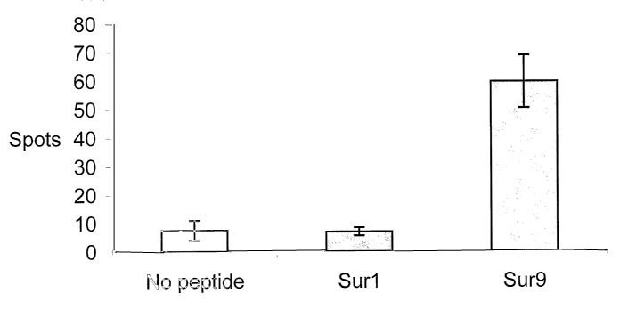
Fig. 1 illustrates T-cell response as measured in an ELISPOT in patient CLL1
to no peptide,
Surf (LTLGEFLKL, SEQ ID NO:10) peptide and Sur9 (ELTLGEFLKL, SEQ ID NO:3)
peptide.
PBLs were stimulated once with peptide before plated at 6x10⁵ cells per well
in duplicate.
The average number of spots per peptide was calculated using a CCD scanning
device and
a computer system,
Fig. 2 illustrates T-cell response as measured in an ELISPOT in patient CLL1
to no peptide,
the peptide analogue Sur1L2 (LLLGEFLKL, SEQ ID NO:4), and the peptide analogue
Sur1M2 (LMLGEFLKL, SEQ ID NO:5). PBLs were stimulated once with peptide before
plated
at 104 cells per well in duplicate. The average number of spots per peptide
was calculated
using a CCD scanning device and a computer system,
Fig. 3 shows responses as measured in an ELISPOT in patient CLL2 and CLL3 to
no peptide
(black bar), the Suri (LTLGEFLKL, SEQ ID NO:10) peptide (grey bar), the Sur9
(ELTLGEFLKL, SEQ ID NO:3) peptide (white bar), the analogue peptide Sur1L2
(LLLGEFLKL, SEQ ID NO:4) (light grey bar), and the analogue peptide Sur1M2
(LMLGEFLKL, SEQ ID NO:5) (dark grey bar). Each experiment was performed with
105 cells
per well in duplicate, and the average number of spots was calculated,
Fig. 4 represents T cells that were isolated from tumour infiltrated lymph
nodes from pa-
tient Melt, Me12, and Me13, stimulated once in vitro and analyzed in an
ELISPOT assay for
response to no peptide (black bar) the peptides Suri (LTLGEFLKL, SEQ ID NO:10)
(grey
bar) and Sur9 (ELTLGEFLKL, SEQ ID NO:3) (white bar). Each experiment was
performed in
duplicate with 105 cells per well. In each experiment two wells without
addition of peptide
was also included. The average number of spots per peptide was calculated for
each
patient,
Fig. 5 shows functional activity of survivin specific CTLs. CTLs were isolated
from a mela-
noma infiltrated lymph node using survivin coated magnetic beads. (A) Specific
lysis of
melanoma cell lines; the HLA-A2 positive FM3 (triangle) and the HLA-A2
negative FM45
(square). (B) Specific lysis of breast cancer cell lines; the HLA-A2 positive
MCF-7 (triangle)
and the HLA-A2 negative BT-20 (square),
Fig. 6 shows frequency of survivin reactive CTLs in PBL from breast cancer
patients. Reac-
tivity was examined in three breast cancer patients (top, middle, and bottom,
respectively)
by the ELISPOT. For each patient the assays were performed in the absence of
peptide, in
the presence of surf peptide, in the presence of sur9, and in the presence of
the modified
sur1M2 peptide. 1 x 10⁴ effector cells per well were used. The graph depicts
the
quantification of reactive cells; grey columns represent the average number of
IFN-y
producing cells,
Fig. 7 illustrates HLA-35 binding of survivin-derived peptides and analysis of
the peptide-
mediated recovery of HLA-B35 molecules by survivin-derived peptides. Lysates
of meta-
bolically labeled T2-B35 cells were incubated at 4 C in the presence of 50, 5,
0.5, 0.05 and
CA 02513104 2005-07-11
WO 2004/067023 PCT/DK2004/000062
19
0.005 mM of peptide. The recovery of HLA-B35 was analyzed in an assembly assay
and
quantified subsequent to IEF-gel electrophoresis, using ImageGauge
phosphorimager soft-
ware (FUJI photo film Co., LTD., Japan). The C50 value is the concentration of
the peptide
required for half-maximal binding to HLA-B35,
Fig. 8 shows spontaneous T-cell responses observed in PBLs from cancer
patients. A) The
number of IFNy spot forming cells measured in ELISPOT assay without peptide
(white
bars), with sur51-59 (black bars) or sur46-54 (gray bars), among in vitro
stimulated PBLs
from patient CLL5 (105 cells/well), HEM12 (105 cells/well), and HEM8 (5x10⁴
cells/well). B)
The number of spot forming cells among 1.7x10⁵ PBLs from HEM12, cultured for
10 days
with peptide-pulsed matured autologous dendritic cells. The columns represent
the
average of two measurements,
Fig. 9 demonstrates spontaneous T-cell responses against native and modified
survivin
peptides in melanoma patients. A) The number of spot forming cells measured in
ELISPOT
assay against sur51-59 and sur51Y9 from patient FM25 in PBLs (4xl03cells/well)
and TILs
(7x10⁴ cells/well) as well as TILs from FM45 (104 cells/well). B) The number
of spot form-
ing cells measured in ELISPOT assay against sur46 and sur46Y9 measured in TILs
from
FM74 (5x10³ cells/well). The columns represent the average of two measurements
with the
non-specific IFNy release subtracted,
Fig. 10 illustrates binding affinity of survivin-derived peptides to HLA-A1.
Class I MHC
heavy chain bands were quantified on a Phosphorimager. The mount of stabilized
HLA-A1
heavy chain is directly related to the binding affinity of the added peptide.
The peptide-
mediated recovery of HLA-A1 (arbitrary units) induced by 40, 4, 0.4, 0.04 M
of Sur93-
101 (line), Sur93T2 (square), Sur49-58 (circle) or Influenza A, 13131 591-599
(triangle),
Fig. 11 shows spontaneous responses against HLA-A1 restricted peptides.
Spontaneous T-
cell responses against survivin-derived peptides as measured by ELISPOT assay.
The
average number of peptide specific IFNy spots formed in response to Sur92-101,
Sur38Y9,
Sur47Y10, and Sur93T2 among 5x10⁴ in vitro stimulated PBL or TIL from melanoma
patients. The peptide specific responses showed were observed among analyses
of 6 PBL
samples and 3 TIL samples from melanoma (Mel) patients. Non-specific IFNy
spots are
subtracted. Bars: range of duplicates,
Fig. 12 shows spontaneous responses against HLA-All restricted peptides.
Spontaneous T-
cell responses against survivin-derived peptides as measured by the ELISPOT
assay. The
average number of peptide specific IFNy spots formed in response to Sur53-62
among
5x10⁴ in vitro stimulated PBL or TIL from cancer patients. The peptide
specific responses
showed were observed among analyses of 5 melanoma (Mel) patients (5 PBL, 1
TIL) and 2
CLL (CLL) patients (PBL). Non-specific IFNy spots are subtracted. Bars: range
of
duplicates,
Fig. 13 illustrates spontaneous responses against HLA-A3 restricted peptides.
Spontaneous
CA 02513104 2005-07-11
WO 2004/067023 PCT/DK2004/000062
T-cell responses against survivin-derived peptides as measured by the ELISPOT
assay. The
average number of peptide specific IFNy spots formed in response to Sur18K10
among
5x10⁴ in vitro stimulated PBL or TIL from melanoma patients. The peptide
specific
responses showed were observed among analyses of 23 PBL samples and 4 TIL
samples
5 from melanoma (Mel) patients. Non-specific IFNy spots are subtracted. Bars:
range of
duplicates,
Fig. 14 illustrates spontaneous responses against HLA-A2 restricted peptides.
Spontaneous
T cell responses against survivin-derived peptides as measured by the ELISPOT
assay. The
10 average number of peptide specific IFNy spots formed in response to the
11mer peptide,
Sur18-28 among 5x10⁴ in vitro stimulated PBL from cancer patients. The peptide
specific
responses showed were observed among analyses of 10 PBL samples from 2
melanoma
(Mel), 6 CLL (CLL), and 2 mamma carcinoma (MC) patients. Non-specific IFNy
spots are
subtracted. Bars: range of duplicates,
Fig. 15 illustrates spontaneous T cell responses against survivin-derived
peptides as
measured by the ELISPOT assay. The average number of peptide specific IFNy
spots
formed in response to sur6-14 (LPPAWQPFL) among 105 in vitro stimulated PBL
from five
melanoma patients (me125, me126, mel3, mel6, me139), two CLL patients (CLL1,
CLL54)
and 2 breast cancer patients (breastll, breast 15). Non-specific IFNy spots
are subtracted,
Fig. 16 illustrates the laboratory values of stable detection of LDH,
cholinesterase,
creatinine, hemoglobin, leucocytes and thrombocytes following vaccination
therapy of four
patients (ARW, = KN, - WWE, ^ GB), and
Fig. 17 demonstrates kinetic analysis of immunity to survivin peptides
assessed by IFNy
ELISPOT. PBMCs were obtained before the first DC vaccination and three months
thereafter. The numbers of IFNy spot-forming cells above background are
depicted.
In the following table, amino acid sequences for peptides used herein and
their respective
SEQ ID NOs are listed:
SEQ ID DESIGNATION SEQUENCE
NO:
1 Sur6 FLKLDRERA
2 Sur8 TLPPAWQPFL
3 Sur9 ELTLGEFLKL
4 Surf L2 LLLGEFLKL
5 SurlM2 LMLGEFLKL
6 Sur 46-54 CPTENEPDL
7 Sur51-59 EPDLAQCFF
8 Sur46Y9 CPTENEPDY
CA 02513104 2005-07-11
WO 2004/067023 PCT/DK2004/000062
21
9 sur5lY9 EPDLAQCFY
Surf LTLGEFLKL
11 C1 ILKEPVHGV
12 Sur2 RAIEQLAAM
13 Sur3 KVRRAIEQL
14 Sur4 STFKNWPFL
Sur5 SVKKQFEEL
16 Sur? TAKKVRRAI
17 Sur10 ETAKKVRRAI
18 Sur 6-14 LPPAWQPFL
19 Sur 11-19 QPFLKDHRI
Sur 34-43 TPERMAEAGF
21 C24 YPLHEQHQM
22 Surl4-22 LKDHRISTF
23 Sur38-46 MAEAGFIHC
24 Sur93-101 FEELTLGEF
Sur47-56 PTENEPDLAQ
26 Sur49-58 ENEPDLAQCF
27 Sur92-101 QFEELTLGEF
28 Cl VSDGGPNLY
29 surl4Y9 LKDHRISTY
sur93Y9 FEELTLGEY
31 sur92Y9 QFEELTLGEY
32 sur34Y9 TPERMAEAGY
33 sur49Y9 ENEPDLAQCY
34 Sur92T2 QTEELTLGEF
Sur92S2 QSEELTLGEF
36 Sur93T2 FTELTLGEF
37 Sur93S2 FSELTLGEF
38 Sur38Y9 MAEAGFIHY
39 Sur47Y10 PTENEPDLAY
Sur 5-13 TLPPAWQPF
41 Sur 53-61 DLAQCFFCF
42 Sur 54-62 LAQCFFCFK
43 Sur 95-103 ELTLGEFLK
44 Sur 112-120 KIAKETNNK
Sur 13-22 FLKDHRISTF
47 Sur 53-62 DLAQCFFCFK
Sur 103-112 KLDRERAKNK
51 Sur 112-121 KIAKETNNKK
52 Sur 113-122 IAKETNNKKK
53 C3 ILRGSVAHK
54 Sur5K9 TLPPAWQPK
Sur53K9 DLAQCFFCK
56 Sur54L2 LLQCFFCFK
CA 02513104 2005-07-11
WO 2004/067023 PCT/DK2004/000062
22
57 Surl3K9 FLKDHRISTK
58 Surl8K10 RISTFKNWPK
59 Sur113L2 ILKETNNKKK
60 SurEx3-A3-1 TIRRKNLRK
61 SurEx3-A3-2 PTIRRKNLRK
62 Sur2b-A3-1 RITREEHKK
63 C4 AVFDRKSDAK
64 C6 QPRAPIRPI
65 C7 RPPIFIRRL
66 Sur4-14 PTLPPAWQPFL
67 Sur18-28 RISTFKNWPFL
68 Sur54-64 LAQCFFCFKEL
69 Sur86-96 FLSVKKQFEEL
70 Sur88-98 SVKKQFEELTL
71 Sur103-113 KLDRERAKNKI
72 Ebv, BMLF1 GLCTLVAML
73 Hiv, P01 ILKEPVHGV
74 Influenza A, ILRGSVAHK
nucleoprotein265-
273
EXAMPLE 1
Identification of a cytotoxic T-lymphocyte response to the apoptosis inhibitor
protein survivin in cancer patients
Summary
Using CTL epitopes derived from survivin, specific T-cell reactivity against
such antigens in
peripheral blood from chronic lymphatic leukemia (CLL) patients and in tumor-
infiltrated
lymph nodes from melanoma patients by ELISPOT analysis have been studied. CTL
re-
sponses to survivin-derived peptide epitopes were detected in three out of six
melanoma
patients and in three out of four CLL patients. No T-cell reactivity was
detected in PBL from
six healthy controls. Thus, survivin-derived peptides may serve as important
and widely
applicable targets for anti-cancer immunotherapeutic strategies.
Introduction
The survivin protein was scanned for the presence of HLA-A*0201 (HLA-A2)
binding pep-
tide motifs and after successful identification, the peptides were used to
test for specific T-
cell reactivity in leukemia and melanoma patients by ELISPOT assay. In both
patient co-
horts CTL responses to two survivin-derived peptide epitopes were detected,
whereas no
CA 02513104 2005-07-11
WO 2004/067023 PCT/DK2004/000062
23
T-cell reactivity could be detected in the healthy controls. These data
suggest that survivin
represent a widely expressed tumor antigen recognized by autologous T cells.
Materials and Methods
Patients and normal controls
Peripheral vein blood samples from 4 patients diagnosed with CLL (designated
CLL1-4) and
blood samples from 6 normal individuals were collected into heparinised tubes.
PBLs were
isolated using Lymphoprep separation and frozen in fetal calf serum (FCS) with
10% di-
methylsulphoxide. Additionally, T lymphocytes from tumor-infiltrated lymph
nodes were
obtained from 6 melanoma patients (designated mell-6). Freshly resected lymph
nodes
were minced into small fragments, crushed to release cells into culture and
cryopreserved.
PBLs were available from 4 of the melanoma patients. All individuals included
were HLA-A2
positive as determined by FACS analysis using the HLA-A2 specific antibody
BB7.2. The
antibody was purified from hybridoma supernatant. Patient samples were
obtained from
the State University Hospital, Herlev, Denmark. Informed consent was obtained
from the
patients prior to any of theses measures.
Survivin-derived peptides
All peptides were obtained from Research Genetics (Huntsville, AL, USA) and
provided at
>90% purity as verified by HPLC and MS analysis. The peptides used are listed
in Table 1.
Table 1. Peptides examined in this study and their binding affinity to HLA-A2
Name Proteina Sequence SEQ ID NO: C50 (l M)b
C1 HIV-1 P01476-484 ILKEPVHGV 11 0.7
Sur? Survivin96_104 LTLGEFLKL 10 >100
Sur2 Survvivin133-141 RAIEQLAAM 12 Not binding
Sur3 Survivin130-138 KVRRAIEQL 13 >100
Sur4 Survivin20_28 STFKNWPFL 14 Not binding
SurS Survivin88_96 SVKKQFEEL 15 Not binding
Sur6 Survivin101.109 FLKLDRERA 1 30
Sur7 Survivin122-135 TAKKVRRAI 16 Not binding
Sur8 Survivin5_14 TLPPAWQPFL 2 30
Sur9 Survivin95-104 ELTLGEFLKL 3 10
Sur10 Survivin126-135 ETAKKVRRAI 17 Not binding
SurlL2 LLLGEFLKL 4 1
SurlM2 LMLGEFLKL 5 1
aThe value range listed in subscript indicates the position of the peptide in
the survivin se-
quence as disclosed in US 6.245.523
CA 02513104 2005-07-11
WO 2004/067023 PCT/DK2004/000062
24
bThe C50 value is the concentration of the peptide required for half maximal
binding to
HLA-A2 determined as described below
Assembly assay for peptide binding to class I MHC molecules
Assembly assays for binding of the synthetic peptides to class I MHC molecules
metaboli-
cally labeled with [35S]-methionine were carried out as described (12,13). The
assembly
assay is based on stabilization of the class I molecules after loading of
peptide to the pep-
tide transporter deficient cell line T2. Subsequently, correctly folded stable
MHC heavy
chains are immunoprecipitated using conformation-dependent antibodies. After
IEF elec-
trophoresis, gels were exposed to phosphorimager screens, and peptide binding
was
quantified using the Imagequant PhosphorImager program (Molecular Dynamics,
Sunnyvale, CA).
Antigen stimulation of PBLs
To extend the sensitivity of the ELISPOT assay, PBLs were stimulated once in
vitro prior to
analysis (14,15). Fresh and previously frozen PBL5 gave similar results in the
ELISPOT as-
say. On day 0, PBLs or crushed lymph node were thawed and plated in 2 ml/well
at a con-
centration of 2x10⁶ cells in 24-well plates (Nunc, Denmark) in AIM V medium
(Life Tech-
nologies, Roskilde, Denmark), 5% heat-inactivated human serum and 2 mM of L-
glutamine
in the presence of 10 M of peptide. In each experiment a well without peptide
was in-
cluded. Two days later 300 IU/ml recombinant interleukin-2 (IL-2) (Chiron,
Ratingen,
Germany) was added to the cultures. The cultured cells were tested for
reactivity in the
ELISPOT assay on day 12.
ELISPOT assay
The ELISPOT assay used to quantify peptide epitope-specific interferon-y-
releasing effector
cells was performed as in (16). Briefly, nitrocellulose bottomed 96-well
plates (MultiScreen
MAIP N45, Millipore, Hedehusene, Denmark) were coated with anti-IFN-y antibody
(1-D1K,
Mabtech, Nacka, Sweden). The wells were washed, blocked by AIM V medium, and
cells
were added in duplicates at different cell concentrations. Peptides were then
added to each
well and the plates were incubated overnight. On the following day, medium was
discarded
and the wells were washed prior to addition of biotinylated secondary antibody
(7-B6-1-
Biotin, Mabtech). The plates were incubated for 2 hours, washed and Avidin-
enzyme con-
jugate (AP-Avidin, Calbiochem, Life Technologies) was added to each well.
Plates were in-
cubated at RT for 1 hour and the enzyme substrate NBT/BCIP (Gibco, Life
Technologies)
was added to each well and incubated at room temperature for 5-10 min. The
reaction was
terminated by washing with tap water upon the emergence of dark purple spots.
The spots
were counted using the Alphalmager System (Alpha Innotech, San Leandro, CA.
USA) and
the peptide specific CTL frequency could be calculated from the numbers of
spot-forming
cells. The assays were all performed in duplicate for each peptide antigen.
CA 02513104 2005-07-11
WO 2004/067023 PCT/DK2004/000062
Results
Binding of survivin derived peptides to HLA-A2
5 The amino acid sequence of the survivin protein was screened for the most
probable HLA-
A2 nona- and decamer peptide epitopes, using the main HLA-A2 specific anchor
residues
(17). Ten survivin-derived peptides were synthesized and examined for binding
to HLA-A2.
An epitope from HIV-1 po1476-484 (ILKEPVHGV, SEQ ID NO:11) (Table 1) was used
as a
positive control. The peptide concentration required for half maximal
recovering of class I
10 MHC (C50 value) was 0.7 M for the positive control. In comparison, the
peptide designated
Sur9 (ELTLGEFLKL, SEQ ID NO:3) bound at an affinity of C50 = 10 M. The
peptides
designated Sur6 (FLKLDRERA, SEQ ID NO:1) and Sur8 (TLPPAWQPFL, SEQ ID NO:2),
re-
spectively bound to HLA-A2 at C50 = 30 M, whereas Surf (LTLGEFLKL, SEQ ID
NO:10)
and Sur3 (KVRRAIEQL, SEQ ID NO:13) bound weaker (C50 >100 M). Five of the
peptides
15 examined (Sur2, Sur4, Sur5, Sur7, and Sur10) did not bind to HLA-A2.
Since Surf is a weak HLA-A2 binder, two analogue peptides designated Sur1L2
and
Sur1M2, respectively in which a better anchor residue (leucine or methionine)
replaced the
native threonine at position 2 were synthesized. Both of these peptides bind
with almost
20 similar high affinity to HLA-A2 as the positive control (C50 M).
CTL response to survivin in CLL patients
PBLs from four HLA-A2 positive CLL patients were stimulated once in vitro
before exami-
25 nation in the ELISPOT assay. This procedure was chosen to extend the
sensitivity of the
ELISPOT. All of the above 10 survivin-derived peptides were included in the
first line of ex-
periments. Responses were detected to Surf and Sur9 and only data for these
peptides
are given in the figures. Fig. 1 shows CTL reactivity to Suri and Sur9 as
determined in pa-
tient CLL1. Each spot represents a peptide reactive, INF--y-producing cell.
The average
number of spots per peptide was calculated using a CCD scanning device and a
computer
system. Fifty-two Sur9 peptide specific spots (after subtraction of spots
without added
peptide) per 6x10⁵ were detected in the CLL1 patient (Fig. 1). No response was
detected to
the weak HLA-A2 binding peptide Surf, however the patient responded strongly
to the
strong HLA-A2 binding peptide analogue Sur1M2 (35 peptide specific spots per
104 cells)
(Fig. 2). No response was detected to the other strong HLA-A2 binding peptide
analogue
SurlL2 in this patient (Fig. 2). Patient CLL2 responded strongly to Sur9 (128
peptide spe-
cific spots per 105 cells) and weakly to Surf (22 peptide specific spots per
105 cells) (Fig.
3). The response to the Sur1L2 analogue was only slightly increased relative
to the natural
epitope, whereas the patient responded similarly strongly to the Sur1M2
peptide as to the
decamer peptide Sur9. In patient CLL3 a weak response to Sur9 was observed
(Fig. 3). No
response to Surf or the modified Suri peptides were observed in the patient.
No survivin
responses were detected in the last patient CLL4 (data not shown). PBLs from 6
healthy
HLA-A2 positive controls were analyzed to investigate whether a response to
survivin could
CA 02513104 2005-07-11
WO 2004/067023 PCT/DK2004/000062
26
be detected in healthy individuals. No response was observed in any of the
controls to any
of the survivin-derived peptides.
CTL response to survivin in melanoma patients
T lymphocytes isolated from tumour infiltrated lymph nodes from HLA-A2
positive mela-
noma patients were examined. The freshly resected lymph node was minced into
small
fragments and crushed to release cells into culture. Cells were stimulated
once with pep-
tide in vitro before examination in the ELISPOT assay. Survivin specific T
cells were de-
tected in three of the six patients analyzed. A strong Sur9 response was
detected in pa-
tient Me12 and Me13. A weaker response to the Surf peptide was also detected
in these
patients (Fig. 4). In Melt the response to the weakly binding peptide Suri was
stronger
than the response to the stronger HLA-A2 binder Sur9 (Fig. 4). No response was
detected
in the tumor-infiltrated lymph nodes from the last three melanoma patients
(Mel4-6). PBLs
from two of the survivin reacting patients, Mell and Me12, and from two of the
non-reac-
ting patients, Me14 and Mel5, were examined. No response could be detected to
either
Sur9 or Surf in PBLs from any of these patients (data not shown).
EXAMPLE 2
Spontaneous cytotoxic T-cell responses to survivin-derived MHC class I-
restricted T-cell epitopes in situ and ex vivo in cancer patients
Summary
Spontaneous cytotoxic T-cell responses to survivin-derived MHC class I
restricted T-cell
epitopes were demonstrated in situ as well as ex vivo in breast cancer,
leukemia, and
melanoma patients. Moreover, survivin reactive T cells isolated by magnetic
beads coated
with MHC/peptide complexes were cytotoxic to HLA-matched tumours of different
tissue
types. Being a universal tumor antigen, survivin may serve as a widely
applicable target
for anti-cancer immunotherapy.
Materials and Methods
Construction of HLA-peptide complexes for T-cell staining and T-cell sorting
A recognition site for enzymatic biotinylation using biotin protein ligase
(BirA) in fusion
with the 5'-end of the extracellular domains of HLA A*0201 (residues 1-275)
was ex-
pressed in E. coli BL21 (DE3). The recombinant protein was purified by size-
(Sephadex
G25, Pharmacia) and ion exchange (mono-Q, Pharmacia) chromatography from
inclusion
bodies solubilised in 8 M urea. The HLA A*0201 was folded in vitro by dilution
in the pre-
sence of the modified survivin peptide Sur1M2 (LMLGEFLKL, SEQ ID NO:5) or the
MAA
peptide gp100154-163, and subsequently biotinylated as described previously
(35, 36).
CA 02513104 2005-07-11
WO 2004/067023 PCT/DK2004/000062
27
After gel filtration on a Pharmacia Sephadex G25 column to remove unbound
biotin, the
protein was multimerised with streptavidin-FITC conjugated dextran molecules
(kindly
provided by L. Winther, DAKO, Denmark) to generate multivalent HLA-dextran
compounds
for immunohistochemistry. The HLA A*0201 construct was a kind gift of Dr. Mark
M. Davis
(Dept. of Microbiology and Immunology, Stanford University, Palo Alto, CA).
Cell separa-
tion was performed as previously described (37). Briefly, 5 x 10⁶ streptavidin-
conjugated
magnetic beads (Dynal, Oslo, Norway) were washed twice in 200 pl cold PBS,
0.5pg pep-
tide/A*0201 monomers were added and the mixture incubated for 15 min. at room
tem-
perature. After two washes these beads were mixed with PBLs at a ratio of 1:10
and sub-
sequently incubated for 1 h followed by a precipitation of bead-bound cells in
a magnetic
field. The precipitation step was repeated once.
Immunohistochemistry stainings
For staining with FITC-conjugated multimeric peptide/MHC complexes, tissue
sections were
dried overnight and subsequently fixed in cold acetone for 5 min. All
incubation steps were
performed at room temperature and in the dark: (i) 45 min. of the primary
antibody
(1:100 diluted), (ii) Cy 3-conjugated goat anti-mouse (1:500 diluted; code 115-
165-100,
Jackson ImmunoResearch, obtained from Dianova, Hamburg, Germany) for 45 min.
and
finally (iii) the multimers for 75 min. Between each step the slides were
washed two times
for 10 min. in PBS/BSA 0.1%. The slides were mounted in vectashield and kept
in the re-
frigerator until observed under the confocal microscope.
Cytotoxicity assay
Conventional [51Cr]-release assays for CTL-mediated cytotoxicity were carried
out as de-
scribed in (13). Target cells were autologous EBV-transformed B-cell lines,
the HLA-A2
positive breast cancer cell line MCF-7 (available at ATCC), the HLA-A2
positive melanoma
cell line FM3 (38), the HLA-A2 negative breast cancer cell line BT-20
(available from ATCC)
and the HLA-A2 negative melanoma cell line FM45 (38). All cancer cell lines
expressed sur-
vivin as examined by RT-PCR (data not shown).
ELISPOT assay
The ELISPOT assay was used to quantify peptide epitope-specific IFN-y
releasing effector
cells and has been described previously (39). Briefly, nitrocellulose bottomed
96-well
plates (MultiScreen MAIP N45, Millipore) were coated with an anti-IFN-y
antibody (1-D1K,
Mabtech, Sweden) and non-specific binding was blocked using AIM V (GibcoBRL,
Life
Technologies Inc., Gaithersburg, MD, USA). Lymphocytes were added at different
cell con-
centrations together with the specific peptides and T2 cells and incubated
overnight at
37 C. Following two washes the biotinylated detection antibody (7-B6-1-Biotin,
Mabtech)
was added. Specific binding was visualised using alkaline phosphatase-avidin
together with
the respective substrate (GibcoBRL). The reaction was terminated upon the
appearance of
dark purple spots, which were quantitated using the Alphalmager System (Alpha
Innotech,
CA 02513104 2005-07-11
WO 2004/067023 PCT/DK2004/000062
28
San Leandro, CA, USA). The peptides used for the ELISPOT were Surl, Sur9 and
the Surl
analogue peptide Sur1M2 as described in Example 1.
Results
In situ staining of HLA-A2/survivin reactive T cells
In Example 1 two survivin-derived peptide epitopes recognized by T cells in
leukemia and
melanoma, i.e., Surl were identified. The weak binding affinity of Surl to HLA-
A2 was im-
proved substantially by replacing threonine at position 2 with a better anchor
residue
(methionine; SurlM2). This measure enabled the construction of stable HLA-
A2/peptide
complexes. These complexes were multimerised using dextran molecules, which
were
conjugated with streptavidin and FITC. Multimerised MHC-complexes were used to
stain
acetone-fixed, frozen material. Using a confocal laser microscope, SurlM2/HLA-
A*0201
reactive CTL5 could readily be detected in situ in the tumor microenvironment.
We de-
picted such cells in the primary tumor and the sentinel lymph node of a stage
III mela-
noma patient as well as in a primary breast cancer lesion. To ensure the
specificity of the
staining, a series of negative controls was carried out. Neither the use of
peptide/HLA-
dextran multimers with peptides derived from the melanoma differentiation
antigen gp100
on the same tumour, nor SurlM2/HLA-dextran multimers in case of a tumour
sample
obtained from an HLA-A2 negative donor resulted in a positive staining.
Isolated survivin reactive CTLs lyse tumour cell lines of different origin
To characterise the functional capacity of survivin-reactive CTLs, these cells
were isolated
by means of magnetic beads coated with HLA-A2/SurlM2-complexes (36). A freshly
re-
sected melanoma infiltrated lymph node was minced into small fragments and
crushed to
release cells into culture. Cells were stimulated once with peptide in vitro
prior to isolation.
One day after isolation IL-2 was added, and on day 5 the capacity of these
cells to kill tu-
mour cells was tested either by ELISPOT or in standard 51Cr release assays.
First, by
means of ELISPOT analysis it was possible to establish that CTLs isolated
using the modi-
fied SurlM2/HLA-A2-complex also responded to the native Surf peptide (data not
shown).
Second, the cytotoxicity of the survivin reactive CTLs against the HLA-A2
positive mela-
noma cell-line FM3 (Fig. 5 A) and the HLA-A2 positive breast-cancer cell line
MCF-7 (Fig. 5
B) was tested. The isolated T cells effectively lysed both HLA-A*0201 cell
lines. In con-
trast, no cytotoxicity was observed against the HLA-A2 negative melanoma cell
line FM45
(Fig. 5 A) or the HLA-A2 negative breast cancer cell line BT-20 (Fig. 5 B).
Survivin reactivity measured in PBL by ELISPOT
The presence of survivin reactive T cells in PBLs from ten HLA-A2 positive
breast cancer
patients was examined by the ELISPOT. Before analysis, PBLs were stimulated
once in vi-
tro to extend the sensitivity of the assay. Reactivity to the following
survivin peptides was
examined: Surf, Sur9 and Sur1M2. Survivin specific T cells were detected in
six out of the
CA 02513104 2005-07-11
WO 2004/067023 PCT/DK2004/000062
29
ten HLA-A2 positive breast cancer patients. Representative examples are given
in Fig. 6. In
PBL5 from two patients a response against Suri and the modified analogue
Sur1M2, but
not against Sur9 (Fig. 6, top, middle) was detected, in three patients a
response to Sur9
was detected, but not to Surl or Sur1M2 (Fig. 6 bottom), and one patient
responded only
to SurlM2. In contrast, no survivin responses were detected in PBLs from 20
healthy HLA-
A2 positive donors. Similarly, PBLs from fourteen HLA-A2 positive melanoma
patients were
examined. Survivin responses were present in seven of these patients (Table
2). Two
patients responded to the Sur9 peptide, three to the Sur1M2 peptide, one to
both Suri
and SurM2, and one to all three peptides. In Example 1, T-cell response to
survivin in 3
chronic lymphatic leukemia (CLL) patients was tested (Table 2; CLL1, CLL2,
CLL3). These
studies were extended using PBLs from three additional CLL patients. Notably,
all patients
produced a T-cell response to at least one survivin epitope (Table 2; CLL5,
CLL6, CLL7). In
addition, PBLs from one patient suffering from chronic myeloid leukemia (CML)
was
examined. In this patient, a response to all three peptides was identified
(data not shown).
The data are summarized in Table 2.
Table 2. Patients with survivin peptide-specific T lymphocytes in PBLs as
measured by
ELISPOT
Melanoma a)
Patient Suri Sur9 Sur1M2
P4 - - 97
P11 - - 112
P13 - - 71
P15 61 - 101
P17 - 172 -
P39 - 127 -
P64 112 70 128
Breast cancer b)
Patient Surf Sur9 SurlM2
B1 122 - 208
B2 67 - 72
B3 - 54 -
B4 - 45 -
B5 - 19 -
B6 - - 24
CLL c)
Patient Surf Sur9 Sur1M2
CLL1 - 27 320
CLL2 - 39 -
CLL3 23 127 122
CLLS - 100 124
CLL6 - 121 360
CLL7 68 132 174
CA 02513104 2005-07-11
WO 2004/067023 PCT/DK2004/000062
a) Frequency of reactive cells per 104; 14 patients examined.
b) Frequency of reactive cells per 104; 10 patients examined.
c) Frequency of reactive cells per 105; 7 patients examined.
5
EXAMPLE 3
HLA-B35-restricted immune responses to survivin-derived peptides in cancer pa-
10 tients
Summary
In this study, two survivin-derived epitopes, which are restricted to HLA-B35
were identi-
15 fied and characterized. Specific T-cell reactivity against both of these
epitopes was present
in the peripheral blood from patients with different haematopoietic
malignancies and mela-
noma. Substitutions of the C-terminal anchor residue improved the recognition
by tumor
infiltrating lymphocytes from melanoma patients. Furthermore, spontaneous
cytotoxic T-
cell responses to survivin in situ in a primary melanoma lesion was
demonstrated. These
20 epitopes extends the applicability of future vaccine strategies based on
survivin peptides in
relation to malignancies as well as the HLA profile of the patients involved.
In Examples 1 and 2, HLA-A2 restricted survivin-derived T-cell epitopes were
studied.
Since HLA-A2 is only expressed in about 30% of the Caucasian population (63),
peptide
25 epitopes restricted to other HLA class I molecules need to be identified to
extend the frac-
tion of patients that could be treated. In this study, two novel T-cell
epitopes from survivin
restricted to HLA-B35, which is expressed in 9% of the Caucasian population
(63), were
identified, and spontaneous immune responses to these survivin peptides were
detected in
patients with different haematopoietic malignancies and melanoma.
Materials and Methods
Patients
Peripheral vein blood samples from cancer patients were collected, PBLs were
isolated
using Lymphoprep separation, HLA-typed (Department of Clinical Immunology,
University
Hospital, Copenhagen) and frozen in FCS with 10% DMSO. Ten HLA-B35 positive
patients
were selected for further analysis. These patients suffered from melanoma,
CLL, follicular
lymphoma (FL), diffuse large B-cell lymphomas (DLBCL) and Multiple Myeloma
(MM), re-
spectively. At the time blood samples were collected patients had not been
medically
treated within the previous four months. Additionally, tumor-infiltrating
lymphocytes (TIL)
isolated from lymph nodes were collected from three of the melanoma patients
and frozen
in FCS with 10% DMSO.
CA 02513104 2005-07-11
WO 2004/067023 PCT/DK2004/000062
31
Peptides
Seven synthetic survivin-derived peptides were used in this study: Sur6-14,
Sur11-19,
Sur34-43, Sur46-54, Sur51-59, Sur46Y9, Sur51Y9, and one EBV-derived peptide,
EBNA3A
457-466 (63). All peptides were obtained from Research Genetics (Huntsville,
AL) and pro-
vided at >90% purity, as verified by HPLC and MC analyses. The peptides are
listed in Ta-
ble 3 below.
Table 3. HLA-B35 binding of survivin-derived peptides
Name Protein and position Sequence SEQ ID NO: C50 ( M)
Sur6-14 Survivin6-14 LPPAWQPFL 18 >100
Sur11-19 Survivinll-19 QPFLKDHRI 19 Not binding
Sur34-43 Survivin34-43 TPERMAEAGF 20 >100
Sur46-54 Survivin46-54 CPTENEPDL 6 20
Sur51-59 Survivin51_59 EPDLAQCFF 7 13
Sur46Y9 Modified peptide CPTENEPDY 8 4
Sur51Y9 Modified peptide EPDLAQCFY 9 1.5
C24 EBNA3A458-466 YPLHEQHQM 21 0.8
Assembly assay for peptide binding to MHC class I molecules
The assembly assay described in Examples 1 and 2 was used to measure binding
affinity of
the synthetic peptides to HLA-B35 molecules metabolically labeled with
[S35]methionine.
Briefly, the assay is based on peptide-mediated stabilization of empty HLA
molecules re-
leased, upon cell lysis, from the TAP deficient cell line T2, stably
transfected with HLA-B35
(kindly provided by Dr J. Haurum, Symphogen ApS, Lyngby, Denmark). Stably
folded HLA-
molecules were immunoprecipitated using the conformation-dependent mAb W6/32.
The
HLA molecules were separated by IEF electrophoresis, gels were exposed to phos-
phorimager screens (Imaging plate, FUJI photo film Co., LTD., Japan), analyzed
and the
amount of correctly folded HLA molecules were quantified using ImageGauge phos-
phorimager software (FUJI photo film Co., LTD., Japan).
Antigen stimulation of PBLs
To extend the sensitivity of the ELISPOT assay, lymphocytes were stimulated
once in vitro
with peptide prior to analysis (14, 15). PBLs or TILs were thawed and
stimulated with
50 M of the individual peptide epitopes in 96-well plates for 2 Ii at 26 C
(5x10⁵-106 cells
per peptide), and pooled for further 10 days of culture at 37 C in x-vivo with
5% human
serum (HS), in 24 well plates (Nunc, Roskilde, Denmark), with 2x10⁶ cells per
well. At the
second day of incubation 40 g/ml IL-2 (Apodan A/S, Denmark) were added. At
day 10,
the cultured cells were tested for reactivity in the ELISPOT assay.
The ELISPOT assay
CA 02513104 2005-07-11
WO 2004/067023 PCT/DK2004/000062
32
The ELISPOT assay used to quantify peptide specific, IFN-y releasing effector
cells in PBLs
or TILs collected from cancer patients was performed as described in Example
1. Briefly,
nitrocellulose-bottomed 96-well plates (MultiScreen MAIP N45; Millipore,
Hedehusene,
Denmark) were coated with mAb against human IFN-y, 7.5 g/ml (1-D1K; Mabtech,
Nacka,
Sweden). Wells were washed and blocked in x-vivo (x-vivo 15TM BioWhittacker,
Molecular
Applications Aps, Denmark) and cells were added in duplicates at different
concentrations.
For antigen presentation, 104 T2-B35 cells, with and without 10 pM peptide,
were added
per well. Plates were incubated overnight, the cells discarded, and wells
washed prior to
addition of biotinylated secondary antibody (7-B6-1-Biotin; Mabtech). Plates
were incu-
bated 2 h at room temperature, washed and avidin-alkaline phosphatase
conjugate was
added (AP-Avidin; Calbiochem, Life Technologies, Inc.). After 1 h of
incubation at room
temperature, the enzyme substrate nitroblue tetrazolium/5-bromo-4-chloro-3-
indolyl
phosphate (Code No.K0598, DakoCytomation Norden A/S) was added, and dark
purple
spots emerged in 3-7 min. The reaction was terminated by washing with tap
water. Spots
were counted using the Alpha Imager System (Alpha Innotech, San Leandro, CA),
and the
frequency of peptide specific T cells were calculated from the number of spot
forming cells.
All assays were performed in duplicates for each peptide antigen, and
lymphocytes cul-
tured in the same well, were tested in equal cell numbers with and without
peptide, to
measure the number of peptide specific cells in the culture.
Maturation of dendritic cells (DCs)
Adherent cells were isolated from PBLs after 2 h of culture. These were
cultured for 10 ad-
ditional days in RPMI 1640 (GibcoTM Invitrogen corporation, UK) with 10% FCS.
800 ng/ml
GM-CSF (PreproTech, London, UK) and 40 ng/ml IL-4 (PreproTech) were added
every third
day. At day 10, DCs were matured for 24 h by adding 50 ng/ml TNF-a
(PreproTech). After
maturation, DCs were released and pulsed with 20 M peptide in the presence of
3 g/ml
(32-microglobulin for 2 h at 26 C.
Isolation of peptide specific T cells
Antigen specific cells were isolated using sur5lY9/HLA-B35-coated magnetic
beads as de-
scribed in Example 2. Biotinylated monomers of HLA-B35 with sur51Y9 (obtained
from
Prolmmune, Oxford, UK) were coupled to streptavidin coated magnetic beads
(Dynabeads
M-280, Dynal A/S, Oslo, Norway) by incubating 2.5 fag monomers with 5x10⁶
beads in 40
l PBS for 20 min. at room temperature. The magnetic complexes were washed
three
times in PBS, using a magnetic device (Dynal A/S, Oslo, Norway) and
subsequently mixed
with PBLs at a ratio of 1:10 in PBS with 5% BSA, and rotated very gently for 1
h. Antigen
specific CD8+ T cells associating with the magnetic complexes were gently
washed two or
three times. Isolated cells were resuspended several times in x-vivo
supplemented with
5% human serum and incubated for 2 h before the magnetic beads were released
and re-
CA 02513104 2005-07-11
WO 2004/067023 PCT/DK2004/000062
33
moved from the cell suspension. The isolated antigen specific CD8+ T cells
were used in
ELISPOT assay to analyze the cross-reactivity between the native and modified
peptide.
TCR clonotype mapping by denaturing gradient gel electrophoresis (DGGE)
DGGE clonotype mapping of the human TCR BV regions 1-24 has been described in
details
(66). Briefly, RNA was isolated using the Purescript Isolation Kit (Gentra
Systems Inc. MN)
and transcribed cDNA was amplified by PCR using primers for the variable
regions of the
TCR beta chains in conjunction with a common constant region primer. The
computer pro-
gram MELT87 was used to ensure that the amplified DNA molecules were suited
for DGGE
analysis provided a 50 bp GC-rich sequence (GC-clamp) was attached to the 5'-
end of the
constant region primer. DGGE analysis was done in 6% polyacrylamide gels
containing a
gradient of urea and formamide from 20% to 80%. Electrophoresis was performed
at 160
V for 4.5 hours in 1x TAE buffer at a constant temperature of 54 C.
Immunohistochemistry stainings
Multimerised peptide/HLA complexes were used to identify antigen specific T
cells in situ in
tumor lesions of cancer patients using the procedure described in Example 2.
Biotinylated
sur5lY9/HLA-B35 monomer was supplied by Proimmune limited, Oxford, UK. The bi-
otinylated monomers of sur51Y9/HLA-B35 were multimerised with streptavidin-
FITC-con-
jugated dextran molecules (kindly provided by L. Winther, DAKO, Glostrup,
Denmark) to
generate multivalent HLA-dextran compounds for immunohistochemistry. Tissue
sections
were dried overnight and subsequently fixed in cold acetone for 5 min. All the
incubation
steps were performed in the dark at room temperature: (a) 45 min of the
primary anti-
body (1:100 diluted) (b) Cy 3-conjugated goat-anti-mouse antibody (1:500
diluted; code
115-165-100; Jackson ImmunoResearch, obtained from Dianova, Hamburg, Germany)
for
45 min; and finally (c) the multimers for 75 min. Between each step, the
slides were
washed two times for 10 min in PBS/BSA 0.1%. The slides were mounted in
vectashield
and kept in the refrigerator until observed under the confocal microscope
(Leica).
Results
Identification of HLA-B35 binding survivin-derived peptides
The amino acid sequence of survivin was screened for nonameric and decameric
peptides
with anchor residues, according to the peptide-binding motif of HLA-B35 (67).
Five pep-
tides were selected containing proline as the N-terminal anchor in position 2
and phenyl-
alanine, leucine, isoleucine or tyrosine as C-terminal anchor residues (Table
3). Assembly
assay revealed two peptides, sur51-59 (EPDLAQCFF, SEQ ID NO:7) and sur46-54
(CPTENEPDL, SEQ ID NO:6) that were able to stabilise HLA-B35 efficiently.
Additionally,
two peptides, sur34-43 (TPERMAEAGF, SEQ ID NO:20) and sur6-14 (LPPAWQPFL, SEQ
ID
NO:18) showed a weak stabilization, whereas the remaining peptide did not
stabilize HLA-
B35 at all. The peptide concentration required for half maximal recovery of
HLA-B35 (C50)
CA 02513104 2005-07-11
WO 2004/067023 PCT/DK2004/000062
34
was estimated at 13 M for sur51-59 and 20 M for sur46-54. In comparison, the
positive
control-epitope C24 from EBNA3A458-466 (YPLHEQHQM, SEQ ID NO:21) had an
estimated
C50 value of 0,81AM.
To enhance the binding affinity of sur46-54 and sur51-59 the C-terminal amino
acid was
replaced with tyrosine, a better anchor residue (67). The recovery of HLA-B35
mediated by
the modified peptides was analyzed in the assembly assay, and C50 values were
estimated
at 1.51AM for sur51Y9 and 41AM for sur46Y9 (Fig. 7).
Spontaneous immune responses against native peptide epitopes
Initially, five patients were analyzed for spontaneous immune responses to the
four native
HLA-B35 binding peptides sur51-59, sur46-54, sur34-43 and sur6-14. These five
patients
had different haematopoietic malignancies: HEM8 and HEM18 suffered from MM,
HEM12
from FL, HEMS had DLBCL, and CLL5 had CLL.
INF-y ELISPOT assays were performed on PBLs after 10 days of in vitro
stimulation to de-
tect peptide precursor CTLs. Spontaneous immune responses were detected
against two of
the native HLA-B35 binding peptides, sur51-59 and sur46-54. Two patients,
HEM12 and
CLLS showed a response to both sur51-59 and sur46-54, whereas HEM8 only showed
a
response to sur51-59 (figure 8A and B). No response could be detected in the
two
remaining patients, HEMS and HEM18, and no response could be detected to the
poorly
binding peptides sur34-46 and sur6-14 in any patients.
An alternative approach to in vitro stimulation was used in patient HEM12,
i.e. PBLs were
co-cultured with matured autologous dendritic cells pulsed with sur51-59 to
stimulate a
CTL response in vitro. PBLs from this culture showed strong reactivity towards
sur51-59 in
ELISPOT (figure 8B).
Increased recognition of modified peptides
As described above, peptide modifications to enhance the HLA-B35 affinity
resulted in a 5-
10-fold higher affinity for HLA-B35 relative to the native peptides. A group
of five mela-
noma patients were analyzed for spontaneous immune responses to both the
native and
modified peptides by means of ELISPOT assay. PBL samples were analyzed after
in vitro
stimulation, whereas TIL samples were analyzed directly. Spontaneous immune
responses
were observed in either PBLs or TILs from three of the five patients. FM25
showed reacti-
vity against sur51-59 and sur51Y9 in both PBL and TIL samples (Fig. 9A). FM45
responded
only to the modified peptide sur51Y9, with a strong response detectable in
TILs. No PBL5
were available from this patient (figure 9A). FM74 showed a strong response to
sur46Y9 in
TIL, but no response to the native peptide was detectable (figure 9B). A weak
response to
sur46Y9 was also observed in PBLs from FM74 (data not shown).
Cross-reactivity between the native and modified peptide
CA 02513104 2005-07-11
WO 2004/067023 PCT/DK2004/000062
The high affinity of sur51Y9 to HLA-B35 enables the production of stable
monomers of
HLA-B35 with sur51Y9. Having established the presence of survivin reactive T
lymphocytes
in tumor infiltrated lymph nodes and PBLs from different cancer patients,
magnetic beads
5 were coated with such HLA-B35/Sur51Y9-complexes and these were used to
isolate sur-
vivin peptide reactive T lymphocytes from PBL from patient CLL5. This patient
showed a
strong response to sur51-59. Beads were tightly bound to the cell surface of
the specific
cells, as visualized by microscopy (data not shown), permitting precipitation
of antigen
specific cells by a magnetic field. The isolated sur51Y9 specific cells
responded strongly to
10 sur51-59, (figure 9), whereas no response could be detected in the
remaining PBLs (data
not shown). The isolation was analyzed by the RT-PCR/DGGE based TCR clonotype
mapping. This technique allows the analysis for T-cell clonality in complex
cell populations,
even if only small numbers of cells are available. These analyses showed that
8 distinct
clones were isolated (data not shown).
Antigen specific T cells present in situ in a melanoma lesions
Sur51Y9/HLA-B35 monomers were multimerised using dextran molecules conjugated
with
streptavidin and FITC. Multimerised MHC-complexes were used to stain acetone-
fixed, fro-
zen material using the procedure described in Example 2. Antigen specific
cells were visu-
alized using a confocal laser microscope. Sections of primary melanoma from
three pa-
tients were analyzed, and Sur5lY9/HLA-B35-reactive CTLs could readily be
detected in situ
in the tumor microenvironment in one of the patients. Co-staining with a mAb
against
granzyme B showed that these survivin specific CTLs released granzyme B,
exerting
cytotoxic activity, HLA-B35 negative melanoma patients were used as controls
(data not
shown).
EXAMPLE 4
Identification of novel survivin-derived CTL epitopes with different HLA-A-
restriction profiles
Summary
Novel HLA-A1-, HLA-A2-,HLA-A3- and HLA-A11-restricted survivin epitopes were
characterised on the basis of CTL responses in cancer patients. These epitopes
significantly
increase the number of patients eligible for immunotherapy based on survivin-
derived
peptides. Additionally, the collective targeting of several restriction
elements is likely to
decrease the risk of immune escape by HLA-allele loss.
Materials and Methods
CA 02513104 2005-07-11
WO 2004/067023 PCT/DK2004/000062
36
Patients
Patient samples were received from the University of Wurzburg, Germany and the
University Hospital in Herlev, Denmark. Informed consent was obtained from the
patients
prior to any of these measures. Tissue typing was conducted at Department of
Clinical
Immunology, University Hospital, Copenhagen, Denmark. Peripheral blood
lymphocytes
(PBL) from cancer patients with melanoma, mamma carcinoma, and chronic
lymphocytic
leukemia (CLL) were isolated using Lymphoprep separation and frozen in fetal
calf serum
(FCS) with 10% dimethylsulphoxide. Furthermore, T lymphocytes from primary
lesions and
from tumor infiltrated lymph nodes from melanoma patients were obtained.
Freshly
resected tumor tissue was minced into small fragments, and crushed to release
tumor-
infiltrating lymphocytes (TIL) for cryopreservation.
Peptides
All peptides were purchased from Invitrogen (Carlsbad, CA, USA) and provided
at >80%
purity as verified by HPLC and MS analysis. All peptides used are listed in
Table 4, Example
5 below.
Cell lines
The human T2 cell line is a TAP1 and TAP2 defective hybrid of the B-LCL.174
and the T-
LCL CEM cells and thus only express low levels of HLA class I molecules (HLA-
A*0201 and
HLA-B*5101) at the cell surface. T2 cells transfected with HLA-A*0301 were
kindly
provided by Dr A McMicheael, IMM, John Radcliffe Hospital, Oxford. T2 cells
transfected
with HLA-A*1101 were kindly provided by Dr M Masucci, MTC, Karolinska
Institute,
Stockholm, Sweden. The BM36.1 cell line is also defective in TAP function and
has a similar
phenotype as T2 with low expression of HLA class I (HLA-A*0101, HLA-B*3501) at
the
surface. The BM36.1 cells were kindly provided by Dr A Ziegler, Humboldt
University,
Berlin, Germany.
Assembly assay for peptide binding to MHC class I molecules
The binding affinity of synthetic peptides (Invitrogen, Carlsbad, CA, USA) to
HLA-Al, -A2, -
A3, or -All molecules metabolically labeled with [35S] -methionine was
measured in the
assembly assay, as described previously (12). The assay is based on peptide-
mediated
stabilization of empty HLA molecules released upon cell lysis, from the TAP-
deficient cell
lines. Stably folded HLA-molecules were immune-precipitated using the HLA
class I-
specific, conformation-dependent mAb W6/32, and separated by isoelectric
focusing (IEF)
gel electrophoresis. MHC heavy chain bands were quantified using the
ImageGauge
Phosphorimager program (FUJI photo film Co., Carrollton, TX, USA). The
intensity of the
band is proportional to the amount of peptide-bound class I MHC complex
recovered
during the assay. Subsequently, the extent of stabilization of the HLA-
molecule is directly
related to the binding affinity of the added peptide. The peptide
concentration used to
analyze the recovery of the HLA-molecules was 40, 4, 0.4, 0.04 pM for HLA-AI
and HLA-
CA 02513104 2005-07-11
WO 2004/067023 PCT/DK2004/000062
37
All, and 100, 10, 1, 0.1, 0.01 M for HLA-A2 and HLA-A3. The C50 value was
subsequently
calculated for each peptide as the peptide concentration sufficient for half
maximal
stabilization.
Antigen stimulation of PBL
To extend the sensitivity of the ELISPOT assay, PBL were stimulated once in
vitro prior to
analysis. At day 0, PBL or crushed lymph nodes were thawed and plated as 2 x
106 cells in
2 ml/well in 24-well plates (Nunc, Roskilde, Denmark) in x-vivo medium (Bio
Whittaker,
Walkersville, Maryland), 5% heat-inactivated human serum, and 2 mM of L-
glutamine in
the presence of 10 pM of peptide. Two days later 20 IU/ml recombinant
interleukin-2 (IL-
2) (Chiron, Ratingen, Germany) was added to the cultures. The cultured cells
were tested
for reactivity in the ELISPOT on day 10.
ELISPOT assay
The ELISPOT assay was used to quantify peptide epitope-specific interferon-y
releasing
effector cells as previously described (16). Briefly, nitrocellulose bottomed
96-well plates
(MultiScreen MAIP N45, Millipore, Hedehusene, Denmark) were coated with anti-
IFN-y
antibody (1-D1K, Mabtech, Nacka, Sweden). The wells were washed, blocked by X-
vivo
medium, and the cells were added in duplicates at different cell
concentrations. The
peptides were then added to each well and the plates were incubated overnight.
The
following day, media was discarded and the wells were washed prior to addition
of
biotinylated secondary antibody (7-B6-1-Biotin, Mabtech). The plates were
incubated for 2
hours, washed, and avidin-alkaline phosphatase conjugate (Calbiochem, Life
Technologies,
Inc. San Diego, CA, USA) was added to each well. The plates were incubated at
room
temperature for one hour, washed, and the enzyme substrate NBT/BCIP
(DakoCytomation
Norden A/S, Glostrup, Denmark) was added to each well and incubated at RT for
5-10 min.
Upon the emergence of dark purple spots, the reaction was terminated by
washing with
tap-water. The spots were counted using the ImmunoSpot Series 2.0 Analyzer
(CTL
Analyzers, LLC, Cleveland, US) and the peptide specific CTL frequency could be
calculated
from the numbers of spot-forming cells. All assays were performed in
duplicates for each
peptide antigen.
Results
Identification of HLA-A1 restricted survivin epitopes
Binding of survivin-derived peptides to HLA-A1
The amino acid sequence of the survivin protein was screened for the most
probable HLA-
Al nonamer or deca-mer peptide epitopes, using the main HLA-A1 anchor
residues,
aspartic acid (D), glutamic acid (E) at position 3 and tyrosine (Y),
phenylalanine (F) at the
CA 02513104 2010-01-27
38
C-terminus. Accordingly, six survivin-derived peptides were synthesized and
examined for
binding to HLA-Al (table 4). Additionally, the two peptides Sur38-46
(MAEAGFIHC)(SEQ ID
NO:23) and Sur47-56 (PTENEPDLAQ) (SEQ ID NO:25) was included, in spite they
only
contain one of the main anchors, since both were identified as possible good
binders by
the predictive algorithm by Rammensee et at.
C50 values were estimated for each peptide as the peptide concentration
needed for half maximal stabilization of HLA-A1 (table 4). However, only one
of these
peptides Sur92-101 (QFEELTLGEF) (SEQ ID NO:27) bound with almost similar high
affinity
as a known positive control epitope from the Influenza A protein, basic
polymerise 1 (PSI)
(VSDGGPNLY) as exemplified in figure 10. Sur93-101 (FEELTLGEF) (SEQ ID NO:24)
had a
low binding affinity for HLA-A1, whereas none of the other peptides analyzed
bound to
HLA-A1 (Table 4). Consequently, we synthesized a number of analogue peptides
In which
better anchor residues replaced the natural amino acids. We modified the two
peptkles
Sur38-46 (MAEAGFIHC) (SEQ ID NO:23) and Sur47-56 (PTENEPDLAQ) (SEQ ID NO:25)
Introducing tyrosine (Y) instead of cysteine (C) or glutamine (Q) respectively
at the C-
terminus. Both of the modified peptides bound strongly to HLA-A1 (table 4).
Additionally,
we substituted the amino acids at position 2 with the auxiliary anchors
threonine (T) or
serine (S) In the two peptides Sur92-101 and Sur93-101. These modifications
did not have
a positive effect of the binding of Sur92-101 to HLA-A1. In contrast, the
Sur93T2
(FTELTLGEF) (SEQ ID NO:36) bound with high affinity to HIA-Al (Table 4).
Figure 10
Illustrates the binding of the native low affinity peptide Sur93-101, the high
affinity
modified peptide Sur93T2 and the non-binding peptide Sur49-58 as compared to
the
positive control epitope from Influenza. Finally, we modified Sur14-22, Sur34-
43, Sur49-
58, SurS1-59, Sur92-101, and Sur93-101 with tyrosine (Y) at the C-terminus,
however
this did not improve binding affinity to HLA-Al for any of these peptides
(data not shown).
HLA-AI restricted CTL responses against survivin-derived peptides in cancer
patients
PBL from six melanoma patients and TIL from three melanoma patients were
analyzed for
the presence of CTL specific against any of the four high affinity survivin
deduced peptides
Sur38Y9, Sur47Y10, Sur92-101, and Sur93T2 by means of ELISPOT. T-cell
reactivity
against at least one of the survivin-derived peptides was observed In three
PBL samples
and one TIL sample from the total of nine patients analyzed. As seen in figure
11, PBL
from one patient, Me1.A1-3 hosted a T-cell response against all four peptides,
Sur38Y9,
Sur47Y10, Sur92-101, and Sur93T2. McI.A1-2 showed responses against Sur47Yl0,
Sur92-1Q1 and Sur93T2, whereas In Mel.A1-1/TIL and Mel.A1-4/PBL responses were
observed against Sur47Y10 and Sur93T2, respectively (figure 11).
In addition, ten melanoma patients were tested for immune reactivity against
the native
peptides Sur93-101, Sur38-46 and Sur47-56 by means of EUSPOT; however, no
peptide:-
specific responses were detected in any of these patients (data not shown).
Identification of H A=A11 restricted survivin epitones
Binding of survivin-derived peptides to HL 4-Al I
CA 02513104 2005-07-11
WO 2004/067023 PCT/DK2004/000062
39
The amino acid sequence of the survivin protein was screened for nonamer or
deca-mer
peptides with binding motifs corresponding to that of the HLA-A3 super-family,
including
HLA-A3 and HLA-All. Peptide sequences with the main anchor residues, leucine
(L) in
position 2 and lysine (K) at the C-terminus, were chosen together with peptide
sequences
having related amino acids at these positions according to the predictive
algorithm by
Rammensee et al. (table 4).
Thirteen peptides were predicted from the protein sequence of survivin and
analyzed for
binding to HLA-All and HLA-A3. Three of these peptides, Sur53-62 (DLAQCFFCFK)
(SEQ
ID NO:47), Sur54-62 (LAQCFFCFK) (SEQ ID NO:42) and Sur112-120 (KIAKETNNK) (SEQ
ID NO:44) bound HLA-All with high affinity, comparable to the viral epitope
from EBV
nuclear antigen 4 (AVFDRKSDAK) (SEQ ID NO:63). In addition, one peptide,
Sur112-121
(KIAKETNNKK) (SEQ ID NO:51) bound weakly to HLA-All (Table 4).
HLA-All restricted CTL responses against survivin -derived peptides in cancer
patients
PBL from five melanoma patients and two CLL patients were tested for T-cell
reactivity
against the four HLA-A11 binding peptides Sur53-62; Sur54-62, Sur112-120, and
Sur112-
121. We were able to detect responses against the survivin-derived peptide
Sur53-62 in
PBL from two of the melanoma patients, Mel.A11-1, Mel.A11-2, by means of
ELISPOT
(figure 12). Additionally, we were able to detect Sur53-62 specific T-cells
among tumor
infiltrating lymphocytes (TIL) from a tumor infiltrated lymph node in patient
Mel.A11-2
(figure 12). In the patient Mel.A11-1 a strong immune response against the
survivin
peptide Sur53-62 was observed in five different blood samples taken over a
period of two
years (data not shown).
Identification of HLA-A3 restricted survivin epitopes
Binding of survivin-derived peptides to HLA-A3
The survivin-derived peptides predicted for binding to the HLA-A3 super-family
were
additionally analyzed for the binding to HLA-A3. Only two of the peptides
Sur112-120
(KIAKETNNK) (SEQ ID NO:44) and Sur112-121 (I<IAKETNNKK) (SEQ ID NO:57) bound
HLA-A3 with high affinity, similar to the viral epitope, Influenza A
nucleoprotein 265-273
(ILRGSVAHK) (SEQ ID NO:74) (Table 4), Furthermore, two peptides Sur53-62
(DLAQCFFCFK) (SEQ ID NO:47) and Sur95-103 (ELTLGEFLK) (SEQ ID NO:43) bound
weakly to HLA-A3.
Some of the peptides with no detectable binding were modified in an attempt to
increase
the binding affinity for HLA-A3. Thus, we synthesized two analogue peptides of
Sur54-62
and Sur113-122 in which a better anchor residue leucine (L) replaced the
natural alanine
(A) at position 2. Sur54L2 (LLQCFFCFK) (SEQ ID NO:56) bound HLA-A3 with high
affinity,
whereas Sur113L2 (ILKETNNKKK) (SEQ ID NO:59) only bound weakly (Table 4). In
addition, we synthesized four analogue peptides of SurS-13, Sur13-22, Sur18-
27, and
CA 02513104 2005-07-11
WO 2004/067023 PCT/DK2004/000062
Sur53-61 in which the better anchor residue lysine (K) replaced the natural
phenylalanine
(F) at the C-terminus. Sur5K9 (TLPPAWQPK) (SEQ ID NO:54) and Sur18K10
(RISTFKNWPK) (SEQ ID NO:58) bound to HLA-A3 with high affinity, whereas the
substitutions had no detectable effect on the binding to HLA-A3 of Sur13K9
(FLKDHRISTK)
5 (SEQ ID NO:57) and Sur53K9 (DLAQCFFCK) (SEQ ID NO:55) compared to the native
analogues.
HLA-A3 restricted CTL responses against survivin-derived peptides in cancer
patients
10 Nine samples from melanoma patients (five PBL and four TIL) were analyzed
for immune
reactivity against the two native high affinity HLA-A3 binding peptides Sur112-
120 and
Sur112-121, as well as the two native, weak binding peptides Sur53-62 and
Sur95-103.
However, no immune responses against these peptides could be detected by
ELISPOT in
any of the patients. Subsequently, the same patients were analyzed for
spontaneous
15 immune reactivity against the three high affinity, modified survivin-
derived peptides,
Sur5K9, Sur18K10, and Sur54L2. CTL reactivity was detected against Sur18K10 in
TIL
samples from three patients, Mel.A3-1, Mel.A3-2, Mel.A3-3 (figure 13). No
responses were
detected against the two other peptides, Sur5K9 and Sur54L2. To further verify
these
responses, PBL from additional eighteen melanoma patients were analyzed for
CTL
20 reactivity against Sur18K10. Three responding patients, Mel.A3-4, Mel.A3-5,
and Mel.A3-6,
were found among these, resulting in a total of six responding patients among
the twenty-
seven patients analyzed (figure 13).
Identification of a novel HLA-A2 restricted survivin epitope
Binding of 11-mer survivin-derived peptides to HLA-A2
The amino acid sequence of the survivin protein was screened for the most
probable HLA-
A2 11-mer peptide epitopes, using the main HLA-A2 specific anchor residues.
Six survivin
deduced peptides were synthesized and examined for binding to HLA-A2. None of
the
peptides examined bound with similar high affinity as a known positive control
epitope
from Epstein-Barr virus BMLF280_288 peptide (GLCTLVAML) (SEQ ID NO:72) (Table
4). The
peptide concentration required for half maximal recovery of HLA-A2 (C50 value)
was 0.9 M
for the positive control. In comparison, the peptides Sur18-28 (RISTFKNWPFL)
(SEQ ID
NO:67) and Sur86-96 (FLSVKKQFEEL) (SEQ ID NO:69) bound weakly to HLA-A2 (C50 =
69
pM and 72 pM respectively). However, the two known HLA-A2-restricted survivin
epitopes
bound in a similar way weakly to HLA-A2; Sur95-104 (ELTLGEFLKL) (SEQ ID NO:43)
bound with intermediate affinity (C50 = 10 M) whereas Sur96-104 (LTLGEFLKL)
(SEQ ID
NO:10) bound only weakly (C50 >100 M). The remaining four 11-mer peptides
examined
(Sur4-14 (PTLPPAWQPFL) (SEQ ID NO:66), Sur54-64 (LAQCFFCFKEL) (SEQ ID NO:68),
Sur88-98 (SVKKQFEELTL) (SEQ ID NO:70), and Sur103-113 (KLDRERAKNKI) (SEQ ID
NO:74)) did not bind to HLA-A2.
HLA-A2 restricted CTL responses against survivin-derived peptides in cancer
patients
CA 02513104 2005-07-11
WO 2004/067023 PCT/DK2004/000062
41
PBL from ten cancer patients (two melanoma (Mel), six CLL (CLL), and two mamma
carcinoma (MC) patients) was initially analyzed to investigate whether the two
weak
binding 11mer peptides, Sur18-28 and Sur86-96 were presented by HLA-A2 and
recognized by the immune system of cancer patients. CTL responses against
Sur18-28
were found in PBL from two of the ten patients analyzed (CLL-1, CLL-2, figure
14),
whereas no responses could be detected against Sur86-96 (data not shown). To
further
verify these Sur18-28 specific responses, PBL from additional twelve patients
(seven
melanoma, one CLL, and four mamma carcinoma patients) were analyzed for CTL
reactivity against this peptide. Among these, four patients (CLL-3, MC-1, MC-
2, Mel.A2-1)
had Sur18-28 specific immune activity detectable by ELISPOT (Figure 14). Thus,
altogether PBL from six out of twenty-two patients analyzed hosted a CTL
response against
Sur18-28.
Identification of HLA-B7 restricted survivin epitopes
Binding of survivin derived peptides to HLA-B7
The amino acid sequence of the survivin protein was screened for peptides of
nine to ten
amino acids, with anchor residues according to the peptide binding motif of
HLA-B7. Five
peptides were selected and analyzed for their ability to stabilize HLA-B7 in
the assembly
assay. C50 values were estimated for each peptide as the peptide concentration
needed for
half maximal stabilization of HLA-B7 (table 4). Two survivin-derived peptides,
sur6-14
(LPPAWQPFL) (SEQ ID NO:18) and sur11-19 (QPFLKDHRI) (SEQ ID NO:19) stabilized
HLA-
B7 weakly, with C50 values above 100 tM; whereas sur46-54 (CPTENEPDL) (SEQ ID
NO:6),
sur51-59 (EPDLAQCFF) (SEQ ID NO:7), and sur34-43 (TPERMAEAGF) (SEQ ID NO:20)
did
not bind to HLA-B7 (table 4).
HLA-B7 restricted CTL responses against survivin derived peptides in cancer
patients
HLA-B7 positive PBL from five melanoma patients (mel25, mel26, mel3, mel6,
mel39), two
CLL patients (CLL1, CLL54) and 2 breast cancer patients (breastll, breast 15)
were tested
for T-cell reactivity against the weak HLA-B7 binding peptides sur6-14
(LPPAWQPFL) (SEQ
ID NO:18)and sur11-19 (QPFLKDHRI) (SEQ ID NO:19). We were able to detect a
strong
spontaneous CTL response against the survivin derived peptide sur6-14 in PBL
in a CLL
patient and in a breast cancer patient (figure 15). Additionally, we were able
to detect a
weak response against this peptide in the melanoma patient mel3 (figure 15).
Summary of HLA allele-restricted immune responses to survivin-derived peptides
in cancer
patients
A range of survivin-derived peptides comprising 9-11 amino acid residues were
tested for
binding to the following HLA alleles: HLA-A1, HLA-A3, HLA-A11 and HLA-B7 using
the as-
CA 02513104 2005-07-11
WO 2004/067023 PCT/DK2004/000062
42
sembly assay for peptide binding to MHC class I molecules described in the
preceding ex-
amples. In addition, several of the peptides were tested for their capacity to
elicit a CTL
immune response using the ELISPOT assay as also described above.
A summary of the results, including results obtained in the previous examples,
are given in
the below Table 4:
Table 4. C5Q and ELISPOT data for selected survivin-derived peptide
HLA Peptide SEQ ID Footnot
allele length Position Sequence C50 (pM) Remarks NO: es
HLA-A1 9mer Sur14-22 LKDHRISTF NB 22
Sur51-59 EPDLAQCFF NB 7
Sur38-46 MAEAGFIHC NB 23
Sur93-101 FEELTLGEF >100 24
10mer Sur34-43 PERMAEAGF NB 20
Sur47-56 PTENEPDLAQ NB 25
Sur49-58 ENEPDLAQCF NB 26
Sur92-101 QFEELTLGEF 2 27
Control SDGGPNLY 0.8 28
peptide C1
Modified sur14Y9 LKDHRISTY NB 29
peptides
sur51Y9 EPDLAQCFY Weak 9
binding
sur93Y9 FEELTLGEY NB 30
sur92Y9 QFEELTLGEY NB 31
sur34Y9 TPERMAEAGY NB 32
sur49Y9 ENEPDLAQCY NB 33
Sur92T2 QTEELTLGEF 2 34
Sur92S2 QSEELTLGEF 100 35
Sur93T2 FTELTLGEF 1 36
Sur93S2 FSELTLGEF 30 37
Sur38Y9 MAEAGFIHY 0.8 38
Sur47Y10 PTENEPDLAY 0.4 39
HLA-A2 Sur4-14 PTLPPAWQPFL NB 66
Sur18-28 RISTFKNWPFL 69 67
Sur54-64 LAQCFFCFKEL NB 68
Sur86-96 FLSVKKQFEEL 72 69
Sur88-98 SVKKQFEELTL NB 70
Sur103-113 KLDRERAKNKI NB 71
Control EBV,BMLF1 GLCTLVAML 3 72
peptide
CA 02513104 2005-07-11
WO 2004/067023 PCT/DK2004/000062
43
HIV, Pol ILKEPVHGV 0.2 73
HLA-A3 9mer Sur 5-13 LPPAWQPF NB 40
Sur 53-61 DLAQCFFCF NB 41
Sur 54-62 LAQCFFCFK NB 42
Sur 95-103 ELTLGEFLK >100 43
Sur 112-120 KIAKETNNK 2 44 i
10mer Sur 13-22 FLKDHRISTF NB 45
Sur 18-27 RISTFKNWPF NB 46
Sur 53-62 DLAQCFFCFK 100 47 ii
Sur 84-93 CAFLSVKKQF NB 48
Sur 101-110 FLKLDRERAK NB 49
Sur 103-112 KLDRERAKNK NB 50
Sur 112-121 KIAKETNNKK 1 51
Sur 113-122 IAKETNNKKK NB 52
Control C3 ILRGSVAHK 0.1-0.3 53
peptide
Modified Sur5K9 LPPAWQPK 2 54
peptides
Sur53K9 DLAQCFFCK NB 55
Sur54L2 LLQCFFCFK 1 56
Surl3K9 FLKDHRISTK NB 57
Surl8KlO RISTFKNWPK 0.02 58
Surll3L2 ILKETNNKKK >100 59
SurEx3-A3-1 IRRKNLRK 0.5 60 iii
SurEx3-A3-2 PTIRRKNLRK NB 61
Sur2b-A3-1 RITREEHKK NB 62
Influenza A, ILRGSVAHK 0.1 74
Control nucleoprotein
peptide 265-273
HLA- Sur 5-13 LPPAWQPF NB 40
11 9mer
Sur 53-61 DLAQCFFCF NB 41
Sur 54-62 LAQCFFCFK 0.4 42
Sur 95-103 ELTLGEFLK NB 43
Sur 112-120 KIAIETNNK 1 44
10mer Sur 13-22 FLKDHRISTF NB 45
Sur 18-27 RISTFKNWPF NB 46
Sur 53-62 DLAQCFFCFK 5 47
Sur 84-93 CAFLSVKKQF NB 48
Sur 101-110 FLKLDRERAK NB 49
Sur 103-112 KLDRERAKNK NB 50
Sur 112-121 KIAKETNNKK >100 51 iv
CA 02513104 2005-07-11
WO 2004/067023 PCT/DK2004/000062
44
Sur 113-122 IAKETNNKKK NB 52
Control C4 AVFDRKSDAK 0.2 63
peptide
HLA-B7 9mer Sur 6-14 LPPAWQPFL >100 18 v
Sur 11-19 QPFLKDHRI >100 19
Sur 46-54 CPTENEPDL NB 6
Sur 51-59 EPDLAQCFF NB 7
10mer Sur 34-43 PERMAEAGF NB 20
Control C6 QPRAPIRPI 0.1 64
peptides
C7 RPPIFIRRL 0.5 65
'An response was observed against the peptide Sur112-120 in one lymphoma
patient
(HEM34) by means of ELISPOT. "Responses were detected against the peptide
Sur53-62 in
3 lymphoma patients (HEMS, 11, 34) by means of ELISPOT. 'VA weak response was
observed in a melanoma patient (FM-TIL95) by means of ELISPOT. v"A response
was
observed against Sur112-121 in a melanoma patient (PM6), most evident in
metastatic
lymph-node suspension, and weaker in the TIL from primary tumor and PBL by
means of
ELISPOT.
"'An response against the peptide Sur6-14 was observed in a CLL patient
(CLL9), and a
weaker response was observed in a lymphoma patient by means of elispot (HEM
21) (data
not shown.
EXAMPLE 5
Therapeutic trial procedures using survivin-derived peptides as immunogens
Summary
Five heavily pretreated stage IV melanoma patients were vaccinated with the
modified
HLA-A2-restricted survivin epitope, namely the sur1M2 peptide, presented by
autologous
dendritic cells in a compassionate use setting. Four of the patients mounted
strong T-cell
response to this epitope as measured by ELISPOT assay. Furthermore, in situ
peptide/HLA-
A2 multimer staining revealed the infiltration of survivin reactive cells into
both visceral
and soft tissue metastases. Notably, vaccination associated toxicity was not
observed. The
data demonstrate that it is feasible to induce T-cell response against
survivin, even in late
stage melanoma patients, and that these vaccinations are well tolerated.
Materials and Methods
Patient eligibility criteria and treatment regimen
CA 02513104 2005-07-11
WO 2004/067023 PCT/DK2004/000062
All clinical procedures were in accordance with the Declaration of Helsinki
and all patients
provided informed consent prior to therapy. Stage IV cutaneous or uveal
melanoma
patients were eligible when their disease was progressive despite at least two
different
chemo-, immuno, or chemoimmunotherapies. In addition, a patients had to be 18
years or
5 older, express HLAA*0201, and suffer from measurable disease validated by
cranial,
thoracic and abdominal computed tomography scans. Patients' Karnofsky index
had to be
60% or better. No systemic chemo-, and/or immunotherapy was allowed within 4
week
prior to vaccination. Important exclusion criteria were evidence of CNS
metastases, active
autoimmune or infectious diseases, pregnancy and lactation, as well as
significant
10 psychiatric abnormality. Peptide pulsed dendritic cells were generated as
previously
described (82). Briefly, PBMCs from leukapheresis were isolated on
LymphoprepTM
(Nycomed Pharma), frozen in aliquots and stored in liquid nitrogen. One week
prior to
vaccination, PBMCs were thawed, washed and cultured in medium containing
gentamycin,
glutamine and heat inactivated autologous plasma. On day 1 and 5, IL-4 and GM-
CSF were
15 added. To differentiate mature DCs, TNF-y and prostaglandin E2 were added
on day 6. On
day 7, cells displaying phenotypical and morphological characteristics of
mature DCs, i.e. a
veiled appearance and = 75% CD83 expression, were pulsed with a modified
survivin-
derived HLA-A2 restricted survivin96_104 epitope, LMLGEFLKL (SEQ ID NO
10)(Clinalfa,
Switzerland) 14. Cells were only used for vaccination if microbial tests of
samples taken
20 from cultures on days 1 and 5 proved to be sterile.
Patients were vaccinated at 7-day intervals for the first two vaccinations
followed by 28-
day intervals for further vaccinations. A total of 10-20 x 106 mature,
survivin96-104 pulsed
DCs were resuspended in PBS, containing 1% human serum albumin, and injected
25 intradermally in aliquots of 1.5 x 10⁶ DCs per injection site in the
ventromedial regions of
the thighs close to the regional lymph nodes. Limbs where draining lymph nodes
had been
removed and/or irradiated were excluded. Leukapheresis was repeated after 5
vaccinations
in absence of severe deterioration of patient's state of health or occurrence
of CNS
metastases.
Measurement of clinical and immunological responses
CT scans were performed prior to vaccination and every three months thereafter
or in case
of severe clinical signs of disease progression. Immunological responses were
monitored
by the ELISPOT assay, using PBMCs obtained every three months, to detect
survivin96-104
specific IFN-y release. To extend the sensitivity of the ELISPOT assay, PBMCs
were
stimulated once in vitro at a concentration of 1 x 10⁶ cells per ml in 24-well
plates (Nunc,
Denmark) in X-vivo medium (Bio Whittaker, Walkersville, Maryland),
supplemented with
5% heat-inactivated human serum and 2 mM of L-glutamine in the presence of 10
pM of
peptide. Two days later, 40 IU/ml recombinant interleukin-2 (IL-2) (Chiron,
Ratingen,
Germany) were added. After 10 days the cells were tested for reactivity. To
this end,
nitrocellulose bottomed 96-well plates (MultiScreen MAIP N45, Millipore,
Glostrup,
Denmark) were coated with an anti-IFN-y antibody (1-D1K, Mabtech, Sweden).
Lymphocytes were added at 104 - 105 cells in 200 pl X-vivo medium per well
together with
CA 02513104 2005-07-11
WO 2004/067023 PCT/DK2004/000062
46
104 T2-cells and the relevant peptides at a final concentration of 2 pM. After
an overnight
incubation at 37 C and two washes, the biotinylated detection antibody (7-B6-1-
Biotin,
Mabtech, Sweden) was added; its specific binding was visualised using alkaline
phosphatase-avidin together with the respective substrate (GibcoBRL). The
reaction was
terminated upon the appearance of dark purple spots, which were quantitated
using the
Alphalmager System (Alpha Innotech, San Leandro, CA, USA).
Survivin96-1o4/HLA-A*0201 reactive CD8+ T lymphocytes were also tracked in
situ both at
the vaccination sites as well as in visceral, soft tissue, or cutaneous
metastases by means
of multimeric survivin96_1o4/HLA-A*0201 complexes. Vaccination sites were
excised 24 h
after intradermal injection in all patients, whereas metastatic lesions were
only removed in
selected patients, if easily accessible (patients KN and GB), or removed
during a curative
intent (patient WW). The staining procedure for multimeric peptide/MHC
complexes has
been described recently (68). The multimeric survivin96.1o4/HLA-A*0201
complexes were
generated by introduction of a recognition site for enzymatic biotinylation at
the 5' end of
the extracellular domains of HLA-A*0201 (residues 1-275). The recombinant
protein was
purified by size-exclusion (Sephadex G25, Pharmacia, Erlangen, Germany) and
ionexchange (mono-Q, Pharmacia) chromatography and folded in vitro by dilution
in
presence of the respective peptides and 32-microglobulin. After gel filtration
on a
Sephadex G25 column, the protein was multimerized with streptavidin-FITC
conjugated to
dextran molecules (kindly provided by L. Winther, DAKO, Copenhagen, Denmark)
to
generate multivalent HLA-dextran complexes. Cryopreserved sections of the
respective
samples were dried over-night and subsequently fixed in cold acetone for 5
min. All
incubation steps were performed in the dark at room temperature as follows:
(i) 45 min of
an anti-CD8 antibody (1:100, clone HIT8a, Pharmingen, San Diego, CA), (ii) Cy3-
conjugated goat antimouse (1:500 diluted; code 115-165-100, Dianova, Hamburg,
Germany) for 45 min and finally (iii) the multimers for 75 min. Between each
step the
slides were washed twice for 10 min in PBS/BSA 0.1%. Finally, slides were
mounted in
vectashield and observed under a Leica Confocal Microscope (TCS 4D, Leica,
Mannheim,
Germany).
Results
Patient characteristics, toxicity and clinical course
Five far-advanced stage IV melanoma patients were enrolled, two suffering from
uveal
melanoma, one from soft tissue melanoma and the remaining two from cutaneous
melanoma. Due to the manifestation of symptomatic brain metastases, one
patient was
taken off therapy after only two vaccinations. The other four patients
received up to 15
vaccinations. One patient died from cardiac arrest in tumor free status after
surgical
resection of remaining metastases. Another patient was taken off therapy after
10
vaccinations because of appearance of visceral metastases (RW). One patient
remained on
study after 15 vaccinations. Detailed patient characteristics, previous
therapy, number of
vaccinations and survival status are summarized in table 5.
CA 02513104 2005-07-11
WO 2004/067023 PCT/DK2004/000062
47
No major toxicities occurred. Thus, hemoglobin, leucocytes and thrombocytes,
as well as
lactate dehydrogenase, creatinine and cholinesterase were not influenced by
the
vaccination therapy (Fig. 16). No signs of systemic or local toxicity were
observed at the
injection sites. Furthermore, there was no detection of impaired wound
healing,
hemorrhagic disorders, cardiac dysfunction, vasculitis or inflammatory bowel
disease.
In one patient (WW), pre-existing liver metastases could be stabilized under
vaccination
therapy, but a new adrenal metastasis still occurred. Unfortunately, this
patient died due
to cardiac arrest, even though tumor-free after curative surgery. A brain
metastasis was
detected in patient PB only 4 weeks after initiation of vaccination.
Therefore, this patient
had to be excluded from further vaccinations after only two DC injections. The
other three
patients demonstrated slow progression of metastatic disease without
substantial
impairment in their general state of health. Remarkably, for patient KN, an
overall survival
of 13 months (from vaccination start to death) could be achieved despite a
heavy
metastatic load and fast disease progression at the start of vaccination.
Patient GB
remained on protocol 14 months after initiation of vaccination with survivin-
peptide pulsed
DCs. It should be noted, however, that both patients (RW and GB) received
additional
localized treatment for tumor control, either radiation of subcutaneous tumors
(RW) or
local chemotherapy (GB).
Survivin -specific CDS+ T cell responses
To monitor the kinetics of cytotoxic T cell responses, PBMCs obtained prior to
and three
months after vaccination were tested for reactivity to the modified
survivin96_104 epitope by
ELISPOT for IFN-y. Before analysis, PBMCs were stimulated once in vitro to
extend the
sensitivity of this assay. In all four patients tested, an induction of
survivin96.104 reactive T
cells was evident (Fig. 17). Analysis for reactivity to other HLA-A*0201
restricted survivin
peptides, i.e. the non-modified survivin96-104 and the adjacent Sur9 epitope,
demonstrated
a T cell response against these peptides in two of the patients (KN and
RW)(data not
shown).
The prognostic and clinical value of measurements of tumor-specific T-cell
responses in
peripheral blood has been questioned repeatedly; thus, we also tested for the
presence of
survivin96.104/HLA-A*0201 reactive CD8+ T lymphocytes among tumor infiltrating
lymphocytes in situ by peptide/MHC multimer staining. To validate the method,
we first
analyzed tissue samples from delayed type hypersensitivity reactions occurring
at the
vaccination site within 24 hrs. This analysis confirmed earlier observations
that intradermal
injections of peptide-pulsed DC induce a strong peptide-specific inflammatory
T-cell
infiltrate. Subsequently, the peptide/MHC multimer staining procedure was
applied on soft
tissue and visceral metastases, which revealed the presence of
survivin96.104/HLA-A*0201
reactive cells among the CD8+ infiltrate. This observation suggests that the
vaccination
does not only induce T cell with the desired specificity, but also endows them
with the
necessary homing capacity.
CA 02513104 2005-07-11
WO 2004/067023 PCT/DK2004/000062
48
U) U)
C C m U)
n O j.-, 0
(0 L (0 O C C O .u
C E 0 0 0 U w E
0
o u E E a) i o N E
L-
Z) rq D -4
((00 > + ri ri "6 U n -r I-
C
0
a=+
(II
0
O U
Z -
> ,-(
3: fo
0 C C 0 C C U N (I) ,tõi ,"( ^
E vi 0 (n 0 n41 3 (0 fl 3 fn ( a) a) C a)
E 3 (0 C ) o aa)) Q- C o C N na U)
C In (0 a) 0) C C O (0 LQ d O E c ro
0 4-1 fu o 0 > ra n B 3 a o w a) o C a3i E
L- W C: U) fu Ln
'C C 4 fu m 41
.u v C Q)
U) c: u
a) fu -T r) fu 0 L-
U CL 0 C a E CL a C E E a .0 E 2 E > CL n
a)
C
0
o a) o
n C C
N
N u
U) (0 4--' 4--'
fu Q) 0)
a) Ln 442
> > 0 > 0
- U)
O
J a .~ 0-
U 4--l C ~v L. E C
(0 C
4-1 0- C 0 E U M (NB
C
U) 0)
L
E 0 vo (0
41
C 1 1 _
v -C E - ..C C\ fu fu
u c C c v= Q
O a. (B 2. (o .2 a--' _Q (a
L
(0 Z) i0 ~= z C V) 4~ in 4= (0 ..Q sue. i O a)
75 LL D
E (,) E
o 0
a) a)
0 4.1
0 N 0 0 0) U (0
a) u- 4-)
a) a)
Q) a_ J N . C ,-, O 4"1 C U) > _0 U) r0 - [Z- (O
to 0
E
L- E C > U)
41 4 0 0 OL L (~
(0 10 s 0 (a L
41 fu
0
(0 a) E n c a E
u 0 T, N E
(
0 i0 a) a) () a)
E fO -Fu -Fu
x
E <U) v - in N N U)
U)
Sri .~
a) C
as
41
I- C C7 Y CL
SUBSTITUTE SHEET (RULE 26)
CA 02513104 2005-07-11
WO 2004/067023 PCT/DK2004/000062
49
REFERENCES
1. Van den Eynde, B. J. and Boon, T. Tumor antigens recognized by T
lymphocytes.
Int.J.Clin.Lab.Res., 27: 81-86, 1997.
2. Rosenberg, S. A. Development of cancer immunotherapies based on
identification of the
genes encoding cancer regression antigens. J.Natl.Cancer Inst., 20;88: 1635-
1644, 1996.
3. Marchand, M., van Baren, N., Weynants, P., Brichard, V., Dreno, B.,
Tessier, M. H.,
Rankin, E., Parmiani, G., Arienti, F., Humblet, Y., Bourlond, A., Vanwijck,
R., Lienard, D.,
Beauduin, M., Dietrich, P. Y., Russo, V., Kerger, J., Masucci, G., Jager, E.,
De Greve, J.,
Atzpodien, J., Brasseur, F., Coulie, P. G., van der Bruggen, P., and Boon, T.
Tumor regres-
sions observed in patients with metastatic melanoma treated with an antigenic
peptide en-
coded by gene MAGE-3 and presented by HLA- Al. Int.J.Cancer, 80: 219-230,
1999.
4. Brossart, P., Stuhler, G., Flad, T., Stevanovic, S., Rammensee, H. G.,
Kanz, L., and
Brugger, W. Her-2/neu-derived peptides are tumor-associated antigens expressed
by hu-
man renal cell and colon carcinoma lines and are recognized by in vitro
induced specific
cytotoxic T lymphocytes. Cancer Res., 58: 732-736, 1998.
5. Brossart, P., Heinrich, K. S., Stuhler, G., Behnke, L., Reichardt, V. L.,
Stevanovic, S.,
Muhm, A., Rammensee, H. G., Kanz, L., and Brugger, W. Identification of HLA-A2-
re-
stricted T-cell epitopes derived from the MUC1 tumor antigen for broadly
applicable vac-
cine therapies. Blood, 93: 4309-4317, 1999.
6. Vonderheide, R. H., Hahn, W. C., Schultze, J. L., and Nadler, L. M. The
telomerase
catalytic subunit is a widely expressed tumor-associated antigen recognized by
cytotoxic T
lymphocytes. Immunity., 10: 673-679, 1999.
7. LaCasse, E. C., Baird, S., Korneluk, R. G., and MacKenzie, A. E. The
inhibitors of apop-
tosis (IAPs) and their emerging role in cancer. Oncogene, 17: 3247-3259, 1998.
8. Altieri, D. C., Marchisio, P. C., and Marchisio, C. Survivin apoptosis: an
interloper be-
tween cell death and cell proliferation in cancer. Lab Invest, 79: 1327-1333,
1999.
9. Ambrosini, G., Adida, C., Sirugo, G., and Altieri, D. C. Induction of
apoptosis and inhibi-
tion of cell proliferation by survivin gene targeting. J.Biol.Chem., 273:
11177-11182, 1998.
10. Grossman, D., McNiff, J. M., Li, F., and Altieri, D. C. Expression and
targeting of the
apoptosis inhibitor, survivin, in human melanoma. J.Invest Dermatol., 113:
1076-1081,
1999.
CA 02513104 2005-07-11
WO 2004/067023 PCT/DK2004/000062
11. Grossman, D., McNiff, J. M., Li, F., and Altieri, D. C. Expression of the
apoptosis in-
hibitor, survivin, in nonmelanoma skin cancer and gene targeting in a
keratinocyte cell
line. Lab Invest, 79: 1121-1126, 1999.
5 12. Andersen, M. H., Sondergaard, I., Zeuthen, J., Elliott, T., and Haurum,
J. S. An assay
for peptide binding to HLA-Cw*0102. Tissue Antigens., 54: 185-190, 1999.
13. Andersen, M. H., Bonfill, J. E., Neisig, A., Arsequell, G., ndergaard, I.,
Neefjes, J.,
Zeuthen, J., Elliott, T., and Haurum, J. S. Phosphorylated Peptides Can Be
Transported by
10 TAP Molecules, Presented by Class I MHC Molecules, and Recognized by
Phosphopeptide-
Specific CTL. J.Immunol., 163: 3812-3818, 1999.
14. McCutcheon, M., Wehner, N., Wensky, A., Kushner, M., Doan, S., Hsiao, L.,
Calabresi,
P., Ha, T., Tran, T. V., Tate, K. M., Winkelhake, J., and Spack, E. G. A
sensitive ELISPOT
15 assay to detect low-frequency human T lymphocytes. J.Immunol.Methods, 210:
149-166,
1997.
15. Pass, H. A., Schwarz, S. L., Wunderlich, J. R., and Rosenberg, S. A.
Immunization of
patients with melanoma peptide vaccines: immunologic assessment using the
ELISPOT as-
20 say. Cancer J.Sci.Am., 4: 316-323, 1998.
16. Berke, Z., Andersen, M. H., Pedersen, M., Fugger, L., Zeuthen, J., and
Haurum, J. S.
Peptides spanning the junctional region of both the abl/bcr and the bcr/abl
fusion proteins
bind common HLA class I molecules. Leukemia, 14: 419-426, 2000.
17. Falk, K., Rotzschke, 0., Stevanovic, S., Jung, G., and Rammensee, H. G.
Allele-specific
motifs revealed by sequencing of self-peptides eluted from MHC molecules.
Nature, 351:
290-296, 1991.
18. Cornelison, T. L. Human papillomavirus genotype 16 vaccines for cervical
cancer pro-
phylaxis and treatment. Curr.Opin.Oncol., 12: 466-473, 2000.
19. Lee, S. P., Chan, A. T., Cheung, S. T., Thomas, W. A., CroomCarter, D.,
Dawson, C.
W., Tsai, C. H., Leung, S. F., Johnson, P. J., and Huang, D. P. CTL control of
EBV in naso-
pharyngeal carcinoma (NPC): EBV-specific CTL responses in the blood and tumors
of NPC
patients and the antigen-processing function of the tumor cells. J,Immunol,,
165: 573-
582, 2000.
20. Swana, H. S., Grossman, D., Anthony, J. N., Weiss, R. M., and Altieri, D.
C. Tumor
content of the antiapoptosis molecule survivin and recurrence of bladder
cancer.
N.Engl.J.Med., 341: 452-453, 1999.
21. Salgaller, M. L., Afshar, A., Marincola, F. M., Rivoltini, L., Kawakami,
Y., and Rosen-
berg, S. A. Recognition of multiple epitopes in the human melanoma antigen
gplOO by pe-
CA 02513104 2005-07-11
WO 2004/067023 PCT/DK2004/000062
51
ripheral blood lymphocytes stimulated in vitro with synthetic peptides. Cancer
Res., 55:
4972-4979, 1995.
22. Salgaller, M. L., Marincola, F. M., Cormier, J. N., and Rosenberg, S. A.
Immunization
against epitopes in the human melanoma antigen gp100 following patient
immunization
with synthetic peptides. Cancer Res., 56: 4749-4757, 1996.
23. Valmori, D., Fonteneau, J. F., Lizana, C. M., Gervois, N., Lienard, D.,
Rimoldi, D., Jon-
geneel, V., Jotereau, F., Cerottini, J. C., and Romero, P. Enhanced generation
of specific
tumor-reactive CTL in vitro by selected Melan-A/MART-1 immunodominant peptide
ana-
logues. J.Immunol., 160: 1750-1758, 1998.
24. Pardoll, D. M. Cancer vaccines. Nat.Med., 4: 525-531, 1998.
25. Kugler, A., Stuhler, G., Walden, P., Zoller, G., Zobywalski, A., Brossart,
P., Trefzer, U.,
Ullrich, S., Muller, C. A., Becker, V., Gross, A. J., Hemmerlein, B., Kanz,
L., Muller, G. A.,
and Ringert, R. H. Regression of human metastatic renal cell carcinoma after
vaccination
with tumor cell-dendritic cell hybrids. Nat.Med., 6: 332-336, 2000.
26. Becker, J. C., Guldberg, P., Zeuthen, J., Br cker, E. B., and thor
Straten, P. Accumula-
tion of identical T cells in melanoma and vitiligo-like leukoderma.
J.Invest.Dermatol., 113:
1033-1038, 1999.
27. Rohayem, J., Diestelkoetter, P., Weigle, B., Oehmichen, A., Schmitz, M.,
Mehlhorn, J.,
Conrad, K., and Rieber, E. P. Antibody response to the tumor-associated
inhibitor of
apoptosis protein survivin in cancer patients. Cancer Res.
2000.Apr.1.;60.(7.):1815.-7.,
60: 1815-1817.
28. Adida, C., Haioun, C., Gaulard, P., Lepage, E., Morel, P., Briere, J.,
Dombret, H.,
Reyes, F., Diebold, J., Gisselbrecht, C., Salles, G., Altieri, D. C., and
Molina, T. J. Prognos-
tic significance of survivin expression in diffuse large B-cell lymphomas.
Blood, 96: 1921-
1925,2000.
29. Islam, A., Kageyama, H., Takada, N., Kawamoto, T., Takayasu, H., Isogai,
E., Ohira,
M., Hashizume, K., Kobayashi, H., Kaneko, Y., and Nakagawara, A. High
expression of
Survivin, mapped to 17q25, is significantly associated with poor prognostic
factors and
promotes cell survival in human neuroblastoma. Oncogene, 19: 617-623, 2000.
30. Kawasaki, H., Altieri, D. C., Lu, C. D., Toyoda, M., Tenjo, T., and
Tanigawa, N. Inhibi-
tion of apoptosis by survivin predicts shorter survival rates in colorectal
cancer. Cancer
Res., 58: 5071-5074, 1998.
31. Schmitz, M., Diestelkoetter, P., Weigle, B., Schmachtenberg, F.,
Stevanovic, S.,
Ockert, D., Rammensee, H. G., and Rieber, E. P. Generation of survivin-
specific CD8+ T
CA 02513104 2005-07-11
WO 2004/067023 PCT/DK2004/000062
52
effector cells by dendritic cells pulsed with protein or selected peptides.
Cancer Res., 60:
4845-4849, 2000.
32. Andersen, M. H., Pedersen, L. 0., Becker, J. C., and thor Straten, P.
Identification of a
Cytotoxic T Lymphocyte Response to the Apoptose Inhibitor Protein Survivin in
Cancer Pa-
tients. Cancer Res., 61: 869-872, 2001.
33. Lee, K. H., Panelli, M. C., Kim, C. J., Riker, A. I., Bettinotti, M. P.,
Roden, M. M.,
Fetsch, P., Abati, A., Rosenberg, S. A., and Marincola, F. M. Functional
dissociation be-
tween local and systemic immune response during anti-melanoma peptide
vaccination.
J.Immunol., 161: 4183-4194, 1998.
34. Rosenberg, S. A., Yang, J. C., Schwartzentruber, D. J., Hwu, P.,
Marincola, F. M.,
Topalian, S. L., Restifo, N. P., Dudley, M. E., Schwarz, S. L., Spiess, P. J.,
Wunderlich, J.
R., Parkhurst, M. R., Kawakami, Y., Seipp, C. A., Einhorn, J. H., and White,
D. E. Immu-
nologic and therapeutic evaluation of a synthetic peptide vaccine for the
treatment of pa-
tients with metastatic melanoma. Nat.Med., 4: 321-327, 1998.
35. Altman, J. D., Moss, P. A., Goulder, P. J. R., Barouch, D. H., McHeyzer
Williams, M. G.,
Bell, J. I., McMichael, A. J., and Davis, M. M. Phenotypic analysis of antigen-
specific T lym-
phocytes. Science, 274: 94-96, 1996.
36. Schrama, D., Andersen, M. H., Terheyden, P., Schroder, L., Pedersen, L.
0., thor
Straten, P., and Becker, J. C. Oligoclonal T-Cell Receptor Usage Of Melanocyte
Differentia-
tion Antigen-reactive T Cells in Stage IV Melanoma Patients. Cancer Res., 61:
493-496,
2001.
37. Luxembourg, A. T., Borrow, P., Teyton, L., Brunmark, A. B., Peterson, P.
A., and Jack-
son, M. R. Biomagnetic isolation of antigen-specific CD8+ T cells usable in
immunotherapy.
Nat.Biotechnol., 16: 281-285, 1998.
38. Kirkin, A. F., Reichert Petersen, T., Olsen, A. C., Li, L., thor Straten,
P., and Zeuthen,
J. Generation of human-melanoma specific T lymphocyte clones defining novel
cytolytic
targets with panels of newly established melanoma cell lines. Cancer Immu-
nol.Immunother., 41: 71-81, 1995.
39. Scheibenbogen, C., Lee, K. H., Mayer, S., Stevanovic, S., Moebius, U.,
Herr, W.,
Rammensee, H. G., and Keilholz, U. A sensitive ELISPOT assay for detection of
CD8+ T
lymphocytes specific for HLA class I-binding peptide epitopes derived from
influenza pro-
teins in the blood of healthy donors and melanoma patients. Clin.Cancer Res.,
3: 221-226,
1997.
CA 02513104 2005-07-11
WO 2004/067023 PCT/DK2004/000062
53
40. thor Straten, P., Guldberg, P., Gronbaek, K., Zeuthen, J., and Becker, J.
C. In Situ T-
Cell Responses against Melanoma Comprise High Numbers of Locally Expanded T-
Cell
Clonotypes. J.Immunol., 163: 443-447, 1999.
41. Kessler, J. H., Beekman, N. J., Bres-Vloemans, S. A., Verdijk, P., van
Veelen, P. A.,
Kloosterman-Joosten, A. M., Vissers, D. C., ten Bosch, G. J., Kester, M. G.,
Sijts, A.,
Wouter, D. J., Ossendorp, F., Offringa, R., and Melief, C. J. Efficient
identification of novel
HLA-A(*)0201-presented cytotoxic T lymphocyte epitopes in the widely expressed
tumor
antigen PRAME by proteasome-mediated digestion analysis. J.Exp.Med., 193: 73-
88, 2001.
42. de Vries, T. J., Fourkour, A., Wobbes, T., Verkroost, G., Ruiter, D. J.,
and van Muijen,
G. N. Heterogeneous expression of immunotherapy candidate proteins gplOO, MART-
1,
and tyrosinase in human melanoma cell lines and in human melanocytic lesions.
Cancer
Res., 57: 3223-3229, 1997.
43. Jager, E., Ringhoffer, M., Karbach, J., Arand, M., Oesch, F., and Knuth,
A. Inverse re-
lationship of melanocyte differentiation antigen expression in melanoma
tissues and CD8+
cytotoxic-T-cell responses: evidence for immunoselection of antigen-loss
variants in vivo.
Int.J.Cancer, 66: 470-476, 1996.
44. Cormier, J. N., Abati, A., Fetsch, P., Hijazi, Y. M., Rosenberg, S. A.,
Marincola, F. M.,
and Topalian, S. L. Comparative analysis of the in vivo expression of
tyrosinase, MART-
1/Melan-A, and gp100 in metastatic melanoma lesions: implications for
immunotherapy.
J.Immunother., 21: 27-31, 1998.
45. Riker, A., Cormier, J., Panelli, M., Kammula, U., Wang, E., Abati, A.,
Fetsch, P., Lee, K.
H., Steinberg, S., Rosenberg, S., and Marincola, F. Immune selection after
antigen-specific
immunotherapy of melanoma. Surgery, 126: 112-120, 1999.
46. Maeurer, M. J., Gollin, S. M., Martin, D., Swaney, W., Bryant, J.,
Castelli, C., Robbins,
P., Parmiani, G., Storkus, W. J., and Lotze, M. T. Tumor escape from immune
recognition:
lethal recurrent melanoma in a patient associated with downregulation of the
peptide
transporter protein TAP-1 and loss of expression of the immunodominant MART-
1/Melan-A
antigen. J.Clin.Invest, 98: 1633-1641, 1996.
47. Grossman, D., Kim, P. J., Schechner, J. S., and Altieri, D. C. Inhibition
of melanoma
tumor growth in vivo by survivin targeting. Proc.Natl.Acad,Sci.U.S.A, 98: 635-
640, 2001.
48. Tamm, I., Wang, Y., Sausville, E., Scudiero, D. A., Vigna, N., Oltersdorf,
T., and Reed,
J. C. IAP-family protein survivin inhibits caspase activity and apoptosis
induced by Fas
(CD95), Bax, caspases, and anticancer drugs. Cancer Res., 58: 5315-5320, 1998.
49. Monzo, M., Rosell, R., Felip, E., Astudillo, J., Sanchez, J. J., Maestre,
J., Martin, C.,
Font, A., Barnadas, A., and Abad, A. A novel anti-apoptosis gene: Re-
expression of sur-
CA 02513104 2005-07-11
WO 2004/067023 PCT/DK2004/000062
54
vivin messenger RNA as a prognosis marker in non-small-cell lung cancers.
J.Clin.Oncol.,
17: 2100-2104, 1999.
50. Nakagawara, A. Molecular basis of spontaneous regression of neuroblastoma:
role of
neurotrophic signals and genetic abnormalities. Hum.Cell, 11: 115-124, 1998.
51. Renkvist, N., Castelli, C., Robbins, P. F., and Parmiani, G. A listing of
human tumor
antigens recognized by T cells. Cancer Immunol Immunother., 50: 3-15, 2001.
52. Melief, C. J., Toes, R. E., Medema, J. P., van der Burg, S. H., Ossendorp,
F., and Of-
fringa, R. Strategies for immunotherapy of cancer. Adv.Immunol., 75:235-82.:
235-282,
2000.
53. Gilboa, E. The makings of a tumor rejection antigen. Immunity., 11: 263-
270, 1999.
54. Li, F., Ambrosini, G., Chu, E. Y., Plescia, J., Tognin, S., Marchisio, P.
C., and Altieri, D.
C. Control of apoptosis and mitotic spindle checkpoint by survivin. Nature,
396: 580-584,
1998.
55. Zaffaroni, N. and Daidone, M. G. Survivin expression and resistance to
anticancer
treatments: perspectives for new therapeutic interventions. Drug
Resist.Updat., 5: 65-72,
2002.
56. Shinozawa, I., Inokuchi, K., Wakabayashi, I., and Dan, K. Disturbed
expression of the
anti-apoptosis gene, survivin, and EPR-1 in hematological malignancies.
Leuk.Res, 24:
965-970, 2000.
57. Granziero, L., Ghia, P., Circosta, P., Gottardi, D., Strola, G., Geuna,
M., Montagna, L.,
Piccoli, P., Chilosi, M., and Caligaris-Cappio, F. Survivin is expressed on
CD40 stimulation
and interfaces proliferation and apoptosis in B-cell chronic lymphocytic
leukemia. Blood,
97: 2777-2783, 2001.
58. Ambrosini, G., Adida, C., and Altieri, D. C. A novel anti-apoptosis gene,
survivin, ex-
pressed in cancer and lymphoma. Nat.Med., 3: 917-921, 1997.
59. Altieri, D. C. Validating survivin as a cancer therapeutic target.
Nat.Rev.Cancer, 3: 46-
54, 2003.
60. Olie, R. A., Simoes-Wust, A. P., Baumann, B., Leech, S. H., Fabbro, D.,
Stahel, R. A.,
and Zangemeister-Wittke, U. A novel antisense oligonucleotide targeting
survivin expres-
sion induces apoptosis and sensitizes lung cancer cells to chemotherapy.
Cancer Res, 60:
2805-2809, 2000.
61. Andersen, M. H. and thor Straten, P. Survivin--a universal tumor antigen.
His-
tol.Histopathol., 17: 669-675, 2002.
CA 02513104 2005-07-11
WO 2004/067023 PCT/DK2004/000062
62. Andersen, M. H., Pedersen, L. 0., Capeller, B., Brr, cker, E. B., Becker,
J. C., and thor,
S. P. Spontaneous cytotoxic T-cell responses against survivin-derived MHC
class I-re-
stricted T-cell epitopes in situ as well as ex vivo in cancer patients. Cancer
Res, 61: 5964-
5 5968, 2001.
63. Currier, J. R., Kuta, E. G., Turk, E., Earhart, L. B., Loomis-Price, L.,
Janetzki, S., Fer-
rari, G., Birx, D. L., and Cox, J. H. A panel of MHC class I restricted viral
peptides for use
as a quality control for vaccine trial ELISPOT assays. J.Immunol.Methods, 260:
157-172,
10 2002.
64. Elvin, J., Potter, C., Elliott, T., Cerundolo, V., and Townsend, A. A
method to quantify
binding of unlabeled peptides to class I MHC molecules and detect their allele
specificity. J
Immunol Methods, 158: 161-171, 1993.
65. Ruppert, J., Sidney, J., Celis, E., Kubo, R. T., Grey, H. M., and Sette,
A. Prominent role
of secondary anchor residues in peptide binding to HLA-A2.1 molecules. Cell,
74: 929-937,
1993.
66. thor Straten, P., Barfoed, A., Seremet, T., Saeterdal, I., Zeuthen, J.,
and Guldberg, P.
Detection and characterization of alpha-beta-T-cell clonality by denaturing
gradient gel
electrophoresis (DGGE). Biotechniques, 25: 244-250, 1998.
67. Rammensee, H., Bachmann, J., Emmerich, N. P., Bachor, O. A., and
Stevanovic, S.
SYFPEITHI: database for MHC ligands and peptide motifs. Immunogenetics, 50:
213-219,
1999.
68. Schrama, D., Pedersen Ls, L. 0., Keikavoussi, P., Andersen, M. H.,
Straten, P. P.,
Brocker, E. B., Kampgen, E., and Becker, J. C. Aggregation of antigen-specific
T cells at
the inoculation site of mature dendritic cells. J.Invest Dermatol., 119: 1443-
1448, 2002.
69. Mahotka, C., Wenzel, M., Springer, E., Gabbert, H. E., and Gerharz, C. D.
Survivin-
deltaEx3 and survivin-2B: two novel splice variants of the apoptosis inhibitor
survivin with
different antiapoptotic properties. Cancer Res., 59: 6097-6102, 1999.
70. Hicklin, D. J., Marincola, F. M., and Ferrone, S. HLA class I antigen
downregulation in
human cancers: T-cell immunotherapy revives an old story. Mol.Med.Today, 5:
178-186,
1999,
71. Seliger, B., Cabrera, T., Garrido, F., and Ferrone, S. HLA class I antigen
abnormalities
and immune escape by malignant cells. Semin.Cancer Biol., 12: 3-13, 2002.
72. Sette, A., Vitiello, A., Reherman, B., Fowler, P., Nayersina, R., Kast, W.
M., Melief, C.
J., Oseroff, C., Yuan, L., Ruppert, J., and . The relationship between class I
binding affinity
CA 02513104 2005-07-11
WO 2004/067023 PCT/DK2004/000062
56
and immunogenicity of potential cytotoxic T cell epitopes. J.Immunol., 153:
5586-5592,
1994.
73. Moudgil, K. D. and Sercarz, E. E. Can antitumor immune responses
discriminate be-
tween self and nonself? Immunol.Today, 15: 353-355, 1994.
74. Parkhurst, M. R., Salgaller, M. L., Southwood, S., Robbins, P. F., Sette,
A., Rosenberg,
S. A., and Kawakami, Y. Improved induction of melanoma-reactive CTL with
peptides from
the melanoma antigen gplOO modified at HLA-A*0201-binding residues.
J.Immunol., 157:
2539-2548, 1996.
75. Guichard, G., Zerbib, A., Le Gal, F. A., Hoebeke, J., Connan, F., Choppin,
J., Briand, J.
P., and Guillet, J. G. Melanoma peptide MART-1(27-35) analogues with enhanced
binding
capacity to the human class I histocompatibility molecule HLA-A2 by
introduction of a
beta-amino acid residue: implications for recognition by tumor-infiltrating
lymphocytes.
J.Med.Chem., 43: 3803-3808, 2000.
76. Clay, T. M., Custer, M. C., McKee, M. D., Parkhurst, M., Robbins, P. F.,
Kerstann, K.,
Wunderlich, J., Rosenberg, S. A., and Nishimura, M. I. Changes in the fine
specificity of
gp100(209-217)-reactive T cells in patients following vaccination with a
peptide modified
at an HLA-A2.1 anchor residue. J.Immunol., 162: 1749-1755, 1999.
77. Melief, C. J., van der Burg, S. H., Toes, R. E., Ossendorp, F., and
Offringa, R. Effective
therapeutic anticancer vaccines based on precision guiding of cytolytic T
lymphocytes.
Immunol.Rev., 188: 177-182, 2002.
78. Jager, E., Ringhoffer, M., Altmannsberger, M., Arand, M., Karbach, J.,
Jager, D.,
Oesch, F., and Knuth, A. Immunoselection in vivo: independent loss of MHC
class I and
melanocyte differentiation antigen expression in metastatic melanoma.
Int.J.Cancer, 71:
142-147, 1997.
79. Thurner, B., Haendle, I., Roder, C., Dieckmann, D., Keikavoussi, P.,
Jonuleit, H.,
Bender, A., Maczek, C., Schreiner, D., von den, D. P., Brocker, E. B.,
Steinman, R. M.,
Enk, A., Kampgen, E., and Schuler, G. Vaccination with mage-3A1 peptide-pulsed
mature,
monocyte-derived dendritic cells expands specific cytotoxic T cells and
induces regression
of some metastases in advanced stage IV melanoma. J.Exp.Med,, 190: 1669-1678,
1999.
80. Yee, C., Thompson, J. A., Roche, P., Byrd, D. R., Lee, P. P., Piepkorn,
M., Kenyon, K.,
Davis, M. M., Riddell, S. R., and Greenberg, P. D. Melanocyte destruction
after antigen-
specific immunotherapy of melanoma: direct evidence of t cell-mediated
vitiligo.
J.Exp.Med., 192: 1637-1644, 2000.
81. Simon, R. M., Steinberg, S. M., Hamilton, M., Hildesheim, A., Khleif, S.,
Kwak, L. W.,
Mackall, C. L., Schlom, J., Topalian, S. L., and Berzofsky, J. A. Clinical
trial designs for the
CA 02513104 2005-07-11
WO 2004/067023 PCT/DK2004/000062
57
early clinical development of therapeutic cancer vaccines. J.Clin.Oncol., 19:
1848-1854,
2001.
CA 02513104 2005-07-11
WO 2004/067023 PCT/DK2004/000062
1/20
SEQUENCE LISTING
<110> Straten, Eivind Per Thor
Andersen, Mads Hald
<120> SURVIVIN-DERIVED PEPTIDES AND USE
THEREOF
<130> 31757PC01
<150> US 60/352,284
<151> 2003-01-30
<160> 85
<170> FastSEQ for Windows Version 4.0
<210> 1
<211> 9
<212> PRT
<213> Artificial Sequence
<220>
<223> Sur53K9 peptide
<400> 1
Phe Leu Lys Leu Asp Arg Glu Arg Ala
1 5
<210> 2
<211> 10
<212> PRT
<213> Artificial Sequence
<220>
<223> Sur53K9 peptide
<400> 2
Thr Leu Pro Pro Ala Trp Gin Pro Phe Leu
1 5 10
<210> 3
<211> 10
<212> PRT
<213> Artificial Sequence
<220>
<223> Sur53K9 peptide
<400> 3
Glu Leu Thr Leu Gly Glu Phe Leu Lys Leu
1 5 10
CA 02513104 2005-07-11
WO 2004/067023 PCT/DK2004/000062
2/20
<210> 4
<211> 9
<212> PRT
<213> Artificial Sequence
<220>
<223> Sur53K9 peptide
<400> 4
Leu Leu Leu Gly Glu Phe Leu Lys Leu
1 5
<210> 5
<211> 9
<212> PRT
<213> Artificial Sequence
<220>
<223> Sur53K9 peptide
<400> 5
Leu Met Leu Gly Glu Phe Leu Lys Leu
1 5
<210> 6
<211> 9
<212> PRT
<213> Artificial Sequence
<220>
<223> Sur53K9 peptide
<400> 6
Cys Pro Thr Glu Asn Glu Pro Asp Leu
1 5
<210> 7
<211> 9
<212> PRT
<213> Artificial Sequence
<220>
<223> Sur53K9 peptide
<400> 7
Glu Pro Asp Leu Ala Gln Cys Phe Phe
1 5
<210> 8
<211> 9
<212> PRT
<213> Artificial Sequence
<220>
CA 02513104 2005-07-11
WO 2004/067023 PCT/DK2004/000062
3/20
<223> Sur53K9 peptide
<400> 8
Cys Pro Thr Glu Asn Glu Pro Asp Tyr
1 5
<210> 9
<211> 9
<212> PRT
<213> Artificial Sequence
<220>
<223> Sur53K9 peptide
<400> 9
Glu Pro Asp Leu Ala Gln Cys Phe Tyr
1 5
<210> 10
<211> 9
<212> PRT
<213> Artificial Sequence
<220>
<223> Sur53K9 peptide
<400> 10
Leu Thr Leu Gly Glu Phe Leu Lys Leu
1 5
<210> 11
<211> 9
<212> PRT
<213> Artificial Sequence
<220>
<223> Sur53K9 peptide
<400> 11
Ile Leu Lys Glu Pro Val His Gly Val
1 5
<210> 12
<211> 9
<212> PRT
<213> Artificial Sequence
<220>
<223> Sur53K9 peptide
<400> 12
Arg Ala Ile Glu Gln Leu Ala Ala Met
1 5
CA 02513104 2005-07-11
WO 2004/067023 PCT/DK2004/000062
4/20
<210> 13
<211> 9
<212> PRT
<213> Artificial Sequence
<220>
<223> Sur53K9 peptide
<400> 13
Lys Val Arg Arg Ala Ile Glu Gln Leu
1 5
<210> 14
<211> 9
<212> PRT
<213> Artificial Sequence
<220>
<223> Sur53K9 peptide
<400> 14
Ser Thr Phe Lys Asn Trp Pro Phe Leu
1 5
<210> 15
<211> 9
<212> PRT
<213> Artificial Sequence
<220>
<223> Sur53K9 peptide
<400> 15
Ser Val Lys Lys Gln Phe Glu Glu Leu
1 5
<210> 16
<211> 9
<212> PRT
<213> Artificial Sequence
<220>
<223> Sur53K9 peptide
<400> 16
Thr Ala Lys Lys Val Arg Arg Ala Ile
1 5
<210> 17
<211> 10
<212> PRT
<213> Artificial Sequence
<220>
<223> Sur53K9 peptide
CA 02513104 2005-07-11
WO 2004/067023 PCT/DK2004/000062
5/20
<400> 17
Glu Thr Ala Lys Lys Val Arg Arg Ala Ile
1 5 10
<210> 18
<211> 9
<212> PRT
<213> Artificial Sequence
<220>
<223> Sur53K9 peptide
<400> 18
Leu Pro Pro Ala Trp Gin Pro Phe Leu
1 5
<210> 19
<211> 9
<212> PRT
<213> Artificial Sequence
<220>
<223> Sur53K9 peptide
<400> 19
Gln Pro Phe Leu Lys Asp His Arg Ile
1 5
<210> 20
<211> 10
<212> PRT
<213> Artificial Sequence
<220>
<223> Sur53K9 peptide
<400> 20
Thr Pro Glu Arg Met Ala Glu Ala Gly Phe
1 5 10
<210> 21
<211> 9
<212> PRT
<213> Artificial Sequence
<220>
<223> Sur53K9 peptide
<400> 21
Tyr Pro Leu His Glu Gln His Gln Met
1 5
<210> 22
CA 02513104 2005-07-11
WO 2004/067023 PCT/DK2004/000062
6/20
<211> 9
<212> PRT
<213> Artificial Sequence
<220>
<223> Sur53K9 peptide
<400> 22
Leu Lys Asp His Arg Ile Ser Thr She
1 5
<210> 23
<211> 9
<212> PRT
<213> Artificial Sequence
<220>
<223> Sur53K9 peptide
<400> 23
Met Ala Glu Ala Gly Phe Ile His Cys
1 5
<210> 24
<211> 9
<212> PRT
<213> Artificial Sequence
<220>
<223> Sur53K9 peptide
<400> 24
She Glu Glu Leu Thr Leu Gly Glu She
1 5
<210> 25
<211> 10
<212> PRT
<213> Artificial Sequence
<220>
<223> Sur53K9 peptide
<400> 25
Pro Thr Glu Asn Glu Pro Asp Leu Ala Gin
1 5 10
<210> 26
<211> 10
<212> PRT
<213> Artificial Sequence
<220>
<223> Sur53K9 peptide
CA 02513104 2005-07-11
WO 2004/067023 PCT/DK2004/000062
7/20
<400> 26
Glu Asn Glu Pro Asp Leu Ala Gln Cys Phe
1 5 10
<210> 27
<211> 10
<212> PRT
<213> Artificial Sequence
<220>
<223> Sur53K9 peptide
<400> 27
Gln Phe Glu Glu Leu Thr Leu Gly Glu Phe
1 5 10
<210> 28
<211> 9
<212> PRT
<213> Artificial Sequence
<220>
<223> Sur53K9 peptide
<400> 28
Val Ser Asp Gly Gly Pro Asn Leu Tyr
1 5
<210> 29
<211> 9
<212> PRT
<213> Artificial Sequence
<220>
<223> Sur53K9 peptide
<400> 29
Leu Lys Asp His Arg Ile Ser Thr Tyr
1 5
<210> 30
<211> 9
<212> PRT
<213> Artificial Sequence
<220>
<223> Sur53K9 peptide
<400> 30
Phe Glu Glu Leu Thr Leu Gly Glu Tyr
1 5
<210> 31
<211> 10
CA 02513104 2005-07-11
WO 2004/067023 PCT/DK2004/000062
8/20
<212> PRT
<213> Artificial Sequence
<220>
<223> Sur53K9 peptide
<400> 31
Gln She Glu Glu Leu Thr Leu G1y Glu Tyr
1 5 10
<210> 32
<211> 10
<212> PRT
<213> Artificial Sequence
<220>
<223> Sur53K9 peptide
<400> 32
Thr Pro Glu Arg Met Ala Glu Ala Gly Tyr
1 5 10
<210> 33
<211> 10
<212> PRT
<213> Artificial Sequence
<220>
<223> Sur53K9 peptide
<400> 33
Glu Asn Glu Pro Asp Leu Ala Gln Cys Tyr
1 5 10
<210> 34
<211> 10
<212> PRT
<213> Artificial Sequence
<220>
<223> Sur53K9 peptide
<400> 34
Gln Thr Glu Glu Leu Thr Leu Gly Glu Phe
1 5 10
<210> 35
<211> 10
<212> PRT
<213> Artificial Sequence
<220>
<223> Sur53K9 peptide
<400> 35
CA 02513104 2005-07-11
WO 2004/067023 PCT/DK2004/000062
9/20
Gln Ser Glu Glu Leu Thr Leu Gly Glu Phe
1 5 10
<210> 36
<211> 9
<212> PRT
<213> Artificial Sequence
<220>
<223> Sur53K9 peptide
<400> 36
Phe Thr Glu Leu Thr Leu Gly Glu Phe
1 5
<210> 37
<211> 9
<212> PRT
<213> Artificial Sequence
<220>
<223> Sur53K9 peptide
<400> 37
Phe Ser Glu Leu Thr Leu Gly Glu Phe
1 5
<210> 38
<211> 9
<212> PRT
<213> Artificial Sequence
<220>
<223> Sur53K9 peptide
<400> 38
Met Ala Glu Ala Gly Phe Ile His Tyr
1 5
<210> 39
<211> 10
<212> PRT
<213> Artificial Sequence
<220>
<223> Sur53K9 peptide
<400> 39
Pro Thr Glu Asn Glu Pro Asp Leu Ala Tyr
1 5 10
<210> 40
<211> 9
<212> PRT
CA 02513104 2005-07-11
WO 2004/067023 PCT/DK2004/000062
10/20
<213> Artificial Sequence
<220>
<223> Sur53K9 peptide
<400> 40
Thr Leu Pro Pro Ala Trp Gln Pro Phe
1 5
<210> 41
<211> 9
<212> PRT
<213> Artificial Sequence
<220>
<223> Sur53K9 peptide
<400> 41
Asp Leu Ala Gln Cys Phe Phe Cys Phe
1 5
<210> 42
<211> 9
<212> PRT
<213> Artificial Sequence
<220>
<223> Sur53K9 peptide
<400> 42
Leu Ala Gln Cys Phe Phe Cys Phe Lys
1 5
<210> 43
<211> 9
<212> PRT
<213> Artificial Sequence
<220>
<223> Sur53K9 peptide
<400> 43
Glu Leu Thr Leu Gly Glu Phe Leu Lys
1 5
<210> 44
<211> 9
<212> PRT
<213> Artificial Sequence
<220>
<223> Sur53K9 peptide
<400> 44
Lys Ile Ala Lys Glu Thr Asn Asn Lys
CA 02513104 2005-07-11
WO 2004/067023 PCT/DK2004/000062
11/20
1 5
<210> 45
<211> 10
<212> PRT
<213> Artificial Sequence
<220>
<223> Sur53K9 peptide
<400> 45
She Leu Lys Asp His Arg Ile Ser Thr She
1 5 10
<210> 46
<211> 10
<212> PRT
<213> Artificial Sequence
<220>
<223> Sur53K9 peptide
<400> 46
Arg Ile Ser Thr Phe Lys Asn Trp Pro She
1 5 10
<210> 47
<211> 10
<212> PRT
<213> Artificial Sequence
<220>
<223> Sur53K9 peptide
<400> 47
Asp Leu Ala Gln Cys She She Cys She Lys
1 5 10
<210> 48
<211> 10
<212> PRT
<213> Artificial Sequence
<220>
<223> Sur53K9 peptide
<400> 48
Cys Ala She Leu Ser Val Lys Lys Gln She
1 5 10
<210> 49
<211> 10
<212> PRT
<213> Artificial Sequence
CA 02513104 2005-07-11
WO 2004/067023 PCT/DK2004/000062
12/20
<220>
<223> Sur53K9 peptide
<400> 49
Phe Leu Lys Leu Asp Arg Glu Arg Ala Lys
1 5 10
<210> 50
<211> 10
<212> PRT
<213> Artificial Sequence
<220>
<223> Sur53K9 peptide
<400> 50
Lys Leu Asp Arg Glu Arg Ala Lys Asn Lys
1 5 10
<210> 51
<211> 10
<212> PRT
<213> Artificial Sequence
<220>
<223> Sur53K9 peptide
<400> 51
Lys Ile Ala Lys Glu Thr Asn Asn Lys Lys
1 5 10
<210> 52
<211> 10
<212> PRT
<213> Artificial Sequence
<220>
<223> Sur53K9 peptide
<400> 52
Ile Ala Lys Glu Thr Asn Asn Lys Lys Lys
1 5 10
<210> 53
<211> 9
<212> PRT
<213> Artificial Sequence
<220>
<223> Sur53K9 peptide
<400> 53
Ile Leu Arg Gly Ser Val Ala His Lys
1 5
CA 02513104 2005-07-11
WO 2004/067023 PCT/DK2004/000062
13/20
<210> 54
<211> 9
<212> PRT
<213> Artificial Sequence
<220>
<223> Sur53K9 peptide
<400> 54
Thr Leu Pro Pro Ala Trp Gln Pro Lys
1 5
<210> 55
<211> 9
<212> PRT
<213> Artificial Sequence
<220>
<223> Sur53K9 peptide
<400> 55
Asp Leu Ala Gln Cys Phe Phe Cys Lys
1 5
<210> 56
<211> 9
<212> PRT
<213> Artificial Sequence
<220>
<223> Sur53K9 peptide
<400> 56
Leu Leu Gln Cys Phe Phe Cys Phe Lys
1 5
<210> 57
<211> 10
<212> PRT
<213> Artificial Sequence
<220>
<223> Sur53K9 peptide
<400> 57
Phe Leu Lys Asp His Arg Ile Ser Thr Lys
1 5 10
<210> 58
<211> 10
<212> PRT
<213> Artificial Sequence
CA 02513104 2005-07-11
WO 2004/067023 PCT/DK2004/000062
14/20
<220>
<223> Sur53K9 peptide
<400> 58
Arg Ile Ser Thr Phe Lys Asn Trp Pro Lys
1 5 10
<210> 59
<211> 10
<212> PRT
<213> Artificial Sequence
<220>
<223> Sur53K9 peptide
<400> 59
Ile Leu Lys Glu Thr Asn Asn Lys Lys Lys
1 5 10
<210> 60
<211> 9
<212> PRT
<213> Artificial Sequence
<220>
<223> Sur53K9 peptide
<400> 60
Thr Ile Arg Arg Lys Asn Leu Arg Lys
1 5
<210> 61
<211> 10
<212> PRT
<213> Artificial Sequence
<220>
<223> Sur53K9 peptide
<400> 61
Pro Thr Ile Arg Arg Lys Asn Leu Arg Lys
1 5 10
<210> 62
<211> 9
<212> PRT
<213> Artificial Sequence
<220>
<223> Sur53K9 peptide
<400> 62
Arg Ile Thr Arg Glu Glu His Lys Lys
1 5
CA 02513104 2005-07-11
WO 2004/067023 PCT/DK2004/000062
15/20
<210> 63
<211> 10
<212> PRT
<213> Artificial Sequence
<220>
<223> Sur53K9 peptide
<400> 63
Ala Val Phe Asp Arg Lys Ser Asp Ala Lys
1 5 10
<210> 64
<211> 9
<212> PRT
<213> Artificial Sequence
<220>
<223> Sur53K9 peptide
<400> 64
Gin Pro Arg Ala Pro Ile Arg Pro Ile
1 5
<210> 65
<211> 9
<212> PRT
<213> Artificial Sequence
<220>
<223> Sur53K9 peptide
<400> 65
Arg Pro Pro Ile Phe Ile Arg Arg Leu
1 5
<210> 66
<211> 11
<212> PRT
<213> Artificial Sequence
<220>
<223> Sur53K9 peptide
<400> 66
Pro Thr Leu Pro Pro Ala Trp Gln Pro Phe Leu
1 5 10
<210> 67
<211> 11
<212> PRT
<213> Artificial Sequence
<220>
CA 02513104 2005-07-11
WO 2004/067023 PCT/DK2004/000062
16/20
<223> Sur53K9 peptide
<400> 67
Arg Ile Ser Thr Phe Lys Asn Trp Pro Phe Leu
1 5 10
<210> 68
<211> 11
<212> PRT
<213> Artificial Sequence
<220>
<223> Sur53K9 peptide
<400> 68
Leu Ala Gln Cys She She Cys She Lys Glu Leu
1 5 10
<210> 69
<211> 11
<212> PRT
<213> Artificial Sequence
<220>
<223> Sur53K9 peptide
<400> 69
She Leu Ser Val Lys Lys Gln She Glu Glu Leu
1 5 10
<210> 70
<211> 11
<212> PRT
<213> Artificial Sequence
<220>
<223> Sur53K9 peptide
<400> 70
Ser Val Lys Lys Gln Phe Glu Glu Leu Thr Leu
1 5 10
<210> 71
<211> 11
<212> PRT
<213> Artificial Sequence
<220>
<223> Sur53K9 peptide
<400> 71
Lys Leu Asp Arg Glu Arg Ala Lys Asn Lys Ile
1 5 10
CA 02513104 2005-07-11
WO 2004/067023 PCT/DK2004/000062
17/20
<210> 72
<211> 9
<212> PRT
<213> Artificial Sequence
<220>
<223> Sur53K9 peptide
<400> 72
Gly Leu Cys Thr Leu Val Ala Met Leu
1 5
<210> 73
<211> 8
<212> PRT
<213> Artificial Sequence
<220>
<223> Sur53K9 peptide
<400> 73
Leu Lys Glu Pro Val His Gly Val
1 5
<210> 74
<211> 9
<212> PRT
<213> Artificial Sequence
<220>
<223> Sur53K9 peptide
<400> 74
Ile Leu Arg Gly Ser Val Ala His Lys
1 5
<210> 75
<211> 10
<212> PRT
<213> Artificial Sequence
<220>
<223> Sur53K9 peptide
<400> 75
Arg Leu Gln Glu Glu Arg Thr Cys Lys Val
1 5 10
<210> 76
<211> 10
<212> PRT
<213> Artificial Sequence
<220>
<223> Sur53K9 peptide
CA 02513104 2005-07-11
WO 2004/067023 PCT/DK2004/000062
18/20
<400> 76
Gln Leu Cys Pro Ile Cys Arg Ala Pro Val
1 5 10
<210> 77
<211> 10
<212> PRT
<213> Artificial Sequence
<220>
<223> Sur53K9 peptide
<400> 77
Arg Leu Ala Ser Phe Tyr Asp Trp Pro Leu
1 5 10
<210> 78
<211> 10
<212> PRT
<213> Artificial Sequence
<220>
<223> Sur53K9 peptide
<400> 78
Leu Leu Arg Ser Lys Gly Arg Asp Phe Val
1 5 10
<210> 79
<211> 10
<212> PRT
<213> Artificial Sequence
<220>
<223> Sur53K9 peptide
<400> 79
Val Leu Glu Pro Pro Gly Ala Arg Asp Val
1 5 10
<210> 80
<211> 10
<212> PRT
<213> Artificial Sequence
<220>
<223> Sur53K9 peptide
<400> 80
Pro Leu Thr Ala Glu Val Pro Pro Glu Leu
1 5 10
<210> 81
CA 02513104 2005-07-11
WO 2004/067023 PCT/DK2004/000062
19/20
<211> 9
<212> PRT
<213> Artificial Sequence
<220>
<223> Sur53K9 peptide
<400> 81
Ser Leu Gly Ser Pro Val Leu Gly Leu
1 5
<210> 82
<211> 9
<212> PRT
<213> Artificial Sequence
<220>
<223> Sur53K9 peptide
<400> 82
Gln Ile Leu Gly Gln Leu Arg Pro Leu
1 5
<210> 83
<211> 9
<212> PRT
<213> Artificial Sequence
<220>
<223> Sur53K9 peptide
<400> 83
Leu Thr Ala Glu Val Pro Pro Glu Leu
1 5
<210> 84
<211> 9
<212> PRT
<213> Artificial Sequence
<220>
<223> Sur53K9 peptide
<400> 84
Gly Met Gly Ser Glu Glu Leu Arg Leu
1 5
<210> 85
<211> 9
<212> PRT
<213> Artificial Sequence
<220>
<223> Sur53K9 peptide
CA 02513104 2005-07-11
WO 2004/067023 PCT/DK2004/000062
20/20
<400> 85
Glu Leu Pro Thr Pro Arg Arg Glu Val
1 5