Note: Descriptions are shown in the official language in which they were submitted.
CA 02513721 2011-04-21
METHOD AND APPARATUS FOR REDUCING TISSUE DAMAGE
AFTER ISCHENUC INJURY
Background
[0002] The reduction or cessation of blood flow to a vascular bed accounts for
a
variety of clinical events that require immediate intervention and restitution
of
adequate perfusion to the jeopardized organ or tissue. Different tissues can
withstand differing degrees of ischemic injury. However, tissues may progress
to
irreversible injury and cellular necrosis if not reperfused.
[0003] Impaired perfusion of cardiac tissue (ischemia) results in a loss of
the
heart's ability to function properly as the tissue becomes oxygen and energy
deprived. Permanent injury is directly related to the duration of the oxygen
deficit
the myocardium experiences. Ischemia occurs when blood flow to an area of
cells is
insufficient to support normal metabolic activity. Surgical and percutaneous
revascularization techniques following acute myocardial infarction (Ml) are
highly
effective at treating ischemic myocardial tissue. In the case of an acute MI,
the
main blood flow is stopped by the blockage of a coronary artery and the tissue
is
perfused only through collateral arteries. If the ischemic condition persists
for an
extended period, the damage to cells within the ischemic zone progresses to
irreversible injury and cellular necrosis. Reperfusion is the term used to
describe the
act of reestablishing blood flow and oxygen supply to ischemic tissue.
Reperfusion
is essential to the future survival of cells within an ischemic area.
Reperfusion may
be achieved by a blood flow recanalization therapy, generally including one of
coronary angioplasty, administration of a thrombolytic drug, coronary artery
bypass
surgery, or the like.
1
CA 02513721 2005-05-06
WO 2004/043510 PCT/US2003/035948
[0004] Timely reperfusion of ischemic myocardium limits infarct size and early
reperfusion with angioplasty or thrombolytic therapy provides benefits of
reduced
myocardial damage, improved ventricular function, and reduced mortality in
patients
with acute MI. Myocardial salvage can however be compromised by such
complications as coronary reocclusion and severe residual coronary stenosis.
[0005] Reperfusion of the ischemic myocardium does not alone return full
functioning of the myocardium. In fact, it is well known that reperfusion
itself can
cause damage to many cells that survived the ischemic event. Studies have
shown
that reperfusion may accelerate death of irreversibly injured myocardium, and
may
also compromise survival of jeopardized, but still viable myocytes salvaged by
reperfusion. These so-called reperfusion injuries may represent more than 50%
of
ultimate infarct size. A number of cellular mechanisms are believed to be
responsible for ischemia-induced reperfusion injury. Development of adjuvant
treatments to protect the post-ischemic myocardium and maximize benefits of
coronary reperfusion has thus become a major target of modern cardiovascular
research.
[0006] Compounds capable of minimizing and containing ischemic or
reperfusion damage represent important therapeutic agents. In the past years,
it has
been demonstrated that the mortality rates following myocardial infarction and
reperfusion can be further improved by delivery of drugs which optimize energy
transfer in the post-ischemic heart tissue. For example, an arterial infusion
of a
combination of glucose, insulin, and potassium (GIK) after an acute myocardial
infarction and reperfusion has been shown to provide an impact on the injured
but
viable myocardium tissue and reduced mortality.
[0007] The high level of insulin created by the arterial infusion of GIK has
been
shown to improve ischemic and post-ischemic myocardial systolic and diastolic
function as well as improving coronary vasodilatation. The provision of
insulin also
preserves and restores myocardial glycogen stores. GIK also decreases
circulating
levels of arterial free fatty acids (FFAs) and myocardial FFA uptake. High FFA
levels are toxic to ischemic myocardium and are associated with increased
membrane damage, arrhythmias, and decreased cardiac function. Thus, there are
2
CA 02513721 2005-05-06
WO 2004/043510 PCT/US2003/035948
many mechanism by which insulin can reduce ischemic injury. However, when
insulin is delivered systemically by arterial infusion, the insulin stimulates
glucose
and potassium uptake throughout the body and thus reduces glucose and
potassium
levels in the blood to unsafe levels resulting in hypoglycemia and
hypokolemia.
GIK therapy thus involves administration of glucose and potassium along with
the
insulin to mitigate the undesirable systemic side effects of systemic insulin
administration and requires careful monitoring of glucose and potassium
levels.
[0008] In general, the compounds which have been used for reducing tissue
damage after acute myocardial infarction have been delivered systemically,
such as
by arterial infusion. Systemic delivery of these compounds have significant
drawbacks including the requirement for additional administration of
protective
agents to prevent damage to non-target tissues caused by the systemic
delivery, i.e.
requirement for delivery of glucose and potassium with an insulin infusion.
Other
drawbacks include the requirement for continuous administration and
supervision,
suboptimal delivery to the ischemic area, patient discomfort, high dosages
required
for systemic delivery, and side effects of the systemic delivery and high
dosages.
Summary of the Invention
[0009] The present invention relates to the local delivery of therapeutic
agents
which reduce myocardial tissue damage due to ischemia. The therapeutic agents
are
delivered locally to the myocardial tissue and over an administration period
sufficient to achieve reduction in ischemic injury of the myocardial tissue.
[00010] In accordance with one aspect of the invention, a method for reducing
tissue damage following ischemic injury includes identifying an implantation
site
within a blood vessel; delivering an expandable medical device containing a
drug
which preserves myocardial cell viability into the blood vessel to the
selected
implantation site; implanting the medical device at the implantation site; and
locally
delivering a therapeutic agent from the expandable medical device to tissue at
the
implantation site and to the blood vessels downstream of the implantation site
over
an administration period sufficient to reduce ischemic injury of the
surrounding
myocardial cells.
3
CA 02513721 2005-05-06
WO 2004/043510 PCT/US2003/035948
[00011] In accordance with another aspect of the invention, a method of
delivering insulin locally to myocardial tissue to reduce tissue damage
following
myocardial infarction and reperfusion includes identifying an occlusion site
within a
blood vessel; treating the occlusion site to achieve reperfusion; and locally
delivering insulin to the tissue at or near the treated occlusion site and
downstream
of the occlusion site to reduce ischemic injury.
[00012] In accordance with an additional aspect of the invention, an
implantable
medical device for delivering insulin locally to myocardial tissue inclues an
implantable medical device configured to be implanted within a coronary artery
and
a therapeutic dosage of insulin in a biocompatible polymer affixed to the
implantable
medical device, wherein the therapeutic dosage of insulin is released to the
myocardial tissue at a therapeutic dosage and over an administration period
effective
to reduce ischemic injury of the myocardial tissue.
[00013] In accordance with a further aspect of the invention, an implantable
medical device for delivering a therapeutic agent locally to myocardial tissue
includes an implantable medical device configured to be implanted within a
coronary artery, and a therapeutic dosage of a therapeutic agent for treatment
of
ischemic injury following acute myocardial infarction. The therapeutic agent
is
affixed to the implantable medical device in a manner such that the
therapeutic agent
is released to the myocardial tissue at a therapeutic dosage and over an
administration period effective to reduce ischemic injury of the myocardial
tissue.
[00014] In accordance with another aspect of the invention, a stent for
delivering
insulin locally to myocardial tissue includes a substantially cylindrical
expandable
device body configured to be implanted within a blood vessel, and a
therapeutic
dosage of insulin in a biocompatible polymer affixed to the implantable
medical
device body.
Brief Description of the Drawings
[00015] The invention will now be described in greater detail with reference
to
the preferred embodiments illustrated in the accompanying drawings, in which
like
elements bear like reference numerals, and wherein:
4
CA 02513721 2005-05-06
WO 2004/043510 PCT/US2003/035948
[00016] FIG. 1 is a cross-sectional perspective view of a portion of an
expandable
medical device implanted in the lumen of an artery with a therapeutic agent
arranged
for delivery to the lumen of the artery;
[00017] FIG. 2 is a perspective view of an expandable medical device showing a
plurality of openings;
[00018] FIG. 3 is an expanded side view of a portion of the expandable medical
device of FIG. 2;
[00019] FIG. 4 is an enlarged cross-section of an opening illustrating a
therapeutic agent for directional delivery to a lumen of a blood vessel;
[00020] FIG. 5 is an enlarged cross-section of an opening illustrating a first
therapeutic agent provided for delivery to a lumen of the blood vessel and a
second
therapeutic agent provided for delivery to a wall of the blood vessel; and
[00021] FIG. 6 is an enlarged cross-section of an opening illustrating first
and
second therapeutic agents for delivery to a lumen of the blood vessel.
Detailed Description
[00022] The present invention relates to method and apparatus for treatment of
acute ischemic syndromes including acute myocardial infarction. The methods
and
devices provide for delivery of therapeutic agents locally to the myocardial
tissue to
limit the necrotic zone in ischemic injury. The local delivery of the
therapeutic
agents avoid the need for systemic delivery and associated need to administer
additional protective agents to prevent damage to non-target tissues.
[00023] First, the following terms, as used herein, shall have the following
meanings:
[00024] The terms "drug" and "therapeutic agent" are used interchangeably to
refer to any therapeutically active substance that is delivered to a bodily
conduit of a
living being to produce a desired, usually beneficial, effect.
[00025] The term "matrix" or "biocompatible matrix" are used interchangeably
to
refer to a medium or material that, upon implantation in a subject, does not
elicit a
detrimental response sufficient to result in the rejection of the matrix. The
matrix
typically does not provide any therapeutic responses itself, though the matrix
may
CA 02513721 2005-05-06
WO 2004/043510 PCT/US2003/035948
contain or surround a therapeutic agent, and/or modulate the release of the
therapeutic agent into the body. A matrix is also a medium that may simply
provide
support, structural integrity or structural barriers. The matrix may be
polymeric,
non-polymeric, hydrophobic, hydrophilic, lipophilic, amphiphilic, and the
like. The
matrix may be bioresorbable or non-bioresorbable.
[00026] The term "bioresorbable" refers to a matrix, as defined herein, that
can
be broken down by either chemical or physical process, upon interaction with a
physiological environment. The matrix can erode or dissolve. A bioresorbable
matrix serves a temporary function in the body, such as drug delivery, and is
then
degraded or broken into components that are metabolizable or excretable, over
a
period of time from minutes to years, preferably less than one year, while
maintaining any requisite structural integrity in that same time period.
[00027] The term "ischemia" refers to local hypoxia resulting from obstructed
blood flow to an affected tissue.
[00028] The term "ischemic injury" as used herein refers to both injury due to
obstructed blood flow and reperfusion injury caused by removal of the
obstruction.
[00029] The term "openings" includes both through openings and recesses.
[00030] The term "pharmaceutically acceptable" refers to the characteristic of
being non-toxic to a host or patient and suitable for maintaining the
stability of a
beneficial agent and allowing the delivery of the beneficial agent to target
cells or
tissue.
[00031] The term "polymer" refers to molecules formed from the chemical union
of two or more repeating units, called monomers. Accordingly, included within
the
term "polymer" may be, for example, dimers, trimers and oligomers. The polymer
may be synthetic, naturally-occurring or semisynthetic. In preferred form, the
term
"polymer" refers to molecules which typically have a Mw greater than about
3000
and preferably greater than about 10,000 and a Mw that is less than about 10
million,
preferably less than about a million and more preferably less than about
200,000.
Examples of polymers include but are not limited to, poly-a-hydroxy acid
esters
such as, polylactic acid (PLLA or DLPLA), polyglycolic acid, polylactic-co-
glycolic
acid (PLGA), polylactic acid-co-caprolactone; poly (block-ethylene oxide-block-
6
CA 02513721 2005-05-06
WO 2004/043510 PCT/US2003/035948
lactide-co-glycolide) polymers (PEO-block-PLGA and PEO-block-PLGA-block-
PEO); polyethylene glycol and polyethylene oxide, poly (block-ethylene oxide-
block-propylene oxide-block-ethylene oxide); polyvinyl pyrrolidone;
polyorthoesters; polysaccharides and polysaccharide derivatives such as
polyhyaluronic acid, poly (glucose), polyalginic acid, chitin, chitosan,
chitosan
derivatives, cellulose, methyl cellulose, hydroxyethylcellulose,
hydroxypropylcellulose, carboxymethylcellulose, cyclodextrins and substituted
cyclodextrins, such as beta-cyclo dextrin sulfo butyl ethers; polypeptides,
and
proteins such as polylysine, polyglutamic acid, albumin; polyanhydrides;
polyhydroxy alkonoates such as polyhydroxy valerate, polyhydroxy butyrate, and
the
like.
[00032] The term "primarily" with respect to directional delivery, refers to
an
amount greater than about 50% of the total amount of beneficial agent provided
to a
blood vessel.
[00033] The term "restenosis" refers to the renarrowing of an artery following
an
angioplasty procedure which may include stenosis following stent implantation.
Methods for Locally Delivering Drugs to Preserve Myocardial Cell Viability
[00034] Implantable medical devices in the form of stents when implanted
directly at or near a site of a previously occluded blood vessel can be used
to deliver
therapeutic agents to the myocardial tissue at and downstream of the
implantation
site. The delivery of the agent locally at the ischemic injury site improves
the
viability of the cells by reducing ischemic injury to the myocardial cells
including
reperfusion injury which may occur upon return of blood flow to the ischemic
tissue.
In cases where reperfusion therapy is performed by angioplasty, a stent is
often
delivered to the reopened occlusion site. A drug delivery stent for delivery
of a
therapeutic agent for treatment of ischemic injury can be implanted at the
implantation site in the traditional manner after angioplasty. The drug
delivery stent
for delivery of the therapeutic agent implanted at or near the occlusion site
following
reperfusion therapy provides the advantage of reduction of ischemic injury
including
7
CA 02513721 2012-12-27
WO 2004/043510 PCTIUS2003/035948
reduction of reperfusion injury without the difficulties associated with
systemic
delivery of the therapeutic agent.
[00035] The metabolic mechanisms of reperfusion injury are not completely
clear. Lack of oxygen and accumulation of metabolic products change the energy
transfer in the tissue. After reperfusion, the oxidation of glucose remains
depressed,
as does contractile function. In addition, reperfusion damage occurs due to
the
inflammatory response. Reperfused ischemic tissue attracts leukocytes which
release proteolytic enzymes and oxidants that in turn promote further
inflammation
followed by eventual healing and scarring. Therefore, anti-inflammatory drugs
that
dampen the inflammatory response can reduce reperfusion injury. Protease
inhibitors, antioxidants, vasodilators, and other cardio-protective agents can
also
improve tissue function following reperfusion. Vasodilators when delivered
downstream of an occlusion, either acute or nonacute, can expand vessel
dimensions
and thus increase blood flow to an ischemic area.
[00036] The drugs which are particularly well suited for the reduction of
ischemic
injury following acute myocardial infarction or other ischemic injuries
include, but
are not limited to, vasodilators, such as adenosine, and dipyridamole; nitric
oxidedonors
inlcuding nicorandil; prostaglandins and their derivatives; antioxidants;
membrane stabilizing
agents; anti-TNF compounds; anti-inflamatories including
dexamethasone,Aspirin,
pirfenidone, meclofenamic acid, and tranilast; hypertension drugs including
Beta
blockers, ACE inhibitors, and calcium channel blockers; anti-metabolites, such
as 2-
CdA; vasoactive substances including vasoactive intestinal polypeptides (VIP);
insulin; cell sensitizers to insulin including glitazones, P par agonists, and
metformin; protein kinases; antisense oligonucleotides including resten-NG;
immuno-suppressants including sirolimus, everolimus, tacrolimus, etoposide,
and
mitoxantrone; antithrombins; antiplatelet agents including tirofiban,
eptifibatide, and
abciximab; cardio protectants including, VIP, pituitary adenylate cyclase-
activating
peptide (PACAP), apoA-I milano, amlodipine, cilostaxone, and
thienopyridine; anti-leukocytes; cyclooxygenase inhibitors including COX-1 and
COX-2 inhibitors; and petidose inhibitors which increase glycolitic metabolism
including omnipatrilat.
8
CA 02513721 2005-05-06
WO 2004/043510 PCT/US2003/035948
[00037] Agents for the treatment of ischemic injury may also be delivered
using a
gene therapy-based approach in combination with an expandable medical device.
Gene therapy refers to the delivery of exogenous genes to a cell or tissue,
thereby
causing target cells to express the exogenous gene product. Genes are
typically
delivered by either mechanical or vector-mediated methods. Mechanical methods
include, but are not limited to, direct DNA microinjection, ballistic DNA-
particle
delivery, liposome-mediated transfection, and receptor-mediated gene transfer.
Vector-mediated delivery typically involves recombinant virus genomes,
including
but not limited to those of retroviruses, adenoviruses, adeno-associated
viruses,
herpesviruses, vaccinia viruses, picomaviruses, alphaviruses, and
papovaviruses .
[00038] According to one aspect of the invention, a stent or other local
delivery
device is used for local delivery of insulin following acute MI and
reperfusion.
Insulin is a hormone which improves glycolic metabolism and ATP production.
Insulin also may act as a vasodilator, an anti-inflammatory, and an
antiplatelet agent.
Thus, insulin acts by several mechanisms to decrease infarct size by reducing
inflammation, slowing the rate of ischemic necrosis, decreasing circulating
levels of
FFA and myocardial FFA uptake, restoring myocardial glycogen stores and
improving contractile function.
[00039] The insulin for use in the present invention can be human, non-human,
or
synthetic and can be complete or fragments. Preferably the insulin is a
stable, short
acting form which is resistant to radiation. Insulin in its crystalline form
may be used
for improved resistance to radiation. When the insulin is combined with a
polymer
an agent may be added to preserve bioactivity. Insulin has been found to
retain its
bioactivity for administration periods of at least 24 hours when delivered in
poly(lactide-co-glycolide) (PLGA). For substantially longer administration
periods,
an antacid or other agent may be used to maintain a required pH for continued
bioactivity from a PLGA matrix.
[00040] In one example, insulin can be combined with a hydrogel or proto-
hydrogel matrix. The insulin/hydrogel is loaded into the openings of a stent
and
dehydrated. Rehydration of the hydrogel causes the hydrogel to swell and
allows
the insulin to be released from the hydrogel.
9
CA 02513721 2005-05-06
WO 2004/043510 PCT/US2003/035948
[00041] Although the delivery of insulin from a stent has been described
herein
primarily for delivery to the lumen of a blood vessel for reducing ischemic
injury,
insulin may also be delivered murally from the stent to treat restenosis.
[00042] According to another aspect of the invention, a stent or other local
delivery device is used for local delivery of VIP following acute MI and
reperfusion.
VIP is a neuropeptide which is naturally released by the heart during coronary
occlusion and exerts a protective effect on the heart. VIP acts as a
vasodilator, a
platelet inhibitor, and an antiproliferative. VIP acts by inhibiting the
production of
pro-inflammatory agents and stimulating the production of anti-inflammatory
cytokines in activated macrophages.
[00043] In one embodiment of the invention, a drug which is suited for the
reduction of ischemic injury is delivered at or near the site of a reopened
occlusion
following myocardial infarction or other acute ischemic syndromes. The
delivery of
the drug at or near the site of the previous occlusion allows the drug to be
delivered
by the blood flow downstream to the reperfused tissue. The drug can be
delivered
by a stent containing drug in openings in the stent as described further
below. The
drug can also be delivered by a drug coated stent, an implant, microspheres, a
catheter, coils, or other local delivery means.
[00044] For example, microspheres, coils, lyposomes, or other small drug
carriers
can be delivered locally at or near the site of a previous occlusion with a
catheter or
drug delivery stent. These small drug carriers are released and pass
downstream into
the myocardium where they may implant themselves delivering the drug directly
to
the ischemic tissue.
[00045] The drug can be released over an administration period which is
dependent on the mode of action of the drug delivered. For example, insulin
may be
delivered over an administration period of from a few minutes up to weeks,
preferably insulin is delivered over a period of at least 1 hour, more
preferably at
least 2 hours, and more preferably about 10-48 hours. In another example, a
fast
acting vasodilator, such as adenosine or a derivative thereof, may be
delivered over a
shorter administration period of a few seconds to a few minutes.
CA 02513721 2005-05-06
WO 2004/043510 PCT/US2003/035948
[00046] In one example, the drug for reduction of ischemic injury is delivered
from a stent primarily in a luminal direction with minimal drug being
delivered
directly from the stent in the direction of the vessel wall. This stent may be
placed
alone in the occlusion or may be placed in addition to another stent (bare
stent or
drug delivery stent) placed in connection with an angioplasty procedure. The
stent
for delivery of ischemic injury treatment agent may be placed within or
adjacent
another previously placed stent. The implantation site for the stent may be at
or near
the site of the occlusion. An implantation site may also be selected at or
near a
location of a plaque rupture site or a vessel narrowing.
[00047] The present invention is also particularly well suited for the
delivery of a
second therapeutic agent primarily from a mural side of a stent in addition to
the first
agent delivered primarily from the luminal side of the stent for reduction of
ischemic
injury. The primarily murally delivered agents may include antineoplastics,
antiangiogenics, angiogenic factors, antirestenotics, anti-thrombotics, such
as
heparin, antiproliferatives, such as paclitaxel and Rapamycin and derivatives
thereof.
[00048] In the dual agent example, a drug suited for the reduction of ischemic
injury is delivered primarily luminally from a stent while a drug for the
treatment of
restenosis is delivered primarily murally from the stent. In one likely
example, the
first drug for the reduction of ischemic injury is delivered at a first
delivery rate for a
first administration period, such as over a period of about 1 to about 24
hours, while
the second drug for the treatment of restenosis is delivered at a second
delivery rate
for a second administration period, such as over a period of about 2 days or
longer,
preferably about 3 days or longer, and more preferably about 10 days or
longer.
[00049] In another dual agent delivery example, two agents for treatment of
ischemic injury are both delivered primarily luminally. The two agents may be
delivered over different administration periods depending on the mode of
action of
the agents. For example, a fast acting agent may be delivered over a short
period of
a few minutes while a slower acting agent is delivered over several hours or
days.
[00050] In another example, the local delivery of a therapeutic agent suited
for the
reduction of ischemic injury is used in combination with one or more
systemically
delivered therapeutic agents. For example, when insulin is delivered locally
to the
11
CA 02513721 2005-05-06
WO 2004/043510 PCT/US2003/035948
site of a previously occluded blood vessel, glucose and/or potassium can be
delivered systemically if needed. However, a much smaller amount of
systemically
administered glucose and/or potassium will be needed than in the case of
systemically administered insulin. In addition, glucose and/or potassium may
be
delivered locally by the same drug delivery stent as the insulin or by another
local
delivery vehicle, such as another stent, catheter, or implant.
[00051] Some of the therapeutic agents for use with the present invention
which
may be transmitted primarily luminally, primarily murally, or both include,
but are
not limited to, antiproliferatives, antithrombins, immunosuppressants,
antilipid
agents, anti-inflammatory agents, antineoplastics, antiplatelets, angiogenic
agents,
anti-angiogenic agents, vitamins, antimitotics, metalloproteinase inhibitors,
NO
donors, estradiols, anti-sclerosing agents, and vasoactive agents, endothelial
growth
factors, estrogen, beta blockers, AZ blockers, hormones, statins, insulin
growth
factors, antioxidants, membrane stabilizing agents, calcium antagonists,
retenoid,
bivalirudin, phenoxodiol, etoposide, ticlopidine, dipyridamole, and trapidil
alone or
in combinations with any therapeutic agent mentioned herein. Therapeutic
agents
also include peptides, lipoproteins, polypeptides, polynucleotides encoding
polypeptides, lipids, protein-drugs, protein conjugate drugs, enzymes,
oligonucleotides and their derivatives, ribozymes, other genetic material,
cells,
antisense, oligonucleotides, monoclonal antibodies, platelets, prions,
viruses,
bacteria, and eukaryotic cells such as endothelial cells, stem cells, ACE
inhibitors,
monocyte/macrophages or vascular smooth muscle cells to name but a few
examples. The therapeutic agent may also be a pro-drug, which metabolizes into
the
desired drug when administered to a host. In addition, therapeutic agents may
be
pre-formulated as microcapsules, microspheres, microbubbles, liposomes,
niosomes,
emulsions, dispersions or the like before they are incorporated into the
therapeutic
layer. Therapeutic agents may also be radioactive isotopes or agents activated
by
some other form of energy such as light or ultrasonic energy, or by other
circulating
molecules that can be systemically administered. Therapeutic agents may
perform
multiple functions including modulating angiogenesis, restenosis, cell
proliferation,
thrombosis, platelet aggregation, clotting, and vasodilation. Anti-
inflammatories
12
CA 02513721 2005-05-06
WO 2004/043510 PCT/US2003/035948
include non-steroidal anti-inflammatories (NSAID), such as aryl acetic acid
derivatives, e.g., Diclofenac; aryl propionic acid derivatives, e.g.,
Naproxen; and
salicylic acid derivatives, e.g., aspirin, Diflunisal. Anti-inflammatories
also include
glucocoriticoids (steroids) such as dexamethasone, prednisolone, and
triamcinolone.
Anti-inflammatories may be used in combination with antiproliferatives to
mitigate
the reaction of the tissue to the antiproliferative.
[00052] Some of the agents described herein may be combined with additives
which preserve their activity. For example additives including surfactants,
antacids,
antioxidants, and detergents may be used to minimize denaturation and
aggregation
of a protein drug, such as insulin. Anionic, cationic, or nonionic detergents
may be
used. Examples of nonionic additives include but are not limited to sugars
including
sorbitol, sucrose, trehalose; dextrans including dextran, carboxy methyl (CM)
dextran, diethylamino ethyl (DEAE) dextran; sugar derivatives including D-
glucosaminic acid, and D-glucose diethyl mercaptal; synthetic polyethers
including
polyethylene glycol (PEO) and polyvinyl pyrrolidone (PVP); carboxylic acids
including D-lactic acid, glycolic acid, and propionic acid; detergents with
affinity for
hydrophobic interfaces including n-dodecyl-(3-D-maltoside, n-octyl-(3-D-
glucoside,
PEO-fatty acid esters (e.g. stearate (myrj 59) or oleate), PEO-sorbitan-fatty
acid
esters (e.g. Tween 80, PEO-20 sorbitan monooleate), sorbitan-fatty acid esters
(e.g.
SPAN 60, sorbitan monostearate), PEO-glyceryl-fatty acid esters; glyceryl
fatty acid
esters (e.g. glyceryl monostearate), PEO-hydrocarbon-ethers (e.g. PEO-10 oleyl
ether; triton X-100; and Lubrol. Examples of ionic detergents include but are
not
limited to fatty acid salts including calcium stearate, magnesium stearate,
and zinc
stearate; phospholipids including lecithin and phosphatidyl choline; CM-PEG;
cholic acid; sodium dodecyl sulfate (SDS); docusate (AOT); and taumocholic
acid.
Implantable Medical Devices with Openings
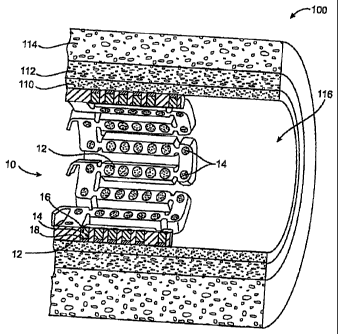
[00053] FIG. 1 illustrates an expandable medical device 10 in the form of a
stent
implanted in a lumen 116 of an artery 100. A wall of the artery 100 includes
three
distinct tissue layers, the intima 110, the media 112, and the adventitia 114.
When
the expandable medical device 10 is implanted in an artery at an occlusion
site, a
13
CA 02513721 2011-04-21
therapeutic agent delivered from the expandable medical device to the lumen
116 of
the artery 100 is distributed locally to the tissue at the site of the
occlusion and
downstream by the blood flow.
[00054] One example of an expandable medical device 10, as shown in FIGS. 1-
3, includes large, non-deforming struts 12, which can contain openings 14
without
compromising the mechanical properties of the struts, or the device as a
whole. The
non-deforming struts 12 may be achieved by the use of ductile hinges 20 which
are
described in detail in U.S. Patent No. 6,241,762; The openings 14 serve as
large,
protected reservoirs for delivering various beneficial agents to the device
implantation site.
[00055] The relatively large, protected openings 14, as described above, make
the
expandable medical device of the present invention particularly suitable for
delivering large amounts of therapeutic agents, larger molecules or genetic or
cellular agents, and for directional delivery of agents. The large non-
deforming
openings 14 in the expandable device 10 form protected areas or receptors to
facilitate the loading of such an agent, and to protect the agent from
abrasion,
extrusion, or other degradation during delivery and implantation.
[00056] FIG. 1 illustrates an expandable medical device for directional
delivery of
a therapeutic agent 16. The openings 14 contain the therapeutic agent 16 for
delivery to the lumen 116 of the blood vessel and an optional barrier layer 18
in or
adjacent the mural side of the openings.
[00057] The volume of beneficial agent that can be. delivered using openings
14 is
about 3 to 10 times greater than the volume of a 5 micron coating covering a
stent
with the same stent/vessel wall coverage ratio. This much larger beneficial
agent
capacity provides several advantages. The larger capacity can be used to
deliver
multi-drug combinations, each with independent release profiles, for improved
efficacy. Also, larger capacity can be used to provide larger quantities of
less
aggressive drugs and to achieve clinical efficacy without the undesirable side-
effects
of more potent drugs, such as retarded healing of the endothelial layer.
14
CA 02513721 2011-04-21
[00058] FIG. 4 shows across section of a portion of a medical device 10 in
which
one or more beneficial agents have been loaded into an opening 14 in multiple
layers. Although multiple discrete layers are shown for ease of illustration,
the
layers may be discrete layers with independent compositions or blended to form
a
continuous polymer matrix and agent inlay. For example, the layers can be
deposited separately in layers of a drug, polymer, solvent composition which
are
then blended together in the openings by the action of the solvent. The agent
may be
distributed within an inlay uniformly or in a concentration gradient. Examples
of
some methods of creating such layers and arrangements of layers are described
in
U.S. Patent Publication No. 2002/0082680, published on June 27, 2002. The use
of drugs in combination with polymers within the openings 14 allows the
medical
device 10 to be designed with drug release kinetics tailored to the specific
drug
delivery profile desired.
[00059] According to one example, the total depth of the opening 14 is about
50
to about 140 microns, and the typical layer thickness would be, about 2 to
about 50
microns, preferably about 12 microns. Each typical layer is thus individually
about
twice as thick as the typical coating applied to surface-coated stents. There
can be at
least two and preferably about five to twelve such layers in a typical
opening, with a
total beneficial agent thickness about 4 to 28 times greater than a typical
surface
coating. According to one embodiment of the present invention, the openings
have
an area of at least 5 x 10⁻⁶ square inches, and preferably at least 10 x 10-6
square
inches.
[00060] In the example of FIG. 4, the mural side of the openings are provided
with a barrier layer 18 which is a layer of polymer or other material having
an
erosion rate which is sufficiently slow to allow substantially all of the
therapeutic
agent in the therapeutic agent layers 16 to be delivered from the luminal side
of the
opening prior to complete erosion of the barrier layer. The barrier layer 18
prevents
loss of the beneficial agent during transport, storage, and during the stent
implantation procedure. However, the barrier layer 18 may be omitted where
mural
and lumina] delivery of the agent is acceptable.
CA 02513721 2005-05-06
WO 2004/043510 PCT/US2003/035948
[00061] In one example, the barrier layer 18 and/or the cap layer 22 may be
formed by a material soluble in a different solvent from the therapeutic agent
layers
to prevent intermixing of layers. For example, where one or more layers of
therapeutic agent and matrix have been deposited in the openings in a solvent
(e.g.
Insulin and PVP in water), it may be desirable to select a different polymer
and
solvent combination (e.g. PLGA in DMSO) for the barrier layer to prevent the
therapeutic agent from mixing into the barrier layer. Another layer, such as a
cap
layer may be formed by a third non-mixing polymer and solvent combination
(e.g.
PLGA in anisole). In addition to the barrier layer and cap layer, other
therapeutic
agent layers, protective layers, or separating layers may also be formed of
non-
mixing polymer/solvent systems in this manner.
[00062] A cap layer 22 can be provided which serves as a seal during filling
of
the openings. The cap layer 22 is preferably a rapidly degrading biocompatible
material.
[00063] Since each layer of both the barrier layer 18 and therapeutic agent 16
is
created independently, individual chemical compositions and pharmacokinetic
properties can be imparted to each layer. Numerous useful arrangements of such
layers can be formed, some of which will be described below. Each of the
layers
may include one or more agents in the same or different proportions from layer
to
layer. Changes in the agent concentration between layers can be used to
achieve a
desired delivery profile. For example, a decreasing release of drug for about
24
hours can be achieved. In another example, an initial burst followed by a
constant
release for about one week can be achieved. Substantially constant release
rates over
time period from a few hours to months can be achieved. The layers may be
solid,
porous, or filled with other drugs or excipients.
[00064] FIG. 5 is a cross sectional view of a portion of an expandable medical
device 10 including two or more therapeutic agents. Dual agent delivery
systems
such as that shown in FIG. 5 can deliver two or more therapeutic agents in
different
directions for the treatment of different conditions or stages of conditions.
For
example, a dual agent delivery system may deliver different agents in the
luminal
16
CA 02513721 2005-05-06
WO 2004/043510 PCT/US2003/035948
and mural directions for treatment of ischemia and restenosis from the same
drug
delivery device.
[00065] In FIG. 5, an antirestenotic agent 32 is provided at the mural side of
the
device 10 in one or more layers and a therapeutic agent 36 for reducing
ischemic
injury is provided at the luminal side of the device in one or more layers. A
separating layer 34 can be provided between the agent layers. A separating
layer 34
can be particularly useful when the administration periods for the two agents
are
substantially different and delivery of one of the agents will be entirely
completed
while the other agent continues to be delivered. The separating layer 34 can
be any
biocompatible material, which is preferably degradable at a rate which is
equal to or
longer than the longer of the administration periods of the two agents.
[00066] FIG. 6 illustrates an expandable medical device 10 including an inlay
40
formed of a biocompatible matrix with first and second agents provided in the
matrix for delivery according to different agent delivery profiles. As shown
in FIG.
6, a first drug illustrated by Os is provided in the matrix with a
concentration
gradient such that the concentration of the drug is highest adjacent the
barrier layer
18 at the mural side of the opening and is lowest at the luminal side of the
opening.
The second drug illustrated by As is relatively concentrated in an area close
to the
luminal side of the opening. This configuration illustrated in FIG. 6 results
in
delivery of two different agents with different delivery profiles from the
same inlay
40. The two different agents can be agents which treat ischemic injury by
different
modes of action, such as insulin and VIP.
[00067] In the embodiments described above, the therapeutic agent can be
provided in the expandable medical device in a biocompatible matrix. The
matrix
can be bioerodible as those described below or can be a permanent part of the
device
from which the therapeutic agent diffuses. One or more barrier layers,
separating
layers, and cap layers can be used to separate therapeutic agents within the
openings
or to prevent the therapeutic agents from degradation or delivery prior to
implantation of the medical device.
17
CA 02513721 2005-05-06
WO 2004/043510 PCT/US2003/035948
EXAMPLES
Example 1
[00068] In this example, a drug delivery stent substantially equivalent to the
stent
illustrated in FIGS. 2 and 3 having an expanded size of about 3 mm X 17 mm is
loaded with insulin in the following manner. The stent is positioned on a
mandrel
and an optional quick degrading layer is deposited into the openings in the
stent.
The quick degrading layer is low molecular weight PLGA provided on the luminal
side to protect the subsequent layers during transport, storage, and delivery.
The
layers described herein are deposited in a dropwise manner and are delivered
in
liquid form by use of a suitable organic solvent, such as DMSO, NMP, or DMAc.
A
plurality of layers of insulin and low molecular weight PLGA matrix are then
deposited into the openings to form an inlay of drug for the reduction of
ischemic
injury. The insulin and polymer matrix are combined and deposited in a manner
to
achieve a drug delivery profile which results in about 70% of the total drug
released
in about the first 2 hours, about 80% released in about 8 hours, and
essentially 100%
released in about 24 to about 48 hours. A barrier layer of moderate or high
molecular weight PLGA, a slow degrading polymer, is deposited over the insulin
layers to prevent the insulin from migrating to the mural side of the stent
and the
vessel walls. The degradation rate of the barrier layer is selected so that
the cap
layer does not degrade substantially until after the about 24-48 hour
administration
period.
[00069] The insulin dosage provided on the stent described is about 230
micrograms. The dosage has been calculated based on reported studies on
systemic
infusions of insulin which are estimated to deliver to the heart about 10
micrograms
of insulin over a 24 hour period. The total dosage on the stent may range from
about
micrograms to about 800 micrograms, preferably about 200 to about 400
micrograms.
18
CA 02513721 2005-05-06
WO 2004/043510 PCT/US2003/035948
Example 2
[00070] In this example, a drug delivery stent substantially equivalent to the
stent
illustrated in FIGS. 2 and 3 having an expanded size of about 3 mm X 17 mm is
loaded with insulin with a total dosage of about 230 micrograms in the
following
manner. The stent is positioned on a mandrel and an optional quick degrading
layer
is deposited into the openings in the stent. The quick degrading layer is
PLGA. A
plurality of layers of insulin and a poloxamer block copolymer of PEO and PPO
(Pluronic F 127) are then deposited into the openings to form an inlay of drug
for the
reduction of ischemic injury. The insulin and polymer matrix are combined at a
ratio of about 33:67 and deposited in a manner to achieve a drug delivery
profile
similar to that described in Example 1. A barrier layer of high molecular
weight
PLGA, a slow degrading polymer, is deposited over the insulin layers to
prevent the
insulin from migrating to the mural side of the stent and the vessel walls.
The
degradation rate of the barrier layer is selected so that the cap layer does
not degrade
substantially until after the about 24-48 hour administration period.
Example 3
[00071] In this example, a drug delivery stent substantially equivalent to the
stent
illustrated in FIGS. 2 and 3 having an expanded size of about 3 mm X 17 mm is
loaded with insulin with a total dosage of about 230 micrograms and with
paclitaxel
with a total dosage of about 10-30 micrograms in the following manner. The
stent is
positioned on a mandrel and an optional quick degrading layer is deposited
into the
openings in the stent. The quick degrading layer is PLGA. A plurality of
layers of
insulin and low molecular weight PLGA are then deposited into the openings to
form an inlay of drug for the reduction of ischemic injury. The insulin and
polymer
matrix are combined and deposited in a manner to achieve a drug delivery
profile
similar to that described in Example 1. A plurality of layers of high
molecular
weight PLGA, a slow degrading polymer, and paclitaxel are deposited over the
insulin layers to provide delivery of the paxlitaxel to the mural side of the
stent and
the vessel walls. The resorbtion rate of the paxlitaxel layers is selected so
that these
19
CA 02513721 2005-05-06
WO 2004/043510 PCT/US2003/035948
layers deliver paclitaxel continuously over an administration period of about
2 or
more days.