Note: Descriptions are shown in the official language in which they were submitted.
CA 02513794 2012-10-17
METHOD OF COATING STENTS TO IMPROVE THE STABILITY OF THE
THERAPEUTIC AGENT CONTAINED THEREIN
BACKGROUND OF THE INVENTION
1. Field of the Invention
The present invention relates to methods for coating stents, and
more particularly to methods for coating stents with therapeutic agents.
2. Discussion of the Related Art
Many individuals suffer from circulatory disease caused by a
progressive blockage of the blood vessels that profuse the heart and other
major organs. More severe blockage of blood vessels in such individuals
often leads to hypertension, ischemic injury, stroke, or myocardial
infarction. Atherosclerotic lesions, which limit or obstruct coronary blood
flow, are the major cause of ischemic heart disease. Percutaneous
transluminal coronary angioplasty is a medical procedure whose purpose is
to increase blood flow through an artery. Percutaneous transluminal
coronary angioplasty is the predominant treatment for coronary vessel
stenosis. The increasing use of this procedure is attributable to its
relatively high success rate and its minimal invasiveness compared with
coronary bypass surgery. A limitation associated with percutaneous
transluminal coronary angioplasty is the abrupt closure of the vessel, which
may occur immediately after the procedure and restenosis, which occurs
gradually following the procedure. Additionally, restenosis is a chronic
problem in patients who have undergone saphenous vein bypass grafting.
The mechanism of acute occlusion appears to involve several factors and
may result from vascular recoil with resultant closure of the artery and/or
deposition of blood platelets and fibrin along the damaged length of the
newly opened blood vessel.
CA 02513794 2012-10-17
Restenosis after percutaneous transluminal coronary angioplasty is
a more gradual process initiated by vascular injury. Multiple processes,
including thrombosis, inflammation, growth factor and cytokine release, cell
proliferation, cell migration and extracellular matrix synthesis each
contribute to the restenotic process.
While the exact mechanism of restenosis is not completely
understood, the general aspects of the restenosis process have been
identified. In the normal arterial wall, smooth muscle cells proliferate at a
low rate, approximately less than 0.1 percent per day. Smooth muscle
cells in the vessel walls exist in a contractile phenotype characterized by
eighty to ninety percent of the cell cytoplasmic volume occupied with the
contractile apparatus. Endoplasmic reticulum, Golgi, and free ribosomes
are few and are located in the perinuclear region. Extracellular matrix
surrounds the smooth muscle cells and is rich in heparin-like
glycosylaminoglycans, which are believed to be responsible for maintaining
smooth muscle cells in the contractile phenotypic state (Campbell and
Campbell, 1985, Phenotypic Modulation of Smooth Cells in Primary Culture",
(Table of Contents), Chapter 2, Volume 1, pp. 39-52).
Upon pressure expansion of an intracoronary balloon catheter
during angioplasty, smooth muscle cells within the vessel wall become
injured, initiating a thrombotic and inflammatory response. Cell derived
growth factors such as platelet derived growth factor, basic fibroblast
growth factor, epidermal growth factor, thrombin, etc., released from
platelets, invading macrophages and/or leukocytes, or directly from the
smooth muscle cells provoke a proliferative and migratory response in
medial smooth muscle cells. These cells undergo a change from the
contractile phenotype to a synthetic phenotype characterized by only a few
contractile filament bundles, extensive rough endoplasmic reticulum, Golgi
and free ribosomes. Proliferation/migration usually begins within one to
two days' post-injury and peaks several days thereafter (Campbell and
Campbell, Cell Biology of Smooth Muscle in Culture: Implications for
2
CA 02513794 2012-10-17
Atherogenesis", Inter. Angio, 6 pp. 73 (1987); CLOWES, A.W.et al., "Kinetics
of Cellular Proliferation after Arterial Injury", Laboratory Investigation,
Vol. 52, No.
6, pp. 611-616, 1985); SCHWARTZ, S.M. et al., Significance of Quiescent
Smooth Muscle Migration in the Injured Rat Carotid Artery, Circ. Res., 56,
1985,
pp. 139-145.
Daughter cells migrate to the intimal layer of arterial smooth muscle
and continue to proliferate and secrete significant amounts of extracellular
matrix proteins. Proliferation, migration and extracellular matrix synthesis
continue until the damaged endothelial layer is repaired at which time
proliferation slows within the intima, usually within seven to fourteen days
post-injury. The newly formed tissue is called neointima. The further
vascular narrowing that occurs over the next three to six months is due
primarily to negative or constrictive remodeling.
Simultaneous with local proliferation and migration, inflammatory
cells adhere to the site of vascular injury. Within three to seven days post-
injury, inflammatory cells have migrated to the deeper layers of the vessel
wall. In animal models employing either balloon injury or stent
implantation, inflammatory cells may persist at the site of vascular injury
for
at least thirty days (Tanaka et al., Sustained Activation of Vascular Cells
and
Leukocytes in the Rabbit Aorta after Balloon Injury", Circulation Vol. 88 p.
1788
(1993); Edelman et al., "Pathobiologic Responses to Stenting", American
Journal of Cardiology Vol. 91, Issue 7, Suppl. 1 (April 1998) pp. 4E-6E).
Inflammatory cells therefore are present and may contribute to both the
acute and chronic phases of restenosis.
Numerous agents have been examined for presumed anti-
proliferative actions in restenosis and have shown some activity in
experimental animal models. Some of the agents which have been shown
to successfully reduce the extent of intimal hyperplasia in animal models
include: heparin and heparin fragments (Clowes, A.W. and Karnovsky M.,
Nature 265: 25-26, 1977; Guyton, J.R. et al., Circ. Res., 46: 625-634,
3
CA 02513794 2012-10-17
1980; Clowes, A.W. and Clowes, M.M., Lab. Invest. 52: 611-616, 1985;
Clowes, A.W. and Clowes, M.M., Circ. Res. 58: 839-845, 1986; Majesky et
al., Circ. Res. 61: 296-300, 1987; Snow et al., Am. J. Pathol. 137: 313-330,
1990; Okada, T. et al., Neurosurgery 25: 92-98, 1989), colchicine (Currier,
J.W. et al., Circ. 80: 11-66, 1989), taxol (Sollot, S.J. et al., J. Clin.
Invest.
95: 1869-1876, 1995), angiotensin converting enzyme (ACE) inhibitors
(Powell, J.S. et al., Science, 245: 186-188, 1989), angiopeptin (Lundergan,
C.F. et al. Am. J. Cardiol. 17(Suppl. B):132B-136B, 1991), cyclosporin A
(Jonasson, L. et al., Proc. Natl., Acad. Sci., 85: 2303, 1988), goat-anti-
rabbit PDGF antibody (Ferns, G.A.A., et al., Science 253: 1129-1132,
1991), terbinafine (Nemecek, G.M. et al., J. Pharmacol. Exp. Thera. 248:
1167-1174, 1989), trapidil (Liu, M.W. et al., Circ. 81: 1089-1093, 1990),
tranilast (Fukuyama, J. et al., Eur. J. Pharmacol. 318: 327-332, 1996),
interferon-gamma (Hansson, G.K. and Holm, J., Circ. 84: 1266-1272,
1991), rapamycin (Marx, S.O. et al., Circ. Res. 76: 412-417, 1995), steroids
(Colburn, M.D. et al., J. Vasc. Surg. 15: 510-518, 1992), see also Berk,
B.C. et al., J. Am. Coll. Cardiol. 17: 111B-117B, 1991), ionizing radiation
(Weinberger, J. et al., Int. J. Rad. Onc. Biol. Phys. 36: 767-775, 1996),
fusion toxins (Farb, A. et al., Circ. Res. 80: 542-550, 1997) antisense
oligionucleotides (Simons, M. et al., Nature 359: 67-70, 1992) and gene
vectors (Chang, M.W. et al., J. Clin. Invest. 96: 2260-2268, 1995). Anti-
proliferative action on smooth muscle cells in vitro has been demonstrated
for many of these agents, including heparin and heparin conjugates, taxol,
tranilast, colchicine, ACE inhibitors, fusion toxins, antisense
oligionucleotides, rapamycin and ionizing radiation. Thus, agents with
diverse mechanisms of smooth muscle cell inhibition may have therapeutic
utility in reducing intimal hyperplasia.
However, in contrast to animal models, attempts in human
angioplasty patients to prevent restenosis by systemic pharmacologic
means have thus far been unsuccessful. Neither aspirin-dipyridamole,
ticlopidine, anti-coagulant therapy (acute heparin, chronic warfarin, hirudin
or hirulog), thromboxane receptor antagonism nor steroids have been
4
CA 02513794 2012-10-17
effective in preventing restenosis, although platelet inhibitors have been
effective in preventing acute reocclusion after angioplasty (Mak and Topol,
Clinical Trials to Prevent Restenosis after Percutaneous Coronary
Revascularization, Annals New York Academy of Sciences, 1997, pp. 255-
288; "Clinical Trials to Prevent Restenosis after Percutaneous Coronary
Revascularization", Department of Card iolog, Cleveland Clinical
Foundation, Ohio p. 255 (1991); Lang et al., Effects of Okadaic Acid and
ATPyS on Cell Length and Ca2+ Channel Currents Recorded in Single
Smooth Muscle Cells of the Guinea-pig Taenia Caeci, Br. J. Pharmacol.,
104, 1991, pp. 331-336; Popma et al., "Clinical Trials of Restenosis After
Coronary Angioplasty", Journal of the American Heart Association
(Circulation), 84:1426-1436 (1991). The
platelet GP Ilb/Illa receptor,
antagonist, Reopro is still under study but Reopro has not shown
definitive results for the reduction in restenosis following angioplasty and
stenting. Other agents, which have also been unsuccessful in the
prevention of restenosis, include the calcium channel antagonists,
prostacyclin mimetics, angiotensin converting enzyme inhibitors, serotonin
receptor antagonists, and anti-proliferative agents. These agents must be
given systemically, however, and attainment of a therapeutically effective
dose may not be possible; anti-proliferative (or anti-restenosis)
concentrations may exceed the known toxic concentrations of these agents
so that levels sufficient to produce smooth muscle inhibition may not be
reached (Mak and Topol, Clinical Trials to Prevent Restenosis after
Percutaneous Coronary Revascularization, Annals New York Academy of
Sciences, 1997, pp. 255-288; Lang et al., Effects of Okadaic Acid and
ATPyS on Cell Length and Ca2+ Channel Currents Recorded in Single
Smooth Muscle Cells of the Guinea-pig Taenia Caeci, Br. J. Pharmacol.,
104, 1991, pp. 331-336; Popma et al., Clinical Trials of Restenosis After
Coronary Angioplasty", Journal of the American Heart Association
(Circulation), 84:1426-1436 (1991).
Additional clinical trials in which the effectiveness for preventing
restenosis utilizing dietary fish oil supplements or cholesterol lowering
5
CA 02513794 2012-10-17
agents has been examined showing either conflicting or negative results so
that no pharmacological agents are as yet clinically available to prevent
post-angioplasty restenosis (Mak and Topol, Clinical Trials to Prevent
Restenosis after Percutaneous Coronary Revascularization, Annals New
York Academy of Sciences, 1997, pp. 255-288; Franklin and Faxon,
"Pharmacologic Prevention of Restenosis After Coronary Angioplasty:
Review of the Randomized Clinical Trials", Coronary Artery Disease, Vol.
4, No. 3 (March 1993) Serruys, P.W. et al., Evaluation of Ketanserin in the
Prevention of Restenosis after Percutaneous Transluminal Coronary
Angioplasty. A Multicenter Randomized Double-blind Placebo-controlled
Trial, Circulation, 88, 1993, pp. 1588-1601); SERRUYS, P.W. et al.,
Heparin-Coated Palmaz-Schatz Stents in Human Coronary Arteries: Early
Outcome of the Benestent-Il Pilot Study, Circulation, Vol. 93(3), 1996, pp.
412-422. Recent observations suggest that the antilipid/antioxident agent,
probucol, may be useful in preventing restenosis but this work requires
confirmation (Tardif et al., "Probucol and Multivitamins in the Prevention of
Restenosis After Coronary Angioplasty", New England Journal of Medicine,
Volume 337:365-'372 (1997); Yokoi, et al., "Effectiveness of an Antioxidant
in Preventing Restenosis After Percutaneous Translumina! Coronary
Angioplasty: The Probucol Angioplasty Restenosis Trial", JACC, Vol. 30,
No. 4 p. 855 (1997). Probucol is presently not approved for use in the
United States and a thirty-day pretreatment period would preclude its use
in emergency angioplasty. Additionally, the application of ionizing radiation
has shown significant promise in reducing or preventing restenosis after
angioplasty in patients with stents (Teirstein et al., "Catheter-Based
Radiotherapy to Inhibit Restenosis after Coronary Stenting", New England
Journal of Medicine, Vol. 336, p. 1679 (1997). Currently, however, the
most effective treatments for restenosis are repeat angioplasty,
atherectomy or coronary artery bypass grafting, because no therapeutic
agents currently have Food and Drug Administration approval for use for
the prevention of post-angioplasty restenosis.
6
CA 02513794 2013-08-16
Unlike systemic pharmacologic therapy, stents have proven useful
in significantly reducing restenosis. Typically, stents are balloon-
expandable slotted metal tubes (usually, but not limited to, stainless steel),
which, when expanded within the lumen of an angioplastied coronary
artery, provide structural support through rigid scaffolding to the arterial
wall. This support is helpful in maintaining vessel lumen patency. In two
randomized clinical trials, stents increased angiographic success after
percutaneous transluminal coronary angioplasty, by increasing minimal
lumen diameter and reducing, but not eliminating, the incidence of
restenosis at six months (Serruys et al., "A Comparison of Balloon-
Expandable-Stent Implantation with Balloon Angioplasty in Patients with
Coronary Artery Disease", New England Journal of Medicine, Volume
331:489-495 (1994); Fischman et al., A Randomized Comparison of
Coronary-Stent Placement and Balloon Angioplasty in the Treatment of
Coronary Artery Disease", The New England Journal of Medicine, Volume
331:496-501 (1994).
Additionally, the heparin coating of stents appears to have the
added benefit of producing a reduction in sub-acute thrombosis after stent
implantation (Serruys et al., Heparin-Coated Palmaz-Schatz Stents in
Human Coronary Arteries: Early Outcome of the Benestent-II Pilot Study,
Circulation, Vol. 93(3), 1996, pp. 412-422). Thus, sustained mechanical
expansion of a stenosed coronary artery with a stent has been shown to
provide some measure of restenosis prevention, and the coating of stents
with heparin has demonstrated both the feasibility and the clinical
usefulness of delivering drugs locally, at the site of injured tissue.
As stated above, the use of heparin coated stents demonstrates the
feasibility and clinical usefulness of local drug delivery; however, the
manner in which the particular drug or drug combination is affixed to the
local delivery device will play a role in the efficacy of this type of
treatment.
For example, the processes and materials utilized to affix the drug/drug
combinations to the local delivery device should not interfere with the
operations of the drug/drug combinations. In addition, the processes and
materials utilized should be biocompatible and maintain the drug/drug
7
CA 02513794 2012-10-17
combinations on the local device through delivery and over a given period
of time. For example, removal of the drug/drug combination during delivery
of the local delivery device may potentially cause failure of the device.
Accordingly, there exists a need for drug/drug combinations and
associated local delivery devices for the prevention and treatment of
vascular injury causing intimal thickening which is either biologically
induced, for example, atherosclerosis, or mechanically induced, for
example, through percutaneous transluminal coronary angioplasty. In
addition, there exists a need for maintaining the drug/drug combinations on
the local delivery device through delivery and positioning as well as
ensuring that the drug/drug combination is released in therapeutic dosages
over a given period of time.
A variety of stent coatings and compositions have been proposed
for the prevention and treatment of injury causing intimal thickening. The
coatings may be capable themselves of reducing the stimulus the stent
provides to the injured lumen wall, thus reducing the tendency towards
thrombosis or restenosis.
Alternately, the coating may deliver a
pharmaceutical/therapeutic agent or drug to the lumen that reduces
smooth muscle tissue proliferation or restenosis. The mechanism for
delivery of the agent is through diffusion of the agent through either a bulk
polymer or through pores that are created in the polymer structure, or by
erosion of a biodegradable coating.
Both bioabsorbable and biostable compositions have been reported
as coatings for stents. They generally have been polymeric coatings that
either encapsulate a pharmaceutical/therapeutic agent or drug, e.g.
rapamycin, taxol etc., or bind such an agent to the surface, e.g. heparin-
coated stents. These coatings are applied to the stent in a number of
ways, including, though not limited to, dip, spray, or spin coating
processes.
8
CA 02513794 2012-10-17
While the selection of an appropriate therapeutic agent and an
appropriate coating in which to incorporate the therapeutic agent is
important, maintaining the stability of the agent is also important.
Accordingly, there
exists a need for developing a process for coating the implantable medical
device that incorporates steps to stabilize the therapeutic agent.
SUMMARY OF THE INVENTION
The process of the present invention provides a means for
overcoming the difficulties associated with the coating of implantable
medical devices with therapeutic agents.
In accordance with one aspect, the present invention is directed to a
process for coating implantable medical devices. The method comprises
applying a primer coating on the implantable medical devices, including the
application of a parylene layer and annealing the parylene layer to reduce
autoxidation initiators, preparing a basecoat solution comprising polymers
and a therapeutic agent and applying the basecoat solution to the
implantable medical devices coated with parylene, the basecoat solution
being prepared with and applied utilizing a process to reduce the presence
and exposure of the basecoat solution to oxygen, raising the glass
transition temperature of the therapeutic agent and creating a coating
morphology to protect the therapeutic agent from autoxidation,
preparing a topcoat solution comprising at least one polymer and
applying the topcoat solution to the implantable medical devices coated
with the basecoat solution, the topcoat solution being prepared with and
applied utilizing a process to reduce the presence and exposure of the
topcoat solution to oxygen, raising the glass transition temperature of the
therapeutic agent and creating a coating morphology to protect the
therapeutic agent from autoxidation and finally processing the implantable
medical devices, including inspecting, packaging and sterilizing the coated
9
CA 02513794 2012-10-17
medical devices, the final processing including protecting the therapeutic
agent from autoxidation, reducing the presence of and exposure of all
materials to free radicals and reducing the presence of and exposure of all
materials to oxygen.
The process of the present invention incorporates a number of steps
to increase the stability of the therapeutic agent, including protecting the
therapeutic agent from autoxidation by increasing the glass transition
temperature of the agent, reducing the presence of and/or exposure of
various materials utilized to free radicals and autoxidation initiators and
reducing the presence of and/or exposure of the various materials to
oxygen.
BRIEF DESCRIPTION OF THE DRAWINGS
The foregoing and other features and advantages of the invention
will be apparent from the following, more particular description of preferred
embodiments of the invention, as illustrated in the accompanying drawings.
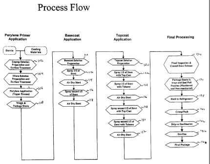
Figure 1 is a flow chart of a first exemplary embodiment of a process
for coating stents in accordance with the present invention.
Figure 2 is a flow chart of a second exemplary embodiment of a
process for coating stents in accordance with the present invention.
Figure 3 is a flow chart of a third exemplary embodiment of a
process for coating stents in accordance with the present invention.
Figure 4 is a flow chart of a fourth exemplary embodiment of a
process for coating stents in accordance with the present invention.
DETAILED DESCRIPTION OF THE PREFERRED EMBODIMENTS
The present invention is directed to a process for coating stents or
other implantable medical devices with one or more therapeutic agents,
CA 02513794 2012-10-17
such as a rapamycin. One exemplary process is set forth in the flow chart
of Figure 1. The first part of the process comprises the primer application.
In the exemplary embodiment, the first step in the process is surface
preparation and treatment, step 102. This step involves utilizing a cleaning
solution to remove endotoxins from the stents to be coated. The cleaning
solution may comprise any number of cleaning solutions, for example, a
high pH solution such as a potassium hydroxide solution containing
silicates. The next step is also a surface preparation and treatment step,
step 104. In this step a silane solution is utilized to prepare the surfaces
of
the stents for the deposition of a primer layer. The next step is the
application of the primer itself, step 106. In this exemplary embodiment,
parylene is applied to the stents utilizing a vapor deposition process. Once
the parylene is applied, the stents are packaged and weighed, step 108.
Once the stents are weighed, they are placed in containers or vials. The
vials may be formed from any number of suitable materials. In the
exemplary embodiment, the vials are formed from polypropylene.
The second part of the process comprises the basecoat application.
The first step in the second part of the process is the preparation of the
basecoat, step 110. The
basecoat may comprise any suitable
biocompatible polymers and therapeutic agents. The therapeutic agents
and polymers should preferably be compatible. In the
exemplary
embodiment, the basecoat solution comprises polyethylene co-
vinylacetate, polybutylmethacrylate and a rapamycin, such as sirolimus.
The solution is prepared in a. standard reactor. The solution is decanted
into smaller containers for the next step. The next step is the coating of
the stents, step 112. In this step, the stents are coated with the basecoat
solution. The stents may be coated in any suitable manner. In the
exemplary embodiment, the stents are coated utilizing a spray coating
technique. Nitrogen is utilized as the carrier gas for the basecoat solution.
In step 112, one half of the stent is coated and then air dried in step 114.
The half coated stents are dried at a relative humidity of about thirty to
about fifty-five percent for about thirty minutes. The air temperature is held
11
CA 02513794 2012-10-17
at about room temperature. The air in the drying chamber is continuously
recirculated. Upon completion of the drying step 114, the second half of
the stent is coated, step 116 and then dried again in step 118. Steps 116
and 118 are identical to steps 112 and 114.
The third part of the process comprises the topcoat application. The
first step in the third part of the process is the preparation of the topcoat
solution, step 120. The preparation of the topcoat comprises preparing a
solution of polybutylmethacrylate. Once the solution is prepared and
decanted into a spraying container, one-half of the stent is coated, step
122. The next step of the process is another coating step, step 124. In
this coating step, the half of the stents that have been topcoated are
sprayed with toluene. The spraying of toluene has a polishing effect on the
topcoat and also facilitates elution control of the therapeutic agent from the
polymeric coating. Once step 124 is complete, the stents are air dried,
step 126, under the same conditions as in steps 114 and 118. Steps 128,
130 and 132 are the same as steps 122, 124 and 126 but for the second
half of the stents.
The fourth and final part of the exemplary process comprises the
final processing. The first step in the fourth part of the process is final
inspection and coated stent release, step 134. Each of the stents is
inspected for defects. Various inspection techniques such as microscopy
may be utilized to determine if the stents meet various rigorous standards.
The next step in the process is packaging, step 136. The stents are put
into trays and sealed in pouches. In this exemplary embodiment, the trays
are PETG trays. Once the stents are packaged, they are refrigerated, step
138. The stents are maintained at a temperature from about five degrees
centigrade to about eight degrees centigrade. Wider ranges may be
utilized. The next step in the process is the crimping and packaging of
each of the stents, step 140. In this step, the stents are positioned on the
delivery device and crimped to the desired size. Once positioned on the
delivery devices, the whole system is packaged and shipped to a location
12
CA 02513794 2012-10-17
for sterilization, steps 142 and 144. The systems are sterilized utilizing
ethylene oxide, but other suitable sterilization processes may be utilized.
The final step of the process is final packaging, step 146.
A number of process modifications may be utilized to address
autoxidation. Autoxidation occurs when there is a fuel, in this case the
therapeutic agent, an ignition of the fuel, in this case radicals, and
finally,
there is oxygen or oxygen containing compounds. The first process
modification includes protecting the active pharmaceutic ingredient or
therapeutic agent, sirolimus, from autoxidation. One way in which to
protect the active pharmaceutic ingredient is to raise its glass transition
temperture, tg. A higher glass transition temperature leads to a more
stable therapeutic agent at room temperature. Amorphous substances act
like sponges and will pick up other compounds such as solvents. Sirolimus
is an amorphous therapeutic agent. Accordingly, in order to make an
amorphous therapeutic agent more stable, one has to raise its glass
transition temperature and since solvents lower the glass transition
temperature, the minimization of exposure to residual solvents is required.
Ways in which to reduce or minimize exposure to residual solvents include
keeping extraneous solvents away from the coating, for example, cleaning
agents and solvent bottles, and storing stents in an environment that is
substantially solvent free, for example, away from freshly coated stents
and/or from solutions. Preferably, the therapeutic agents or stents coated
with therapeutic agents are stored in stability chambers. In addition, a
higher glass transition temperature may be achieved by increasing the
removal of residual solvents post coating. This may be accomplished by
allowing more time for residual solvent removal post coating, by applying
vacuum conditions and heat to enhance residual solvent removal and by
allowing short-term moisture exchange (presence of humidity) to enhance
residual solvent removal. The long-term exposure to relative humidity is
preferably controlled because humidity may act as a plasticizer. Vacuum
packaging and packaging under inert gas of the finished goods addresses
this concern. Also, the three domain coating morphology, i.e., three
13
CA 02513794 2012-10-17
different zones of polymer and therapeutic agent offers only some
protection of the therapeutic agent from oxygen. Accordingly, the spraying
conditions may be modified to control or affect the coating morphology, for
example, low humidity and spray head distance. The steps of the process
that may be modified to accomplish these improvements include steps
112, 114, 116, 118, 126, 132, 138 and 146 as illustrated in Figure 2.
Another process modification comprises reducing the presence of
and/or exposure to free radicals and, autoxidation initiators. This may be
accomplished by utilizing materials with minimal free radicals, for example,
polypropylene vials may be utilized rather than PETG trays, and utilizing
tools to assist in the crimping and packaging stage that are fabricated from
inert materials. This may also be accomplished by parylene annealing to
reduce parylene radicals. The steps of the process that may be modified
to accomplish these improvements include steps 104 and 136 as illustrated
in Figure 3.
Yet another process modification comprises reducing the presence
of and/or exposure to oxygen. This may be accomplished by having
improved controls of raw materials, improved coating solution mixing and
handling, and improved coatings. Improved control of raw materials
includes solvents such as THE with low hydroperoxides and an active
pharmaceutic ingredient with minimal handling. Improved coating solution
mixing and handling includes inert gas blanketing to reduce dissolved
oxygen and the minimization of all decanting steps. Improved coating
includes spraying in a nitrogen rich environment, vacuum oven, purging
with inert gas after annealing and vacuum packaging, and/or packaging
under inert gas of works in progress and finished goods. The steps of the
process that may be modified to accomplish these improvements include
steps 101, 110, 112, 116, 120, 136 and 146 as illustrated in Figure 4.
It is important to note that although stents are discussed in detail
herein, the local delivery of drug/drug combinations may be utilized to treat
14
CA 02513794 2012-10-17
=
a wide variety of conditions utilizing any number of medical devices, or to
enhance the function and/or life of the device. For example, intraocular
lenses, placed to restore vision after cataract surgery is often compromised
by the formation of a secondary cataract. The latter is often a result of
cellular overgrowth on the lens surface and can be potentially minimized by
combining a drug or drugs with the device. Other medical devices which
often fail due to tissue in-growth or accumulation of proteinaceous material
in, on and around the device, such as shunts for hydrocephalus, dialysis
grafts, colostomy bag attachment devices, ear drainage tubes, leads for
pace makers and implantable defibrillators can also benefit from the
device-drug combination approach. Devices which serve to improve the
structure and function of tissue or organ may also show benefits when
combined with the appropriate agent or agents. For example, improved
osteointegration of orthopedic devices to enhance stabilization of the
implanted device could potentially be achieved by combining it with agents
such as bone-morphogenic protein. Similarly other surgical devices,
sutures, staples, anastomosis devices, vertebral disks, bone pins, suture
anchors, hemostatic barriers, clamps, screws, plates, clips, vascular
implants, tissue adhesives and sealants, tissue scaffolds, various types of
dressings, bone substitutes, intraluminal devices, and vascular supports
could also provide enhanced patient benefit using this drug-device
combination approach. Essentially, any type of medical device may be
coated in some fashion with a drug or drug combination which enhances
treatment over use of the singular use of the device or pharmaceutical
agent.
In addition to various medical devices, the coatings on these
devices may be used to deliver therapeutic and pharmaceutic agents
including: antiproliferative/antimitotic agents including natural products
such as vinca alkaloids (i.e. vinblastine, vincristine, and vinorelbine),
paclitaxel, epidipodophyllotoxins (i.e. etoposide, teniposide), antibiotics
(dactinomycin (actinomycin D) daunorubicin, doxorubicin and idarubicin),
anthracyclines, mitoxantrone, bleomycins, plicamycin (mithramycin) and
CA 02513794 2012-10-17
mitomycin, enzymes (L-asparaginase which systemically metabolizes L-
asparagine and deprives cells which do not have the capacity to synthesize
their own asparagine); antiplatelet agents such as G(GP) Ilb/Illa inhibitors
and vitronectin receptor antagonists; antiproliferative/antimitotic alkylating
agents such as nitrogen mustards (mechlorethamine, cyclophosphamide
and analogs, melphalan, chlorambucil), ethylenimines and
methylmelamines (hexamethylmelamine and thiotepa), alkyl sulfonates-
busulfan, nitrosoureas (carmustine (BCNU) and analogs, streptozocin),
triazenes ¨ dacarbazinine (DTIC);
antiproliferative/antimitotic
antimetabolites such as folic acid analogs (methotrexate), pyrimidine
analogs (fluorouracil, floxuridine, and cytarabine), purine analogs and
related inhibitors (mercaptopurine, thioguanine, pentostatin and 2-
chlorodeoxyadenosine {cladribine}), platinum coordination complexes
(cisplatin, carboplatin), procarbazine,
hydroxyurea, mitotane,
aminoglutethimide; hormones (i.e. estrogen); anticoagulants (heparin,
synthetic
heparin salts and other inhibitors of thrombin); fibrinolytic agents (such as
tissue plasminogen activator, streptokinase and urokinase), aspirin,
dipyridamole, ticlopidine, clopidogrel, abciximab; antimigratory;
antisecretory (breveldin); anti-inflammatory: such as adrenocortical
steroids (cortisol, cortisone, fludrocortisone, prednisone, prednisolone, 6a-
methylprednisolone, triamcinolone, betamethasone, and dexamethasone),
non-steroidal agents (salicylic acid derivatives i.e. aspirin; para-
aminophenol derivatives i.e. acetominophen; indole and indene acetic
acids (indomethacin, sulindac, and etodalac), heteroaryl acetic acids
(tolmetin, diclofenac, and ketorolac), arylpropionic acids (ibuprofen and
derivatives), anthranilic acids (mefenamic acid, and meclofenamic acid),
enolic acids (piroxicam, tenoxicam, phenylbutazone, and
oxyphenthatrazone), nabumetone, gold compounds (auranofin,
aurothioglucose, gold sodium thiomalate); immunosuppressives:
(cyclosporine, tacrolimus (FK-506), sirolimus (rapamycin), azathioprine,
mycophenolate mofetil); angiogenic agents: vascular endothelial growth
factor (VEGF), fibroblast growth factor (FGF); angiotensin receptor
16
CA 02513794 2013-08-16
blockers; nitric oxide donors; anti-sense oligionucleotides and
combinations thereof; cell cycle inhibitors, mTOR inhibitors, and growth
factor receptor signal transduction kinase inhibitors; retenoids; cyclin/CDK
inhibitors; HMG co-enzyme reductase inhibitors (statins); and protease
inhibitors.
Although shown and described is what is believed to be the most
practical and preferred embodiments, it is apparent that departures from
specific designs and methods described and shown will suggest
themselves to those skilled in the art and may be used without departing
from the scope of the invention. The present invention is not restricted to
the particular constructions described and illustrated, but should be
constructed to cohere with all modifications that may fall within the scope
of the appended claims.
17