Note: Descriptions are shown in the official language in which they were submitted.
CA 02513857 2005-07-27
LOW DEPLOYMENT FORCE DELIVERY DEVICE
BACKGROUND OF THE INVENTION
1. Field of the Invention
The present invention relates to delivery devices, and more particularly,
to low deployment force delivery devices for self-deploying intraluminal
devices.
2. Discussion of the Related Art
An aneurysm is an abnormal dilation of a layer or layers of an arterial wall,
usually caused by a systemic collagen synthetic or structural defect. An
abdominal aortic aneurysm is an aneurysm in the abdominal portion of the
aorta,
usually located in or near one or both of the two iliac arteries or near the
renal
arteries. The aneurysm often arises in the infrarenal portion of the diseased
aorta, for example, below the kidneys. A thoracic aortic aneurysm is an
aneurysm in the thoracic portion of the aorta. When left untreated, the
aneurysm
may rupture, usually causing rapid fatal hemorrhaging.
Aneurysms may be classified or typed by their position as well as by the
number of aneurysms in a cluster. Typically, abdominal aortic aneurysms may
be classified into five types. A Type I aneurysm is a single dilation located
between the renal arteries and the iliac arteries. Typically, in a Type I
aneurysm,
the aorta is healthy between the renal arteries and the aneurysm and between
the aneurysm and the iliac arteries.
A Type II A aneurysm is a single dilation located between the renal
arteries and the iliac arteries. In a Type I1 A aneurysm, the aorta is healthy
between the renal arteries and the aneurysm, but not healthy between the
aneurysm and the iliac arteries. In other words, the dilation extends to the
aortic
bifurcation. A Type II B aneurysm comprises three dilations. One dilation is
1
CA 02513857 2005-07-27
located between the renal arteries and the iliac arteries. Like a Type II A
aneurysm, the aorta is healthy between the aneurysm and the renal arteries,
but
not healthy between the aneurysm and the iliac arteries. The other two
dilations
are located in the iliac arteries between the aortic bifurcation and the
bifurcations
between the external iliacs and the internal iliacs. The iliac arteries are
healthy
between the iliac bifurcation and the aneurysms. A Type II C aneurysm also
comprises three dilations. However, in a Type II C aneurysm, the dilations in
the
iliac arteries extend to the iliac bifurcation.
A Type 111 aneurysm is a single dilation located between the renal arteries
and the iliac arteries. In a Type III aneurysm, the aorta is not healthy
between
the renal arteries and the aneurysm. In other words, the dilation extends to
the
renal arteries.
A ruptured abdominal aortic aneurysm is presently the thirteenth leading
cause of death in the United States. The routine management of abdominal
aortic aneurysms has been surgical bypass, with the placement of a graft in
the
involved or dilated segment. Although resection with a synthetic graft via
transperitoneal or retroperitoneal procedure has been the standard treatment,
it
is associated with significant risk. For example, complications include
perioperative myocardial ischemia, renal failure, erectile impotence,
intestinal
ischemia, infection, lower limb ischemia, spinal cord injury with paralysis,
aorta-
enteric fistula, and death. Surgical treatment of abdominal aortic aneurysms
is
associated with an overall mortality rate of five percent in asymptomatic
patients,
sixteen to nineteen percent in symptomatic patients, and is as high as fifty
percent in patients with ruptured abdominal aortic aneurysms.
Disadvantages associated with conventional surgery, in addition to the
high mortality rate, include an extended recovery period associated with the
large
surgical incision and the opening of the abdominal cavity, difficulties in
suturing
the graft to the aorta, the loss of the existing thrombosis to support and
reinforce
the graft, the unsuitability of the surgery for many patients having abdominal
aortic aneurysms, and the problems associated with performing the surgery on
2
CA 02513857 2005-07-27
an emergency basis after the aneurysm has ruptured. Further, the typical
recovery period is from one to two weeks in the hospital and a convalescence
period, at home, ranging from two to three months or more, if complications
ensue. Since many patients having abdominal aortic aneurysms have other
chronic illnesses, such as heart, lung, liver and/or kidney disease, coupled
with
the fact that many of these patients are older, they are less than ideal
candidates
for surgery.
The occurrence of aneurysms is not confined to the abdominal region.
While abdominal aortic aneurysms are generally the most common, aneurysms
in other regions of the aorta or one of its branches are possible. For
example,
aneurysms may occur in the thoracic aorta. As is the case with abdominal
aortic
aneurysms, the widely accepted approach to treating an aneurysm in the
thoracic aorta is surgical repair, involving replacing the aneurysmal segment
with
a prosthetic device. This surgery, as described above, is a major undertaking,
with associated high risks and with significant mortality and morbidity.
Over the past five years, there has been a great deal of research directed
at developing less invasive, endovascular, i.e., catheter directed, techniques
for
the treatment of aneurysms, specifically abdominal aortic aneurysms. This has
been facilitated by the development of vascular stents, which can and have
been
used in conjunction with standard or thin-wall graft material in order to
create a
stent-graft or endograft. The potential advantages of less invasive treatments
have included reduced surgical morbidity and mortality along with shorter
hospital and intensive care unit stays.
Stent-grafts or endoprostheses are now Food and Drug Administration
(FDA) approved and commercially available. Their delivery procedure typically
involves advanced angiographic techniques performed through vascular
accesses gained via surgical cut down of a remote artery, which may include
the
common femoral or brachial arteries. Over a guidewire, the appropriate size
introducer will be placed. The catheter and guidewire are passed through the
aneurysm. Through the introducer, the stent-graft will be advanced to the
3
CA 02513857 2005-07-27
appropriate position. Typical deployment of the stent-graft device requires
withdrawal of an outer sheath while maintaining the position of the stent-
graft
with an inner-stabilizing device. Most stent-grafts are self-expanding;
however,
an additional angioplasty procedure, e.g., balloon angioplasty, may be
required
to secure the position of the stent-graft. Following the placement of the
stent-
graft, standard angiographic views may be obtained.
Due to the large diameter of the above-described devices, typically
greater than twenty French (3F=1 mm), arteriotomy closure typically requires
open surgical repair. Some procedures may require additional surgical
techniques, such as hypogastric artery embolization, vessel ligation, or
surgical
bypass in order to adequately treat the aneurysm or to maintain blood flow to
both lower extremities. Likewise, some procedures will require additional
advanced catheter directed techniques, such as angioplasty, stent placement
and embolization, in order to successfully exclude the aneurysm and
efficiently
manage leaks.
While the above-described endoprostheses represent a significant
improvement over conventional surgical techniques, there is a need to improve
the endoprostheses, their method of use and their applicability to varied
biological conditions. Accordingly, in order to provide a safe and effective
alternate means for treating aneurysms, including abdominal aortic aneurysms
and thoracic aortic aneurysms, a number of difficulties associated with
currently
known endoprostheses and their delivery systems must be overcome. One
concern with the use of endoprostheses is the prevention of endo-leaks and the
disruption of the normal fluid dynamics of the vasculature. Devices using any
technology should preferably be simple to position and reposition as
necessary,
should preferably provide an acute, fluid tight seal, and should preferably be
anchored to prevent migration without interfering with normal blood flow in
both
the aneurysmal vessel as well as branching vessels. In addition, devices using
the technology should preferably be able to be anchored, sealed, and
maintained in bifurcated vessels, tortuous vessels, highly angulated vessels,
partially diseased vessels, calcified vessels, odd shaped vessels, short
vessels,
4
CA 02513857 2005-07-27
and long vessels. In order to accomplish this, the endoprostheses should
preferably be highly durable, extendable and re-configurable while maintaining
acute and long-term fluid tight seals and anchoring positions.
The endoprostheses should also preferably be able to be delivered
percutaneously utilizing catheters, guidewires and other devices which
substantially eliminate the need for open surgical intervention. Accordingly,
the
diameter of the endoprostheses in the catheter is an important factor. This is
especially true for aneurysms in the larger vessels, such as the thoracic
aorta. In
addition, the delivery force required to deliver the endoprosthesis should
preferably be minimized to reduce the risk of damage to the endoprosthesis.
SUMMARY OF THE INVENTION
The low deployment force delivery device of the present invention
overcomes the limitations associated with currently utilized devices as
briefly
described above.
In accordance with one aspect, the present invention is directed to a low
deployment force delivery apparatus for intraluminal devices. The delivery
apparatus comprises an inner tube having a proximal region and a distal
region, the distal region being configured to receive an intraluminal device
and
a sheath having a proximal end and a distal end positioned concentrically
around at least a portion of the inner tube, the distal end comprising an
inner
layer at least partially covering the intraluminal device and an outer layer,
the
inner layer and outer layer being connected at the distal end of the sheath
and
configured for relative movement therebetween. .
5
CA 02513857 2005-07-27
BRIEF DESCRIPTION OF THE DRAWINGS
The foregoing and other features and advantages of the invention will be
apparent from the following, more particular description of preferred
embodiments of the invention, as illustrated in the accompanying drawings.
Figure 1 is a diagrammatic representation of an exemplary low
deployment force delivery system in accordance with the present invention.
Figure 2 is a diagrammatic representation of an exemplary deployment
force delivery system with the stmt partially deployed in accordance with the
present invention.
Figure 3 is a diagrammatic representation of an alternate exemplary low
deployment force delivery system in accordance with the present invention.
Figure 4 is a diagrammatic representation of an exemplary deployment
force delivery system with the stent partially deployed in accordance with the
present invention.
DETAILED DESCRIPTION OF THE PREFERRED EMBODIMENTS
Various endoprosthesis assemblies, which include expandable stents
and/or stent grafts, have been proposed or developed for use in association
with angioplasty treatments and other medical procedures such as aneurysm
repair. The endoprosthesis assembly is generally percutaneously routed to a
treatment site and the scent and/or stent graft is expanded to maintain or
restore the patency of a body passageway such as a blood vessel or bile
duct or to create a new passageway. A stent is typically cylindrical in shape
comprising an expandable open frame. The stent and/or stent graft will
typically expand either by itself (self-expanding stents) or will expand upon
exertion of an outwardly directed radial force on an inner surface of the
stent
6
CA 02513857 2005-07-27
frame by a balloon catheter or the like. The deployment of a stent or stent
graft is substantially the same as is described in detail subsequently.
Accordingly, there is a need for a self-expanding stent or stent graft
delivery system which is able to navigate tortuous passageways, which
prevents the stent or stent graft from becoming embedded therein, which
allows the physician to more easily and accurately deploy the stent or stent
graft within the target area.
Stents for endovascular implantation into a blood vessel or the like, to
maintain or restore the patency of the passageway, have been deployed
percutaneously to minimize the invasiveness associated with surgical
exposure of the treatment site during coronary artery bypass. Percutaneous
deployment is initiated by an incision into the vascular system of the
patient,
typically into the femoral artery. A tubular or sheath portion of an
introduces is
inserted through the incision and extends into the artery. The introduces has
a central lumen which provides a passageway through the patient's skin and
artery wall into the interior of the artery. An outwardly tapered hub portion
of
the introduces remains outside the patient's body to prevent blood from
leaking out of the artery along the outside of the sheath. The introduces
lumen includes a valve to block blood flow out of the artery through the
introduces passageway. A distal end of a guide wire is passed through the
introduces passageway into the patient's vasculature. The guide wire is
threaded through the vasculature until the inserted distal end extends just
beyond the treatment site. The proximal end of the guide wire extends
outside the introduces.
For endovascular deployment, a stmt, in an unexpended or constricted
configuration, is crimped onto a deflated balloon portion of a balloon
catheter. The balloon portion is normally disposed near a distal end of the
balloon catheter. The catheter has a central lumen extending its entire
length. The distal end of the balloon catheter is threaded onto the proximal
end of the guide wire. The distal end of the catheter is inserted into the
CA 02513857 2005-07-27
introducer lumen and the catheter is pushed along the guide wire until the
stent reaches the treatment site. At the treatment site, the balloon is
inflated
causing the stent to radially expand and assume an expanded configuration.
When the stent is used to reinforce a portion of the blood vessel wall, the
stent is expanded such that its outer diameter is approximately ten percent to
twenty percent larger than the inner diameter of the blood vessel at the
treatment site, effectively causing an interference fit between the stent and
the blood vessel that inhibits migration of the stent. The balloon is deflated
and the balloon catheter is withdrawn from the patient's body. The guide
I0 wire is similarly removed. Finally, the introducer is removed from the
artery.
An example of a commonly used stent is given in U.S. Patent Number
4,733,665 filed by Palmaz on November 7, 1985. Such stents are often
referred to as balloon expandable stents. Typically the stent is made from a
solid tube of stainless steel. Thereafter, a series of cuts are made in the
wall of
the stent. The stent has a first smaller diameter which permits the stent to
be
delivered through the human vasculature by being crimped onto a balloon
catheter. The stent also has a second or expanded diameter. The expanded
diameter is achieved through the application, by the balloon catheter
positioned
in the interior of the tubular shaped member, of a radially outwardly directed
force.
However, such "balloon expandable" stents are often impractical for use
in some vessels such as superficial arteries, like the carotid artery. The
carotid
artery is easily accessible from the exterior of the human body. A patient
having a balloon expandable stent made from stainless steel or the like,
placed
in their carotid artery might be susceptible to sever injury through day to
day
activity. A sufficient force placed on the patient's neck, such as by falling,
could cause the stent to collapse, resulting in injury to the patient. In
order to
prevent this, self-expanding stents have been proposed for use in such
vessels. Self-expanding stents act similarly to springs and will recover to
their
expanded or implanted configuration after being crushed.
s
CA 02513857 2005-07-27
One type of self-expanding stent is disclosed in U.S. Patent Number
4,665,771. The disclosed stent has a radially and axially flexible, elastic
tubular
body with a predetermined diameter that is variable under axial movement of
ends of the body relative to each other and which is composed of a plurality
of
individually rigid but flexible and elastic thread elements defining a
radially self-
expanding helix. This type of stent is known in the art as a "braided stent"
and
is so designated herein. Placement of such stents in a body vessel can be
achieved by a device which comprises an outer catheter for holding the stent
at
its distal end, and an inner piston which pushes the stent forward once it is
in
position.
Other types of self-expanding stents use alloys such as Nitinol (Ni-Ti
alloy), which have shape memory and/or superelastic characteristics in medical
devices which are designed to be inserted into a patient's body. The shape
memory characteristics allow the devices to be deformed to facilitate their
insertion into a body lumen or cavity and then be heated within the body so
that
the device returns to its original shape. Superelastic characteristics on the
other hand generally allow the metal to be deformed and restrained in the
deformed condition to facilitate the insertion of the medical device
containing
the metal into a patient's body, with such deformation causing the phase
transformation. Once within the body lumen the restraint on the superelastic
member can be removed, thereby reducing the stress therein so that the
superelastic member can return to its original un-deformed shape by the
transformation back to the original phase.
Alloys having shape memory/superelastic characteristics generally have
at least two phases. These phases are a martensite phase, which has a
relatively low tensile strength and which is stable at relatively low
temperatures,
and an austenite phase, which has a relatively high tensile strength and which
is stable at temperatures higher than the martensite phase.
When stress is applied to a specimen of a metal, such as Nitinol,
exhibiting superelastic characteristics at a temperature above which the
9
CA 02513857 2005-07-27
austenite is stable (i.e. the temperature at which the transformation of
martensite phase to the austenite phase is complete), the specimen deforms
elastically until it reaches a particular stress level where the alloy then
undergoes a stress-induced phase transformation from the austenite phase to
the martensite phase. As the phase transformation proceeds, the alloy
undergoes significant increases in strain but with little or no corresponding
increases in stress. The strain increases while the stress remains essentially
constant until the transformation of the austenite phase to the martensite
phase is complete. Thereafter, further increase in stress is necessary to
cause
further deformation. The martensitic metal first deforms elastically upon the
application of additional stress and then plastically with permanent residual
deformation.
If the load on the specimen is removed before any permanent
deformation has occurred, the martensitic specimen will elastically recover
and
transform back to the austenite phase. The reduction in stress first causes a
decrease in strain. As stress reduction reaches the level at which the
martensite phase transforms back into the austenite phase, the stress level in
the specimen will remain essentially constant (but substantially less than the
constant stress level at which the austenite transforms to the martensite)
until
the transformation back to the austenite phase is complete, i.e. there is
significant recovery in strain with only negligible corresponding stress
reduction. After the transformation back to austenite is complete, further
stress
reduction results in elastic strain reduction. This ability to incur
significant
strain at relatively constant stress upon the application of a load and to
recover
from the deformation upon the removal of the load is commonly referred to as
superelasticity or pseudoelasticity. It is this property of the material which
makes it useful in manufacturing tube cut self-expanding stents. The prior art
makes reference to the use of metal alloys having superelastic characteristics
in medical devices which are intended to be inserted or otherwise used within
a
patient's body. See for example, U.S. Patent Number 4,665,905 to Jervis and
U.S. Patent Number 4,925,445 to Sakamoto et al.
~o
CA 02513857 2005-07-27
Designing delivery systems for delivering self-expanding stents has
proven difficult. One example of a prior art self-expanding stent delivery
system is shown in U.S. Patent Number 4,580,568 to Gianturco. This patent
discloses a delivery apparatus which uses a hollow sheath, like a catheter.
The sheath is inserted into a body vessel and navigated therethrough so that
its distal end is adjacent the target site. The stent is then compressed to a
smaller diameter and loaded into the sheath at the sheath's proximal end. A
cylindrical flat end pusher, having a diameter almost equal to the inside
diameter of the sheath is inserted into the sheath behind the stent. The
pusher
is then used to push the stent from the proximal end of the sheath to the
distal
end of the sheath. Once the stent is at the distal end of the sheath, the
sheath
is pulled back, while the pusher remains stationary, thereby exposing the
stent
and allowing it to expand within the vessel.
However, delivering the stent through the entire length of the catheter
may cause many problems, including possible damage to a vessel or the stent
during its travel. In addition, it is often difficult to design a pusher
having
enough flexibility to navigate through the catheter, but also enough stiffness
to
push the stent out of the catheter. Therefore, it was determined that pre-
loading the stent into the distal and of the catheter, and then delivering the
catheter through the vessel to the target site may be a better approach. fn
order to ensure proper placement of the stent within catheter, it is often
preferred that the stent be pre-loaded at the manufacturing site. Except this
in
itself has posed some problems. Because the catheter exerts a significant
force on the self-expanding stent, which keeps it from expanding, the stent
may
tend to become imbedded within the wall of the catheter. When this happens,
the catheter has difficulty sliding over the stent during delivery. This
situation
can result in the stent becoming stuck inside the catheter, or could damage
the
stent during delivery.
Another example of a prior art self-expanding stent delivery system is
given in U.S. Patent Number 4,732,152 to Wallsten et al. This patent discloses
a probe or catheter having a self expanding stent pre-loaded into its distal
end.
11
CA 02513857 2005-07-27
The stent is first placed within a flexible hose and compressed before it is
loaded into the catheter. When the stent is at the delivery site the catheter
and
hose are withdrawn over the stent so that it can expand within the vessel.
However, withdrawing the flexible hose over the stent during expansion could
also cause damage to the stent.
Accordingly, there is a need for a self-expanding stent or stent graft
delivery system which is able to navigate tortuous passageways, which
prevents the stent or stent graft from becoming embedded therein, which
allows the physician to more easily and accurately deploy the stent or stent
graft within the target area.
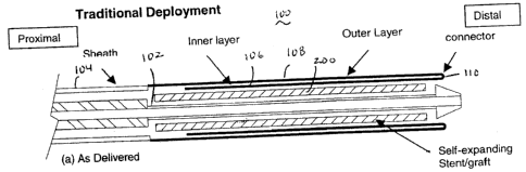
Referring to Figure 1, there is illustrated an exemplary delivery device in
accordance with the present invention. For ease of explanation, only the
distal
region of the device is illustrated as the proximal region may be
substantially
similar to traditional delivery devices. The low deployment force delivery
device 100 of the present invention comprises an inner tube or shaft 102 and
an outer sheath 104. The inner tube or shaft 102 includes a guidewire lumen.
A stent or stent graft 200 is positioned over the distal end of the inner tube
or
shaft 102 and held in position by the outer sheath 104. The distal end of the
outer sheath 104 comprises an inner layer 106, an outer layer 108 and a
connector 110. In this exemplary embodiment, the connector 110 is simply a
folded region between the inner and outer layers 106, 108. However, in
alternate exemplary embodiments, the connector 110 may comprise any
suitable device or means for allowing relative movement between the inner and
outer layers 106, 108 as described in detail subsequently. The inner tube or
shaft 702 and the outer sheath 104 may comprise any suitable, biocompatible
materials utilized in delivery catheters. For example, the inner shaft 102 may
comprise high density polyethylene and the outer sheath 104 may comprise
braided NylonT"". The inner and outer layers may comprise any suitable
material, and preferably comprise a very supple but strong material such as
woven DacronT"". However, it is important to note that the inner and outer
layers may comprise different materials.
12
CA 02513857 2005-07-27
In operation, the physician retracts the outer sheath 104 to deploy the
stent or stent graft 200. Upon retraction of the outer sheath 104, the sheath
104 pulls the outer layer 108 which then inverts the inner layer 106 to expose
the stent or stent graft 200. As illustrated, once the inner layer 106 is
completely inverted, the remaining portion of the stmt or stent graft 200 is
deployed in the usual fashion.
Figure 3 illustrates an alternate exemplary embodiment of a low
deployment force delivery device 300 that allows for the most distal portion
of
the stent or stent graft 200 to be deployed last as may be the case for an
abdominal aortic aneurysm repair stent graft device that comprises distal
barbs. In this exemplary embodiment, the inner layer 106 comprises a distal
end section 302 that loops through the lumen of the stent or stent graft and
1S attaches to the distal tip 112 of the inner tube 102. In this manner, until
the
distal end section 302 is uncurled, the distal end of the stent or stent graft
remains unexpanded as illustrated in Figure 4.
In each of these embodiments, the inner layer tapers larger from its
most proximal end to its distal end to allow smooth and easy deployment. In
addition, at least one of the inner and outer layers may be coated with a
lubricious material.
Although shown and described is what is believed to be the most
practical and preferred embodiments, it is apparent that departures from
specific designs and methods described and shown will suggest themselves to
those skilled in the art and may be used without departing from the spirit and
scope of the invention. The present invention is not restricted to the
particular
constructions described and illustrated, but should be constructed to cohere
with all modifications that may fall within the scope for the appended claims.
13