Note: Descriptions are shown in the official language in which they were submitted.
. . . . .. .... ...:.. _. .. , . .... . ... .. .. .:.. . _. . ..... , , .. . F
.:.... , , .. .. ... õ . . . .. . _ . .
CA 02513917 2009-07-30
ARTICULATING IlVIPLANT SYSTEM
FIELD OF THE INVENTION
The present invention relates to an articulating implant system for fixation
to a bone.
Specifically, the present invention provides an articulating implant system
for replacing the distal
ulna.
BACKGROUND OF THE INVENTION
Both the proximal and distal radioulnar joints are synovial joints. The
proximal joint lies
between the head of the radius and the radial notch of the ulna. The distal
radioulnar joint is
separated from the wrist by an articular disc that extends from the base of
the ulnar styloid
process to the radius.
The distal radioulnar joint is a pivot-joint formed between the head of the
ulna and the ulnar
notch on the lower end of the radius. The articular surfaces are connected
together by the volar
radioulnar ligament, the dorsal radioulnar ligament, and the articular disk.
The volar radioulnar
ligament is a narrow band of fibers extending from the anterior margin of the
ulnar notch of the
radius to the front of the head of the ulna. The dorsal radioulnar ligament
extends between
corresponding surfaces on the dorsal aspect of the articulation. The articular
disk is triangular in
shape, and is placed transversely beneath the head of the ulna, binding the
lower ends of the ulna
and radius firmly together. Its periphery is thicker than its center, which is
occasionally
perforated. It is attached by its apex to a depression between the styloid
process and the head of
the ulna; and by its base, which is thin, to the prominent edge of the radius,
which separates the
ulnar notch from the carpal articular surface. Its margins are united to the
ligaments of the wrist-
joint. Its upper surface, smooth and concave, articulates with the head of the
ulna, forming an
arthrodial joint; its under surface, also concave and smooth, forms part of
the wrist-joint and
1
CA 02513917 2005-07-20
WO 2004/071357 PCT/US2004/003517
articulates with the triangular bone and medial part of the lunate. Both
surfaces are clothed by
synovial membrane; the upper, by that of the distal radioulnar articulation,
the under, by that of
the wrist.
The radius articulates in pronation and supination on the distal ulna. The
ulna, a relatively
straight forearm bone linked to the wrist, translates dorsal-palmarly to
accept the modestly
bowed radius. Since the sigmoid fossa socket in most wrists is relatively
flat, ligaments are
required to support the distal ulna. These ligaments include the triangular
fibrocartilage (TFC),
the extensor carpi ulnaris (ECU) subsheath, and the ulnar collateral ligament
complex. The
stabilizing elements of the triangular fibrocartilage (TFC), extensor carpi
ulnaris (ECU)
subsheath, and the ulnar collateral complex are well recognized along with the
importance of a
distal ulna component (ulnar head) for transfer of compressive loads between
the ulnar carpus
and the distal ulna across the distal radioulnar joint. The distal radioulnar
joint shares loading
forces that occur with forearm rotation and gripping. The arc of pronation and
supination
averages 150 to 160 degrees with the most useful portion being between 80
degrees pronation
and 45 degrees supination.
One of the most common fractures in humans is fracture of the distal radius.
Inherent bony
instability, soft tissue danlage, and frequent associated injuries make the
distal radius fractures
very difficult to treat. Distal radius fractures are usually caused by a fall
on an outstretched
hand. When a person falls on an outstretched hand, the hand suddenly becomes
rigid, and the
monlentum from the fall will cause both a twisting force and a compressing
force on the
forearm. The kind of injury these forces are likely to cause depends on the
age of the person
who is injured. In children, and in older adults, such a fall is likely to
result in a fracture of the
radius. Distal radius fractures may also result from direct trauma such as
might occur during an
auto accident.
There are several types of fractures. A non-displaced fracture is one in which
the bone cracks
and the broken pieces stay in alignment. A torus or ripple fracture bends the
back of the radius
away from the growth plate. A displaced fracture is one in which the bone
breaks in two or more
pieces that move out of aligmnent. Such a break may be extremely painful and
produces a
2
CA 02513917 2005-07-20
WO 2004/071357 PCT/US2004/003517
deformity that is easily seen. An open or compound fracture is one in which
the ends of the bone
are displaced and pierce the skin. In these cases, there is a significant risk
of infection.
For displaced broken bones to heal properly and without serious complications,
they need to be
set and held in place for the body to repair and replace the damaged bone. The
process usually
takes between 4 and 12 weeks. Some fractures may be set without surgery, the
bones being held
in place first with a splint and then, after healing has started, with a cast.
If the bones are
seriously displaced, however, or if there is damage that needs to be repaired,
surgery may be
needed and the bones may need to be held together with pins or wire.
Closed treatment inethods including casting, pins and plaster, and external
fixation have
frequently yielded unsatisfactory results. Treatment using formal open
reduction and internal
fixation with the conventional plate system, when achieving anatoniic
reduction and early
mobilization, has produced some promising results. The value of imnlediate
mobilization of the
injured joints is clear.
With distal radial fractures, muscles may gradually weaken from lack of use
during bone
healing. A patient may need physical therapy in order to regain proper use of
the wrist.
Ligament disruption, ulnar styloid fractures, and fractures into the distal
radioulnar joint are
comnlon occurrences following fractures of the distal radius and other
rotational instability
injuries of the foreami. Fracture or dislocation involving the distal
radioulnar joint often results
in a loss of forearm rotation related to either instability or incongruity
between the sigmoid fossa
of the distal radius and the ulnar head. A variety of different fractures
involving the distal radius
may cause this condition including the Colles' fracture and the Galeazzi
fractures.
When there is loss of stability of the distal radioulnar joint, there is
subsequent weakness in grip
and pinch as well as potential loss of forearm rotation. Instability may also
be associated with
either an injury to the triangular fibrocartilage or to the ulnar styloid.
When instability is present,
a number of ligament reconstructive procedures have been devised to assist in
treating the
3
CA 02513917 2005-07-20
WO 2004/071357 PCT/US2004/003517
unstable distal ulna. Unfortunately, ligament reconstruction of the distal
ulna is often incomplete
in restoring stability, and joint replacement is often necessary.
Where there is an incongruity of the joint surface involving either the
articulation of the ulnar
head with the sigmoid fossa of the distal radius, or if there is a significant
ulnar impaction
syndrome between the distal articular surface of the head of the ulna and the
ulna carpus, a joint
replacement may be necessary. Specifically, this may include either joint
replacement of the
distal ulna or operative procedures designed to shorten the ulna or resect all
or part of the distal
ulna (i.e. Darrach, Bowers, or matched resection procedures).
Implants or prostheses are employed for restoring damaged upper and lower
extremity bones
such as fingers, wrists, elbows, knees and ankles of human patients. These
prostheses are
especially useful in the reconstruction of joints which, for example, have
been dainaged by
pathological conditions such as rheuinatoid arthritis, degenerative arthritis,
aseptic necrosis, and
for treating trauma which may have a debilitating effect on articular joints.
There are three types of arthroplasties: 1) unconstrained, 2) semi-constrained
and 3) fully
constrained. A comnion flaw with all of these current joint replacement
designs is the inability to
reconstruct and re-attach the replaced joint's vital native capsular and
ligainentous restraints,
which dictate, in large measure, the behavior and stability of the joint
(i.e., its kinematics).
The primary reasons for wrist replacement surgery are to relieve pain and to
maintain function in
the wrist and hand. The primary indications, therefore, for reconstruction of
the distal radioulnar
joint by prosthetic replacement (ulnar head replacement only) are generally
related to a fracture
of the distal ulna or a fracture extending into the distal radioulnar joint
producing post-traumatic
arthritis. Degenerative arthritis from other causes is also a primary
indication. This is
considered if there is associated arthritis and an ulnar shortening procedure
is contraindicated.
Osteoarthritis, the most common form of arthritis, results from a gradual
wearing away of the
cartilage covering on bones. A third condition for primary ulna replacement is
rheumatoid
arthritis with a painful and unstable distal radioulnar joint. Rheumatoid
arthritis is a chronic
4
CA 02513917 2005-07-20
WO 2004/071357 PCT/US2004/003517
inflammatory disease of the joints that results in pain, stiffness and
swelling. Rheumatoid
arthritis usually affects several joints on both the right and left sides of
the body. Both forms of
arthritis may affect the strength of the fmgers and hand, making it difficult
to grip or pinch. In
some cases, fusing the wrist bones together will reduce or eliminate pain and
improve grip
strength. However, if the bones are fused together, the ability of the wrist
to move and bend is
lost. Wrist replacement surgery may enable retention or recovery of wrist
movements. In these
situations, prosthetic replacement of the distal ulna with soft tissue
advancement may be
beneficial.
A distal ulnar prosthesis is also suitable to correct a previous resection of
the distal ulna that has
failed. Such will be the case for both partial resection of the joint
articular surface and complete
resection of the distal ulna. When faced with failed distal ulna resection,
one has options
towards reconstruction without restoring the distal radioulnar joint (DRUJ).
For example, a
failed distal ulna may be corrected by a pronator quadratus interposition, or,
if there has been
only a partial resection, a fusion of the distal radioulnar joint combined
with a proximal
pseudarthrosis (Suave-Kapandji procedure). These procedures, however, do not
restore the
nonnal DRUJ function of motion or load transfer and may be associated with
instability of the
distal ulna and proximal impingement of the ulna on the distal radius. In
these cases, a distal
ulna prosthesis is generally preferable. A distal ulnar prosthesis is also
suitable to correct a
previous prosthetic replacement such as a silicone ulnar head replacement
which has failed.
A distal ulnar prosthesis attachable to a soft tissue pocket including the
triangular fibrocartilage,
ECU subsheath, and ulnar collateral ligament complex to thereby maintain
distal radioulnar joint
stability, which aligns anatomically with the sigmoid fossa of the distal
radius and is
isosymmetric with the anatomic center of rotation of the forearm, and that
allows for a normal
forearm rotation of approximately 150-170 degrees would be desirable. More
specifically, it
would be desirable to have such a modular distal ulnar prosthesis wherein
there is no risk of
separation of the two components (the stem and the head) due to biomechanical
forces from the
tissues attached by suture to the implant.
5
CA 02513917 2005-07-20
WO 2004/071357 PCT/US2004/003517
SUMMARY OF THE INVENTION
The present invention is directed to an articulating implant system for
fixation to a bone. The
implant system comprises two components: a fixation component and an
articulating component.
The fixation component has first and second ends. The first end of the
fixation component is
configured for fixation to a bone. This may be, for example, a stem. The
second end of the
fixation component is configured for operative attachment to the articulating
component. Suture
attachment means are provided at or near the second end of the fixation
component. This may
be, for example, by provision of holes for receiving sutures, the holes
positioned through an
extension provided at the second end of the fixation coinponent.
The articulating component is configured for articulating against bone or
cartilage. Optionally,
an area of the articulating coinponent may have a porous surface for ingrowth.
This area would
preferably be near the suture attachment means of the fixation component. A
coimecting taper
means may be provided at a first end thereof. The articulating component is
configured for
operative attachment to the fixation component. This may be done by, for
example, a Morse
taper. It is preferable that the suture attachment means of the fixation
component cooperate with
the articulating coinponent such that the attachment means is provided at a
suitable location near
the articulating surface.
In one embodiment, an extension is provided at the second end of the fixation
component.
Suture holes are provided at both the distal and the proximal ends of the
extension. A bore is
provided through the articulating component such that the extension of the
fixation component
passes therethrough, the suture hole provided at the distal end of the
extension extending through
the bore, the suture holes provided at the proximal end of the extension
failing to pass through
the bore. Thus, upon assembly, the implant system has attachment means at both
ends of the
articulating component.
The articulating implant system of the present invention is particularly
suited to a modular ulnar
implant for implantation after a resection of the distal ulna. In the
embodiment of a modular
6
CA 02513917 2005-07-20
WO 2004/071357 PCT/US2004/003517
ulnar implant, the fixation component is a stem and the articulating component
is a head.
Generally, the modular ulnar implant comprises an eccentric head and a stem,
the stem having
suture holes for receiving sutures to anchor the implant to soft tissues that
are exposed after
resection of the distal ulna. Preferably, the stem attaches to the head via a
morse taper. These
soft tissues include the ulna collateral capsule, the triangular
fibrocartilage, and the extensor
carpi ulnaris subsheath.
The head is offset from the stem, is triangulated to reproduce normal anatomy,
and has an
approximately 200 arc for mating with the radial sigmoid notch. The head
includes a bore
extending completely therethrough for receiving an extension from the stem.
Additionally, the
head may include a drainage hole and instrument interface on its distal
surface to allow effective
in vivo assembly and rotational positioning. Optionally, the head is covered,
at least near the
triangulated portion, with an ingrowth coating to promote ingrowth with the
soft tissues.
The stem has first and second ends. The first stem end is configured for
fixation to a bone, is
tapered to match the ulnar canal anatomy and is preferably fluted for
effective fixation in the
canal. The second stem end is configured for operative attachment to the head.
The stem
includes a platform near the head interface at the second stem end to prevent
subsidence into the
ulnar canal. The stem includes suture holes for receiving sutures to anchor
the implant to soft
tissues. The suture holes anchor the implant to the triangular soft tissues. A
stem extender collar
may be used to add additional resection height. The stem may include an
instrument interface
for positioning control.
In one embodiment, the stem includes an extension at the second end,
preferably centrally
located on a morse taper. The extension is configured for receipt by the bore
in the head. At
least one suture hole is provided at the distal end of the extension for
receiving sutures, the suture
hole being accessible after the head has been placed on the stem. Suture holes
may also be
provided on the platform near the stem second end, near the head interface. In
this embodiment,
the suture holes in the platform and in the extension anchor the implant to
the triangular soft
tissues.
7
CA 02513917 2005-07-20
WO 2004/071357 PCT/US2004/003517
The articulating implant system allows for attachment of tissues near a
surface or location of an
articulating component without attaching the suture to that component. This
allows independent
rotation and orientation of the articulating component, a head in a modular
ulnar implant system,
with respect to the tissue suture attachment. Forces or constraints of the
tissue attachment do not
affect the orientation or behavior of the articulating component. The implant
also allows for
more versatility of the suture attachment by not being constrained to the non-
articulating area of
the articulating component.
In the case of a typical ulnar implant, there is risk of component separation
due to the rotation of
the implant relative to the surrounding tissue that could be suture attached
to the implant. With
the present design, the risk of component separation is eliminated. Using the
modular ulnar
implant of the invention, there is no risk of separation of the two components
(the stem and the
head) due to biomechanical forces from the tissues attached by suture to the
implant. By having
the suture attachment means on the fixation component, a stem in a modular
ulnar implant
system, forces from the suture tissue attachment are transferred directly
through the fixation
component to the bone and not through the connection of the articulating
component to the
fixation coinponent.
8
CA 02513917 2005-07-20
WO 2004/071357 PCT/US2004/003517
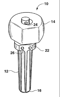
BRIEF DESCRIPTION OF THE DR.AWINGS
Figure 1 illustrates an embodiment of a modular ulnar implant in accordance
with the present
invention.
Figure 2 illustrates the stem of the modular ulnar implant of Figure 1.
Figure 3 illustrates the head of the modular ulnar implant of Figure 1.
DETAILED DESCRIPTION OF THE INVENTION
The present invention is directed to an articulating implant system for
fixation to a bone: The
implant system comprises two components: a fixation component and an
articulating component.
Figure 1 illustrates a modular ulnar implant 10 in accordance with the
articulating implant
system of the present invention. The iinplant 10 is intended to be an
anatomical replacement for
the distal ulna after its resection. The modular ulnar implant 10 includes a
fixation component
and an articulating component. Specifically, the fixation coniponent is a stem
12 and the
articulating component is a head 14. Preferably, the stem 12 attaches to the
head 14 via a morse
taper.
With particular reference to Figure 1, the stem 12, or fixation component, is
elongated and
formed with first and second ends, 16 and 18. The first stem end 16 is
configured for fixation to
a bone, specifically, for insertion into the intramedullary canal of the
distal ulna to thereby
anchor the modular ulnar implant to the distal ulna. The stem 12 is tapered to
match the ulnar
canal anatomy and facilitate insertion of the first stem end 16 into the
intramedullary canal of the
distal ulna. Preferably, the stem 12 is provided with flutes 20 for to prevent
rotation of the stem
12 in the intramedullary canal of the distal ulna, thereby facilitating
effective fixation of the
implant 10 in the canal. The second stem end 18 may be formed with a roughened
or porous
surface to enable a cement-free joint between the stem and the distal ulna.
Alternately, or in
addition to such surface, cement may be used to anchor the stem 12 in the
intramedullary canal
of the distal ulna.
9
CA 02513917 2005-07-20
WO 2004/071357 PCT/US2004/003517
The second stem end 18 is configured for operative attachment to the head 14.
The stem 12
includes a platform 22 near the head interface at the second stem end 18 to
prevent subsidence
into the ulnar canal or excessive penetration of the stem 12 into the
intramedullary canal of the
distal ulna.
The stem 12 includes, at or near the second stem end 18, suture holes 24 and
26 for receiving
sutures to anchor the implant 10 to soft tissues. Sutures threaded through the
suture holes 24 and
26 anchor the implant to the triangular soft tissues that are exposed after
resection of the distal
ulna. These soft tissues include the ulna collateral capsule, the triangular
fibrocartilage, and the
extensor carpi ulnaris subsheatli. A stem extender collar may be used to add
additional resection
height. The stem 12 may include an instrument interface for positioning
control.
As more clearly seen in Figure 2, the stem 12 includes an extension 28 at the
second end 18,
preferably centrally located and formed as a morse taper. The extension 28 is
configured for
receipt by a bore in the head 14. At least one suture hole 24 is provided at a
distal end 30 of the
extension 28 for receiving sutures, the suture hole 24 being accessible after
the head 14 has been
placed on the stem 12. Suture holes 26 may also be provided on the platforin
22 near the stem
second end 18, near the head interface. In this embodiment, the suture holes
26 and 24 in the
platform aiid in the extension, respectively, anchor the implant 10 to the
triaiigular soft tissues.
The head 14, or articulating component, as shown in Figure 3, is configured
for articulating
against bone or cartilage. With reference to Figure 1, it can be seen that the
head 14 is offset
from the stem 12 after the implant 10 has been assembled. The head 14 is
triangulated to
reproduce normal anatomy, and has an approximately 200 arc for mating with
the radial sigmoid
notch. The head includes a bore 32 extending completely tlierethrough for
receiving an
extension from the stem. In alternate embodiinents, the head 14 may include a
bore extending
less than completely therethrough for mating with the second end 18 of the
stem 12.
Additionally, the head 14 may include a drainage hole and instrument interface
on its distal
surface to allow effective in vivo assembly and rotational positioning.
Optionally, the head 14 is
CA 02513917 2005-07-20
WO 2004/071357 PCT/US2004/003517
covered, at least near the triangulated portion, with an ingrowth coating, or
is otherwise provided
with a porous surface, to promote ingrowth with the soft tissues.
The second end 18 of the stem 12 and the bore 32 of the head 14 are
complementary so as to
provide a secure fit between the head 14 and the stem 12 when the second stem
end 18 is
inserted in the bore 32 of the head 14. Thus, for example, the bore 32 is
provided through the
head 14 such that the extension 28 of the stem 12 passes completely
therethrough, with both the
extension 28 of the stem 12 and the bore 14 being Morse tapers. In the
embodiment shown in
Figure 1, insertion of the stem extension 28 through the bore 32 results in
the suture hole 24
provided at the distal end 30 of the extension 28 extending through the bore
32 while the suture
holes 26 provided at the proximal end of the extension 34 fail to pass through
the bore 32. Thus,
upon assembly, the implant 10 has suture holes at both ends of the head 14.
Thus, using the modular ulnar implant of the invention, the irnplant is
attached to the soft tissues
via the stem (or fixation component). By having the suture attachment means on
the fixation
component, a stem in a modular ulnar implant system, forces from the suture
tissue attachment
are transferred directly through the fixation component to the bone and not
through the
connection of the articulating component to the fixation component. As a
result, there is no risk
of separation of the head and stem due to biomechanical forces from the
tissues attached by
suture to the implant.
In order to implant an ulnar implant in accordance with the articulating
implant system of the
present invention, the distal ulna is first exposed. The distal ulna may be
exposed by making an
incision along the medial shaft of the distal ulna in line with the ulnar
styloid. Alternatively, a
dorsal incision centered over the distal radioulnar joint in line with the
fourth metacarpal may be
used to expose the distal ulna. Once exposed, a template may be placed against
the distal ulna
and located distally over the articular surface of the distal ulna to mark the
prescribed resection
length. The distal ulna is resected, by, for example, using an oscillating
saw, exposing the
intramedullary canal and the soft tissues that formerly surrounded the distal
ulna. Once exposed,
the intramedullary canal is identified and reamed to accommodate an
appropriately sized stem.
11
CA 02513917 2005-07-20
WO 2004/071357 PCT/US2004/003517
Prior to implantation of the ulnar implant, a trial stem and trial head may be
used to verify
anatomical alignment and to ensure that the proper resection length has been
achieved. After
removal of the trial stem and trial head, the stem of the stem may be inserted
into the
intramedullary canal of the distal ulna to anchor the implant to the distal
ulna. Specifically, the
first end of the stem is inserted into the intramedullary canal, for example
by using an impactor
until the platform contacts the distal ulna. The fit between the stem and the
distal ulna may be
assessed by applying a distal traction on the stem. Any movement of the stem
in the
intramedullary canal of the distal ulna indicates that a firm fit has not been
obtained. If a firm fit
is not obtained between the stem and the distal ulna after inipaction of the
stem, a bone cement
such as polymethylmethacrylate may be used to cement the stem to the distal
ulna.
Once the stem has been secured within the distal ulna, the head may be
impacted onto the stem.
Specifically, the extension at the second stem end may be inserted into the
bore formed in the
head. The head may be advanced onto the stem with an impactor until a secure
fit is obtained
between the head and the stem.
The stem may then be sutured to the soft tissue formerly surrounding the
distal ulna, specifically
the ulna collateral capsule, the triangular fibrocartilage, and tlie extensor
carpi ulnaris subsheath
using the suture holes formed in the stem. Non-absorbable sutures may be used.
By having the
sutures attach to the implant through the fixation component, forces from the
suture tissue
attachment are transferred directly through the fixation component to the
bone, thereby reducing
or eliminating the risk of component separation due to such forces. After the
head is attached to
the stem and the stem has been sutured to the soft tissue, the sutures may be
tied into the capsular
sleeve surrounding the implant and the subcutaneous tissues and skin may be
closed over the
implant.
While a preferred embodiment of the present invention has been described, it
should be
understood that various changes, adaptations, and modifications may be made
therein without
departing from the spirit of the invention and the scope of the appended
claims..
12